Embed Size (px)
Citation preview
NSC 224 MEDICAL BIOCHEMISTRY II
1
NATIONAL OPEN UNIVERSITY OF NIGERIA
SCHOOL OF HEALTH SCIENCES
COURSE CODE: NSC 224 COURSE TITLE : MEDICAL BIOCHEMISTRY I1 COURSE UNITS: 3(2-1-2) COURSE DEVELOPER: Dr. O Emma-Okon, PhD Dr. J.O. Areola, PhD COURSE CONTENT EDITOR : Dr O.O. Irinoye Dr T.O. Oladogba COURSE REVIEWER : Dr. S. A Isa Dr. M S Enaji Dr. S Rabiu
OURSE GUIDE
NSC 224 MEDICAL BIOCHEMISTRY II
2
National Open University of Nigeria Headquarters University Village Plot 91, Cadastral Zone, NnamdiAzikiwe Express way Jabi, Abuja Lagos Office 14/16 Ahmadu Bello Way Victoria Island, Lagos e-mail: [email protected] website: www.nou.edu.ng Published by National Open University of Nigeria Print 2017 ISBN: ISBN: 978-058-009-3 All Rights Reserved
NSC 224 MEDICAL BIOCHEMISTRY II
3
COURSE CONTENTS PAGE
General Introduction 5 Course Information 5 Course Overview 6 Course Objectives 6 Course Implementation - Doing the Course 7 Course Requirements and Expectations of You 7 Equipment and Software Needed to Access Course 7 Number and Places of Meeting (Online, Face-To-Face, Laboratory Practical) 7 Online Discussion Forum 7 Course Evaluation 8 Grading Criteria 8 Schedule of Assignments with Dates 9 Reference Textbooks for the Course 9
NSC 224 MEDICAL BIOCHEMISTRY II
4
GENERAL INTRODUCTION Like Medical Biochemistry 1, Medical Biochemistry II is also a subset of general biochemistry that focused on the essential components required for the basic understanding of allied and health sciences, including nursing sciences. The course deals with intermediary metabolism and biological oxidation. This course introduces the students to different biochemical pathways that break down complex set of ingested molecules to functional materials and energy to meet the body’s needs. These pathways constitute what is known as Metabolism. Metabolism is the sum of all of the chemical reactions that take place in an organism. Metabolic reactions are either catabolic or anabolic. Catabolic reactions break down large molecules and release energy, while anabolic reactions large molecules and release energy, while anabolic reactions synthesize larger molecules and require energy. Clinical conditions that are associated with impairment of the metabolic pathway were also discussed. COURSE AIM The aim of this course is to introduce students to the basic understanding of intermediary metabolism and biological oxidation. COURSE OBJECTIVES At the end of this course, students should be able to understand the different metabolic pathways that breakdown molecules and produce energy. The different pathways that are anabolic and those that are catabolic. The different clinical conditions associated with metabolic pathways. WORKING THROUGH THE COURSE The blended learning mode would be adopted in teaching this course: 70% online and 30% face-to-face. Only students that have registered for this course would be allowed access to study materials and interactive sessions. Study materials (hard and soft copies) would be tailored point-by-point to address the scope of each module. As part of the learning objectives, students are expected to attempt Tutor Marked Assignments (TMA) and other Self-Assessment Questions (SAQ) provided at the end of each module preparatory to in-tests and final examinations to be undertaken at the end of the semester. For periodical evaluation of learning and tutor-student interaction, students are expected to keep a portfolio where all completed assignments are stored. COURSE MATERIALS The following course materials would be provided: Lecture notes
NSC 224 MEDICAL BIOCHEMISTRY II
5
Power point slides Pre-recorded videos and Logbooks for virtual laboratory demonstrations STUDY UNITS NSC 224 (Medical Biochemistry II) is a 3 credit units course comprising of three (3) modules and 9 study units. . TEXT BOOKS AND REFERENCES Murry, R. K., Bender, D. A., Bothan, K.M., Kennelly, P. J., Rodwell, V. W. and Well, P. A. (2015). Harper’s Illustrated Biochemistry (30th Edition) McGraw-Hill Medical. Nelson, D. L. and Cox, M. M. (2012). Lehninger Principles of Biochemistry (6th edition) WH Freeman. Pamela, C. C., Richard, A. H. and Denise, R. F. (2013). Lippincott’s Illustrated Reviews Biochemistry (6th edition) Lippincott Williams & Wilkins. Marks' Essentials of Medical Biochemistry: A clinical approach. 2nd Edition Copyright 2007 Lippincott Williams & Wilkins. Garrett and Grisham Biochemistry, 2nd Edition. Harcourt College Pub Lippincott Biochemistry Fourth Edition (2010). Robert K. Murray, MD, PhD. ‘Harper’s Illustrated Biochemistry’. Twenty-Eighth Edition. 2009 Devlin T.M. (2010) Textbook of Biochemistry with Clinical Correlation 7thEdition. JohnWiley & SonsInc. ASSIGNMENT FILE Assignments would be given at the end of each module and submission time would be spelt out accordingly. TUTOR MARKED ASSIGNMENT To be provided by tutor at the end of each module in addition to the ones already spelt out in the course material.
NSC 224 MEDICAL BIOCHEMISTRY II
6
FINAL EXAMINATION AND GRADING The final written examination will come up at the end of the semester comprising essay and objective questions covering all the contents covered in the course. The final examination will amount to 60% of the total grade for the course. GRADING CRITERIA Grades will be based on the following Percentages
Tutor Marked Individual Assignments 10% Computer marked Assignment 10% Group assignment 5% Discussion Topic participation 5% Laboratory practical 10% End of Course examination 60%
Total 100% GRADING SCALE
A = 70-100 B = 60 - 69 C= 50 - 59 F = < 49
COURSE MARKING SCHEME Course marking scheme would be provided by the tutor. HOW TO GET THE MOST FROM THIS COURSE
Students are expected to participate in all interactions with the tutor and ask questions during lectures
Materials recommended for further reading should be used to augment study guides provided by the tutor.
Importantly, timely submission of assignments and returned marked scripts should be used for self-evaluation
Regular participation in online discussion forums and study groups to compare notes and share ideas
Keep in touch with information outlets provided at study centers, Department and NOUN websites
FACILITATORS, TUTORS AND TUTORIALS Information on Profile of facilitators and tutors for the course would be provided at the Department and NOUN websites. Schedule for tutorials would be arranged by facilitators and tutors from time to time during the semester.

NSC 224 MEDICAL BIOCHEMISTRY II
7
SUMMARY The Course NSC 224 (Medical Biochemistry II) is a compulsory course for Nursing Science students. The course content is prepared in modules and units. It covers the basic requirements for teaching medical biochemistry to Nursing Science students. It is expected that the foundation provided in NSC 224 would find relevance as students advance in their course of study. The minimum pass grade is 50% made up of assignments and examination components.
NSC 224 MEDICAL BIOCHEMISTRY II
8
CONTENTS PAGE
Module 1- CARBOHYDRATES METABOLISM
Contents Unit1: Glycolysis Unit2: Glycolysis 2 Unit 3: Tricarboxylic Acid Cycle
Module 2- FATTY ACID METABOLISM
Contents Unit 1: Fatty Acid Oxidation Unit 2: Fatty Acid Oxidation 2
Module 3- VITAMINS AND TRACE ELEMENTS
Contents Unit 1: The Fat Soluble Vitamins Unit 2: The Fat Soluble Vitamins Unit 3: Trace Elements
Module 4- AMINO ACID METABOLISM
Contents Unit 1 Sources of Amino Acids Unit 2 Disorders of Amino acid Metabolism Unit 3 Diabetes Mellitus
COURSE GUIDEC
NSC 224 MEDICAL BIOCHEMISTRY II
9
Module 1: CARBOHYDRATE METABOLISM This module introduces the students to the major concept in the metabolism of carbohydrates. Carbohydrates are the major source of energy for the living cells. Glucose is the central molecule in carbohydrate metabolism since all the major pathways of carbohydrates metabolism are connected with it. The important pathways of carbohydrates metabolism and associated disorders will be discussed. Module Objectives At the end of this module students should be able to:
i. Explain the concept of digestion and absorption ii. Outline the major pathways for carbohydrates metabolism.
iii. Discuss the regulation of the major metabolic pathways. iv. Highlight the clinical conditions associated with the pathways.
MAIN CONTENTS Unit 1: Glycolysis Unit 2: Glycolysis 2 Unit 3: Tricarboxylic Acid Cycle
UNIT ONE- GLYCOLYSIS
CONTENT 1.0 Introduction 2.0 Objectives 3.0 Main Content 3.1 The glycolytic pathway 3.2 The stages of glycolytic pathway 4.0 Conclusion 5.0 Summary 6.0 Tutor Marked Assignments 6.1 Activity 6.2 Tutor Marked Tests 7.0 Reference and other resources
1.0 Introduction Glycolysis is the sequence of reactions that converts glucose into pyruvate with the production of ATP. In aerobic organisms, glycolysis is the prelude to the citric acid cycle and the electron transport chain, where most of the energy contained in glucose is released. Under aerobic conditions, pyruvate enters the mitochondria where it is completely oxidized to CO2 and H2O.If the supply of oxygen is insufficient, e.g. in actively contracting muscle, pyruvate is converted into Lactate. In some anaerobic organisms, pyruvate is transformed into ethanol. The formation of ethanol and Lactate from Glucose are examples of fermentations. Reactions of glycolysis take place in the
NSC 224 MEDICAL BIOCHEMISTRY II
10
cytosol. Glycolysis is sometimes called the Embden Meyerhof pathway, after Gustav Embden and Otto Meyerhof who made significant contributions to its elucidation in 1940.
2.0 Objectives At the end of this unit, you should be able to:
i. Explain the concept of Glycolysis ii. State the importance of the glycolytic pathway iii. Enumerate the different reactions which make up the pathway and the enzymes
which catalyze these reactions
3.0 Main Content 3.1 Overview of digestion and absorption of Carbohydrates Digestion is the hydrolysis of complex food molecules into smaller water soluble molecules that can easily be absorbed by the gastrointestinal tract for utilization by the cells. The organs of the digestive tract usually have a large reserve capacity. Pancreas for example secretes enzymes five to ten folds higher than required for digestion of foods ingested. Digestion of carbohydrates usually starts in the mouth, where food material comes in contact with saliva during mastication. Saliva contains a carbohydrate splitting enzyme called salivary amylase (ptyalin). The enzyme hydrolyzes α- (1,4) glycosidic linkages in starch, glycogen and dextrins, producing smaller molecules like maltose, glucose and disaccharides maltotriose. Ptyalin action stops in stomach when pH falls to 3.0. No carbohydrate splitting enzymes are available in gastric juice. HCl may hydrolyze some dietary sucrose to equal amounts of glucose and fructose. Food reaches the duodenum from stomach where it meets the pancreatic juice. Pancreatic juice contains a carbohydrate-splitting enzyme pancreatic amylase. Like ptyalin it also requires Cl- for activity. The enzyme hydrolyzes α-(1,4) glycosidic linkage situated well inside polysaccharide molecule. Other criteria and end products of action are similar of ptyalin. The final digestion of di- and oligosaccharides to monosaccharides primarily occurs at the mucosal lining of the upper jejunum. This is carried out by oligosaccharidases and disaccharidases. Lactase, maltase and sucrase are example of disaccharidases. The principal monosaccharides produced by the digestion process are glucose, fructose and galactose. Glucose account for nearly 80% of the total monosaccharides. The absorption of sugars mostly takes place in the duodenum and upper jejunum of small intestine. Two mechanisms are involved in absorption of sugars. Simple Diffusion and active transport mechanism. Simple diffusion is dependent on sugar concentration gradients between the intestinal lumen, mucosal cells and blood plasma. All the monosaccharides are probably absorbed to some extent by simple ‘passive’ diffusion. On the other hand, Glucose and galactose are absorbed very rapidly and hence it has been suggested that they are absorbed actively and it requires energy. Glucose and Na+share the same transport system (symport) called sodium-dependent glucose transporter. Fructose
NSC 224 MEDICAL BIOCHEMISTRY II
11
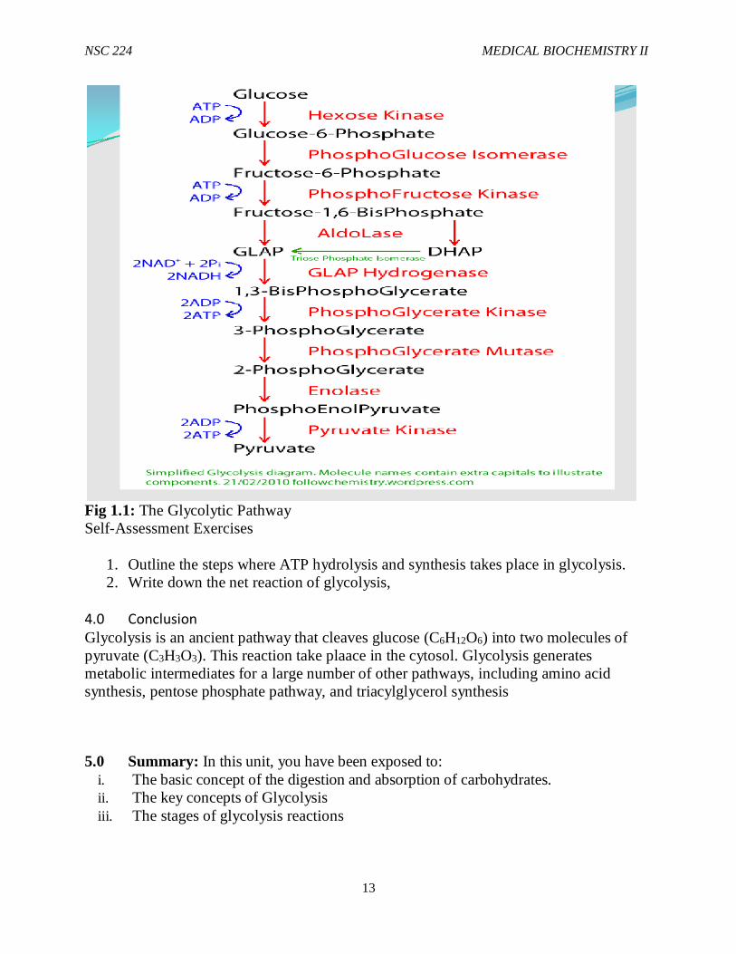
absorption is also rapid but not so much as compared to glucose and galactose but it is definitely faster than pentoses. Hence fructose is not absorbed by simple diffusion alone and it is suggested that some mechanism facilitates its transport, called as” facilitated transport”. 3.2 The glycolytic pathway The word glycolysis is derived from the Greek glykys meaning sweet and lysis meaning splitting. Glycolysis is a linear, 10-step pathway that converts glucose, a six-carbon monosaccharide, to two molecules of pyruvate (CH3COCO2 –). Glycolysis takes place entirely in the cytosol, whereas, pyruvate oxidation occurs in the mitochondrial matrix where ATP is generated. Oxygen is not required for glycolysis in the cytosol (anaerobic) but it is necessary for aerobic respiration in the mitochondrial matrix where the O2 serves as the terminal electron acceptor. Glycolysis is an ancient pathway that cleaves glucose (C6H12O6) into two molecules of pyruvate (C3H3O3). Under aerobic conditions, the pyruvate is completely oxidized by the citrate cycle to generate CO2, whereas, under anaerobic (lacking O2) conditions, it is either converted to lactate, or to ethanol + CO2 (fermentation). The glycolytic pathway consists of ten enzymatic steps organized into two stages. In Stage 1, two ATP are invested to “prime the pump,” and in Stage 2, four ATP are produced to give a net ATP yield of two moles of ATP per mole of glucose. Three glycolytic enzymes catalyze highly exergonic reactions (G<<0) which drive metabolic flux through the pathway; these enzymes are regulated by the energy charge in the cell (ATP requirements). The three enzymes are hexokinase, phosphofructokinase 1, and pyruvate kinase. Glycolysis generates metabolic intermediates for a large number of other pathways, including amino acid synthesis, pentose phosphate pathway, and triacylglycerol synthesis.
3.3 The stages of glycolytic pathway The glycolytic pathway can be divided into three stages.
Stage One The conversion of Glucose to fructose 1,6 Biphosphate. This stage comprises of 3 steps- a phosphorylation, an isomerization and another phosphorylation.
i. Glucose is phosphorylated by ATP to form glucose 6-phosphate. This reaction is catalyzed by Hexokinase (An enzyme that transfers a phosphoryl group from ATP to an acceptor is called a kinase) . Glucose + ATP Glucose 6-phosphate + ADP + Pi
NSC 224 MEDICAL BIOCHEMISTRY II
12
ii. Glucose 6-phosphate is isomerized to Fructose 6-phosphate. The reaction is catalyzed by Phospho glucose isomerase.
iii. Fructose 6-phosphate is phosphorylated by ATP to Fructose 1,6-biphosphate. Fructose 6- phosphate + ATP Fructose 1,6-biphosphate +ADP + H+
This reaction is catalyzed by phosphofructokinase, an allosteric enzyme. The pace of glycolysis is critically dependent on the level of this enzyme. Its catalytic activity is controlled by ATP and other metabolites.
Stage Two This stage of glycolysis consists of 4 steps, starting with the splitting of Fructose 1,6 biphosphate to yield glyceraldehyde 3-phosphate and dihydroxyacetone phosphate. The remaining steps in glycolysis involve 3 carbon units rather than 6 carbon units.
(iva) Fructose1,6-biphosphate Dihydroxyacetonephosphate+ Glyceraldehyde 3-phosphate The reaction is catalyzed by aldolase. The 2 products formed are isomers. Dihydroxyacetone phosphate is a ketose while glyceraldehydes 3-phosphate is an aldose. The reaction proceeds readily from DAP to TPI through the action of the enzyme triose phosphate isomerase. Thus, 2 molecules of glyceraldehyde 3- phosphate are formed from one molecule of Fructose 1,6 biphosphate.
( v ) Gl yc e ra ld eh yd e 3 -p ho sph a te D ih yd ro x yace to n e pho sph a te .
( v i ) Conversion of glyceraldehydes 3-phosphate to 1,3 –diphosphoglycerate, catalysed by glyceraldehyde 3-phosphate dehydrogenase GLY 3-P + NAD+ + PI 1,3 DPG + NADH + H+
( v i i ) 1,3 Diphosphoglycerate is converted to 3-phophoglycerate, and ATP is generated. The rxn is catalyzed by phosphoglycerate kinase. 1,3 Diphosphoglycerate + ADP 3-Phosphoglycerate + ATP
Stage Three In this stage, three steps are involved leading to the generation of pyruvate
(viiia) 3-phosphoglycerate is converted to 2-phosphoglycerate, through the action of 2-phosphoglyceromutase.
(ixa) 2-phosphoglycerate is converted to phosphoenolpyruvate by Enolase 2-Phosphoglycerate Phosphoenol pyruvate + H2O
(xa) PEP is converted to Pyruvate with the generation of ATP, the rxn being catalyzed by pyruvate kinase. PEP + ADP + Pi Pyruvate + ATP The net reaction in the conversion of Glucose into pyruvate is Glucose + 2Pi + 2ADP + 2NAD+ 2Pyruvate + 2ATP + 2NADH + 2H+ + 2H2O
NSC 224 MEDICAL BIOCHEMISTRY II
13
Fig 1.1: The Glycolytic Pathway Self-Assessment Exercises
1. Outline the steps where ATP hydrolysis and synthesis takes place in glycolysis. 2. Write down the net reaction of glycolysis,
4.0 Conclusion Glycolysis is an ancient pathway that cleaves glucose (C6H12O6) into two molecules of pyruvate (C3H3O3). This reaction take plaace in the cytosol. Glycolysis generates metabolic intermediates for a large number of other pathways, including amino acid synthesis, pentose phosphate pathway, and triacylglycerol synthesis
5.0 Summary: In this unit, you have been exposed to: i. The basic concept of the digestion and absorption of carbohydrates. ii. The key concepts of Glycolysis iii. The stages of glycolysis reactions
NSC 224 MEDICAL BIOCHEMISTRY II
14
6.0 Tutor Marked Assignments
i. Explain the basic concept of Glycolysis ii. State the importance of the glycolytic pathway iii. Enumerate the different reactions which make up the pathway and the enzymes
which catalyze these reactions
7.0 References and Further Reading Katherine, M. A. Rogers and William N. Scott (2011). Nurses! Test yourself in anatomy and physiology Kathryn, A. Booth, Terri. D. Wyman (2008). Anatomy, physiology, and pathophysiology for allied health Keith L.M, Persuade T.V.N (2006). The Developing Human Clinically Oriented Embryology; 8th Edition Lippincott Williams & Wilkins Kent, M. Van De Graff, R.WardRhees, Sidney P. (2010). Schaum’s outline of human anatomy and physiology 3rd edition. Philip, T. (2012). Seeley’s principles of anatomy & physiology 2nd edition. Sadler, T.W (2004). Langman’s Medical Embryology 9th edition.
NSC 224 MEDICAL BIOCHEMISTRY II
15
UNIT TWO- GLYCOLYSIS II
CONTENT 1.0 Introduction 2.0 Objectives 3.0 Main Content 3.1 Consumption and generation of ATP in Glycolysis 3.2 Regulation of Glycolysis 3.3 Regulation of Glycolysis 3.4 Clinical conditions associated with impaired Glycolysis 4.0 Conclusion 5.0 Summary 6.0 Tutor Marked Assignments 7.0 Reference and other resources
1.0 Introduction We continue our discussion of the glycolytic pathway. 2 ATP molecules are produced in the course of the pathway. However, more ATP is produced when pyruvate is completely oxidized to CO2 and H2O in the mitochondria. The glycolytic pathway is regulated through the activities of 3 enzymes that catalyze its irreversible reactions. However, the most important control element of glycolysis is the enzyme phosphofrctokinase (PFK), the enzyme catalyzing the first irreversible step unique to the pathway. Pyruvate has 3 fates- It may be converted to acetyl coA, ethanol or Lactate. Clinical conditions associated with impaired glycolysis include Lactic acidosis and Pyruvate kinase deficiency.
2.0 Objectives At the end of this unit, you should be able to:
i. Give the gross and net ATP yield of glycolysis ii. List the glycolytic regulatory enzymes and their corresponding effectors. iii. Explain the fates of Pyruvate iv. List and explain some disorders of Glycolytic pathway
3.0 Main Content
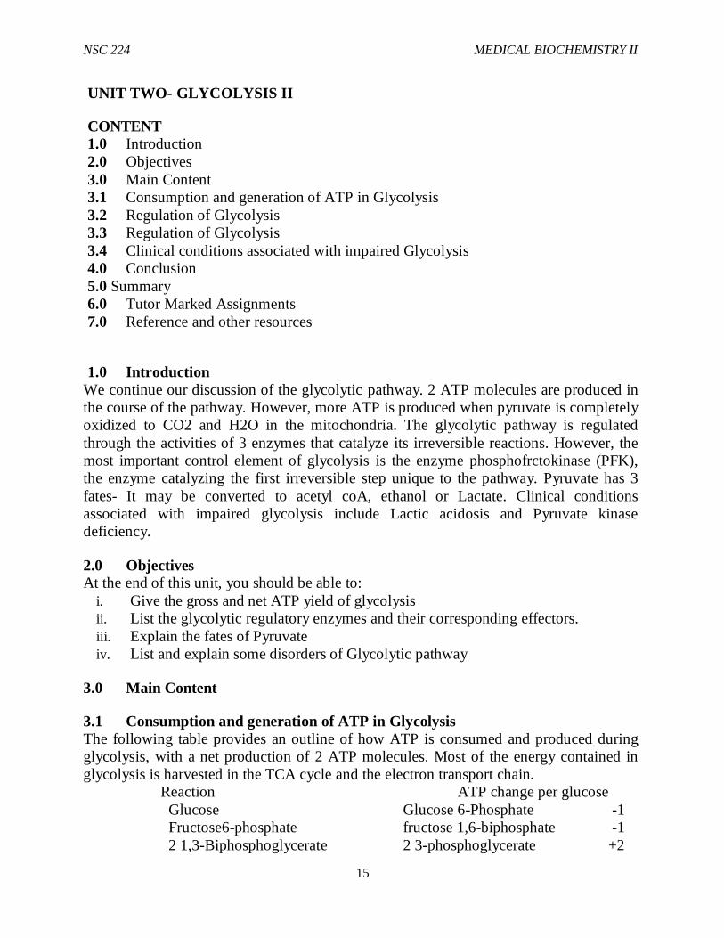
3.1 Consumption and generation of ATP in Glycolysis The following table provides an outline of how ATP is consumed and produced during glycolysis, with a net production of 2 ATP molecules. Most of the energy contained in glycolysis is harvested in the TCA cycle and the electron transport chain. Reaction ATP change per glucose
Glucose Glucose 6-Phosphate -1 Fructose6-phosphate fructose 1,6-biphosphate -1 2 1,3-Biphosphoglycerate 2 3-phosphoglycerate +2
NSC 224 MEDICAL BIOCHEMISTRY II
16
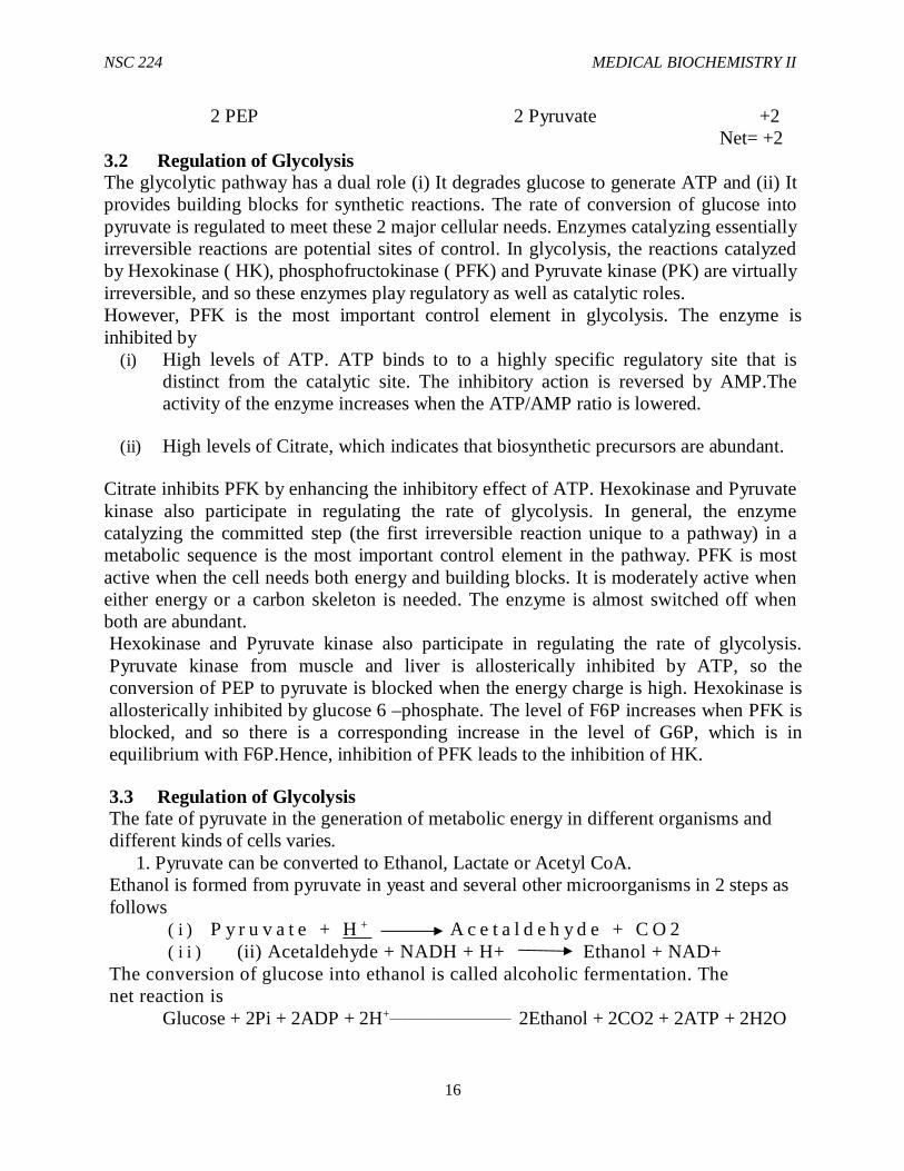
2 PEP 2 Pyruvate +2 Net= +2
3.2 Regulation of Glycolysis The glycolytic pathway has a dual role (i) It degrades glucose to generate ATP and (ii) It provides building blocks for synthetic reactions. The rate of conversion of glucose into pyruvate is regulated to meet these 2 major cellular needs. Enzymes catalyzing essentially irreversible reactions are potential sites of control. In glycolysis, the reactions catalyzed by Hexokinase ( HK), phosphofructokinase ( PFK) and Pyruvate kinase (PK) are virtually irreversible, and so these enzymes play regulatory as well as catalytic roles. However, PFK is the most important control element in glycolysis. The enzyme is inhibited by
(i) High levels of ATP. ATP binds to to a highly specific regulatory site that is distinct from the catalytic site. The inhibitory action is reversed by AMP.The activity of the enzyme increases when the ATP/AMP ratio is lowered.
(ii) High levels of Citrate, which indicates that biosynthetic precursors are abundant.
Citrate inhibits PFK by enhancing the inhibitory effect of ATP. Hexokinase and Pyruvate kinase also participate in regulating the rate of glycolysis. In general, the enzyme catalyzing the committed step (the first irreversible reaction unique to a pathway) in a metabolic sequence is the most important control element in the pathway. PFK is most active when the cell needs both energy and building blocks. It is moderately active when either energy or a carbon skeleton is needed. The enzyme is almost switched off when both are abundant. Hexokinase and Pyruvate kinase also participate in regulating the rate of glycolysis. Pyruvate kinase from muscle and liver is allosterically inhibited by ATP, so the conversion of PEP to pyruvate is blocked when the energy charge is high. Hexokinase is allosterically inhibited by glucose 6 –phosphate. The level of F6P increases when PFK is blocked, and so there is a corresponding increase in the level of G6P, which is in equilibrium with F6P.Hence, inhibition of PFK leads to the inhibition of HK.
3.3 Regulation of Glycolysis The fate of pyruvate in the generation of metabolic energy in different organisms and different kinds of cells varies.
1. Pyruvate can be converted to Ethanol, Lactate or Acetyl CoA. Ethanol is formed from pyruvate in yeast and several other microorganisms in 2 steps as follows
( i ) P y r u v a t e + H+ A c e t a l d e h y d e + C O 2 ( i i ) (ii) Acetaldehyde + NADH + H+ Ethanol + NAD+
The conversion of glucose into ethanol is called alcoholic fermentation. The net reaction is
Glucose + 2Pi + 2ADP + 2H+ ___________________ 2Ethanol + 2CO2 + 2ATP + 2H2O
NSC 224 MEDICAL BIOCHEMISTRY II
17
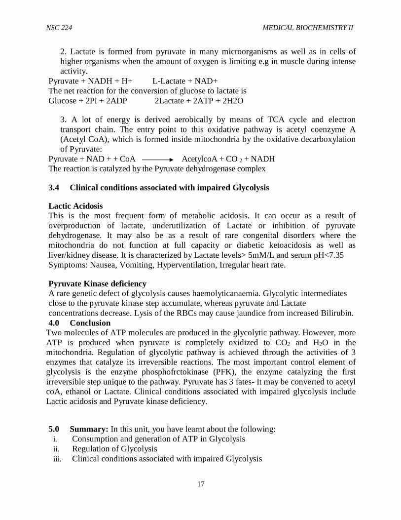
2. Lactate is formed from pyruvate in many microorganisms as well as in cells of higher organisms when the amount of oxygen is limiting e.g in muscle during intense activity.
Pyruvate + NADH + H+ L-Lactate + NAD+ The net reaction for the conversion of glucose to lactate is Glucose + 2Pi + 2ADP 2Lactate + 2ATP + 2H2O
3. A lot of energy is derived aerobically by means of TCA cycle and electron transport chain. The entry point to this oxidative pathway is acetyl coenzyme A (Acetyl CoA), which is formed inside mitochondria by the oxidative decarboxylation of Pyruvate:
Pyruvate + NAD + + CoA AcetylcoA + CO 2 + NADH The reaction is catalyzed by the Pyruvate dehydrogenase complex
3.4 Clinical conditions associated with impaired Glycolysis
Lactic Acidosis This is the most frequent form of metabolic acidosis. It can occur as a result of overproduction of lactate, underutilization of Lactate or inhibition of pyruvate dehydrogenase. It may also be as a result of rare congenital disorders where the mitochondria do not function at full capacity or diabetic ketoacidosis as well as liver/kidney disease. It is characterized by Lactate levels> 5mM/L and serum pH<7.35 Symptoms: Nausea, Vomiting, Hyperventilation, Irregular heart rate.
Pyruvate Kinase deficiency A rare genetic defect of glycolysis causes haemolyticanaemia. Glycolytic intermediates close to the pyruvate kinase step accumulate, whereas pyruvate and Lactate concentrations decrease. Lysis of the RBCs may cause jaundice from increased Bilirubin. 4.0 Conclusion
Two molecules of ATP molecules are produced in the glycolytic pathway. However, more ATP is produced when pyruvate is completely oxidized to CO2 and H2O in the mitochondria. Regulation of glycolytic pathway is achieved through the activities of 3 enzymes that catalyze its irreversible reactions. The most important control element of glycolysis is the enzyme phosphofrctokinase (PFK), the enzyme catalyzing the first irreversible step unique to the pathway. Pyruvate has 3 fates- It may be converted to acetyl coA, ethanol or Lactate. Clinical conditions associated with impaired glycolysis include Lactic acidosis and Pyruvate kinase deficiency.
5.0 Summary: In this unit, you have learnt about the following: i. Consumption and generation of ATP in Glycolysis ii. Regulation of Glycolysis iii. Clinical conditions associated with impaired Glycolysis
NSC 224 MEDICAL BIOCHEMISTRY II
18
6.0 Tutor Marked Assignments i. Give the gross and net ATP yield of glycolysis
ii. List the glycolytic regulatory enzymes and their corresponding effectors. iii. Explain the fates of Pyruvate. iv. List and explain some disorders of Glycolytic pathway
7.0 References and Further reading
Katherine, M. A. Rogers and William N. Scott (2011). Nurses! Test yourself in anatomy and physiology Kathryn, A. Booth, Terri. D. Wyman (2008). Anatomy, physiology, and pathophysiology for allied health Keith L.M, Persuade T.V.N (2006). The Developing Human Clinically Oriented Embryology8th Edition Lippincott Williams & Wilkins Kent, M. Van De Graff, R.WardRhees, Sidney P. (2010). Schaum’s outline of human anatomy and physiology 3rd edition. Philip, T. (2012). Seeley’s principles of anatomy & physiology 2nd edition. Sadler, T.W (2004). Langman’s Medical Embryology 9th edition.
NSC 224 MEDICAL BIOCHEMISTRY II
19
UNIT THREE- TRICARBOXYLIC ACID CYCLE
CONTENTS 1.0 Introduction 2.0 Objective 3.0 Main Content 3.1 Description of TCA cycle 3.2 The Amphibolic Nature of the TCA Cycle 3.3 The Anaplerotic Nature of the TCA Cycle 3.4 The relationship between TCA cycle and Beriberi 3.5 Summary of oxidative phosphorylation 3.6 Inhibitors of electron transport chain 4.0 Conclusion 5.0 Summary 6.0 Tutor Marked Assignments 6.1 Activity
6.2 Tutor Marked Tests 7.0 Reference and other resources 1.0 Introduction The citric acid cycle was discovered by Hans krebs in 1937 and received Nobel prize for the discovery in 1953. The cycle was therefore named after him as kreb’s cycle. The cycle is also known as citric acid cycle or tricarboxylic acid cycle. The citric acid cycle is the final common pathway for the oxidation of fuel molecules (protein, fatty acids and carbohydrates) to energy, carbon dioxide and water. Without this cycle, most of the food we eat cannot be converted to energy. Most of these fuel molecules are metabolized to acetyl Coenzyme A (acetyl CoA) or intermediates of the cycle. The acetyl CoA generated is then fed into the cycle and condenses with oxaloacetate. Two molecules of CO2 is liberated, the energy released is conserved in the reduced electron carriers NADH and FADH2. In the final stage the conserved energy is released and stored as ATP.
2.0 Objectives At the end of this unit, you should be able to:
i. Describe TCA cycle in detail ii. Explain the Amphibolic nature of the TCA cycle iii. Explain the Anaplerotic nature of the TCA cycle iv. Describe the relationship between this cycle and Beriberi (a neurological disease) v. Give the summary of oxidative phosphorylation vi. Give examples and describe the inhibition of electron transport chain
3.0 Main Content 3.1 Description of TCA cycle In eukaryotes, TCA cycle takes place in the mitochondria because all the enzymes of the cycle are located inside the mitochondria matrix. The TCA cycle is an important source of
NSC 224 MEDICAL BIOCHEMISTRY II
20
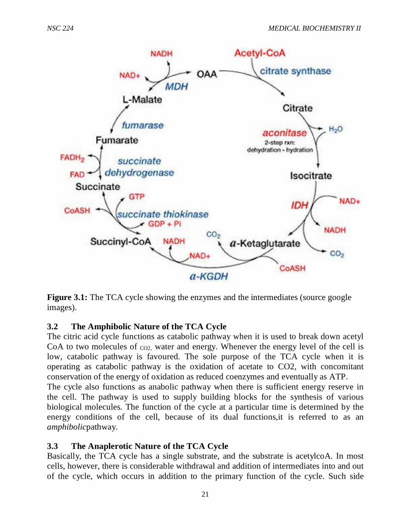
precursors or building blocks for the synthesis of molecules such as amino acids, purine bases, cholesterol and porphyrins. The cycle starts when a four-carbon compound (oxaloacetate) condenses with a two carbon acetyl unit of acetyl coA to yield a six carbon tricarboxylic acetate (citrate). In a cyclic series of reactions (figure 2.1) the isomer of citrate (isocitrate) is oxidativelydecarboxylated (one molecule of CO2 is released). The resulting five carbon compound, α-ketoglutarate is also oxidativelydecarboxylated (another molecule of CO2 is also released) to yield a four-carbon compound (succinate). Oxaloacetate, the starting material is eventually regenerated through the formation of fumarate and malate. Oxaloacetate’s function in kreb’s cycle can be described as catalytic in nature because the compound participates in the oxidation reaction and it is regenerated at the end of the cycle. Carboxylation of pyruvate is the major source of oxaloacetate as the starting materials for the cycle.
NSC 224 MEDICAL BIOCHEMISTRY II
21
Figure 3.1: The TCA cycle showing the enzymes and the intermediates (source google images).
3.2 The Amphibolic Nature of the TCA Cycle The citric acid cycle functions as catabolic pathway when it is used to break down acetyl CoA to two molecules of CO2, water and energy. Whenever the energy level of the cell is low, catabolic pathway is favoured. The sole purpose of the TCA cycle when it is operating as catabolic pathway is the oxidation of acetate to CO2, with concomitant conservation of the energy of oxidation as reduced coenzymes and eventually as ATP. The cycle also functions as anabolic pathway when there is sufficient energy reserve in the cell. The pathway is used to supply building blocks for the synthesis of various biological molecules. The function of the cycle at a particular time is determined by the energy conditions of the cell, because of its dual functions,it is referred to as an amphibolicpathway.
3.3 The Anaplerotic Nature of the TCA Cycle Basically, the TCA cycle has a single substrate, and the substrate is acetylcoA. In most cells, however, there is considerable withdrawal and addition of intermediates into and out of the cycle, which occurs in addition to the primary function of the cycle. Such side
NSC 224 MEDICAL BIOCHEMISTRY II
22
reactions serve two main purposes: one, to provide for the synthesis of compounds derived from any of several intermediates of the cycle and to replenish the supply of intermediates in the cycle as needed to prevent the shutting down of the cycle
Oxaloacetate and α-ketoglutarate are used in the synthesis of several amino acids. Citrate is the source of the acetyl coA in the cytosol, which is used for the synthesis of fats, other lipids and some amino acids. These are some of the major drains on the TCA cycle. Reactions that replenish the intermediates in the TCA cycle are termed Anaplerotic. One of such reactions is the conversion of pyruvate to oxaloacetate. It is not necessary to replenish the intermediate that is used in a biosynthetic pathway directly, as the replenishment of any intermediate will occur by a feeding-in process at any point in the cycle. For example, when carbohydrates are being metabolized, the TCA cycle intermediates are replenished by production of oxaloacetate from pyruvate.
3.4 The relationship between TCA cycle and Beriberi Beriberi, a neurological and cardiovascular disorder is caused by a dietary deficiency of thiamin also called vitamin B1. Beriberi is also occasionally seen in alcoholics who are severely malnurished and thus thiamine deficient. The disease is characterized by neurologic and cardiac symptoms such as pain in the limbs and distorted skin sensation. The heart may be enlarged and may eventually lead to paralysis. Which biochemical processes might be affected by a deficiency of thiamine? Thiamine pyrophosphate is the prosthetic group of two important TCA cycle enzymes; pyruvate dehydrogenase and α-ketoglutatrate dehydrogenase. In beriberi, the levels of pyruvate and α-ketoglutarate in the blood are higher than normal. The reason why vitamin B1 deficiency leads to neurological disorders is because the nervous system relies essentially on glucose as its only fuel. In contrast, most other tissues can use fat as a source of fuel (Acetyl CoA) for the citric acid cycle. The pyruvate dehydrogenase complex is required to convert pyruvate (the end product of glycolysis) to Acetyl CoA. When the enzymes are inactivated due to thiamine deficiency, energy production in the nervous system is shut down. The consequence of this are the symptoms of beriberi listed above.
3.5 Summary of oxidative phosphorylation The reduced coenzymes (NADH is called the reduced form of nicotinamide adenine dinucleotide and FADH2 is called the reduced form of flavine adenine dinucleotide) derived from the TCA cycle are themselves oxidized when they released their protons and electrons. The electrons are transferred to oxygen, the final electron acceptor through a complex chain of electron-carrying molecules known as the electron transport chain. During the electron transferring process, large amount of energy is released and it is conserved in the form of ATP. This process is called oxidative phosphorylation.
3.6 Inhibitors of electron transport chain Inhibitors of electron transport chain were found to be useful asbarbiturate drugs, antibiotics and insecticides especially those that are selective. Examples include: Amytal
NSC 224 MEDICAL BIOCHEMISTRY II
23
(a barbiturate drug), rotenone (a plant product commonly used as an insecticide) and piericidin A and oligomycin (antibiotics) block the electron flow through the respiratory chain and thereby shut down energy production in their respective targets. 4.0 Conclusion Carbohydrates are the major source of energy for the living cells. Glucose is the central molecule in carbohydrates metabolism. Glucose is oxidized in glycolysis either anaerobically or aerobically. Acetyl CoA is produced from pyruvate which is completely oxidized in the TCA cycle. Thus, complete oxidation of one mole of glucose generates 38 ATP molecules.
5.0 Summary In this unit, you have learnt about the following:
i. Description of TCA cycle ii. Amphibolic Nature of the TCA Cycle iii. Anaplerotic Nature of the TCA Cycle iv. Relationship between TCA cycle and Beriberi v. Summary of oxidative phosphorylation vi. Inhibitors of electron transport chain
6.0 Tutor Marked Assignments
i. Explain the Amphibolic nature of the TCA cycle ii. Explain the Anaplerotic nature of the TCA cycle iii. Describe the relationship between this cycle and Beriberi (a neurological disease) iv. Give the summary of oxidative phosphorylation v. Give examples and describe the inhibition of electron transport chain
7.0 References and Further reading
Katherine, M. A. Rogers and William N. Scott (2011). Nurses! Test yourself in anatomy and physiology Kathryn, A. Booth, Terri. D. Wyman (2008). Anatomy, physiology, and pathophysiology for allied health Keith L.M, Persuade T.V.N (2006). The Developing Human Clinically Oriented Embryology 8th Edition Lippincott Williams & Wilkins Kent, M. Van De Graff, R.WardRhees, Sidney P. (2010). Schaum’s outline of human anatomy and physiology 3rd edition. Philip, T. (2012). Seeley’s principles of anatomy & physiology 2nd edition. Sadler, T.W (2004). Langman’s Medical Embryology 9th edition.
NSC 224 MEDICAL BIOCHEMISTRY II
24
Module-2: FATTY ACID OXIDATION
Module Objective: At the end of this module, you should be able to discuss the following:
i. The concept of Fatty Acid Oxidation. ii. How fatty acids are activated for oxidation
iii. Entry of fatty acyl coA into mitochondria.
Contents Unit 1: Fatty Acid Oxidation Unit 2: Fatty Acid Oxidation II
UNIT ONE- FATTY ACID OXIDATION
CONTENT 1.0 Introduction 2.0 Objectives 3.0 Main Content 3.1 Fatty Acid Activation 3.2 β-Oxidation of Fatty Acids 3.3 Net ATP Yield from Palmitate Oxidation 4.0 Conclusion 5.0 Summary 6.0 Tutor Marked Assignments 6.1 Activity 6.2 Self-assessment Question 7.0 References and other resources
1.0 Introduction Fatty acids serve as a more efficient source of energy than carbohydrates. This is because they are reduced and anhydrous. The energy yield from 1 g of fatty acids is approximately 9Kcal, compared to 4Kcal for CHOs. Since the hydrocarbon portion of FAs is hydrophobic, these molecules can be stored in a relatively anhydrous environment. CHOs are more highly hydrated. If the human body relied on CHOs to store energy, then a person will need to carry 31kg of hydrated glycogen to have the energy equivalent to 5kg of fat. Hibernating animals provide a good example for utilizing fat reserves as fuel e.g bears hibernate for about 7 months, during which it derives its energy from fat stores. Utilization of fatty acids for energy production varies significantly from tissue to tissue and depends on the metabolic status of the tissue/ organ i.e fed or fasted, exercising or at rest. They are a major source of energy in cardiac and skeletal muscle, while the brain utilizes them poorly due to limited transport across the blood-brain barrier. Red blood
NSC 224 MEDICAL BIOCHEMISTRY II
25
cells cannot oxidize fatty acid because they lack mitochondria. During prolonged fasting, the liver converts acetyl coA generated by FA oxidation and amino acid breakdown to ketone bodies which become a major fuel. 2.0 Objectives At the end of this unit, you should be able to:
i. Describe the activation of fatty acids and the transport of fatty acyl coAs into the mitochondria, and list the steps of β-oxidation
ii. Outline the steps involved in β-oxidation iii. Calculate the net ATP yield from Palmitate oxidation.
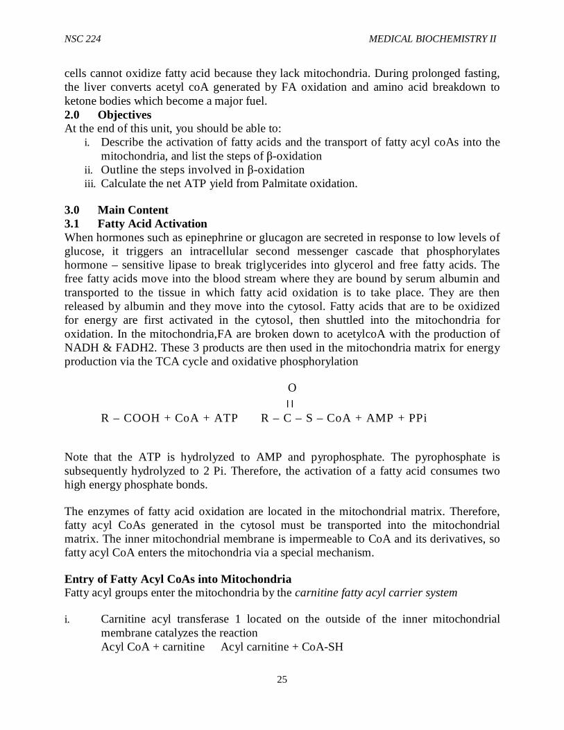
3.0 Main Content 3.1 Fatty Acid Activation When hormones such as epinephrine or glucagon are secreted in response to low levels of glucose, it triggers an intracellular second messenger cascade that phosphorylates hormone – sensitive lipase to break triglycerides into glycerol and free fatty acids. The free fatty acids move into the blood stream where they are bound by serum albumin and transported to the tissue in which fatty acid oxidation is to take place. They are then released by albumin and they move into the cytosol. Fatty acids that are to be oxidized for energy are first activated in the cytosol, then shuttled into the mitochondria for oxidation. In the mitochondria,FA are broken down to acetylcoA with the production of NADH & FADH2. These 3 products are then used in the mitochondria matrix for energy production via the TCA cycle and oxidative phosphorylation
O ׀ ׀
R – COOH + CoA + ATP R – C – S – CoA + AMP + PPi
Note that the ATP is hydrolyzed to AMP and pyrophosphate. The pyrophosphate is subsequently hydrolyzed to 2 Pi. Therefore, the activation of a fatty acid consumes two high energy phosphate bonds.
The enzymes of fatty acid oxidation are located in the mitochondrial matrix. Therefore, fatty acyl CoAs generated in the cytosol must be transported into the mitochondrial matrix. The inner mitochondrial membrane is impermeable to CoA and its derivatives, so fatty acyl CoA enters the mitochondria via a special mechanism.
Entry of Fatty Acyl CoAs into Mitochondria Fatty acyl groups enter the mitochondria by the carnitine fatty acyl carrier system
i. Carnitine acyl transferase 1 located on the outside of the inner mitochondrial membrane catalyzes the reaction Acyl CoA + carnitine Acyl carnitine + CoA-SH
NSC 224 MEDICAL BIOCHEMISTRY II
26
ii. Carninitine acyl translocase transports the acyl carnitine across the inner mitochondrial membrane into the matrix, and simultaneously transports free carnitine to the cytosol.
iii. In the matrix, carnitine acyl transferase II resynthesizes the fatty acyl CoA and releases free carnitine. Acyl carnitine + CoA Acyl CoA + carnitine
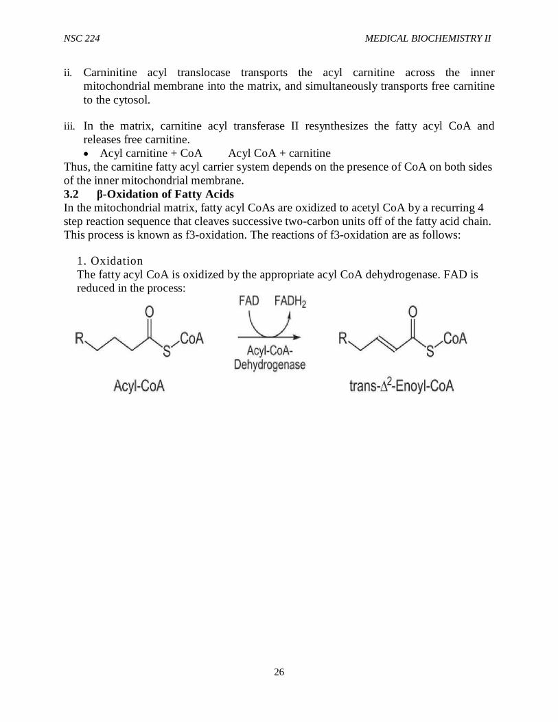
Thus, the carnitine fatty acyl carrier system depends on the presence of CoA on both sides of the inner mitochondrial membrane. 3.2 β-Oxidation of Fatty Acids In the mitochondrial matrix, fatty acyl CoAs are oxidized to acetyl CoA by a recurring 4 step reaction sequence that cleaves successive two-carbon units off of the fatty acid chain. This process is known as f3-oxidation. The reactions of f3-oxidation are as follows:
1. Oxidation The fatty acyl CoA is oxidized by the appropriate acyl CoA dehydrogenase. FAD is reduced in the process:
NSC 224 MEDICAL BIOCHEMISTRY II
27
The mitochondrion contains at least 4 dehydrogenases specific for fatty acyl CoAs of different chain lengths. They are very long chain, long chain, medium chain and short chain acyl-CoA dehydrogenases (VLCAD, LCAD, MCAD and SCAD). VL CAD – oxidizes straight chain acyl-CoA from C 12 – C 24. M CAD has broad chain length specificity but is most active with C6 and C8 substrates. S CAD order of preferred C4 > C6 > C8 LCAD is involved in initiating the oxidation of branched chain FA .
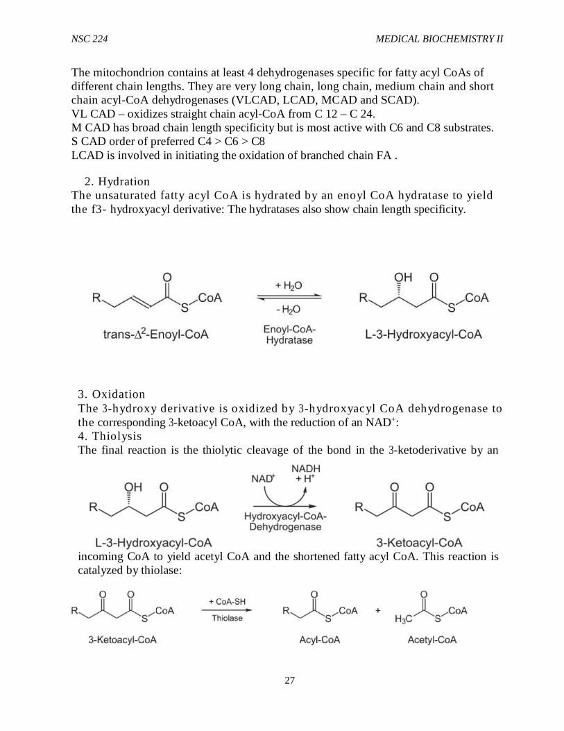
2. Hydration The unsaturated fatty acyl CoA is hydrated by an enoyl CoA hydratase to yield the f3- hydroxyacyl derivative: The hydratases also show chain length specificity.
3. Oxidation The 3-hydroxy derivative is oxidized by 3-hydroxyacyl CoA dehydrogenase to the corresponding 3-ketoacyl CoA, with the reduction of an NAD+: 4. Thiolysis The final reaction is the thiolytic cleavage of the bond in the 3-ketoderivative by an
incoming CoA to yield acetyl CoA and the shortened fatty acyl CoA. This reaction is catalyzed by thiolase:
NSC 224 MEDICAL BIOCHEMISTRY II
28
The shortened fatty acid chain is now ready for the next cycle of 3-oxidation. A feature unique to the oxidation of a long chain FA is that the enoyl CoA hydratase, 3-hydroxyacyl. CoA DH and 3-ketothiolase steps are all cat by a membrane bond complex of the 3 enzymes called a trifunctional protein. This complex is different from the enzyme that catalyzes oxidation of medium and short chain acyl CoAs, all of which are soluble proteins in the mitochondria matrix.
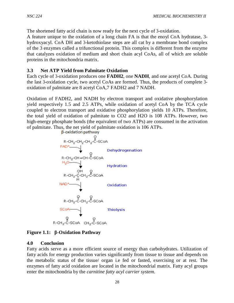
3.3 Net ATP Yield from Palmitate Oxidation Each cycle of 3-oxidation produces one FADH2, one NADH , and one acetyl CoA. During the last 3-oxidation cycle, two acetyl CoAs are formed. Thus, the products of complete 3-oxidation of palmitate are 8 acetyl CoA,7 FADH2 and 7 NADH.
Oxidation of FADH2, and NADH by electron transport and oxidative phosphorylation yield respectively 1.5 and 2.5 ATPs, while oxidation of acetyl CoA by the TCA cycle coupled to electron transport and oxidative phosphorylation yields 10 ATPs. Therefore, the total yield of oxidation of palmitate to CO2 and H2O is 108 ATPs. However, two high-energy phosphate bonds (the equivalent of two ATPs) are consumed in the activation of palmitate. Thus, the net yield of palmitate oxidation is 106 ATPs.
Figure 1.1: β-Oxidation Pathway
4.0 Conclusion Fatty acids serve as a more efficient source of energy than carbohydrates. Utilization of fatty acids for energy production varies significantly from tissue to tissue and depends on the metabolic status of the tissue/ organ i.e fed or fasted, exercising or at rest. The enzymes of fatty acid oxidation are located in the mitochondrial matrix. Fatty acyl groups enter the mitochondria by the carnitine fatty acyl carrier system.
NSC 224 MEDICAL BIOCHEMISTRY II
29
5.0 Summary In this unit, you have learnt about the following:
i. The process of Fatty Acid Activation ii. β-Oxidation of Fatty Acids iii. Net ATP Yield from Palmitate Oxidation
6.0 Tutor Marked Assignment i. Explain why fatty acids are more efficient than carbohydrates as fuel molecules ii. Write balanced equations for the first β-oxidation cycle of palmitate.
iii. Describe the activation of fatty acids and the transport of fatty acyl coAs into the mitochondria.
7.0 References and Further Reading
Katherine, M. A. Rogers and William N. Scott (2011). Nurses! Test yourself in anatomy and physiology
Kathryn, A. Booth, Terri. D. Wyman (2008). Anatomy, physiology, and pathophysiology for allied health
Keith L.M, Persuade T.V.N (2006). The Developing Human Clinically Oriented Embryology 8th Edition Lippincott Williams & Wilkins
Kent, M. Van De Graff, R.WardRhees, Sidney P. (2010). Schaum’s outline of human anatomy and physiology 3rd edition. Philip, T. (2012). Seeley’s principles of anatomy & physiology 2nd edition.
NSC 224 MEDICAL BIOCHEMISTRY II
30
UNIT TWO- FATTY ACID OXIDATION 2 CONTENT
1.0 Introduction 2.0 Objectives 3 .0 Main con ten t 3.1 Oxidation of Unsaturated Fatty Acids 3.2 Oxidation of Fatty Acids Containing an Odd Number of Carbons 3.3 α- and ω-Oxidation of Fatty Acids 3.4 Regulation of fatty acid oxidation 3.5 Ketone Bodies 3.6 Clinical Aspects 4.0 Conclusion 5.0 Summary 6.0 Tutor Marked Assignments 6.1 Activity 6.2 Tutor Marked Tests
1.0 Introduction Although β-oxidation is the major pathway of fatty acid oxidation, it is limited to the oxidation of even numbered saturated fatty acids. Other fatty acids (Unsaturated FA, those with odd no of carbon atoms) require modification of the Beta oxidation pathway. Also, Animal tissues contain minor pathways that involve oxidation of fatty acids at the α- and ω- carbons. The products of α and ω oxidation can enter 3-oxidation. Ketone bodies serve as alternate sources of fuel during periods of fasting or starvation and in conditions when the body is unable to utilize glucose, as occurs in Diabetes mellitus. Disorders of fatty acid oxidation include carnitine deficiency, Refsum’s disease, Jamaican vomiting sickness, Zellweger;s disease and ketoacidosis.
Other Mechanisms of Fatty Acid Oxidation Most fatty acids (especially saturated fatty acids) can be oxidized by the 3- oxidation pathway described in the previous study. However, fatty acids that contain an odd number of carbon atoms, certain unsaturated fatty acids, and methylated fatty acids require modifications of the 3-oxidation sequence.
2.0 Objectives At the end of this unit, you should be able to:
i. Describe pathways for oxidation of unsaturated fatty acids and fatty acids with an odd number of carbon atoms
ii. Describe α and ω oxidation of fatty acids iii. Outline the process by which fatty acid oxidation is regulated
NSC 224 MEDICAL BIOCHEMISTRY II
31
iv. Explain how ketone bodies are utilized for energy production v. Describe some disorders of Fatty acid oxidation
3.1 Main Content 3.1 Oxidation of Unsaturated Fatty Acids While the double bond that is generated between the α and β carbons in the first step of β-oxidation is in the trans configuration,the double bond in most unsaturated fatty acids are cis, and yield the D-isomer when hydrated by the hydratase. These D isomers are converted to the L-isomers by a racemase. β- oxidation then proceeds normally. If a double bond in an unsaturated fatty acid is located between the β and γ carbon atoms instead of between the α and β carbons, the fatty acid cannot directly enter β-oxidation. Instead, an isomerase moves the double bond to the correct position, and β-oxidation proceeds. If a double bond in an unsaturated fatty acid is located between the β and γ carbon atoms instead of between the α and β carbons, the fatty acid cannot directly enter β-oxidation. Instead, an isomerase moves the double bond to the correct position, and β-oxidation proceeds.
3.2 Oxidation of Fatty Acids Containing an Odd Number of Carbons
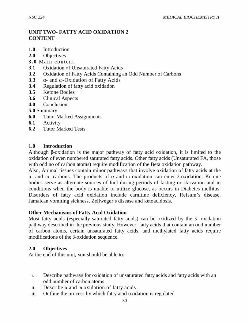
Fatty acids that contain and odd number of carbons are oxidized by β-oxidation, with the successive removal of two-carbon units, until a final three-carbon propionyl CoA is obtained. Propionyl CoA is utilized as shown in Figure 7-15. First, propionyl CoA is carboxylated to D-methylmalonyl CoA by propionyl CoA carboxylase. Methylmalonyl CoA. racemase then converts D-methylmalonyl CoA to L-methylmalonyl CoA. Finally, L-methyl CoA undergoes rearrangement by methlmalonyl CoA mutase to yield succinyl CoA. As a result of entering the TCA cycle, succinyl CoA may be oxidized to CO2 and H2O or used as a precursor for glconeogenesis.
NSC 224 MEDICAL BIOCHEMISTRY II
32
Figure: Metabolism of propionylcoA
3.3 α- and ω -Oxidation of Fatty Acids Animal tissues contain minor pathways that involve oxidation of fatty acids at the α- and ω-carbons. The products of α and ω oxidation can enter β-oxidation.
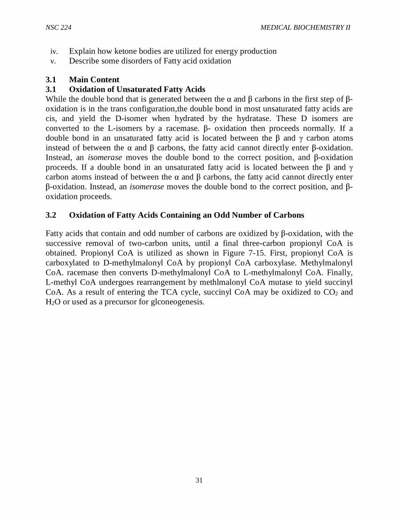
α-Oxidation During their metabolism, some fatty acids are hydroxylated on C-2 (the α carbon). The resulting α-hydroxy derivatives may be further oxidized to yield CO2 and fatty acids consisting of one less carbon atom, which may then be metabolized by 3-oxidation. α -Oxidation, which occurs in the endoplasmic reticulum, is especially important in the oxidation of methylated fatty acids. It is a method of generating odd-chain fatty acids.
NSC 224 MEDICAL BIOCHEMISTRY II
33
Figure: α-oxidation of phytanic acid
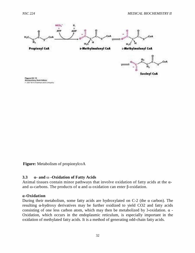
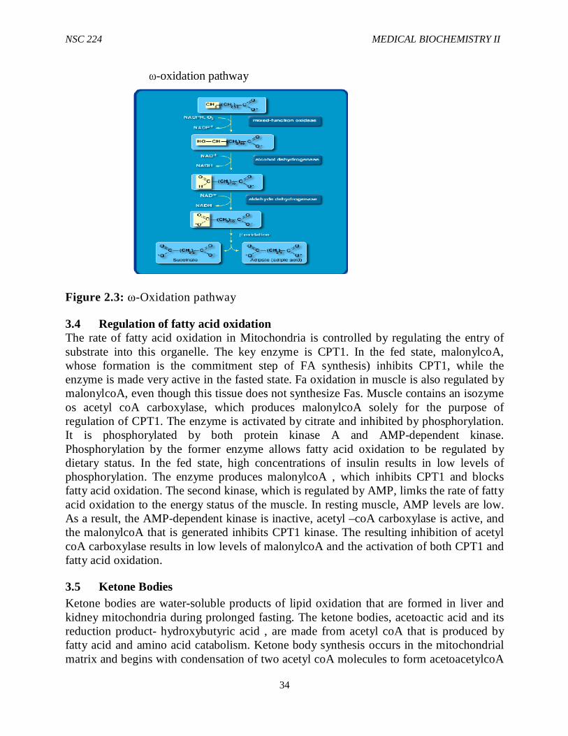
ω-Oxidation Fatty acids that are hydroxylated on the terminal carbon can undergo ω-oxidation. The ω-hydroxy group is converted to an ω-carboxy group, yielding an α, ω-dicarboxylic fatty acid. If this dicarboxylic fatty acid enters β-oxidation, it can be oxidized from both ends. ω-Oxidation of fatty acids has been detected on the endoplasmic reticulum of liver cells.
NSC 224 MEDICAL BIOCHEMISTRY II
34
ω-oxidation pathway
Figure 2.3: ω-Oxidation pathway
3.4 Regulation of fatty acid oxidation The rate of fatty acid oxidation in Mitochondria is controlled by regulating the entry of substrate into this organelle. The key enzyme is CPT1. In the fed state, malonylcoA, whose formation is the commitment step of FA synthesis) inhibits CPT1, while the enzyme is made very active in the fasted state. Fa oxidation in muscle is also regulated by malonylcoA, even though this tissue does not synthesize Fas. Muscle contains an isozyme os acetyl coA carboxylase, which produces malonylcoA solely for the purpose of regulation of CPT1. The enzyme is activated by citrate and inhibited by phosphorylation. It is phosphorylated by both protein kinase A and AMP-dependent kinase. Phosphorylation by the former enzyme allows fatty acid oxidation to be regulated by dietary status. In the fed state, high concentrations of insulin results in low levels of phosphorylation. The enzyme produces malonylcoA , which inhibits CPT1 and blocks fatty acid oxidation. The second kinase, which is regulated by AMP, limks the rate of fatty acid oxidation to the energy status of the muscle. In resting muscle, AMP levels are low. As a result, the AMP-dependent kinase is inactive, acetyl –coA carboxylase is active, and the malonylcoA that is generated inhibits CPT1 kinase. The resulting inhibition of acetyl coA carboxylase results in low levels of malonylcoA and the activation of both CPT1 and fatty acid oxidation.
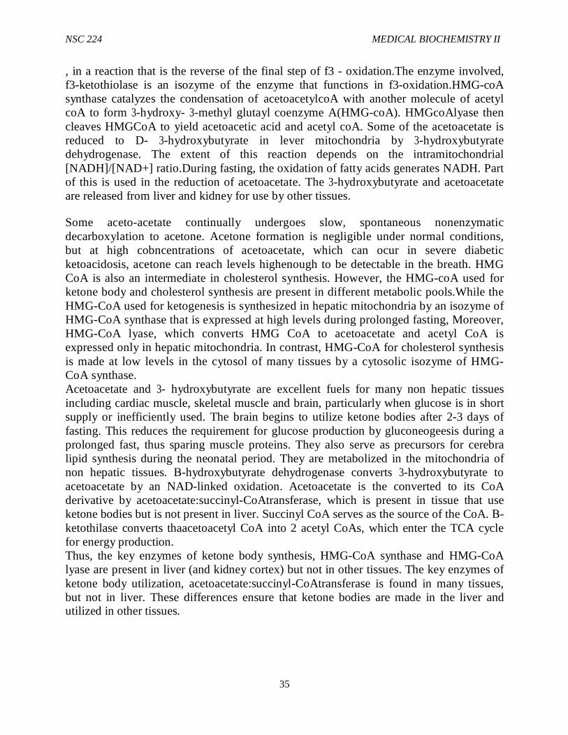
3.5 Ketone Bodies Ketone bodies are water-soluble products of lipid oxidation that are formed in liver and kidney mitochondria during prolonged fasting. The ketone bodies, acetoactic acid and its reduction product- hydroxybutyric acid , are made from acetyl coA that is produced by fatty acid and amino acid catabolism. Ketone body synthesis occurs in the mitochondrial matrix and begins with condensation of two acetyl coA molecules to form acetoacetylcoA
NSC 224 MEDICAL BIOCHEMISTRY II
35
, in a reaction that is the reverse of the final step of f3 - oxidation.The enzyme involved, f3-ketothiolase is an isozyme of the enzyme that functions in f3-oxidation.HMG-coA synthase catalyzes the condensation of acetoacetylcoA with another molecule of acetyl coA to form 3-hydroxy- 3-methyl glutayl coenzyme A(HMG-coA). HMGcoAlyase then cleaves HMGCoA to yield acetoacetic acid and acetyl coA. Some of the acetoacetate is reduced to D- 3-hydroxybutyrate in lever mitochondria by 3-hydroxybutyrate dehydrogenase. The extent of this reaction depends on the intramitochondrial [NADH]/[NAD+] ratio.During fasting, the oxidation of fatty acids generates NADH. Part of this is used in the reduction of acetoacetate. The 3-hydroxybutyrate and acetoacetate are released from liver and kidney for use by other tissues.
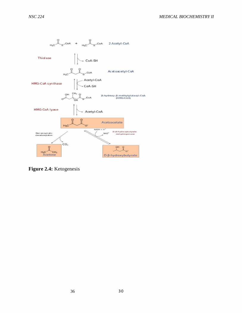
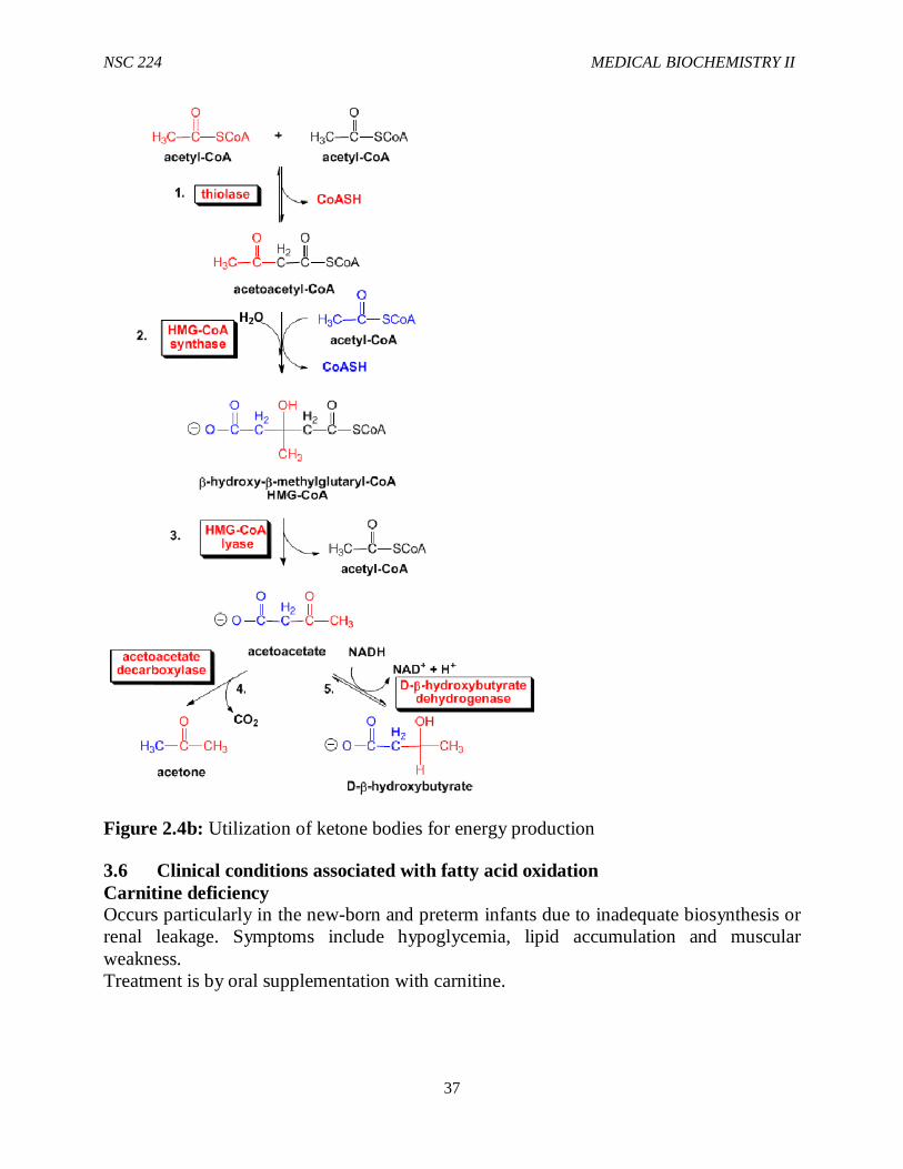
Some aceto-acetate continually undergoes slow, spontaneous nonenzymatic decarboxylation to acetone. Acetone formation is negligible under normal conditions, but at high cobncentrations of acetoacetate, which can ocur in severe diabetic ketoacidosis, acetone can reach levels highenough to be detectable in the breath. HMG CoA is also an intermediate in cholesterol synthesis. However, the HMG-coA used for ketone body and cholesterol synthesis are present in different metabolic pools.While the HMG-CoA used for ketogenesis is synthesized in hepatic mitochondria by an isozyme of HMG-CoA synthase that is expressed at high levels during prolonged fasting, Moreover, HMG-CoA lyase, which converts HMG CoA to acetoacetate and acetyl CoA is expressed only in hepatic mitochondria. In contrast, HMG-CoA for cholesterol synthesis is made at low levels in the cytosol of many tissues by a cytosolic isozyme of HMG-CoA synthase. Acetoacetate and 3- hydroxybutyrate are excellent fuels for many non hepatic tissues including cardiac muscle, skeletal muscle and brain, particularly when glucose is in short supply or inefficiently used. The brain begins to utilize ketone bodies after 2-3 days of fasting. This reduces the requirement for glucose production by gluconeogeesis during a prolonged fast, thus sparing muscle proteins. They also serve as precursors for cerebra lipid synthesis during the neonatal period. They are metabolized in the mitochondria of non hepatic tissues. Β-hydroxybutyrate dehydrogenase converts 3-hydroxybutyrate to acetoacetate by an NAD-linked oxidation. Acetoacetate is the converted to its CoA derivative by acetoacetate:succinyl-CoAtransferase, which is present in tissue that use ketone bodies but is not present in liver. Succinyl CoA serves as the source of the CoA. Β-ketothilase converts thaacetoacetyl CoA into 2 acetyl CoAs, which enter the TCA cycle for energy production. Thus, the key enzymes of ketone body synthesis, HMG-CoA synthase and HMG-CoA lyase are present in liver (and kidney cortex) but not in other tissues. The key enzymes of ketone body utilization, acetoacetate:succinyl-CoAtransferase is found in many tissues, but not in liver. These differences ensure that ketone bodies are made in the liver and utilized in other tissues.
NSC 224 MEDICAL BIOCHEMISTRY II
36
30
Figure 2.4: Ketogenesis
NSC 224 MEDICAL BIOCHEMISTRY II
37
Figure 2.4b: Utilization of ketone bodies for energy production
3.6 Clinical conditions associated with fatty acid oxidation Carnitine deficiency Occurs particularly in the new-born and preterm infants due to inadequate biosynthesis or renal leakage. Symptoms include hypoglycemia, lipid accumulation and muscular weakness. Treatment is by oral supplementation with carnitine.
NSC 224 MEDICAL BIOCHEMISTRY II
38
Refsum’s disease An autosomal recessive neurologic disorder of lipid metabolism which occurs due to a metabolic defect that results in accumulation of phytanic acid and phytol (dietary lipids derived from chlorophyll.) Affected individuals may display retinitis pigmentosa, diminished deep tendon reflexes and incoordination.
Zellweger’s syndrome Occurs in individuals with a rare inherited absence of peroxisomes in all tissues. They accumulate C26-C38 polyenoic acids in brain tissue and also exhibit a generalized loss of peroxisomal functions. The disease causes severe neurological symptoms, and most patients die in the first year of life.
Jamaican Vomiting Sickness Caused by eating the unripe fruit of the akee tree which contains the toxin hypoglycin. This inactivates medium and short chain acyl-CoA dehydrogenase, inhibiting 3- oxidation and causing hypoglycemia and is caused by a lack of mitochondrial medium chain acyl CoA dehydrogenase.
Ketoacidosis Higher than normal quantities of ketone bodies present in blood or urine constitute ketonemia and ketonuriarespectively.The overall condition is called ketosis. It is pathologic in Diabetes Mellitus. Non pathologic forms of ketosis are found under conditions of high fat feeding and after severe exercise in the post absorptive phase.
4.0 Conclusion In conclusion, oxidation of fatty acids involves activation of the fatty acid which occurs in the cytosol. The activated fatty acid is then transported to the mitochondria. Β-oxidation proper takes place in the mitochondrial matrix. Thus, fatty acids are oxidized by most tissues in the body. The ketone bodies supplies energy to certain organs, particularly the brain, heart and skeletal muscle under specific conditions including fasting, caloric restriction or sleep.
5.0 Summary In this unit, you have learnt about the following:
i. Oxidation of Unsaturated Fatty Acids ii. Oxidation of Fatty Acids Containing an Odd Number of Carbons iii. α- and w-Oxidation of Fatty Acids iv. Regulation of fatty acid oxidation v. Ketone Bodies vi. Clinical Aspects
NSC 224 MEDICAL BIOCHEMISTRY II
39
6.0 Tutor Marked Assignments i. Describe pathways for oxidation of unsaturated fatty acids and fatty acids with an
odd number of carbon atoms ii. Describe α and w oxidation of fatty acids iii. Outline the process by which fatty acid oxidation is regulated iv. Explain how ketone bodies are utilized for energy production v. Describe some disorders of Fatty acid oxidation
7.0 References and further reading Katherine, M. A. Rogers and William N. Scott (2011). Nurses! Test yourself in anatomy and physiology Kathryn, A. Booth, Terri. D. Wyman (2008). Anatomy, physiology, and pathophysiology for allied health Keith L.M, Persuade T.V.N (2006). The Developing Human Clinically Oriented Embryology 8th Edition Lippincott Williams & Wilkins Kent, M. Van De Graff, R.WardRhees, Sidney P. (2010). Schaum’s outline of human anatomy and physiology 3rd edition. Philip, T. (2012). Seeley’s principles of anatomy & physiology 2nd edition. Sadler, T.W (2004). Langman’s Medical Embryology 9th edition.
NSC 224 MEDICAL BIOCHEMISTRY II
40
MODULE- THREE-VITAMINS AND TRACE ELEMENTS
Introduction Vitamins are group of organic nutrients that are required in minute (small) quantities for normal metabolism. Generally they cannot be synthesized by the body and therefore be supplied in the diet. Vitamins are classified based on their solubility as water soluble or fat soluble. Water Soluble include the B complex vitamins, Vitamin C, Biotin, Folic acid and Pantothenic acid. The fat soluble vitamins include vitamins A, D, E and K. Most of these vitamins act as coenzymes. A coenzyme is a non protein component of an enzyme (cofactor) which is organic in nature. Many of coenzymes are vitamins derivatives, and are essential in the activity of the enzyme.
Module Objective: At the end of this module, you should be able to discuss the following in details:
i. The types of Fat Soluble Vitamins ii. The sources of Fat Soluble Vitamins iii. The functions of Fat Soluble vitamins iv. The deficiency associated with Fat Soluble vitamins v. Trace elements and their biological functions
CONTENTS Unit 1: The Fat Soluble Vitamins Unit 2: The Fat Soluble Vitamins Unit 3: Trace Elements
UNIT ONE- THE FAT SOLUBLE VITAMINS
CONTENT 1.0 Introduction 2.0 Objectives 3.0 Main Content 3.1 Vitamin A or Retinol 3.2 Description of Vitamin D 3.3 Description of Vitamin E 3.4 Description of Vitamin K 4.0 Conclusion 5.0 Summary 6.0 Tutor Marked Assignments 6.1 Activity 7.0 References and other resource
NSC 224 MEDICAL BIOCHEMISTRY II
41
1.0 Introduction Vitamins are organic molecules that are required in small quantity for a variety of biochemical functions and which generally cannot be synthesized in the body but must be supplied in the diet or as supplement. These molecules serve the same roles in nearly all forms of life. Some are synthesized by intestinal micro organisms but in quantity that are not sufficient to meet our need. Human beings require at least 12 vitamins in the diet for various biochemical activities. Deficiency of vitamins can generate diseases in all organisms requiring them for important biochemical reactions. Vitamins can be grouped according to whether they are soluble in water or in non polar solvents. Water soluble vitamins function as coenzyme, while fat soluble vitamins participate in diverse processes such as blood clotting and vision.
2.0 Objectives At the end of this unit, you should be able to:
i. Define fat soluble vitamins (FSV) and list all the FSV ii. Describe vitamin A iii. Describe vitamin D iv. Describe vitamin E v. Describe vitamin K vi. Give a detail description of Fat soluble vitamins
3.0 Main Content 3.1 Vitamin A (Retinol) Two groups of compounds have vitamin A activity; the first group is called retinoid which comprises retinol, retinal and retinoic acid. They are preformed vitamin A, found only in foods of animal origin. The second group is carotenoid, found only in plants; they are composed of β-carotenes and related compounds. Carotenoids are cleaved in the intestinal mucosa by carotene dioxygenase to yield retinal which is reduced to retinol. Retinol is stored in the liver of animals as lipid ester. Vitamin A is heat stable but sensitive to ultraviolet light (UV). This is why it is not good to put your palm oil in the sun when you want to liquefy it; application of little heat is better. This is a common practice in many African communities; it is not a good practice because the ultraviolet rays in the sun destroy the vitamin A present in the oil. Next time you see anyone doing this, please correct the person.
Sources of Vitamin A The richest dietary sources of preformed vitamin A (retinol) are fish liver oils also known as cod liver oil. Other sources include liver of animals, milk and dairy products, dark green vegetables, yellow or red fruits, carrot and tomatoes. Palm-oil is the richest dietary source of carotenoids.
Functions of Vitamin A Roles of vitamin a in vision- The role of vitamin A in vision was discovered by George Wald, who received the Nobel Prize in 1943 for this discovery. Vision is based on the

NSC 224 MEDICAL BIOCHEMISTRY II
42
absorption of light by photoreceptor cells in the eye. These cells are sensitive to light in a relatively narrow region of the electromagnetic spectrum, with wavelengths between 300 and 850nm. Vertebrates have two kinds of photoreceptor cells, called rods and cones because of their distinctive shapes. Cones function in bright light and are responsible for colour vision, whereas rods function in dim light but do not perceive colour. A human retina contains about 3 million cones and 100 million rods. Rods are slender elongated structures densely packed with phtoreceptors molecules. The photosensitive molecule is often called a visual pigment because it is highly coloured owing to its ability to absorb light. The photoreceptor molecule in rods is rhodopsin, which consists of the protein opsin covalently linked to 11-cis-retinal that serves as a prosthetic group. Iodopsin is the photoreceptor molecule present in the cones.
Gene expression and tissue differentiation Another important function of vitamin A is the control of cell differentiation. All-trans-retinoic acid and 9-cis-retinoic acid regulate growth, development and tissue differentiation; they have different actions in different tissues. Retinoic acid binds to nuclear receptor and regulates the transcription of specific genes. There are two families of nuclear retinoid receptors: the retinoic acid receptors (RAR) bind All-trans-retinoic or 9-cis-retinoic acid and the retinoid X receptors (RXR) bind only 9-cis-retinoic acid.
Deficiency of Vitamin A Vitamin A deficiency is the most important preventable cause of blindness in the world. The earliest sign of deficiency is a loss of sensitivity to green light, followed by impairment to adapt to dim light followed by night blindness. More prolonged deficiency leads to xerophthalmia (dry and keratinization of the cornea and blindness). Growth retardation, dermatitis, anorexia and hypogeusia .Vitamin A also has an important role in differentiation of immune system cells. Mild deficiency of vitamin A leads to increased susceptibility to infectious diseases.
Toxicity Vitamin A Human body has a limited capacity to metabolize vitamin A, and excessive intakes leads to accumulation beyond the capacity of binding protein (opsin), so that unbound vitamin A causes tissue damage. Excess vitamin A is teratogenic to pregnant women (it can cause congenital deformity in the fetus). Symptoms of toxicity affect the central nervous system and this include headache, nausea, anorexia (lack of appetite) and ataxia (defective muscular control leading to staggering). Liver and bones are also affected. Excessive dryness of skin is also a symptom of vitamin A toxicity.
NSC 224 MEDICAL BIOCHEMISTRY II
43
3.2 Description of Vitamin D Vitamin D is also known as cholecalciferol. Vitamin D could be thought of as a hormone rather than vitamin because it can be synthesized in the body; it is released into the blood circulation like hormones and has biochemical effects on target organs. Vitamin D is included in the list of vitamins, as it becomes an essential dietary factor when endogenous synthesis is inadequate to meet the physiological requirements. This condition is common in the temperate region where there may not be enough sunlight for greater part of a year. It is also common among women in some Arab nations where virtually all the parts of a woman’s body is covered thereby preventing vitamin D synthesis in the skin. There are two forms of vitamin D: The naturally produced vitamin D3 or cholecalciferol is the form obtained from animal sources in the diet or made in the skin. It is produced in the skin from ultraviolet activation of 7-dehydrocholesterol. Artificially produced vitamin D2 or ergocalciferol, is the form made in the laboratory by irradiating the plant sterol, ergosterol and it is the form most readily available for pharmaceutical use. In the temperate regions, the plasma concentration of vitamin D is highest at the end of summer and lowest at the end of winter. In the tropical regions, vitamin D deficiency is not common due to availability of sunlight for most part of the year.
Dietary sources of vitamin D The most reliable dietary sources of vitamin D are fortified foods. Milk for example is fortified to a level of 400 international units per quart. The recommended daily intake of vitamin D is 400 IU, irrespective of age. Other sources include the liver, cod liver oil and egg yolk.
Functions of vitamin D Its main function is the regulation of calcium metabolism and absorption. Vitamin D is converted to calcitriol (1,25-dihydrxycholecalciferol), the active hormone by hydroxylation reactions in the liver and kidneys. It binds to receptor, structurally similar to the steroid receptors to form a complex that functions as a transcription factor thereby regulating gene expression.
Vitamin D deficiency Vitamin D deficiency in childhood produces rickets, a disease characterized by inadequate calcification of cartilage and bone as a result of poor absorption of calcium. Similar problems occur in adult as a result of demineralization of bone, especially in women who have little exposure to sunlight. In adults, vitamin D deficiency leads to softening and weakening of bones, a condition called osteomalacia. Osteoporosis is another bone disease associated with old age. It is described as the loss of bone density caused by excessive absorption of calcium and phosphorus from the bone.
Toxicity of vitamin D High concentration of vitamin D in the plasma can lead to contraction of blood vessels and calcinosis, i.e. the calcification of soft tissues such as the liver and kidneys. This has serious consequences on health and can lead to death.
NSC 224 MEDICAL BIOCHEMISTRY II
44
3.3 Description of Vitamin E Vitamin E, also known as tocopherol is the generic name for two families of compounds. Tocopherols (α, β, γ and δ type) differ in the number and position of the methyl groups on the ring. The α-tocopherol is the most potent and it is usual to express vitamin E intake in terms of milligrams α-tocopheerol equivalent. Tocotrienols are structurally related to tocopherols but they are less potent and also contain unsaturated hydrocarbon side chains.
Dietary sources of vitamin E The major dietary sources of vitamin E are fats and oils with different tocopherol content. The richest sources are: Soya and corn oils (50-150 mg/100gm) and Palm oil (20-70 g/100gm), Coconut and olive oils are relatively low in vitamin E content (1-10mg/100gm). The major site of vitamin E storage is the adipose tissue. Synthetic vitamins E are also available as dietary supplements.
Functions of vitamin E Vitamin E does not have a precisely defined metabolic function. It acts as lipid-soluble antioxidant in cell membranes. Its antioxidant functions include radical chain-breaking and free radical trapping in cell membrane and plasma lipoprotein by reacting with the lipid peroxide radicals formed by peroxidation of poly unsatureated fatty acids.
Vitamin E deficiency Vitamin E deficiency in human is rare. The only known symptom of vitamin E deficiency is haemolytic anaemia due to an increased red blood cell fragility and damage
Toxicity of vitamin E There is no record of vitamin E toxicity but because it is fat soluble, too much of it may be toxic. Unlike the other fat soluble vitamins such as vit A, K and D, vit E does not seem to have any known toxic effects.
3.4 Description of Vitamin K Vitamin K derived its name from German word koagulation. The most important function of vitamin K is its role in the synthesis of blood clotting proteins, hence it is called blood clotting vitamin. There are two naturally occurring forms of vitamin K, the first is phylloquinone; the normal dietary source found in green vegetables. The second is known as menaquinones, synthesized by the intestinal bacteria of children but adult cannot synthesize vitamin K. Menadiones and menadioldiacetate are synthetic compounds that can be metabolized to phylloquinone.
Dietary sources of vitamin K Excellent sources of vitamin K are cabbage and green vegetables, other sources are tomatoes, cheese, meat and egg yolk.
NSC 224 MEDICAL BIOCHEMISTRY II
45
Functions of vitamin K Synthesis of blood clotting proteins- vitamin K has been known for many years to be essential for the synthesis of prothrombin and several other clotting factors. Vitamin K participates in the carboxylation of glutamate residues to γ-carboxyglutamate, which makes modified glutamic acid a much stronger chelator of Ca2+. The results of studies of the abnormal prothrombin synthesized in the absence of vit K or in the presence of vitamin K antagonists (these are the compounds that prevent vitamin K from performing its functions) such as dicoumarol, revealed the mode of action of this vitamin. Dicoumarol is found in spoiled sweet clover leaves and causes a fatal hemorrhagic disease in cattle fed on this hay. This coumarin derivative is used clinically as an anticoagulant to prevent thromboses in patients prone to clot formation. Dicoumarol and such related vitamin K antagonists as warfarin also serve as effective rat poisons.
Deficiency of vitamin K The clinical manifestation of a Vitamin K deficiency is hemorrhage, when there is little cut, bleeding may continue for a very long time.
Toxicity of vitamin K High doses of the naturally occurring fat soluble form of Vitamin K (K1) appear to be non-toxic, but the water-soluble forms of menadione (K3) have produced serious side effects in high doses, especially in newborn infants. Large doses of menadione given to newborns or their mothers during labour have resulted in hemolytic anemia. Premature infants have less tolerance to exces Vitamin K than full-term infants do. Adult toxicity signs are primarily circulatory and involve a variety of cardiac and pulmonary signs.
4.0 Conclusion
In conclusion, vitamins play a very important role in the body. The fat soluble vitamins are obtained from animal and plant sources. Deficiency of one vitamin may be associated with a particular disease condition in the body.
5.0 Summary: In this unit, you have been taken through the following: i. Vitamin A or Retinol ii. Description of Vitamin D iii. Description of Vitamin E iv. Description of Vitamin K
6.0 Tutor Marked Assignments i. Define fat soluble vitamins (FSV) and list all the FSV ii. Describe vitamin A iii. Describe vitamin D iv. Describe vitamin E v. Describe vitamin K
NSC 224 MEDICAL BIOCHEMISTRY II
46
UNIT TWO- WATER SOLUBLE VITAMINS
CONTENT 1.0 Introduction 2.1 Objectives 3.0 Main Content 3.1 Vitamin B1 (Thiamine) 3.2 Vitamin B2 (Riboflavin) 3.3 Vitamin B3 (Niacin) 3.4 Vitamin B5 (Pantothenic acid) 3.5 Vitamin B6 (Pyridoxine) 3.6 Vitamin H (Biotin) 3.7 Vitamin B9 (Folic acid) 3.8 Vitamin B12 3.9 Vitamin C (Ascorbic acid) 4.0 Conclusion 5.0 Summary 6.0 Tutor Marked Assignments 6.1 Activity 6.2 Tutor Marked Tests 7.0 References and other resources
NSC 224 MEDICAL BIOCHEMISTRY II
47
1.0 Introduction All water soluble vitamins are readily soluble in water and are absorbed without the involvement of fat. Excess intakes are excreted in the urine but some of them have some side effects. Some of the vitamins are stored for a short period while many are stored in the body for up to 2 months. These water soluble vitamins function as co-enzymes; they must be metabolically converted to their active forms for them to be functional but vitamins C and biotin are used directly without conversion.
2.0 Objectives At the end of this unit, you should be able to:
i. List the biochemical functions, deficiency and natural sources of vitamin B1 (Thiamine).
ii. List the biochemical functions, deficiency and natural sources of vitamin B2 iii. Describe the biochemical functions, deficiency and natural sources of vitamin B3
(Niacin) iv. List the biochemical functions, deficiency and natural sources of vitamin B5
(Panthotenic acid) v. Enumerate the biochemical functions, deficiency and natural sources of vitamin B6
(Pyridoxine) vi. List the biochemical functions, deficiency and natural sources of vitamin H
(Biotin) vii. Itemise the biochemical functions, deficiency and natural sources of vitamin B9
(Folic acid) viii. Describe the biochemical functions, deficiency and natural sources of vitamin B12 ix. List the biochemical functions, deficiency and natural sources of vitamin C
3.0 Main Content 3.1 Vitamin B1 (Thiamine) Functions of vitamin B1
i. Thiamine reacts with ATP to form Thiamine pyrophosphate (TPP), the active form of vitamin B1 in biochemical reactions.
ii. TPP is a co-enzyme in the decarboxylation of pyruvate and α-ketoglutarate and also in transketolase and transaldolase reactions.
iii. Decarboxylation of pyruvate is a crucial reaction in nervous system; this was explained in section 2.4.
Deficiency of vitamin B1 i. Thiamine deficiency is associated with elevated levels of pyruvate and lactate. ii. Its deficiency also causes a clinical disorder known as Beriberi, Oedema and an
array of other abnormalities.
NSC 224 MEDICAL BIOCHEMISTRY II
48
Natural sources of vitamin B1 i. Thiamine is available in a wide variety of foods making its deficiency very rare.
Some of the sources include unpolished rice, whole grains such as maize, yeast, nuts and potatoes.
ii. It is easily destroyed by heat, so the thiamine content of foods is lowered by cooking.
Since Vitamin B1 functions in the release of energy from fuel molecules, its requirement is linked to caloric intake. The recommended daily allowance (RDA) of Thiamine is 1.0-1.5mg for most individuals. However higher quantity is required during pregnancy. Dietary deficiency is common in people with Anorexia, diarrhea, alcoholics and post-operative patients
3.2 Vitamin B2 (Riboflavin) Functions of vitamin B2
i. Riboflavin is the electron carrier co-enzymes that exist as FAD and FMN. ii. They function in a variety of redox reactions catalysed by oxidases, reductases and
dehydrogenases (fatty acid synthesis, TCA cycle and amino acid synthesis) iii. They are necessary for aerobic respiration and tissue maintenance.
Deficiency of Riboflavin Riboflavin deficiency is widespread but not fatal. Deficiency is characterized by cheilosis, desquamation and inflammation of the tongue, when this happen the person will not be able to take food containing pepper.
Natural sources of Riboflavin Riboflavin is found in a wide variety of foods, especially liver, kidney and green leafy vegetables. The major sources are milk and dairy products. Because of its intense yellow colour, it is used as food additives. The RDA for riboflavin is 1.2mg, but pregnant and lactating mother may require more.
3.3 Vitamin B3 (Niacin) Functions of vitamin B3 Niacin (also known as Nicotinic acid) and its amide derivative nicotinamide are the precursor of the co-enzymes NAD+ and NADP+. They are essential coenzymes in numerous cellular reactions such as lipid biosynthesis, the pentose phosphate pathway and amino acid metabolism.
Deficiency of vitamin B3 Niacin deficiency causes the disease pellagra. The major symptoms are photosensitive dermatitis on the skin, impaired digestion, diarrhea and mental confusion.
NSC 224 MEDICAL BIOCHEMISTRY II
49
Natural sources of Vitamin B3 Sufficient quantity can be synthesized endogenously from tryptophan. (Take note of the vitamins that are synthesized in sufficient quantity, we have mentioned vitamin D). It is present in meats, fish and nuts. The RDA for Niacin is 13mg. Excess niacin can cause liver damage.
3.4 Vitamin B5 (Pantothenic acid) Functions of vitamin B5
i. Pantothenate is a precursor of co-enzyme A, that participates in acyl transfer reactions.
ii. Coenzyme A is the carrier for acetyl groups obtained from the degradation of carbohydrates and lipids.
iii. Coenzyme A also carries the acetyl and malonyl groups used in fatty acid synthesis.
Deficiency i. Vitamin B5 deficiency has not been observed in humans. No RDA has been set for
Vitamin B5 yet. Most individuals ingest between 5 and 20mg of pantothenate per day. 7.4.3 Natural sources
ii. Beef liver, peanuts, Soybeans and wheat germ are all rich in pantothenate. 3.5 Vitamin B6 (Pyridoxine) Functions of vitamin B6
i. The biologically active forms of pyridoxine are pyridoxamine phosphate and pyridoxal phosphate and are mostly involved in amino acid metabolism.
ii. They function in various transamination and oxidation reactions. Some women use it for premenstrual pain.
Deficiency of vitamin B6 i. The deficiency symptoms include irritability, mental confusion, convulsions
(in infants), peripheral neuropathy and inflammation of the mouth.
Natural Sources i. Since the pyridoxine coenzymes are involved primarily with amino acid
metabolism their daily requirement varies with the daily protein intake. The vitamin is present in meat, fish and fruits. The RDA is 2.2mg for men and 2.0mg for women.
3.6 Vitamin H (Biotin)
Functions of Biotin i. Biotin which is used by cells without modification is the coenzyme for cellular
carboxylation reactions. ii. The carboxylation of acetyl CoA to malonyl CoA in preparation for fatty acid
synthesis is an example of biotin dependent carboxylation.
NSC 224 MEDICAL BIOCHEMISTRY II
50
Deficiency of Biotin i. Its deficiency has not been observed in humans on a normal diet. Raw egg
contains a protein called avidin that binds biotin very tightly. Biotin deficiency can occur in a person who takes raw eggs regularly.
ii. It is synthesized by the intestinal flora in amounts believed sufficient to meet the daily needs of man (This is the third vitamin that is synthesized in sufficient quantity by man).
Natural Sources The intake of the average healthy adult is 100 to 300µg/day. Sources include liver,
peanuts etc.
3.7 Vitamin B9 (Folic acid) Functions of Folic acid
i. Folic acid, also called folicin is converted to tetrahydrofolate, which is the primary carrier of one carbon units in the cell.
Deficiency Symptoms associated with folic acid deficiency are megaloblastic and macrocylic anemia, growth failure. All the symptoms disappear upon administration of folic acid. Natural sources
i. Folic acid is synthesized by the intestinal bacteria, so the daily dietary requirement is difficult to measure. The RDA for healthy individuals has been set at 400µg with higher allowances for pregnant and lactating women. Good sources of folic acid include Beef, Chicken, liver, peanuts and brewer’s yeast.
3.8 Vitamin B12
Functions of vitamin B12 i. Vitamin B12 consists of cobalamin and its active derivatives, including
cyanocobalamin and methyl cobalamin. ii. Cobalamin is an essential cofactor in the conversion of homocysteine to
methionine and in the conversion of methyl malonylcoA to succinyl-coA.
Deficiency i. Vitamin B12 deficiency impairs the metabolism of folic acid and this disturbs red
blood cell synthesis. ii. The clinical symptoms of vitamin B12 deficiency are collectively called
pernicious anemia and include megaloblastic anemia, changes in the intestinal mucosa and in extreme cases, neuropathy.
Natural Sources i. The vitamin B12 RDA for healthy individuals has been set at 3µg. The actual
daily intake for most individuals is from 5 to 15µg/day. ii. Most vitamin B12 in the body is concentrated in the liver.
NSC 224 MEDICAL BIOCHEMISTRY II
51
iii. In general foods derived from animals contain more of vitamin B12 than foods derived from plants.
3.9 Vitamin C (Ascorbic acid) Functions of vitamin C
i. The active forms of Vitamin C are ascorbic and dehydroascorbic acids. It is known as antiscurvy agent.
ii. The symptoms of scurvy are swollen gum, loss of teeth, skin lesions, pain and weakness (legs).
iii. Vitamin C participates in hydroxylation reactions and also promotes intestinal absorption of iron. They participate in amino acid metabolism, the synthesis of norepinephrine and the synthesis of collagen.
Deficiency of vitamin C i. Vitamin C deficiency causes scurvy. Other symptoms of scurvy include impaired
wound healing and changes in bone development and growth. ii. Many of these symptoms appear to be the result of the defective collagen
synthesized in the absence of Vitamin C.
NSC 224 MEDICAL BIOCHEMISTRY II
52
iii. Deficiency of vitamin C can also cause impaired growth in children. In severe cases, skin becomes rough, dry and develop hyperkeratotic (scaly) changes in the hair follicles.
iv. Drugs known to increase the urinary excretion of ascorbate include aspirin and barbiturates. Chronc diarrhea may contribute to vitamin C deficiency.
Natural Sources i. Good sources of vitamin C are peppers, papaya and citrus fruits. The RDA for
vitamin C is 60mg, based on the amount required to prevent or cure scurvy. More Vitamin C can be required during pregnancy and lactation.
4.0 Conclusion Water-soluble vitamins are soluble in aqueous solutions and are often used as cofactors by many enzymes. The water soluble vitamins also have common properties besides their solubility property. Since they are water soluble excess can be excreted through urine. Hypervitaminosis may not cause toxicity. Most of these vitamins act as coenzymes.
5.0 Summary In this study unit, you have been exposed to the following in details: i. Vitamin B1 (Thiamine) ii. Vitamin B2 (Riboflavin) iii. Vitamin B3 (Niacin) iv. Vitamin B5 (Pantothenic acid) v. Vitamin B6 (Pyridoxine) vi. Vitamin H (Biotin) vii. Vitamin B9 (Folic acid) viii. Vitamin B12 ix. Vitamin C (Ascorbic acid) 6.0 Tutor Marked Assignments
i. List the biochemical functions, deficiency and natural sources of vitamin B1 (Thiamine).
ii. List the biochemical functions, deficiency and natural sources of vitamin B2 iii. Describe the biochemical functions, deficiency and natural sources of vitamin B3
(Niacin) iv. List the biochemical functions, deficiency and natural sources of vitamin B5
(Panthotenic acid) v. Enumerate the biochemical functions, deficiency and natural sources of vitamin
B6 (Pyridoxine) vi. List the biochemical functions, deficiency and natural sources of vitamin H
(Biotin) vii. Itemise the biochemical functions, deficiency and natural sources of vitamin B9
(Folic acid) viii. Describe the biochemical functions, deficiency and natural sources of vitamin B12 ix. List the biochemical functions, deficiency and natural sources of vitamin C
4 3
NSC 224 MEDICAL BIOCHEMISTRY II
53
7.0 References and Further reading Katherine, M. A. Rogers and William N. Scott (2011). Nurses! Test yourself in anatomy and physiology Kathryn, A. Booth, Terri. D. Wyman (2008). Anatomy, physiology, and pathophysiology for allied health Keith L.M, Persuade T.V.N (2006). The Developing Human Clinically Oriented Embryology 8th Edition Lippincott Williams & Wilkins Kent, M. Van De Graff, R.WardRhees, Sidney P. (2010). Schaum’s outline of human anatomy and physiology 3rd edition. Philip, T. (2012). Seeley’s principles of anatomy & physiology 2nd edition. Sadler, T.W (2004). Langman’s Medical Embryology 9th edition.
NSC 224 MEDICAL BIOCHEMISTRY II
54
UNIT THREE- TRACE ELEMENTS CONTENT 1.0 Introduction 2.0 Objectives 3.0 Main Content 3.1 Macro vs Microminerals 3.2 Trace Elements 4.0 Conclusion 5 .0 Summary 6.0 Tutor Marked Assignments 6.1 Activity 6.2 Tutor Marked Tests 7.0 References and other resources
1.0 Introduction Dietary minerals are the chemical elements required by living organisms, other than the four elements carbon, hydrogen, nitrogen, and oxygen present in common organic molecules. For any nutrient, there is a range of intake between that which is clearly inadequate, leading to clinical deficiency disease, and that which is so much in excess of the body’s metabolic capacity that there may be signs of toxicity. Between the 2 extremes, there is a level of intake that is adequate for normal health and the maintenance of metabolic integrity. Individuals do not have the same requirement for nutrients, even when calculated on the basis of body size or energy expenditure. Therefore, in order to assess the adequacy of diets, it is necessary to set a reference level of intake high enough to ensure that no one either suffers from deficiency or is at risk of toxicity. Many of the essential minerals are widely distributed in foods, and most people eating a mixed diet are likely to receive adequate intakes, although supplements can be used when some requirements are not adequately met by the diet, or when chronic or acute deficiencies arise from pathology or injury, etc.
2.0 Objectives At the end of this unit, you should be able to:
i. Distinguish between Macro and Microminerals ii. List important microminerals (Trace Elements) and enumerate the functions, food
sources, required daily allowance, deficiency and toxicity symptoms of each trace mineral studied.
3.0 Main Content 3.1 Macro vs Microminerals Dietary Minerals include the macro and microminerals. Macrominerals are required in the diet in large amounts (>100mg/day). They represent about 80% of body organic matter and include calcium, phosphorus, potassium, sulfur, sodium, chlorine, and magnesium.
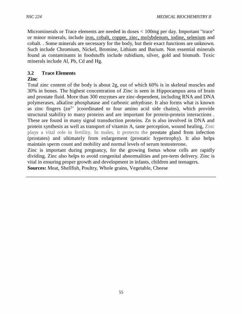
NSC 224 MEDICAL BIOCHEMISTRY II
55
Microminerals or Trace elements are needed in doses < 100mg per day. Important "trace" or minor minerals, include iron, cobalt, copper, zinc, molybdenum, iodine, selenium and cobalt. . Some minerals are necessary for the body, but their exact functions are unknown. Such include Chromium, Nickel, Bromine, Lithium and Barium. Non essential minerals found as contaminants in foodstuffs include rubidium, silver, gold and bismuth. Toxic minerals include Al, Pb, Cd and Hg. 3.2 Trace Elements Zinc Total zinc content of the body is about 2g, out of which 60% is in skeletal muscles and 30% in bones. The highest concentration of Zinc is seen in Hippocampus area of brain and prostate fluid. More than 300 enzymes are zinc-dependent, including RNA and DNA polymerases, alkaline phosphatase and carbonic anhydrase. It also forms what is known as zinc fingers (zn2+ )coordinated to four amino acid side chains), which provide structural stability to many proteins and are important for protein-protein interactions . These are found in many signal transduction proteins. Zn is also involved in DNA and protein synthesis as well as transport of vitamin A, taste perception, wound healing, Zinc plays a vital role in fertility. In males, it protects the prostate gland from infection (prostates) and ultimately from enlargement (prostatic hypertrophy). It also helps maintain sperm count and mobility and normal levels of serum testosterone. Zinc is important during pregnancy, for the growing foetus whose cells are rapidly dividing. Zinc also helps to avoid congenital abnormalities and pre-term delivery. Zinc is vital in ensuring proper growth and development in infants, children and teenagers. Sources: Meat, Shellfish, Poultry, Whole grains, Vegetable, Cheese
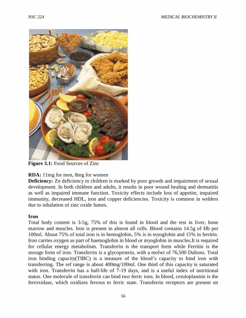
NSC 224 MEDICAL BIOCHEMISTRY II
56
Figure 3.1: Food Sources of Zinc
RDA: 11mg for men, 8mg for women Deficiency: Zn deficiency in children is marked by poor growth and impairment of sexual development. In both children and adults, it results in poor wound healing and dermatitis as well as impaired immune function. Toxicity effects include loss of appetite, impaired immunity, decreased HDL, iron and copper deficiencies. Toxicity is common in welders due to inhalation of zinc oxide fumes.
Iron Total body content is 3-5g, 75% of this is found in blood and the rest in liver, bone marrow and muscles. Iron is present in almost all cells. Blood contains 14.5g of Hb per 100ml. About 75% of total iron is in hemoglobin, 5% is in myoglobin and 15% in ferritin. Iron carries oxygen as part of haemoglobin in blood or myoglobin in muscles.It is required for cellular energy metabolism. Transferrin is the transport form while Ferritin is the storage form of iron. Transferrin is a glycoprotein, with a molwt of 76,500 Daltons. Total iron binding capacity(TIBC) is a measure of the blood’s capacity to bind iron with transferring. The ref range is about 400mg/100ml. One third of this capacity is saturated with iron. Transferrin has a half-life of 7-19 days, and is a useful index of nutritional status. One molecule of transferrin can bind two ferric ions. In blood, ceruloplasmin is the ferroxidase, which oxidizes ferrous to ferric state. Transferrin receptors are present on
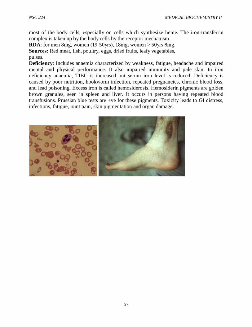
NSC 224 MEDICAL BIOCHEMISTRY II
57
most of the body cells, especially on cells which synthesize heme. The iron-transferrin complex is taken up by the body cells by the receptor mechanism. RDA: for men 8mg, women (19-50yrs), 18mg, women > 50yrs 8mg. Sources: Red meat, fish, poultry, eggs, dried fruits, leafy vegetables, pulses. Deficiency: Includes anaemia characterized by weakness, fatigue, headache and impaired mental and physical performance. It also impaired immunity and pale skin. In iron deficiency anaemia, TIBC is increased but serum iron level is reduced. Deficiency is caused by poor nutrition, hookworm infection, repeated pregnancies, chronic blood loss, and lead poisoning. Excess iron is called hemosiderosis. Hemosiderin pigments are golden brown granules, seen in spleen and liver. It occurs in persons having repeated blood transfusions. Prussian blue tests are +ve for these pigments. Toxicity leads to GI distress, infections, fatigue, joint pain, skin pigmentation and organ damage.
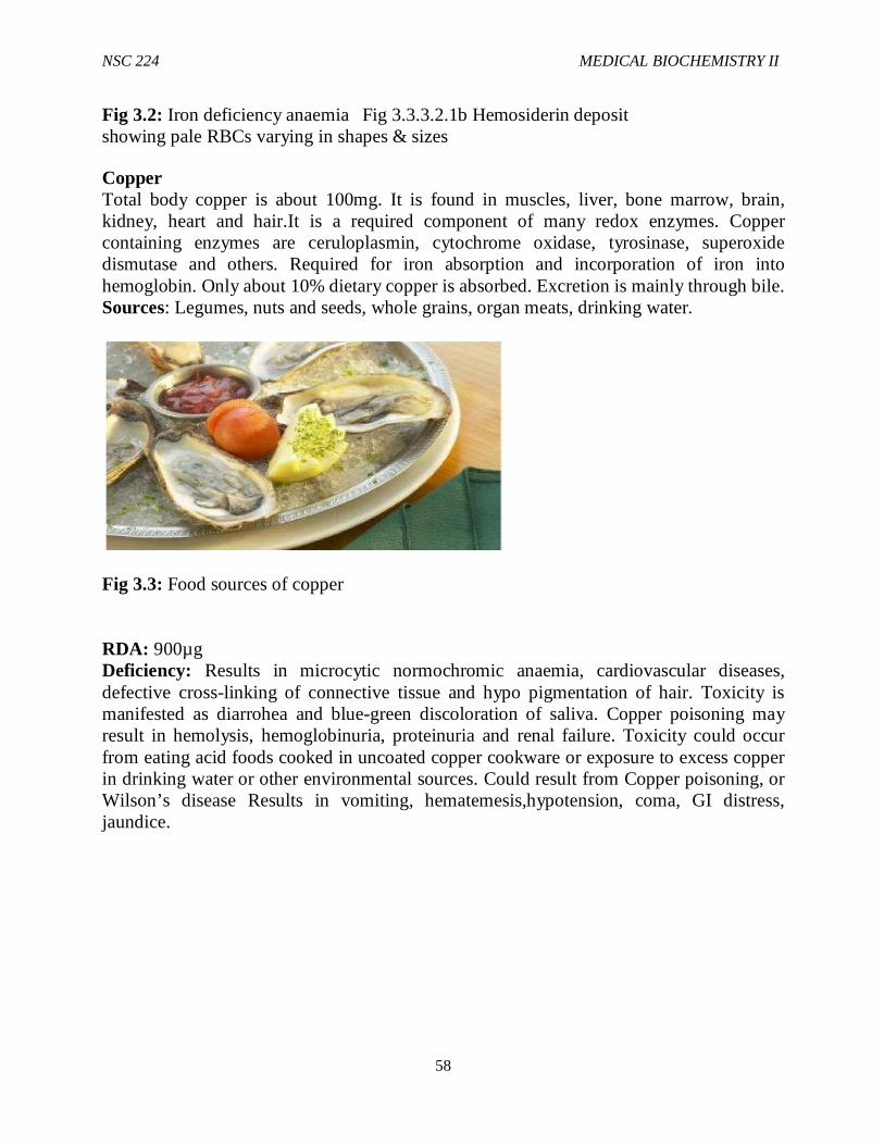
NSC 224 MEDICAL BIOCHEMISTRY II
58
Fig 3.2: Iron deficiency anaemia Fig 3.3.3.2.1b Hemosiderin deposit showing pale RBCs varying in shapes & sizes
Copper Total body copper is about 100mg. It is found in muscles, liver, bone marrow, brain, kidney, heart and hair.It is a required component of many redox enzymes. Copper containing enzymes are ceruloplasmin, cytochrome oxidase, tyrosinase, superoxide dismutase and others. Required for iron absorption and incorporation of iron into hemoglobin. Only about 10% dietary copper is absorbed. Excretion is mainly through bile. Sources: Legumes, nuts and seeds, whole grains, organ meats, drinking water.
Fig 3.3: Food sources of copper
RDA: 900µg Deficiency: Results in microcytic normochromic anaemia, cardiovascular diseases, defective cross-linking of connective tissue and hypo pigmentation of hair. Toxicity is manifested as diarrohea and blue-green discoloration of saliva. Copper poisoning may result in hemolysis, hemoglobinuria, proteinuria and renal failure. Toxicity could occur from eating acid foods cooked in uncoated copper cookware or exposure to excess copper in drinking water or other environmental sources. Could result from Copper poisoning, or Wilson’s disease Results in vomiting, hematemesis,hypotension, coma, GI distress, jaundice.
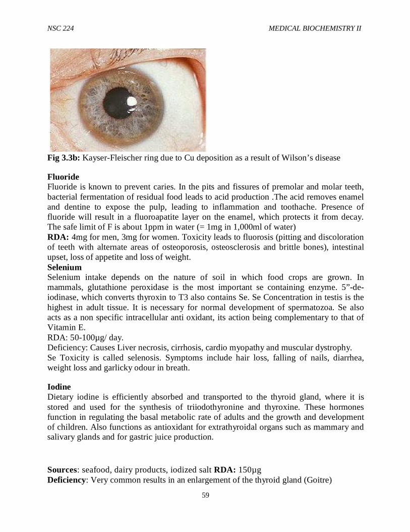
NSC 224 MEDICAL BIOCHEMISTRY II
59
Fig 3.3b: Kayser-Fleischer ring due to Cu deposition as a result of Wilson’s disease
Fluoride Fluoride is known to prevent caries. In the pits and fissures of premolar and molar teeth, bacterial fermentation of residual food leads to acid production .The acid removes enamel and dentine to expose the pulp, leading to inflammation and toothache. Presence of fluoride will result in a fluoroapatite layer on the enamel, which protects it from decay. The safe limit of F is about 1ppm in water (= 1mg in 1,000ml of water) RDA: 4mg for men, 3mg for women. Toxicity leads to fluorosis (pitting and discoloration of teeth with alternate areas of osteoporosis, osteosclerosis and brittle bones), intestinal upset, loss of appetite and loss of weight. Selenium Selenium intake depends on the nature of soil in which food crops are grown. In mammals, glutathione peroxidase is the most important se containing enzyme. 5”-de-iodinase, which converts thyroxin to T3 also contains Se. Se Concentration in testis is the highest in adult tissue. It is necessary for normal development of spermatozoa. Se also acts as a non specific intracellular anti oxidant, its action being complementary to that of Vitamin E. RDA: 50-100µg/ day. Deficiency: Causes Liver necrosis, cirrhosis, cardio myopathy and muscular dystrophy. Se Toxicity is called selenosis. Symptoms include hair loss, falling of nails, diarrhea, weight loss and garlicky odour in breath.
Iodine Dietary iodine is efficiently absorbed and transported to the thyroid gland, where it is stored and used for the synthesis of triiodothyronine and thyroxine. These hormones function in regulating the basal metabolic rate of adults and the growth and development of children. Also functions as antioxidant for extrathyroidal organs such as mammary and salivary glands and for gastric juice production.
Sources: seafood, dairy products, iodized salt RDA: 150µg Deficiency: Very common results in an enlargement of the thyroid gland (Goitre)
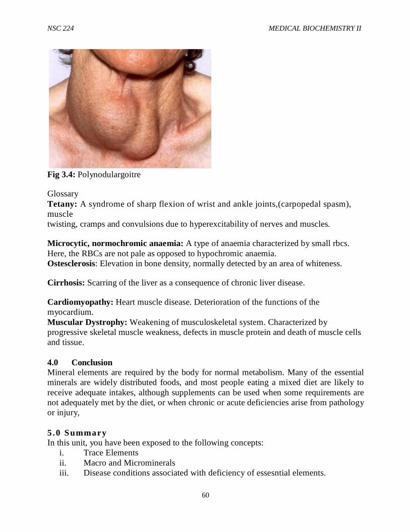
NSC 224 MEDICAL BIOCHEMISTRY II
60
Fig 3.4: Polynodulargoitre
Glossary Tetany: A syndrome of sharp flexion of wrist and ankle joints,(carpopedal spasm), muscle twisting, cramps and convulsions due to hyperexcitability of nerves and muscles.
Microcytic, normochromic anaemia: A type of anaemia characterized by small rbcs. Here, the RBCs are not pale as opposed to hypochromic anaemia. Ostesclerosis: Elevation in bone density, normally detected by an area of whiteness.
Cirrhosis: Scarring of the liver as a consequence of chronic liver disease.
Cardiomyopathy: Heart muscle disease. Deterioration of the functions of the myocardium. Muscular Dystrophy: Weakening of musculoskeletal system. Characterized by progressive skeletal muscle weakness, defects in muscle protein and death of muscle cells and tissue. 4.0 Conclusion Mineral elements are required by the body for normal metabolism. Many of the essential minerals are widely distributed foods, and most people eating a mixed diet are likely to receive adequate intakes, although supplements can be used when some requirements are not adequately met by the diet, or when chronic or acute deficiencies arise from pathology or injury, 5.0 Summary In this unit, you have been exposed to the following concepts:
i. Trace Elements ii. Macro and Microminerals iii. Disease conditions associated with deficiency of essesntial elements.
NSC 224 MEDICAL BIOCHEMISTRY II
61
6.0 Tutor Marked Assignments i. Distinguish between Macro and Microminerals ii. List important microminerals (Trace Elements) and enumerate the functions, food
sources, required daily allowance, deficiency and toxicity symptoms of each trace mineral studied.
7.0 References and further reading Katherine, M. A. Rogers and William N. Scott (2011). Nurses! Test yourself in anatomy and physiology Kathryn, A. Booth, Terri. D. Wyman (2008). Anatomy, physiology, and pathophysiology for allied health Keith L.M, Persuade T.V.N (2006). The Developing Human Clinically Oriented Embryology 8th Edition Lippincott Williams & Wilkins Kent, M. Van De Graff, R.WardRhees, Sidney P. (2010). Schaum’s outline of human anatomy and physiology 3rd edition. Philip, T. (2012). Seeley’s principles of anatomy & physiology 2nd edition. Sadler, T.W (2004). Langman’s Medical Embryology 9th edition.
NSC 224 MEDICAL BIOCHEMISTRY II
62
MODULE 4- AMINO ACID METABOLISM Introduction Amino acids are the monomeric units of proteins. Metabolism of amino acids involves the breakdown of amino acids and how amino acids are synthesized. Breakdown of amino acids involves the removal of amino group and utilization of carbon skeleton for the production of other essential biological molecules in the cell. Part of this is also used in the synthesis of other amino acids required. Module Objective: At the end of this module, you should be able to discuss the following in details:
i. Sources of Amino Acids ii. Digestion and adsorption of proteins iii. Disorders of Amino acid Metabolism iv. Diabetes Mellitus
CONTENTS Unit 1 Sources of Amino Acids Unit 2 Disorders of Amino acid Metabolism Unit 3 Diabetes Mellitus UNIT ONE- SOURCES OF AMINO ACIDS CONTENTS 1.0 Introduction 2.0 Objectives 3.0 Main Content 3.1 Sources of Amino Acids 3.2 Fate of Free Amino Acids 3.3 Digestion of Dietary Protein 3.4 Degradation of endogenous proteins 3.5 Catabolism of amino acids 4.0 Conclusion 5.0 Summary 6.0 Tutor Marked Assignments 6.1 Activity 7.0 Tutor Marked Tests
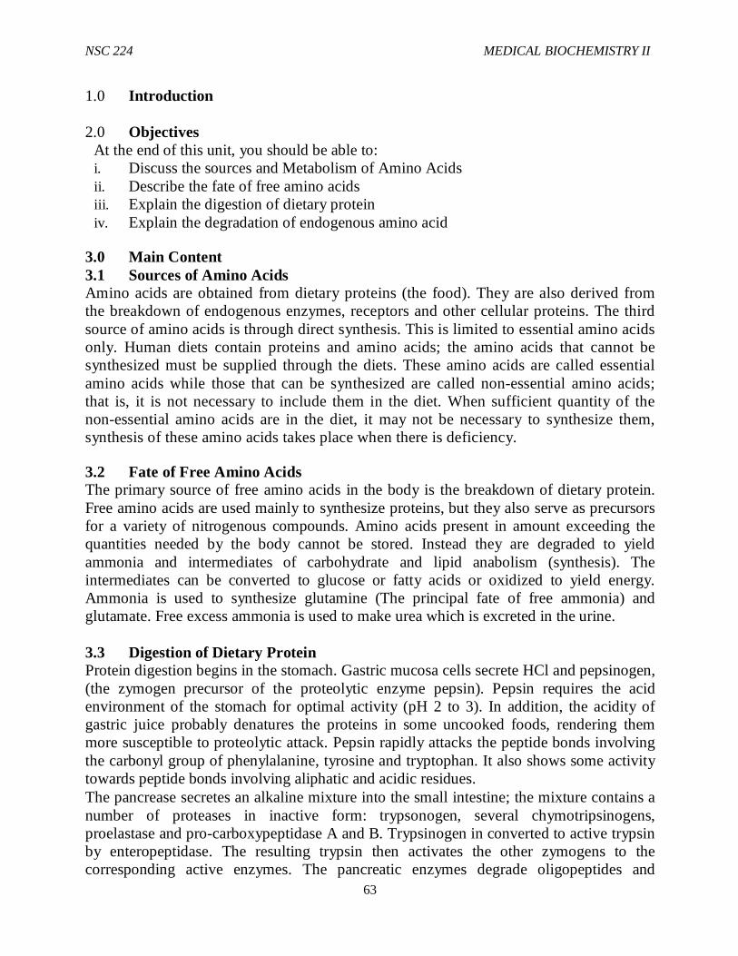
NSC 224 MEDICAL BIOCHEMISTRY II
63
1.0 Introduction 2.0 Objectives
At the end of this unit, you should be able to: i. Discuss the sources and Metabolism of Amino Acids ii. Describe the fate of free amino acids iii. Explain the digestion of dietary protein iv. Explain the degradation of endogenous amino acid
3.0 Main Content 3.1 Sources of Amino Acids Amino acids are obtained from dietary proteins (the food). They are also derived from the breakdown of endogenous enzymes, receptors and other cellular proteins. The third source of amino acids is through direct synthesis. This is limited to essential amino acids only. Human diets contain proteins and amino acids; the amino acids that cannot be synthesized must be supplied through the diets. These amino acids are called essential amino acids while those that can be synthesized are called non-essential amino acids; that is, it is not necessary to include them in the diet. When sufficient quantity of the non-essential amino acids are in the diet, it may not be necessary to synthesize them, synthesis of these amino acids takes place when there is deficiency.
3.2 Fate of Free Amino Acids The primary source of free amino acids in the body is the breakdown of dietary protein. Free amino acids are used mainly to synthesize proteins, but they also serve as precursors for a variety of nitrogenous compounds. Amino acids present in amount exceeding the quantities needed by the body cannot be stored. Instead they are degraded to yield ammonia and intermediates of carbohydrate and lipid anabolism (synthesis). The intermediates can be converted to glucose or fatty acids or oxidized to yield energy. Ammonia is used to synthesize glutamine (The principal fate of free ammonia) and glutamate. Free excess ammonia is used to make urea which is excreted in the urine. 3.3 Digestion of Dietary Protein Protein digestion begins in the stomach. Gastric mucosa cells secrete HCl and pepsinogen, (the zymogen precursor of the proteolytic enzyme pepsin). Pepsin requires the acid environment of the stomach for optimal activity (pH 2 to 3). In addition, the acidity of gastric juice probably denatures the proteins in some uncooked foods, rendering them more susceptible to proteolytic attack. Pepsin rapidly attacks the peptide bonds involving the carbonyl group of phenylalanine, tyrosine and tryptophan. It also shows some activity towards peptide bonds involving aliphatic and acidic residues. The pancrease secretes an alkaline mixture into the small intestine; the mixture contains a number of proteases in inactive form: trypsonogen, several chymotripsinogens, proelastase and pro-carboxypeptidase A and B. Trypsinogen in converted to active trypsin by enteropeptidase. The resulting trypsin then activates the other zymogens to the corresponding active enzymes. The pancreatic enzymes degrade oligopeptides and
NSC 224 MEDICAL BIOCHEMISTRY II
64
polypeptide to free amino acids and short oligopeptides which are transported into the intestinal mucoasa cells. Trypsin cleaves the peptide bonds involving the carboxyl group of lysine or arginine. Chymotrypsins are most active on peptide bonds involving the carboxyl groups of phenylalanine, tyrosine and tryptophan. Elastase is active toward peptide bonds involving neutral aliphatic amino acids. Carboxypepetidase A releases aliphatic and aromatic amino acids one at a time from the c-terminus. Carboxypeptidase B releases c-terminal lysine or arginine residues. The digestion of small peptides is completed within the intestinal mucosal cells which contain aminopeptidase and dipeptidases. Aminopeptidases release one amino acid at a time from the N-terminus. Prolidase is a dipeptidase specific for dipeptides containing proline in the carboxyl position. Protein turnover Most intra-cellular proteins are undergoing continual breakdown and synthesis. The rate of turnover of these proteins is variable and usually will vary depending on the nature of the protein and the metabolic state of the individual. Tissues with high rate of protein degradation are the uterine tissue during pregnancy and skeletal muscle during starvation Two major pathways are involved in protein turnover; one is carried out by proteases in lysosomes and a second major pathway involves a ubiquitin dependent pathway working in conjunction with a macromolecular protease complex called aproteosome. The amino acids released in this process can then enter into the same pathways as the amino acids derived from the diet. Nitrogen balance The major dietary source of N is protein. Excess amino acids are not stored, rather degraded and their N is disposed of in Urea, ammonia, uric acid or creatinine in urine with small amounts in fecal matter. Thus, Nitrogen balance: Nitrogen intake = Nitrogen excretion Nitrogen balance can either be positive or negative nitrogen balance. Positive nitrogen balance: intake > excretion Negative nitrogen balance: excretion > intake Situations associated with positive nitrogen balance include growth of children, pregnancy and wound healing. While negative nitrogen balance includes starvation, malnutrition and disease conditions like burns and surgery.
3.4 Degradation of endogenous proteins Endogenous proteins contribute to the supply of free amino acid in two ways. Proteins are degraded in cells by free lysosomal proteases; in addition proteins in the gastric and pancreatic secretions are digested and absorbed in the same way as dietary proteins.
NSC 224 MEDICAL BIOCHEMISTRY II
65
Essential amino acids Non essential amino acids Phenylalanine Glycine Tryptophan Alanine Hisidine Proline Valine Tyrosine Isoleucine Serine Arginine Cysteine Threonine Glutamic acid Methionine Glutamine Lysine Aspartic acid Leucine Asparagines
Tissue Distribution of amino acid metabolism The principal site of amino acid metabolism is the liver, which is able to take up all the amino acids from the circulation. Muscle shows a preference for the uptake of valine, isoleucine and leucine, brain preferentially takes up valine. Therefore, muscle and brain contribute significantly to the metabolism of branched chain amino acids. Ammonia produced as a result of amino acids degradation in the tissues is excreted in the urine but alanine and serine are transported to the liver for further metabolism.
3.5 Catabolism of amino acids Amino acids are degradation in two steps: Removal of the α-amino group and conversion of the carbon chain to an intermediate of lipid and carbohydrate metabolism.
Removal of the α-amino group (Nitrogen). Removal of the amino group from amino acids is accomplished by variety of processes such as transamination, oxidative and non oxidative deamination.
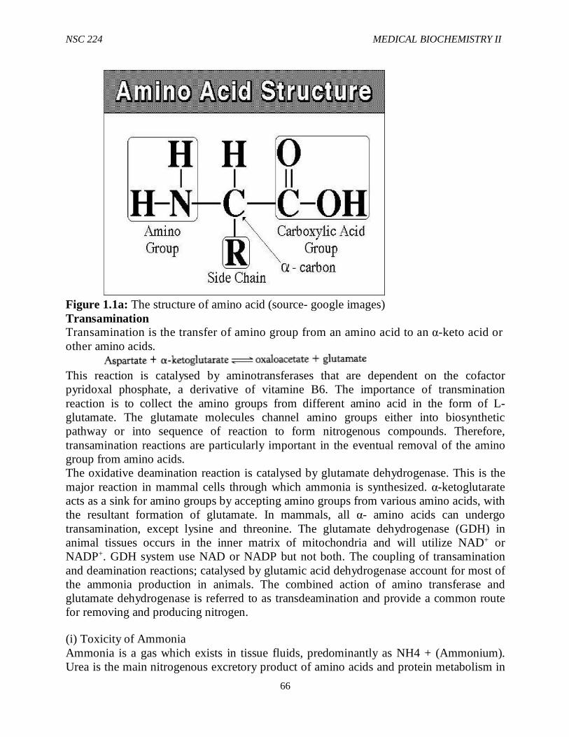
NSC 224 MEDICAL BIOCHEMISTRY II
66
Figure 1.1a: The structure of amino acid (source- google images) Transamination Transamination is the transfer of amino group from an amino acid to an α-keto acid or other amino acids.
This reaction is catalysed by aminotransferases that are dependent on the cofactor pyridoxal phosphate, a derivative of vitamine B6. The importance of transmination reaction is to collect the amino groups from different amino acid in the form of L-glutamate. The glutamate molecules channel amino groups either into biosynthetic pathway or into sequence of reaction to form nitrogenous compounds. Therefore, transamination reactions are particularly important in the eventual removal of the amino group from amino acids. The oxidative deamination reaction is catalysed by glutamate dehydrogenase. This is the major reaction in mammal cells through which ammonia is synthesized. α-ketoglutarate acts as a sink for amino groups by accepting amino groups from various amino acids, with the resultant formation of glutamate. In mammals, all α- amino acids can undergo transamination, except lysine and threonine. The glutamate dehydrogenase (GDH) in animal tissues occurs in the inner matrix of mitochondria and will utilize NAD+ or NADP+. GDH system use NAD or NADP but not both. The coupling of transamination and deamination reactions; catalysed by glutamic acid dehydrogenase account for most of the ammonia production in animals. The combined action of amino transferase and glutamate dehydrogenase is referred to as transdeamination and provide a common route for removing and producing nitrogen.
(i) Toxicity of Ammonia Ammonia is a gas which exists in tissue fluids, predominantly as NH4 + (Ammonium). Urea is the main nitrogenous excretory product of amino acids and protein metabolism in
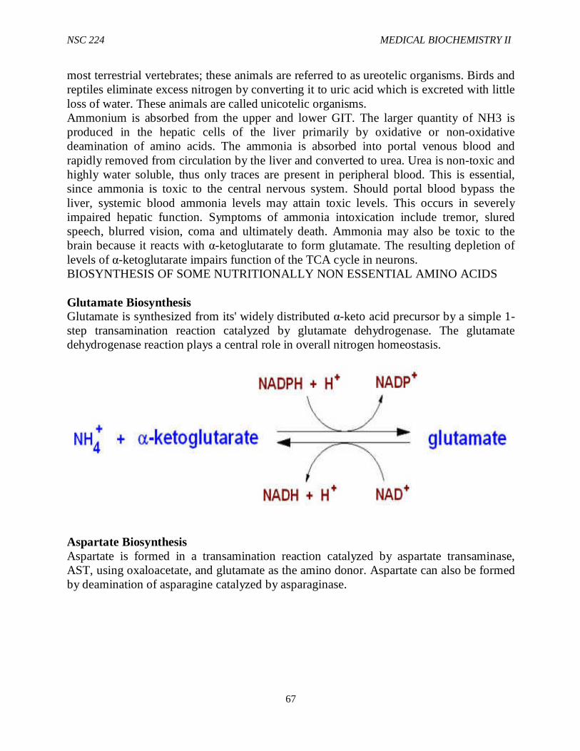
NSC 224 MEDICAL BIOCHEMISTRY II
67
most terrestrial vertebrates; these animals are referred to as ureotelic organisms. Birds and reptiles eliminate excess nitrogen by converting it to uric acid which is excreted with little loss of water. These animals are called unicotelic organisms. Ammonium is absorbed from the upper and lower GIT. The larger quantity of NH3 is produced in the hepatic cells of the liver primarily by oxidative or non-oxidative deamination of amino acids. The ammonia is absorbed into portal venous blood and rapidly removed from circulation by the liver and converted to urea. Urea is non-toxic and highly water soluble, thus only traces are present in peripheral blood. This is essential, since ammonia is toxic to the central nervous system. Should portal blood bypass the liver, systemic blood ammonia levels may attain toxic levels. This occurs in severely impaired hepatic function. Symptoms of ammonia intoxication include tremor, slured speech, blurred vision, coma and ultimately death. Ammonia may also be toxic to the brain because it reacts with α-ketoglutarate to form glutamate. The resulting depletion of levels of α-ketoglutarate impairs function of the TCA cycle in neurons. BIOSYNTHESIS OF SOME NUTRITIONALLY NON ESSENTIAL AMINO ACIDS Glutamate Biosynthesis Glutamate is synthesized from its' widely distributed α-keto acid precursor by a simple 1-step transamination reaction catalyzed by glutamate dehydrogenase. The glutamate dehydrogenase reaction plays a central role in overall nitrogen homeostasis.
Aspartate Biosynthesis Aspartate is formed in a transamination reaction catalyzed by aspartate transaminase, AST, using oxaloacetate, and glutamate as the amino donor. Aspartate can also be formed by deamination of asparagine catalyzed by asparaginase.
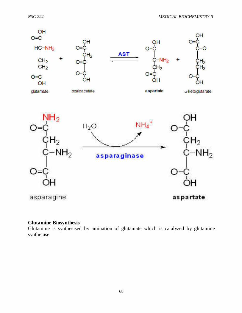
NSC 224 MEDICAL BIOCHEMISTRY II
68
Glutamine Biosynthesis Glutamine is synthesised by amination of glutamate which is catalyzed by glutamine synthetase
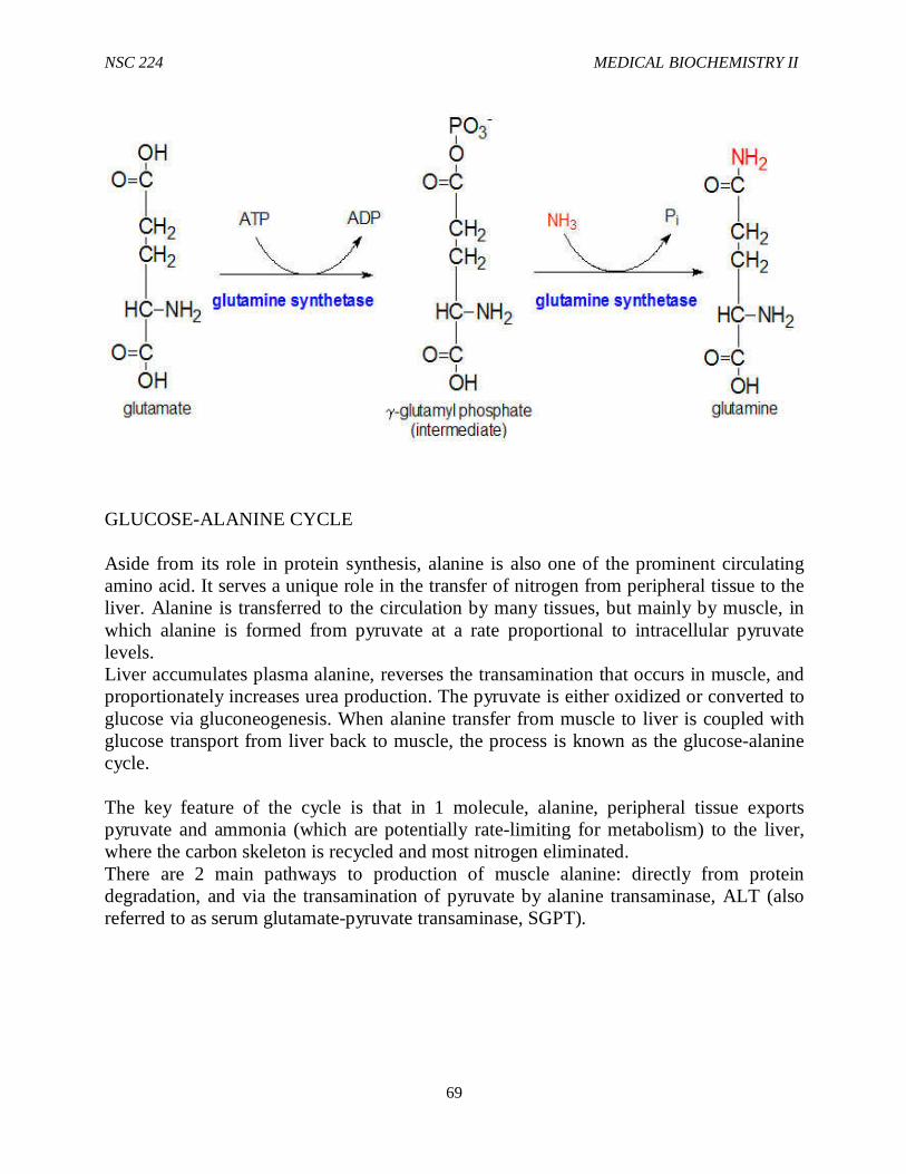
NSC 224 MEDICAL BIOCHEMISTRY II
69
GLUCOSE-ALANINE CYCLE Aside from its role in protein synthesis, alanine is also one of the prominent circulating amino acid. It serves a unique role in the transfer of nitrogen from peripheral tissue to the liver. Alanine is transferred to the circulation by many tissues, but mainly by muscle, in which alanine is formed from pyruvate at a rate proportional to intracellular pyruvate levels. Liver accumulates plasma alanine, reverses the transamination that occurs in muscle, and proportionately increases urea production. The pyruvate is either oxidized or converted to glucose via gluconeogenesis. When alanine transfer from muscle to liver is coupled with glucose transport from liver back to muscle, the process is known as the glucose-alanine cycle. The key feature of the cycle is that in 1 molecule, alanine, peripheral tissue exports pyruvate and ammonia (which are potentially rate-limiting for metabolism) to the liver, where the carbon skeleton is recycled and most nitrogen eliminated. There are 2 main pathways to production of muscle alanine: directly from protein degradation, and via the transamination of pyruvate by alanine transaminase, ALT (also referred to as serum glutamate-pyruvate transaminase, SGPT).
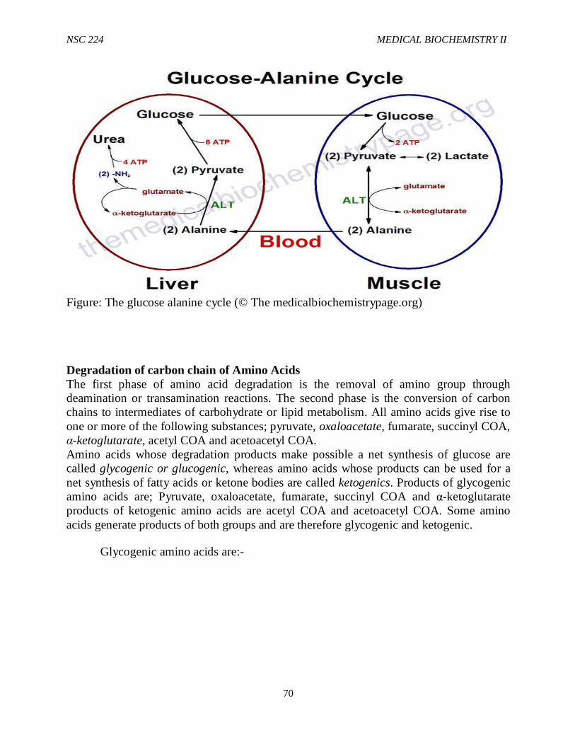
NSC 224 MEDICAL BIOCHEMISTRY II
70
Figure: The glucose alanine cycle (© The medicalbiochemistrypage.org)
Degradation of carbon chain of Amino Acids The first phase of amino acid degradation is the removal of amino group through deamination or transamination reactions. The second phase is the conversion of carbon chains to intermediates of carbohydrate or lipid metabolism. All amino acids give rise to one or more of the following substances; pyruvate, oxaloacetate, fumarate, succinyl COA, α-ketoglutarate, acetyl COA and acetoacetyl COA. Amino acids whose degradation products make possible a net synthesis of glucose are called glycogenic or glucogenic, whereas amino acids whose products can be used for a net synthesis of fatty acids or ketone bodies are called ketogenics. Products of glycogenic amino acids are; Pyruvate, oxaloacetate, fumarate, succinyl COA and α-ketoglutarate products of ketogenic amino acids are acetyl COA and acetoacetyl COA. Some amino acids generate products of both groups and are therefore glycogenic and ketogenic.
Glycogenic amino acids are:-
NSC 224 MEDICAL BIOCHEMISTRY II
71
Alamine Asparagines Aspartate Arginine Cysteine Glutamate Glutamine Histidine Isoleucine
Methionine Proline Serine Thronine Tyrosine Tryptophan Valine Phenylalanine
NSC 224 MEDICAL BIOCHEMISTRY II
72
Ketogenic amino acids are leucine and lysine Glycogenic and ketogenic amino acids are: isolencine, phenytalanine, tyrosine and tryptophan. Historically, an amino acid was classified as glycogenic or ketogenic depending whether its administration to diabetic animals resulted in an increase in urinary excretion of glucose or of ketone bodies respectively. In animals that are neither fasted nor diabetic, intermediates of amino acid degradation are oxidized by the TCA cycle to produce energy.
4.0 Conclusion Protein digestion begins in the stomach. Gastric mucosa cells secrete HCl and pepsinogen, The amino acids and small peptides are transported into the intestinal cells of the brush border by a family of amino acid specific transports many of which require Na+
The first phase of amino acid degradation is the removal of amino group through deamination or transamination reactions. The second phase is the conversion of carbon chains to intermediates of carbohydrate or lipid metabolism. 5.0 Summary: in this unit, you have been taken through the following:
i. Sources of Amino Acids ii. Fate of Free Amino Acids iii. Digestion of Dietary Protein iv. Degradation of endogenous proteins v. Catabolism of amino acids
6.0 Tutor Marked Assignments i. Discuss the sources and Metabolism of Amino Acids ii. Describe the fate of free amino acids iii. Explain the digestion of dietary protein iv. Explain the degradation of endogenous amino acid
7.0 References and further reading Murray, R.K., Bender, D.A., Botham, K., M., Kennelly, P.J., Rodwell V.W. and
Well, P.A., (2012). Harper’s Illustrated Biochemistry (29th Edition) McGraw-Hill Medical
Devlin T.M. (2010) Textbook of Biochemistry with Clinical Correlation 7thEdition. JohnWiley & SonsInc.
Nelson, D.L. and Cox M.M., (2009) Lehninger Principles of Biochemistry 4thedition
Katherine, M. A. Rogers and William N. Scott (2011). Nurses! Test yourself in anatomy and physiology
Kathryn, A. Booth, Terri. D. Wyman (2008). Anatomy, physiology, and pathophysiology for allied health
Keith L.M, Persuade T.V.N (2006). The Developing Human Clinically Oriented Embryology 8th Edition Lippincott Williams & Wilkins
NSC 224 MEDICAL BIOCHEMISTRY II
73
Kent, M. Van De Graff, R.Ward Rhees, Sidney P. (2010). Schaum’s outline of human anatomy and physiology 3rd edition.
Philip, T. (2012). Seeley’s principles of anatomy & physiology 2nd edition. Sadler, T.W (2004). Langman’s Medical Embryology 9th edition. The Medical Biochemistry Page. themedicalbiochemistrypage.org
NSC 224 MEDICAL BIOCHEMISTRY II
74
UNIT TWO - DISORDERS OF AMINO ACID METABOLISM
1.0 Introduction 2.0 Objectives 3.0 Main Content 3.1 Phenylketonuria (PKU) 3.2 Albinism 3.3 Alkoptonuria 3.4 Maple syrup Urine disease 4.0 Conclusion 5.0 Summary 6.0 Tutor Marked Assignment 6.1 Activity 6.2 Tutor Marked Tests 7.0 References and other resources
1.0 Introduction Disorder of amino acid metabolism results from deficiency or complete absence of a particular enzyme required to complete the metabolism of some amino acids. All of them are genetically inherited diseases. Some of the disorders are as follows:
2.0 Objectives At the end of this unit, you should be able to explain the following Amino acid metabolism disorders in details.
i. Phenylketonuria (PKU) ii. Albinism iii. Alkoptonuria iv. Maple syrup Urine disease
3.0 Main Content 3.1 Phenylketonuria (PKU) – PKU occurs when phenylalanine hydroxylase activity is absent or significantly reduced (this enzyme is responsible for the degradation of phenylalanine). Phenylalanine (PA) accumulates, which stimulates the metabolism of PA by alternative pathways resulting in urinary excretion of high amounts of phenyllactate, phenylpyruvate, o-hydroxylphenylacetate, and phenyl acetylglutamine as well as phenylalanine. It may lead to neurological problems. 3.2 Albinism – Melanin is the black pigment of human skin, it is synthesized inside the melanocytes from tyrosine. In the first 2 steps of melanin formation, tyrosine is converted to dihydroxy phenylalanine (DOPA) and then to DOPA quinone by tyrosinase. DOPA quinone is then converted to melanin. If there is tyrosinase deficiency, DOPA quinine will not be converted to melanin. Therefore, deficiency of tyrosinase is the cause of certain types of albinism. It is also responsible for gray hair; hair becomes grey in the absence of melanin. Albinism is an example of inborn errors of metabolism.
NSC 224 MEDICAL BIOCHEMISTRY II
75
3.3 Alkoptonuria– In alkaptonuria, the enzyme homogentisate oxidase required for tyrosine metabolism is deficient. High amounts of homogentisate are excreted in the urine which causes the urine to darken gradually or rapidly due to oxidation of homogenetisate. Clinical conditions resulting from this disorder is called ochronosis and in later years arthritis.
3.4 Maple syrup Urine disease – The first 2 steps in the degradation of branched chain amino acids such as valine, leucine and isoleucine are identical (Transamination followed by oxidative decarboxylation). The decarboxylation reaction is catalysed by the branched chain α-keto acid dehydrogenase complex. A genetic deficiency of this enzyme causes elevated levels of valine, isoleucine and leucine and their α-keto acids in the blood and urine. The high concentration of α-keto acids gives the urine of these patients a maple syrup odour, leading to the name maple syrup urine disease for the disorder. Clinical symptoms are poor feeding after the 1st week of life, vomiting, muscular hypertonicity and sometimes convulsion.
4.0 Conclusion
5.0 Summary: In this unit, you have been taken through the following Amino acid Metabolism disorders.
i. Phenylketonuria (PKU) ii. Albinism iii. Alkoptonuria iv. Maple syrup Urine disease
6.0 Tutor Marked Assignments
Discuss in details the following Amino acid metabolism disorders. i. Phenylketonuria (PKU) ii. Albinism iii. Alkoptonuria iv. Maple syrup Urine disease
7.0 References and further reading Murray, R.K., Bender, D.A., Botham, K., M., Kennelly, P.J., Rodwell V.W. and
Well, P.A., (2012). Harper’s Illustrated Biochemistry (29th Edition) McGraw-Hill Medical
Devlin T.M. (2010) Textbook of Biochemistry with Clinical Correlation 7thEdition. JohnWiley & SonsInc.
Nelson, D.L. and Cox M.M., (2009) Lehninger Principles of Biochemistry 4thedition
Katherine, M. A. Rogers and William N. Scott (2011). Nurses! Test yourself in anatomy and physiology
NSC 224 MEDICAL BIOCHEMISTRY II
76
Kathryn, A. Booth, Terri. D. Wyman (2008). Anatomy, physiology, and pathophysiology for allied health
Keith L.M, Persuade T.V.N (2006). The Developing Human Clinically Oriented Embryology 8th Edition Lippincott Williams & Wilkins
Kent, M. Van De Graff, R.Ward Rhees, Sidney P. (2010). Schaum’s outline of human anatomy and physiology 3rd edition.
Philip, T. (2012). Seeley’s principles of anatomy & physiology 2nd edition. Sadler, T.W (2004). Langman’s Medical Embryology 9th edition. The Medical Biochemistry Page. themedicalbiochemistrypage.org
NSC 224 MEDICAL BIOCHEMISTRY II
77
UNIT THREE- DIABETES MELLITUS
CONTENTS 1.0 Introduction 2.0 Objectives 3.0 Main Content 3.1 Diabetes Mellitus 3.2 Classification of Diabetes 3.3 Diagnosis of Diabetes 4.0 Conclusion 5.0 Summary 6.0 Tutor Marked Assignments 6.1 Activity 6.2 Tutor Marked Tests 7.0 References and other resources
1.0 Introduction Diabetes mellitus is a complex disease that affects several hundred million people. Diabetes is characterized by an elevated level of glucose in the blood and in the urine. Glucose is excreted in the urine when the blood glucose level exceeds the reabsorptive capacity of the renal tubules. Water accompanies the excreted glucose, and so an untreated diabetic in the acute phase of the disease is hungry and thirsty. The loss of glucose depletes the carbohydrate stores, which leads to the breakdown of fat and protein. The mobilization of fats results in the formation of large amounts of acetyl CoA. Ketone bodies (acetoacetate, acetone, and hydroxybutyrate) are formed when acetyl CoA cannot enter the citric acid cycle because there is insufficient oxaloacetate.( oxaloacetate is derived from glucose and some amino acids). The excretion of ketone bodies impairs the acid-base balance and causes further dehydration, which may lead to coma and death in the acute phase of the disease in an untreated diabetic.
2.0 Objectives At the end of this unit, you should be able to:
i. Describe the metabolic changes that accompany Diabetes Mellitus ii. Explain the classification of Diabetes Mellitus and distinguish the different types of Diabetes in
terms of metabolic features, symptoms and time of onset iii. Describe how Diabetes mellitus is diagnosed and the mode of action of medications used in
treatment of Diabetes.
3.0 Main Content
3.1 Diabetes Mellitus Diabetes mellitus, caused by a deficiency in the secretion or action of insulin, is a relatively common disease: nearly 6% of the United States population shows some degree of abnormality in glucose metabolism that is indicative of diabetes or a tendency toward the condition. There are two major clinical
NSC 224 MEDICAL BIOCHEMISTRY II
78
classes of diabetes mellitus: type I diabetes, or insulin dependent diabetes mellitus (IDDM), and type II diabetes, or non-insulin-dependent diabetes mellitus (NIDDM), also called insulin-resistant diabetes. Type I Diabetes In type I diabetes, the disease begins early in life and quickly becomes severe. This disease responds to insulin injection, because the metabolic defect stems from a paucity of pancreatic _ cells and a consequent inability to produce sufficient insulin. IDDM requires insulin therapy and careful, lifelong control of the balance between dietary intake and insulin dose. Characteristic symptoms of type I (and type II) diabetes are excessive thirst and frequent urination (polyuria), leading to the intake of large volumes of water (polydipsia) (“diabetes mellitus” means “excessive excretion of sweet urine”). These symptoms are due to the excretion of large amounts of glucose in the urine, a condition known as glucosuria. Type II Diabetes Type II diabetes is slow to develop (typically in older, obese individuals), and the symptoms are milder and often go unrecognized at first. This is really a group of diseases in which the regulatory activity of insulin is defective: insulin is produced, but some feature of the insulin-response system is defective. These individuals are insulin-resistant. The connection between type II diabetes and obesity (discussed below) is an active area of research. Individuals with either type of diabetes are unable to take up glucose efficiently from the blood; recall that insulin triggers the movement of GLUT4 glucose transporters to the plasma membrane of muscle and adipose tissue. Another characteristic metabolic change in diabetes is excessive but incomplete oxidation of fatty acids in the liver. The acetyl-CoA produced by _ oxidation cannot be completely oxidized by the citric acid cycle, because the high [NADH]/[NAD_] ratio produced by f3 - oxidation inhibits the cycle (recall that three steps convert NAD_ to NADH). Accumulation of acetyl-CoA leads to overproduction of the ketone bodies acetoacetate and f3-hydroxybutyrate, which cannot be used by extrahepatic tissues as fast as they are made in the liver. In addition to - f3 -hydroxybutyrate and acetoacetate, the blood of diabetics also contains acetone, which results from the spontaneous decarboxylation of acetoacetate: Acetone is volatile and is exhaled, and in uncontrolled diabetes, the breath has a characteristic odor sometimes mistaken for ethanol. A diabetic individual who is experiencing mental confusion due to high blood glucose is occasionally misdiagnosed as intoxicated, an error that can be fatal. The overproduction of ketone bodies, called ketosis, results in greatly increased concentrations of ketone bodies in the blood (ketonemia) and urine (ketonuria).
NSC 224 MEDICAL BIOCHEMISTRY II
79
3.2 Classification of Diabetes
Type 1 DM About 10% of all diabetic persons are classified as type I. It usually appears in childhood or teenage, but it is not limited to young people. In this disease, there is very low or complete absence of insulin production by the pancreas, because of defective f3-cell function, the result of an autoimmune process. There is severe derangement of carbohydrate, lipid and protein metabolism, leading to
i. Hyperglycemia :As a result of inability of insulin-dependent tissues to take up glucose and from accelerated hepatic gluconeogenesis from amino acids derived from muscle protein
ii. Hypertriglyceridemia: Because VLDL and Chylomicrons cannot be cleared from the blood by lipoprotein lipase, whose expression is dependent upon insulin.
iii. Severe episodes of Ketoacidosis: This results from increased lipolysis in adipose tissue which increases plasma fatty acid levels and ketone body production by the liver.
In type 1 DM, every tissue plays the catabolic role that it was designed to play in starvation, in spite of adequate or even excess fuel from the gut. This results ultimately in death unless insulin is administered. Administered insulin promotes glucose uptake and inhibits gluconeogenesis, lipolysis and proteolysis. However, it is necessary to adjust the insulin dose to variable dietary intake and physical activity. tight control of blood sugar requires several injections of insulin per day and close blood sugar monitoring by the patient.
Type 2 Diabetes mellitus Nearly 90% of diabetic persons are of type 2.It is also called adult-onset diabetes because it usually occurs in middle aged to elderly people. Aging, physical inactivity, western culture lifestyle, obesity and family history of diabetes are risk factors. It typically occurs in the setting of metabolic syndrome, which also includes abdominal obesity, hypertension and hyperlipidemia Type 2 diabetics are resistant to insulin and have insufficient production of insulin to overcome the resistance. The majority of patients are obese, and although their insulin levels are high, they are not as high as those of non diabetic but similarly obese persons. Recent work implicate increased level of tumor necrosis factorα (TNFα) and resistin as well as reduced secretion of adiponectin as a cause of insulin resistance.The greater the adipose tissue mass, the greater the production of TNFα which impairs insulin action. Uncontrolled glucose production by the liver and low uptake by the skeletal muscle results in hyperglycemia.. Hypertriglyceridemia is also seen, as a result of increased VLDL. Ketoacidosis is rare in this type of diabetes. Diet alone can control the disease in the obese diabetic as insulin receptors increase and post – receptor abnormalities improve, which will increase tissue sensitivity to insulin and glucose tolerance.
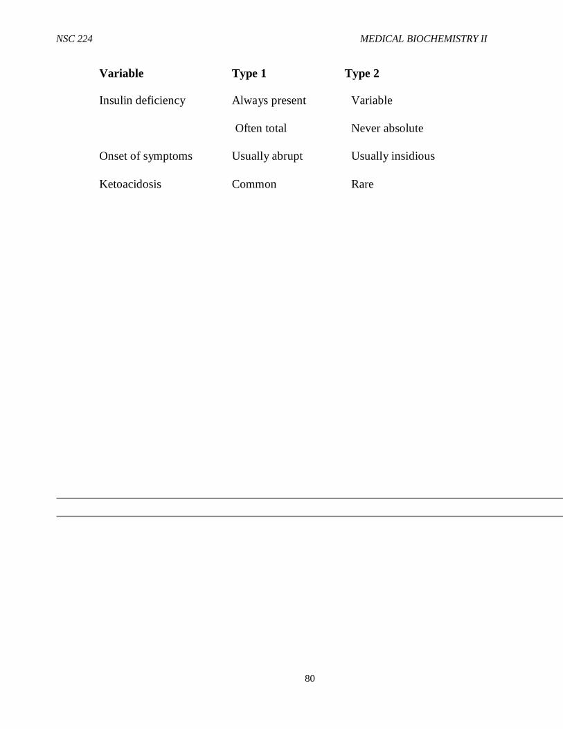
NSC 224 MEDICAL BIOCHEMISTRY II
80
Variable Type 1 Type 2
Insulin deficiency Always present Variable
Often total Never absolute
Onset of symptoms Usually abrupt Usually insidious
Ketoacidosis Common Rare
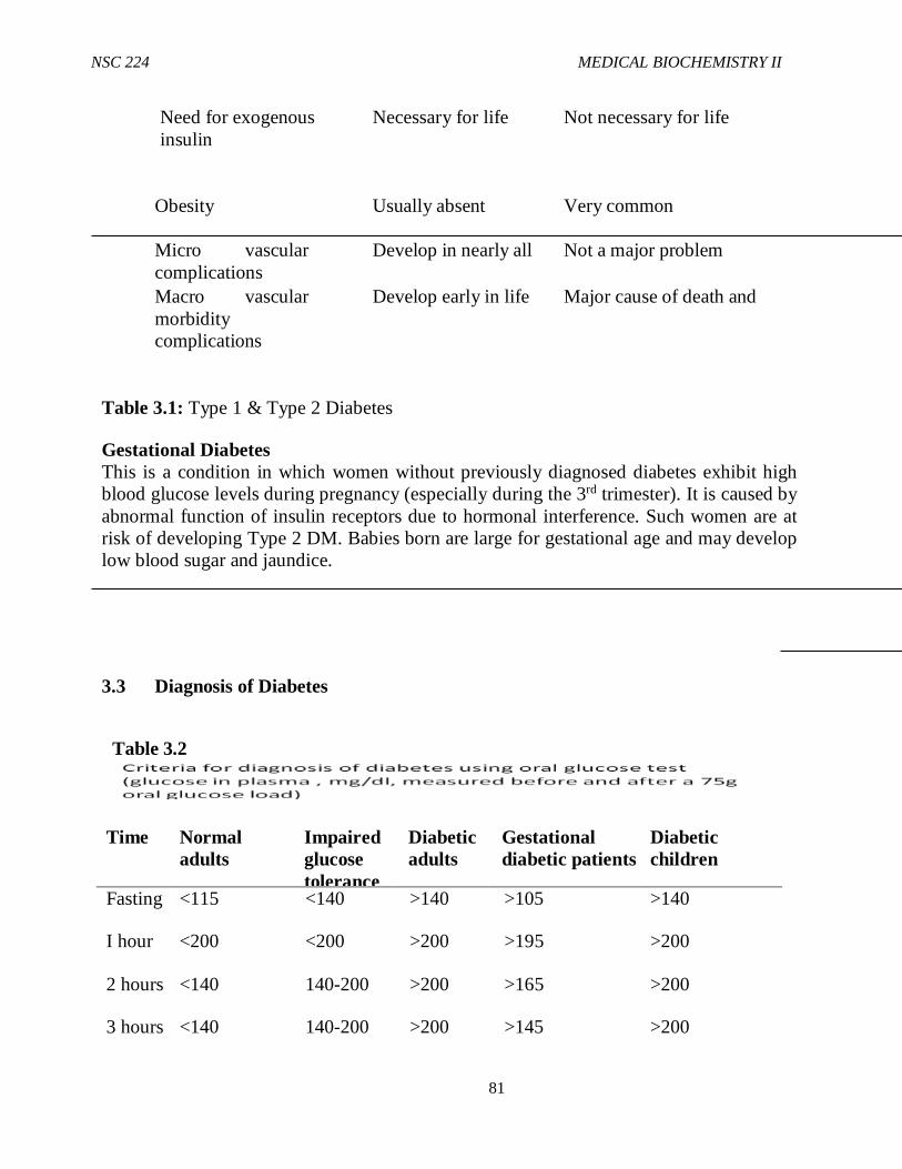
NSC 224 MEDICAL BIOCHEMISTRY II
81
Need for exogenous Necessary for life Not necessary for life insulin
Obesity Usually absent Very common
Micro vascular Develop in nearly all Not a major problem complications Macro vascular Develop early in life Major cause of death and morbidity complications
Table 3.1: Type 1 & Type 2 Diabetes
Gestational Diabetes This is a condition in which women without previously diagnosed diabetes exhibit high blood glucose levels during pregnancy (especially during the 3rd trimester). It is caused by abnormal function of insulin receptors due to hormonal interference. Such women are at risk of developing Type 2 DM. Babies born are large for gestational age and may develop low blood sugar and jaundice.
3.3 Diagnosis of Diabetes
Table 3.2
Time Normal adults
Impaired glucose tolerance
Diabetic adults
Gestational diabetic patients
Diabetic children
Fasting <115 <140 >140 >105 >140
I hour <200 <200 >200 >195 >200
2 hours <140 140-200 >200 >165 >200
3 hours <140 140-200 >200 >145 >200
NSC 224 MEDICAL BIOCHEMISTRY II
82
Measurement of glycated haemoglobin (Hemoglobin A1c) is also used for diagnosis of Diabetes. The reaction occurs by a non enzymatic reaction between glucose and the amino-terminal valine of the Hb α chain and is favoured by high glucose levels. The concentration of Hb1c is a good index of glucose level control. Glycation of proteins may contribute to the development of complications in Diabetes. An HbA1c of 6.5% is recommended as the cut off point for diagnosing diabetes. Medications are usually targeted at controlling hyperglycemia. These Medications include
i. Thiazolidinediones: Sensitizes peripheral tissues to insulin action ii. Metformin : Reduces hepatic gluconeogenesis iii. Sulfonyl Ureas: Stimulate insulin secretion from β cells.
Insulin administration is essential for treatment of type 1 DM and is often used in selected patients with type 2 diabetes. 4.0 Conclusion
Diabetes mellitus is a complex disease that affects several hundred million people. Diabetes is characterized by an elevated level of glucose in the blood and in the urine. Diabetes mellitus, caused by a deficiency in the secretion or action of insulin, is a relatively common disease. There are two major clinical classes of diabetes mellitus: type I diabetes, or insulin dependent diabetes mellitus (IDDM), and type II diabetes, or non-insulin-dependent diabetes mellitus (NIDDM), also called insulin-resistant diabetes.
5.0 Summary: In this unit, you have been taken through the following:
i. Concept of Diabetes Mellitus ii. Classification of Diabetes iii. Diagnosis of Diabetes
6.0 Tutor Marked Assignments i. ii. Describe the metabolic changes that accompany Diabetes Mellitus iii. Explain the classification of Diabetes Mellitus and distinguish the different types of
Diabetes in terms of metabolic features, symptoms and time of onset iv. Describe how Diabetes mellitus is diagnosed and the mode of action of
medications used in treatment of Diabetes.
NSC 224 MEDICAL BIOCHEMISTRY II
83
7.0 Reference and further reading Katherine, M. A. Rogers and William N. Scott (2011). Nurses! Test yourself in
anatomy and physiology Kathryn, A. Booth, Terri. D. Wyman (2008). Anatomy, physiology, and
pathophysiology for allied health Keith L.M, Persuade T.V.N (2006). The Developing Human Clinically Oriented
Embryology 8th Edition Lippincott Williams & Wilkins Kent, M. Van De Graff, R.WardRhees, Sidney P. (2010). Schaum’s outline of human
anatomy and physiology 3rd edition. Philip, T. (2012). Seeley’s principles of anatomy & physiology 2nd edition. Sadler, T.W (2004). Langman’s Medical Embryology 9th edition.