Embed Size (px)
Citation preview

This content has been downloaded from IOPscience. Please scroll down to see the full text.
Download details:
IP Address: 148.81.44.178
This content was downloaded on 21/10/2013 at 09:47
Please note that terms and conditions apply.
Novel ZnO/MgO/Fe2O3 composite optomagnetic nanoparticles
View the table of contents for this issue, or go to the journal homepage for more
2013 J. Phys.: Condens. Matter 25 194105
(http://iopscience.iop.org/0953-8984/25/19/194105)
Home Search Collections Journals About Contact us My IOPscience

IOP PUBLISHING JOURNAL OF PHYSICS: CONDENSED MATTER
J. Phys.: Condens. Matter 25 (2013) 194105 (7pp) doi:10.1088/0953-8984/25/19/194105
Novel ZnO/MgO/Fe2O3 compositeoptomagnetic nanoparticles
I Kaminska, B Sikora, K Fronc, P Dziawa, K Sobczak, R Minikayev,W Paszkowicz and D Elbaum
Institute of Physics, Polish Academy of Sciences, 32/46 Aleja Lotnikow 02-668 Warsaw, Poland
E-mail: [email protected]
Received 17 October 2012, in final form 10 February 2013Published 24 April 2013Online at stacks.iop.org/JPhysCM/25/194105
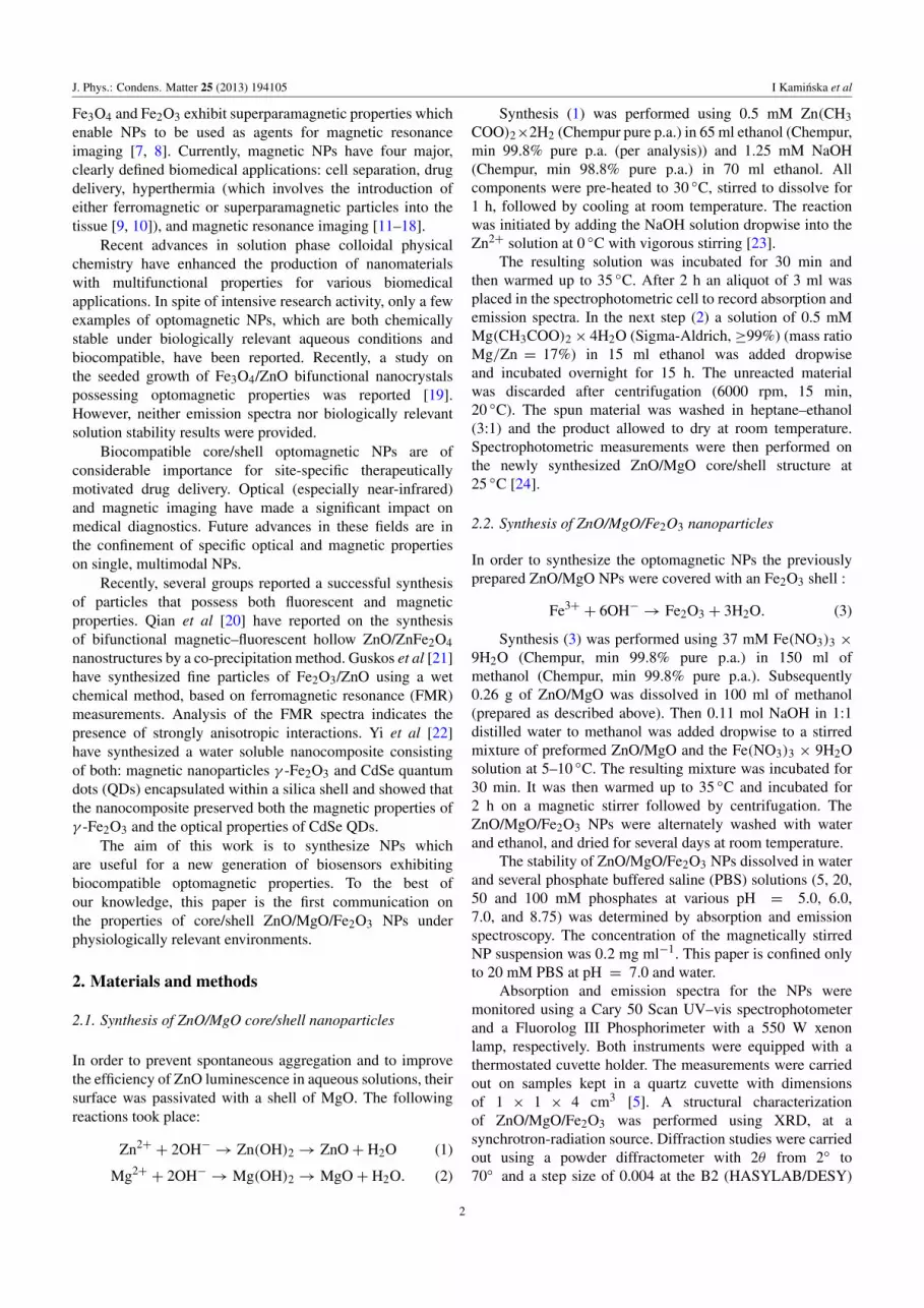
AbstractA facile sol–gel synthesis of novel ZnO/MgO/Fe2O3 nanoparticles (NPs) is reported and theirperformance is compared to that of ZnO/MgO. Powder x-ray diffraction (XRD) patternsreveal the crystal structure of the prepared samples. The average particle size of the samplewas found to be 4.8 nm. The optical properties were determined by UV–vis absorption andfluorescence measurements. The NPs are stable in biologically relevant solutions (phosphatebuffered saline (PBS), 20 mM, pH = 7.0) contrary to ZnO/MgO NPs which degrade in thepresence of inorganic phosphate. Superparamagnetic properties were determined with asuperconducting quantum interference device (SQUID). Biocompatible and stable in PBSZnO/MgO/Fe2O3 core/shell composite nanocrystals show luminescent and magneticproperties confined to a single NP at room temperature (19–24 ◦C), which may render thematerial to be potentially useful for biomedical applications.
(Some figures may appear in colour only in the online journal)
1. Introduction
Multimodal nanostructures can be a source of valuableinformation, leading to an understanding of the processesresponsible for medical pathology, early diagnostics and forthe ultimate treatment of diseases. Theranostics, a fusionof therapeutic and diagnostic strategies, is a relatively new,dynamically growing field of medicine. The future successof this field directly depends on the development of anew generation of biocompatible, luminescent, magnetic andphoto-triggered ‘smart’ theranostic agents. An example ofsuch an agent is a subject of this study.
ZnO/MgO heterostructures have been useful in thestudy of chemical sensors, optical devices, scanning probesand heterojunction materials with quantum confinementeffects [1]. Bera et al [2] have reported MgO as anideal candidate for a shell material in ZnO passivation.ZnO/MgO core/shell structures have optical properties whichcan be potentially useful for bio-imaging applications. Whilefunctionalized by beta-cyclodextrin and Texas Red, theZnO/MgO nanostructures are effective donors in fluorescenceresonance energy transfer to the aromatic acceptor [3].
They are relatively unstable in a physiologically relevantenvironment. However, those nanoparticles (NPs) coated witha magnetic Fe2O3 shell and specific antibodies can be usefulin a rational design of diagnostic probes for the detectionand the quantitative determination of specific biomarkers. Inaddition, a bio-reactive Fe2O3 shell is capable of generatingreactive oxygen species (ROS), which can be potentiallyuseful for site-specific cellular toxicity [4].
We have recently reported on the mechanism of ZnO NPsol–gel synthesis and confirmed that while the initial rapidnucleation and growth is kinetically controlled, subsequentnanocrystal growth is thermodynamically controlled throughdiffusion limited Ostwald coarsening [5]. Thus by applyingsolution phase colloidal methodology, nanostructure sizescan be tailored to match the dimensions of biological cells(10–100 µm), viruses (20–450 nm) or proteins (5–50 nm).The application of sol–gel technology to Fe doped ZnOnanofibers obtained by an electrospinning method has beenrecently reported by us [6]. The magnetic nanomaterials canbe manipulated with an external magnetic field gradient,and therefore can be concentrated in the proximity oftarget delivery. Suitable magnetic NPs can be synthesizedto resonantly respond to an alternating magnetic field. Both
10953-8984/13/194105+07$33.00 c© 2013 IOP Publishing Ltd Printed in the UK & the USA
J. Phys.: Condens. Matter 25 (2013) 194105 I Kaminska et al
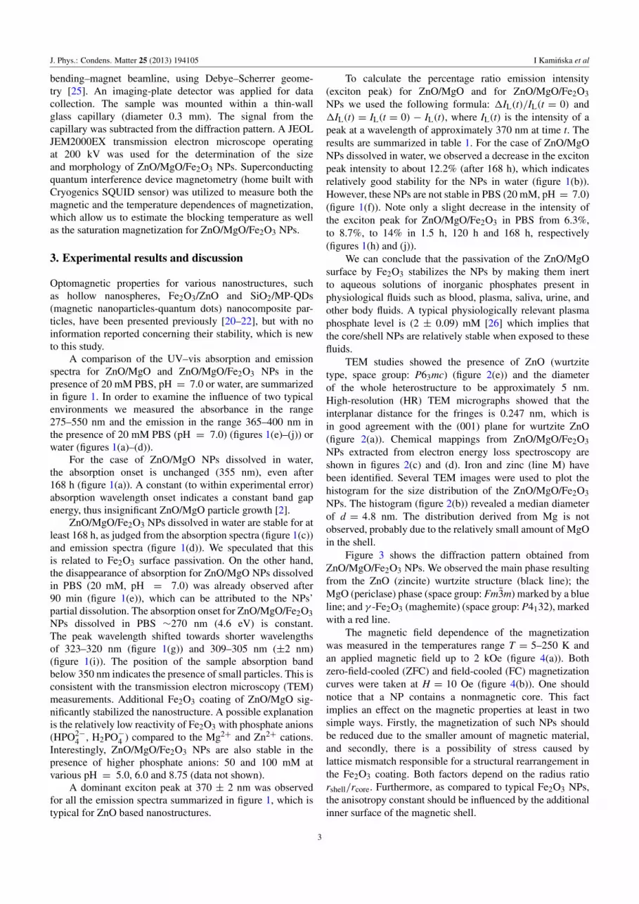
Fe3O4 and Fe2O3 exhibit superparamagnetic properties whichenable NPs to be used as agents for magnetic resonanceimaging [7, 8]. Currently, magnetic NPs have four major,clearly defined biomedical applications: cell separation, drugdelivery, hyperthermia (which involves the introduction ofeither ferromagnetic or superparamagnetic particles into thetissue [9, 10]), and magnetic resonance imaging [11–18].
Recent advances in solution phase colloidal physicalchemistry have enhanced the production of nanomaterialswith multifunctional properties for various biomedicalapplications. In spite of intensive research activity, only a fewexamples of optomagnetic NPs, which are both chemicallystable under biologically relevant aqueous conditions andbiocompatible, have been reported. Recently, a study onthe seeded growth of Fe3O4/ZnO bifunctional nanocrystalspossessing optomagnetic properties was reported [19].However, neither emission spectra nor biologically relevantsolution stability results were provided.
Biocompatible core/shell optomagnetic NPs are ofconsiderable importance for site-specific therapeuticallymotivated drug delivery. Optical (especially near-infrared)and magnetic imaging have made a significant impact onmedical diagnostics. Future advances in these fields are inthe confinement of specific optical and magnetic propertieson single, multimodal NPs.
Recently, several groups reported a successful synthesisof particles that possess both fluorescent and magneticproperties. Qian et al [20] have reported on the synthesisof bifunctional magnetic–fluorescent hollow ZnO/ZnFe2O4nanostructures by a co-precipitation method. Guskos et al [21]have synthesized fine particles of Fe2O3/ZnO using a wetchemical method, based on ferromagnetic resonance (FMR)measurements. Analysis of the FMR spectra indicates thepresence of strongly anisotropic interactions. Yi et al [22]have synthesized a water soluble nanocomposite consistingof both: magnetic nanoparticles γ -Fe2O3 and CdSe quantumdots (QDs) encapsulated within a silica shell and showed thatthe nanocomposite preserved both the magnetic properties ofγ -Fe2O3 and the optical properties of CdSe QDs.
The aim of this work is to synthesize NPs whichare useful for a new generation of biosensors exhibitingbiocompatible optomagnetic properties. To the best ofour knowledge, this paper is the first communication onthe properties of core/shell ZnO/MgO/Fe2O3 NPs underphysiologically relevant environments.
2. Materials and methods
2.1. Synthesis of ZnO/MgO core/shell nanoparticles
In order to prevent spontaneous aggregation and to improvethe efficiency of ZnO luminescence in aqueous solutions, theirsurface was passivated with a shell of MgO. The followingreactions took place:
Zn2++ 2OH−→ Zn(OH)2 → ZnO+ H2O (1)
Mg2++ 2OH−→ Mg(OH)2 → MgO+ H2O. (2)
Synthesis (1) was performed using 0.5 mM Zn(CH3COO)2×2H2 (Chempur pure p.a.) in 65 ml ethanol (Chempur,min 99.8% pure p.a. (per analysis)) and 1.25 mM NaOH(Chempur, min 98.8% pure p.a.) in 70 ml ethanol. Allcomponents were pre-heated to 30 ◦C, stirred to dissolve for1 h, followed by cooling at room temperature. The reactionwas initiated by adding the NaOH solution dropwise into theZn2+ solution at 0 ◦C with vigorous stirring [23].
The resulting solution was incubated for 30 min andthen warmed up to 35 ◦C. After 2 h an aliquot of 3 ml wasplaced in the spectrophotometric cell to record absorption andemission spectra. In the next step (2) a solution of 0.5 mMMg(CH3COO)2 × 4H2O (Sigma-Aldrich, ≥99%) (mass ratioMg/Zn = 17%) in 15 ml ethanol was added dropwiseand incubated overnight for 15 h. The unreacted materialwas discarded after centrifugation (6000 rpm, 15 min,20 ◦C). The spun material was washed in heptane–ethanol(3:1) and the product allowed to dry at room temperature.Spectrophotometric measurements were then performed onthe newly synthesized ZnO/MgO core/shell structure at25 ◦C [24].
2.2. Synthesis of ZnO/MgO/Fe2O3 nanoparticles
In order to synthesize the optomagnetic NPs the previouslyprepared ZnO/MgO NPs were covered with an Fe2O3 shell :
Fe3++ 6OH−→ Fe2O3 + 3H2O. (3)
Synthesis (3) was performed using 37 mM Fe(NO3)3 ×
9H2O (Chempur, min 99.8% pure p.a.) in 150 ml ofmethanol (Chempur, min 99.8% pure p.a.). Subsequently0.26 g of ZnO/MgO was dissolved in 100 ml of methanol(prepared as described above). Then 0.11 mol NaOH in 1:1distilled water to methanol was added dropwise to a stirredmixture of preformed ZnO/MgO and the Fe(NO3)3 × 9H2Osolution at 5–10 ◦C. The resulting mixture was incubated for30 min. It was then warmed up to 35 ◦C and incubated for2 h on a magnetic stirrer followed by centrifugation. TheZnO/MgO/Fe2O3 NPs were alternately washed with waterand ethanol, and dried for several days at room temperature.
The stability of ZnO/MgO/Fe2O3 NPs dissolved in waterand several phosphate buffered saline (PBS) solutions (5, 20,50 and 100 mM phosphates at various pH = 5.0, 6.0,7.0, and 8.75) was determined by absorption and emissionspectroscopy. The concentration of the magnetically stirredNP suspension was 0.2 mg ml−1. This paper is confined onlyto 20 mM PBS at pH = 7.0 and water.
Absorption and emission spectra for the NPs weremonitored using a Cary 50 Scan UV–vis spectrophotometerand a Fluorolog III Phosphorimeter with a 550 W xenonlamp, respectively. Both instruments were equipped with athermostated cuvette holder. The measurements were carriedout on samples kept in a quartz cuvette with dimensionsof 1 × 1 × 4 cm3 [5]. A structural characterizationof ZnO/MgO/Fe2O3 was performed using XRD, at asynchrotron-radiation source. Diffraction studies were carriedout using a powder diffractometer with 2θ from 2◦ to70◦ and a step size of 0.004 at the B2 (HASYLAB/DESY)
2
J. Phys.: Condens. Matter 25 (2013) 194105 I Kaminska et al
bending–magnet beamline, using Debye–Scherrer geome-try [25]. An imaging-plate detector was applied for datacollection. The sample was mounted within a thin-wallglass capillary (diameter 0.3 mm). The signal from thecapillary was subtracted from the diffraction pattern. A JEOLJEM2000EX transmission electron microscope operatingat 200 kV was used for the determination of the sizeand morphology of ZnO/MgO/Fe2O3 NPs. Superconductingquantum interference device magnetometry (home built withCryogenics SQUID sensor) was utilized to measure both themagnetic and the temperature dependences of magnetization,which allow us to estimate the blocking temperature as wellas the saturation magnetization for ZnO/MgO/Fe2O3 NPs.
3. Experimental results and discussion
Optomagnetic properties for various nanostructures, suchas hollow nanospheres, Fe2O3/ZnO and SiO2/MP-QDs(magnetic nanoparticles-quantum dots) nanocomposite par-ticles, have been presented previously [20–22], but with noinformation reported concerning their stability, which is newto this study.
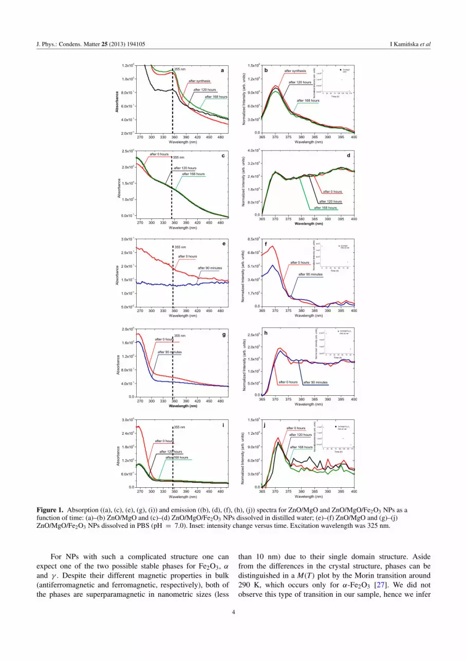
A comparison of the UV–vis absorption and emissionspectra for ZnO/MgO and ZnO/MgO/Fe2O3 NPs in thepresence of 20 mM PBS, pH = 7.0 or water, are summarizedin figure 1. In order to examine the influence of two typicalenvironments we measured the absorbance in the range275–550 nm and the emission in the range 365–400 nm inthe presence of 20 mM PBS (pH = 7.0) (figures 1(e)–(j)) orwater (figures 1(a)–(d)).
For the case of ZnO/MgO NPs dissolved in water,the absorption onset is unchanged (355 nm), even after168 h (figure 1(a)). A constant (to within experimental error)absorption wavelength onset indicates a constant band gapenergy, thus insignificant ZnO/MgO particle growth [2].
ZnO/MgO/Fe2O3 NPs dissolved in water are stable for atleast 168 h, as judged from the absorption spectra (figure 1(c))and emission spectra (figure 1(d)). We speculated that thisis related to Fe2O3 surface passivation. On the other hand,the disappearance of absorption for ZnO/MgO NPs dissolvedin PBS (20 mM, pH = 7.0) was already observed after90 min (figure 1(e)), which can be attributed to the NPs’partial dissolution. The absorption onset for ZnO/MgO/Fe2O3NPs dissolved in PBS ∼270 nm (4.6 eV) is constant.The peak wavelength shifted towards shorter wavelengthsof 323–320 nm (figure 1(g)) and 309–305 nm (±2 nm)(figure 1(i)). The position of the sample absorption bandbelow 350 nm indicates the presence of small particles. This isconsistent with the transmission electron microscopy (TEM)measurements. Additional Fe2O3 coating of ZnO/MgO sig-nificantly stabilized the nanostructure. A possible explanationis the relatively low reactivity of Fe2O3 with phosphate anions(HPO2−
4 , H2PO−4 ) compared to the Mg2+ and Zn2+ cations.Interestingly, ZnO/MgO/Fe2O3 NPs are also stable in thepresence of higher phosphate anions: 50 and 100 mM atvarious pH = 5.0, 6.0 and 8.75 (data not shown).
A dominant exciton peak at 370 ± 2 nm was observedfor all the emission spectra summarized in figure 1, which istypical for ZnO based nanostructures.
To calculate the percentage ratio emission intensity(exciton peak) for ZnO/MgO and for ZnO/MgO/Fe2O3NPs we used the following formula: 1IL(t)/IL(t = 0) and1IL(t) = IL(t = 0) − IL(t), where IL(t) is the intensity of apeak at a wavelength of approximately 370 nm at time t. Theresults are summarized in table 1. For the case of ZnO/MgONPs dissolved in water, we observed a decrease in the excitonpeak intensity to about 12.2% (after 168 h), which indicatesrelatively good stability for the NPs in water (figure 1(b)).However, these NPs are not stable in PBS (20 mM, pH = 7.0)(figure 1(f)). Note only a slight decrease in the intensity ofthe exciton peak for ZnO/MgO/Fe2O3 in PBS from 6.3%,to 8.7%, to 14% in 1.5 h, 120 h and 168 h, respectively(figures 1(h) and (j)).
We can conclude that the passivation of the ZnO/MgOsurface by Fe2O3 stabilizes the NPs by making them inertto aqueous solutions of inorganic phosphates present inphysiological fluids such as blood, plasma, saliva, urine, andother body fluids. A typical physiologically relevant plasmaphosphate level is (2 ± 0.09) mM [26] which implies thatthe core/shell NPs are relatively stable when exposed to thesefluids.
TEM studies showed the presence of ZnO (wurtzitetype, space group: P63mc) (figure 2(e)) and the diameterof the whole heterostructure to be approximately 5 nm.High-resolution (HR) TEM micrographs showed that theinterplanar distance for the fringes is 0.247 nm, which isin good agreement with the (001) plane for wurtzite ZnO(figure 2(a)). Chemical mappings from ZnO/MgO/Fe2O3NPs extracted from electron energy loss spectroscopy areshown in figures 2(c) and (d). Iron and zinc (line M) havebeen identified. Several TEM images were used to plot thehistogram for the size distribution of the ZnO/MgO/Fe2O3NPs. The histogram (figure 2(b)) revealed a median diameterof d = 4.8 nm. The distribution derived from Mg is notobserved, probably due to the relatively small amount of MgOin the shell.
Figure 3 shows the diffraction pattern obtained fromZnO/MgO/Fe2O3 NPs. We observed the main phase resultingfrom the ZnO (zincite) wurtzite structure (black line); theMgO (periclase) phase (space group: Fm3m) marked by a blueline; and γ -Fe2O3 (maghemite) (space group: P4132), markedwith a red line.
The magnetic field dependence of the magnetizationwas measured in the temperatures range T = 5–250 K andan applied magnetic field up to 2 kOe (figure 4(a)). Bothzero-field-cooled (ZFC) and field-cooled (FC) magnetizationcurves were taken at H = 10 Oe (figure 4(b)). One shouldnotice that a NP contains a nonmagnetic core. This factimplies an effect on the magnetic properties at least in twosimple ways. Firstly, the magnetization of such NPs shouldbe reduced due to the smaller amount of magnetic material,and secondly, there is a possibility of stress caused bylattice mismatch responsible for a structural rearrangement inthe Fe2O3 coating. Both factors depend on the radius ratiorshell/rcore. Furthermore, as compared to typical Fe2O3 NPs,the anisotropy constant should be influenced by the additionalinner surface of the magnetic shell.
3
J. Phys.: Condens. Matter 25 (2013) 194105 I Kaminska et al
Figure 1. Absorption ((a), (c), (e), (g), (i)) and emission ((b), (d), (f), (h), (j)) spectra for ZnO/MgO and ZnO/MgO/Fe2O3 NPs as afunction of time: (a)–(b) ZnO/MgO and (c)–(d) ZnO/MgO/Fe2O3 NPs dissolved in distilled water; (e)–(f) ZnO/MgO and (g)–(j)ZnO/MgO/Fe2O3 NPs dissolved in PBS (pH = 7.0). Inset: intensity change versus time. Excitation wavelength was 325 nm.
For NPs with such a complicated structure one canexpect one of the two possible stable phases for Fe2O3, αand γ . Despite their different magnetic properties in bulk(antiferromagnetic and ferromagnetic, respectively), both ofthe phases are superparamagnetic in nanometric sizes (less
than 10 nm) due to their single domain structure. Asidefrom the differences in the crystal structure, phases can bedistinguished in a M(T) plot by the Morin transition around290 K, which occurs only for α-Fe2O3 [27]. We did notobserve this type of transition in our sample, hence we infer
4

J. Phys.: Condens. Matter 25 (2013) 194105 I Kaminska et al
Figure 2. (a) HRTEM images for ZnO/MgO/Fe2O3 NPs. (b) Size distributions for ZnO/MgO/Fe2O3 NPs. Electron energy lossspectroscopy chemical mapping for ZnO/MgO/Fe2O3 NPs. (c) Zinc mapping. (d) Iron mapping. (e) Electron diffraction patterncorresponding to a wurtzite crystal structure.
Table 1. Change of the emission intensity for ZnO/MgO and for ZnO/MgO/Fe2O3 NPs (details in the text).
Environment Sample
1IL(t) (arb. u.)
t = 90 min t = 120 h t = 168 h
Water ZnO/MgO Not tested 5.6% 12.2%ZnO/MgO/Fe2O3 0% 0% 0%
PBS ZnO/MgO 31.9% Not stable Not stableZnO/MgO/Fe2O3 6.3% 8.7% 14.0%
Figure 3. XRD patterns (Cu Kα1, λ = 1.5406 A) for theZnO/MgO/Fe2O3 nanocrystalline powder.
that the shell is built from the γ -phase. It can be stabilized bycubic MgO—the middle part of the structure.
Magnetization dependences on temperature (figure 4(b))show a behavior known for typical superparamagnetic NPs.
Interestingly, the slight difference of 1 K between both theaverage and the maximal blocking temperatures, TB andTB,max respectively, suggests a small dispersion of NP sizes.The magnetic anisotropy constant can be calculated fromthe relation K = 25kBVB/TB, where kB is the Boltzmannconstant, TB is the average blocking temperature and VB isthe corresponding volume of NPs. For an average diameterestimated from TEM studies (d = 4.8 nm), the value of K is2.6×106 erg cm−3, which is relatively high. One should keepin mind that this estimation is not dedicated to heterostructureswith magnetic shells.
Hysteretic behavior is well observed below the blockingtemperature TB = 43 K, while above this point the NPs reveala superparamagnetic response in applied magnetic fields.Saturation of magnetization (MS) in the Fe2O3 NPs can beattained at magnetic fields much higher than 2 kOe [28].However, the experimental points M(H) at temperaturesabove TB are well fitted by a Langevin function, giving thehighest saturation magnetization at TB equal to 55 emu g−1.This value is around 0.6 × MS, as compared to bulkγ -Fe2O3 [29].
In order to demonstrate the magnetic properties ofZnO/MgO/Fe2O3 NPs at room temperature they were
5

J. Phys.: Condens. Matter 25 (2013) 194105 I Kaminska et al
Figure 4. (a) Mass magnetization as a function of applied external magnetic field measured for various temperatures in the rangeT = 5–250 K. (b) ZFC and FC magnetization curves as well as their difference MFC −MZFC for ZnO/MgO/Fe2O3 NPs.
Figure 5. ZnO/MgO/Fe2O3 NPs dissolved in distilled water. (a) Ina magnetic field the particles are attracted to the side of the vessel(indicated by dashed ovals). (b) Image of ZnO/MgO/Fe2O3 NPsafter the removal of or without the magnetic field.
suspended in distilled water and inserted between neodymiummagnets (in a magnetic field of approximately 600 mT). Themagnetic field attracts NPs, as shown in figure 5(a). Theremoval of the external magnetic field dispersed the NPshomogeneously in the suspension and no visible aggregateswere observed [30]. As one can see, superparamagnetismenables the NPs to respond to an applied magnetic fieldwithout any permanent magnetization and thus to bere-dispersed rapidly when the magnetic field is removed(figure 5(b)) [31].
4. Conclusions
In this work, using an efficient and low cost sol–gel method,we synthesized sizable quantities of ZnO/MgO/Fe2O3 NPsof a defined geometry, crystalline structure, and optical andmagnetic properties. The fabricated material has potential,after proper biological passivation, in biomedical applications.The ZnO/MgO/Fe2O3 NPs of mean diameter 4.8 nm,while in aqueous solutions, maintain their optical andsuperparamagnetic properties at room temperature. Theoptomagnetic dual properties are potentially useful for
biological labeling imaging and separation in an externalmagnetic field of biomolecules attached to the NPs. Inaddition, the material could be used for diagnosticallyrelevant biosensing when passivated with a biologically activemolecule capable of binding specifically with a pathologicaltarget. Our ZnO based heterostructure material is shown to bestable for seven days in physiologically relevant environmentsand is potentially biologically compatible.
Acknowledgments
We wish to thank Dr Tony Bell for assistance withthe operation of the B2 powder diffraction beamline atHASYLAB/DESY.
This work has been supported by grant NN 518 424036from the Ministry of Science and Higher Education andInnovative Economy grant POIG.01.01.02-00-008/0.
References
[1] Plank N O V, Snaith H J, Ducati C, Bendall J S,Schmidt-Mende L and Welland M E 2008 Nanotechnology19 465603
[2] Bera D L, Qian P and Holloway H 2008 J. Phys. D: Appl.Phys. 41 182002
[3] Rakshit S and Vasudevan S 2008 ACS Nano 2 1473[4] Zhao J, You H, Zhang Y and Yuan J 2011 Toxic effects of
nano-Fe2O3 on liver and kidney cells of rats 5th Int. Conf.on Bioinformatics and Biomedical Engineering (ICBBE)(Wuhan, China, 10–12 May) pp 1–4
[5] Sikora B, Fronc K, Kaminska I, Baranowska-Korczyc A,Sobczak K, Dłuzewski P and Elbaum D 2012 J. Sol–GelSci. Technol. 61 197
[6] Baranowska-Korczyc A, Reszka A, Sobczak K, Sikora B,Dziawa P, Aleszkiewicz M, Kłopotowski Ł, Paszkowicz W,Dłuzewski P, Kowalski B J, Kowalewski T A, Sawicki M,Elbaum D and Fronc K 2012 J. Sol–Gel Sci. Technol.61 494
[7] Lowery T 2009 Nanomaterials-based magnetic sensors switchbiosensors Nanomaterials for the Life Sciences vol 1, edC S S R Kumar (Weinheim: Wiley-VCH) pp 31–54
[8] Pankhurst Q A, Connolly J, Jones S K and Dobson J 2003J. Phys. D: Appl. Phys. 36 R167
6

J. Phys.: Condens. Matter 25 (2013) 194105 I Kaminska et al
[9] Brusentsov N A 1990 Mendeleev Chem. J. 35 98[10] Chan D C F, Kirpotin D B and Bunn P A 1993 J. Magn.
Magn. Mater. 122 374[11] Hernando A, Crespo P and Garcıa M A 2005 Sci. World J.
5 972[12] Tartaj P, Morales M P, Ventemillas-Verdaguer S,
Gonzalez-Carreno T and Serna C J 2003 J. Phys. D: Appl.Phys. 36 R182
[13] Berry C C 2005 J. Mater. Chem. 15 543–7[14] Saiyed Z M, Telang S D and Ramchand C N 2003 Biomagn.
Res. Technol. 1 2[15] Curtis A 2003 Europhys. News 34 210–1[16] Brusentsov N A, Gogosov V V, Brusentsova T N,
Sergeev A V, Jurchenko N Y, Kuznetsov A A,Kuznetsov O A and Shumakov L I 2001 J. Magn. Magn.Mater. 225 113
[17] Jordan A, Scholz R, Maier-Hauff K, Johannsen M, Wust P,Nadobny J, Schirra H, Schmidt H, Deger S, Loening S,Lanksch W and Felix R 2001 J. Magn. Magn. Mater.225 118
[18] Wang Y X, Hussain S M and Krestin G P 2001 Eur. Radiol.11 2319–31
[19] Chiu W, Khiew P, Cloke M, Isa D, Lim H, Tan T, Huang N,Radiman S, Abd-Shukor R, Hamid M A A andChia Ch 2010 J. Phys. Chem. C 114 8212–8
[20] Qian H S, Hu Y, Li Z Q, Yang X Y, Li L Ch, Zhang X T andXu R 2010 J. Phys. Chem. C 114 17455–9
[21] Guskos N, Glenis S, Zolnierkiewicz G, Typek J, Sibera D,Kaszewski J, Moszynski D, Łojkowski W andNarkiewicz U 2010 Physica B 405 4054–8
[22] Yi D K, Selvan S T, Lee S S, Papaefthymiou G C,Kundaliya D and Ying J Y 2005 J. Am. Chem. Soc.127 4990–1
[23] Lee J J, Bang J and Yang H 2009 J. Phys. D: Appl. Phys.42 025305
[24] Sikora B, Fronc K, Kaminska I, Koper K, Stepien P andElbaum D 2013 J. Phys.: Condens. Matter 25 194104
[25] Knapp M, Baehtz C, Ehrenberg H and Fuess H 2004J. Synchrotron Radiat. 11 328–34
[26] Goodman J and Bessman A N 1975 Am. J. Med. Sci.270 447–51
[27] Morin F 1950 J. Phys. Rev. 78 819–20[28] Coey J M D 1971 Phys. Rev. Lett. 27 1140[29] Singh V, Seehra M S, Bali S, Eyring E M, Shah N,
Huggins F E and Huffman G P 2011 J. Phys. Chem. Solids72 1373–6
[30] Cao X, Prozorov R, Koltypin Yu, Kataby G, Felner I andGedanken A 1997 J. Mater. Res. 12 2
[31] Ma Z, Guan Y and Liu H 2006 J. Magn. Magn. Mater.301 469–77
7


