Embed Size (px)
Citation preview

Nitric Oxide Release in Human Aortic Endothelial CellsMediated by Delivery of Amphiphilic Polysiloxane Nanoparticles
to Caveolae
Takehiro Nishikawa,*,† Norio Iwakiri,‡ Yoshiro Kaneko,‡ Akihiko Taguchi,†
Kazuhito Fukushima,† Hidezo Mori,† Nobuhiro Morone,§ and Jun-ichi Kadokawa*,‡
National Cardiovascular Center Research Institute, 5-7-1, Fujishirodai, Suita, Osaka 565-8565, Japan,National Center of Neurology and Psychiatry, National Institute of Neuroscience, 4-1-1,
Ogawahigashimachi, Kodaira, Tokyo 187-8502, Japan, and Graduate School of Science and Engineering,Kagoshima University, 1-21-40 Korimoto, Kagoshima, Kagoshima 890-0065, Japan
Received February 1, 2009; Revised Manuscript Received June 8, 2009
Microdomains such as lipid raft and caveolae are organized as functional compartments in plasma membrane ofcells. In this study, we note the functional platform of caveolae with dual functions, internalization of externalsubstances and cell signalings leading to nitric oxide release, and hypothesize that the switching of enzyme activityof endothelial nitric oxide synthase can be achieved by targeting caveolae with nanoparticles. We preparedpolysiloxane nanoparticles and studied cellular uptake of the nanoparticles and its concomitant influence on thenitric oxide release in human aortic endothelial cells. We found that polysiloxane nanoparticles were endocytosedvia caveolae in human aortic endothelial cells and that enhanced nitric oxide release was followed by the cellularuptake of the nanoparticles. Furthermore, we confirmed that endothelial nitric oxide synthase was activated duringcellular uptake of the nanoparticles. These findings support our idea that delivery of the polymeric nanoparticlesto endothelial cells can lead to the induction of nitric oxide release.
Introduction
With the recent progress in structural biology, it has beenrevealed that microdomains such as lipid raft and caveolae areorganized as functional compartments in plasma membrane ofcells.1 Caveolae in particular are abundant in the plasmamembrane of endothelial cells, occupying 15% of total cellvolume and function as platforms for signaling and transporting.2
In relation with pathology, it is pointed out that a lost functionof caveolae is involved in various cardiovascular diseases suchas vascular dysfunction, atherosclerosis, and hypertrophy.3 Forinstance, hypertension is one of the risk factors for variouscardiovascular diseases such as atherosclerosis, ischemic heartfailure, stroke, and chronic renal failure. Blood pressure iscontrolled using antihypertensive drugs, which are the majormedication for treating hypertension. Nitric oxide (NO) is a keysubstance in the vasorelaxation process and plays a crucial rolein the regulation of blood pressure.4 Direct control of nitric oxide(NO) production in vascular endothelium can be a novel strategyof the medication for hypertension and can lead to theimprovement of endothelial functions.5 NO molecules arereleased from vascular endothelium, are diffused to media, theouter layer of smooth muscle cells, and trigger the activationof soluble guanylate cyclase that leads to smooth muscle cellrelaxation.6 NO is synthesized by endothelial nitric oxidesynthase (eNOS) that is embedded in caveolae and is activatedby external stimuli such as bioactive substances and dynamicenvironmental factors.7 Caveolae are characteristic flask-shaped
invaginations of plasma membrane with diameters of 50-100nm and work as functional platforms for internalization ofextracellular materials (endocytosis) and cell signalings leadingto NO production.3
In the past studies about vascular drug delivery, it was notedthat caveolae can provide a possible pathway for drug deliverycoupled with caveoli-mediated endocytosis.8 To date, nanopar-ticles as drug carriers have been extensively studied with regardsto delivery, drug loading, drug release, in vivo circulation, andtoxicological properties.9 However, whether such a nanoparticlecause effects on the functions of cells, tissues, and organ remainsstill unclear. Considering the endocytic pathways of cells, it isknown that four basic mechanisms, macropinocytosis, clathrin-mediated endocytosis, caveolin-mediated endocytosis, and clath-rin- and caveolin-independent endocytosis, are involved inpinocytosis (endocytosis in all mammalian cells).10 Bioactivesubstances and serum proteins circulating in bloodstream havebeen found to be external stimuli for the activation of signaltransduction. Although it was reported that artificial nanopar-ticles could influence a cell function of macrophages upon theassociation with lipopolysaccharide,11 the artificial nanoparticleshave not been considered and discussed in terms of the externalstimuli as an input signal at specific membrane microdomainof cells for the activation of signal transduction, so far.Therefore, we have an interest in the influence of cellular uptakeof nanoparticles on the signal transductions leading to theexpression of cell functions. As the first attempt, we preparednanoparticles from amphiphilic polysiloxane and studied cellularuptake of the nanoparticles and its concomitant influence onNO release in human aortic endothelial cells.
In this research, we chose polysiloxane as a polymericmaterial for the preparation of nanoparticles, because siloxanebackbone with self-repair property is quite stable in physiologi-cal conditions12 and nanoparticles of polysiloxane are expected
* To whom correspondence should be addressed. Tel.: 81-6-6833-5012 (T.N.); 81-99-285-7743 (J.-i.K.). Fax: 81-6-6872-7485 (T.N.); 81-99-285-3253 (J.-i.K.). E-mail: [email protected] (T.N.); [email protected] (J.-i.K.).
† National Cardiovascular Center Research Institute.‡ Kagoshima University.§ National Center of Neurology and Psychiatry.
Biomacromolecules 2009, 10, 2074–20852074
10.1021/bm900128x CCC: $40.75 2009 American Chemical SocietyPublished on Web 07/07/2009

to be resistant to biological degradation in both extracellularand cytoplasmic environments. Silicone derivatives are knownto be very attractive materials, because they exhibit low toxicityand unique physical properties. Therefore they have been widelyused as versatile products such as foam stabilizer, rubber, paint,fiber, glass, and textile.13 In spite of the usage of polysiloxanein wide fields, polysiloxane has not been studied so far in termsof polymeric materials for nanoparticles that are aimed at drugdelivery system. Although biodegradable polymers have beenextensively studied as polymeric materials for nanocarriers, arecent review article indicates the toxicological problemsassociated with degradation products of biodegradable polymericcarriers.14 Polysiloxane has noteworthy properties with regardto degradation, stability and durability, based on a self-repairmechanism where silanol groups generated by the degradationof siloxane backbone can condense each other to form newsiloxane bonds.12 Polysiloxane with stability and inertness isexpected to achieve reduced toxicity and longer circulation interms of their usage as a drug carrier. Here, we describenanoparticle formation of amphiphilic polysiloxane, cellularuptake of polysiloxane nanoparticles via caveolae of membranemicrodomains, and influence of polysiloxane nanoparticles oncellular function; nitric oxide release in human aortic endothelialcells.
Experimental Section
Materials. Amphiphilic polysiloxane (Am-PAPS) was prepared bythe method described in our previous report.15 Am-PAPS was labeledwith fluorescein thioisocyanate and was named Flu-Am-PAPS. Rabbitanti-caveolin-1 IgG was purchased from Sigma (St. Louis, MI). Mouseanti-eNOS IgG and mouse antiphospho-Ser1177-eNOS IgG waspurchased from BD Transduction Laboratories (Franklin Lakes, NJ).Rabbit anti-fluorescein IgG was purchased from Molecular Probes(Eugene, OR). Anti-rabbit IgG conjugated with gold nanoparticles (5nm in diameter) was purchased from GE healthcare. TRITC-labeledanti-rabbit IgG and TRITC-labeled anti-mouse IgG were purchased fromMolecular Probes (Eugene, OR). HRP conjugated antimouse IgG waspurchased from Cell Signaling Technology (Danvers, Mass). Mono-sulfo-N-hydroxy-succinimido nanogold (NANOGOLD) was purchasedfrom Nanoprobes (Yaphank, NY). Gold nanoparticle labeled Am-PAPS(Au-Am-PAPS) was prepared by coupling NANOGOLD to theamphiphilic polysiloxane. All other reagents were purchased fromGibco, Nacalai Tesque, Sigma, or Wako Pure Chemicals unlessotherwise indicated.
Nanoparticles Suspension. Aqueous suspension of each amphiphilicpolysiloxane (Am-PAPS, Flu-Am-PAPS, or Au-Am-PAPS) was pre-pared by dispersing it (initial concentration: 1 mg/mL) into cell culturemedium (EGM-2; Cambrex) and sonication with bath-type ultrasoundwasher (25 W and 40 kHz for 5 min). The suspension was passedthrough the membrane filters with the pore size of 0.45 µm (Millex-GV; Millipore) and 0.22 µm (Millex-GV; Millipore) for sterilization.Concentration of Flu-Am-PAPS was determined by fluorescencespectroscopy and was 0.1 mg/mL that was based on calibration withfluorescein-labeled sugar conjugated PAPS (1 mg/mL dissolved inEGM-2).
Measurements. The 1H NMR spectra (600 MHz) were recordedusing a JEOL ECA600 spectrometer. Fluorescence spectra for quantita-tive analysis were obtained on a fluorescence spectrometer (RF-5300PC: Shimadzu) using a quartz cuvette (1 mm path length). The dynamiclight scattering (DLS) measurement was performed on a Zetasizer 3000(Malvern Instruments). Morphological study of nanoparticles wascarried out by scanning electron microscope (Hitachi S-4100 electronmicroscope). The observation of plasma membrane and cytosol ofhuman aortic endothelial cells was performed by a transmission electronmicroscope (Tecnai G2 Sphera, FEI, U.S.A.). Fluorescence imagingwas performed on a IX-71 (Olympus) equipped with a fluorescence
mirror unit, U-MNIBA3 (band-pass filter from 470 to 495 nm forexcitation light and using a long pass filter from 510 to 550 nm foremission light) for the detection of fluorescein emission and U-MWIG3(band-pass filter from 530 to 550 nm for excitation light and using along pass filter >575 nm for emission light) for the detection ofrhodamine emission. High-pressure mercury lamp (USH-1030 L:Olympus) was used as a light source for the fluorescence microscopyand was powered by a power supply (BH2-RFL-T3: Olympus).
Cell Culture Experiment. Human aortic endothelial cells (HAECs)were purchased as cryopreserved samples of third passage (Lot: 4F1350)from Cambrex, Wakersville, MD. The HAECs used in the experimentwere fourth passage. Polystyrene dishes (φ35 mm, Iwaki) were filledwith 2 mL of a supplemented culture medium (EGM-2; Cambrex) andequilibrated in a 37 °C, 5% CO2 humidified incubator for 30 min beforecell seeding. After the frozen cells were thawed at 37 °C, 70 µL of thecell suspension (8.0 × 10⁵ cells/mL, viability: 85% (determined bytrypan blue exclusion test)) were seeded in the culture dishes. The cell-seeded plates were placed in a 37 °C, 5% CO2 humidified incubator.The HAECs were cultured for 72 h before the medium exchange withnanoparticle suspension. For fluorescence microscopy observation, thecells were rinsed with PBS (warmed at 37 °C), fixed by immersinginto 10% formaldehyde neutral buffer solution (Nacalai tesque) at roomtemperature (22 °C) for 15 min and washed three times with PBS(Gibco). Fluorescence images used for the quantitative analysis of thecellular uptake of nanoparticles were taken at the constant exposuretime to compare fluorescence intensity of the images at prescribedincubation time. Fluorescence images of the cells were taken by afluorescence microscope (IX71; Olympus) equipped with a CCD camera(DP70; Olympus). To perform quantitative evaluation of fluorescenceintensity, the fluorescence images were taken at the same exposure time(1/6.0 s). The exposure time was automatically measured by theoperating software of DP70. The appropriate exposure time was chosenunder the condition that over exposed images should be avoided tocalculate fluorescence intensity of the images as correct as possible.The excess over exposure and under exposure could be prevented whenthe exposure time was set at 1/6.0 s that was measured when the HAECsexposed to Flu-Am-PAPS for 6 h were photographed by DP70.Fluorescence intensity of the incorporated nanoparticles of Flu-Am-PAPS was measured by integrating the fluorescence intensity observedat each pixel of the fluorescence images using image analysis software(Fluoview ver. 5.0; Olympus).
Cytotoxicity Assay. Cytotoxicity of polysiloxane nanoparticles wereassessed using Cell Count Reagent SF (Nacalai Tesque, Kyoto) as acolorimetric indicator for living cells. HAECs were seeded in each wellof 96-well plates and were incubated in a 37 °C, 5% CO2 humidifiedincubator for 24 h before cytotoxicity assay. Cell number in each wellwas adjusted by stepwise 2-fold dilution of HAECs cell suspension.The cell number per well ranged from 5000 to 40000 cells. The totalamount of the cell suspension including growth medium was 100 µLper well. The growth medium was replaced with 100 µL of a suspensionof polysiloxane nanoparticle (1 mg/mL) for cytotoxicity testing. After6 or 24 h incubation, each well was rinsed with 100 µL of growthmedium to remove polysiloxane nanoparticles and was filled with 100µL of fresh growth medium. Then, 10 µL of Cell Count Reagent SFsolution containing WST-8 (5 mM),16 colorimetric indicator, was addedto each well to evaluate cell viability. HAECs were incubated withWST-8 for 4 h. WST-8 is converted to water-soluble formazan by anelectron mediator coupled to the intracellular reduction of NAD+.Absorbance of each well was measured at 450 nm using a microplatereader (Model 680, Bio-Rad, Hercules, CA). Cell viability was evaluatedby the following equation: (cell viability) ) (Abs for samples to betested - Abs for blank)/(Abs for control - Abs for blank) × 100,“Abs” stands for absorbance of each well at 450 nm.
Immunostaining. To visualize the localization of caveolin-1 andendothelial nitric oxide synthase (eNOS) in HAECs, each antigen wasstained by immunological method using primary antibodies (rabbit anti-caveolin-1 IgG; Sigma and mouse anti-eNOS IgG; BD) and fluores-
Nitric Oxide Release of Endothelial Cells Biomacromolecules, Vol. 10, No. 8, 2009 2075
cence labeled secondary antibodies (TRITC labeled anti-rabbit IgG andTRITC labeled anti-mouse IgG; Molecular Probes). For immunostain-ing, cells were fixed by 10% formaldehyde neutral buffer solution(Nacalai tesque) at room temperature (22 °C) for 15 min and permeatedwith 0.1% PBS (pH 7.2, Gibco) solution of Triton X-100 (Sigma) for5 min at 20 °C. After 1 h of blocking with 1% normal goat serumsolution, HAECs were incubated with a primary antibody (200 timesdilution with 0.1% normal goat serum solution) for 1 h. The HAECstreated with primary antibody were incubated with fluorescent dyelabeled secondary IgG for 1 h. Fluorescence images of the cells weretaken by a fluorescence microscope (IX71; Olympus) equipped with aCCD camera (DP70; Olympus) and confocal laser scanning microscope(FV 100; Olympus).
Transmission Electron Microscopy (TEM). Two types of speci-mens, ultrathin section and rapid-freeze, deep-etch, freeze-replica, wereprepared for the observation of fine structure of plasma membrane andcytosol in cells using transmission electron microscopy. HAECs weregrown on carbon-coated sapphire glass with a diameter of 5 mm for 2days after inoculation. Specimens for ultrathin section were preparedby the following method. For the TEM observation of polysiloxanenanoparticles in caveolae, HAECs were exposed to gold nanoparticlelabeled Am-PAPS (Au-Am-PAPS) dispersed in growth medium (EGM-2) for 3 h. After a 3 h incubation with Au-Am-PAPS, the HAECsattached on coverslips were washed with NaHCa buffer (30 mMHEPES, 100 mM NaCl, 2 mM CaCl2, pH 7.3) and treated with chemicalfixation reagent (2.5% glutaraldehyde (GA), 150 mM sucrose in PBSbuffer). In the case of the immunodetection of caveolae in HAECs,HAECs were fixed with NaHCa buffer containing 4% paraformaldehydefor 1 h, then treated with a quenching solution containing 50 mM lysine,50 mM glycine, and 50 mM ammonium chloride, and permeated with0.1% NaHCa buffer solution of Triton X-100 for 1 min. After 1 h ofblocking with 1% BSA solution, HAECs were incubated with a primaryantibody to caveolin-1 (200 times dilution with 0.1% BSA solution)for 1 h. The HAECs treated with anti-caveolin-1 IgG were incubatedwith 10 nm colloidal gold conjugated anti-rabbit IgG (10 times dilutionwith 0.1% BSA solution) for 1 h and treated with PBS containing 2.5%glutaraldehyde for 15 min. These fixed specimens of HAECs werepostfixed with Osmium (0.1%) in 0.1 M PBS buffer. The cells wererinsed with PBS and distilled water, dehydrated with ethanol, andembedded in epoxy resin (EPON 812, TAAB Laboratories EquipmentLtd., UK) by polymerization for 72 h at 70 °C. Ultrathin sections ofthe embedded HAECs were cut at thickness of 70 nm with anultramicrotome (Reichert-Nissei Ultracut N, Nissei Sangyo Co., Tokyo,Japan), mounted on electron microscope grids, stained with uranylacetate/lead citrate, and then observed by TEM (Tecnai G2 Sphera,FEI, USA).
Specimens for the rapid-freeze, deep-etch, freeze-replica wereprepared by the following method.17 After a 1 h incubation withfluorescein-labeled nanoparticles (Flu-Am-PAPS), the HAECs werewashed with the mammalian ringer solution (155 mM NaCl, 3 mMKCl, 2 mM CaCl2, 1 mM MgCl2, 3 mM NaH2PO4, and 5 mM HEPESbrought to pH 7.4 with NaOH, plus 10 mM glucose). Immediately afterbeing unroofed from the apical cell membrane, the basal cell membranewas fixed for 15 min in 1% paraformaldehyde/0.25% glutaraldehydein buffer A (70 mM KCl, 5 mM MgCl2, 3 mM EGTA, 30 mM HEPESbuffer adjusted at pH 7.4 with KOH) and washed with the NaHCabuffer for 10 min three times. To identify the nanoparticles attached tothe undercoat structure of plasma membrane, the detached basal sideof plasma membrane of HAECs was labeled by treating with a primaryantibody against the fluorescein molecule and a secondary antibodyconjugated with 5 nm diameter colloidal gold. The labeled specimenwas further fixed in 2% GA buffer on ice for 15 min. The specimenwas washed in distilled water for 1 min before rapid freezing. Thecoverslip attached to the basal side of the HAECs was set on the plungertip of the rapid freezing device (Polaron, U.S.A.) with the cytoplasmicsurface of the plasma membrane down. The coverslip was fallen ontoa polished pure copper block, which was precooled by liquid helium.
The frozen coverslip was immersed in liquid nitrogen and wastransferred into the freeze etching shadowing chamber (Bal-TecBAF060, Liechtenstein). The cytoplasmic surface was deeply etchedand rotary shadowed with platinum/carbon at an angle of 22° from thesurface and with carbon from the top. The replica was removed fromthe coverslip in aqueous solution of 1% hydrofluoric acid. After thereplica was washed with distilled water, the replica was mounted onmesh copper grid coated with polyvinyl Formvar (Nisshin EM, Japan).Finally, the sample grid was observed by TEM.
Nitric Oxide (NO) Detection. NO detection was carried out byreferencing the previous literature.18 HAECs were exposed to Dulbec-co’s modified Eagle medium (phenol red free DMEM, Invitrogen)containing 50 µM diaminorhodamine-4 acetoxymethyl ester (DAR-4M AM; Daiichi pure chemicals) for 10 min. The DAR-4 M AM loadedsample was washed with DMEM (1 mL) and filled with 2 mL ofDMEM for post incubation. Post incubation was performed for 15 minin a 37 °C, 5% CO2 humidified incubator. The sample was rinsed withDMEM (1 mL) twice and filled with 1 mL of DMEM. Then, the dishwas fixed on a microscope stage with adhesive tape. Fluorescenceimages were captured to monitor NO release by fluorescence micro-scope (IX-71 equipped with a temperature controlled stage (MicrowarmPlate, Kitazato Supply) and a CCD camera (DP-70) Olympus) afterthe addition of DMEM (in case of resting state or after the cellularuptake of nanoparticles) or DMEM solution of bradykinin (1 µM) (incase of stimulation). Image capture was carried out every 30 s by initial180 s of monitoring, then every 60 s by the end of monitoring at 900 s.
Western Blotting. HAECs cultured on a plastic culture dish (Iwaki,φ35 mm) were lysed with 40 µL of RIPA lysis buffer (Rockland,Gilbertsville, PA) for 5 min at 4 °C and scraped from the dish surfacewith a cell scraper. The lysates were collected and then centrifuged toremove insoluble materials at 10000g for 20 min. The supernatant wascollected in a test tube. Protein concentration in the supernatant wasdetermined by the Bio-Rad Protein Assay (Bio-Rad Laboratories,Hercules, CA) based on the method of Bradford. The supernatant wasmixed with Laemmli sample buffer (Bio-Rad, Hercules, CA) containing5% (V/V) �-mercaptoethanol and heated at 100 °C for 2 min. Thereduced protein solutions, including 2 µg of the extracted proteins, wereloaded into each well of 5-20% gradient SDS-PAGE gel (e-PAGEL,ATTO, Tokyo). The gel was run at 20 mA constant current in anelectrophoresis chamber (AE6500, ATTO, Tokyo) plugged with apower supply (AE8135, ATTO, Tokyo). The proteins on a gel weretransferred to a PVDF membrane (Clear Blot Membrane-P, ATTO,Tokyo) at 140 mA in a semi-dry electrophoretic transfer cell (AE6678,ATTO, Tokyo). The PVDF membrane was blocked with a blockingbuffer (Blocking One-P, Nacalai Tesque, Kyoto) for 30 min at 20 °Cand then incubated with a primary antibody (1000 times dilution withCan-Get-Signal; solution 1; Toyobo, Osaka) to each target protein(eNOS, phosphorylated eNOS at Ser1177, �-Tubulin) overnight at 4°C. The target protein transferred on the PVDF membrane was reactedwith horseradish peroxidase (HRP) conjugated secondary antibody (CellSignaling, Danvers, Mass; 1000 times dilution with Can-Get-Signal(solution 2)) for 1 h at 20 °C and detected using colorimetric detectionreagent (Ez West Blue, ATTO, Tokyo). The reagent contains 3,3′,5,5′-tetramethylbendidine (TMB), colorimetric reagent, and hydrogenperoxide, substrate for HRP. TMB yields blue color when oxidizedwith hydrogen peroxide (catalyzed by HRP). Blue bands indicatingthe target proteins appeared on a PVDF membrane after a PVDFmembrane was treated with Ez West Blue. Colorimetric intensity ofthe bands on a PVDF membrane was measured by image capture usinga digital scanner (GT-X700, EPSON, Tokyo) and image analysissoftware (Fluoview ver. 5.0; Olympus, Tokyo).
Results and Discussion
Polysiloxane Nanoparticles. In our previous report15 wesynthesized a novel amphiphilic polysiloxane and found thatthe amphiphilic polysiloxane formed nanoparticles with diam-eters of several tens of nanometers in water. In addition to the
2076 Biomacromolecules, Vol. 10, No. 8, 2009 Nishikawa et al.
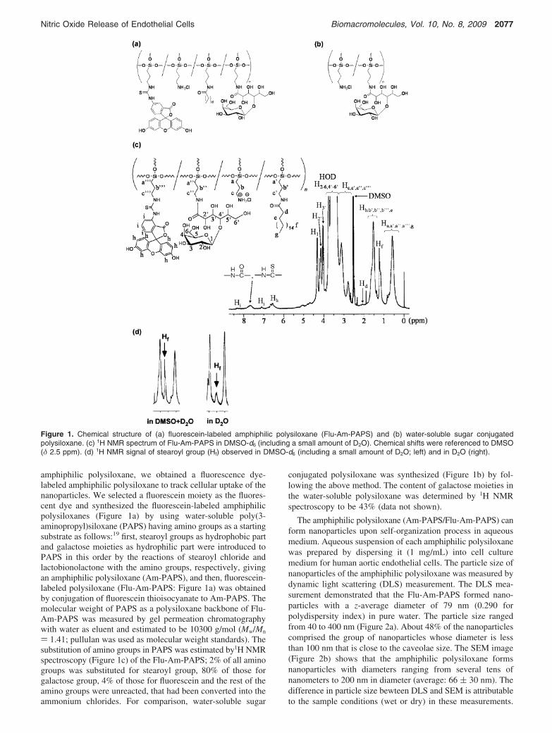
amphiphilic polysiloxane, we obtained a fluorescence dye-labeled amphiphilic polysiloxane to track cellular uptake of thenanoparticles. We selected a fluorescein moiety as the fluores-cent dye and synthesized the fluorescein-labeled amphiphilicpolysiloxanes (Figure 1a) by using water-soluble poly(3-aminopropyl)siloxane (PAPS) having amino groups as a startingsubstrate as follows:19 first, stearoyl groups as hydrophobic partand galactose moieties as hydrophilic part were introduced toPAPS in this order by the reactions of stearoyl chloride andlactobionolactone with the amino groups, respectively, givingan amphiphilic polysiloxane (Am-PAPS), and then, fluorescein-labeled polysiloxane (Flu-Am-PAPS: Figure 1a) was obtainedby conjugation of fluorescein thioisocyanate to Am-PAPS. Themolecular weight of PAPS as a polysiloxane backbone of Flu-Am-PAPS was measured by gel permeation chromatographywith water as eluent and estimated to be 10300 g/mol (Mw/Mn
) 1.41; pullulan was used as molecular weight standards). Thesubstitution of amino groups in PAPS was estimated by1H NMRspectroscopy (Figure 1c) of the Flu-Am-PAPS; 2% of all aminogroups was substituted for stearoyl group, 80% of those forgalactose group, 4% of those for fluorescein and the rest of theamino groups were unreacted, that had been converted into theammonium chlorides. For comparison, water-soluble sugar
conjugated polysiloxane was synthesized (Figure 1b) by fol-lowing the above method. The content of galactose moieties inthe water-soluble polysiloxane was determined by 1H NMRspectroscopy to be 43% (data not shown).
The amphiphilic polysiloxane (Am-PAPS/Flu-Am-PAPS) canform nanoparticles upon self-organization process in aqueousmedium. Aqueous suspension of each amphiphilic polysiloxanewas prepared by dispersing it (1 mg/mL) into cell culturemedium for human aortic endothelial cells. The particle size ofnanoparticles of the amphiphilic polysiloxane was measured bydynamic light scattering (DLS) measurement. The DLS mea-surement demonstrated that the Flu-Am-PAPS formed nano-particles with a z-average diameter of 79 nm (0.290 forpolydispersity index) in pure water. The particle size rangedfrom 40 to 400 nm (Figure 2a). About 48% of the nanoparticlescomprised the group of nanoparticles whose diameter is lessthan 100 nm that is close to the caveolae size. The SEM image(Figure 2b) shows that the amphiphilic polysiloxane formsnanoparticles with diameters ranging from several tens ofnanometers to 200 nm in diameter (average: 66 ( 30 nm). Thedifference in particle size bewteen DLS and SEM is attributableto the sample conditions (wet or dry) in these measurements.
Figure 1. Chemical structure of (a) fluorescein-labeled amphiphilic polysiloxane (Flu-Am-PAPS) and (b) water-soluble sugar conjugatedpolysiloxane. (c) 1H NMR spectrum of Flu-Am-PAPS in DMSO-d6 (including a small amount of D2O). Chemical shifts were referenced to DMSO(δ 2.5 ppm). (d) 1H NMR signal of stearoyl group (Hf) observed in DMSO-d6 (including a small amount of D2O; left) and in D2O (right).
Nitric Oxide Release of Endothelial Cells Biomacromolecules, Vol. 10, No. 8, 2009 2077

The nanoparticles had a slightly negative zeta potential (-0.9mV) in the cell culture medium. The nanoparticle of Am-PAPSdispersed in phosphate buffered saline did not exhibit consider-able change in particle size after one week storage at 37 °C(Figure S1a in Supporting Information). Furthermore, particlesize measurement using DLS was carried out on the polysiloxanenanoparticles dispersed in cell culture medium containing 5%fetal bovine serum and found that one week storage ofpolysiloxane nanoparticles in serum containing culture mediumat 37 °C caused little change in average particle size and particlesize distribution (Figure S1b in Supporting Information). Thesedata suggest that nanoparticles of Am-PAPS are stable inphysiological condition. According to our recent report,15 theNMR spectroscopy for the long alkyl side chains of theamphiphilic polysiloxane indicated that the signal intensity ofprotons in long alkyl chains become weak when the amphiphilicpolysiloxane dissolved in D2O was measured. Figure 1c showsthat the signal observed around δ 1.25 ppm is ascribed to theprotons of stearoyl chains attached to Flu-Am-PAPS and thesignal intensity of the peak at δ 1.25 ppm of Flu-Am-PAPSdissolved in D2O is weakened to the 76% of that of the peak atδ 1.25 ppm measured in DMSO (Figure 1d). The decrease inthe intensity of the proton signal results from the restrictedmolecular motion of long alkyl chain and indicates that themolecular aggregation of Flu-Am-PAPS is driven by inter/intrahydrophobic association between stearoyl groups attached tothe polysiloxane backbone.
Cellular Uptake of Nanoparticles by Human AorticEndothelial Cells. Cellular uptake of the nanoparticles byHAECs was observed by fluorescence microscopy. HAECs werecultured for 72 h to obtain confluent culture of HAECs aftercell suspension of HAECs was seeded on tissue culture dishes.At 72 h after cell seeding, cell culture medium was replacedwith the polymer suspension of Flu-Am-PAPS. Then the HAECswere cultured in the polymer suspension for 15 min, 1, 3, 6,
24, 48, 72, and 144 h to monitor cellular uptake of thenanoparticles of Flu-Am-PAPS. The polymer suspension in cellculture dishes was exchanged daily during the incubation withFlu-Am-PAPS. Clearance of the incorporated nanoparticles fromHAECs was observed after the nanoparticle suspension inculture dishes was replaced with EGM-2, growth medium forHAECs. For fluorescence microscopy observation, the cells wererinsed with 37 °C phosphate buffered saline (PBS), then werefixed by immersing into 10% formaldehyde neutral buffersolution at room temperature for 15 min and washed three timeswith PBS. Fluorescence images of the cells were taken by afluorescence microscope equipped with a CCD camera at eachincubation time. To evaluate cellular uptake of nanoparticlesquantitatively, fluorescence intensity (F.I.) per image wasdetermined by integrating the brightness at each pixel offluorescence image using an image analysis software. The F.I.was normalized in a ratio of the F.I. at each incubation timeover the F.I. at 6 h of incubation (the relative fluorescenceintensity). The time course of the relative F.I. was plotted onthe graph, Figure 3a (closed circles: uptake of nanoparticles;open circles: clearance of nanoparticles). HAECs incorporatedprogressively nanoparticles of Flu-Am-PAPS until 72 h ofincubation and reached saturation thereafter until the end ofobservation at 144 h of incubation. Decline of F.I. after theremoval of Flu-Am-PAPS from the cell culture dishes at 6 h ofincubation indicates that the incorporated nanoparticles of Flu-Am-PAPS were gradually excreted from HAECs. However,40% of the incorporated nanoparticles were still trapped incytosol of HAECs at 138 h of post incubation (144 h of totalincubation time). Meanwhile, the relative F.I. for the uptakedropped to only 2% of the control level (left bar in Figure 3b:6 h at 37 °C) when HAECs were exposed to the nanoparticlesat 4 °C of the incubation temperature for 6 h (middle bar inFigure 3b). Considerable cell detachment was not observed evenafter 6 h of the incubation at 4 °C. This means that cell cultureof HAECs was maintained at a low culture temperature andthe HAECs were alive. The temperature triggered dramaticdecrease in the relative F.I. suggests that the nanoparticles aremainly incorporated into HAECs by endocytosis, not byadsorption to cell membrane. Endocytosis was restored afterthe HAECs exposed to low temperature environment were putback to the regular culture condition (cell culture at 37 °C).The reduced relative F.I. corresponding to the nanoparticleuptake at 4 °C increased to 90% of the relative F.I. that wasmeasured after 6 h of incubation with Flu-Am-PAPS nanopar-ticles at 37 °C (left (incubation at 37 °C for 6 h) and right(incubation at 4 °C for 6 h and postincubation at 37 °C for 6 h)bars in Figure 3b). To ensure that the cellular uptake study wascarried out under the condition that the dose of polysiloxanenanoparticles had little toxicity on HAECs, cytotoxicity of thepolysiloxane nanoparticles in HAECs was evaluated by colo-rimetric cell viability assay using WST-8 as an indicator. Cellviability of HAECs was 95% when HAECs were cultured inthe growth medium containing 1 mg/mL of Flu-Am-PAPS for24 h. This suggests that the cellular uptake study was carriedout under the condition that the polysiloxane nanoparticles didnot exhibit cytotoxicity toward HAECs at 1 mg/mL of Flu-Am-PAPS.
Caveolae as Endocytic Pathway for Nanoparticles. En-docytosis is one of the important cell activities in internalizationof various extracellular substances.20 Endocytic pathways havebeen taken into account in targeted delivery of drug to vasculartissue. Caveolae are flask-shaped invaginations of cell membranewith diamters of 50-100 nm and are thought to function as
Figure 2. Particle size and morphology of nanoparticles of fluorescein-labeled amphiphilic polysiloxane (Flu-Am-PAPS). (a) Particle sizehistogram of nanoparticles of Flu-Am-PAPS. The nanoparticles weredispersed in pure water. (b) SEM image of Flu-Am-PAPS. Scale bar:200 nm.
2078 Biomacromolecules, Vol. 10, No. 8, 2009 Nishikawa et al.
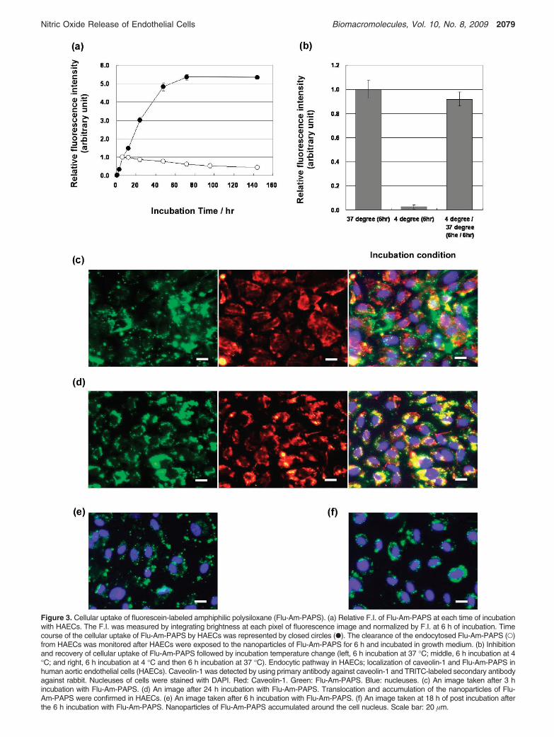
Figure 3. Cellular uptake of fluorescein-labeled amphiphilic polysiloxane (Flu-Am-PAPS). (a) Relative F.I. of Flu-Am-PAPS at each time of incubationwith HAECs. The F.I. was measured by integrating brightness at each pixel of fluorescence image and normalized by F.I. at 6 h of incubation. Timecourse of the cellular uptake of Flu-Am-PAPS by HAECs was represented by closed circles (b). The clearance of the endocytosed Flu-Am-PAPS (O)from HAECs was monitored after HAECs were exposed to the nanoparticles of Flu-Am-PAPS for 6 h and incubated in growth medium. (b) Inhibitionand recovery of cellular uptake of Flu-Am-PAPS followed by incubation temperature change (left, 6 h incubation at 37 °C; middle, 6 h incubation at 4°C; and right, 6 h incubation at 4 °C and then 6 h incubation at 37 °C). Endocytic pathway in HAECs; localization of caveolin-1 and Flu-Am-PAPS inhuman aortic endothelial cells (HAECs). Caveolin-1 was detected by using primary antibody against caveolin-1 and TRITC-labeled secondary antibodyagainst rabbit. Nucleuses of cells were stained with DAPI. Red: Caveolin-1. Green: Flu-Am-PAPS. Blue: nucleuses. (c) An image taken after 3 hincubation with Flu-Am-PAPS. (d) An image after 24 h incubation with Flu-Am-PAPS. Translocation and accumulation of the nanoparticles of Flu-Am-PAPS were confirmed in HAECs. (e) An image taken after 6 h incubation with Flu-Am-PAPS. (f) An image taken at 18 h of post incubation afterthe 6 h incubation with Flu-Am-PAPS. Nanoparticles of Flu-Am-PAPS accumulated around the cell nucleus. Scale bar: 20 µm.
Nitric Oxide Release of Endothelial Cells Biomacromolecules, Vol. 10, No. 8, 2009 2079
platforms for endocytosis and signaling.21 The caveolae-mediated pathway is prominent and ubiquitous endocyticmechanisms in vascular endothelial cells.8 Figure 3c and d showfluorescence images of HAECs that incorporated fluorescein-labeled polysiloxane nanoparticles (green) and were immun-ostained for caveolin-1 (primary antibody against caveolin-1,TRITC-labeled secondary antibody against rabbit; red), whichcomprises the caveolae. Fluorescence image taken at 3 h ofincubation time (Figure 3c) demonstrates that some of the greenemissions from nanoparticles (left image in Figure 3c) areoverlapped with red emissions from caveolin-1 (middle imagein Figure 3c) to make yellow area (right image in Figure 3c).The yellow overlaps suggest that nanoparticles are localized atthe caveolae. At 24 h of incubation time, nanoparticles (leftimage in Figure 3d) were localized as ring-like patterns ofoverlapped area around nucleuses of HAECs (right image inFigure 3d). This indicates that the nanoparticles of Flu-Am-PAPS were trapped into caveolae in plasma membrane andtransported into cytosol of HAECs by 24 h of incubation. Theperi-nuclear localization of the nanoparticles (Figure 3f) wasalso observed when HAECs were exposed to the nanoparticlesfor 6 h (Figure 3e) and incubated for 18 h after the mediumcontaining nanoparticles was changed to growth mediumcontaining no nanoparticles. The fluorescence imaging dataindicate the following process for the uptake of the nanopar-ticles: (i) the nanoparticles are trapped at caveolae in plasmamembrane, (ii) the nanoparticles are internalized into cytosol,and (iii) the nanoparticles are localized at peri-nuclear region.Furthermore, internalization of the nanoparticles into the cytosolof HAECs was confirmed by confocal laser scanning micros-copy. The X-Y plane images of HAECs were taken as opticalslices with 0.2 µm thick along Z-axis by confocal laser scanningmicroscope. Figure 4a shows fluorescence intensity profiles ofFlu-Am-PAPS along the Z-direction in HAECs. The intensityprofiles were obtained by integrating the brightness at each pixelover the whole area of each image, normalizing the intensity tothe intensity at basal position (Z ) 0 µm) and plotting thenormalized intensity as a function of depth in a cell. Theintenisty maximum was confirmed in each profile (arrow headsin the graph) and indicates the spatio and temporal position ofthe nanoparticles in HAECs. Although each profile has twomaxima at Z ) 0 µm and at other position in Z-axis respectively,the intensity maximum that we are discusing here is locatedabove the basal level. The position of the intensity maximumshifted gradually from the apical side (Z ) 3 µm) to the basalside (Z ) 0-1 µm) of HAECs; Z ) 2.4-2.6 µm at 1 h, Z )1.1-1.9 µm at 3 h, Z ) 0.5-1.6 µm at 6 h, and Z ) 0.4-1.2µm at 24 h. Thus, the peak shift clearly shows that thenanoparticles were trapped at the plasma membrane andinternalized into the cytosol of HAECs.
The nanoparticles of the amphiphilic polysiloxane can fit intocaveolae in the plasma membrane of HAECs, because thehistogram of the particle size (Figure 2 (a)) for the nanoparticlesof Flu-Am-PAPS indicates that at least 48% of the totalpopulation of the nanoparticles have diameters in the range of10-100 nm, that is comparable to the dimension of caveolae(50-100 nm in both opening diameter and depth). Fluorescencemicroscope observation demonstrated the colocalization ofthe nanoparticles and caveolae (Figure 3c and d). To specifythe endocytic pathway for the uptake of the nanoparticles weattempted to observe caveolae in HAECs by TEM. Specimensfor TEM were prepared by the methods described in thematerials and methods after HAECs were incubated withnanoparticles of gold nanoparticle labeled Am-PAPS (Au-Am-
PAPS) or Flu-Am-PAPS at 37 °C for 3 h. The nanoparticles(Flu-Am-PAPS) uptaken by HAECs were detected by immu-nolabeling using anti-fluorescein IgG as a primary antibody anda secondary antibody attached to a gold nanoparticle (diameter:5 nm). Here we observed TEM specimens by following the twodifferent methods: conventional ultrathin section and rapid-freeze deep-etch immunoreplication.22 The ultrathin sectionimage (Figure 4b) shows that caveolae (round-shape structureswith diameters of about 100 nm: indicated by a square in Figure4b) are incorporated into cytosol and that dark dots (5-10 nmin diameter) of gold nanoparticles attached to polysiloxane areconfirmed in the inner perimeter of caveolae vesicles (indicatedby arrows in the inset of Figure 4b). Figure 4c shows theultrathin section image of cytosol of the intact HAECs (nottreated with the nanoparticles of Au-Am-PAPS), showingcaveolae in cytosol of HAECs. Caveolae in cytosol of HAECswere detected by immunostaining of caveolin-1 using rabbit anti-caveolin-1 IgG and gold nanoparticles (10 nm of diameter)conjugated anti-rabbit IgG. Gold nanoparticles with 10 nm ofdiameter were specifically accumulated around the outer pe-rimeter of round-shape structures with diameters of about 100nm (Figure 4c). The result of immunodetection of caveolaeindicates that the round shape structures in cytosol of HAECsare caveolae and provides an evidence that nanoparticles of Au-Am-PAPS are trapped in caveolae. Thus, the ultrathin sectionimage of HAECs indicates that the nanoparticles of Au-Am-PAPS are trapped in caveolae at the plasma membrane ofHAECs and are internalized into cytosol of HAECs. In additionto the nanoparticles trapped in caveolae, dark spots with adiameter of 5-10 nm (surrounded by circle in Figure 4b) wereconfirmed in cytosol of HAECs (Figure 4b). These spots areassigned to gold nanoparticles attached to amphiphilic polysi-loxane (Au-Am-PAPS). The different localization of polysi-loxane nanoparticles in cytosol of HAECs suggests that thereare at least two different pathways in the cellular uptake ofpolysiloxane nanoparticles: caveolae and the other pathways,including clathrin-coated pits and macropinocytosis. Further-more, we observed the undercoat structure on the cytoplasmicsurface of the upper cell membrane using rapid-freeze deep-etch immunoreplication so as to obtain a detailed view of thecellular uptake of the nanoparticles via caveolae as mainendocytic pathways for the uptake of polysiloxane nanoparticles.Fine structures of plasma membrane such as filamentous netlikestructure of actin filaments, clathrin coated pits, and caveolaecan be preserved by rapid freezing at the cooling speed of 105
°C/sec in specimen preparation. The extreme cooling speed doesnot cause any ice nucleation that can damage the organizedstructure of the membrane skeleton. In a recent study by Moroneand Kusumi,22 they applied the rapid-freeze replication methodto the electron microscopy observation of the membrane skeletonand succeeded in viewing the fine structure of the membraneskeleton with nanometer scale resolution and demonstrating thatthe membrane skeleton mesh corresponds to the membranecompartment model that was suggested by the study of thediffusion of membrane molecules. Figure 4d and e are TEMimages of the undercoat structure of the bottom cell membraneof HAECs before and after the exposure to the nanoparticles ofFlu-Am-PAPS. The images were taken using a specimen thatwas processed by the rapid-freeze and deep-etch method.Caveolae (the characteristic striated round structures: 50-100nm in diameter), clathrin-coated pits (the characteristic basket-like structures: 100-200 nm in diameter), and actin filamentswere confirmed in the upper cell membrane of the intact HAECs(Figure 4d). The nanoparticles of Flu-Am-PAPS endocytosed
2080 Biomacromolecules, Vol. 10, No. 8, 2009 Nishikawa et al.

Figure 4. Internalization of nanoparticles of Flu-Am-PAPS in HAECs. (a) F.I. profiles of Flu-Am-PAPS in HAECs. F.I. was calculated by integratingbrightness at each pixel over an optical section of confocal fluorescence image. The F.I. is plotted against the Z-axis position of each opticalsection in a confocal fluorescence image. Arrow heads indicates the intensity maximum in the profiles. The intensity maximum can reflect themost probable location of nanoparticles of Flu-Am-PAPS in HAECs at each incubation time. TEM observation of caveolae-mediated endocytosisin HAECs. Ultrathin sections were cut out from embedded HAECs in the direction parallel to the culture dish surface. In the ultrathin sectionimage (b) the nanoparticles of Au-Am-PAPS were incorporated into cytosol of HAECs. Small black dots derived from gold nanoparticles attachedto the amphiphilic polysiloxane with diameter of 5-10 nm are confirmed in the inner perimeter of caveolae (indicated by square). The inset isa two-times magnified image of the area indicated by square and shows clearly the small black dots in caveolae (indicated by arrows); (c)immunodetection of caveolae structures in cytosol of HAECs. Caveolae were confirmed by specific accumulation of 10 nm gold nanoparticlesof secondary IgG (dark black dots) around the round structures with diameters of 100 nm in cytosol. Scale bars in Figure (b) and (c) represent200 nm. Scale bars in the insets are 100 nm in (a) and 200 nm in (b). Rapid-freeze, deep-etch immunoreplication TEM images ((d) and (e))show the undercoat structure of the upper cell membrane enriched in caveolae (indicated by arrow heads), (d), and the gold nanoparticles(white spots in circles) indicating the existence of the Flu-Am-PAPS in the caveolae-enriched area, (e). Other fine structures in (d) are clathrincoated pits (indicated by circle with broken line) and filamentous actin (indicated by arrow). Scale bar: 100 nm.
Nitric Oxide Release of Endothelial Cells Biomacromolecules, Vol. 10, No. 8, 2009 2081
by HAECs were detected by indirect immunolabeling methodwith 5 nm diameter colloidal gold particles. Figure 4e showsthat the gold particles for immunolabeling of Flu-Am-PAPS thatwere identified as the dispersed white spots in the TEM imageappeared on the internal face of the upper cell membrane andthat the gold particles were localized in the area where caveolaewere densely accumulated (Figure 4e). The localization of goldparticles in cell membrane means that the nanoparticles of Flu-Am-PAPS are localized in the caveolae accumulated area ofthe upper cell membrane and strongly suggests that caveolaecan function as pathways in endocytosis when the nanoparticlesof Flu-Am-PAPS are endocytosed into HAECs.
As for the particle size of the endocytosed nanoparticles, weextracted the endocytosed nanoparticles (Flu-Am-PAPS) fromHAECs by using a surfactant free protein extraction methodand measured the size of the extracted nanoparticles by DLS.According to the histogram of the particle size of the extractednanoparticles in cytoplasmic fraction of HAECs (the histogramdata are attached to Supporting Information as Figure S2), thehistogram peak corresponding to the intact nanoparticles ofFlu-Am-PAPS (the histogram peak at 150 nm in Figure S2b)shifted to smaller particle size (the histogram peak at 100 nmin Figure S2a) after the endocytosis into HAECs. This suggeststhat the nanoparticles of Flu-Am-PAPS comparable to thecaveolae size are preferentially endocytosed by HAECs. Fur-thermore, we performed a DLS measurement of the collectedsample including serum proteins of growth medium, nanopar-ticles of Flu-Am-PAPS, and cell debris after the incubation withHAECs and found that the histogram peak of the polysiloxanenanoparticles shifted from 150 nm (before incubation) to 250nm (the histogram peak in Figure S2c) after the incubation withHAECs. This suggests that the polysiloxane nanoparticles thatare larger than the caveolae size are more difficult to beendocytosed and remain after the incubation with HAECs. Fromthe above experiment data of immunofluorescence microscopy,electron microscopy, and particle size measurement, it can beconcluded that nanoparticles of amphiphilic polysiloxanescomparable to caveolae size are endocytosed via caveolae inplasma membrane of HAECs.
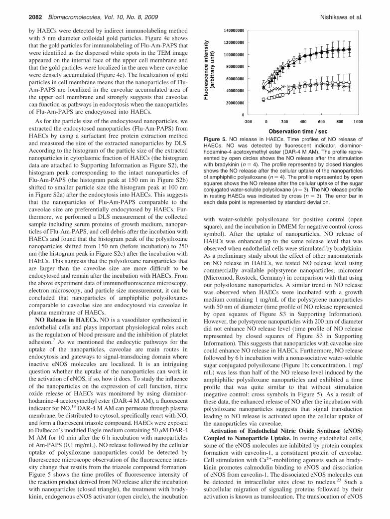
NO Release in HAECs. NO is a vasodilator synthesized inendothelial cells and plays important physiological roles suchas the regulation of blood pressure and the inhibition of plateletadhesion.7 As we mentioned the endocytic pathways for theuptake of the nanoparticles, caveolae are main routes inendocytosis and gateways to signal-transducing domain whereinactive eNOS molecules are localized. It is an intriguingquestion whether the uptake of the nanoparticles can work inthe activation of eNOS, if so, how it does. To study the influenceof the nanoparticles on the expression of cell function, nitricoxide release of HAECs was monitored by using diaminor-hodamine-4 acetoxymethyl ester (DAR-4 M AM), a fluorescentindicator for NO.18 DAR-4 M AM can permeate through plasmamembrane, be distributed to cytosol, specifically react with NO,and form a fluorescent triazole compound. HAECs were exposedto Dulbecco’s modified Eagle medium containing 50 µM DAR-4M AM for 10 min after the 6 h incubation with nanoparticlesof Am-PAPS (0.1 mg/mL). NO release followed by the cellularuptake of polysiloxane nanoparticles could be detected byfluorescence microscope observation of the fluorescence inten-sity change that results from the triazole compound formation.Figure 5 shows the time profiles of fluorescence intensity ofthe reaction product derived from NO release after the incubationwith nanoparticles (closed triangle), the treatment with brady-kinin, endogenous eNOS activator (open circle), the incubation
with water-soluble polysiloxane for positive control (opensquare), and the incubation in DMEM for negative control (crosssymbol). After the uptake of nanoparticles, NO release ofHAECs was enhanced up to the same release level that wasobserved when endothelial cells were stimulated by bradykinin.As a preliminary study about the effect of other nanomaterialson NO release in HAECs, we tested NO release level usingcommercially available polystyrene nanoparticles, micromer(Micromod, Rostock, Germany) in comparison with that usingour polysiloxane nanoparticles. A similar trend in NO releasewas observed when HAECs were incubated with a growthmedium containing 1 mg/mL of the polystyrene nanoparticleswith 50 nm of diameter (time profile of NO release representedby open squares of Figure S3 in Supporting Information).However, the polystyrene nanoparticles with 200 nm of diameterdid not enhance NO release level (time profile of NO releaserepresented by closed squares of Figure S3 in SupportingInformation). This suggests that nanoparticles with caveolae sizecould enhance NO release in HAECs. Furthermore, NO releasefollowed by 6 h incubation with a nonassociative water-solublesugar conjugated polysiloxane (Figure 1b; concentration, 1 mg/mL) was less than half of the NO release level induced by theamphiphilic polysiloxane nanoparticles and exhibited a timeprofile that was quite similar to that without stimulation(negative control: cross symbols in Figure 5). As a result ofthese data, the enhanced release of NO after the incubation withpolysiloxane nanoparticles suggests that signal transductionleading to NO release is activated upon the cellular uptake ofthe nanoparticles via caveolae.
Activation of Endothelial Nitric Oxide Synthase (eNOS)Coupled to Nanoparticle Uptake. In resting endothelial cells,some of the eNOS molecules are inhibited by protein complexformation with caveolin-1, a constituent protein of caveolae.Cell stimulation with Ca2+-mobilizing agonists such as brady-kinin promotes calmodulin binding to eNOS and dissociationof eNOS from caveolin-1. The dissociated eNOS molecules canbe detected in intracellular sites close to nucleus.23 Such asubcellular migration of signaling proteins followed by theiractivation is known as translocation. The translocation of eNOS
Figure 5. NO release in HAECs. Time profiles of NO release ofHAECs. NO was detected by fluorescent indicator, diaminor-hodamine-4 acetoxymethyl ester (DAR-4 M AM). The profile repre-sented by open circles shows the NO release after the stimulationwith bradykinin (n ) 4). The profile represented by closed trianglesshows the NO release after the cellular uptake of the nanoparticlesof amphiphilic polysiloxane (n ) 4). The profile represented by opensquares shows the NO release after the cellular uptake of the sugarconjugated water-soluble polysiloxane (n ) 3). The NO release profilein resting HAECs was indicated by cross (n ) 3). The error bar ineach data point is represented by standard deviation.
2082 Biomacromolecules, Vol. 10, No. 8, 2009 Nishikawa et al.

is followed by the activation of eNOS in endothelial cells.23
Phosphorylation of eNOS on Ser1177 occurs concomitantly inthe activation process of eNOS.24 Here we studied the influenceof nanoparticle uptake of HAECs on the eNOS activation withrespect to the intracellular translocation and the phosphorylationof eNOS. Immunofluorescence imaging of HAECs usingmonoclonal antibody to eNOS demonstrated that eNOS boundto cell membrane (cell periphery: Figure 6a) translocate to thecell cytosol and the peri-nuclear region upon 5 min stimulationwith 1 µM of bradykinin (Figure 6b). The translocation of eNOSwas confirmed as ring-like patterns around nucleuses after theHAECs were exposed to nanoparticles of Am-PAPS for 6 h(Figure 6c). This suggests that caveolae-mediated endocytosisof nanoparticles in HAECs can be coupled to the eNOSactivation and can work as an external stimuli in a signaltransduction where membrane-bound eNOS are activated.Phosphorylation of eNOS at Ser1177 (P-eNOS) was detected byWestern blot analysis. Figure 7a shows that phosphorylation ofeNOS occurred when HAECs were incubated with the nano-particles of Am-PAPS. The target proteins (eNOS and P-eNOS)transferred to PVDF membranes were detected by colorimetricreaction of TMB substrate. The protein levels of the targetproteins were evaluated by measuring the intensity of thereaction product stained on the blotted bands. In Figure 7b thetarget protein levels were plotted against time of the incubationwith nanoparticles. The total amount of eNOS graduallydecreased to 65% of the initial amount of eNOS at 6 h ofincubation while HAECs were incubated with polysiloxanenanoparticles (the time profile of eNOS: closed squares in Figure7b). �-Tubulin, a loading control for Western blot analysis,exhibited a slight decrease in expression level during theincubation with polysiloxane nanoparticles (the time profile of�-tubulin: closed circles in Figure 7b), although the same amountof extracted proteins (2 µg) was applied to each lane in SDS-PAGE. This indicates that incubation with polysiloxane nano-particles influences the expression level of eNOS in HAECs.On the other hand, the P-eNOS (Ser1177) level graduallyincreased by 1.3 fold at 1 h of incubation and by 1.6-fold at
3 h from the initial phosphorylation level of eNOS (the timeprofile of P-eNOS: closed triangles in Figure 7b). The expressionlevel of P-eNOS reached saturation point at 6 h of incubationwith polysiloxane nanoparticles. Thus, the activation of eNOSwas followed by the cellular uptake of polysiloxane nanopar-ticles and the expression level of P-eNOS was maintained aslong as HAECs were exposed to the nanoparticles. Furthermore,the expression levels of target proteins (eNOS, P-eNOS, and�-tubulin) were back to the initial level of each protein (timeprofiles (from 3 to 6 h of incubation time) indicated by opensymbols and dotted lines in Figure 7b) when polysiloxanenanoparticles were removed from culture medium and HAECswere incubated in a fresh growth medium for 3 h after theremoval of nanoparticles. These data suggest that phosphory-lation of eNOS at Ser1177 is stimulated by interaction withnanoparticles and the enhanced level of phosphorylation ismaintained transiently during the interaction with polysiloxanenanoparticles. Meanwhile, any eNOS translocation was not
Figure 6. Translocation of eNOS in HAECs in response to bradykininand upon cellular uptake of the nanoparticles of Am-PAPS. (a)Representative image of eNOS localization in resting HAECs (ar-rowhead). eNOS was redistributed from the cell periphery to intra-cellular sites near the nucleus (arrowheads in (b) and (c)) whenHAECs were treated with 1 µM bradykinin for 5 min, (b), or wereincubated with the nanoparticles of Am-PAPS (0.1 mg/mL) for 6 h,(c). eNOS did not translocate to peri-nuclear region of HAECs whenHAECs were exposed to sugar conjugated water-soluble polysiloxane(1 mg/mL) for 6 h, (d). Scale bar in each image is 20 µm.
Figure 7. Nanoparticles of amphiphilic polysiloxane (Am-PAPS)stimulate phosphorylation of eNOS. HAECs were incubated withnanoparticles of Am-PAPS for the time period indicated. (a) Celllysates were analyzed by Western blot with antibodies for phospho-rylated eNOS-Ser1177, eNOS, and �-tubulin. (b) The bands of targetproteins were quantified by measuring the colorimetric intensity. Thegraph shows the time course of the expression level of the targetproteins (2, 4: P-eNOS, 9, 0: e-NOS, and b, O: �-tubulin). The timecourses represented by closed symbols demonstrate the expressionlevel of the proteins of interest during the incubation with polysiloxanenanoparticles. The time course of the proteins of interest after theremoval of polysiloxane nanoparticles at 3 h of incubation are depictedby open symbols and dotted lines. Each data point represents themeans ( SE (n ) 3).
Nitric Oxide Release of Endothelial Cells Biomacromolecules, Vol. 10, No. 8, 2009 2083

observed at all when HAECs were exposed to the water-solublesugar conjugated polysiloxane (concentration: 1 mg/mL) for 24 h(Figure 6d). According to the result of fluorescence imageanalysis for the estimation of the amount of the endocytosedpolymers, the amount of the incorporated water-soluble pol-ysiloxane at 24 h of the incubation was approximately 15% ofthat of the incorporated fluorescein-labeled nanoparticles (Flu-Am-PAPS) at 6 h of the incubation time (Figure S4 is availableat Supporting Information). The water-soluble polysiloxane wascertainly endocytosed by HAECs; however, the eNOS activationdid not occur. These results suggest that the types of thepolymers (nonassociative polymer chains or nanoparticles ofamphiphilic polymers) exposed to cells and the pathways(caveolae or clathrin coated pits) selected in endocytosis areimportant factors that can influence the signal transduction inthe regulation of cell function. Thus, we assume that the deliveryof the nanoparticles targeted to caveolae facilitates eNOSactivation (subcellular translocation and phosphorylation atSer1177) and the activated eNOS promotes NO release. Thedetails of the mechanism underlying the eNOS activation uponthe caveolae-mediated endocytosis of our polysiloxane nano-particles is still under investigation and will be reportedelsewhere.
Conclusion
In conclusion, the nanoparticles of amphiphilic polysiloxanewere endocytosed via caveolae in human aortic endothelial cellsand the uptake of the nanoparticles promoted nitric oxide releasein HAECs. Caveolae is a membrane microdomain where varioussignal transduction molecules are accumulated and externalstimuli are processed. Endothelial nitric oxide synthase is oneof the constituent molecules in signal transduction and plays asignificant role in regulation of vasorelaxation by synthesizingnitric oxide. Some of eNOS molecules are bound to caveolin-1and deactivated in caveolae. Activation of eNOS is triggeredin vivo by binding of physiological active molecules such asbradykinin, angiotensin-II, and estrogen to their correspondingreceptors in endothelial cells. According to the recent study byManiatis et al., eNOS-dependent NO production is coupled tocaveolae-mediated endocytosis induced by albumin bindingprotein gp60 (albumin receptor).25 In their case, NO releasestarted just after gp60 activation by adding BSA and lasted upto 20 min. Phosphorylation of eNOS accompanied by gp60activation was confirmed at just 30 s after the addition of BSA.This means that BSA uptake via caveolae in rat lung microvas-cular endothelial cells is quickly processed as an external signaland leads to NO release. In our study, a delay of 15-60 minbetween the beginning of the nanoparticle uptake and the onsetof the induced phosphorylation of eNOS was observed. Thiskind of delay was rather observed in the phosphorylation ofeNOS that was stimulated by shear stress to bovine aorticendothelial cells.26 Sustained phosphorylation of eNOS isanother feature of eNOS activation that is followed by nano-particle uptake of HAECs. We found that the phosphorylationof eNOS lasted while HAECs were exposed to nanoparticlesfor 6 h. The phosphorylation of eNOS induced by gp60activation lasted at most for 30 min. Furthermore, there wasconsiderable difference in the working concentration of stimu-lants for the activation of eNOS between BSA (5 mg/mL) andour nanoparticles (0.1 mg/mL or less). We think that there aresome different mechanisms in the activation of eNOS inducedby caveolae-mediated endocytosis between former study usingBSA and our current study using artificial nanoparticles. In anycase, nanoparticle uptake by HAECs stimulates the activation
of eNOS and raises the NO production. This means thatnanoparticles can be regarded as extracellular signals. Concern-ing the influence of the interaction between nanomaterials withcells on the expression of cell function, a study on the behaviorof bovine carotid arterial endothelial cells cultured on polyure-thane nanocomposites demonstrated that cell migration couldbe regulated by the surface morphological change induced byblending gold nanoparticles with polyurethane and revealed thatthe promoted migration was associated with up-regulation ofeNOS expression via the activation of PI3K/Akt signalingpathway.27 The key phenomena explaining the promoted cellmigration is signal transduction associated with the interactionbetween cells and the nanostructured surface and concomitantrearrangement of cytoskeleton. Meanwhile, our study deals withsignal transduction associated with endocytosis of nanoparticlesthat are targeted to membrane microdomains of cells. As far aswe know, this is the first demonstration that nitric oxide releasein HAECs can be induced by the caveolae-mediated cellularuptake of artificial nano materials of synthetic polymer and thatthe uptake itself can work as external stimuli leading to theexpression of a cell function. Thus, nanoparticle can work asan artificial signal substance whose signaling characteristics maybe tuned by molecular design of constituting amphiphilicpolymers as well as the nanoparticles can be used as nanocarriersin drug delivery system. In the drug delivery system thenanocarrier itself should be inert to targeted cells and tissues.However, as we demonstrated in this study, nanoparticles caninfluence cell functions; nanoparticles may not only promotethe pharmacological effects of delivered drugs but also causeundesirable effects in the target tissues or cells. The moleculardesign of constituent molecules for nanoparticles and theinteraction between nanoparticles and cells should be consideredmore carefully in terms of the activation of cell functions.Nevertheless, we expect that targeting delivery of nanoparticlesincluding our polysiloxane nanoparticles to caveolae is apotential and novel medication to hypertension based on theregulation of NO release by switching of eNOS activation in asingle cell.
Acknowledgment. T.N. thanks Professor Mitsuru Akashiand Dr. Takami Akagi of Osaka University for allocatinginstrument time of dynamic light scattering measurement, Dr.Tetsuji Yamaoka and Dr. Atsushi Mahara of NationalCardiovascular Center Research Institute for allocatinginstrument time of fluorescence spectroscopy measurement,Dr. Tsutomu Furuzono of National Cardiovascular CenterResearch Institute for allocating instrument time of microplatereader in protein assay, and Mrs. Mina Kaneko for technicalwork in cell culture experiments. Part of this research wasfinancially supported by Terumo Life Science Foundation infiscal year of 2008.
Supporting Information Available. Supplementary data ofnanoparticle size change in physiological condition, size ofnanoparticles endocytosed, and cellular uptake of water-solublepolysiloxane. This material is available free of charge via theInternet at http://pubs.acs.org.
References and Notes(1) Simons, K.; Ikonen, E. Nature 1997, 387, 569–572.(2) Minshall, R. D.; Sessa, W. C.; Stan, R. V.; Anderson, R. G. W.; Malik,
A. B. Am. J. Physiol. 2003, 285, L1179–L1183.(3) Gratton, J.-P.; Bernatchez, P.; Sessa, W. C. Circ. Res. 2004, 94, 1408–
1417.(4) Wyatt, A. W.; Steinert, J. R.; Mann, G. E. Biochem. Soc. Symp. 2004,
71, 143–156.
2084 Biomacromolecules, Vol. 10, No. 8, 2009 Nishikawa et al.
(5) Vallance, P.; Chan, N. Heart 2001, 85, 342–350.(6) McIntosh, D. P.; Tan, X.-Y.; Oh, P.; Schnitzer, J. E. Proc. Natl. Acad.
Sci. U.S.A. 2002, 99, 1996–2001.(7) Michel, T. Braz. J. Med. Biol. Res. 1999, 32, 1361–1366.(8) Muro, S.; Koval, M.; Muzykantov, V. Curr. Vasc. Pharmacol. 2004,
2, 281–299.(9) Sanvicens, N.; Marco, M. P. Trends Biotechnol. 2008, 26, 425–433.
(10) Conner, S.; Scmid, S. L. Nature 2003, 422, 37–44.(11) Cruz, T.; Gaspar, R.; Donato, A.; Lopes, C. Pharm. Res. 1997, 14,
73–79.(12) Mark, J. E.; Allcock, H. R.; West, R. In Inorganic Polymers, 2nd ed.;
Oxford University Press: New York, 2005; pp 154-199.(13) Kichler, A.; Sabourault, N.; Decor, R.; Leborgne, C.; Schmutz, M.;
Valleix, A.; Danos, O.; Wagner, A.; Mioskowski, C. J. ControlledRelease 2003, 93, 403–414.
(14) Moghimi, S. M.; Hunter, A. C.; Murray, J. C. Pharmacol. ReV. 2001,53, 283–318.
(15) Beppu, K.; Kaneko, Y.; Kadokawa, J.; Mori, H.; Nishikawa, T. Polym.J. 2007, 39, 1065–1070.
(16) Tominaga, H.; Ishiyama, M.; Ohseto, F.; Sasamoto, K.; Hamamoto,T.; Suzuki, K.; Watanabe, M. Anal. Commun. 1999, 36, 47–50.
(17) Heuser, J. Traffic 2000, 1, 545–552.(18) Kojima, H.; Hirotani, M.; Nakatsubo, N.; Kikuchi, K.; Urano, Y.; Higuchi,
T.; Hirata, Y.; Nagano, T. Anal. Chem. 2001, 73, 1967–1973.
(19) Kaneko, Y.; Iyi, N.; Kurashima, K.; Matsumoto, T.; Fujita, T.;Kitamura, K. Chem. Mater. 2004, 16, 3417–3423.
(20) Durin, G.; Cottin, S.; Blanc, E.; Rees, A. R.; Temsamani, J. J. Biol.Chem. 2003, 278, 31192–31201.
(21) Drab, M.; Verkade, P.; Elger, M.; Kasper, M.; Lohn, M.; Lauterbach,B.; Menne, J.; Lindschau, C.; Mende, F.; Luft, F. C.; Schedl, A.; Haller,H.; Kurzchalia, T. V. Science 2001, 293, 2449–2452.
(22) Morone, N.; Fujiwara, T.; Murase, K.; Kasai, R.; Ike, H.; Yuasa, S.;Usukura, J.; Kusumi, A. J. Cell Biol. 2006, 174, 851–862.
(23) Prabhakar, P.; Thatte, H. S.; Goetz, R. M.; Cho, M. R.; Golan, D. E.;Michel, T. J. Biol. Chem. 1998, 273, 27383–27388.
(24) Fleming, I.; Busse, R. Am. J. Physiol. 2003, 284, R1–R12.(25) Maniatis, N.; Brovkovych, V.; Allen, S. E.; John, T. A.; Shajahan,
A. N.; Tiruppathi, C.; Vogel, S. M.; Skidgel, R. A.; Malik, A. B.;Minshall, R. D. Circ. Res. 2006, 99, 870–877.
(26) Boo, Y. C.; Sorescu, G.; Boyd, N.; Shiojima, I.; Walsh, K.; Du, J.;Jo, H. J. Biol. Chem. 2002, 277, 3388–3396.
(27) Hung, H. S.; Wu, C. C.; Chien, S.; Hsu, S. H. Biomaterials 2009, 30,1502–1511.
BM900128X
Nitric Oxide Release of Endothelial Cells Biomacromolecules, Vol. 10, No. 8, 2009 2085


