Embed Size (px)
Citation preview

The FASEB Journal • Research Communication
Nitric oxide elicits functional MMP-13 protein-tyrosinenitration during wound repair
Tania R. Lizarbe,* Concepcion Garcıa-Rama,* Carlos Tarın,* Marta Saura,†
Enrique Calvo,* Juan Antonio Lopez,* Carlos Lopez-Otın,‡ Alicia R. Folgueras,‡
Santiago Lamas,§ and Carlos Zaragoza*,1
*Centro Nacional de Investigaciones Cardiovasculares, Madrid, Spain; †Departamento de Fisiologıa,Facultad de Medicina, Universidad de Alcala, Madrid, Spain; ‡Departamento de Bioquimica, InstitutoUniversitario de Oncologia, Universidad de Oviedo, Oviedo, Spain; and §Centro de InvestigacionesBiologicas, Instituto “Reina Sofıa” de Investigaciones Nefrologicas, Madrid, Spain
ABSTRACT Nitric oxide (NO) plays a critical role inwound healing, in part by promoting angiogenesis.However, the precise repair pathways affected by NOare not well defined. We now show that NO regulatesmatrix metalloproteinase-13 (MMP-13) release duringwound repair. We find that normally MMP-13 is keptinside endothelial cells by an association with caveo-lin-1. However, nitration of MMP-13 on tyrosine resi-due Y338 causes it to dissociate from caveolin-1 and bereleased from endothelial cells. We next explored thefunctional significance of MMP-13 nitration in vivo.Skin injury increases nitration of MMP-13 in mice. Skinwounds in inducible nitric oxide synthase knockoutmice release less MMP-13 and heal more slowly thanskin wounds in wild-type mice. Conversely, skin woundsin caveolin-1 knockout mice have increased NO pro-duction, increased MMP-13 nitration, and acceleratedwound healing. Collectively, our data reveal a newpathway through which NO modulates wound repair:nitration of MMP-13 promotes its release from endo-thelial cells, where it accelerates angiogenesis andwound healing.—Lizarbe, T. R., Garcıa-Ram, C., Tarın,C., Saura, M., Calvo, E., Lopez, J. A., Lopez-Otın, C.,Folgueras, A. R., Lamas, S., Zaragoza, C. Nitric oxideelicits functional MMP-13 protein-tyrosine nitrationduring wound repair. FASEB J. 22, 000–000 (2008)
Key Words: metalloproteinases � collagenase-3 � inducible ni-tric oxide synthase � caveolin-1 � wound healing
Wound healing is an orchestrated task carried outin response to varied stimuli by several cell types (1–3),triggered by the release of several cytokines, growthfactors, and radicals such as reactive oxygen species (4)and nitric oxide (NO). Mice lacking endothelial NOsynthase have shown defects in wound healing andangiogenesis (5). However, the molecular mechanismsthrough which these molecules induce healing are stillunder investigation.
A critical determinant of cell migration is the balancethat exists between extracellular matrix deposition anddegradation. Matrix metalloproteinases (MMPs) are
extracellular matrix degrading enzymes that play essen-tial roles during tissue development, atherosclerosis,ovarian function, arthritis, osteoarthritis, cancer, angio-genesis, and wound healing, processes that all involvecell migration (6–11). The matrix metalloproteinaseMMP-13 is expressed by several cell types, including thevascular endothelium (12). MMP-13 forms a complex atthe cell surface with caveolin-1, the major proteincomponent of caveolae (13).
The role of caveolin-1 in cell migration has beenproposed (14). We have identified a region within theC-terminal domain as the MMP-13 sequence that bindsto caveolin-1, and we provide in vitro and in vivoevidence that NO triggers MMP-13 release and cellmotility through posttranslational nitration of a ty-rosine residue adjacent to this region.
MATERIALS AND METHODS
Reagents
General cell culture supplies were from BD Biosciences(Madrid, Spain), calf serum was from BioWhittaker (Verviers,Belgium), and cell culture gelatin and antibiotics were fromSigma (St. Louis, MO, USA). Autoradiography film was fromKodak (Rochester, NY, USA; polyvinylidene difluoride pro-tein transfer membranes were from Millipore (Iberica,Spain); and horseradish perioxidase-conjugated secondaryantibodies, the enhanced chemiluminescence immunoblotdetection system, and protein A/G-Sepharose were from GEHealthCare (Alcobendas, Spain). EDTA-free protease inhib-itor cocktail tablets were from Roche (Madrid, Spain). Opti-mem and Lipofectamine were from Gibco-Life Technologies(Gaithersburg, MD, USA). Fluorsave coverslip mounting so-lution was from Calbiochem (CN Biosciences, Nottingham,UK). The NO donor DEA-NO was from Alexis (Alexis Bio-chemicals, San Diego, CA, USA). Primary antibodies wereobtained as follows: rabbit anti-human MMP-13 from Calbio-chem; goat-anti-human-MMP-13 from Santa Cruz Biotechnol-ogies (Santa Cruz, CA, USA); anti-caveolin-1 from BD Trans-
1 Correspondence: Fundacion CNIC, Melchor FernandezAlmagro 3, 28029 Madrid, Spain. E-mail: [email protected]
doi: 10.1096/fj.07-103804
10892-6638/08/0022-0001 © FASEB
The FASEB Journal article fj.07-103804. Published online May 21, 2008.

duction Laboratories (BD Biosciences); anti-FLAG fromSigma; and mouse monoclonal anti-ICAM-2 and VWF fromTransduction Laboratories (BD Biosciences). The protein-nitration inhibitor peptide RYEYA was synthesized by Sigma.
Animals
Inducible nitric oxide synthase (iNOS) null mice and caveo-lin-1 null mice were purchased from The Jackson Laboratory(Bar Harbor, ME, USA). MMP-13 null mice were kindlydonated by S. Krane (Center for Immunology and Inflamma-tory Diseases, Massachusetts General Hospital and HarvardMedical School, Charlestown, MA, USA). The background ofall mice was C57BL/6, and therefore, C57BL/6 wild-typemice were used for control purposes. No differences in sizeand weight were detected in these mice. All animals werehoused in our animal facilities in isolated rooms.
Cells
Bovine aortic endothelial cells (BAECs) and murine aorticendothelial cells (MAECs) were grown on gelatin (13).MAECs were selected by fluorescence-activated cell sorterwith anti-ICAM-2 antibody, and culture purity was verifiedby double confocal immunofluorescence staining for VWFand ICAM-2. The purity of MAECs used in experimentsexceeded 99%.
Plasmids and cell transfection
Epitope-tagged constructs encoding full-length MMP-13, par-tial and mutant MMP-13 sequences, and caveolin-1 wereexpressed in endothelial cells and in bacteria as indicated. Inparticular, we expressed the following MMP-13 polypeptides:full-length MMP-13 (amino acids 1–471); MMP-13 lacking thesignal peptide (amino acids 20–471); MMP-13 lacking thepropeptide domain (amino acids 112–471); MMP-13 catalyticdomain (amino acids 112–267); MMP-13 hemopexin domain(HP; amino acids 281–471); and MMP-13 HP domain dele-tion mutant (lacking amino acids 347–360).
GST-fusion proteins were expressed with pGEX-4T2 (GEHealthCare). Recombinant proteins were purified as de-scribed previously (13). For mammalian expression, cDNAswere cloned in p3XFLAG-myc-CMV-24 (Sigma). Targeteddeletion of HP domain was performed with the QuickChange Site-Directed Mutagenesis Kit (Stratagene, La Jolla,CA, USA).
For exogenous protein expression, cells were transientlytransfected using Lipofectamine 2000 Reagent with the ap-propriate mammalian plasmids as described previously (13).Expression was monitored by confocal immunofluorescencemicroscopy and immunoblotting with the correspondingantibodies.
Wound-healing assay
Cells were grown in 6-well plates, and a straight cut was madeacross the confluent monolayer with a scalpel. Endothelialcell movement into the denuded area was monitored overtime by microscopy as described previously (13).
Mouse skin repair and immunohistofluoresence
Mice were anesthetized, and after the dorsal hair was shavedand the skin was cleaned with ethanol, full-thickness exci-sional skin wounds were performed on the dorsal middle lineusing an 8 mm biopsy punch (Accuderm, Ft. Lauderdale, FL,USA). The healing was monitored by taking photographs atthe indicated time points, and the area unoccupied by skinwas calculated for each time point, using the Image J software(U.S. National Institutes of Health, Bethesda, MD, USA).Results were presented as percentage of original wound areaat time point 0.
Skin biopsies were obtained from the back wounds ofC57BL/6 wild-type iNOS null mice, MMP-13 null mice, andcaveolin-1 null mice. Samples were embedded in paraffin,and 4 �m thick serial sections were subjected to eosin-hematoxylin staining or confocal immunofluorescence mi-croscopy with anti-MMP-13 and anti-nitrotyrosine antibodiesas described previously (13).
Immunoblotting
Cell lysis and immunoblotting were performed as describedpreviously (13).
Immunoprecipitation
Cells were disrupted with RIPA buffer, and lysates wereprecleared with the appropriate control immunoglobulin G(IgG) together with protein A-Sepharose. Immunoprecipita-tion was performed as described previously (13)
Confocal microscopy
Proteins were detected by confocal microscopy in culturedcells and paraffin-embedded tissue sections as describedpreviously (13).
Affinity binding assays
Synthetic biotinylated peptides corresponding to the caveolinscaffolding domain (CSD) and to the caveolin-1 bindingdomain of MMP-13 (see Table 1) were purchased from
TABLE 1. Peptide sequences
Peptide Sequence
MMP-13 peptidesa
CSD-BD AYEHPSHDLIFIFRGRKFWALNGYDCSD-BD DEL YEHPSHDLIWALNGYCSD-BD scramble AYEHPSHDLIRRGFFFKIKWALNGYD
Caveolin-1 peptide, CSD FHGIWKASFTTFTVTKYWFYRLLProtein-nitration inhibitor peptide RYEYA
a Sequences based on residues 337–361, which span the 8-amino acid putative caveolin-1 bindingsequence in the HP domain (underlined).
2 Vol. 22 September 2008 LIZARBE ET AL.The FASEB Journal

Sigma. Peptides were immobilized on neutravidin-loadedcolumns (Ultralink Immobilized Neutravidin Protein Plus;Pierce, Rockford, IL, USA), and recombinant purified pro-teins or cell extracts were passed through them. Columnswere washed 3 times with 0.05% Tween 20 in PBS, andproteins were then eluted with 0.05 N NaOH for detection byimmunoblot.
Metalloproteinase activity
MMP-13 activity was measured as the degradation of a fluo-rescent substrate from Calbiochem (CN Biosciences) as de-scribed by the manufacturer. The substrate is highly efficientfor MMP-13, with low efficiency for MMP-1 and MMP-8.
Characterization of nitration of peptide HP
One milligram of biotinylated HP peptide was treated with100 nM of ONOO� for 10 min at room temperature. NitratedHP peptide was digested by addition of modified porcinetrypsin (sequencing grade; Promega, Madison, WI, USA) at afinal concentration of 0.1 mg. Insolution digestion proceededat 37°C for 30 min. The resulting tryptic peptides were onlineinjected onto a C-18 reversed-phase nanocolumn (DiscoveryBIO Wide Pore; Supelco, Bellafonte, PA, USA) and analyzedin a continuous acetonitrile gradient. A flow rate of �300nl/min was used to elute peptides from the reversed-phasenanocolumn to an electrospray ion source coupled to an iontrap mass spectrometer (Esquire HCT; Bruker Daltonics,Bremen, Germany) for real-time ionization and fragmenta-tion.
Statistical analysis
Unless otherwise specified, data are expressed as means � sd,and experiments were performed at least 3 times in duplicate.Whenever comparisons were made with a common control,comparisons were made with ANOVA followed by Dunnett’smodification of the t test. The level of statistical significancewas defined as P � 0.05. Error bars represent mean � sd.
RESULTS
NO-mediated wound healing in mice is dependent onprotein tyrosine nitration
To investigate the underlying effect of NO in woundrepair (15, 16), we found that in iNOS null mice,cutaneous wound healing was significantly delayed withrespect to wild-type mice (Fig. 1A, C; WT vs. iNOS). Bycontrast, blockage of protein nitration in excisionalwounds (17; Table 1), prompted a similar pattern ofwound closure in both animals (Fig. 1B, C; WT-N-Tyr-Ivs. iNOS-N-Tyr-I), pointing toward nitration as onemechanism elicited by NO during wound repair.
MMP-13 nitration is involved in NO-mediatedwound healing
We found that MMP-13 activity was induced in skinsamples from wild-type injured animals with respect toiNOS null mice. However, tyrosine nitration inhibitionsignificantly reduced MMP-13 activity in both strains
(Fig. 2A). In addition to MMP-13 activity, a correlationbetween protein nitration and wound healing wasdetected by confocal microscopy of skin biopsies frominjured animals at day 6 posttreatment in which asubstantial accumulation of nitrated MMP-13 in wild-type mice with respect to iNOS knockouts was detected,and this effect was reversed by protein nitration inhibi-tion (Fig. 2B).
Figure 1. Lack of NO inhibits wound healing in mice. A)Wounds (8 mm) were made on the backs of wild-type (WT)and iNOS null mice, and area unoccupied by skin wasmonitored over time. Results are presented as percentage ofinitial wounded area (n�10 mice/time point; mean�sd;*P�0.05 vs. time-matched WT). B) Same as in A and woundswere treated with N-tyrosine (N-Tyr) inhibitor peptide (100�M) every day for 10 days (n�10 mice/time point). C)Eosin-hematoxylin staining of skin biopsies harvested 10 daysafter injury from WT mice (WT), iNOS null mice (iNOS), WTmice treated with the inhibitor peptide (WT-N-Tyr-I), andiNOS null mice treated with the inhibitor peptide (iNOS-N-Tyr-I). ES, Eschar; G, granulation tissue; HPE, hyperprolifera-tive epithelium. View: �4 (left panels); �10 (right panels).
3NO AND MMP-13 ACTIVATION IN WOUND REPAIR

The relevance of MMP-13 was also confirmed inMMP-13 null mice, detecting a significant delay ofwound healing with respect to their wild-type counter-parts. Interestingly, wound healing could not be furtherimpaired by inhibition of protein tyrosine nitration inMMP-13 null mice, indicating the importance ofMMP-13 nitration in wound repair (Fig. 2C).
Tyrosine nitration of MMP-13 mediates NO-inducedMMP-13 secretion and endothelial cell migrationduring wound healing
We performed endothelial wound healing assays inBAECs in which we found that peroxynitrite (100nmol/L) stimulated migration over control cells in atime-dependent manner, and this effect was preventedby the inhibition of nitration (Fig. 3A). In addition,
peroxynitrite also increased secreted MMP-13 activity inthe conditioned media of injured cultures (Fig. 3B).
Tyrosine nitration of secreted MMP-13 was assayed bycrossed coimmunoprecipitation of culture superna-tants with anti-nitrotyrosine and anti-MMP-13 antibod-ies, detecting nitration of MMP-13 in the culture mediaof injured endothelial monolayers, with the signalstrength increasing with the length of exposure to 100nmol/L peroxynitrite (Fig. 3C).
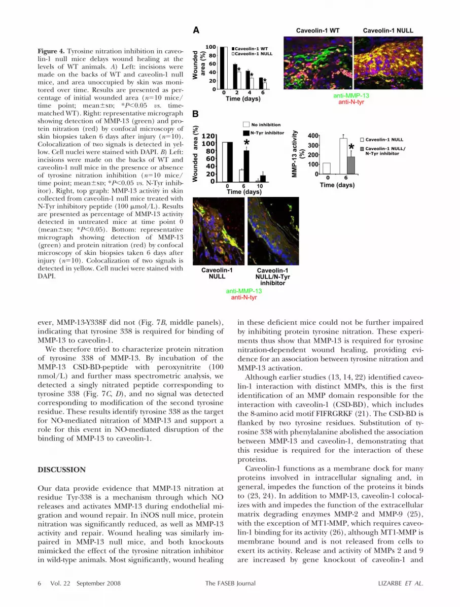
Lack of caveolin-1 increases injury-dependentnitrogen stress and accelerates skin wound healingin mice
We and others found that MMP-13 binds to caveolin-1(13) and that lack of caveolin-1 induces cell migration(18–20). To evaluate the relationship between MMP-13and caveolin-1 in wound closure, we performed woundhealing assays in caveolin-1 null mice. We found asignificant acceleration of skin healing in caveolin-1null mice, as compared with their wild-type counter-parts (Fig. 4A, graph), detecting a significant increaseof MMP-13 nitration (Fig. 4A, right panels) andMMP-13 activity (Fig. 4B, right graph) by day 6 afterinjury. In contrast, nitration inhibition induced woundclosure delay (Fig. 4B, left graph), MMP-13 nitrationreduction, and MMP-13 activity inhibition (Fig. 4B,bottom panels).
MMP-13 is bound to the cell membrane withcaveolin-1 through the MMP HP-like domain
To explore the mechanism of NO-mediated MMP-13nitration, we mapped the precise location at which-MMP-13 binds to caveolin-1 in the plasma membranebefore secretion. We generated FLAG-taggedpolypeptides containing the following MMP-13 do-mains (Fig. 5A, top scheme): Full-length MMP-13,MMP-13 lacking the signal peptide (SP), MMP-13lacking the propeptide domain (PP), the MMP-13catalytic domain (CD), MMP-13 C-terminal domain(CTD), MMP-13 HP, and a variant of MMP-13 HPdomain lacking amino acids 338 to 360 (HP*). Wefound that MMP-13 HP-like (FLAG-HP) was thesingle fragment of MMP-13 that shows colocalizationwith endogenous caveolin-1 (Fig. 5B, top panels), asdetected by confocal microscopy and by crossed-coimmunoprecipitation.
For caveolin-1 ligands, a consensus binding motif hasbeen proposed in which any three aromatic amino acidresidues (Ø) are distributed as follows: ØXØXXXXØ,(21). We identified a sequence with this structure(FIFRGRKF) in the MMP-13 HP domain at positions347–354 (Fig. 5A, bottom scheme). Confocal microscopy,as well as coimmunoprecipitation assays showed that aversion of the MMP-13 HP domain in which these 8amino acids were deleted (FLAG-HP*) did not colocalizewith caveolin-1 (Fig. 5B, bottom panels; Fig. 5C, right).
Figure 2. Nitrated MMP-13 is involved in NO-mediated woundhealing. A) MMP-13 activity in skin collected from injured WTand iNOS null mice treated with N-Tyr inhibitory peptide asin Fig. 1B. Results are presented as absorbance units(mean�sd; *P�0.05 WT 0 h vs. WT 6 h). B) Nitration ofproteins (N-Tyr, green) and MMP-13 (red) detection byconfocal microscopy of skin biopsies taken from WT andiNOS-deficient mice, treated or not every day with the N-Tyrinhibitory peptide and collected 6 days after injury. (n�10mice/time point). Cell nuclei were detected with DAPI.Merged panels show colocalization (yellow). C) Incisionswere made on the backs of WT and MMP-13 null mice,treated or not with the N-Tyr inhibitory peptide, and the areaunoccupied by skin was monitored over time. Results arepresented as percentage of initial wounded area (n�10mice/time point; mean�sd; *P�0.05 vs. N-Tyr inhibition).
4 Vol. 22 September 2008 LIZARBE ET AL.The FASEB Journal

2NO disrupts interaction between the MMP-13 HPdomain and the scaffolding domain of caveolin-1
We exposed endothelial cells expressing FLAG-HP tothe NO donor DEA-NO (10 or 100 nmol/L, liberating15 nmol and 150 nmol of NO, respectively), findinginhibition of the capacity to interact with caveolin-1 ateither DEA-NO concentration, as detected by crossedcoimmunoprecipitation (Fig. 5D) and confocal micros-copy (Fig. 5E).
To further dissect the contribution of NO, we carriedout affinity-binding reactions with biotinylated peptidescorresponding to wild-type and mutant sequences ofthe putative CSD-binding domain (CSD-BD) ofMMP-13 (Table 1). Columns bearing the wild-typepeptide (CSD-BD) retained caveolin-1 from BAEC ly-sates (Fig. 6A, lane 2), but peptides in which theconsensus 8-amino acid CSD binding sequence wasdeleted (CSD-BD DEL) or scrambled (CSD-BD scram-ble) did not (Fig. 6A, lanes 3 and 4). When peptideswere pretreated with 100 nmol/L DEA-NO, caveolin-1was not retained by any of the columns (Fig. 6A, lanes5–8). Similar results were obtained with MAEC lysates(Fig. 6B).
We next tested the ability of columns bearing abiotinylated caveolin-1 scaffolding domain (CSD) pep-tide, used to bind to other proteins, to bind purified
recombinant GST-fusion versions of MMP-13. Full-length MMP-13 (GST-MMP-13) and the MMP-13-HPdomain (GST-HP) bound to the CSD, but no interac-tion was detected with the MMP-13 HP domain lackingthe CSD-BD (GST-HP CSD-BD DEL) (Fig. 6C, lanes 1and 3 vs. lane 5). Incubation of GST-fusion proteinswith 100 nmol/L DEA-NO, significantly reduced bind-ing of GST-MMP-13 (Fig. 6C, lane 1 vs. lane 2) andcompletely blocked binding of GST-HP (Fig. 6C, lane 3vs. lane 4). These results demonstrate that MMP-13 andcaveolin-1 interact through the CSD-BD and the CSD,respectively, and that NO disrupts the complex.
MMP-13 tyrosine 338 is required for binding tocaveolin-1 and is a target of NO-mediated proteintyrosine nitration
Interestingly, we found that flanking the CSD-BD do-main were two tyrosine residues present at positions338 and 360 of the MMP-13, respectively (Fig. 7A).Endothelial cells were transfected with FLAG-taggedwild-type MMP-13 (MMP-13 WT) or with one of twoFLAG-tagged MMP-13 constructs containing a pointmutation at one of the tyrosine residues: MMP-13-Y338F and MMP-13-Y360F (Fig. 7B), detecting thatexogenously MMP-13 WT and MMP-13-Y360F werebound caveolin-1 (Fig. 7B, left and right panels); how-
Figure 3. NO induces activation ofMMP-13 by protein tyrosine nitra-tion during endothelial woundhealing. A) Wound healing assay inBAECs treated with peroxynitrite(100 nmol/L) and the protein nitra-tion inhibitor (N-Tyr-Inhib). Cell mi-gration was monitored over 4 h. Re-sults are expressed as increase inmigration relative to control cultures(n�3; mean�sd; *P�0.05 vs. con-trol). Left: photomicrographs of arepresentative assay. B) MMP-13 ac-tivity assay in culture supernatantsharvested from injured BAECstreated with 100 nmol/L peroxyni-trite (n�3; mean�sd; *P�0.05 vs.control harvested 2 h after injury;@P�0.05 vs. control harvested 4 hafter injury). C) Crossed coimmuno-precipitation of culture supernatants
from BAECs treated as in B. Culture supernatants were immunoprecipitated (IP) with anti-nitrotyrosine antibody (N-Tyr) andwith anti-MMP-13 antibody, and proteins were detected by immunoblot (IB). As loading control, immunoprecipitatedsupernatants with anti-nitrotyrosine were also detected with anti-nitrotyrosine antibody (n�3).
5NO AND MMP-13 ACTIVATION IN WOUND REPAIR
ever, MMP-13-Y338F did not (Fig. 7B, middle panels),indicating that tyrosine 338 is required for binding ofMMP-13 to caveolin-1.
We therefore tried to characterize protein nitrationof tyrosine 338 of MMP-13. By incubation of theMMP-13 CSD-BD-peptide with peroxynitrite (100nmol/L) and further mass spectrometric analysis, wedetected a singly nitrated peptide corresponding totyrosine 338 (Fig. 7C, D), and no signal was detectedcorresponding to modification of the second tyrosineresidue. These results identify tyrosine 338 as the targetfor NO-mediated nitration of MMP-13 and support arole for this event in NO-mediated disruption of thebinding of MMP-13 to caveolin-1.
DISCUSSION
Our data provide evidence that MMP-13 nitration atresidue Tyr-338 is a mechanism through which NOreleases and activates MMP-13 during endothelial mi-gration and wound repair. In iNOS null mice, proteinnitration was significantly reduced, as well as MMP-13activity and repair. Wound healing was similarly im-paired in MMP-13 null mice, and both knockoutsmimicked the effect of the tyrosine nitration inhibitorin wild-type animals. Most significantly, wound healing
in these deficient mice could not be further impairedby inhibiting protein tyrosine nitration. These experi-ments thus show that MMP-13 is required for tyrosinenitration-dependent wound healing, providing evi-dence for an association between tyrosine nitration andMMP-13 activation.
Although earlier studies (13, 14, 22) identified caveo-lin-1 interaction with distinct MMPs, this is the firstidentification of an MMP domain responsible for theinteraction with caveolin-1 (CSD-BD), which includesthe 8-amino acid motif FIFRGRKF (21). The CSD-BD isflanked by two tyrosine residues. Substitution of ty-rosine 338 with phenylalanine abolished the associationbetween MMP-13 and caveolin-1, demonstrating thatthis residue is required for the interaction of theseproteins.
Caveolin-1 functions as a membrane dock for manyproteins involved in intracellular signaling and, ingeneral, impedes the function of the proteins it bindsto (23, 24). In addition to MMP-13, caveolin-1 colocal-izes with and impedes the function of the extracellularmatrix degrading enzymes MMP-2 and MMP-9 (25),with the exception of MT1-MMP, which requires caveo-lin-1 binding for its activity (26), although MT1-MMP ismembrane bound and is not released from cells toexert its activity. Release and activity of MMPs 2 and 9are increased by gene knockout of caveolin-1 and
Figure 4. Tyrosine nitration inhibition in caveo-lin-1 null mice delays wound healing at thelevels of WT animals. A) Left: incisions weremade on the backs of WT and caveolin-1 nullmice, and area unoccupied by skin was moni-tored over time. Results are presented as per-centage of initial wounded area (n�10 mice/time point; mean�sd; *P�0.05 vs. time-matched WT). Right: representative micrographshowing detection of MMP-13 (green) and pro-tein nitration (red) by confocal microscopy ofskin biopsies taken 6 days after injury (n�10).Colocalization of two signals is detected in yel-low. Cell nuclei were stained with DAPI. B) Left:incisions were made on the backs of WT andcaveolin-1 null mice in the presence or absenceof tyrosine nitration inhibition (n�10 mice/time point; mean�sd; *P�0.05 vs. N-Tyr inhib-itor). Right, top graph: MMP-13 activity in skincollected from caveolin-1 null mice treated withN-Tyr inhibitory peptide (100 �mol/L). Resultsare presented as percentage of MMP-13 activitydetected in untreated mice at time point 0(mean�sd; *P�0.05). Bottom: representativemicrograph showing detection of MMP-13(green) and protein nitration (red) by confocalmicroscopy of skin biopsies taken 6 days afterinjury (n�10). Colocalization of two signals isdetected in yellow. Cell nuclei were stained withDAPI.
6 Vol. 22 September 2008 LIZARBE ET AL.The FASEB Journal

impeded by its reexpression, whereas CSD peptidein-hibits cell migration associated with these proteases(14), supporting our finding as a general mechanismshared by other MMPs. In addition, in caveolin-1-deficient mice wound repair was accelerated, and it wasassociated with increased nitrotyrosine staining andincreased MMP-13 activity at the wound site. Theincreased nitration may arise from increased endothe-lial NO synthase activity, since this is normally re-
strained by binding to caveolin-1 and is increased incaveolin-1-deficient animals (27).
Our results suggests that nitration might be requiredfor dissociation from caveolin-1 and proteolytic activa-tion of unbound MMP-13. However, we cannot excludeinvolvement of NO-induced S-nitrosylation, as de-scribed for MMP-9 activation in the CNS (28). Anotherpossibility is that MMP-13 also interacts with a yetundefined partner, and dissociation also requires NO.
Figure 5. NO disrupts theinteraction between caveo-lin-1 and MMP-13 at the pro-tease HP-like domain. A)Top: schematic representa-tion of MMP-13 domainstructure. See Materials and
Methods for definitions. Bottom: putative binding sequence for the interac-tion with caveolin-1. B) Confocal microscopy of BAECs showing expression ofendogenous caveolin-1 (green) and expression of ectopic FLAG-taggedMMP-13 HP-like domain (FLAG-HP, red) or a deletion variant (FLAG-HP*,red). Merged panels show protein colocalization (yellow) and DAPI stainingof cell nuclei (blue; n�3). C) Crossed coimmunoprecipitation betweenendogenous caveolin-1 and exogenous FLAG-HP (left) or FLAG-HP* (right;n�3). D) Confocal microscopy of BAECs treated as indicated with DEA-NO,showing the expression of endogenous caveolin-1 (green) and exogenously
expressed FLAG-tagged MMP-13 HP domain (FLAG-HP, red). Merged panels show colocalization (yellow; n�3). E) Crossedcoimmunoprecipitation of exogenous FLAG-HP (left panels) and endogenous caveolin-1 (right panels) from lysates ofBAECs treated as indicated with 100 nmol/L DEA-NO (n�3). FLAG-HP-negative lanes represent nontransfected cells.IP-negative lanes are whole lysate inputs.
Figure 6. Caveolin-1 binds to the CSD-binding domainlocated within the HP domain of MMP-13. A) BAEClysates were passed through neutravidin columns towhich the following synthetic biotinylated peptides werefixed: CSD-BD, CSD-BD DEL, or CSD-BD scramble (seeTable 1 for peptide details). Control: empty column (nobound peptide). Where indicated, biotinylated MMP-13peptides were preincubated with 100 nmol/L DEA-NO.After being washed, bound proteins were eluted andimmunobloted with anti-caveolin-1 antibody. (n�3). B)Affinity binding to CSD-BD as in A of MAEC lysates(n�3). C) Affinity binding of recombinant purifiedN-terminal GST-fusion proteins GST-MMP-13, GST-HP,and a variant GST-HP CSD-BD DEL. GST-fusion pro-
teins were passed through neutravidin columns bearing a biotynylated peptide corresponding to the CSD. Whereindicated, GST-fusion proteins were preincubated with 100 nmol/L DEA-NO. Columns were washed, and eluted proteinswere separated in 15% SDS-polyacrilamide gels and immunobloted with anti-GST antibody.
7NO AND MMP-13 ACTIVATION IN WOUND REPAIR

Previous studies (21) show that at least four of thefive residues critical for binding to the caveolin-1 CSDare aromatic (CSD residues F89, F92, W98, and F99),suggesting that the interactions are mainly hydropho-bic. Modeling of the three-dimensional structure of thehuman MMP-13 HP domain (29) predicts that residuesF347, F349, and F354, together with Y338, form ahydrophobic pocket oriented toward the outer surfaceof the HP domain. The relative positioning of thesearomatic side chains suggests that CSD F89 mightinteract with MMP-13 Y338 through hydrophobic andvan der Waals forces. Nitration of MMP-13 Y338 wouldthen abolish this interaction, probably by reducing thehydrophobicity and thereby disrupting the MMP-13 HPhydrophobic pocket, and this may be a general mech-anism for dissociation from caveolin-1.
Tyrosine nitration modulates the activity of severalproteins and is associated with many pathological situ-ations, including Alzheimer’s, Parkinson’s (30), andcardiovascular diseases (31). Protein tyrosine nitrationof MMP-13 by physiologically relevant concentrationsof peroxynitrite at Tyr 338 provides a mechanisticexplanation of how NO disrupts the MMP-13/caveo-lin-1 complex and initiates cell migration (13, 32, 33).
Activation of cell migration through NO-inducedprotein nitration has implications for the understand-ing of endothelial activation in a range of physiologicaland pathological situations, including angiogenesis,
atherosclerosis and cancer, and the ability to identifysuch early activation events may provide markers of theinitial stages of endothelial dysfunction.
This study was supported by the following institutions andagencies: Ministerio de Educacion y Ciencia, ProgramaRamon y Cajal (to C.Z. and M.S.); Plan Nacional de I�D�I(SAF 2005-06025 to C.Z. and SAF 2006-02410 to S.L.); theEuropean Union (FEDER 2FD97–1432, to S.L.); the SpanishSociety of Nephrology (grant-in-aid 2001, to C.Z. and S.L.);and the Ministerio de Sanidad y Consumo (Red tematica deinvestigacion cooperativa RECAVA C03/01, to C.Z. and S.L.).We thank Dr. S. Bartlett for valuable and significant assistancewith the manuscript.
REFERENCES
1. Navarro, A., Anand-Apte, B., and Parat, M. O. (2004) A role forcaveolae in cell migration. FASEB J. 18, 1801–1811
2. Gerhardt, H., and Betsholtz, C. (2005) How do endothelial cellsorientate? EXS 3–15
3. Noiri, E., Lee, E., Testa, J., Quigley, J., Colflesh, D., Keese, C. R.,Giaever, I., and Goligorsky, M. S. (1998) Podokinesis in endo-thelial cell migration: role of nitric oxide. Am. J. Physiol. 274,C236–C244
4. Yung, L. M., Leung, F. P., Yao, X., Chen, Z. Y., and Huang, Y.(2006) Reactive oxygen species in vascular wall. Cardiovasc.Hematol. Disord. Drug Targets 6, 1–19
5. Lee, P. C., Salyapongse, A. N., Bragdon, G. A., Shears, L. L., 2nd,Watkins, S. C., Edington, H. D., and Billiar, T. R. (1999)
Figure 7. NO induces nitration of tyrosine 338 of MMP-13, required for the binding to caveolin-1. A) Schematic representationof MMP-13 caveolin-1 binding domain. B) Crossed coimmunoprecipitation of lysates from BAECs expressing FLAG-tagged WTMMP-13 (MMP-13-WT) or point-mutated versions in which tyrosines at positions 338 or 360 were substituted by proline(MMP-13-Y338F and MMP-13-Y360F). Lane 1: immunoprecipitation with anti-FLAG; lane 2: immunoprecipitation withanti-caveolin-1 (n�3). C) Left: extracted ion chromatogram (EIC) of digested CSD-BD peptides. N-terminally biotinylated (B)CSD-BD peptide was treated with 100 �mol/L ONOO�, digested with trypsin, and analyzed by electrospray ionization-ion trapmass spectrometry as described in Materials and Methods. Chart shows EIC for the ions at m/z 557.2 Da (doubly-chargedpeptide KFWALNGYD) and 695.3 Da (triply charged peptide BAYEHPSHDLIFIFR) and their corresponding tyrosine-nitratedpeptides (m/z 572.2 and 710.3 Da). These peptides correspond to C- and N-terminal portions of the CSD-BD, respectively. Thetwo unmodified peptides were detected, but the only nitrated ion detected was m/z 710.3 Da, containing the nitrated Tyr-338.Right inset: EIC chart for the ion at m/z 710.3 Da, as a result of treatment with 100 nmol/L ONOO�; no signal was detectedfor peptide containing second nitrated tyrosine at any ONOO� concentration. D) Protein sequencing and characterization ofdigested CSD-BD peptides. MS/MS spectra from ions at m/z 695.3 (top panel) and 710.3 Da (bottom panel), correspondingto unmodified and tyrosine-nitrated BAYEHPSHDLIFIFR. Diagram shows fragment ions corresponding to main fragmentationseries (�, water loss; ��, doubly charged fragments). Numbered boxes indicate nitration revealed by both b and y series.
8 Vol. 22 September 2008 LIZARBE ET AL.The FASEB Journal

Impaired wound healing and angiogenesis in eNOS-deficientmice. Am. J. Physiol. 277, H1600–H1608
6. Newby, A. C. (2006) Matrix metalloproteinases regulate migra-tion, proliferation, and death of vascular smooth muscle cells bydegrading matrix and non-matrix substrates. Cardiovasc. Res. 69,614–624
7. Lynch, C. C., and Matrisian, L. M. (2002) Matrix metallopro-teinases in tumor-host cell communication. Differentiation 70,561–573
8. Ravanti, L., and Kahari, V. M. (2000) Matrix metalloproteinasesin wound repair (review). Int. J. Mol. Med. 6, 391–407
9. Kuzuya, M., and Iguchi, A. (2003) Role of matrix metallopro-teinases in vascular remodeling. J. Atheroscler. Thromb. 10, 275–282
10. Inada, M., Wang, Y., Byrne, M. H., Rahman, M. U., Miyaura, C.,Lopez-Otin, C., and Krane, S. M. (2004) Critical roles forcollagenase-3 (Mmp13) in development of growth plate carti-lage and in endochondral ossification. Proc. Natl. Acad. Sci.U. S. A. 101, 17192–17197
11. Zucker, S., Cao, J., and Chen, W. T. (2000) Critical appraisal ofthe use of matrix metalloproteinase inhibitors in cancer treat-ment. Oncogene 19, 6642–6650
12. Zaragoza, C., Balbin, M., Lopez-Otin, C., and Lamas, S. (2002)Nitric oxide regulates matrix metalloprotease-13 expression andactivity in endothelium. Kidney Int. 61, 804–808
13. Lopez-Rivera, E., Lizarbe, T. R., Martinez-Moreno, M., Lopez-Novoa, J. M., Rodriguez-Barbero, A., Rodrigo, J., Fernandez,A. P., Alvarez-Barrientos, A., Lamas, S., and Zaragoza, C. (2005)Matrix metalloproteinase 13 mediates nitric oxide activation ofendothelial cell migration. Proc. Natl. Acad. Sci. U. S. A. 102,3685–3690
14. Williams, T. M., Medina, F., Badano, I., Hazan, R. B., Hutchin-son, J., Muller, W. J., Chopra, N. G., Scherer, P. E., Pestell, R. G.,and Lisanti, M. P. (2004) Caveolin-1 gene disruption promotesmammary tumorigenesis and dramatically enhances lung me-tastasis in vivo. Role of Cav-1 in cell invasiveness and matrixmetalloproteinase (MMP-2/9) secretion. J. Biol. Chem. 279,51630–51646
15. Weller, R. (2003) Nitric oxide: a key mediator in cutaneousphysiology. Clin. Exp. Dermatol. 28, 511–514
16. Yamasaki, K., Edington, H. D., McClosky, C., Tzeng, E., Liz-onova, A., Kovesdi, I., Steed, D. L., and Billiar, T. R. (1998)Reversal of impaired wound repair in iNOS-deficient mice bytopical adenoviral-mediated iNOS gene transfer. J. Clin. Invest.101, 967–971
17. Ye, Y., Quijano, C., Robinson, K. M., Ricart, K. C., Strayer, A. L.,Sahawneh, M. A., Shacka, J. J., Kirk, M., Barnes, S., Accavitti-Loper, M. A., Radi, R., Beckman, J. S., and Estevez, A. G. (2007)Prevention of peroxynitrite-induced apoptosis of motor neu-rons and PC12 cells by tyrosine-containing peptides. J. Biol.Chem. 282, 6324–6337
18. Hassan, G. S., Williams, T. M., Frank, P. G., and Lisanti, M. P.(2006) Caveolin-1-deficient aortic smooth muscle cells show cellautonomous abnormalities in proliferation, migration, and en-dothelin-based signal transduction. Am. J. Physiol. Heart Circ.Physiol. 290, H2393–2401
19. Kim, H. P., Wang, X., Nakao, A., Kim, S. I., Murase, N., Choi,M. E., Ryter, S. W., and Choi, A. M. (2005) Caveolin-1 expres-sion by means of p38beta mitogen-activated protein kinasemediates the antiproliferative effect of carbon monoxide. Proc.Natl. Acad. Sci. U. S. A. 102, 11319–11324
20. Zhang, W., Razani, B., Altschuler, Y., Bouzahzah, B., Mostov,K. E., Pestell, R. G., and Lisanti, M. P. (2000) Caveolin-1 inhibitsepidermal growth factor-stimulated lamellipod extension andcell migration in metastatic mammary adenocarcinoma cells(MTLn3). Transformation suppressor effects of adenovirus-mediated gene delivery of caveolin-1. J. Biol. Chem. 275, 20717–20725
21. Couet, J., Li, S., Okamoto, T., Ikezu, T., and Lisanti, M. P.(1997) Identification of peptide and protein ligands for thecaveolin-scaffolding domain. Implications for the interaction ofcaveolin with caveolae-associated proteins. J. Biol. Chem. 272,6525–6533
22. Labrecque, L., Nyalendo, C., Langlois, S., Durocher, Y., Roghi,C., Murphy, G., Gingras, D., and Beliveau, R. (2004) Src-mediated tyrosine phosphorylation of caveolin-1 induces itsassociation with membrane type 1 matrix metalloproteinase.J. Biol. Chem. 279, 52132–52140
23. Stan, R. V. (2005) Structure of caveolae. Biochim. Biophys. Acta1746, 334–348
24. Pike, L. J. (2005) Growth factor receptors, lipid rafts andcaveolae: an evolving story. Biochim. Biophys. Acta 1746, 260 –273
25. Puyraimond, A., Fridman, R., Lemesle, M., Arbeille, B., andMenashi, S. (2001) MMP-2 colocalizes with caveolae on thesurface of endothelial cells. Exp. Cell. Res. 262, 28–36
26. Galvez, B. G., Matias-Roman, S., Yanez-Mo, M., Vicente-Man-zanares, M., Sanchez-Madrid, F., and Arroyo, A. G. (2004)Caveolae are a novel pathway for membrane-type 1 matrixmetalloproteinase traffic in human endothelial cells. Mol. Biol.Cell 15, 678–687
27. Razani, B., Engelman, J. A., Wang, X. B., Schubert, W., Zhang,X. L., Marks, C. B., Macaluso, F., Russell, R. G., Li, M., Pestell,R. G., Di Vizio, D., Hou, H., Jr., Kneitz, B., Lagaud, G., Christ,G. J., Edelmann, W., and Lisanti, M. P. (2001) Caveolin-1 nullmice are viable but show evidence of hyperproliferative andvascular abnormalities. J. Biol. Chem. 276, 38121–38138
28. Gu, Z., Kaul, M., Yan, B., Kridel, S. J., Cui, J., Strongin, A., Smith,J. W., Liddington, R. C., and Lipton, S. A. (2002) S-nitrosylationof matrix metalloproteinases: signaling pathway to neuronal celldeath. Science 297, 1186–1190
29. Gomis-Ruth, F. X., Gohlke, U., Betz, M., Knauper, V., Murphy,G., Lopez-Otin, C., and Bode, W. (1996) The helping hand ofcollagenase-3 (MMP-13): 2.7 A crystal structure of its C-terminalhaemopexin-like domain. J. Mol. Biol. 264, 556–566
30. Ebadi, M., Brown-Borg, H., El Refaey, H., Singh, B. B., Garrett,S., Shavali, S., and Sharma, S. K. (2005) Metallothionein-mediated neuroprotection in genetically engineered mousemodels of Parkinson’s disease. Brain Res. Mol. Brain Res. 134,67–75
31. Turko, I. V., and Murad, F. (2002) Protein nitration in cardio-vascular diseases. Pharmacol. Rev. 54, 619–634
32. Beare, A. H., O’Kane, S., Krane, S. M., and Ferguson, M. W.(2003) Severely impaired wound healing in the collagenase-resistant mouse. J. Invest. Dermatol. 120, 153–163
33. Hartenstein, B., Dittrich, B. T., Stickens, D., Heyer, B., Vu,T. H., Teurich, S., Schorpp-Kistner, M., Werb, Z., and Angel,P. (2006) Epidermal development and wound healing inmatrix metalloproteinase 13-deficient mice. J. Invest. Derma-tol. 126, 486 – 496
Received for publication January 25, 2008.Accepted for publication April 24, 2008.
9NO AND MMP-13 ACTIVATION IN WOUND REPAIR


