Embed Size (px)
Citation preview
RESEARCH Open Access
Nicotine-induced survival signaling in lung cancercells is dependent on their p53 status while itsdown-regulation by curcumin is independentVineshkumar T Puliyappadamba†, Vino T Cheriyan†, Arun Kumar T Thulasidasan, Smitha V Bava,Balachandran S Vinod, Priya R Prabhu, Ranji Varghese, Arathy Bevin, Shalini Venugopal, Ruby John Anto*
Abstract
Background: Lung cancer is the most lethal cancer and almost 90% of lung cancer is due to cigarette smoking.Even though nicotine, one of the major ingredients of cigarette smoke and the causative agent for addiction, isnot a carcinogen by itself, several investigators have shown that nicotine can induce cell proliferation andangiogenesis. We observed that the proliferative index of nicotine is different in the lung cancer cell lines H1299(p53-/-) and A549 (p53+/+) which indicates that the mode of up-regulation of survival signals by nicotine might bedifferent in cells with and without p53.
Results: While low concentrations of nicotine induced activation of NF-�B, Akt, Bcl2, MAPKs, AP1 and IAPs inH1299, it failed to induce NF-�B in A549, and compared to H1299, almost 100 times higher concentration ofnicotine was required to induce all other survival signals in A549. Transfection of WT-p53 and DN-p53 in H1299and A549 respectively, reversed the mode of activation of survival signals. Curcumin down-regulated all the survivalsignals induced by nicotine in both the cells, irrespective of their p53 status. The hypothesis was confirmed whenlower concentrations of nicotine induced NF-�B in two more lung cancer cells, Hop-92 and NCI-H522 with mutantp53 status. Silencing of p53 in A549 using siRNA made the cells susceptible to nicotine-induced NF-�B nucleartranslocation as in A549 DN-p53 cells.
Conclusions: The present study reveals a detrimental role of nicotine especially in lung cancer patients withimpaired p53 status and identifies curcumin as a potential chemopreventive.
BackgroundLung cancer is the most lethal of all cancers worldwidewith a dismal prognosis and 5 year survival rate of <15%. Reports indicate that p53 alterations are the mostcommon genetic events in lung cancer development and50-60% of non-small cell lung cancers (NSCLC) and90% of small cell lung cancers (SCLC) contain p53mutations [1]. The major reason for lung cancer inci-dence is tobacco smoke, which contains nicotine. Thereis no evidence indicating that nicotine is a carcinogenby itself [2]; nevertheless, it has been demonstrated thatnicotine promotes the growth of cancer cell populationsin the lungs [3] by stimulating cell proliferation and
angiogenesis [4,5]. Chronic exposure to nicotine causesmitogenic stimulus and leads to the activation ofgrowth-promoting and antiapoptotic signaling pathwayssuch as PI3K-Akt/mTOR, NF-�B, COX-2, Bcl2, MAPKsetc [6-10]. It has also been shown to induce secretion ofseveral growth factors and upregulate the calpain familyof proteins in lung cancer cells leading to the activationof Raf/MAPK/ERK pathway [11-13]. Moreover, reportsindicate that nicotine inhibits apoptosis induced by var-ious stimuli including chemotherapeutic agents in lungcancer cells where survivin and XIAP are suggested toplay a key role [14].Curcumin, a polyphenol isolated from Curcuma longa,
has been extensively investigated for its cancer chemo-preventive and chemotherapeutic properties, which areattributed mainly to its antioxidant and anti-inflamma-tory potential [15,16]. Curcumin, with its potent anti-
* Correspondence: [email protected]† Contributed equallyIntegrated Cancer Research Program, Division of Cancer Research, RajivGandhi Centre for Biotechnology, Thiruvananthapuram, 695014, Kerala, India
Puliyappadamba et al. Molecular Cancer 2010, 9:220http://www.molecular-cancer.com/content/9/1/220
© 2010 Puliyappadamba et al; licensee BioMed Central Ltd. This is an Open Access article distributed under the terms of the CreativeCommons Attribution License (http://creativecommons.org/licenses/by/2.0), which permits unrestricted use, distribution, andreproduction in any medium, provided the original work is properly cited.
inflammatory property, is expected to exert chemopre-ventive effects on carcinogenesis and has been shown tomodulate numerous transcription factors, proteinkinases etc. that have been linked to the pathophysiol-ogy of cancer [17]. It up-regulates several apoptosisinducing genes such as p53 and p21 [17] and down-reg-ulates pro-survival genes such as NF-�B, Akt, Bcl2 andCyclin D1 [17,18] induced by various stimulants. Thishas been suggested to be the reason for its chemosensi-tizing efficacy [17,19]. Despite the bioavailability of cur-cumin being very low, studies indicate that even at aphysiologically achievable concentration, curcumin issufficient for its chemopreventive and chemotherapeuticactivity [20].p53, which regulates many cellular activities including
cell cycle arrest and apoptosis, is the most commonlymutated gene in human cancers [21]. It has been welldocumented that absence of functional p53 in cells ren-der them resistant to chemotherapy, and restoration ofp53 lessen the tumor occurrence [22]. One major func-tion of p53 is to control DNA replication by inducingp21 protein, which promotes cell cycle arrest by modify-ing the phosphorylation of the cyclin-dependent kinases[23], although regulation of p21 expression independentof p53 also has been reported [24]. Activation of thep53-p21 pathway and induction of both p53 and p21are often reported in response to DNA damaging agentsincluding nicotine [25].The nuclear transcription factor NF-�B is an impor-
tant survival signal which induces the expressions ofdiverse target genes including COX-2, Cyclin D1 andXIAP that promote cell proliferation, regulate apoptosisand stimulate invasion and metastasis [9,26]. Studiesindicate that simultaneous inhibition of p53 and NF-�Btranscriptional activities decide the fate of the cell[27,28]. Previous studies have also demonstrated thatnicotine activates signal transduction pathways relevantto carcinogenesis through the activations of MAPK andPI3-K/Akt pathways [6,7].In the present study we have tried to correlate the
effect of nicotine to the p53 status of lung cancer cells,especially NSCLC cell lines of adenocarcinoma or largecell carcinoma origin. We selected these cells for ourstudy because cigarette smoking is a major risk factor inthe development of NSCLC, which accounts for 80% ofall lung cancers [14]. We report in this study that theinduction of survival signals in lung cancer cells by nico-tine is dependent on the p53 status of the cells and iden-tify p53 status as an important predictor of lung cancerprogression. Additionally, we have attempted in delineat-ing the signaling cross-talk mechanism orchestratingp53-dependent regulation and expression of survival sig-nals such as NF-�B, AP-1, Akt, Bcl2 etc., in the presenceand absence of p53 expression. Our findings underscore
that p53 plays an essential role in regulating survival sig-nals, especially NF-�B, in response to nicotine. We alsopresent data supporting our contention that curcumindown-regulates all the survival signals induced by nico-tine, independent of the p53 status of the cell.
ResultsNicotine induces more proliferation in lung cancer cellslacking p53, which is down-regulated by curcuminEven though nicotine-induced proliferation of lung can-cer cells has been reported [4], our findings show forthe first time that, among the lung cancer cell lines,nicotine induces more proliferation in H1299 lackingp53 especially at lower concentrations (10-9M -10-7M)than A549 with active p53 (maximum at 10-7M -10-6M).Of interest, pre-treatment with a non-toxic concentra-tion of curcumin significantly reduced (p < 0.001) theviability of both lung cancer cells (Fig 1. A-B).
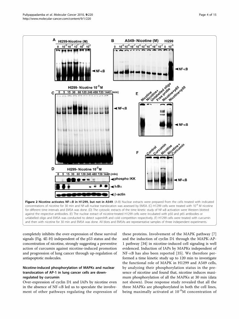
Nicotine induces NF-�B in H1299 cells but not in A549cellsNicotine has been shown to activate NF-�B in cancercells of various origins, including lung [3,8]. We alsohave reported earlier that cigarette smoke inducesstrong activation of NF-�B in H1299 cells [29]. In thepresent study, H1299 and A549 cells were exposed tovarious concentrations of nicotine and the status ofnuclear translocation of NF-�B was studied. Interest-ingly, nicotine could activate NF-�B only in H1299 cells(Fig. 2A) while it failed to induce a significant activationof NF-�B in A549 cells at any of the concentrations stu-died (Fig. 2B). In H1299 cells, lower concentrations (10-9M-10-7M) of nicotine induced maximum activation ofNF-�B and a time kinetics study using 10-8 M nicotineshowed maximum activation at 30 min of exposure tonicotine (Fig. 2C). In congruence, the phosphorylationpattern of IKK and degradation pattern of I�Ba (Fig.2D) correlated with that of the NF-�B activation, attest-ing that nicotine induces NF-�B through the classicpathway. The specificity of the NF-�B band was verifiedby super shift assay and by cold competition (Fig. 2E).The activation of NF-�B induced by nicotine in H1299cells was completely abolished by treatment with 10 μMcurcumin (Fig. 2F).
Nicotine-induced phosphorylation of Akt and overexpression of COX-2 and Cyclin D1 in lung cancer cellsare inhibited by curcuminWe compared the effect of nicotine in A549 and H1299cells in inducing pro-survival molecular entities-Akt,COX-2 and Cyclin D1, which generally get activated inresponse to NF-�B activation. Though nicotine was inef-fective in inducing NF-�B in A549 cells, it effectivelyinduced Akt, COX-2 and Cyclin D1 in both the cells
Puliyappadamba et al. Molecular Cancer 2010, 9:220http://www.molecular-cancer.com/content/9/1/220
Page 2 of 15

(Fig. 3A-C) indicating that nicotine activates these pro-survival signaling molecules independent of NF-�B.However, there was a marked difference in the expres-sion pattern of all these survival signals between theinvestigated cell lines. It was noticed that, while nicotineinduces activation of these survival molecules in therange of 10-9M - 10-7M in H1299 cells, a 10-100 foldhigh concentration of nicotine was needed to induce thesame effect in A549 (Fig. 3A-C). Despite studies report-ing the interdependence and cross-talk between Akt andNF-�B [26], phosphorylation of Akt by PI3K pathwayindependent of NF-�B is also well evidenced [30-33].Similarly, while several reports strongly correlate theinvolvement of NF-�B in Cyclin D1 regulation [30],there are reports which clearly demonstrate the tran-scription of Cyclin D1, independent of NF-�B, in celllines including A549 [32,4]. The protein level expressionof Cyclin D1 in A549 was low compared to that inH1299 cells at all the concentrations studied (Fig. 3C).We infer from these results that nicotine-inducedexpression of these survival signals was severely com-promised in the presence of curcumin in both the inves-tigated cell lines (Fig. 3D-F) and hence we drawattention to the protective role of curcumin against sur-vival and proliferative signals induced by nicotine.
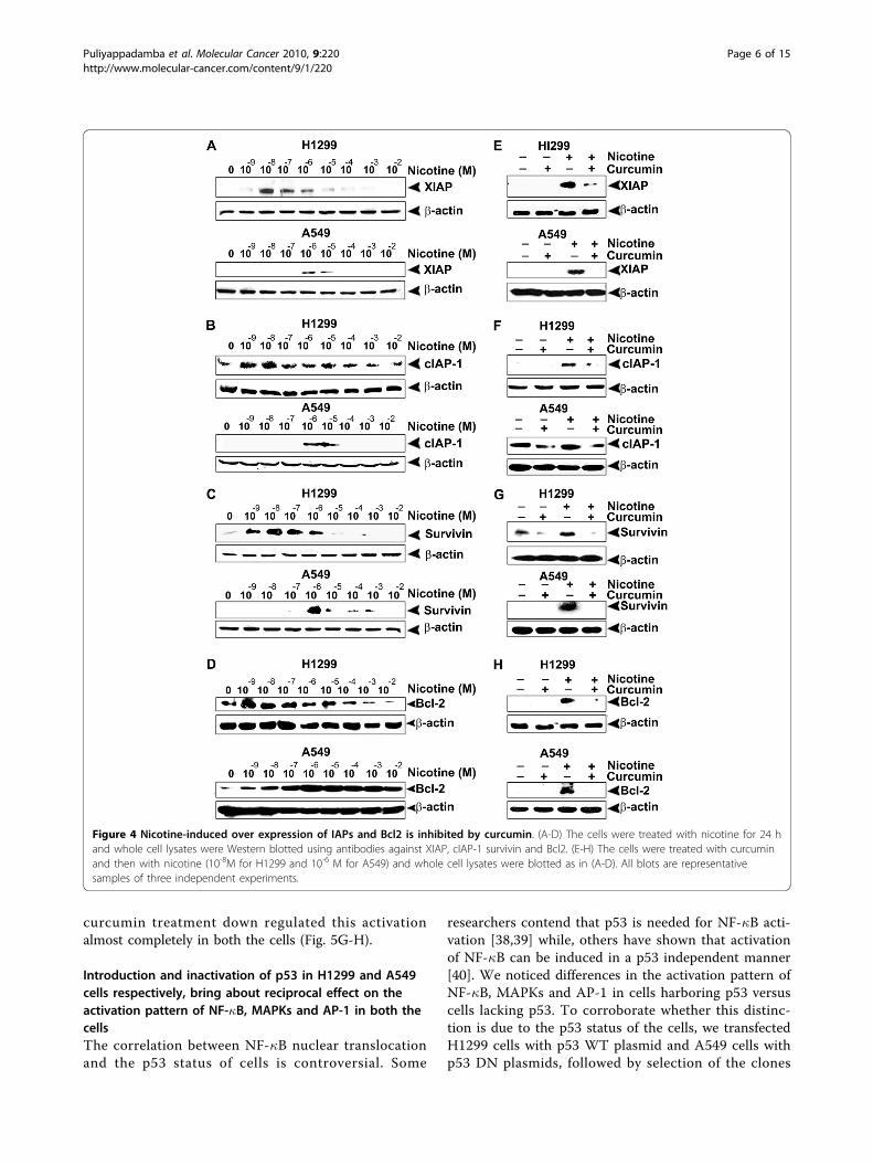
Curcumin prevents the over-expression of IAPs and Bcl2by nicotineThe IAP family of proteins involved in apoptosis regulationis generally activated in response to NF-�B activation [33].Consistent with previous reports indicating that 1 μMnicotine induces the IAP family members, XIAP and survi-vin [14], we also observed the over-expression of XIAP andsurvivin in A549 and H1299 cell lines, albeit difference inthe concentration of nicotine for effective stimulation ofthese proteins. However, we observed robust expression ofcIAP-1 too in both the cell lines by nicotine (Fig. 4A-C),though Dasgupta et al [14] did not observe it in A549 cells.It was inferred that while nicotine induces activation ofthese survival signals in the range 10-9 M-10-7 M in H1299cells, it induces the same level of activation in A549 cells at10-6 M-10-4 M range (Fig. 4A-C). Additionally, becauseBcl2 is considered as a general inhibitor of apoptosis, weevaluated the role of nicotine in its induction above basallevel and tested the effect of curcumin in conferring che-moprotection. As in the case of other survival signals, a 10-100 fold higher concentration of nicotine was necessary toinduce Bcl2 in A549 cells, compared to H1299 cells (Fig.4D) although, curcumin abolished it more or less comple-tely in both the cell lines. Results obtained thus far indicatethat in the investigated lung cancer cell lines, curcumin
Figure 1 Nicotine induces more proliferation in H1299 than in A549, which is down regulated by curcumin. (A-B) The cells were treatedwith nicotine and/or curcumin for 72 h and 24 h respectively for MTT assay and thymidine incorporation assay. Tritiated thymidine (0.5 μCi/well)was added 6 h before the completion of incubation. All error bars indicate standard deviation between three independent experiments.
Puliyappadamba et al. Molecular Cancer 2010, 9:220http://www.molecular-cancer.com/content/9/1/220
Page 3 of 15
completely inhibits the over-expression of these survivalsignals (Fig. 4E-H) independent of the p53 status and theconcentration of nicotine, strongly suggesting a preventiveaction of curcumin against nicotine-induced promotionand progression of lung cancer through up-regulation ofantiapoptotic molecules.
Nicotine-induced phosphorylation of MAPKs and nucleartranslocation of AP-1 in lung cancer cells are down-regulated by curcuminOver-expression of cyclin D1 and IAPs by nicotine evenin the absence of NF-�B led us to speculate the involve-ment of other pathways regulating the expression of
these proteins. Involvement of the MAPK pathway [7]and the induction of cyclin D1 through the MAPK-AP-1 pathway [34] in nicotine-induced cell signaling is wellevidenced. Induction of IAPs by MAPKs independent ofNF-�B has also been reported [35]. We therefore per-formed a time kinetic study up to 120 min to investigatethe functional role of MAPK in H1299 and A549 cells,by analyzing their phosphorylation status in the pre-sence of nicotine and found that, nicotine induces maxi-mum phosphorylation of all the MAPKs at 30 min (datanot shown). Dose response study revealed that all thethree MAPKs are phosphorylated in both the cell lines,being maximally activated at 10-8M concentration of
Figure 2 Nicotine activates NF-�B in H1299, but not in A549. (A-B) Nuclear extracts were prepared from the cells treated with indicatedconcentrations of nicotine for 30 min and NF-�B nuclear translocation was assessed by EMSA. (C) H1299 cells were treated with 10-8 M nicotinefor different time intervals and EMSA was done. (D) The cytosolic extracts of the time kinetic study of NF-�B activation were Western blottedagainst the respective antibodies. (E) The nuclear extract of nicotine-treated H1299 cells were incubated with p50 and p65 antibodies orunlabelled oligo and EMSA was conducted to detect supershift and cold competition respectively. (F) H1299 cells were treated with curcuminand then with nicotine for 30 min and EMSA was done. All blots and EMSAs are representative samples of three independent experiments.
Puliyappadamba et al. Molecular Cancer 2010, 9:220http://www.molecular-cancer.com/content/9/1/220
Page 4 of 15
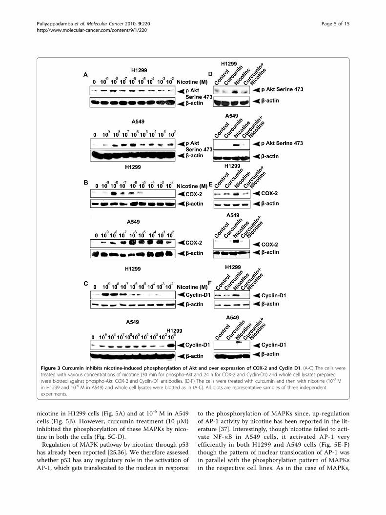
nicotine in H1299 cells (Fig. 5A) and at 10-6 M in A549cells (Fig. 5B). However, curcumin treatment (10 μM)inhibited the phosphorylation of these MAPKs by nico-tine in both the cells (Fig. 5C-D).Regulation of MAPK pathway by nicotine through p53
has already been reported [25,36]. We therefore assessedwhether p53 has any regulatory role in the activation ofAP-1, which gets translocated to the nucleus in response
to the phosphorylation of MAPKs since, up-regulationof AP-1 activity by nicotine has been reported in the lit-erature [37]. Interestingly, though nicotine failed to acti-vate NF-�B in A549 cells, it activated AP-1 veryefficiently in both H1299 and A549 cells (Fig. 5E-F)though the pattern of nuclear translocation of AP-1 wasin parallel with the phosphorylation pattern of MAPKsin the respective cell lines. As in the case of MAPKs,
Figure 3 Curcumin inhibits nicotine-induced phosphorylation of Akt and over expression of COX-2 and Cyclin D1. (A-C) The cells weretreated with various concentrations of nicotine (30 min for phospho-Akt and 24 h for COX-2 and Cyclin-D1) and whole cell lysates preparedwere blotted against phospho-Akt, COX-2 and Cyclin-D1 antibodies. (D-F) The cells were treated with curcumin and then with nicotine (10-8 Min H1299 and 10-6 M in A549) and whole cell lysates were blotted as in (A-C). All blots are representative samples of three independentexperiments.
Puliyappadamba et al. Molecular Cancer 2010, 9:220http://www.molecular-cancer.com/content/9/1/220
Page 5 of 15
curcumin treatment down regulated this activationalmost completely in both the cells (Fig. 5G-H).
Introduction and inactivation of p53 in H1299 and A549cells respectively, bring about reciprocal effect on theactivation pattern of NF-�B, MAPKs and AP-1 in both thecellsThe correlation between NF-�B nuclear translocationand the p53 status of cells is controversial. Some
researchers contend that p53 is needed for NF-�B acti-vation [38,39] while, others have shown that activationof NF-�B can be induced in a p53 independent manner[40]. We noticed differences in the activation pattern ofNF-�B, MAPKs and AP-1 in cells harboring p53 versuscells lacking p53. To corroborate whether this distinc-tion is due to the p53 status of the cells, we transfectedH1299 cells with p53 WT plasmid and A549 cells withp53 DN plasmids, followed by selection of the clones
Figure 4 Nicotine-induced over expression of IAPs and Bcl2 is inhibited by curcumin. (A-D) The cells were treated with nicotine for 24 hand whole cell lysates were Western blotted using antibodies against XIAP, cIAP-1 survivin and Bcl2. (E-H) The cells were treated with curcuminand then with nicotine (10-8M for H1299 and 10-6 M for A549) and whole cell lysates were blotted as in (A-D). All blots are representativesamples of three independent experiments.
Puliyappadamba et al. Molecular Cancer 2010, 9:220http://www.molecular-cancer.com/content/9/1/220
Page 6 of 15
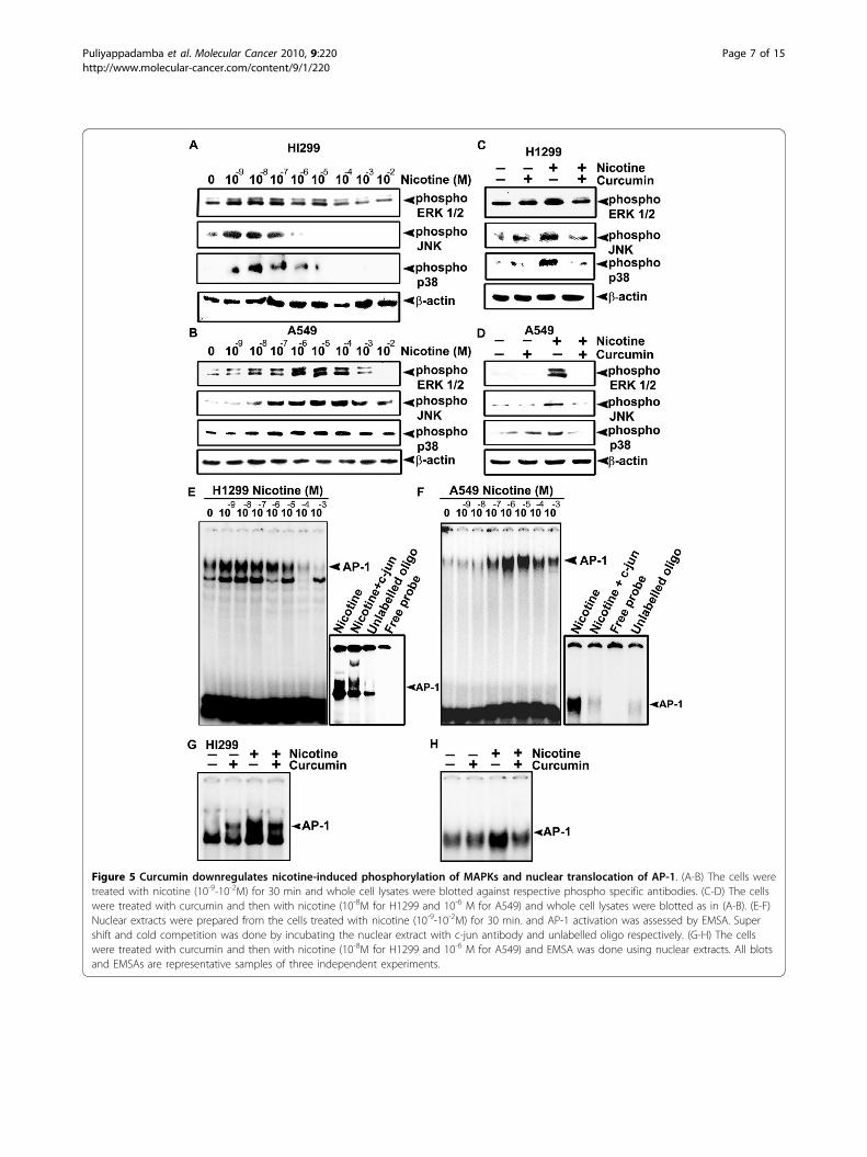
Figure 5 Curcumin downregulates nicotine-induced phosphorylation of MAPKs and nuclear translocation of AP-1. (A-B) The cells weretreated with nicotine (10-9-10-2M) for 30 min and whole cell lysates were blotted against respective phospho specific antibodies. (C-D) The cellswere treated with curcumin and then with nicotine (10-8M for H1299 and 10-6 M for A549) and whole cell lysates were blotted as in (A-B). (E-F)Nuclear extracts were prepared from the cells treated with nicotine (10-9-10-2M) for 30 min. and AP-1 activation was assessed by EMSA. Supershift and cold competition was done by incubating the nuclear extract with c-jun antibody and unlabelled oligo respectively. (G-H) The cellswere treated with curcumin and then with nicotine (10-8M for H1299 and 10-6 M for A549) and EMSA was done using nuclear extracts. All blotsand EMSAs are representative samples of three independent experiments.
Puliyappadamba et al. Molecular Cancer 2010, 9:220http://www.molecular-cancer.com/content/9/1/220
Page 7 of 15
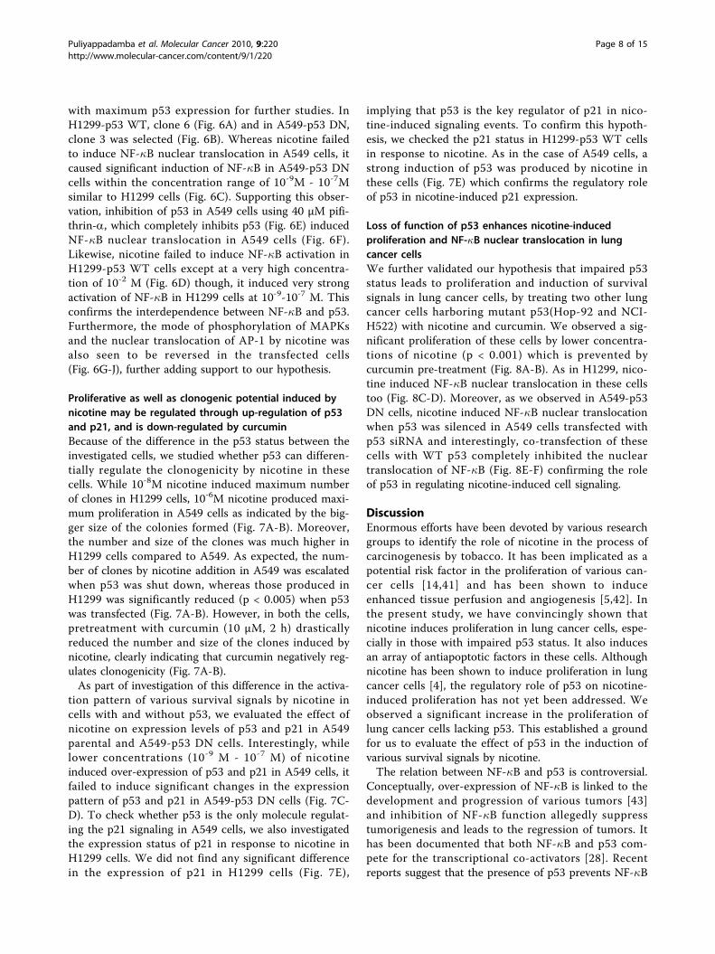
with maximum p53 expression for further studies. InH1299-p53 WT, clone 6 (Fig. 6A) and in A549-p53 DN,clone 3 was selected (Fig. 6B). Whereas nicotine failedto induce NF-�B nuclear translocation in A549 cells, itcaused significant induction of NF-�B in A549-p53 DNcells within the concentration range of 10-9M - 10-7Msimilar to H1299 cells (Fig. 6C). Supporting this obser-vation, inhibition of p53 in A549 cells using 40 μM pifi-thrin-a, which completely inhibits p53 (Fig. 6E) inducedNF-�B nuclear translocation in A549 cells (Fig. 6F).Likewise, nicotine failed to induce NF-�B activation inH1299-p53 WT cells except at a very high concentra-tion of 10-2 M (Fig. 6D) though, it induced very strongactivation of NF-�B in H1299 cells at 10-9-10-7 M. Thisconfirms the interdependence between NF-�B and p53.Furthermore, the mode of phosphorylation of MAPKsand the nuclear translocation of AP-1 by nicotine wasalso seen to be reversed in the transfected cells(Fig. 6G-J), further adding support to our hypothesis.
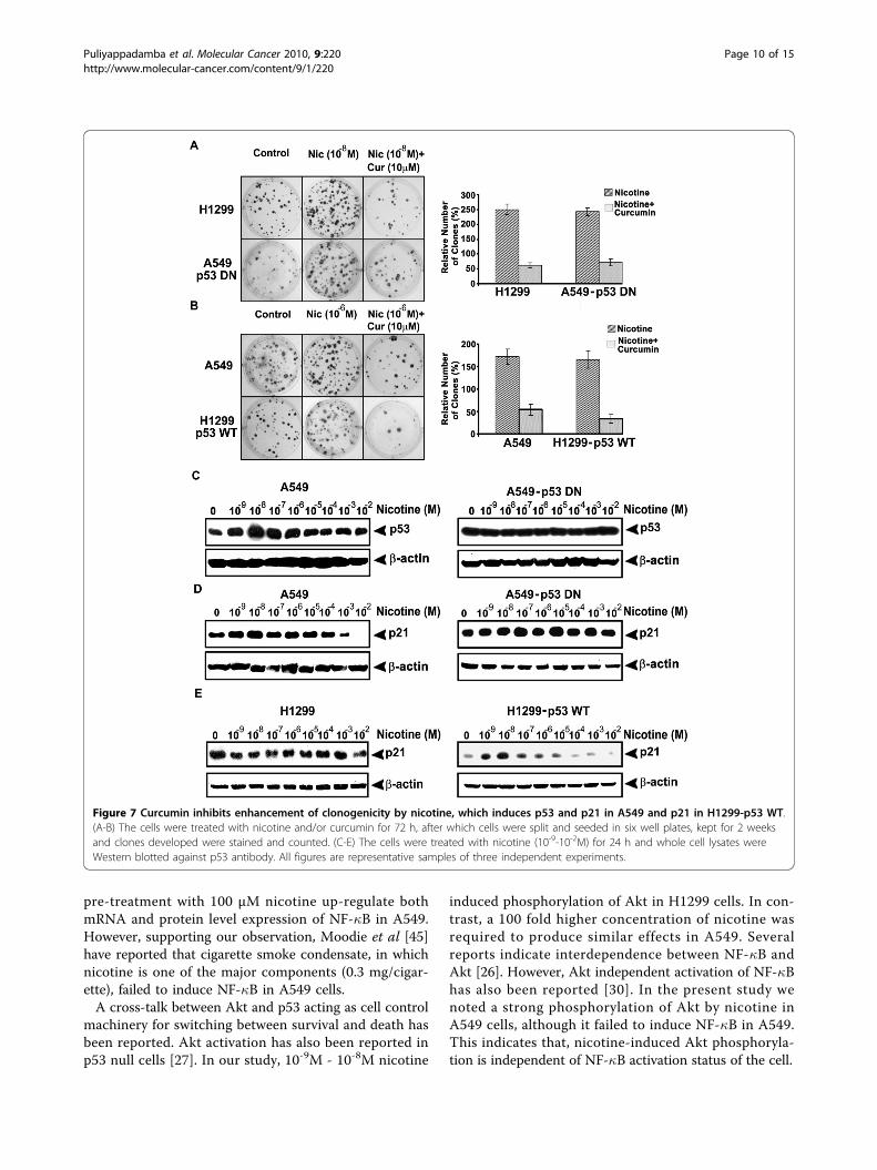
Proliferative as well as clonogenic potential induced bynicotine may be regulated through up-regulation of p53and p21, and is down-regulated by curcuminBecause of the difference in the p53 status between theinvestigated cells, we studied whether p53 can differen-tially regulate the clonogenicity by nicotine in thesecells. While 10-8M nicotine induced maximum numberof clones in H1299 cells, 10-6M nicotine produced maxi-mum proliferation in A549 cells as indicated by the big-ger size of the colonies formed (Fig. 7A-B). Moreover,the number and size of the clones was much higher inH1299 cells compared to A549. As expected, the num-ber of clones by nicotine addition in A549 was escalatedwhen p53 was shut down, whereas those produced inH1299 was significantly reduced (p < 0.005) when p53was transfected (Fig. 7A-B). However, in both the cells,pretreatment with curcumin (10 μM, 2 h) drasticallyreduced the number and size of the clones induced bynicotine, clearly indicating that curcumin negatively reg-ulates clonogenicity (Fig. 7A-B).As part of investigation of this difference in the activa-
tion pattern of various survival signals by nicotine incells with and without p53, we evaluated the effect ofnicotine on expression levels of p53 and p21 in A549parental and A549-p53 DN cells. Interestingly, whilelower concentrations (10-9 M - 10-7 M) of nicotineinduced over-expression of p53 and p21 in A549 cells, itfailed to induce significant changes in the expressionpattern of p53 and p21 in A549-p53 DN cells (Fig. 7C-D). To check whether p53 is the only molecule regulat-ing the p21 signaling in A549 cells, we also investigatedthe expression status of p21 in response to nicotine inH1299 cells. We did not find any significant differencein the expression of p21 in H1299 cells (Fig. 7E),
implying that p53 is the key regulator of p21 in nico-tine-induced signaling events. To confirm this hypoth-esis, we checked the p21 status in H1299-p53 WT cellsin response to nicotine. As in the case of A549 cells, astrong induction of p53 was produced by nicotine inthese cells (Fig. 7E) which confirms the regulatory roleof p53 in nicotine-induced p21 expression.
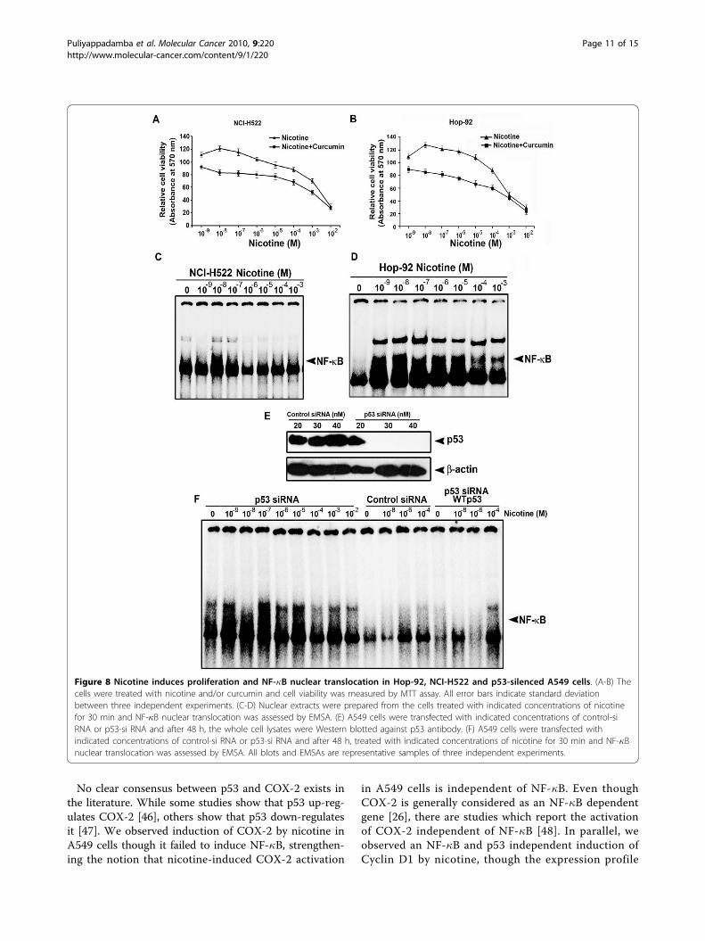
Loss of function of p53 enhances nicotine-inducedproliferation and NF-�B nuclear translocation in lungcancer cellsWe further validated our hypothesis that impaired p53status leads to proliferation and induction of survivalsignals in lung cancer cells, by treating two other lungcancer cells harboring mutant p53(Hop-92 and NCI-H522) with nicotine and curcumin. We observed a sig-nificant proliferation of these cells by lower concentra-tions of nicotine (p < 0.001) which is prevented bycurcumin pre-treatment (Fig. 8A-B). As in H1299, nico-tine induced NF-�B nuclear translocation in these cellstoo (Fig. 8C-D). Moreover, as we observed in A549-p53DN cells, nicotine induced NF-�B nuclear translocationwhen p53 was silenced in A549 cells transfected withp53 siRNA and interestingly, co-transfection of thesecells with WT p53 completely inhibited the nucleartranslocation of NF-�B (Fig. 8E-F) confirming the roleof p53 in regulating nicotine-induced cell signaling.
DiscussionEnormous efforts have been devoted by various researchgroups to identify the role of nicotine in the process ofcarcinogenesis by tobacco. It has been implicated as apotential risk factor in the proliferation of various can-cer cells [14,41] and has been shown to induceenhanced tissue perfusion and angiogenesis [5,42]. Inthe present study, we have convincingly shown thatnicotine induces proliferation in lung cancer cells, espe-cially in those with impaired p53 status. It also inducesan array of antiapoptotic factors in these cells. Althoughnicotine has been shown to induce proliferation in lungcancer cells [4], the regulatory role of p53 on nicotine-induced proliferation has not yet been addressed. Weobserved a significant increase in the proliferation oflung cancer cells lacking p53. This established a groundfor us to evaluate the effect of p53 in the induction ofvarious survival signals by nicotine.The relation between NF-�B and p53 is controversial.
Conceptually, over-expression of NF-�B is linked to thedevelopment and progression of various tumors [43]and inhibition of NF-�B function allegedly suppresstumorigenesis and leads to the regression of tumors. Ithas been documented that both NF-�B and p53 com-pete for the transcriptional co-activators [28]. Recentreports suggest that the presence of p53 prevents NF-�B
Puliyappadamba et al. Molecular Cancer 2010, 9:220http://www.molecular-cancer.com/content/9/1/220
Page 8 of 15
activation though, the mechanism is not clear. However,contradicting these observations, p53-induced NF-�Bactivation has also been reported [44]. It has also beenshown that inhibition of p53 does not inhibit NF-�Bfunction [27].
In our present study we observed a strong activationof NF-�B by nicotine in the p53 null H1299 cell line,while no significant activation of the same was noted inthe p53 proficient A549 cells, at any of the concentra-tions studied. Zhang et al [7] have reported that
Figure 6 Over-expression and silencing of p53 produce reciprocal effect on the activation pattern of NF-�B, MAPKs and AP-1. (A-B)H1299 and A549 cells were transfected with pcDNA3-p53 WT and pcDNA3-p53 DN constructs respectively and the stable clones selected werelysed and Western blotted against p53 antibody. (C-D) H1299-p53 WT and A549-p53 DN cells were treated with nicotine (10-9-10-2M) for 30 min,and EMSA was done using nuclear extracts. (E) A549 cells were treated with different concentrations of pifithrin-a (10-50 μM) for 4 h and thewhole cell lyasates prepared were Western blotted against p53 antibody. (F) A549 cells were treated with 40 μM pifithrin-a for 4 h followed bynicotine (10-9-10-2M) for 30 min and EMSA was done. (G-H) H1299-p53 WT and A549-p53 DN cells were treated with nicotine (10-9-10-2M) for 30min, and EMSA was done. (I-J) H1299 WT-p53 and A549 DN-p53 cells were treated with nicotine (10-9-10-2M) for 30 min and whole cell lysateswere Western blotted against respective phospho specific antibodies. All blots and EMSAs are representative samples of three independentexperiments.
Puliyappadamba et al. Molecular Cancer 2010, 9:220http://www.molecular-cancer.com/content/9/1/220
Page 9 of 15
pre-treatment with 100 μM nicotine up-regulate bothmRNA and protein level expression of NF-�B in A549.However, supporting our observation, Moodie et al [45]have reported that cigarette smoke condensate, in whichnicotine is one of the major components (0.3 mg/cigar-ette), failed to induce NF-�B in A549 cells.A cross-talk between Akt and p53 acting as cell control
machinery for switching between survival and death hasbeen reported. Akt activation has also been reported inp53 null cells [27]. In our study, 10-9M - 10-8M nicotine
induced phosphorylation of Akt in H1299 cells. In con-trast, a 100 fold higher concentration of nicotine wasrequired to produce similar effects in A549. Severalreports indicate interdependence between NF-�B andAkt [26]. However, Akt independent activation of NF-�Bhas also been reported [30]. In the present study wenoted a strong phosphorylation of Akt by nicotine inA549 cells, although it failed to induce NF-�B in A549.This indicates that, nicotine-induced Akt phosphoryla-tion is independent of NF-�B activation status of the cell.
Figure 7 Curcumin inhibits enhancement of clonogenicity by nicotine, which induces p53 and p21 in A549 and p21 in H1299-p53 WT.(A-B) The cells were treated with nicotine and/or curcumin for 72 h, after which cells were split and seeded in six well plates, kept for 2 weeksand clones developed were stained and counted. (C-E) The cells were treated with nicotine (10-9-10-2M) for 24 h and whole cell lysates wereWestern blotted against p53 antibody. All figures are representative samples of three independent experiments.
Puliyappadamba et al. Molecular Cancer 2010, 9:220http://www.molecular-cancer.com/content/9/1/220
Page 10 of 15
No clear consensus between p53 and COX-2 exists inthe literature. While some studies show that p53 up-reg-ulates COX-2 [46], others show that p53 down-regulatesit [47]. We observed induction of COX-2 by nicotine inA549 cells though it failed to induce NF-�B, strengthen-ing the notion that nicotine-induced COX-2 activation
in A549 cells is independent of NF-�B. Even thoughCOX-2 is generally considered as an NF-�B dependentgene [26], there are studies which report the activationof COX-2 independent of NF-�B [48]. In parallel, weobserved an NF-�B and p53 independent induction ofCyclin D1 by nicotine, though the expression profile
Figure 8 Nicotine induces proliferation and NF-�B nuclear translocation in Hop-92, NCI-H522 and p53-silenced A549 cells. (A-B) Thecells were treated with nicotine and/or curcumin and cell viability was measured by MTT assay. All error bars indicate standard deviationbetween three independent experiments. (C-D) Nuclear extracts were prepared from the cells treated with indicated concentrations of nicotinefor 30 min and NF-�B nuclear translocation was assessed by EMSA. (E) A549 cells were transfected with indicated concentrations of control-siRNA or p53-si RNA and after 48 h, the whole cell lysates were Western blotted against p53 antibody. (F) A549 cells were transfected withindicated concentrations of control-si RNA or p53-si RNA and after 48 h, treated with indicated concentrations of nicotine for 30 min and NF-�Bnuclear translocation was assessed by EMSA. All blots and EMSAs are representative samples of three independent experiments.
Puliyappadamba et al. Molecular Cancer 2010, 9:220http://www.molecular-cancer.com/content/9/1/220
Page 11 of 15
was less in A549 cells. Cyclin D1 is also considered asan NF-�B dependent gene [49] although, recent studiesimplicate Cyclin D1 expression through pathways inde-pendent of NF-�B [32].IAP family of proteins is another essential target of
NF-�B [50]. The present study as well as a previousstudy by Dasgupta et al [14], have noticed over-expres-sion of IAPs by nicotine in A549 and H1299 cells. Ofinterest, this is the first study reporting the difference inthe activation pattern of IAPs between both these celllines. Bcl2 is another molecule which has a key role inregulating nicotine-induced survival signaling and hasoften been considered as NF-�B dependent. Interest-ingly, we observed Bcl2 up-regulation in both the celllines, though the pattern of up-regulation variedbetween cell lines. In rapport to our observation, anovel nicotine-stimulated survival pathway that involvesBcl2 phosphorylation through MAPK pathway has beenreported earlier [10].Nicotine also induces p53 [25] which, often regulates
the phosphorylation pattern of MAPKs [36,51]. On thecontrary, another study reports that MAPK activationoccurs only in cells with functional p53 [52], indicating areciprocal interaction between p53 signaling pathway andMAPK pathway. We observed a discrete difference in thephosphorylation pattern of MAPKs in both the cells. Theinvolvement of p53 was further confirmed when thephosphorylation pattern was reversed when p53 wasinactivated in A549 and introduced in H1299 cells.AP-1 is a transcriptional regulator and phosphorylation
of MAPKs leads to nuclear translocation of AP-1 [53].Although NF-�B and AP-1 are regulated by differentmechanisms, several studies indicate that they are acti-vated simultaneously [54]. Nicotine induced nucleartranslocation of AP-1 in both the cell lines, even thoughit failed to induce NF-�B in A549. However, as in thecase of MAPKs, depending on the p53 status of the cellline, there was a significant change in the activation pat-tern of AP-1. In support of the involvement of p53 inAP1 activation, the nuclear translocation pattern of AP-1was reversed in A549-p53DN and H1299-p53WT cells.The ability of nicotine to enhance adherence-indepen-
dent proliferation of tumor cells is well documented[55]. We observed a marked difference in the numberand size of the clones between both H1299 and A549cells treated with nicotine, which was strongly corrobor-ating within the concentration range of nicotine atwhich the survival signals are activated in both the cells,implicating a regulatory role of these survival signals onnicotine-induced enhancement of clonogenic potential.As there was a drastic reduction in the number ofclones on treatment with curcumin in both the cells, itis also evident that curcumin inhibits the effect of nico-tine by down-regulating these survival signals. Nicotine
has a high toxicity in comparison to many other alka-loids such as cocaine and higher doses of nicotine havebeen reported to be lethal (0.5-1.0 mg/kg for adulthumans, and 10 mg for children) [56]. In our study, wealso observed that all the survival signals induced bynicotine are abrogated at higher concentrations suggest-ing that there may be a balance between the pro-survi-val and anti-survival signals induced by this compoundat different concentrations. However the exact concen-tration at which this switching over occurs is stillunclear as we observed the cessation of various survivalsignals at different concentrations of nicotine.Activation of almost all the survival signals investi-
gated in this study is involved in the process of lungcancer progression. It has also been shown that p53 andp21 negatively regulate these survival signals [28,47,23].Induction of p53 and its downstream target p21 bynicotine has been correlated to the inhibition of cellproliferation by nicotine [25]. We observed a stronginduction of p53 as well as p21 by lower concentrationsof nicotine in A549 cells which may be the reason forthe absence of proliferative signals at this concentrationin these cells. A strong induction of p21 was noted inresponse to nicotine in H1299 cells transfected withwild type p53 as well. But we did not observe anychange in the expression of p53 in A549-p53DN cells.This is expected since p53 is non-functional in thesecells, and hence cannot regulate the expression of p21.Similarly, no difference in expression of p21 in responseto nicotine treatment was noted in H1299 cells as wellas A549-p53DN cells, again confirming the role of p53in regulating the nicotine-induced signaling events.Hence, we hypothesize that the induction of p53 andp21 by lower concentrations of nicotine in A549 cellsmay be one of the reasons why nicotine failed to inducethese survival signals at these concentrations in A549,though further studies are required to confirm thishypothesis.Our hypothesis that p53 is the key regulator of nico-
tine-induced survival signaling in lung cancer cells, wasfurther authenticated in two more lung cancer cells withmutant p53 status. Lower concentrations of nicotineinduced proliferation in these cells and pre-treatmentwith curcumin prevented it and, as in H1299, lowerconcentrations of nicotine induced NF-�B nuclear trans-location in these cells too. In addition, when p53 wassilenced in A549 cells by p53 siRNA, as in A549-p53DN cells, nicotine induced a strong nuclear transloca-tion of NF-�B, while co-transfection of these cells withWT p53 completely inhibited the same, substantiatingthe regulatory role of p53 in nicotine signaling.The data obtained from our study is very significant
because the average plasma level concentration of nico-tine in a typical pack per day smoker is between 20-40
Puliyappadamba et al. Molecular Cancer 2010, 9:220http://www.molecular-cancer.com/content/9/1/220
Page 12 of 15
mg [57]. Studies have indicated that chemotherapeuticdrugs like cisplatin, which induce p53 in cells with awild-type p53 gene can induce apoptosis in these cellswhile cells with mutated p53 are unaffected [58]. Sup-porting this observation, Dasgupta et al [14] have shownthat nicotine prevents the cisplatin-induced apoptosis inA549 cells with active p53. We also disclose a detrimen-tal role of nicotine in lung cancer patients with mutantp53 status. Hence our study has proved beyond doubtthat p53 has a significant role in regulating the survivalsignals induced by nicotine in lung cancer cells. It isalso clear that curcumin down-regulates these survivalsignals induced by nicotine in both the cells, indepen-dent of their p53 status.
ConclusionsOur study identifies nicotine as a potential tumor pro-moter, especially in people with impaired p53 status andillustrates a chemopreventive role to curcumin againstnicotine-induced lung cancer progression.
MethodsCell linesH1299 cell line was a gift from Prof. BB Aggarwal, MDAnderson, Houston, A549 and NCI-H522 cells wereprocured from NCCS, Pune, and Hop-92 was obtainedfrom NCI, Bethesda, MD.
ChemicalsAntibodies against p53, I�Ba, XIAP and cIAP-1 andphosphospecific antibodies against-IKK, p38, Akt andp42/44 were purchased from Cell Signalling (Beverly,MA) and those against p50, RelA, pJNK, COX-2 andSurvivin were obtained from Santa Cruz Biotechnology(Santa Cruz, CA). G418 and pifithrin-a were procuredfrom Calbiochem (San Diego, USA). Lipofectamine wasprocured from Invitrogen (Carlsbad, CA) and controlsiRNA and p53 siRNA were procured from Dharmacon,Inc (Chicago, IL). All other chemicals were purchasedfrom Sigma Chemicals (St. Louis, MO).
Mode of treatmentIn all combination treatments 10 μM curcumin wasadded 2 h before nicotine (10-8 M in H1299 and 10-6 Min A549).
MTT AssayProliferative/cytotoxic effect of nicotine and/or curcuminwas determined by MTT assay as described earlier [18].
[3H] Thymidine incorporationFor [3H] thymidine incorporation assay, cells wereseeded in 96 well plates and treated with required con-centrations of the drugs. After 18 h of incubation, [3H]
thymidine was added (0.5 μCi/well) and further incu-bated for 6 h. Then the culture medium was removed,the cells were washed twice with phosphate-buffered sal-ine (PBS), treated with 5% trichloroacetic acid, pelletedand supernatant was removed. Cells were then washedwith ethanol and solubilized with 0.2N NaOH, contentsof each well were mixed with 3 ml scintillation fluid andthe radioactivity was counted using a liquid scintillationcounter [59]. The experiment was repeated thrice andthe error bars indicate standard deviation.
Electrophoretic Mobility Shift Assay (EMSA)To detect activation of NF-�B and AP-1, EMSA wasdone and visualized by a Phosphor Imager (Bio-Rad Per-sonal FX). The specificity of the bands were confirmedby super shift [29].
Isolation of PlasmidsThe plasmids were isolated by the standard alkaline lysismethod [60].
Stable TransfectionH1299 Cells were transfected with pcDNA3-p53WT orthe empty vector pcDNA3 while A549 cells were trans-fected with pcDNA3-p53 DN or the empty vectorpcDNA3 using the calcium-phosphate transfection kit(Life Technologies, Inc.) according to manufacturer’sprotocol and selected using G418 (150 μg/ml) [61].
siRNA TransfectionA549 cells were transiently transfected with controlsiRNA, p53 siRNA and/or WT-p53 plasmid using lipo-fectamine 2000 reagent according to manufactures pro-tocol (Invitrogen).
Western blotThe total protein after treatment with nicotine and/orcurcumin was electroblotted as described earlier [18]and developed by Enhanced Chemiluminescence(Immobilon Western, Miliipore, Billerica, MA).
Clonogenic assayThe cells were treated with nicotine and/or curcuminfor 72 h, as in MTT assay after which the cells weresplit and seeded in six well plates and clonogenic assaywas done as described by Franken et al [62].
Statistical analysisStatistical significance was calculated using one-wayANOVA followed by Tukey post-hoc analysis.
AbbreviationsAP-1: Activator protein 1; Bcl2: B-cell lymphoma 2; cIAP: Cellular Inhibitor ofapoptosis protein; COX2: Cyclooxygenase 2; DN: Dominant Negative; EMSA:
Puliyappadamba et al. Molecular Cancer 2010, 9:220http://www.molecular-cancer.com/content/9/1/220
Page 13 of 15
Electrophoretic mobility shift assay; ERK: Extracellular Signal Regulated Kinase;Fig.: Figure; h: Hour; IAP: Inhibitor of apoptosis protein; I�Ba: Inhibitor ofkappa B protein alpha; IKK: I�B kinase; JNK: c-Jun N-terminal kinase; M: Molar;MAPK-Mitogen activated protein kinases; min: Minute; mTOR: Mammaliantarget of rapamycin; NF-�B: Nuclear factor kappa B; PI3-K-phosphatidylinositol-3-kinase; RelA; Reticuloendotheliosis Viral OncogeneHomolog A; RNA: Ribonucleic acid; siRNA: Small interfering RNA; WT: WildType; XIAP: X-chromosome linked inhibitor of apoptosis; μM: Micro molar
AcknowledgementsWe thank Bert Vogelstein, Philip D Perkins and Ghana Shyam Swarup for thep53 plasmids, Tessy T Maliekal for technical advice and Gayathri LT andJayesh Antony for technical assistance. The authors acknowledge withthanks, Christine Wojewade (USA) and Sophia Margaret Joseph for Editorialcorrections.
Authors’ contributionsAll authors read and approved the final manuscript.VTP and VTC has equally contributed to the in vitro viability assays,transfection and NF-�B and AP-1 studies, SVB and RV contributed to theMAPK studies, AKTT carried out the clonogenic assay, BSV and PRPcontributed to the detection of expression pattern of p53, I�Ba and p-IKK,AB and SV contributed to the expression studies of IAPs and RJA designedand coordinated the study and drafted the manuscript.
Competing interestsThe authors declare that they have no competing interests.
Received: 18 March 2010 Accepted: 20 August 2010Published: 20 August 2010
References1. D’Amico D, Carbone D, Mitsudomi T, Nau M, Fedorko J, Russell E,
Johnson B, Buchhagen D, Bodner S, Phelps R, Gazdar A, Minna JD: Highfrequency of somatically acquired p53 mutations in small-cell lungcancer cell lines and tumors. Oncogene 1992, 7:339-346.
2. Waldum HL, Nilsen OG, Nilsen T, Rørvik H, Syversen V, Sanvik AK,Haugen OA, Torp SH, Brenna E: Long-term effects of inhaled nicotine. LifeSciences 1996, 58:1339-1346.
3. Heeschen C, Weis M, Aicher A, Dimmeler S, Cooke JP: A novel angiogenicpathway mediated by non-neuronal nicotinic acetylcholine receptors. JClin Inves 2002, 110:527-536.
4. Dasgupta P, Rastogi S, Pillai S, Ordonez-Ercan D, Morris M, Haura E,Chellappan S: Nicotine induces cell proliferation by b-arrestin-mediatedactivation of Src and Rb-Raf-1 pathways. J Clin Invest 2006, 116:2208-2217.
5. Egleton RD, Brown KC, Dasgupta P: Angiogenic activity of nicotinicacetylcholine receptors: Implications in tobacco related vasculardiseases. Pharmacol Ther 2009, 121:205-223.
6. West KA, Linnoila IR, Belinsky SA, Harris CC, Dennis PA: Tobaccocarcinogen-induced cellular transformation increases activation of thePhosphatidylinositol 3’-Kinase/Akt pathway in Vitro and in Vivo. CancerRes 2004, 64:446-451.
7. Zhang T, Lu H, Shang X, Tian Y, Zheng C, Wang S, Cheng H, Zhou R:Nicotine prevents the apoptosis induced by menadione in human lungcancer cells. Biochem Biophys Res Comm 2006, 342:928-934.
8. Tsurutani J, Castillo SS, Brognard J, Granville CA, Zhang C, Gills JJ, Sayyah J,Dennis PA: Tobacco components stimulate Akt-dependent proliferationand NF�B-dependent survival in lung cancer cells. Carcinogenesis 2005,26:1182-1195.
9. Shin VY, Wu WK, Chu KM, Wong HP, Lam EK, Tai EK, Koo MW, Cho CH:Nicotine induces cyclooxygenase-2 and vascular endothelial growthfactor receptor-2 in association with tumor-associated invasion andangiogenesis in gastric cancer. Mol Cancer Res 2005, 3:607-615.
10. Mai H, May WS, Gao F, Jin Z, Deng X: A functional role for nicotine in Bcl2phosphorylation and suppression of apoptosis. J Bio Chem 2003,278:1886-1891.
11. Tsai JR, Chong IW, Chen CC, Lin SR, Sheu CC, Hwang JJ: Mitogen-activatedprotein kinase pathway was significantly activated in human bronchialepithelial cells by nicotine. DNA Cell Biol 2006, 25:312-322.
12. Arredondo J, Chernyavsky AI, Grando SA: The nicotinic receptorantagonists abolish pathobiologic effects of tobacco-derivednitrosamines on BEP2D cells. J Cancer Res Clin Oncol 2006, 132:653-663.
13. Arredondo J, Chernyavsky AI, Grando SA: Nicotinic receptors mediatetumorigenic action of tobacco-derived nitrosamines on immortalizedoral epithelial cells. Cancer Biol Ther 2006, 5:511-517.
14. Dasgupta P, Kinkade R, Joshi B, Decook C, Haura E, Chellappan S: Nicotineinhibits apoptosis induced by chemotherapeutic drugs by up-regulatingXIAP and Survivin. Proc Natl Acad Sci 2006, 103:6332-6337.
15. Sharma RA, Gescher AJ, Steward WP: Curcumin: The story so far. Eur JCancer 2005, 41:1955-1968.
16. Oyama Y, Masuda T, Nakata M, Chikahisa L, Yamazaki Y, Miura K,Okagawa M: Protective actions of 5’-n-alkylated curcumins on living cellssuffering from oxidative stress. Eur J Pharmacol 1998, 360:65-71.
17. Aggarwal BB, Kumar A, Aggarwal MS, Shisodia S: Curcumin derived fromturmeric Curcuma longa: a spice for all seasons. In Phytopharmaceuticalsin Cancer Chemoprevention. Edited by: Bagchi D, Preuss HG. Boca Raton:CRC Press; 2005:349-387.
18. Anto RJ, Mukhopadhyay A, Denning K, Aggarwal BB: Curcumin(diferuloylmethane) induces apoptosis through activation of caspase-8,BID cleavage and cytochrome C release: its suppression by ectopicexpression of Bcl-2 and Bcl-xl. Carcinogenesis 2002, 23:143-150.
19. Bava SV, Puliappadamba VT, Deepti A, Nair A, Karunagaran D, Anto RJ:Sensitization of taxol-induced apoptosis by curcumin involves down-regulation of nuclear factor-�B and the serine/threonine kinase Akt andis independent of tubulin polymerization. J Biol Chem 2005,280:6301-6308.
20. Ireson C, Orr S, Jones DJ, Verschoyle R, Lim CK, Luo JL, Howells L,Plummer S, Jukes R, Williams M, Steward WP, Gescher A: Characterizationof metabolites of the chemopreventive agent curcumin in human andrat hepatocytes and in the rat in vivo, and evaluation of their ability toinhibit phorbol ester-induced Prostaglandin E2 production. Cancer Res2001, 61:1058-1064.
21. Arakawa H: p53, apoptosis and axon-guidance molecules. Cell Death Differ2005, 12:1057-1065.
22. Bykov VJ, Issaeva N, Shilov A, Hultcrantz M, Pugacheva E, Chumakov P,Bergman J, Wiman KG, Selivanova G: Restoration of the tumor suppressorfunction to mutant p53 by a low-molecular-weight compound. NatureMed 2002, 8:282-288.
23. Stepulak A, Sifringer M, Rzeski W, Endesfelder S, Gratopp A, Pohl EE,Bittigau P, Felderhoff-Mueser U, Kaindl AM, Bührer C, Hansen HH, Stryjecka-Zimmer M, Turski L, Ikonomidou C: NMDA antagonist inhibits theextracellular signal-regulated kinase pathway and suppresses cancergrowth. Proc Natl Acad Sci 2005, 102:15605-15610.
24. Macleod KF, Sherry N, Hannon G, Beach D, Tokino T, Kinzler K, Vogelstein B,Jacks T: p53-dependent and independent expression of p21 during cellgrowth, differentiation, and DNA damage. Genes Dev 1995, 9:935-944.
25. Berger F, Gage FH, Vijayaraghavan S: Nicotinic receptor-induced apoptoticcell death of hippocampal progenitor cells. J Neurosci 1998, 18:6871-6881.
26. Meng F, Liu L, Chin PC, D’Mello SR: Akt is a downstream target of NF-�B.J Biol Chem 2002, 277:29674-29680.
27. Culmsee C, Siewe J, Junker V, Retiounskaia M, Schwarz S, Camandola S, El-Metainy S, Behnke H, Mattson MP, Krieglstein J: Reciprocal inhibition ofp53 and nuclear factor-�B transcriptional activities determines cellsurvival or death in neurons. J Neurosci 2003, 23:8586-8595.
28. Webster GA, Perkins ND: Transcriptional cross talk between NF-�B andp53. Mol Cell Biol 1999, 19:3485-3495.
29. Anto RJ, Mukhopadhyay A, Shishodia S, Gairola CG, Aggarwal BB: Cigarettesmoke condensate activates nuclear transcription factor-�B throughphosphorylation and degradation of I�Ba: correlation with induction ofcyclooxygenase-2. Carcinogenesis 2002, 23:1511-1518.
30. Sudheerkumar P, Shiras A, Das G, Jagtap JC, Prasad V, Shastry P:Independent activation of Akt and NF-kappaB pathways and their rolein resistance to TNF-a mediated cytotoxicity in gliomas. Mol Carcinog2008, 47:126-136.
31. Biliran H, Banerjee S, Sarkar FH, Thakur A, Bollig A, Ahmed F, Wu J, Sun Y,Liao JD: c-Myc-induced chemosensitization is mediated by suppressionof Cyclin D1 expression and NF-�B activity in pancreatic cancer cells.Clin Cancer Res 2007, 13:2811-2821.
Puliyappadamba et al. Molecular Cancer 2010, 9:220http://www.molecular-cancer.com/content/9/1/220
Page 14 of 15
32. Klein EA, Yang C, Kazanietz MG, Assoian RK: NF�B-independent signalingto the Cyclin D1 gene by Rac. Cell Cycle 2007, 6:1115-1121.
33. Wang CY, Mayo MW, Korneluk RG, Goeddel DV, Baldwin AS Jr: NF-�Bantiapoptosis: induction of TRAF1 and TRAF2 and c-IAP-1 and c-IAP2 tosuppress Caspase-8 activation. Science 1998, 281:1680-1683.
34. Chu M, Guo J, Chen CY: Long-term exposure to nicotine, via Raspathway, induces Cyclin D1 to stimulate G1 cell cycle transition. J BiolChem 2005, 280:6369-6379.
35. Furusu A, Nakayama K, Xu Q, Konta T, Kitamura M: MAP Kinase-dependent,NF�B-independent regulation of inhibitor of apoptosis protein genes byTNF-a. J Cell Physiol 2007, 210:703-710.
36. Zhang H, Shi X, Zhang QJ, Hampong M, Paddon H, Wahyuningsih D,Pelech S: Nocodazole-induced p53-dependent c-Jun N-terminal Kinaseactivation reduces apoptosis in Human colon carcinoma HCT116 Cells. JBiol Chem 2002, 277:43648-43658.
37. Wang X, Bacher B, Hollt V: Nicotine-induced gene expression ofproenkephalin in bovine chromaffin cells. Clin Invest 1994, 72:925-929.
38. Ryan KM, Ernst MK, Rice NR, Vousden KH: Role of NF-kappaB in p53-mediated programmed cell death. Nature 2000, 404:892-897.
39. Bohuslav J, Chen LF, Kwon H, Mu Y, Greene WC: p53 induces NF-�Bactivation by an I�B kinase-independent mechanism involvingphosphorylation of p65 by ribosomal S6 Kinase 1. J Biol Chem 2004,279:26115-26125.
40. Tanaka Y, Ota K, Kameoka M, Itaya A, Yoshihara K: Up-regulation of NF�B-responsive gene expression by ΔNp73a in p53 null cells. Exp Cell Res2006, 312:1254-1264.
41. Chen RJ, Ho YS, Guo HR, Wang YJ: Rapid activation of Stat3 and ERK1/2by nicotine modulates cell proliferation in human bladder cancer cells.Toxicol Sci 2008, 104:283-293.
42. Heeschen C, Jang JJ, Weis M, Pathak A, Kaji S, Hu RS, Tsao PS, Johnson FL,Cooke JP: Nicotine stimulates angiogenesis and promotes tumor growthand atherosclerosis. Nat Med 2001, 7:833-839.
43. Nakshatri H, Nakshatri PB, Martin DA, Goulet RJ Jr, Sledge GW Jr:Constitutive activation of NF-�B during progression of breast cancer tohormone-independent growth. Mol Cell Biol 1997, 17:3629-3639.
44. Kandel ES, Skeen J, Majewski N, Di Cristofano A, Pandolfi PP, Feliciano CS,Gartel A, Hay N: Activation of Akt/Protein Kinase B Overcomes a G2/MCell Cycle Checkpoint Induced by DNA Damage. Mol Cell Biol 2002,22:7831-7841.
45. Moodie FM, Marwick JA, Anderson CS, Szulakowski P, Biswas SK, Bauter MR,Kilty I, Rahman I: Oxidative stress and cigarette smoke alter chromatinremodeling but differentially regulate NF-�B activation andproinflammatory cytokine release in alveolar epithelial cells. FASEB J2004, 18:1897-1899.
46. Han JA, Kim JI, Ongusaha PP, Hwang DH, Ballou LR, Mahale A, Aaronson SA,Lee SW: p53-mediated induction of Cox-2 counteracts p53- or genotoxicstress-induced apoptosis. EMBO J 2002, 21:5635-5644.
47. Subbaramaiah K, Telang N, Ramonetti JT, Araki R, DeVito B, Weksler BB,Dannenberg AJ: Transcription of cyclooxygenase-2 is enhanced intransformed mammary epithelial cells. Cancer Res 1996, 56:4424-4429.
48. Zhang D, Li J, Song L, Ouyang W, Gao J, Huang C: A JNK1/AP-1-Dependent, COX-2 Induction Is Implicated in 12-O-Tetradecanoylphorbol-13-Acetate-Induced Cell Transformation throughRegulating Cell Cycle Progression. Mol Cancer Res 2008, 6:165-174.
49. Guttridge DC, Albanese C, Reuther JY, Pestell RG, Baldwin AS Jr: NF-�Bcontrols cell growth and differentiation through transcriptionalregulation of Cyclin D1. Mol Cell Biol 1999, 19:5785-5799.
50. LaCasse EC, Baird S, Korneluk RG, MacKenzie AE: The inhibitors ofapoptosis (IAPs) and their emerging role in cancer. Oncogene 1998,17:3247-3259.
51. Gulati AP, Yang YM, Harter D, Mukhopadhyay A, Aggarwal BB, Benzil DL,Whysner J, Albino AP, Murali R, Jhanwar-Uniyal M: Mutant human tumorsuppressor p53 modulates the activation of mitogen-activated proteinkinase and nuclear factor-�b, but not c-jun n-terminal kinase andactivated protein-1. Mol Carcinog 2006, 45:26-37.
52. Lee SW, Fang L, Igarashi M, Ouchi T, Lu KP, Aaronson SA: Sustainedactivation of Ras/Raf/mitogen-activated protein kinase cascade by thetumor suppressor p53. Proc Natl Acad Sci 2000, 97:8302-8305.
53. Zhong CY, Zhou YM, Douglas GC, Witschi H, Pinkerton KE: MAPK/AP-1signal pathway in tobacco smoke-induced cell proliferation and
squamous metaplasia in the lungs of rats. Carcinogenesis 2005,26:2187-2195.
54. Fujioka S, Niu J, Schmidt C, Sclabas GM, Peng B, Uwagawa T, Li Z, Evans DB,Abbruzzese JL, Chiao PJ: NF-�B and AP-1 connection: Mechanism of NF-�B-dependent regulation of AP-1 activity. Mol Cell Biol 2004,24:7806-7819.
55. Dasgupta P, Rizwani W, Pillai S, Kinkade R, Kovacs M, Rastogi S, Banerjee S,Carless M, Kim E, Coppola D, Haura E, Chellappan S: Nicotine induces cellproliferation, invasion and epithelial-mesenchymal transition in a varietyof human cancer cell lines. Int J Cancer 2009, 124:36-45.
56. Okamoto M, Kita T, Okuda H, Tanaka T, Nakashima T: Effects of aging onacute toxicity of nicotine in rats. Pharmacol Toxicol 1994, 75:1-6.
57. Benowitz NL, Hall SM, Herning RI, Jacob P III, Jones RT, Osman AL: Smokersof low-yield cigarettes do not consume less nicotine. N Engl J Med 1983,309:139-142.
58. Zamble DB, Jacks T, Lippard SJ: p53-Dependent and -independentresponses to cisplatin in mouse testicular teratocarcinoma cells. Proc NatlAcad Sci 1998, 95:6163-6168.
59. Anto RJ, Maliekal TT, Karunagaran D: L-929 cells harboring ectopicallyexpressed RelA resist curcumin-induced apoptosis. J Biol Chem 2000,275:15601-15604.
60. Bimboim HC, Doly J: A rapid alkaline extraction procedure for screeningrecombinant plasmid DNA. Nucl Acids Res 1979, 7:1513-1523.
61. Anto RJ, Venkatraman M, Karunagaran D: Inhibition of NF-�B sensitizesA431 cells to epidermal growth factor-induced apoptosis, whereas itsactivation by ectopic expression of RelA confers resistance. J Biol Chem2003, 278:25490-25498.
62. Franken NA, Rodermond HM, Stap J, Haveman J, van Bree C: Clonogenicassay of cells in vitro. Nat Protoc 2006, 1:2315-2319.
doi:10.1186/1476-4598-9-220Cite this article as: Puliyappadamba et al.: Nicotine-induced survivalsignaling in lung cancer cells is dependent on their p53 status while itsdown-regulation by curcumin is independent. Molecular Cancer 20109:220.
Submit your next manuscript to BioMed Centraland take full advantage of:
• Convenient online submission
• Thorough peer review
• No space constraints or color figure charges
• Immediate publication on acceptance
• Inclusion in PubMed, CAS, Scopus and Google Scholar
• Research which is freely available for redistribution
Submit your manuscript at www.biomedcentral.com/submit
Puliyappadamba et al. Molecular Cancer 2010, 9:220http://www.molecular-cancer.com/content/9/1/220
Page 15 of 15


