Embed Size (px)
Citation preview

This article appeared in a journal published by Elsevier. The attachedcopy is furnished to the author for internal non-commercial researchand education use, including for instruction at the authors institution
and sharing with colleagues.
Other uses, including reproduction and distribution, or selling orlicensing copies, or posting to personal, institutional or third party
websites are prohibited.
In most cases authors are permitted to post their version of thearticle (e.g. in Word or Tex form) to their personal website orinstitutional repository. Authors requiring further information
regarding Elsevier’s archiving and manuscript policies areencouraged to visit:
http://www.elsevier.com/authorsrights
Author's personal copy
Neuroactive effects of cotinine on the hippocampus: Behavioral and biochemicalparameters
R.B. de Aguiar a,b, G.M. Parfitt a, J. Jaboinski a, D.M. Barros a,*
a Programa de Pós Graduação em Ciências Fisiológicas: Fisiologia Animal Comparada, Instituto de Ciências Biológicas, Universidade Federal do Rio Grande, Av. Itália,Km 8 e Campus Carreiros, Cx. Postal 474, CEP 96201-900, Rio Grande, RS, BrazilbNúcleo de Estudos em Ciências e Matemática, Instituto Federal de Educação, Ciência e Tecnologia Sul-rio-grandense, Pelotas, RS, Brazil
a r t i c l e i n f o
Article history:Received 2 April 2012Received in revised form15 March 2013Accepted 19 March 2013
Keywords:CotinineNicotineHippocampusERK 1/2Rat
a b s t r a c t
The present work evaluated the effects of nicotine (NIC), cotinine (COT), mecamylamine (MEC), meth-yllycaconitine (MLA) and dihydro-beta-eritroidine (DHbE) on memory extinction and the followingbiochemical parameters of the hippocampus: lipid peroxidation (LPO), antioxidant capacity (AC) and thephosphorylation of Extracellular-Signal-Regulated Kinase (ERK 1/2). Young male rats that wereimplanted bilaterally with cannulae were submitted to memory extinction tests sessions, and theirhippocampi were dissected for biochemical assays. The extinction of fear memory was significantlyimproved by both nicotine and its metabolite. Cotinine significantly increased LPO, while nicotinesignificantly decreased it. Antioxidant capacity was increased by all treatments. Our results showed thatcotinine, unlike nicotine, may increase oxidative stress in the hippocampus, but this increase dependsupon the dose used and happens without causing corresponding impairments in cognitive function.Cotinine also increased the phosphorylation of ERK 1/2 in a similar fashion as nicotine. Considering theseresults, it is plausible to wonder to what extent nicotine-attributed effects are really due to the actions ofthis alkaloid and whether they could be due instead to cotinine or to cotinineenicotine interactionswithin the brain.
� 2013 Elsevier Ltd. All rights reserved.
1. Introduction
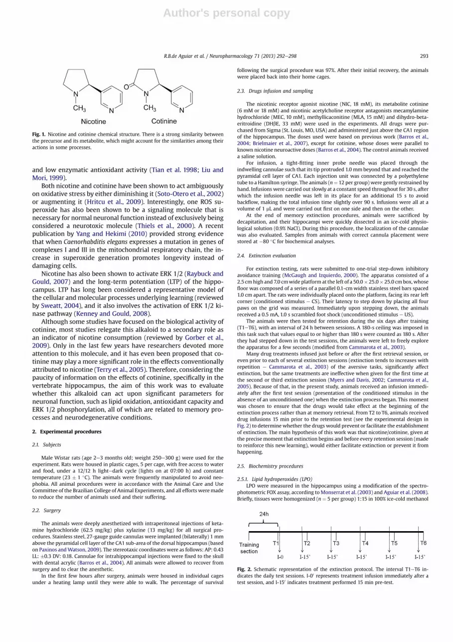
Nicotine is a strongly neuroactive molecule, and one of theprimarily and most abundant metabolites from its oxidation inhumans and in some animal species (e.g., rats and mice) is cotinine(Crooks et al., 1997), which is a nicotine-like molecule (Fig. 1). Thisalkaloid may also be biologically active at nicotine sensitive targets(Crooks et al., 1997; Terry et al., 2005; O’Leary et al., 2008).
After agonist (acetylcholine or nicotine) binding, neuronalnicotinic acetylcholine receptors (nAchR) may activate importantbiochemical cascades within the brain (Barros et al., 2004, 2005;Kenney and Gould, 2008; Boccia et al., 2010; Parfitt et al., 2012).These ionotropic receptors trigger significant cellular alterationsthat contribute to the maintenance of memory performance, and ithas been widely reported that nicotine, when acting at nAchRs,may facilitate working, short-term and long-term memories,among other cognitive functions (Levin and Simon, 1998; Barroset al., 2004; Kenney and Gould, 2008).
It is well known that nicotinemay affect fear motivated learning,and this effect may be studied via the inhibitory avoidance task.These procedures are based on the fact that strong and stablememory traces may be induced in a single training session, if thepresented stimulus is significantly aversive (Whitlock et al., 2006).Nevertheless, fear responses may decrease in their intensitythrough different means, among which is extinction. Fear extinc-tion is a form of learning characterized by a diminution in theamplitude and frequency of a response when the conditionedstimulus that elicits it is not reinforced. Extinction is not about“forgetting” the original fear associated learning, but it is instead aform of active learning that is distinct from acquisition and requiresadditional training to develop (reviewed by Myers and Davis,2002).
Among its many biological actions, it has also been reported thatnicotine interacts with complex I of the brain mitochondrial res-piratory chain and decreases the generation of reactive oxygenspecies (ROS) (Cormier et al., 2001). ROS, such as hydroxyl radical(�OH), establish oxidative stress status in the brain and may causesevere cellular damage. The brain is particularly susceptible tooxidative stress, especially because of its high oxygen consumption,elevated polyunsaturated lipid content and iron concentrations,
* Corresponding author. Tel.: þ55 53 32935170.E-mail address: [email protected] (D.M. Barros).
Contents lists available at SciVerse ScienceDirect
Neuropharmacology
journal homepage: www.elsevier .com/locate/neuropharm
0028-3908/$ e see front matter � 2013 Elsevier Ltd. All rights reserved.http://dx.doi.org/10.1016/j.neuropharm.2013.03.032
Neuropharmacology 71 (2013) 292e298
Author's personal copy
and low enzymatic antioxidant activity (Tian et al. 1998; Liu andMori, 1999).
Both nicotine and cotinine have been shown to act ambiguouslyon oxidative stress by either diminishing it (Soto-Otero et al., 2002)or augmenting it (Hritcu et al., 2009). Interestingly, one ROS su-peroxide has also been shown to be a signaling molecule that isnecessary for normal neuronal function instead of exclusively beingconsidered a neurotoxic molecule (Thiels et al., 2000). A recentpublication by Yang and Hekimi (2010) provided strong evidencethat when Caenorhabditis elegans expresses a mutation in genes ofcomplexes I and III in the mitochondrial respiratory chain, the in-crease in superoxide generation promotes longevity instead ofdamaging cells.
Nicotine has also been shown to activate ERK 1/2 (Raybuck andGould, 2007) and the long-term potentiation (LTP) of the hippo-campus. LTP has long been considered a representative model ofthe cellular andmolecular processes underlying learning (reviewedby Sweatt, 2004), and it also involves the activation of ERK 1/2 ki-nase pathway (Kenney and Gould, 2008).
Although some studies have focused on the biological activity ofcotinine, most studies relegate this alkaloid to a secondary role asan indicator of nicotine consumption (reviewed by Gorber et al.,2009). Only in the last few years have researchers devoted moreattention to this molecule, and it has even been proposed that co-tinine may play a more significant role in the effects conventionallyattributed to nicotine (Terry et al., 2005). Therefore, considering thepaucity of information on the effects of cotinine, specifically in thevertebrate hippocampus, the aim of this work was to evaluatewhether this alkaloid can act upon significant parameters forneuronal function, such as lipid oxidation, antioxidant capacity andERK 1/2 phosphorylation, all of which are related to memory pro-cesses and neurodegenerative conditions.
2. Experimental procedures
2.1. Subjects
Male Wistar rats (age 2e3 months old; weight 250e300 g) were used for theexperiment. Rats were housed in plastic cages, 5 per cage, with free access to waterand food, under a 12/12 h lightedark cycle (lights on at 07:00 h) and constanttemperature (23 � 1 �C). The animals were frequently manipulated to avoid neo-phobia. All animal procedures were in accordance with the Animal Care and UseCommittee of the Brazilian College of Animal Experiments, and all efforts weremadeto reduce the number of animals used and their suffering.
2.2. Surgery
The animals were deeply anesthetized with intraperitoneal injections of keta-mine hydrochloride (62.5 mg/kg) plus xylazine (13 mg/kg) for all surgical pro-cedures. Stainless steel, 27-gauge guide cannulas were implanted (bilaterally) 1 mmabove the pyramidal cell layer of the CA1 sub-area of the dorsal hippocampus (basedon Paxinos andWatson, 2009). The stereotaxic coordinates were as follows: AP: 0.43LL: �0.3 DV: 0.18. Cannulae for intrahippocampal injections were fixed to the skullwith dental acrylic (Barros et al., 2004). All animals were allowed to recover fromsurgery and to clear the anesthetic.
In the first few hours after surgery, animals were housed in individual cagesunder a heating lamp until they were able to walk. The percentage of survival
following the surgical procedure was 97%. After their initial recovery, the animalswere placed back into their home cages.
2.3. Drugs infusion and sampling
The nicotinic receptor agonist nicotine (NIC, 18 mM), its metabolite cotinine(6 mM or 18 mM) and nicotinic acetylcholine receptor antagonists mecamylaminehydrochloride (MEC, 10 mM), methyllicaconitine (MLA, 15 mM) and dihydro-beta-eritroidine (DHbE, 33 mM) were used in the experiments. All drugs were pur-chased from Sigma (St. Louis, MO, USA) and administered just above the CA1 regionof the hippocampus. The doses used were based on previous work (Barros et al.,2004; Brielmaier et al., 2007), except for cotinine, whose doses were parallel toknown nicotine neuroactive doses (Barros et al., 2004). The control animals receiveda saline solution.
For infusion, a tight-fitting inner probe needle was placed through theindwelling cannulae such that its tip protruded 1.0 mm beyond that and reached thepyramidal cell layer of CA1. Each injection unit was connected by a polyethylenetube to a Hamilton syringe. The animals (n¼ 12 per group)were gently restrained byhand. Infusions were carried out slowly at a constant speed throughout for 30 s, afterwhich the infusion needle was left in its place for an additional 15 s to avoidbackflow, making the total infusion time slightly over 90 s. Infusions were all at avolume of 1 mL and were carried out first on one side and then on the other.
At the end of memory extinction procedures, animals were sacrificed bydecapitation, and their hippocampi were quickly dissected in an ice-cold physio-logical solution (0.9% NaCl). During this procedure, the localization of the cannulaewas also evaluated. Samples from animals with correct cannula placement werestored at �80 �C for biochemical analyses.
2.4. Extinction evaluation
For extinction testing, rats were submitted to one-trial step-down inhibitoryavoidance training (McGaugh and Izquierdo, 2000). The apparatus consisted of a2.5 cmhigh and 7.0 cmwide platform at the left of a 50.0� 25.0� 25.0 cmbox,whosefloor was composed of a series of a parallel 0.1-cmwidth stainless steel bars spaced1.0 cm apart. The rats were individually placed onto the platform, facing its rear leftcorner (conditioned stimulus e CS). Their latency to step down by placing all fourpaws on the grid was measured. Immediately upon stepping down, the animalsreceived a 0.5 mA, 1.0 s scrambled foot shock (unconditioned stimulus e US).
The animals were then tested for retention during the six days after training(T1eT6), with an interval of 24 h between sessions. A 180-s ceiling was imposed inthis task such that values equal to or higher than 180 s were counted as 180 s. Afterthey had stepped down in the test sessions, the animals were left to freely explorethe apparatus for a few seconds (modified from Cammarota et al., 2003).
Many drug treatments infused just before or after the first retrieval session, oreven prior to each of several extinction sessions (extinction tends to increases withrepetition e Cammarota et al., 2003) of the aversive tasks, significantly affectextinction, but the same treatments are ineffective when given for the first time atthe second or third extinction session (Myers and Davis, 2002; Cammarota et al.,2005). Because of that, in the present study, animals received an infusion immedi-ately after the first test session (presentation of the conditioned stimulus in theabsence of an unconditioned one) when the extinction process began. This momentwas chosen to ensure that the drugs would take effect at the beginning of theextinction process rather than at memory retrieval. From T2 to T6, animals receiveddrug infusions 15 min prior to the retention test (see the experimental design inFig. 2) to determinewhether the drugs would prevent or facilitate the establishmentof extinction. The main hypothesis of this work was that nicotine/cotinine, given atthe precisemoment that extinction begins and before every retention session (madeto reinforce this new learning), would either facilitate extinction or prevent it fromhappening.
2.5. Biochemistry procedures
2.5.1. Lipid hydroperoxides (LPO)LPO were measured in the hippocampus using a modification of the spectro-
photometric FOX assay, according toMonserrat et al. (2003) and Aguiar et al. (2008).Briefly, tissues were homogenized (n ¼ 5 per group) 1:15 in 100% ice-cold methanol
Fig. 1. Nicotine and cotinine chemical structure. There is a strong similarity betweenthe precursor and its metabolite, which might account for the similarities among theiractions in some processes.
Fig. 2. Schematic representation of the extinction protocol. The interval T1eT6 in-dicates the daily test sessions. I-00 represents treatment infusion immediately after atest session, and I-150 indicates treatment performed 15 min pre-test.
R.B.de Aguiar et al. / Neuropharmacology 71 (2013) 292e298 293

Author's personal copy
and centrifuged at 1000 g, 4 �C, for 10min. Supernatantswere used in the assay. Lipidhydroperoxides were determined using 1 mM FeSO4 (prepared immediately beforeuse), 250 mM H2SO4, 1 mM xylenol orange, deionized water (to complete a finalvolumeof 350mL) and5mL of the hippocampus extract. Cumenehydroperoxide (CHP;0.1 mM; Sigma) was employed as a standard. Themicroplatewas read at 550 nm in auniversal microplate reader (ELx 800, Biotek ELx 800, Winooski, VT, USA).
2.5.2. Antioxidant capacity (AC)To determine antioxidant capacity, the fluorescent probe 20 ,70-dichloro-dihydro-
fluorescein (H2DCFDA, Sigma, USA) was used (Myhre and Fonnum, 2001) with somemodifications. Briefly, tissueswerehomogenized (n¼5per group)1:3w/v in ice-cold40 mM TriseHCl (pH 7.4) buffer, centrifuged at 20,000 g, 4 �C, for 20 min. The su-pernatant was reserved and its protein content determined by Biuret assay for totalproteins (Doles, Goiânia, GO, Brazil). All samples were then adjusted to a final con-centration of 3.32 mg/ml. Samples were placed on microplates with a mediumcontaining the following: (30mMHEPES, 200mMKCl and 1mMMgCl2; pH 7.2), 2,20-azobis (2methylpropionamidine) dihydrochloride (200mMABAP; Aldrich, USA) and16 mMH2DCFDA. For 60min, thefluorescence (resulting fromROS production and thesample’s inability to counteract this oxidative insult) was measured using a Multi-label Counter (VICTOR2 TM; Perkin Elmer, Canada) (excitation and emission wave-lengths of 485 and 520 nm, respectively). Fluorescence counts (FCs) werestandardized by the total protein content of the homogenates. In this assay, a high FCindicates lowantioxidant capacity, while low FC indicates high antioxidant potential.
2.5.3. Phospho-extracellular-signal-regulated kinase (pERK 1/2)Enzyme phosphorylation was determined by colorimetric assay using CASE�
(Cellular Activation of Signaling ELISA from SABiosciences�). To minimize animaluse, before proceeding to in vivo ERK 1/2 determination, in vitro experiments wereperformed with hippocampi extracted from a few untreated (but cannulated) ani-mals to verify the sensitivity of the determination kit in tissue extracts. Subjectswere sacrificed by decapitation, and their hippocampi were quickly dissected in anice-cold phosphate-buffered saline (PBS, pH 7.4) solution. Tissues were gently ho-mogenized in PBS, and the same treatment doses used in the in vivo experimentswere applied. Samples were incubated at 36 �C for 15 min, with rocking.
Samples (n ¼ 5 per group) were then diluted 1:10 in PBS, containing phos-phatase inhibitor cocktail 1 (purchased from SigmaeAldrich) to avoid proteindephosphorylation and were processed as recommended by the manufacturer.Briefly, formaldehyde cell fixing buffer was added to 100 mL of the sample suspen-sion. After that, we proceeded to the treatment with quenching buffer (containing30% H2O2 and 10% NaN3), washing and incubation with primary phospho or totalERK 1/2 antibodies at room temperature. Next, samples were blocked, washed andincubated with HRP-conjugated secondary antibody. After that, washing andtreatment with development, followed by a stop solution, were performed, and theabsorbance of the microplate was quickly read at 450 nm using a universal micro-plate reader (ELx808, BioTek, Winooski, VT, USA).
The microplate was then washed for relative cell number determination. First,cell staining buffer was added to each well and incubated for 30 min at roomtemperature. Following several washing steps, samples were incubated in SDS (1%),and absorbance was read at 595 nm. After in vitro determination results, we per-formed the same steps for the in vivo extracts. Antibody reading was normalized tothe relative cell number to determine the relative extent of target protein phos-phorylation. The phospho-ERKe pERK/cell number ratiowas normalized to the totalERK e tERK/cell number ratio for the same experimental condition.
2.6. Statistics
The use of a 180-s ceiling in the extinction test sessions required usingnonparametric statistics, but the experimental design required a repeated measuresanalysis. Therefore Friedman’s test was chosen as a nonparametric statistical tool,similar to a parametric repeated measures ANOVA. Analyses of variance were fol-lowed using Dunn’sMultiple Comparison Test when appropriate. Data are expressedas the median and the interquartile range (25/75).
When the assumption of variance homogeneity and normality was verified inthe biochemical assays, a one-way ANOVA test of variance followed by NewmansKeuls post hoc tests were performed. In all comparisons, p< 0.05 indicated statisticalsignificance.
Lipid hydroperoxide results were expressed as CHP equivalents per gram of wetweight tissue. Antioxidant capacity was expressed as fluorescence counts (FC) permilligram of protein. The phospho-ERK results are expressed as pERK/tERK ratio.Data are shown as the mean � SEM.
3. Results
3.1. Extinction
Latencies for extinction in the inhibitory avoidance apparatusare shown in Fig. 3aeg. Extinction of aversive memory occurred in
all groups, until the third day of exposure (T3) (CTR ¼ 54.60 48.51/57.25 s; NIC18 16.30 12.94/34.22 s; COT06 ¼ 33.28 22.65/63.74 s;COT18 ¼ 23.93 15.28/26.82 s; MEC ¼ 30.80 13.44/46.60 s;MLA ¼ 53.36 28.48/57.30 DHbE ¼ 22.91 14.02/34.31 s) to thecontext (p < 0.05, vs. T1 of each respective group). When theselatencies were compared to the step-down latencies for theexperimental groups on T3, we found that the NIC18 (p ¼ 0.024, vs.CTR T3), COT18 (p¼ 0.0006, vs. CTR T3) and the DHbE (p¼ 0.038, vs.CTR T3) groups were significantly different from the CTR group.These differences persisted until T6 (p > 0.05, vs. T1 of eachrespective group).
3.2. Lipid hydroperoxides
Fig. 4 shows that treatment with nicotine (443.4 � 5.51 nmolCHP/g) significantly decreased LPO, while treatment with cotinineat a concentration of 6 mM (694.5 � 13.22 nmol CHP/g) signifi-cantly increased LPO when compared to control group(608.3� 4.54 nmol CHP/g). Non-specific antagonist mecamylamine(128.3 � 1.19 nmol CHP/g) provoked the highest decrease in LPOvalues, while the other antagonist (MLA ¼ 589.1 � 51.33 nmol CHP/g; DHbE ¼ 533.9 � 18.52 nmol CHP/g) and cotinine 18 mM(653.2�19.85 nmol CHP/g) did not significantly increase LPOwhencompared to control group.
3.3. Antioxidant capacity
All treatments increased antioxidant capacity in the rat hippo-campus compared to the control group (CTR ¼ 6.84 � 107 � 3.88 �106 FC/mg prot; NIC18 ¼ 5.06 � 107 � 6.77 � 106 FC/mg prot;COT06 ¼ 4.97 � 107 � 4.55 � 106 FC/mg prot; COT18 ¼ 5.12 � 107 �6.73� 106 FC/mg prot; MEC¼ 5.29� 107� 2.24� 106 FC/mg prot), asshown inFig. 5. Interestingly, the specific antagonistsDHbE (a4b2) andMLA (a-7) evoked the highest increases in hippocampal antioxidantcapacity (DHbE ¼ 3.33 � 107 � 2.41 � 106 FC/mg prot and MLA ¼4.10 � 107 � 1.41 �106 FC/mg prot).
3.4. Phospho- extracellular-signal-regulated kinase
In vitro assay results (Fig. 6a) showed that both MEC(0.384 � 0.04) and MLA (0.496 � 0.04) significantly decreased thepERK/tERK ratios compared to the control group (0.639 � 0.04).The results in Fig. 6b for in vivo experiments showed that NIC(0.838� 0.07) and COT06 (0.810� 0.07) both significantly increasedthe pERK/tERK ratios compared to the control group (0.524 � 0.07).However, no significant effects were found in MEC-treated(0.497 � 0.07) or MLA-treated (0.494 � 0.07) animals in vivo. Thepresent work also found that DHbE was the only antagonist tosignificantly reduce ERK 1/2 phosphorylation in both in vitro(0.203 � 0.04) and in vivo (0.141 � 0.07) experiments whencompared to their respective controls. Additionally, cotinine 18 mMwas the only agonist to significantly increase protein phosphoryla-tion in both in vitro (0.938 � 0.04) and in vivo (0.970 � 0.07) exper-iments. No significant differences were found between values foundfrom in vitro (Fig. 6a) and in vivo (Fig. 6b) samples.
4. Discussion
The hippocampus has long been known to be a criticallyimportant structure for attention and memory (Barros et al., 2004;Boccia et al., 2010). Nevertheless, human imaging studies have alsoshown an association between the hippocampus and the extinctionof memory (Milad et al., 2007).
It is well known that exposure to acetylcholine or other nicotinicagonists activate nAChR function in the hippocampus. These
R.B.de Aguiar et al. / Neuropharmacology 71 (2013) 292e298294

Author's personal copy
Fig. 4. Lipid hydroperoxide in the hippocampus. CTR (saline); NIC18 (nicotine 18 mM);COT06 (cotinine 6 mM); COT18 (cotinine 18 mM); MEC (mecamylamine 10 mM); MLA(methyllicaconitine 15 mM) and DHbE (dihydro-beta-eritroidine 33 mM). *NIC18decreased LPO compared to control. #COT06 increased LPO compared to CTR, MEC,MLA and DHbE, but it did not differ from COT18. �MEC10 significantly decreased LPOcompared to control and to all other groups. Neither COT18 nor MLA or DHbE signif-icantly differed from control group (F ¼ 103.56, p < 0.05).
Fig. 5. Hippocampal antioxidant capacity. CTR (saline); NIC18 (nicotine 18 mM);COT06 (cotinine 6 mM); COT18 (cotinine 18 mM); MEC (mecamylamine 10 mM); MLA(methyllicaconitine 15 mM) and DHbE (dihydro-beta-eritroidine 33 mM). N.B.: High FC/mg prot ¼ low antioxidant capacity. *Significant difference compared to control. #Sig-nificant difference compared to control, NIC-, COT- and MEC-treated groups (F ¼ 1557,p < 0.05).
Fig. 3. Effects of the drugs on the extinction of aversive memory. CTR (saline); NIC18 (nicotine 18 mM); COT06 (cotinine 6 mM); COT18 (cotinine 18 mM); MEC (mecamylamine10 mM). Tests were performed for six days, during which animals received their respective treatment. a) Results for the CTR group. A significant decrease in the latency to step downoccurred from the third day on (*, p ¼ 0.0265, vs CTR T2). b) Results for the NIC18 group. A significant decrease occurred from the second day on (*, p ¼ 0.0081, vs NIC18 T2). The T3test also showed a significant difference for the T3 CTR group (#, p ¼ 0.024, vs CTR T3). c) Results for COT06 group. A significant decrease occurred on the second day (*, P ¼ 0.0069,vs COT06 T1). d) Results for the COT18 group. A significant decrease was measured from the second day on (*, p ¼ 0.0022, vs COT18 T1). The T3 test also showed a significantdifference from the T3 CTR group (#, p ¼ 0.0006, vs CTR T3). e) Results for the MEC group. A significant decrease was measured from the second day on (*, p ¼ 0.0265, vs MEC T1). f)Results for the MLA group. A significant and stable decrease was measured from the second day on (*, p ¼ 0.0004, vs MLA T1). g) Results for the DHbE group. A significant decreasewas measured from the second day on (*, p ¼ 0.0011, vs DHbE T1). The T3 test also showed a significant difference for the T3 CTR group (#, p ¼ 0.038, vs CTR T3).
R.B.de Aguiar et al. / Neuropharmacology 71 (2013) 292e298 295

Author's personal copy
receptors are also located in several other brain regions, includingthe amygdala and the cerebral cortex, which may be particularlyimportant for cognitive functioning (Alkondon and Albuquerque,2003). Both the a4b2 and a7 subtypes of the nAchRs have beenimplicated, particularly in learning and memory. The former arefound in areas that bind nicotine with high affinity (e.g., hippo-campus), and the latter are found in distributed areas with high-affinity binding sites for a-bungarotoxin (e.g., hippocampus, cor-tex and ventral tegmental area) (Levin and Simon, 1998).
We have shown that intrahippocampal injections of nicotine,cotinine or DHbE enhancememory extinction because these groupsshow lower step-down latencies on the T3 test in a fear extinctionparadigm. Studies have found that hippocampal nAchR agonists,such as acetylcholine and nicotine, facilitate hippocampal synapticactivity and increase hippocampal LTP (Kenney and Gould, 2008).We have also shown that the systemic administration of cotininemay enhance fear memory extinction (Zeitlin et al., 2012). Re-searchers investigating fear conditioning have found that targetedtreatments that affect acquisition often also affect extinction(reviewedbyMyers andDavis, 2002). Elias et al. (2010) reported thatthe administration of a cholinergic agonist prior to extinction ses-sions enhanced extinction and that this enhancement appeared tobe a long-lasting change, as the enhanced extinction remained after
substance administration ceased. Because agonists such as nicotineand, most likely, its main metabolite cotinine have the capacity tostimulate hippocampal neurons, it is logical to suppose that thesedrugs may improve extinction, as observed in the present study.
Nevertheless, classical nAchRs antagonists (MLA and MEC) didnot impair extinction as could be expected. Previous studies havesuggested that methyllycaconitine impairs retention performancein inhibitory avoidance tasks, regardless of stimulus intensity, andthat its effects are long lasting (Boccia et al., 2010). These publica-tions suggest that a7 nAchRs could be more critical for aversivetasks than a4b2 subtypes, which would corroborate the resultsfound in the present work, where the broad spectrum non-a7nicotinic antagonist mecamylamine and the a7-specific antagonistmethyllycaconitine (a7 specific antagonist) did not significantlyinterfere with extinction. In this study, we found that cotinineincreased lipid peroxidation in the rat hippocampus, while nicotinesignificantly decreased it. It has been reported that a high dose ofnicotine may induce neurotoxicity and stimulate oxidative stress,whereas a reasonably low concentration may act as an antioxidantand play an important role in the neuroprotective effect (Guanet al., 2003; Barros et al., 2007).
Though it is unlikely that the results found in the present studyare dose dependent, as the lowest cotinine dose was the one thatincreased LPO, it is interesting to hypothesize that cotinine mayplay an important role in stimulating the oxidative stress that isassociated with nicotine in published works. This speculationseems even more reasonable once we direct more attention to ourresults: although a higher dose of cotinine did not increase lipidperoxidation in a statistically significant manner, trends toward anincrease in this parameter could be observed in this group.Conversely, nicotine at the same concentration significantlydecreased LPO.
Most works that attribute a pro-oxidant effect to nicotine wereconducted with the peripheral administration of this drug (Soto-Otero et al., 2002; Arrick and Mayhan, 2007; Hritcu et al., 2009).Most of that nicotine was thus likely metabolized before reachingthe brain. Themain organmetabolizing this alkaloid is the liver, andit has been found that 80% of the nicotine absorbed in this fashion ismetabolized by c-oxidation to cotinine in an NADPH-dependent,cytochrome P-450-catalyzed process (Crooks et al., 1997; Tutkaet al., 2005). This observation only strengthens the hypothesis ofcotinine being the main, if not the only, molecule underlying thereported effects.
Several reports have indicated that nicotine has antioxidantproperties that may be intracellular, due to the activation of thenicotinic receptors, or extracellular, when it acts as a radical scav-enger that sequesters iron, thus preventing the Fenton reaction(Newman et al., 2002; Soto-Otero et al., 2002). Our results show anincrease in the antioxidant capacity of animals treated with bothnicotine and cotinine. Such results corroborate past publications(Cormier et al. 2001; Guan et al., 2003), which suggest that nicotineactions upon nAchRs may alter cell redox status in an antioxidant-like, direct or indirect effect. Nevertheless, a stronger increase wasfound in MLA and in DHbE-treated animals. Two possibilities couldexplain this result. First, it is possible that these substances, asspecific antagonists of important nAchRs (Mogg et al., 2002; Damajet al., 1995), decreased neuron function to a level at which very fewROS are produced, so the increase in antioxidant capacity is thus aconsequence of this reduction in brain function. However, in viewof our extinction results, it is more likely that these substances, dueto their chemical structure, acted directly as oxidant scavengingmolecules, thus resulting in the increased antioxidant capacityverified in the subjects.
Despite reports of nicotine’s inhibition of lipid peroxidation inPC12 cells (Guan et al., 2003), which are supported by our LPO
Fig. 6. Phosphorylation of ERK 1/2. CTR (saline); NIC18 (nicotine 18 mM); COT06(cotinine 6 mM); COT18 (cotinine 18 mM); MEC (mecamylamine 10 mM); MLA(methyllicaconitine 15 mM) and DHbE (dihydro-b-eritroidine 33 mM). a) Results forin vitro experiments. *COT18 significantly increased pERK/tERK ratios compared tocontrol and all other groups. #Antagonists MEC and MLA significantly decreased ERKphosphorylation compared to control group. �DHbE provoked the greatest decrease inERK 1/2 phosphorylation. No significant differences from the control group were foundin either NIC18- or COT06-treated animals (F ¼ 29.38 p < 0.05). b) Results for in vivoexperiments. *NIC18 and COT06 significantly increased pERK 1/2 in rat hippocampus,compared to the control group. #COT18 caused the highest increase compared to thecontrol and all other groups. �DHbE provoked the greatest decrease in ERK 1/2 phos-phorylation. No significant differences from control group were found in both MEC-and MLA-treated animals (F ¼ 481.79, p < 0.05).
R.B.de Aguiar et al. / Neuropharmacology 71 (2013) 292e298296
Author's personal copy
results, it has also been reported that treatment with nicotine mayincrease superoxide anion production in cerebral arterioles (Arrickand Mayhan, 2007). Moreover, recent reports (Newman et al.,2002; Sudheer et al., 2007) have shown that this alkaloid signifi-cantly decreases superoxide dismutase (SOD), catalase (CAT) andglutathione peroxidase (GPx) activity, in addition to decreasing thelevels of glutathione (GSH) and vitamins A, E and C. Studies havealso shown that this nicotine-induced decrease in antioxidantstatus may be due to the increased utilization of the previouslymentioned antioxidants to counteract the ROS generated bynicotine.
In the present study, we also found that MEC significantlydecreased LPO. However, this effect could be less related to aneuronal protective effect than to the capacity of this antagonist todecrease neuronal activity (Dajas-Bailador et al., 2002; Steiner et al.,2007) and, by consequence, decrease ROS production.
Another important observation must be made concerningoxidative stress. Although cotinine at 6 mM increased lipid per-oxidation, this effect did not interfere with the ability of thistreatment to facilitate animal performance in our extinction tests.For many years, oxidative stress was exclusively associated withcellular damage resulting from the actions of oxidant or pro-oxidant substances (Ferreira and Matsubara, 1997). More recently,the redox signaling hypothesis is gaining considerable status in thescientific community. This well-documented but still controversialhypothesis argues that redox homeostasis is necessary for cellularfunction and that these mechanisms could act as modulators orsignals in cell physiological processes (Jones, 2008; Finkel, 2011).
It has been hypothesized that ROS can indirectly regulateimportant cellular signaling pathways for the brain, such as themitogen-activated protein kinase (MAPK) superfamily of serine/threonine kinases, which includes the extracellular signal-regulated kinases ERK1 and ERK2, as well as the p38 MAPK(Finkel, 2011). This new insight into redox status questions theprevious mandatory association between pro-oxidants, oxidativestress and cellular damage, substituting redox reactions for possiblecellular modulators, messengers or even essential physiologicalagents (D’Autréaux and Toledano, 2007; Jones, 2008; Finkel, 2011).
In addition to oxidant or antioxidant status, hippocampalfunction is also significantly affected by extracellular signal-regulated kinases (a subfamily of mitogen-activated protein ki-nases eMAPKs) activity, and data have directly implicated changesin ERK in mechanisms for learning disorders in humans (Sweatt,2004; Cesarini et al., 2009).
ERKs are activated by a variety of extracellular agents, whichinclude growth factors, hormones, and neurotransmitters(Rubinfeld and Seger, 2005). Nicotine acting on hippocampalnAChRs may activate the ERK cascade that is involved in the chainof events linking synaptic activity to long-lasting changes associ-ated with gene expression. Nicotine also stimulates changes incalcium influx and in the release of calcium internal stores, whichcan activate not only kinases but also transcription factors such asCREB (reviewed by Gould, 2006).
In the same matter, cotinine has been reported to bind tonicotinic acetylcholine receptors with a high enough level of af-finity for the compound to affect nicotinic responses (Vainio andTuominen, 2001). This capacity may be responsible for the signif-icant improvements in the delayed match-to-sample (DMTS) taskfound in adult rhesus monkeys (Terry et al., 2005) treated with thisalkaloid.
In agreement with these studies, the present work found notonly that cotinine does affect ERK 1/2 phosphorylation but that thiseffect can be also observed both in vitro and in vivo, while this effectwas not observed for nicotine. As nicotine presents a known dualityof effects that is usually dose-dependent, it is possible that the
concentrations used in the in vitro experiments were too high,which ended up provoking a desensitization of the nAchRs (Guanet al., 2006); consequently, no significant increases in ERK 1/2phosphorylation could be registered. Nevertheless, cotininesignificantly increased pERK 1/2 in vitro and in vivo at the sameintensity. This result reinforces the idea that cotinine might be thereal cause (or at least a strong coadjutant) of the nicotine-attributedcellular effect.
It has been shown that classical nicotinic receptor antagonists,such as mecamylamine (Dajas-Bailador et al., 2002; Steiner et al.,2007), the a7-specific nAChR antagonist a-bungarotoxin (a-Bgt),and DHbE (Nakayama et al., 2006), all inhibit the phosphorylationof ERK 1/2 (Dajas-Bailador et al., 2002) and that this inhibition ispersistent when the antagonists are administered prior to nicotinetreatment (Dajas-Bailador et al., 2002; Nakayama et al., 2006;Steiner et al., 2007). Though two antagonists (MEC and MLA) didnot significantly decrease ERK 1/2 phosphorylation during ourin vivo experiments, our results support previous studies of nAChRantagonists that prevent ERK 1/2 phosphorylation because DHbEsignificantly decreased ERK 1/2 phosphorylation both in vivo andin vitro and because MEC and MLA also diminished pERK in thein vitro experiment.
Vainio et al. (2000) reported that both cotinine and nicotinemayenhance the calcium intracellular concentration, but the pre-incubation of the cell culture with cotinine prevents this nicotine-induced augmentation in intracellular Ca2þ and vice versa. Inter-estingly, studies on metanicotine, which is another nicotine-likeagonist, have shown that this substance decreases retention la-tencies and has a significant amnesic effect when administered inhigh doses. The samemetabolite has been shown to yield beneficialeffects on learning and memory (Lippiello et al., 1996; Levin andChristopher, 2002).
As the activation of (nAChRs) and the administration of nicotineor cotinine (Hritcu et al., 2009) could induce quite opposite effects,it is logical to conclude that nicotine, as well as its metabolites andsubstances with similar properties, may act differently dependingon the dose, the moment of administration and the biologicalmodel used. This study provides more evidence that cotinine is nota mere indicator of nicotine consumption but is a probableneuroactive substance with the potential to influence complexlearning processes and pathways. Nevertheless, future studies arenecessary to clarify the real actions of cotinine in the hippocampus,establishing whether part or all of nicotine’s neuronal, pro-oxidant,antioxidant and pro-phosphorylation effects could, in fact, be dueto cotinine or to nicotine-cotinine-associated actions within theCNS, especially when the administration is carried out peripherallyrather than directly on the target structure.
Acknowledgments
We would like to thank the Programa de Pós-Graduação emCiências Fisiológicas d Fisiologia Animal Comparada, BrazilianNational Council for Scientific and Technological Development(CNPq) for financial support (Grant # 474621/2007-3) and Prof.Larissa Costa Kurtz dos Santos for her valuable comments and herreview of the English in the manuscript. D. M. Barros is a researchfellow from the Brazilian agency CNPq, and Juliana Jaboinskireceived an undergraduate fellowship from CNPq.
References
Aguiar, R.B. de, Dickel, O.E., Cunha, R.W., Monserrat, J.M., Barros, D.M., Martinez, P.E.,2008. Estradiol valerate and tibolone: effects upon brain oxidative stress andblood biochemistry during aging in female rats. Biogerontology 9, 285e298.
Alkondon, M., Albuquerque, E.X., 2003. The nicotinic acetylcholine receptor sub-types and their function in the hippocampus and cerebral cortex. In:
R.B.de Aguiar et al. / Neuropharmacology 71 (2013) 292e298 297
Author's personal copy
Descarries, L., Krnjevi�c, K., Steriade, M. (Eds.), 2003. Progress in Brain Research,vol. 145, pp. 109e120 (2004) (Chapter 7).
Arrick, D.M., Mayhan, W.G., 2007. Acute infusion of nicotine impairs nNOS-dependent reactivity of cerebral arterioles via an increase in oxidative stress.J. Appl. Physiol. 103, 2062e2067.
Barros, D.M., Galhardi, F.G., Ferreira, J.L.R., Guterres, L.B., Dickel, O., Geracitano, L.A.,Izquierdo, I., Monserrat, J.M., 2007. The benefits and drawbacks of nicotineexposure in the cortex and hippocampus of old rats. Neurotoxicology 28, 562e568.
Barros, D.M., Ramirez, M.R., Izquierdo, I.A., 2005. Modulation of working, short- andlong-term memory by nicotinic receptors in the basolateral amygdala in rats.Neurobiol. Learn. Mem. 83, 113e118.
Barros, D.M., Ramirez, M.R., Reis, E., Izquierdo, I.A., 2004. Participation of hippo-campal nicotinic receptors in acquisition, consolidation and retrieval of mem-ory for one trial inhibitory avoidance. Neuroscience 126, 651e656.
Boccia, M.M., Blake, M.G., Krawczyk, M.C., Barati, C.M., 2010. Hippocampal alpha7nicotinic receptors modulate memory consolidation of an inhibitory avoidancetask in mice. Neuroscience 171, 531e543.
Brielmaier, J.M., McDonald, C.G., Smith, R.F., 2007. Immediate and long-termbehavioral effects of a single nicotine injection in adolescent and adult rats.Neurotoxicol. Teratol. 29, 74e80.
Cammarota, M., Bevilaqua, L.R.M., Kerr, D., Medina, J.H., Izquierdo, I., 2003. Inhibi-tion of mRNA and protein synthesis in the CA1 region of the dorsal hippo-campus blocks reinstallment of an extinguished conditioned fear response.J. Neurosci. 23, 737e741.
Cammarota, M., Bevilaqua, L.R.M., Rossato, J.I., Ramírez, M.R., Medina, J.H.,Izquierdo, I., 2005. Relationship between short- and long-term memory andshort- and long-term extinction. Neurobiol. Learn. Mem. 84, 25e32.
Cesarini, L., Alfieri, P., Pantaleoni, F., Vasta, I., Cerutti, M., Petrangeli, V., Mariotti1, P.,Leoni, C., Ricci, D., Vicari, S., Selicorni, A., Tartaglia, M., Mercuri, E., Zampino, G.,2009. Cognitive profile of disorders associated with dysregulation of the RAS/MAPK signaling cascade. Am. J. Med. Genet. Part. A 149A, 140e146.
Cormier, A., Morina, C., Zinia, R., Tillementa, J.-P., Lagrue, G., 2001. In vitro effects ofnicotine on mitochondrial respiration and superoxide anion generation. BrainRes. 900, 72e79.
Crooks, P.A., Li, M., Dwoskin, L.P., 1997. Metabolites of nicotine in rat brain afterperipheral nicotine administration: cotinine nornicotine and norcotinine. DrugMetab. Dispos. 25, 47e54.
D’Autréaux, B., Toledano, M.B., 2007. ROS as signalling molecules: mechanisms thatgenerate specificity in ROS homeostasis. Nat. Rev. Mol. Cell Bio. 8, 813e824.
Dajas-Bailador, F.A., Soliakov, L., Wonnacott, S., 2002. Nicotine activates the extra-cellular signal-regulated kinase 1/2 via the a7 nicotinic acetylcholine receptorand protein kinase A, in SH-SY5Y cells and hippocampal neurons. J. Neurochem.80, 520e530.
Damaj, M.I., Welch, S.P., Martin, B.R., 1995. In vivo pharmacological effects ofdihydro-b-erythroidine, a nicotinic antagonist, in mice. Psychopharmacology117, 67e73.
Elias, G.A., Gulick, D., Wilkinson, D.S., Gould, T.J., 2010. Nicotine and extinction offear conditioning. Neuroscience 165, 1063e1073.
Ferreira, A.L.A., Matsubara, L.S., 1997. Radicais livres: conceitos, doenças relaciona-das, sistema de defesa e estresse oxidativo. Rev. Assoc. Med. Bras. 43, 61e68.
Finkel, T., 2011. Signal transduction by reactive oxygen species. J. Cell. Biol. 194, 7e15.
Gorber, S.C., Schofield-Hurwitz, S., Hardt, J., Levasseur, G., Tremblay, M., 2009. Theaccuracy of self-reported smoking: a systematic review of the relationshipbetween self-reported and cotinine-assessed smoking status. Nicotine Tob. Res.11, 12e24.
Gould, T.J., 2006. Nicotine and hippocampus-dependent learning. Mol. Neurobiol.34, 93e107.
Guan, X., Nakauchi, S., Sumikawa, K., 2006. Nicotine reverses consolidated long-term potentiation in the hippocampal CA1 region. Brain Res. 1078, 80e91.
Guan, Z.-Z., Yu, W.-F., Nordberg, A., 2003. Dual effects of nicotine on oxidative stressand neuroprotection in PC12 cells. Neurochem. Int. 43, 243e249.
Hritcu, L., Ciobica, A., Gorgan, L., 2009. Nicotine-induced memory impairment byincreasing brain oxidative stress. Cent. Eur. J. Biol. 4, 335e342.
Jones, D.P., 2008. Radical-free biology of oxidative stress. Am. J. Physiol. Cell. Physiol.295, C849eC868.
Kenney, J.W., Gould, T.J., 2008. Modulation of hippocampus-dependent learning andsynaptic plasticity by nicotine. Mol. Neurobiol. 38, 101e121.
Levin, E.D., Christopher, N.C., 2002. Persistence of nicotinic agonist RJR 204-inducedworking memory improvement in rats. Drug Dev. Res. 55, 97e103.
Levin, E.D., Simon, B.B., 1998. Nicotinic acetylcholine involvement in cognitivefunction in animals. Psychopharmacology 138, 217e230.
Lippiello, P.M., Bencherif, M., Gray, J.A., Peters, S., Grigoryan, G., Hodges, H.,Collins, A.C., 1996. RJR-2403: a nicotinic agonist with CNS selectivity: II. In vivocharacterization. J. Pharmacol. Exp. Ther. 279, 1422e1429.
Liu, J., Mori, A., 1999. Stress, aging and brain oxidative damage. Neurochem. Res. 24,1479e1497.
McGaugh, J.L., Izquierdo, I., 2000. The contribution of pharmacology to research onthe mechanisms of memory formation. Trends Pharmacol. Sci. 21, 208e210.
Milad, M.R., Wright, C.I., Orr, S.P., Pitman, R.K., Quirk, G.J., Rauch, S.L., 2007. Recall offear extinction in humans activates the ventrolateral prefrontal cortex andhippocampus in concert. Biol. Psychiatry 62, 446e454.
Mogg, A.J., Whiteaker, P., Mcintosh, J.M., Marks, M., Collins, A.C., Wonnacott, S.,2002. Methyllycaconitine is a potent antagonist of a-conotoxin-MII sensitivepresynaptic nicotinic acetylcholine receptors in rat striatum. JPET 302, 197e204.
Monserrat, J.M., Geracitano, L.A., Pinho, G.L., Vinagre, T.M., Faleiros, M., Alciati, J.C.,Bianchini, A., 2003. Determination of lipid peroxides in invertebrate tissuesusing the Fe(III) xylenol orange complex formation. Arch. Environ. Contam.Toxicol. 45, 177e183.
Myers, K.M., Davis, M., 2002. Behavioral and neural analysis of extinction. Neuron36, 567e584.
Myhre, O., Fonnum, F., 2001. The effect of aliphatic, naphthenic, and aromatic hy-drocarbons on production of reactive oxygen species and reactive nitrogenspecies in rat brain synaptosome fraction: the involvement of calcium, nitricoxide synthase, mitochondria, and phospholipase A. Biochem. Pharmacol. 62,119e128.
Nakayama, H., Shimoke, K., Isosaki, M., Satoh, H., Yoshizumi, M., Ikeuchi, T., 2006.Subtypes of neuronal nicotinic acetylcholine receptors involved in nicotine-induced phosphorylation of extracellular signal-regulated protein kinase inPC12h cells. Neurosci. Lett. 392, 101e104.
Newman, M.B., Arendash, G.W., Shytlea, R.D., Bickforda, P.C., Tighea, T., Sanberg, P.R.,2002. Nicotine’s oxidative and antioxidant properties in CNS. Life Sci. 71, 2807e2820.
O’Leary, K., Parameswaran, N., McIntosh, J.M., Quik, M., 2008. Cotinine Selectivelyactivates a subpopulation of a3/a6b2 nicotinic receptors in monkey striatum.J. Pharmacol. Exp. Ther. 325, 646e654.
Paxinos, G., Watson, C., 2009. The Rat Brain in Stereotaxic Coordinates e Compact,sixth ed. Elsevier Academic Press, San Diego, ISBN 978-0-12-374243-8.
Parfitt, G.M., Campos, R.C., Barbosa, A.K., Koth, A.P., Barros, D.M., 2012. Participationof hippocampal cholinergic system in memory persistence for inhibitoryavoidance in rats. Neurobiol. Learn. Mem. 97, 183e188.
Raybuck, J.D., Gould, T.J., 2007. Extracellular signal-regulated kinase 1/2 involve-ment in the enhancement of contextual fear conditioning by nicotine. Behav.Neurosci. 121, 1119e1124.
Rubinfeld, H., Seger, R., 2005. A prototype of MAPK signaling. Mol. Biotech. 31,151e174.
Soto-Otero, R., Méndez-Álvarez, E., Hermida-Ameijeiras, Á., López-Realb, A.M.,Labandeira-García, J.L., 2002. Effects of (�)-nicotine and (�)-cotinine on 6-hydroxydopamine-induced oxidative stress and neurotoxicity: relevance forParkinson’s disease. Biochem. Pharmacol. 64, 125e135.
Steiner, R.C., Heath, C.J., Picciotto, M.R., 2007. Nicotine-induced phosphorylation ofERK in mouse primary cortical neurons: evidence for involvement of gluta-matergic signaling and CaMKII. J. Neurochem. 103, 666e678.
Sudheer, A.R., Muthukumaran, S., Kalpana, C., Srinivasan, M., Menon, V.P., 2007.Protective effect of ferulic acid on nicotine-induced DNA damage and cellularchanges in cultured rat peripheral blood lymphocytes: a comparison with N-acetylcysteine. Toxicol. In Vitro 21, 576e585.
Sweatt, J.D., 2004. Mitogen-activated protein kinases in synaptic plasticity andmemory. Curr. Opin. Neurobiol. 14, 311e317.
Terry Jr., A.V., Hernandez, C.M., Hohnadel, E.J., Bouchard, K.P., Buccafusco, J.J., 2005.Cotinine, a neuroactive metabolite of nicotine: potential for treating disordersof impaired cognition. CNS Drug Rev. 11, 229e252.
Thiels, E., Urban, N.N., Gonzalez-Burgos, G.R., Kanterewicz, B.I., Barrionuevo, G.,Chu, C.T., Oury, T.D., Klann, E., 2000. Impairment of long-term potentiation andassociative memory in mice that overexpress extracellular superoxide dis-mutase. J. Neurosci. 20, 7631e7639.
Tian, L., Cai, Q., Wei, H., 1998. Alterations of antioxidant enzymes and oxidativedamage to macromolecules in different organs of rats during aging. Free Radic.Biol. Med. 24, 1477e1484.
Tutka, P., Mosiewicz, J., Wielosz, M., 2005. Pharmacokinetics and metabolism ofnicotine. Pharmacol. Rep. 57, 143e153.
Vainio, P.J., Törnquist, K., Tuominen, R.K., 2000. Cotinine and nicotine inhibit eachother’s calcium responses in bovine chromaffin cells. Toxicol. Appl. Pharmacol.163, 183e187.
Vainio, P.J., Tuominen, R.K., 2001. Cotinine binding to nicotinic acetylcholine re-ceptors in bovine chromaffin cell and rat brain membranes. Nicotine Tob. Res. 3,177e182.
Whitlock, J.R., Heynen, A.J., Shuler, M.G., Bear, M.F., 2006. Learning induces long-term potentiation in the hippocampus. Science 313, 1093e1097.
Yang, W., Hekimi, S., 2010. A mitochondrial superoxide signal triggers increasedlongevity in Caenorhabditis elegans. PLoS Biol. 8, 1e14.
Zeitlin, R., Patel, S., Solomon, R., Tran, J., Weeber, E.J., Echeverria, V., 2012. Cotinineenhances the extinction of contextual fear memory and reduces anxiety afterfear conditioning. Behav. Brain Res. 228, 284e293.
R.B.de Aguiar et al. / Neuropharmacology 71 (2013) 292e298298


