Embed Size (px)
Citation preview
W&M ScholarWorks W&M ScholarWorks
Reports
1981
Morphology of Udonella caligorum Johnston, 1835, and the Morphology of Udonella caligorum Johnston, 1835, and the
position of Udonellidae in the systematics of platyhelminths position of Udonellidae in the systematics of platyhelminths
A. V. Ivanov
Follow this and additional works at: https://scholarworks.wm.edu/reports
Part of the Aquaculture and Fisheries Commons, Marine Biology Commons, Oceanography Commons,
Parasitology Commons, and the Zoology Commons
Recommended Citation Recommended Citation Ivanov, A. V. (1981) Morphology of Udonella caligorum Johnston, 1835, and the position of Udonellidae in the systematics of platyhelminths. Translation series (Virginia Institute of Marine Science) ; no. 25. Virginia Institute of Marine Science, William & Mary. https://scholarworks.wm.edu/reports/31
This Report is brought to you for free and open access by W&M ScholarWorks. It has been accepted for inclusion in Reports by an authorized administrator of W&M ScholarWorks. For more information, please contact [email protected].
MORPHOLOGY OF UDONELLA CALIGORUM JOHNSTON, 1835, AND THE (~ , \ POSITION OF UDONELLIDAE IN THE SYSTEMATICS OF PLATYHELMINTHS
by A. v. Ivanov
Institute of Zoology, Academy of Sciences, USSR
Parasitological Collection of the Institute of Zoology Academy of Sciences, USSR
XIV, Pages 112-163
1952
Edited by Simmons, J. E., of the University of California at Berkeley and by w. J. Hargis, Jr., and David E. Zwerner
of the Virginia Institute of Marine Science
Translated by Kassatkin, Maria and Serge Kassatkin
University of California at Berkeley
Translation Series No. 25
VIRGINIA INSTITUTE OF HARINE SCIENCE College of William and Mary
Gloucester Point, Virginia
Frank o. Perkins Acting Director
1981
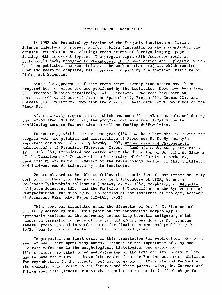
REMARKS ON THE TRANSLATION
In 1958 the Parasitology Section of the Virginia Institute of Marine Science undertook to prepare and/or publish (depending on who accomplished the original translation and editing) translations of foreign language papers dealing with important topics. The program began with Professor Boris E. Bychowsky's book, Monogenetic Trematodes, Their Systematics and Phylogeny, which had been published the year before. The work on that project, which required over two years to complete, was supported in part by the American Institute of Biological Sciences.
Since the appearance of that translation, twenty-five others have been prepared here or elsewhere and published by the Institute. Most have been from the extensive Russian parasitological literature. The rest have been on parasites (4) or fishes (1) from the Spanish (1), French (1), German (2), and Chinese (1) literature. Two from the Russian, dealt with larval molluscs of the Black Sea.
After an early rigorous start which saw some 24 tranlations released during the period from 1961 to 1971, the program lost momentum, largely due to conflicting demands for our time as well as funding difficulties.
Fortunately, within the current year (1981) we have been able to revive the program with the printing and distribution of Professor B. E. Bychowsky's important early work (B. E. Bychowsky, 1937, Ontogenesis and Phytogenetic Relationships of Parasitic Flatworms, Izvest. Acadamia Nauk, SSSR, Ser. Biol. IV: 1353-1383, translated and edited under the direction of Dr. John E. Simmons of the Department of Zoology of the University of California at Berkeley, re-edited by Mr. David E. Zwerner of the Parasitology Section of this Institute, and laid-out and distributed by this Institute.
We are pleased to be able to follow the translation of that important early work with another from the parasitological literature of USSR, by one of Professor Bychowsky's colleagues (Ivanov, A. v., 1952, Morphology of Udonella caligorum Johnston, 1835, and the Position of Udonellidae in the Systematics of Platyhelminths, Parasitological Collection of the Institute of Zoology, Academy of Sciences, USSR, XIV, Pages 112-163, 1952).
This, too, was translated under the direction of Dr. J. E. Simmons and initially edited by him. This paper on the comparative morphology and systematic position of the extremely interesting Udonella caligorum, which occurs on parasitic copepods of the caligid group, was done by Dr. Simmons several years ago and forwarded to us for final treatment and publishing in 1972. Due to various problems, it had to be laid aside.
In preparing the final draft of this translation for publication, Mr. D. E. Zwerner and I have spent many hours. Because of the importance of easy and accurate reference to the morphological, histological and cytological illustrations, so vital to an understanding of the text and its thesis we have had to have the figures redrawn (the copies from the Russian were not sufficient for reproduction in the translation) and to carefully translate and reconcile the symbols, which refer to the figures and their parts. Also, Mr. Zwerner and I have re-edited (several times) the translation to put it in final shape for
ii
publication. This has been a considerable undertaking. Hopefully, all of this effort has produced a published translation which will be of use in the continuing research efforts of the pathobiological (or parasitological) community and of other invertebrate specialists. We and the Institute offer it in this vein.
A key to the abbreviations used in the figures and in the text to refer to anatomical features is provided at the end of the translation.
As editors of the final version, Mr. Zwerner and I are indebted to our typists, Mrs. Mary Fetzer, Mrs. Marcia Hargis, the VIMS Report Center and photographers who assisted in the work. We also wish to thank Ms. Marti German and Mrs. Sylvia Motley who did the printing.
William J. Hargis, Jr. Professor of Marine Science
and David E. Zwerner Assistant Marine Scientist
iii
FOREWORD
Professor A. v. Ivanov's study of Udonella caligorium is unquestionably the most thorough, intensive - and important - ever made of this interesting parasitic flatworm. The body of the work is a very detailed description of the morphology of Udonella with many important and original observations, for example - those of the peculiar and unique nature of the excretory system. Professor Ivanov points out several times the areas in which his observations are limited, and it would be expected that further studies, particularly those making use of such well-developed methods as histochemistry and, perhaps in some cases, result in a modified interpretation. Despite these limitations, Professor Ivanov's study is an essential reference of departure for those planning further research.
Of greatest interest to the editor, and perhaps to many other helminthologists as well, is the marshalling of the descriptive information in order to make a point by point comparison with, particularly, monogeneans and temnocephalans in order to assess the probable affinities of Udonella. It has always been curious that Udonella has for so long been allied with Monogenea almost solely on the basis of general body form and ectocommensalistic (or ectoparasitic, if such it proves to be) mode of life, rather slighting the fact that no oncomiracidium is produced and that development of Udonella, indeed, is remarkably similar to that of many turbellarians. As Ivanov rightly points out, the possibility of convergent similarities resulting from almost identical modes of existence should always be considered in evaluating phylogenetic significance. In the editor 1 s opinion, Professor Ivanov has seized upon critically important features - ontogenesis, lack of chitinoid accessories, and the morphology of the excretory system, in concluding that Udonella is not closely allied with monogeneans.
With regard to the smaller, enigmatic groups of parasitic flatworms, is there reason not to conceive that substantial radiation occurred in the past and that we are left with isolated remnants of a once more diverse fauna - with the tips of the branches, so to speak? To those who, with Miss Hyman, "abhor this raising the ranks" and therefore find the concept of a class Udonelloidea an extreme disposition, it would seem that the only reasonable alternative would be to consider Udonella a very specialized and highly aberrant turbellarian, most closely akin, perhaps, to the rhabdocoeloid, Temnocephala. Certainly, more detailed comparisons should be made with the Scutariellidae.
Marie A. Kassatkin provided the editor with a magnificent translation. He, in turn, consulted Serge Kassatkin for points of clarification. However, the responsibility for any misinterpretations must fall upon the editors. The transliteration scheme of the u.s. Department of Commerce, National Bureau of Standards, Joint Publications Research Service was used, but the editor has altered several names, e.g. Bychowsky, Dogie!, to the more familiar form.
J. E. Simmons
iv
PARASITOLOGICAL COLLECTION OF THE INSTITUTE OF ZOOLOGY, ACADEMY OF SCIENCES, USSR
XIV, Pages 112-163
MORPHOLOGY OF UDONELLA CALIGORUM JOHNSTON, 1835, AND THE POSITION OF UDONELLIDAE IN THE SYSTEMATICS OF PLATYHELMINTHS
By A. v. Ivanov
Institute of Zoology, Academy of Sciences, USSR
Initial Editing by John E. Simmons1
Final Editing and Reconciliation by William J. Hargis, Jr.2 and David E. Zwerner
Translation by Maria A. Kassatkin and Serge Kassatkin3
1952
The platyhelminth, Udonella, lives on parasitic copepod crustaceans and, according to the present system of classification belongs to mongenetic trematodes (Monogena) among which it is usually placed in the group Monopisthocotylea (Fuhrmann, 1928; Bychowsky, 1937; Dawes, 1946; Sproston, 1946). However, the morphology of Udonella has not yet been studied thoroughly by anyone, and a number of unusual features of the structure, ontogenesis and biology of this form cause doubts with regard to its belonging to the Monogena.
Such special characteristics of Udonella which distinguish this form from all other flukes include: 1) the absence of chitinoid hooks on the posterior organ of attachment; 2) the absence of ciliated larvae and metamorphosis; 3) nonparasitic, commensal mode of life which resembles that of the Temnocephala.
Taking all this into consideration, B. E. Bychowsky, who had studied the Monogena for many years, permitted me to use specimens of Udonella collected by him for my morphological studies in order to re-examine the position of this unusual worm in the system. In my work, I frequently made use of the valuable suggestions of v. A. Dogie! and B. E. Bychowsky.
1 Department of Zoology, University of California, Berkeley, California.
2 School of Marine Science and Virginia Institute of Marine Science, College of William and Mary in Virginia, Gloucester Point, Virginia.
3 Lecturer in Slavic Languages and Literature, University of California, Berkeley, California.
1
Haterials and Hethods
The worms which I have studied undoubtedly belong to the species Udonella claigorum Johnston, 1835, which has been known for a long time. All of the material, consisting of several dozens of worms of various ages, was collected in 1946 by B. E. Bychowsky on the southwestern shore of Sakhalin. For fixation he used the fluids of Zenker (with formalin), Bouin, Carnoy and Bend*, as well as mercuric chloride with acetic acid and alcohol.
The study of morphology was done by me on sections stained with ferric hematoxylin, Hansen's hematoxylin, by the Azan method (according to Heidenhain) and according to Mallory. The method of graphic reconstruction was used in many instances.
Taxonomic Remarks
Udonella caligorum is, apparently, the only definite species of this genus. Other species described at various times (Van Beneden and Hesse, 1863; Honticelli, 1889, and others) are synonyms of u. caligorum (Dawes, 1946; Sproston, 1946).
However, Echinella Beneden and Hesse, 1863, Pteronella Beneden and Hesse, 1863, and Calinella Monicelli, 1910, are also sometimes included in the family Udonellidae in addition to Udonella (Braun, 1879-1893; Fuhrmann, 1928). All of these forms live on parasitic copepods (Caligus and Alebion). Their morphology has not yet been studied, it is still not clear how proper it is to isolate them as independent genera.
Habits, Hosts and Geographic Distribution
Almost all of the worms were fixed together with their hosts on which they retained their normal situation. In all instances they were found only on the females of two species of Caligidae, namely: Lepeophtheirus parviventris Wilson, 1905, and L. kareii Yamaguti, 1936. The first host was always removed off the cod (Gadus-morhaua macrocephalus), and the second- off the plaice (Leopsetta obscura).
Adult Udonella caligorum, as a rule, adhere with their organs of attachment to the ovisac of the host, usually on the ventral side of the anterior third. Young immature animals were also found there. Only in isolated instances were adult worms found on the body of a crustacean. For example, only on two female Lepeoptheirus were three adult worms discovered on the ventral surface of the genital segment and only once was an adult worm found on the area lateralis. In contrast, young worms which had recently hatched from eggs were usually localized on the shell of a crustacean at various points, but also on the ventral side. Numerous eggs, sometimes in thick clusters, were always attached to the ventral surface of the genital segment of the host. They occurred extremely rarely in other places around that area (for example, at the posterior edge of the area lateralis, on the main segments of the IV pair of the peraepods or on the base of the ovisacs).
* Transliterated from Russian.
2
Usually, several worms of various ages live on a single crustacean; sometimes, however, greater numbers are present. For example, I counted 36 worms of various ages on one Lepeophtheirus parviventris and 41 worms on one L. kareii, not counting those that had just hatched. Worm-infested crustaceans very often carried numerous Vorticellidae (Peritricha) on their cephalothoraces. Thus, the characteristic location for the adult and middle-aged Udonella caligorum is the anterior part of the ventral side of the ovisacs. Apparently, worms hatching from the eggs are quite agile. At first they remain on the genital segment next to the egg mass, and some crawl to other parts of the host's body, but later they too concentrate on the ovisacs. Further, it is characteristic that their eggs are always deposited on the genital segment of the host. According to Sproston (1946), this indicates a long period of development of Udonella in the egg which is, probably, longer than the development period of the host's eggs.
Since I do not have my own observations on the biology of the worms, I can only cite here the scanty information available in the literature. Sproston (1946) observed the feeding habits of Udonella and showed that the worm eats the mucus secreted by the fish which its host (Caligtis) parasitizes, and picks up pieces of the fish epithelium- remains of the crustacean's food. The customary location of the adult animals on the host is, evidently, connected with the nature of their diet. Caligus eats the mucus and cutaneous epithelium of the fish, scraping it with its cephalothoracic extremities. Small pieces of the epithelium are unavoidably thrown back into the space between the ventral side of the crustacean and the body surface of the fish (Russel, 1925). Always being located on the abdominal side and on the posterior part of the host, Udonella has the most favorable conditions for gathering its food (Sproston, 1946).
Udonella resembles a leech in its movements. Crawling, they alternately attach themselves to the substratum with their anterior glandular depressions and suckers (Sproston, 1946). However, according to B. E. Bychowsky, who observed the behavior of living worms, adult worms are capable of crawling in this manner only if they are artificially detached from the substratum. Usually they remain in the same place and attach themselves so tightly that it is very difficult to detach them without injuring them.
The problem of how a new host becomes infested is not discussed in literature at all. In the absence of a free-swimming larval stage in their development, infestation can, evidently, occur only by direct contact with the hosts. In this connection, the observations by Dawes (1946) are of interest. He states that the hatching of Udonella from the eggs takes place simultaneously with the hatching of the host's larvae. If this is so, it can be imagined that the young Udonella manage to attach themselves to the larvae of the crustacean, accomplishing in this manner, the distribution of the species. On the other hand, Sproston (1946) found mature worms and their egg masses not only on the females, but also on free-swimming young males of Caligus labracis and c. centrodonti. This points to a possibility of the transmission of worms from one host to another during the period of their mating as well. Finally, such transmission is also possible during a casual contact of the Caligus crawling on the fish.
How Udonella behaves during the molting of the host is completely unknown.
According to the observations of U. I. Polyansky, Udonella, just as its host, Caligus, does not occur in winter in the Murmanskaya Oblast.
3
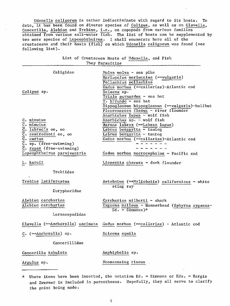
Udonella caligorum is rather indiscriminate with regard to its hosts. To date, it has been found on diverse species of Caligus, as well as on Clavella, Cancerilla, Alebion and Trebius, i.e., on copepods from various families obtained from various salt-water fish. The list of hosts can be supplemented by two more species of Lepeophtheirus. I shall enumerate here all of the crustaceans and their hosts (fish) on which Udonella caligorum was found (see following list).
List of Crustacean Hosts of Udonella, and Fish They Parasitize
Caligus sp.
c. minutus c. minutus
Caligidae
c. labracis oo, oo c. centrodonti oo, oo c. curtus c. sp. (free-swimming) c. rapax (free-swimming) Lepeophtheirus parviventris
L. kareii
Trebiidae
Trebius latifurcatus
Euryphoridae
Alebion carchariae Alebion carchariae
Lernaeopodidae
Holva molva - sea pike Merluccius merluccius (==vulgaris) Pollachius pollachius Gadus morhua (==callarias)-Atlantic cod Sciaena sp. Trigla gurnardus - sea bat T. hirundo - sea bat Hippoglossus hippoglossus (==vulgaris)-halibut Pleuronectes fleaus - river flounder Anarhicha.s lupus - wolf fish Anarhichas sp. - wolf fish Marone labrax (==Labrax lupus) Labrus berggylta - tautog Labrus berggylta - tautog Gadus morhlla (==callarias)-Atlantic cod
Gadus morhua macrocephalus - Pacific cod
Liopsetta obscura - dark flounder
Aetobatus (==Myliobatis) californicus - white sting ray
Carcharias milberti - shark Zygaena malleus - Hammerhead (Sphyrna zygaena
Ed. = Simmons)*
Clavella (==Anchorella) uncinata Gadus morhua (==callarias) - Atlantic cod
c. (==Anchorella) sp. Sciaena aquila
Cancerillidae
Cancerilla tabulata Amphipholis sp.
Argulus sp. Neomaensisg riseus
* Where items have been inserted, the notation Ed. =Simmons or Eds. =Hargis and Zwerner is included in parentheses. llopefully, they all serve to clarify the point being made.
4
Furthermore, Udo~ella caligorum is also characterized by an extremely wide geographical distribution. This species is known from the North Sea, the English Channel, and the Atlantic waters of Europe and North America, as well as from the Mediterranean. According to verbal communication by u. I. Polyansky, Udonella caligorum is common in the Barents Sea in the vicinity of the Hurmansk Biological Station of the Academy of Sciences, USSR. In the Pacific Ocean, this form had been found so far only in its eastern portion, near the shores of California. However, recently, it has also been found by B. E. Bychowsky in the western Pacific - in the Sea of Japan and along the shores of the Southern Kuril Islands. Thus, Udonella caligorum seems to have an interrupted area of distribution. However, this impression may be wrong because of a lack of knowledge of its distribution in most seas of the Northern Hemisphere.
External Morphology
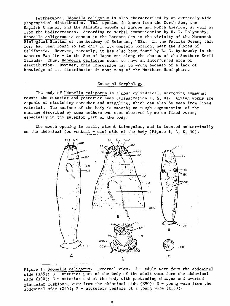
The body of Udonella caligorum is almost cylindrical, narrowing somewhat toward the anterior and posterior ends (Illustration 1, A, D). Livinr; worms are capable of stretching somewhat and wriggling, which can also be seen from fixed material. The surface of the body is smooth; no rough segmentation of the surface described by some authors was ever observed by me on fixed worms, especially in the anterior part of the body.
The mouth opening is small, almost triangular, and is located subterminally on the abdominal (or ventral- eds) side of the body (Figure 1, A, B, MO).
GR MD AGD
•·.·
Q
~EO A
Figure 1. Udonella caligorum. External view. A- adult worm form the abdominal side (X45); B- anterior part of the body of the adult worm form the abdominal side (X90); C - anterior end of the body w·ith protruding pharynx and everted glandular cushions, view from the abdominal side (X90); D- young worm from the abdominal side (X45); E- excretory vesicle of a young worm (X150).
5
In front of it is a small groove-like depression (Figures 1, B, GR) and a small terminal depression (MD). Very often the anterior end of the pharynx can be seen through the mouth opening (Figure 1, A, B, PAE).
Somewhat in front of the mouth, and to its sides, there are glandular suckler-like depressions. They are of a regular oval shape and directed forward and ventro-laterally (Figure 1, A, B, AGC). They are slightly larger than the mouth opening. The delicate edges of the depressions are fimbriated and are equipped with 9-10 small protruding papillae (Figure 1, B SP) which seem to be sensitive. On the bottom of the depressions, frequently we could clearly observe glandular attachment cushions (GCU) of the head glands (see page 5).
Eyes are absent, just as are the special sensitive suckers at the anterior end of the body mentioned by Price (1938).
The body terminates posteriorly with a large terminal sucker-like adhesive disc which is clearly delimited from the trunk (Figure 1, A, D, AOP). The edges of the disc are very thin and give an impression of being webbed; its concave adhesive surface is absolutely smooth and lacks septa. The diameter of the disc is approximately equal to the width of the middle part of the body. Extremely characteristic is the absence of chitinoid equipment on this structure. Normally, the worm adheres securely to the egg sac of the host by means of the concave surface of the disc.
Usually, some of the internal organs can be seen through the integument. The egg-shaped pharynx is the most noticeable. It is located closer to the ventral surface of the body directly behind the mouth (Figure 1, A, B, D, PH). Somewhat in back of the pharynx, also on the abdominal side, and medially, we can see a clear outline of a large ellipsoid egg with a slender filament-like stem at the posterior pole (Figure 1, A, B, CE) in many mature worms. Its position is determined by the movements of the uterus within which it is located; the anterior end is shifted somewhat to the right and the posterior to the left. In young, immature worms numerous follicles of the yolk gland (Figure 1, D, VR) and vesicles of the excretory system are also frequently transparent. The latter are rather large and spherical, and are located in the anterior third part of the body along the sides, but are slightly dorsad (Figure 1, D, EV). In young worms which have been fixed, they stand out in the form of dark spots; on each spot we see a whitish external excretory opening, or nephropore, displaced somewhat posteriorly (Figure 1, E, EO). These openings are also noticeable in the bodies of large mature worms because they are located on the tops of tiny lateral protuberances (Figure 1, A, B, EO).
The genital pore, in the form of a small transverse slit, is located medially posterior to the pharynx (Figure 1, B, GO).
Some authors (Sproston, 1946) observed that Udonella is capable of protruding and exposing its pharynx. However, they did not explain how this was done. In the few instances when an animal was fixed with a projecting pharynx, it could be seen that considerable part of this organ wa's exposed (Figure 1, C, PH). Under these circumstances, the edges of the mouth opening through which the pharynx protrudes are greatly stretched (MO). The front edge of the pharynx, which is directed forward and somewhat ventrally, expands and assumes the shape of a disc (PD).
6
It appears that at the anterior edge of the pharynx there is an annular fold, something like a pharyngeal lip, whose edges are turned in over the pharyngeal mouth when the pharynx is withdrawn (Figure 7, PD), and straightened out in the form of a disc when the pharynx protrudes forward. It is possible that the edges of the disc are capable of moving and probably serve for capturing particles of food.
In the center of the disc of the protruding pharynx we find a small pharyngeal mouth stretched in the medial plane (Figure 1, C, PM). The edges of the disc are equipped with 22 delicate papillae which resemble those along the edges of the glandular adhesive depressions and probably also have a sensory function (SP). Numerous very small papillae are seen on the disc surface arranged around the pharyngeal mouth in regular radial rows.
According to B. E. Bychowsky, the coloration of live worms is brownish.
The length of fixed animals does not exceed 2.7 mm, and the maximum width is 0.6 mm. The diameter of the adhesive organ reaches 0.58 mm, and the length of the pharynx - 0.3 mm.
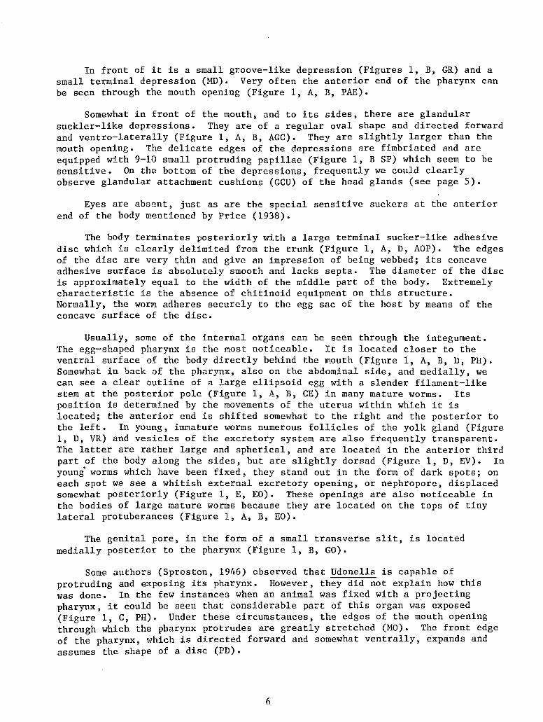
Immediately after emerging from the eggs, young worms are about 0.65 mm long. In appearance they closely resemble adult worms (Figure 2). Their cuticle is strongly cuticularized, just as is that of adult worms. There are no traces of cilia on the epithelium. Through the walls of the body we can see the pharnyx, the intestine, ducts of the head glands, cement glands of the adhesive disc, excretory vesicles and gonads (Figure 2). The adhesive organ, just as in adult worms, has no chitinoid hooks,at all. Thus, Udonella does not have the ciliated larval stage which is so characteristic for all r1onogena. In this respect it is very like the Temnocephala whose emergent or hatching young resemble the adult worm, i.e., there is no metamorphosis.
Figure 2. Udonella caligorum. Young worm at the moment of its emergence from an egg. Sketched by B. E. Bychowsky from a live worm (X73).
The general body shape of Udonella is not much different from that of some Monogena, among which, however, dorsoventrally-flattened shapes predominate. In its appearance, Udonella resembles, at first glance, a monogenetic trematode with a stretched trunk and rounded, sucker-like adhesive disc, for example, a
7
representative of the Monocotylidae (Monocotyle, Heterocotyle~ Tritestis, Loimos, and others).
On the other hand, our worm also resembles some of the Temnocephala in appearance. The latter, however, are characterized by a more-or-less flattened body terminated by a ventral sucker-like organ, and equipped with digitiform tentacles numbering from two (2) to twelve (12). However, Didymorchis have no tentacles, while Scutariella, Monodiscus and Caridinicola, have only a single pair of small papillose tentacles at the anterior of the body. Because of this, the representatives of the first three (3) genera have a great external (superficial? - eds.) resemblance to Udonella. To a lesser degree this may be said of Caridinicola, which- in place of an unpaired sucker-like disc, has a pair of adhesive depressions at its posterior end.
Integuments
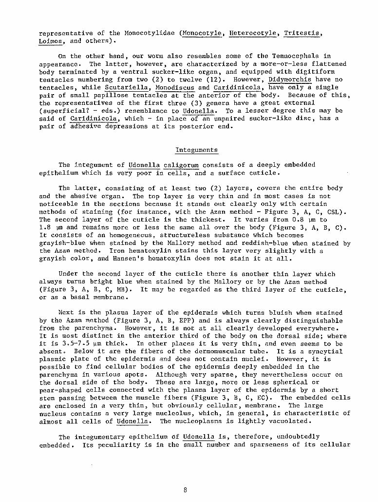
The integument of Udonella caligorum consists of a deeply embedded epithelium which is very poor in cell~nd a surface cuticle.
The latter, consisting of at least two (2) layers, covers the entire body and the ahesive organ. The top layer is very thin and in most cases is not noticeable in the sections because it stands out clearly only with certain methods of staining (for instance, with the Azan method- Figure 3, A, C, CSL). The second layer of the cuticle is the thickest. It varies from 0.8 ].nn to 1.8 ~and remains more or less the same all over the body (Figure 3, A, B, C). It consists of an homogeneous, structureless substance which becomes grayish-blue when stained by the Mallory method and reddish-blue when stained by the Azan method. Iron hematoxylin stains this layer very slightly with a grayish color, and Hansen's hematoxylin does not stain it at all.
Under the second layer of the cuticle there is another thin layer which always turns bright blue when stained by the Mallory or by the Azan method (Figure 3, A, B, C, MB). It may be regarded as the third layer of the cuticle, or as a basal membrane.
Next is the plasma layer of the epidermis which turns bluish when stained by the Azan method (Figure 3, A, B, EPP) and is always clearly distinguishable from the parenchyma. However, it is not at all clearly developed everywhere. It is most distinct in the anterior third of the body on the dorsal side; where it is 3.5-7.5 ~m thick. In other places it is very thin, and even seems to be absent. Below it are the fibers of the dermomuscular tube. It is a syncytial plasmic plate of the epidermis and does not contain nuclei. However, it is possible to find cellular bodies of the epidermis deeply embedded in the parenchyma in various spots. Although very sparse, they nevertheless occur on the dorsal side of the body. These are large, more or less spherical or pear-shaped cells connected with the plasma layer of the epidermis by a short stem passing between the muscle fibers (Figure 3, B, C, EC). The embedded cells are enclosed in a very thin, but obviously cellular, membrane. The large nucleus contains a very large nucleolus, which, in general, is characteristic of almost all cells of Udonella. The nucleoplasma is lightly vacuolated.
The integumentary epithelium ~f Udonella is, therefore, undoubtedly embedded. Its peculiarity is in the small number and sparseness of its cellular
8
bodies. On its surface it forms a well-developed cuticle of at least two layers.
The glands of ectodermal origin (head glands and cement glands of the adhesive organ) will be discussed below in the section on adhesive organs.
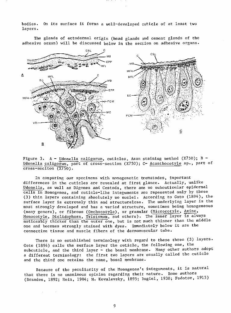
Figure 3. A- Udonella caligorum, cuticles, Azan staining method (X750); B -Udonella caligorum, part of cross-section (X750); C- Acanthocotyle sp., part of cross-seciton (X750).
In comparing our specimens with monogenetic trematodes, important differences in the cuticles are revealed at first glance. Actually, unlike Udonella, as well as Digenea and Cestoda, there are no subcuticular epidermal cells in Monogenea, and cuticle-like integuments are repesented only by three (3) thin layers containing absolutely no nuclei. According to Goto (1894), the surface layer is extremely thin and structureless. The underlying layer is the most strongly developed and has a varied structure, sometimes being homogeneous (many genera), or fibrous (Onchocotyle), or granular (Microcotyle, Axine, Monocotyle, Diclidophora, Tristomum, and others). The inner layer is always noticeably thicker than the outer one, but is not much thinner than the middle one and becomes strongly stained with dyes. Immediately below it are the connective tissue and muscle fibers of the dermomuscular tube.
There is no established terminology with regard to these three (3) layers. Goto (1894) calls the surface layer the cuticle, the following one, the subcuticle, and the third layer - the basal membrane. Many other authors adopt a different terminology: the first two layers are usually called the cuticle and the third one retains the name, basal membrane.
Because of the peculiarity of the Monogenea's integuments, it is natural that there is no unanimous opinion regarding theit nature. Some authors (Brandes, 1892; Rein, 1904; M. Kovalevsky, 1895; Dogie!, 1938; Fedotov, 1915)
9
consider them to be a true cuticle formed by the epithelial cells located in the peripheral layer of the parenchyma. Some others (Braun, 1879-1893; Fuhrmann, 1928; Goto, 1894) do not recognize the presence of any plasmic formations or embedded cells which could be considered as elements of an embedded epithelium under the basal membrane. In accordance with this, it is believed that the cellular epithelial layer in the Monogenea is transformed completely into a cuticle without the formation of integuments of the embedded type (Monticelli, 1893; Fuhrmann, 1928). Finally, there has even been an opinion that the integuments of monogenetic trematodes are represented only by the basal membrane and that, consequently, the epidermis in them is, generally, absent in the adult state (Pratt, 1909; Schneider, 1873; and others).
I feel that the first point of view is correct. I am convinced of this because of the structure of the integuments in the Acanthocotyle sp., which I had the opportunity to study through the kindness of B. E. Bychowsky, using his preparations. Acanthocotyle is a typical representative of monogenetic trematodes which has not been adequately studied histologically. The structure of its integument proved to be extremely interesting and different from that of other Monogenea. They are so primitive that there is absolutely no doubt in interpreting the nature of the integuments of the Monogenea. I shall now describe them. On the dorsal side of the animal there is a two-layered cuticle (Figure 3, C) outside. Next, is a very thin basal membrane under which are the fibers of the dermomuscular tube (MFA, MFL). The surface layer of the cuticle (CSL) is very thin and structureless; because of its ability to be stained strongly by hematoxylin, it is clearly distinguishable from the following, much more substantial light-colored layer (C). The basal membrane is very thin but clearly noticeable (MB).
We have no difficulty in recognizing the usual elements of the typical integuments of the Monogenea in all these layers. But an exceptional peculiarity of Acanthocotyle is the very obvious embedded epithelial cells (EC). They are arranged in a rather thick row in the peripheral layer of the parenchyma which penetrates among them down to the muscle layers (PCM). These are comparatively large, elongated or bulb-like cells with clear boundaries which are connected with the cuticle by their stems. Their inner edges are rounded; they contain rounded nuclei. The height of all embedded cells is not the same; the largest ones are twice as large as the smallest ones.
Brinkmann (1940) observed these cells in Acanthocotyle, also on the dorsal side of the body, but limited himself to a remark that the opinions of the authors on the nature of such "subcuticular cells" do not coincide.
Thus, the integument of Acanthocotyle possesses all the special characteristics of a classical embedded epithelium.
Evidently, this trematode, unlike other Monogenea, has still retained the primitive nature of its integument, which makes it possible to envision the origin of typical integument of monogenetic trematodes.
It becomes absolutely clear that the surface integumentary layers of other Monogenea, which have been studied in this respect, are true cuticular formations and are definitely not metamorphosed cellular epithelia. We can be sure that both of the upper layers are the elements of a true cuticle and the underlying layer is a basement membrane. Other interpretations are hardly
10
possible. In any case, there is not doubt that the original and more primitive state of the integument of the Monogenea was a true embedded epithelium approximately in the same form as we find it in Acanthocotyle. Evidently, in a great majority of the Monogenea, the embedded cells of the epidermis disappeared again. Unicellular cutaneous glands in the peripheral layer of the parenchyma described in some forms are probably what is left of them. On the basis of these considerations, we should compare Udonella with the Monogenea.
In Udonella, the cuticle consists of the same layers as in mongenetic trematodes. In both cases, on the outside we see a strongly-stained, very thin layer under which there is a thicker one which is stained more lightly. In both cases, these two layers are followed by a third one which is probably a basement membrane. But the resemblance is limited just to this, because the typical integuments of the Monogenea have no traces of any embedded cells. Therefore, it would be possible to consider that the integuments of Udonella and Monogenea are basically not comparable. However, this conclusion is not- supported by the structure of the integument in Acanthocotyle in which we see a typical epithelium which is still embedded, although it is limited to the dorsal side of the body. It does not differ essentially from the epithelium of Udonella - only in the great number of the embedded cells.
Evidently, our form, just as Acanthocotyle, is in a more primitive state through which a great majority of monogenetic trematodes have already passed and retained only the cuticle from the embedded epithelium.
In others words, in the structure of this integument, Udonella differs sharply from most of the Monogenea, but, probably, is similar to their closest precursors. The difference is much greater between Udonella and Tremnocephala. The latter have a simple and, apparently, syncytial epithelium which usually forms a single-layered cuticle at its surface which rests on the basement membrane. Sometimes, considerable areas of it retain the ciliated envelope (Didymorchis, Temnocephala dendi, T. minor). Embedded rhabdite glands secreting typical rhabdoids are connected with the epithelium (Bresslau and Reisinger, 1933; Baer, 1931).
Thus, in the structure of its integument, Udonella is much closer to the Monogenea that to Temnocephala in spite of its mode of life, which is similar to the latter.
Musculature
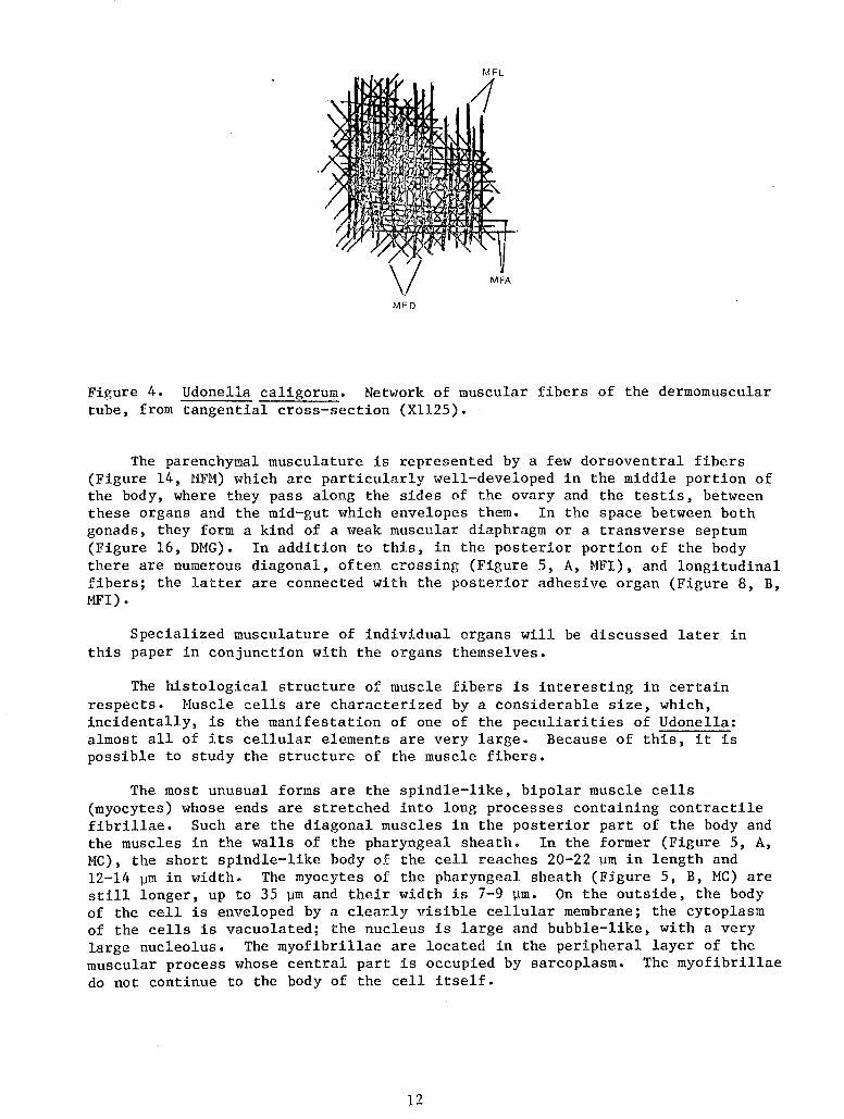
The dermomuscular tube is weakly developed but presents a picture typical of platyhelminths. It is formed by the usual three layers: the external annual layer, the middle diagonal layer and the inner longitudinal layer. The weakest of them is the annular layer consisting of comparatively sparse fibers which are always arranged in one row (Figure 4, MFA). The fibers of the diagonal layer are of the same thickness (MFD) and the fibers of the longitudinal layer are somewhat thicker (HFL). In the posterior half of the body, the longitudinal layer consists of several additional rows of fibers. However, in general, it is noticeable that the dermomuscular tube is developed very weakly.
11
MFL
1
MFA
MFD
Figure 4. Udonella caligorum. Network of muscular fibers of the dermomuscular tube, from tangential cross-section (X1125).
The parenchymal musculature is represented by a few dorsoventral fibers (Figure 14, MFM) which are particularly well-developed in the middle portion of the body, where they pass along the sides of the ovary and the testis, between these organs and the mid-gut which envelopes them. In the space between both gonads, they form a kind of a weak muscular diaphragm or a transverse septum (Figure 16, DMG). In addition to this, in the posterior portion of the body there are numerous diagonal, often crossing (Figure 5, A, MFI), and longitudinal fibers; the latter are connected with the posterior adhesive organ (Figure 8, B, MFI).
Specialized musculature of individual organs will be discussed later in this paper in conjunction with the organs themselves.
The histological structure of muscle fibers is interesting in certain respects. Muscle cells are characterized by a considerable size, which, incidentally, is the manifestation of one of the peculiarities of Udonella: almost all of its cellular elements are very large. Because of this, it is possible to study the structure of the muscle fibers.
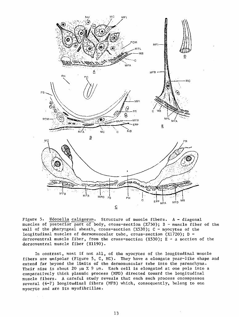
The most unusual forms are the spindle-like, bipolar muscle cells (myocytes) whose ends are stretched into long processes containing contractile fibrillae. Such are the diagonal muscles in the posterior part of the body and the muscles in the walls of the pharyngeal sheath. In the former (Figure 5, A, MC), the short spindle-like body of the cell reaches 20-22 ~m in length and 12-14 ~m in width. The myocytes of the pharyngeal sheath (Figure 5, B, MC) are still longer, up to 35 ~m and their width is 7-9 ~m. On the outside, the body of the cell is enveloped by a clearly visible cellular membrane; the cytoplasm of the cells is vacuolated; the nucleus is large and bubble-like, with a very large nucleolus. The myofibrillae are located in the peripheral layer of the muscular process whose central part is occupied by sarcoplasm. The myofibrillae do not continue to the body of the cell itself.
12
D
EPP MFA
c
Figure 5. Udonella caligorum. Structure of muscle fibers. A- diagonal muscles of posterior part of body, cross-section (X750); B -muscle fiber of the wall of the pharyngeal sheath, cross-section (X530); C - myocytes of the longitudinal muscles of dermomuscular tube, cross-section (Xl720); D -dorsoventral muscle fiber, from the cross-section (X530); E- a section of the dorsoventral muscle fiber (Xll90).
In contrast, most if not all, of the myocytes of the longitudinal muscle fibers are unipolar (Figure 5, C, MC). They have a elongate pear-like shape and extend far beyond the limits of the dermomuscular tube into the parenchyma. Their size is about 20 ~m X 9 ~m. Each cell is elongated at one pole into a comparatively thick plasmic process (MPD) directed toward the longitudinal muscle fibers. A careful study reveals that each such process encompasses several (6-7) longitudinal fibers (MFB) which, consequently, belong to one myocyte and are its myofibrillae.
13
I was unable to examine the myocytes of the diagonal and annular muscles.
The dorsoventral muscles have a different appearance. These are strong fibers crossing the entire body in which the myofibrillae form an external jacket and the central part is occupied by weakly staining homogeneous sarcoplasm (Figure 5, D). Approaching the integument, the fiber widens gradually forming an elongated cone whose base is attached to the skin musculature, and which is covered by a mantle of myofibrillae (~WB) diverging in the distal direction.
When individual areas of such a widened cone are cut in section, it is possible to see clearly the distribution of the rather coarse myofibrillae (Figure 5, E). In cross-section they have an elongated oval shape and, consequently, are ribbon-like. In the peripheral layer of the fiber they are always arranged in a single row, tightly adhering to each other in the narrow part of the fibers. Upon reaching the longitudinal layer of the skin musculature, the distal ends of the myofibrillae gradually thin out and disappear. The cone-like widening of the fiber represents its myocyte (Figure 5, D, MC); here is sarcoplasm in which a large nucleus with a small mucleolus is contained.
A remarkable characteristic of Udonella's musculature is the great stability of its cellular composition. I did not have an opportunity to compare exactly the number of muscular cells in various worms. But being very large and comparatively few in number, they, as can often be seen on exactly oriented cross-secions, are distributed symmetrically, and in equal numbers, on the right and left sides of the body (Figure 5, A, C, HC).
To begin a comparison with other flatworms, we shall mention, first of all, that the position of the layers of the dermomuscular tube coinci.des with that known in the Monogenea and Temnocephala. Certain exceptions, for example the absence of the annular layer in the Hexostoma, Hexabothrium and other Monogenea, are of no signifcance. In general, in all of the cases compared, the position of the layers of the skin musculature fits into the scheme which is usual for the rhabdocoel Turbellaria.
There is only old and scanty information regarding details of structure of the muscular fibers in the Monogenea. In Sphyranura osleri, which has been studied more thoroughly in this respect, the myocytes of the fi.bers of the skin musculature entered deep into the parenchyma (Wright and ~1acCallum, 1887). They have a spindle-like or pear-like shape and are numerous and small. Each of the cells has one process which goes deep into the layers of the dermomascular tube where it connects with an annular longitudinal fiber. These muscular elements, in general, resemble the muscle fibers of the longitudinal musculature of Udonella which have just been described above. In both cases we see fibers of the nematoid type which also occur in the Turbellaria, and particularly often in the Digenea (Bettendorf, 1897).
Information on the muscle fibers of the Temnocephala is even more fragmentary. Definite myocytes are discovered by Baer (1931) who described them as "common for the Platodes." Thin fibrillae project from them and go into the fibers, where they disappear. This description was not accompanied by an illustration and, unfortunately, does not give a clear idea regarding the myocytes. The contractile fiber itself consists of peripheral myofibrillae and a central sarcoplasma.
14
In turbellarians, the muscle fibers are either homogeneous, i.e., they consist of a substance with myofibrillae spread throughout or have-a-cortical fibrillose layer and a central sarcoplasma. Apparently, there are always myocytes which are often represented by a cell located in the parenchyma and connected by processes with one or several contractile fibers. In other instances, the myocyte is reduced to an insignificant plasmic projection containing a nucleus in the fiber itself (Bresslau, 1928-1933). There are transitional stages between these two types of myocytes - the nematoid one, and the one characteristic of the annelids (Bettendorf, 1897).
Both types are also found in Udonella: on the one hand, the myocytes of· the longitudinal skin musculature, and on the other, the myocytes of the fibers of the pharyngeal sheath and dorsoventral muscles.
Thus, we have a definite impression that muscle fibers of our form, just as in the Monogenea, Digenea and, probably, Temnocephala, are within the limits characteristic of the Turbellaria.
Parenchyma
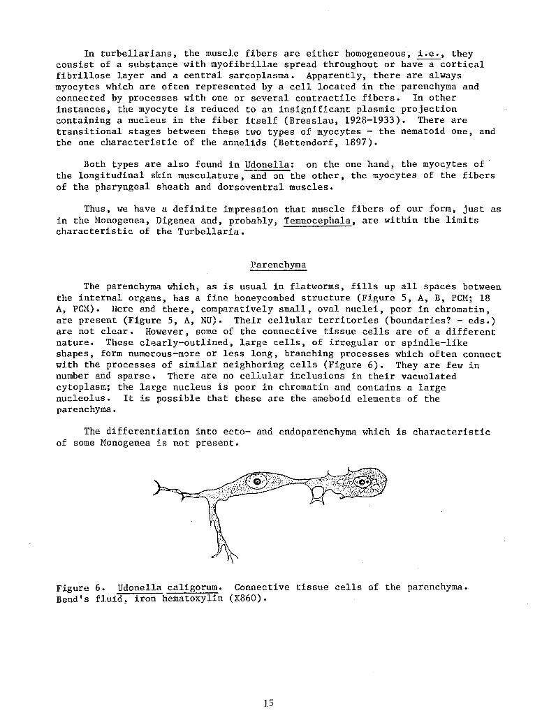
The parenchyma which, as is usual in flatworms, fills up all spaces between the internal organs, has a fine honeycombed structure (Figure 5, A, B, PCM; 18 A, PCM). Here and there, comparatively small, oval nuclei, poor in chromatin, are present (Figure 5, A, NU). Their cellular territories (boundaries? - eds.) are not clear. However, some of the connective tissue cells are of a different nature. These clearly-outlined, large cells, of irregular or spindle-like shapes, form numerous-more or less long, branching processes which often connect with the processes of similar neighboring cells (Figure 6). They are few in number and sparse. There are no cellular inclusions in their vacuolated cytoplasm; the large nucleus is poor in chromatin and contains a large nucleolus. It is possible that these are the ameboid elements of the parenchyma.
The differentiation into ecto- and endoparenchyma which is characteristic of some Monogenea is not present.
Figure 6. Udonella caligorum. Connective tissue cells of the parenchyma. Bend's fluid, iron hematoxylin (X860).
15
Adhesive Organs
The appearance of the anterior adhesive depressions has already been described in this paper. They are connected with powerful clusters of embedded glandular cells and head glands which occupy considerable areas on either side of the pharynx, and even in back of it (Figure 7, GH).
c
Figure 7. Udonella caligorum. Anterior end of body. Frontal section (X250).
Each cluster consists of numerous, pear-shaped unicellular glands whose long ducts lead to the bottom of the adhesive depression. Here, the distal ends of the ducts are arranged close to each other forming a layer which, at first glance, resembles a tall cylindrical epithelium (Figure 7, GCU). However, it does not contain any nuclei, and in reality consists only of the ducts pressed tightly together. In its appearance it somewhat resembles the so-called frontal organ of certain Acoela, which is formed by compressed distal ends of the ducts of frontal glands.
This layer, which represents the bottom of the depression, usually forms tall, thick folds. On its surface there is a thin cuticle which stains strongly with iron hematoxylin and is pierced like a sieve by very fine orifices of the ducts (Figure 7, C).
If we ignore the rather powerful clusters of retractors and protractors (Figure 7, PRO, RET) attached to it, there are no specialized muscles surrounding the depressions. Thus, the depressions are purely glandular formations and do not resemble suckers in any way. Due to the contractions of
16
I
the protractors, the depressions can protrude, at which time the epithelium-like layer everts and transforms into a rounded cushion (Figure 1, C, D, GCU). Evidently by means of these organs the animal is capable of adhering to a substratum with the anterior end of the body. The cushions are pulled back in by means of the above-mentioned retractors.
As has already been pointed out, the edges of the depressions are covered with small papillae which probably have a sensory (tactile) function.
As for the head glands themselves, they do not stain with mucous dyes. Their cytoplasm contains large, irregular vacuoles of secreta (Figure 8, A, SEC) which, evidently, is of a protein nature.
The posterior adhesive organ also cannot be called a sucker despite its disk-like shape and the presence of muscle fibers in it. Its structure is not complicated. On the adhesive surface of the disc there are openings of the ducts of numerous adhesive cement glands whose mass fills up the entire posterior area of the body (Figure 8, B, GC). The glands are large, bulb-shaped or sausage-shaped cells always filled with granular secreta which stains black with iron hematoxylin and becomes bright red when stained by the Azan method or by the Mallory method. The abundant secreta usually obscure the nucleus which lies in the proximal widening of the cell. The gland ducts run parallel to each other (Figure 8, B, GC).
MFRE
Figure 8. Udonella caligorum. A- head glands, cross-section (X750); B -posterior adhesive organ, from the sagittal section (X333).
It is absolutely clear that adhesive function is accomplished exclusively through adhesion by means of the sticky secretion of the glands. This is also supported by the weak development of the musculature of the organ, which excludes the possibility of sucking, and by the absence of any chitinoid hooks or analogous structures.
17
The musculature of the adhesive organ consists of three systems of fibers. The adhesive surface of the organ is covered by a comparatively thick cuticle (Figure 8, B, C) pierced with numerous small pores of the ducts of the cement glands. Under it lie two 'rery weak layers of muscle fibers in the connective tissue, which are a local differentiation of the layers of the dermomuscular tube. The external layer in the central area of the organ is formed by annular fibers (MFEA), and in the peripheral area by radial fibers (MFRE). The arrangement of the fibers of the inner layer is a reverse one - radial fibers in the center (MFRI) and annular ones in the external part (MFIA). Moreover, there are numerous longitudinal muscle fibers connecting the adhesive surface of the organ with the walls of the body in the posterior area of the trunk (MFI). These fibers pass between the glandular cells.
As has already been mentioned, the sucker-like disc of Udonella has absolutely no chitinoid equipment. Price (1938), who observed young animals emerging from the eggs for the first time, remarked on their lack of posterior hooks. According to verbal report by Bychowsky, he made a careful study of the young emerging from the eggs, as well as of the embryos at various stages of development (by crushing the eggs) and found no hooks in any of them. Through observations on my own materials I became convinced that this was true. Thus, it can be considered as proven that the chitinoid accessories are absent at all stages of ontogenesis.
In a discussion of the comparison of the adhesive organs of Udonella with those of other flatworms, I shall mention that the head glands of our form are, undoubtedly, homologous to the frontal glands of many Turbellaria, and to the head glands of the Monogenea, in spite of the existing functional differences. For example, many of the Turbellaria possess a frontal complex of cyanophilic embedded glands (all Acoela, many Rhabdocoela and Alloecoela) which have an attack and defense function. In many Rhabdocoela this complex is represented by pairs of cell clusters which open at the anterior end and secrete formed secreta in the form of rhabdites (Beklemishev, 1937; Bresslau, 1928-1933). In the Temnocephala, in the anterior part of the trunk there are also developed pairs of clusters of unicellular glands which open at the tentacles. Here they play a significant role as cement glands ensuring temporary adhesion of the anterior end of the body (Bresslau and Reisinger, 1933; Pavlovsky, 1937; Baer, 1931). In Caridinicola, they open at the papillose protuberances of the anterior end of the body which resemble very much the anterior adhesive organs of Udonella.
In monogenetic trematodes, in the simplest cases, the anterior adhesive organs are absent and the pairs of the head gland clusters open directly at the anterior end of the body (Monocotylidae, Dactylogyridae). However, most of the Monogenea possess a pair of lateral anterior adhesive organs (Papillose or sucker-like) which are usually called suckers or bothria, depending on the muscular resources and the extent of their separation. In most cases, these organs are connected with complexes of the head glands. The pairs of clusters of typical head glands also occur in the larvae of Monogenea. Characteristic head glands are also present in lycophores - the larvae of Amphilina and Gyrocotyle and in the scolex of pseudophyllidean cestodes. All these structures are correctly homologized by Fuhrmann (1931) with frontal glands of the Turbellaria.
Thus, with respect to the presence as well as the structure, of glandular adhesive organs, Udonella does not differ fundamentally from other flatworms.
18
Head (frontal) glands are a special characteristic of commensal and parasitic Platodes (except Digenea) and are inherited from their turbellarian ancestors.
The posterior adhesive disc of Udonella is of exceptional comparativeanatomic interest. In the rhabdocoele Turbellaria, which are of primary interest to us, this organ is absent. However, it is true that in a number of forms there develop embedded tail cement glands whose secretion ensures temporary adhesion of the posterior end of the body to a substrate. On the contrary, the Temnocephala are characterized by the presence of a well-developed posterior adhesive apparatus. In most forms it :i.s an unpaired, disc-like, more-or-less muscular sometimes stalked organ, shifted somewhat toward the ventral side. It is characterized by muscular deficiency which cannot ensure sucker-like attachment, as well as by strongly developed sement glands, opening at the surface of the organ. Adhesion is achieved by cementing with their· secreta. Chitinoid formations are always absent.
Thus, the posterior adhesive disc of Udonella is similar in all main features to the adhesive organ of the Temnocephala.
The most complete analysis of the posterior adhesive apparatus in the Monogenea from the viewpoint of its evolutionary significance was done by Bychowsky (1937). On the basis of his studies of the larvae, Bychowsky distinguished the primary primitive-type of the adhesive apparatus and justifiably assigned an important phylogenetic significance to it. Thus, the adhesive apparatus of the larvae is represented by two basic forms. One group of the larvae (mostly Monogenea) has from 12 to 16 (more often 14) small marginal hooks of a characteristic structure on their adhesive organ. In the other group of larvae (Octocotylidae, Microcotylidae) 10 marginal hooks of a somewhat different shape develop. Both groups of larvae frequently develop, simultaneously with the marginal hooks or somewhat later, larger paired [one (1)-three (3)] medial hooks (Calceostoma, Nitzschia, Diplorchis, Sphyranura, Octobothrium, Microcotyle, and others).
The primitive form of adhesive apparatus is preserved more or less unchanged in some adult Monogenea such as Protogyrodactylidae, Dactylogyridae and Tetraonchidae (Bychm..rsky, 1937).
Udonella with its sucker-like, glandular, hookless adhesive disc differs essentially from all these forms.
However, as is known, the adhesive apparatus, in most adult Honogenea, varies greatly in its structure and deviates considerably from the primary (or basic - eds.) larval-type. Although the chitinoid equipment is usually preserved, it loses its adhesive significance to a great extent and is replaced functionally by muscular suckers developing on the posterior disc (Polystomidae, Sphyranuridae, Onchocotylidae), by valves (Octocotylidae, Microrotylidae), or by suckers combined with valves (Diclidophoridae). Nothing like this is present in Udonella, which, consequently, differs sharply form these monogenetic trematodes.
Further, in their adult state some Monogenea possess unpaired, sucker-like posterior adhesive discs and in this respect are similar to Udonella, at least at first glance. Thus, in Calceostomidae, Hicrobothriidae, Honocotylidae and Capsalidae the adhesive disc itself grows and changes into a round sucker-like
19
organ. However, basically this type of adhesive apparatus differs little from the primitive state in Protogyrodactylidae, Dactylogyridae and Tetraonchidae because a complete set of the larval hooks is almost always preserved on it. The resemblance (of the Monogenea with sucker-shaped opistohaptors - eds.) to Udonella is superficial, especially because the "sucker" is complicated by radial muscular septa which divide it into a number of depressions or loculi in a number of forms (Monocotylidae, Capsalidae and Enoplocotyle from the Microbothridae).
Finally, we should mention the unusual Acanthocotylidae in which the larval adhesive organ remains in its rudimentary state and a new secondary adhesive disc develops in front of it. The first impression in comparing it with that of Udonella seems to speak in favor of a resemblance, but this again proves to be false. In the Acanthocotylidae (Acanthocotyle), the secondary disc is equipped with radial rows of numerous secondary chitinoid hooks, while the primary disc retains the larval hooks. Thus, comparison of the adhesive posterior apparatus of Udonella and of Monogenea leads to a conclusion that these structures are not comparable.
This conclusion is furter supported if we remember that all the larvae of the Monogenea, without exception, are characterized by unique adhesive discs with a very characteristic set of larval hooks which are absent in the embryos as well as the young of Udonella. According to Sproston (1946), those very few adult Monogenea which have lost their larval chitinoid equipment, such as certain Calceostomidae and Microbothriidae, always possess it in their larval stages.
Digestive Systems
AOP
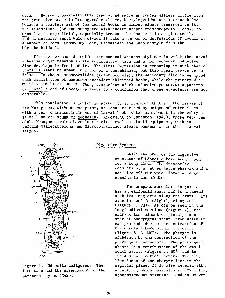
Figure 9. Udonella caligorum. The intestine and the arrangement of the paranephtocytes (X42).
20
Basic features of the digestive apparatus of Udonella have been known for a long time. The instestine consists of a rather large pharynx and a sac-like mid-gut which forms a large opening in the middle.
The compact muscular pharynx has an ellipsoid shape and is arranged with its long axis along the trunk. Its anterior end is slightly elongated (Figure 9, PH). As can be seen in the longitudinal sections (Figure 7), the pharynx lies almost completely in a special pharyngeal sheath from which it can protrude due to the contraction of the muscle fibers within its walls (Figure 5, B, MFI). The pharynx is withdrawn by the contraction of the pharyngeal retractors. The pharyngeal sheath is a continuation of the small mouth cavity (Figure 7, Mel) and is lined with a cuticle layer. The slitlike lumen of the pharynx lies in the sagittal plane; it is also covered with a cuticle, which possesses a very thick, nonhomogeneous structure, and an uneven
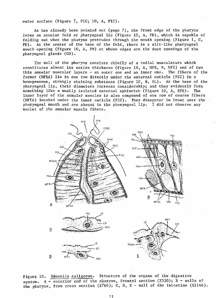
outer surface (Figure 7, PIC; 10, A, PIC).
As has already been pointed out (page 7), the front edge of the pharynx forms an annular fold or pharyngeal lip (Figure 10, A, PD), which is capable of folding out when the pharynx protrudes through the mouth opening (Figure 1, C, PD). At the center of the base of the fold, there is a slit-like pharyngeal mouth opening (Figure 10, A, PM) at whose edges are the duct openings of the pharyngeal glands (GD).
The wall of the pharynx consists chiefly of a radial musculature which constitutes almost its entire thickness (Figure 10, A, MFR, B, MFI) and of two thin annular muscular layers - an outer one and an inner one. The fibers of the former (MFEA) lie in one row directly under the external cuticle (PEC) in a homogeneous, strongly staining substance (Figure 10, B, HL). At the base of the pharyngeal lip, their diameters increase considerably; and they evidently form something like a weakly isolated external sphincter (Figure 10, A, SPH). The inner layer of the annular muscles is also composed of one row of coarse fibers (MFIA) located under the inner cuticle (PIC). They disappear in front near the pharyngeal mouth and are absent in the pharyngeal lip. I did not observe any nuclei of the annular muscle fibers.
PEG
MFI
c
D
Figure 10. Udonella caligorum. Structure of the organs of the digestive system. A- anterior end of the pharynx, frontal section (XS30); B- walls of the pharynx, from cross section (X766); C, D, E- wall of the intestine (X1146).
21
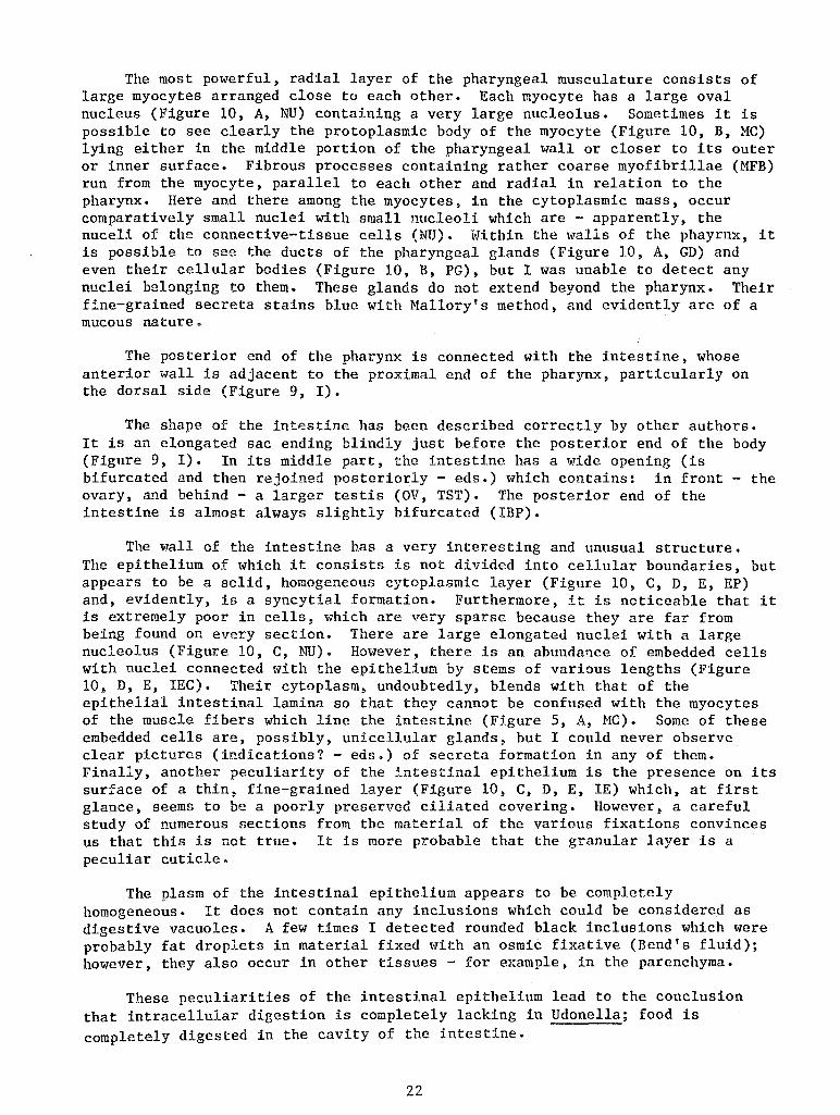
The most powerful, radial layer of the pharyngeal musculature consists of large myocytes arranged close to each other. Each myocyte has a large oval nucleus (Figure 10, A, NU) containing a very large nucleolus. Sometimes it is possible to see clearly the protoplasmic body of the myocyte (Figure 10, B, MC) lying either in the middle portion of the pharyngeal wall or closer to its outer or inner surface. Fibrous processes containing rather coarse myofibrillae (MFB) run from the myocyte, parallel to each other and radial in relation to the pharynx. Here and there among the myocytes, in the cytoplasmic mass, occur comparatively small nuclei with small nucleoli which are - apparently, the nuceli of the connective-tissue cells (NU). Within the walls of the phayrnx, it is possible to see the ducts of the pharyngeal glands (Figure 10, A, GD) and even their cellular bodies (Figure 10, B, PG), but I was unable to detect any nuclei belonging to them. These glands do not extend beyond the pharynx. Their fine-grained secreta stains blue with Mallory's method, and evidently are of a mucous nature.
The posterior end of the pharynx is connected with the intestine, whose anterior wall is adjacent to the proximal end of the pharynx, particularly on the dorsal side (Figure 9, I).
The shape of the intestine has been described correctly by other authors. It is an elongated sac ending blindly just before the posterior end of the body (Figure 9, I). In its middle part, the intestine has a wide opening (is bifurcated and then rejoined posteriorly - eds.) which contains: in front - the ovary, and behind- a larger testis (OV, TST). The posterior end of the intestine is almost always slightly bifurcated (IBP).
The wall of the intestine has a very interesting and unusual structure. The epithelium of which it consists is not divided into cellular boundaries, but appears to be a solid, homogeneous cytoplasmic layer (Figure 10, C, D, E, EP) and, evidently, is a syncytial formation. Furthermore, it is noticeable that it is extremely poor in cells, which are very sparse because they are far from being found on every section. There are large elongated nuclei with a large nucleolus (Figure 10, C, 1ID). However, there is an abundance of embedded cells with nuclei connected with the epithelium by stems of various lengths (Figure 10, D, E, IEC). Their cytoplasm, undoubtedly, blends with that of the epithelial intestinal lamina so that they cannot be confused with the myocytes of the muscle fibers which line the intestine (Figure 5, A, MC). Some of these embedded cells are, possibly, unicellular glands, but I could never observe clear pictures (indications? - eds.) of secreta formation in any of them. Finally, another peculiarity of the intestinal epithelium is the presence on its surface of a thin, fine-grained layer (Figure 10, C, D, E, IE) which, at first glance, seems to be a poorly preserved ciliated covering. However, a careful study of numerous sections from the material of the various fixations convinces us that this is not true. It is more probable that the granular layer is a peculiar cuticle.
The plasm of the intestinal epithelium appears to be completely homogeneous. It does not contain any inclusions which could be considered as digestive vacuoles. A few times I detected rounded black inclusions which were probably fat droplets in material fixed with an osmic fixative (Bend's fluid); however, they also occur in other tissues - for example, in the parenchyma.
These peculiarities of the intestinal epithelium lead to the conclusion that intracellular digestion is completely lacking in Udonella; food is completely digested in the cavity of the intestine.
22
In the lumen of the intestine one can frequently see a homogeneous granular food mass. However, I could never observe any formed elements in it which would make it possible to determine the composition of the food.
In the structure of its pharynx, Udonella is close to the Temnocephala, as well as to the Honogenea. In all cases, this organ is a typical pharynx doliiformis which is also characteristic of the rhabdocoele Turbellaria, Graffilidae and Dalyelliidae. As in Udonella, most of the Temnocephala and many Monogenea have a more-or-less developed pharyngeal sheath which allows the pharynx to be protruded through the mouth opening.
However, there are many differences in the details of its structure. For example, in the Temnocephala it is characterized by a powerful development of the inner annular musculature which frequently forms two powerful sphincters locking the pharynx on both ends. Moreover, there is a longitudinal muscular layer (Baer, 1931; Bresslau and Reisinger, 1933). In our species there is only a .trace of a weakly differentiated anterior sphincter, and the longitudinal muscular fibers are absent.
In typical cases, the Monogenea's pharynx consists, as in our worm, of three muscular layers - internal and external annular layers and a radial layer (Goto, 1894).
Unlike Udonella, the lumen of the pharynx in the Temnocephala and most of the Monogenea is triangular. However, this peculiarity develops independently in various invertebrates in the muscular compartments of the anterior intestine which perform a sucking function (the pharynx of the Nematoda, Hirudinae, Tardigrda, and Pantopoda, the sucking stomach of the Arachnoidea).
However, all of the enumerated differences are of a secondary nature and do not lessen the great fundamental resemblance of Udonella's pharynx, on the one hand, to the pharynx of the Temnocephala and to those of Monogenea and Rhabdocola, on the other.
We must mention another detail which is common to Udonella, Rhabdocoela and Temnocephala. In the latter two, at the apex of the protruding pharynx there often are numerous grasping prickles (Monodiscus), bristles (Scutariella), but more frequently papillae resembling those in the pharynx of Dalyellia and Udonella.
Well-developed pharyngeal glands are as characteristic of the Temnocephala and the Monogenea as they are of the Rhabdocoela. However, in most cases, unlike Udonella, they lie outside of the pharynx and only open into its lumen (the so-called "salivary glands"). However, internal pharyngeal glands occur in the Monogenea and the Rhabdocoela.
The Temnocephala are characterized by a simple intestinal sac, but the intestine of the Honogenea is often somewhat divided. The variety of forms which is observed in the latter is very great. Most of the monogenetic trematodes have t~1o lateral branches which are equipped with rather numerous, more frequently external, branches. However, there occur forms with branches directed medially which join and can produce lateral commissures (Polystomum). In some species, for example in Hicrocotyle reticulata (Microcotylidae), the intestine even has a reticular form. However, it is clear that the divided type of the intestine is of secondary origin. The larvae of the Honogenea always
23
have a simple sac-like intestine which is preserved in many adult, and predominently small, forms (Tetraonchus and Tetraonchoides).
The separation of the intestine into two main branches is, apparently, correlated with the powerful development of the gonads and other organs of the reproductive system in the midsection of the body (Fuhrmann, 1928). Further separation is a result of an overall enlargement of the body and its thickening is due to the necessity for intensification of the distributive function of the intestine and development of numerous dorsoventral muscles. In general, the separation of the intestine in flatworms, as is known, always increases in parallel with the size of the animal (Bresslau, 1928-1933; Beklemishev, 1944). This process occurred repeatedly and independently in the most varied groups (i.e., Polyclada, Triclada, and Crossocoela from Alloecoela, Desmote and Faramacrostomum tricladoides from Rhabdocoela, many Monogenea, Fasciolopsidae, Pronocephalidae and others from Digenea). This is why it is not possible to assign any phylogenetic significance to the differences in the shape of the intestine of Udonella and Monogenea. Having disregarded these differences as insignificant, we can only observe that in the structure of the intestine of Udonella, Temnocephala, and Monogenea, the general features of the structural-type, characteristic of the Rhabdocoela, are most important.
In contrast, there are marked differences in the histology of the intestine. In Temnocephala, the tall columnar epithelium of the intestine closely resembles that of Turbellaria. The ends of cells have no cilia, freely protrude into the lumen of the intestine and, apparently, are capable of energetic ameboid movement and of phagocytizing food particles. Their cytoplasm is usually filled with digestive vascuoles and other inclusions. Between the cells, there occur glandular elements, frequently in the form of embedded cells (Bresslau and Reisinger, 1933; Baer, 1931). This fact indicates the occurrence of lumenal digestion along with intracellular digestion.
According to Goto (1894), mongenetic trematodes have two types of intestinal walls. Some forms (Microcotylidae, Octocotylidae and Diclidophoridae) have no clear uninterrupted epithelium. Individual cells with nuclei are separated from each other by a considerable space and are filled with numerous granules. Other forms (Monocotylidae, Capsalidae, Gyrodactylidae) have an ordinary cuboidal or columnar intestinal epithelium.
Thus, the intestinal epithelium of Udonella is substantially different from that of both groups under comparision. Its unusual structure is the only essential difference in the intestinal apparatus which is exclusively characteristic of Udonella.
Excretory System
I encountered great difficulties in studying the excretory apparatus which is explained not only by the usual difficulties of studying it in sections, but also by certain unusual characteristics of the protonephridial system of Udonella, which do not fit into ordinary morphological and physiolgical schemes.
For this reason I, unfortunately, was unable to clarify completely all the details of the structure of the excretory system. Hm1ever, since it is undoubtedly of great comparative-anatomic i.nterest, I am taking the liberty of publishing the results I did obtain here. Additional studies, particularly on living material, will be necessary to fill in the gaps.
24
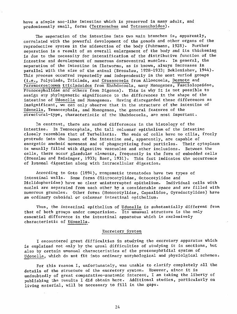
The protonephridial apparatus of Udonella is represented by paired lateral trunks (Figure 11, A, ETA, EPG) whose numerous branches end with terminal cells. The trunks open outside through a pair of urinary vesicles lying laterally in the anterior quarter of the body (EV). However, this usual picture is complicated by the presence of special additional cells and a peculiar development of the canals. Along some of the branches there are special, very unusual, huge cells (PCY). Thus, each of these cells divides the branch into two (2) parts: one - a comparatively short part which connects the cell with the main trunk (PCN) and the other- a very long one (ESC). The latter, apparently, does not have terminal cells on its ramifications, but forms a complex branching system of canals which, finally, open outside independent of the main protonephridial trunks.
The urinary vesicles have a reeular spherical shape and are rather large (Figure 11, A, EV). In adults their diameter reaches 70 ~· On the outside, the vesicle is covered by a thin connective-tissue membrane (Figure 11, B, MB). Its wall consists of vacuolated cytoplasm (EP) and contains only two (2) large nuclei (Figure 11, D, NU), i.e. the entire vesicle is composed of only two (2) cells. Its inner surface is free of cilia. On the ouside of the vesicle there are two (2) - three (3) musculature cells (Figure 11, C, MC) by means of whose contraction the vesicle apparently is empti.ed.
EV
EO
NCC
PCY
A
MFI
c
Figure 11. Udonella caligorum. Organs of the Excretory System. A- scheme of the excretory system (XSO); B, C - excretory vesi.cle, from cross section (X675); D - a section of the excretory vesicle wall with a nucleus (X750); E - excretory opening, from live specimen, illustration by B. E. Bychowsky (XSOO).
25
Each vesicle opens outside through a small nephropore (Figure 11, B, EO) which has no special muscular elements. The nephropores are located on the surface of the body almost laterally, being only slightly positioned dorsally (Figure 11, A, EO).
According to Bychowsky's observations on living worms, the nephropore opens into the vesicle by means of a short funnel-like canal (Figure 11, E, ECO). Sections show that this canal is of a cuticular nature. Namely, it is bounded by the cuticle of the integument which is invaginated at the edges of the nephropore, forming a part of the wall of the excretory vesicle which abuts the nephropore (Figure 11, B, C). Together with the cuticle, the basal membrane (MB1) is also invaginated; the connect:f.ve-tissue membrane of the vesicle (MB) is a continuation of the basal membrane.
The main trunks of the excretory system are represented by two pairs of canals: the comparatively short anterior canals (Figure 11, A, ETA) and the longer posterior canals (EPT). All of them run along the body, occupying a dorso-lateral position. Both pairs turn at the ends in the opposite direction, and their continuations (ETA, EPT) run at some distance parallel to them. I never observed any lateral anastomoses between the right and left canals. In the vicinity of the excretory vesicle, the anterior and posterior trunks on each side of the body join together into a short duct (NCC) which opens into the vesicle.
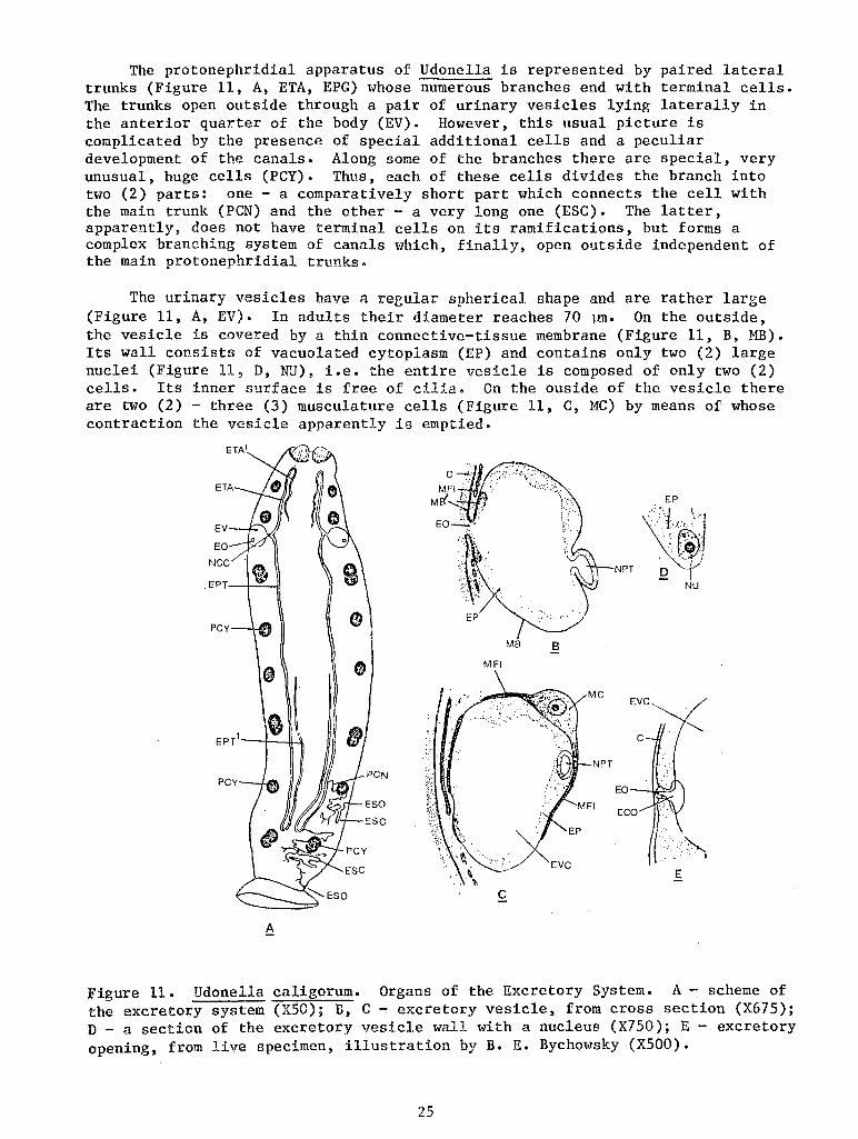
The main trunks are intracellular. Their wall consists of a plasmatic mass (Figure 12, A, PL) which contains large nuclei (NU) and through which runs the canal cavity. The nuclei are sparse and it is possible that their number is constant in each trunk. I could often observe several lumina in the cross-section through the excretory trunk (ET) one of which belonged to the main trunk and others to its branches somewhere in the vicinity. All of them are flanged by a rather thick and clearly delimited layer of tightly packed cytoplasm.
Numerous branches of the main canals run in various directions in the parenchyma. Clarification of their number, position, and the nature of branching was not possible. They are intracellular, just as the main trunks, but their lumina are not bounded by a differentiated layer of cytoplasm (Figure 12, B, ET).
The terminal cells, which I observed many times, probably belong to the smallest cellular elements of Udonella. Their average diameter is about 8-9 m. They are very numerous, which is shown in the schematic Figure 13. The body of the terminal cell has an irregular shape (Figure 12, D, TC) and contains a relatively large oval nucleus (NU) which does not have a nucleolus and is rather rich in chromatin. The tubule on which the cell rests has a very thin, delicate, and structureless wall (ET). The ciliary flame is represented by a long bunch of thin cilia (CC) at the base of which we observe the basal granules blending on the preparation into a strongly-stained strip (CBG).
Now I shall discuss the most unusual and the least understandable of the excretory apparatus.
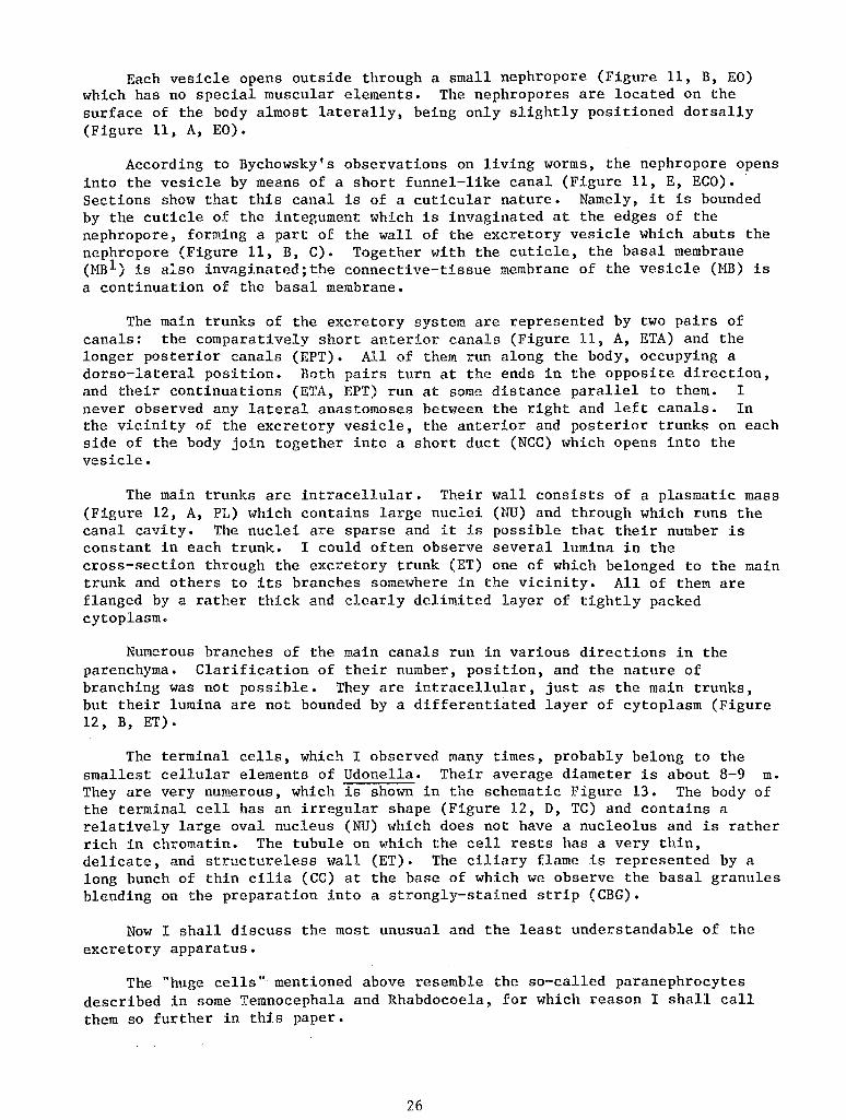
The "huge cells" mentioned above resemble the so-called paranephrocytes described in some Temnocephala and Rhabdocoela, for which reason I shall call them so further in this paper.
26
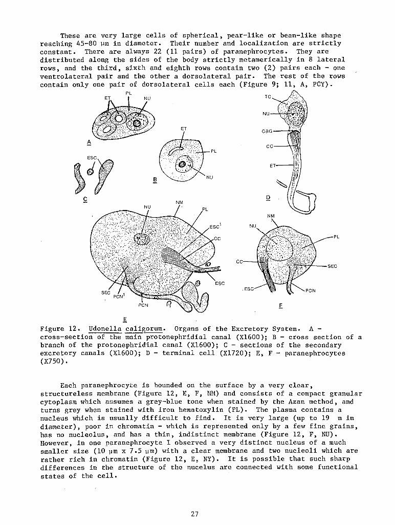
These are very large cells of spherical, pear-like or bean-like shape reaching 45-80 ~m in diameter. Their number and localization are strictly constant. There are always 22 (11 pairs) of paranephrocytes. They are distributed along the sides of the body strictly metamerically in 8 lateral rows, and the third, sixth and eighth rows contain two (2) pairs each- one ventrolateral pair and the other a dorsolateral pair. The rest of the rows contain only one pair of dorsolateral cells each (Figure 9; 11, A, PCY).
PL
ET
A
8
c NM
PL
.~C
f Figure 12. Udone1la caligorum. Organs of the Excretory System. A -cross-section of the main protonephridial canal (X1600); B - cross section of a branch of the protonephridial canal (X1600); C- sections of the secondary excretory canals (X1600); D - terminal cell (X1720); E, F - paranephrocytes (X750).
Each paranephrocyte is bounded on the surface by a very clear, structureless membrane (Figure 12, E, F, tll1) and consists of a compact granular cytoplasm which assumes a grey-blue tone when stained by the Azan method, and turns grey when stained with iron hematoxylin (PL). The plasma contains a nucleus which is usually difficult to find. It is very large (up to 19 min diameter), poor in chromatin- which is represented only by a few fine grains, has no nucleolus, and has a thin, indistinct membrane (Figure 12, F, NU). However, in one paranephrocyte I observed a very distinct nucleus of a much smaller size (10 ~m x 7.5 ~m) with a clear membrane and two nucleoli which are rather rich in chromatin (Figure 12, E, NY). It is possible that such sharp differences in the structure of the nucelus are connected with some functional states of the cell.
27
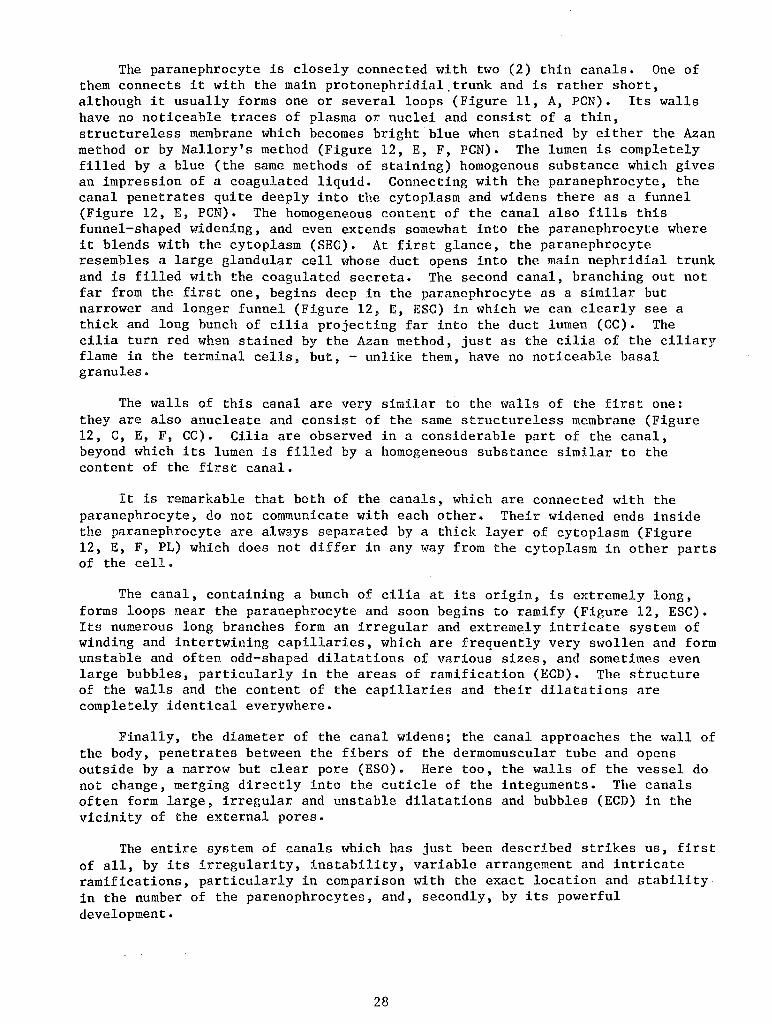
The paranephrocyte is closely connected with two (2) thin canals. One of them connects it with the main protonephridial.trunk and is rather short, although it usually forms one or several loops (Figure 11, A, PCN). Its walls have no noticeable traces of plasma or nuclei and consist of a thin, structureless membrane which becomes bright blue when stained by either the Azan method or by Mallory's method (Figure 12, E, F, PCN). The lumen is completely filled by a blue (the same methods of staining) homogenous substance which gives an impression of a coagulated liquid. Connecting with the paranephrocyte, the canal penetrates quite deeply into the cytoplasm and widens there as a funnel (Figure 12, E, PCN). The homogeneous content of the canal also fills this funnel-shaped widening, and even extends somewhat into the paranephrocyte where it blends with the cytoplasm (SEC). At first glance, the paranephrocyte resembles a large glandular cell whose duct opens into the main nephridial trunk and is filled with the coagulated secreta. The second canal, branching out not far from the first one, begins deep in the paranephrocyte as a similar but narrower and longer funnel (Figure 12, E, ESC) in which we can clearly see a thick and long bunch of cilia projecting far into the duct lumen (CC). The cilia turn red when stained by the Azan method) just as the cilia of the ciliary flame in the terminal cells, but, - unlike them, have no noticeable basal granules.
The walls of this canal are very similar to the walls of the first one: they are also anucleate and consist of the same structureless membrane (Figure 12, C, E, F, CC). Cilia are observed in a considerable part of the canal, beyond which its lumen is filled by a homogeneous substance similar to the content of the first canal.
It is remarkable that both of the canals, which are connected with the paranephrocyte, do not communicate with each other. Their widened ends inside the paranephrocyte are always separated by a thick layer of cytoplasm (Figure 12, E, F, PL) which does not differ in any way from the cytoplasm in other parts of the cell.
The canal, containing a bunch of cilia at its origin, is extremely long, forms loops near the paranephrocyte and soon begins to ramify (Figure 12, ESC). Its numerous long branches form an irregular and extremely intricate system of winding and intertwining capillaries, which are frequently very swollen and form unstable and often odd-shaped dilatations of various sizes, and sometimes even large bubbles, particularly in the areas of ramification (ECD). The structure of the walls and the content of the capillaries and their dilatations are completely identical everyt-1here.
Finally, the diameter of the canal widens; the canal approaches the wall of the body, penetrates between the fibers of the dermomuscular tube and opens outside by a narrow but clear pore (ESO). Here too, the walls of the vessel do not change, merging directly into the cuticle of the integuments. The canals often form large, irregular and unstable dilatations and bubbles (ECD) in the vicinity of the external pores.
The entire system of canals which has just been described strikes us, first of all, by its irregularity, instability, variable arrangement and intricate ramifications, particularly in comparison with the exact location and stability in the number of the parenophrocytes, and, secondly, by its powerful development.
28
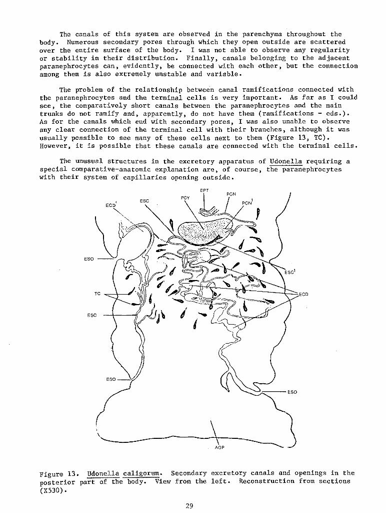
The canals of this system are observed in the parenchyma throughout the body. Numerous secondary pores through which they open outside are scattered over the entire surface of the body. I was not able to observe any regularity or stability in their distribution. Finally, canals belonging to the adjacent paranephrocytes can, evidently, be connected with each other, but the connection among them is also extremely unstable and variable.
The problem of the relationship between canal ramifications connected with the paranephrocytes and the terminal cells is very important. As far as I could see, the comparatively short canals between the paranephrocytes and the main trunks do not ramify and, apparently, do not have them (ramifications- eds.). As for the canals which end with secondary pores, I was also unable to observe any clear connection of the terminal cell with their branches, although it was usually possible to see many of these cells next to them (Figure 13, TC). However, it is possible that these canals are connected with the terminal cells.
The unusual structures in the excretory apparatus of Udonella requiring a special comparative-anatomic explanation are, of course, the paranephrocytes with their system of capillaries opening outside.
Figure 13. Udonella caligorum. Secondary excretory canals and openings in the posterior part of the body. View from the left. Reconstruction from sections (X530).
29
Special large cells which are, undoubtedly, connected with the protonephridial canals have been described in various groups of flatworms. The most complete experimental study of these cells on living worms was done by Reisinger (1922-1923) on the Rhabdocoela, who gave them the name of paranephrocytes. In Kalyptorhynchia (Gyratrix hermaphroditus, Polycystis goettei), they are precisely localized huge cells which appear to be threaded onto the excretory canal which they envelop from all sides. They are characterized by a very large nucleus containing a large nucleolus. Similar paranephrocytes closely resembling them were also discoverd in the Typhloplanidae and Dalyelliidae and are, probably, widespread in other Rhabdocoela.
It is interesting that sometimes (for example, in Polycystis goettei) the canal narrows considerably while passing through the cell, as if it is compressed by it. Its continuation after the emergence from the paranephrocyte produces twigs- capillaries- which end with terminal cells (Reisinger, 1923). As was shown by Reisinger using the intravital staining method, paranephrocytes of the Rhabdocoela are true athrocytes, i.e. excretory cells which extract the excretions from the surrounding parenchyma and excrete them into the protonephridial canal.
The paranephrocytes of Udonella reveal a certain general morphologic resemblance to those of the Rhabdocoela, for example to polycystis, but display substantial differences: firstly, in the nonvacuolated cytoplasm; secondly, in the fact that they completely separate the protonephridial canal into two noncommunicating parts; and, thirdly, in the presence of a powerful bunch of cilia.
Large cells closely connected with the excretory system and, undoubtedly, corresponding to the paranephrocytes of the Rhabdocoela are also known in the Temnocephala (Haswell, 1893; Merton, 1914; Baer, 1929-1931). However, they have been inadequately studied and there is much disagreement regarding them. Merton (1914) is inclined to assume that these cells have a large number of ciliary bunches arranged radially around the nucleus and compares the paranephrocyte of the Temnocephala with the terminal cell of Amphilina which, as is known, is equipped with several ciliary bunches. Baer (1929, 1931) even considers that the paranephrocytes of the Temnocephala are not cells at all but only local swellings of the excretory canal with additional radial tubules in the wall, and states that there is no nucleus in them. Merton's viewpoint seems more correct to me; a nucleus is clearly visible in the paranephrocytes in his illustrations (1914, Table III, Illustration 34).
Each paranephrocyte is enveloped in a thin membrane and is connected with the excretory canal which, possibly, pierces right through it (Merton, 1914; Bresslau and Reisinger, 1933). The constancy in the distribution and number of the paranephrocytes in Temnocephala is an interesting peculiarity \-lhich is shared by Udonella; there are about 20 of them in the former, i.e. the same number as in Udonella.
Finally, cells which resemble the paranephrocytes of Temnocephala somewhat have also been described in the monogenetic trematode, Sphyranura osleri (Wright and MacCallum, 1887). They are very large, with a radial structure of the cytoplasm and are connected at one pole to the excretory canal. However, the nature of these cells is not quite clear and they, probably, are not athrocytes. Evidently, true paranephrocytes are not characteristic of Monogenea.
30
Thus, the paranephrocytes of Udonella differ rather sharply from these cells in Rhabdocoela and Temnocephala, but we have no doubt of their general homology with the latter. It is possible that they retain the excretory functions which have been proven for the paranephrocytes of the Turbellaria and which are very probable for the paranephrocytes of Temnocephala (Bresslau and Reisinger, 1933). The most primitive initial state of paranephrocytes can justifiably be expected in Rhabdocoela.
Then, how could the unusual peculiarities of the excretory apparatus of Udonella, paradoxical at first glance, arise? So far, this question can be answered only hypothetically.
It is quite natural to assume that the paranephrocytes of Udonella formed from the athrocytes of the turbellarian predessors which were close, for example, to the paranephtocytes of Polycystis. The narrowing of the canal running with the cell which was described in Polycystis was intensified and resulted in a complete blocking of the excretory canal by the cell. However, this became possible only after the protonephridial branch equipped with a paranephrocyte began to communicate with the environment through one of several secondary pores.
As a result of this, the paranephrocytes of Udonella divide the entire excretory apparatus into two physiologically independent parts. One part is represented by the main trunks and those of their branches which have no paranephrocytes. Their terminal cells force the excretions to the main trunks and out through the vesicles. This part of the system should, evidently, also include the canals connecting paranephrocytes with the main trunks, and, in part, the paranephrocytes themselves as will be seen later.
The other independent part is represented by all those branches on which paranephrocytes are located and which, in essence, are isolated from the main trunks. These branches have their own motor apparatus in the form of the ciliary cluster of the paranephrocyte, and, probably, terminal cells, and their own system of ducts and external openings. The excretory fluid flows through these ducts in the direction opposite to the flow in the system of the main trunks and independent of it, moving from the paranephrocytes to the secondary pores. Evidently, the two above-mentioned physiological systems differ not only in the direction of the flow of excretions, but there is also a division of the excretory functions between them, i.e., one of them excretes certain substances, and the other - some other substan~ This is indicated by a completely different structure of the walls of the canals in the first and second systems, as well as by a completely different nature of their contents. The protonephridial trunks and ramifications which are not connected with paranephrocytes have comparatively thick plasma walls, and their lumina appear to be completely empty. The canals running from paranephrocytes, whose walls are membrane-like, are always filled with a characteristic coagulated fluid.
As for the paranephrocytes, their passive role of dividing the two physiological excretory systems is quite clear. However, they do have important functions; first of all - that of the motor function of a terminal cell. In fact, the long ciliary cluster of the paranephrocyte is nothing but a ciliary flame. This ciliary flame is turned away from the main protonephridial vessel and is turned in the direction of the canal system opening through secondary pores. The extremely powerful development of this system requires powerful motor accessories. Evidently, the terminal cells, if they are present, are adequate and the paranephrocyte develops its own ciliary flame in addition to
31
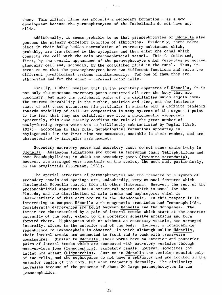
! !
them. This ciliary flame was probably a secondary formation - as a new development because the paranephrocytes of the Turbellaria do not have any cilia.
Additionally, it seems probable to me that paranephrocytes of Udonella also possess the primary excretory function of athrocytes. Evidently, there takes place in their bulky bodies accumulation of excretory substances which, probably, are transformed in the cytoplasm and then enter the canal which connects the cell with the main protonephridial vessel. This is indicated, first, by the overall appearance of the paranephrocyte which resembles an active glandular cell and, secondly, by the coagulated fluid in the canal. Thus, it seems to me that the paranephrocytes have tl~O different functions and serve two different physiological systems simultaneously. For one of them they are athrocytes and for the other - terminal motor cells.
Finally, I shall mention that in the excretory apparatus of Udonella, it is not only the numerous excretory pores scattered all over the body that are secondary, but also the peripheral parts of the capillaries which adjoin them. The extreme instability in the number, position and size, and the intricate shape of all these structures (in particular in animals with a definite tendency towards stability of cellular composition in many systems of the organs!) point to the fact that they are relatively new from a phylogenetic viewpoint. Apparently, this case clearly confirms the rule of the great number of newly-forming organs which was so brilliantly substantiated by Dogie! (1936, 1937). According to this rule, morphological formations appearing in phylogenesis for the first time are numerous, unstable in their number, and are characterized by irregular arrangement.
Secondary excretory pores and excretory ducts do not occur exclusively in Udonella. Analogous formations are known in tapeworms (many Tetraphyllidea and some Pseudophyllidea) in which the secondary pores (foramina secundaria), however, are arranged very regularly on the scolex, the neck and, particularly, on the proglittids (Fuhrmann, 1931).
The special structure of paranephrocytes and the presence of a system of secondary canals and openings are, undoubtedly, very unusual features which distinguish Udonella sharply from all other flatworms. However, the rest of the protonephridial apparatus has a structural scheme which is usual for the Platoda, and the distribution of main trunks and nephropores which is characteristic of this norm occurs in the Rhabdocoela. In this respect it is interesting to compare Udonella with mongenetic trematodes and Temnocephalida. Considerable differences are found between Udonella and the Monogenea. The latter are characterized by a pair of lateral trunks which start at the anterior extremity of the body, extend to the posterior adhesive apparatus and turn forward there. Nephropores with or without an excretory vesicle, are arranged laterally, closer to the anterior end of the body. However, a considerable resemblance to Temnocephala is observed, in which although unlike Udonella, their lateral trunks are connected in front and in back with transverse commissures. Similar to Udonella, these worms have an anterior and posterior pairs of lateral trunks which are connected with excretory vesicles through more-or-less long (Temnocephala), excretory canals; however, sometimes the latter are absent (Didymorchis). Just as in Udonella the vesicles consist only of two cells, and the nephropores do not have a sphincter and are located in the anterior region of the body, but most frequently dorsally. The similarity increases because of the presence of about 20 large paranephrocytes in the
Temnocephalida.
32
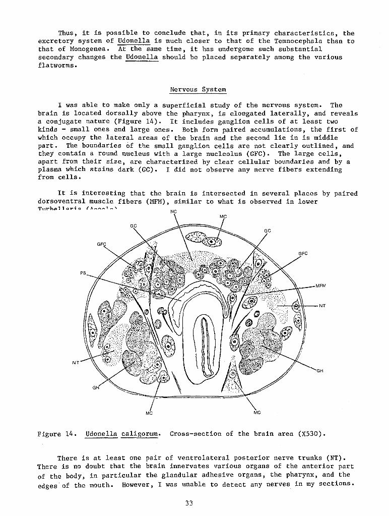
Thus, it is possible to conclude that, in its primary characteristics, the excretory system of Udonella is much closer to that of the Temnocephala than to that of Monogenea. At the same time, it has undergone such substantial secondary changes the Udonella should be placed separately among the various flatworms.
Nervous System
I was able to make only a superficial study of the nervous system. The brain is located dorsally above the pharynx, is elongated laterally, and reveals a conjugate nature (Figure 14). It includes ganglion cells of at least two kinds - small ones and large ones. Both form paired accumulations, the first of which occupy the lateral areas of the brain and the second lie in is middle part. The boundaries of the small ganglion cells are not clearly outlined, and they contain a round nucleus with a large nucleolus (GFC). The large cells, apart from their size, are characterized by clear cellular boundaries and by a plasma which stains dark (GC). I did not observe any nerve fibers extending from cells.
It is interesting that the brain is intersected in several places by paired dorsoventral muscle fibers (MFM), similar to what is observed in lower
NC
NT
Figure 14. Udonella caligorum. Cross-section of the brain area (X530).
There is at least one pair of ventrolateral posterior nerve trunks (NT). There is no doubt that the brain innervates various organs of the anterior part of the body, in particular the glandular adhesive organs, the pharynx, and the edges of the mouth. However, I was unable to detect any nerves in my sections.
33
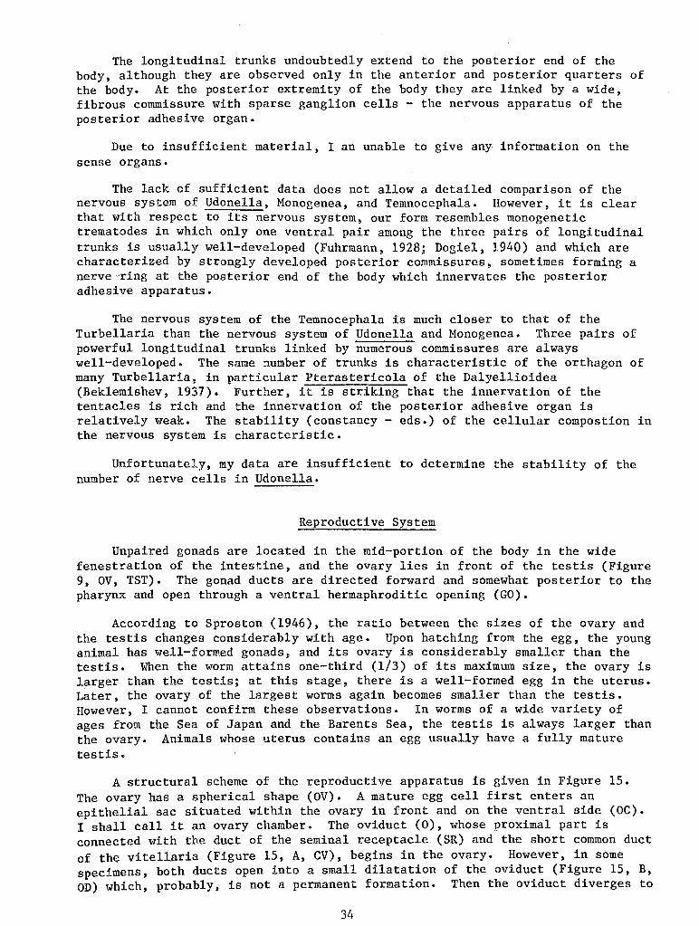
The longitudinal trunks undoubtedly extend to the posterior end of the body, although they are observed only in the anterior and posterior quarters of the body. At the posterior extremity of the body they are linked by a wide, fibrous commissure with sparse ganglion cells - the nervous apparatus of the posterior adhesive organ.
Due to insufficient material, I an unable to give any information on the sense organs.
The lack of sufficient data does not allow a detailed comparison of the nervous system of Udonella, Monogenea, and Temnocephala. However, it is clear that with respect to its nervous system, our form resembles monogenetic trematodes in which only one ventral pair among the three pairs of longitudinal trunks is usually well-developed (Fuhrmann, 1928; Dogiel, 1940) and which are characterized by strongly developed posterior commissures, sometimes forming a nerve ·ring at the posterior end of the body which innervates the posterior adhesive apparatus.
The nervous system of the Temnocephala is much closer to that of the Turbellaria than the nervous system of Udonella and Monogenea. Three pairs of powerful longitudinal trunks linked by numerous commissures are always well-developed. The same number of trunks is characteristic of the orthagon of many Turbellaria, in particular Pterastericola of the Dalyellioidea (Beklemishev, 1937). Further, it is striking that the innervation of the tentacles is rich and the innervation of the posterior adhesive organ is relatively weak. The stability (constancy - eds.) of the cellular compostion in the nervous system is characteristic.
Unfortunately, my data are insufficient to determine the stability of the number of nerve cells in Udonella.
Reproductive System
Unpaired gonads are located in the mid-portion of the body in the wide fenestration of the intestine, and the ovary lies in front of the testis (Figure 9, OV, TST). The gonad ducts are directed forward and somewhat posterior to the pharynx and open through a ventral hermaphroditic opening (GO).
According to Sproston (1946), the ratio between the sizes of the ovary and the testis changes considerably with age. Upon hatching from the egg, the young animal has well-formed gonads, and its ovary is considerably smaller than the testis. When the worm attains one-third (1/3) of its maximum size, the ovary is larger than the testis; at this stage, there is a well-formed egg in the uterus. Later, the ovary of the largest worms again becomes smaller than the testis. However, I cannot confirm these observations. In worms of a wide variety of ages from the Sea of Japan and the Barents Sea, the testis is always larger than the ovary. Animals whose uterus contains an egg usually have a fully mature testis.
A structural scheme of the reproductive apparatus is given in Figure 15. The ovary has a spherical shape (OV). A mature egg cell first enters an epithelial sac situated within the ovary in front and on the ventral side (OC). I shall call it an ovary chamber. The oviduct (0), whose proximal part is connected with the duct of the seminal receptacle (SR) and the short common duct of the vitellaria (Figure 15, A, CV), begins in the ovary. However, in some specimens, both ducts open into a small dilatation of the oviduct (Figure 15, B, OD) which, probably, is not a permanent formation. Then the oviduct diverges to
34
the dorsal side and opens in front of the ovary into a large ootype (OI) which is surrounded by a powerful complex of unicellular glands (OOG, PTG) and continues into the uterus (U). The latter lies in the ventral portion of the body; it is connected by a short duct (UEC) with a small hermaphroditic (or common- eds.) vestibule (atrium genetale commune- AT). Vaginae are absent and there is no genito-intestinal duct (ductus genito-intestinalis).
Vitellaria are represented by numerous lobes which lie in the space between the body wall and the internal organs - the intestine, gonads and gonaducts (Figure 19, VR). They are lacking only in the areas in front of the excretory vesicles and behind the blind end of the intestine. Adjacent lobes of the vitellaria are linked with each other by short connections (VC). A.t the level of the ovary, the vitellaria open into a pair of wide vitelloducts (Figure 15, VD) running to the ventral side where they fuse into the above-mentioned common vitelloduct (Figure 15, A, CV).
The compact testis usually has an ovoid or rounded shape slightly elongated lontigudinally (Figure 9, TST). The seminal duct (Figure 15, SD) begins as a narrow canal on its left side. At first the seminal duct runs forward and ventrally passing between the left vitelloduct and the ovary; then it contiues forward and in the dorsal direction being located above the ootype and, finally, turns to the right. Usually it enlarges here and forms a false seminal vesicle filled with seminal fluid (SV). The seminal duct approaches the eenital vestibule from the dorsal side.
A B
Figure 15. Udonella caligorum. The reproductive system. Reconstructed from sections (Xl33). A- view from the ventral aspect; B- view from the left side.
35
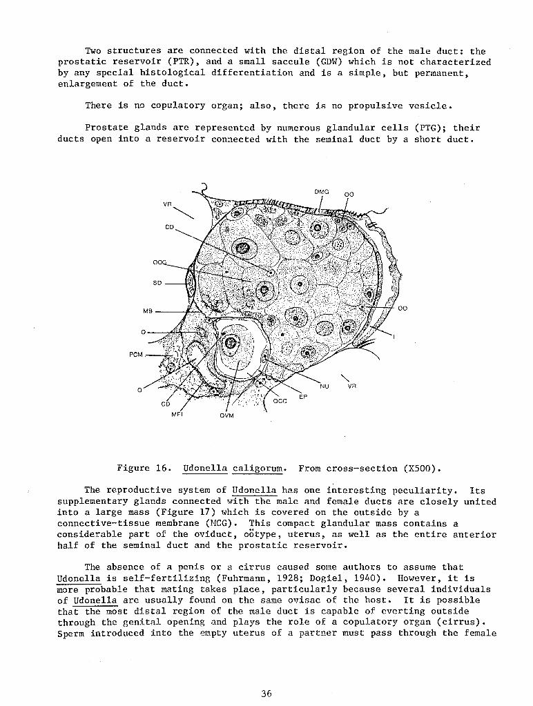
Two structures are connected with the distal region of the male duct: the prostatic reservoir (PTR), and a small saccule (GDW) which is not characterized by any special histological differentiation and is a simple, but permanent, enlargement of the duct.
There is no copulatory organ; also, there is no propulsive vesicle.
Prostate glands are represented by numerous glandular cells (PTG); their ducts open into a reservoir connected with the seminal duct by a short duct.
Figure 16. Udonella caligorum. From cross-section (X500).
The reproductive system of Udonella has one interesting peculiarity. Its supplementary glands connected with the male and female ducts are closely united into a large mass (Figure 17) which is covered on the outside by a connective-tissue membrane (MCG). This compact glandular mass contains a considerable part of the oviduct, ootype, uterus, as well as the entire anterior half of the seminal duct and the prostatic reservoir.
The absence of a penis or a cirrus caused some authors to assume that Udonella is self-fertilizing (Fuhrmann, 1928; Dogiel, 1940). However, it is more probable that mating takes place, particularly because several individuals of Udonella are usually found on the same ovisac of the host. It is possible that the most distal region of the male duct is capable of everting outside through the genital opening and plays the role of a copulatory organ (cirrus). Sperm introduced into the empty uterus of a partner must pass through the female
36
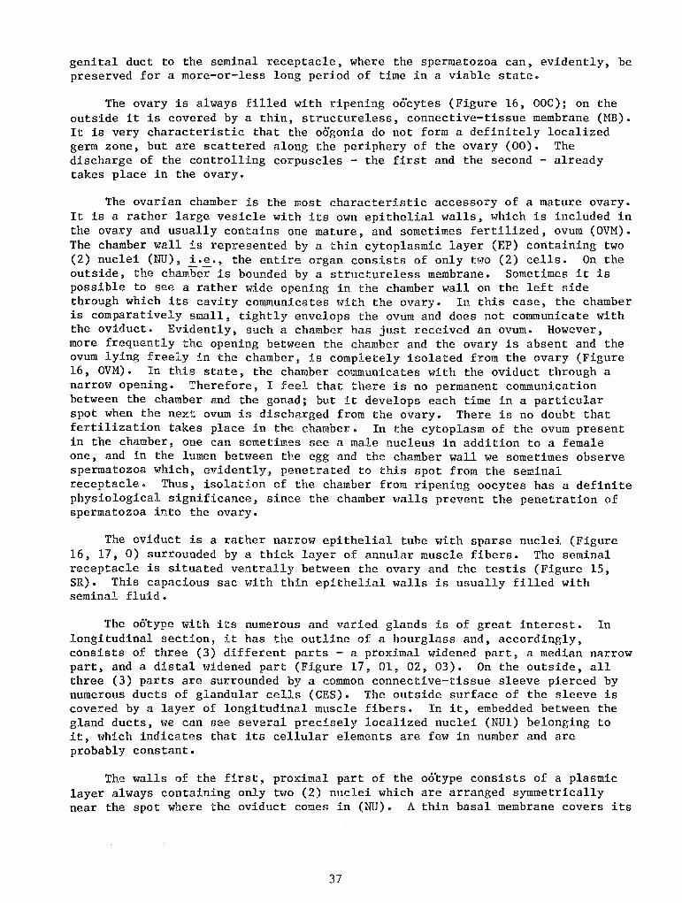
genital duct to the seminal receptacle, where the spermatozoa can, evidently, be preserved for a more-or-less long period of time in a viable state.
The ovary is always filled with ripening oocytes (Figure 16, OOC); on the outside it is covered by a thin, structureless, connective-tissue membrane (MB). It is very characteristic that the oogonia do not form a definitely localized germ zone, but are scattered along the periphery of the ovary (00). The discharge of the controlling corpuscles - the first and the second - already takes place in the ovary.
The ovarian chamber is the most characteristic accessory of a mature ovary. It is a rather large vesicle with its own epithelial walls, which is included in the ovary and usually contains one mature, and sometimes fertilized, ovum (OVM). The chamber wall is represented by a thin cytoplasmic layer (EP) containing two (2) nuclei (NU), i.e., the entire organ consists of only two (2) cells. On the outside, the chamber is bounded by a structureless membrane. Sometimes it is possible to see a rather wide opening in the chamber wall on the left side through which its cavity communicates with the ovary. In this case, the chamber is comparatively small, tightly envelops the ovum and does not communicate with the oviduct. Evidently, such a chamber has just received an ovum. However, more frequently the opening between the chamber and the ovary is absent and the ovum lying freely in the chamber, is completely isolated from the ovary (Figure 16, OVM). In this state, the chamber communicates with the oviduct through a narrow opening. Therefore, I feel that there is no permanent communication between the chamber and the gonad; but it develops each time in a particular spot when the next ovum is discharged from the ovary. There is no doubt that fertilization takes place in the chamber. In the cytoplasm of the ovum present in the chamber, one can sometimes see a male nucleus in addition to a female one, and in the lumen between the egg and the chamber wall we sometimes observe spermatozoa which, evidently, penetrated to this spot from the seminal receptacle. Thus, isolation of the chamber from ripening oocytes has a definite physiological significance, since the chamber walls prevent the penetration of spermatozoa into the ovary.
The oviduct is a rather narrow epithelial tube with sparse nuclei (Figure 16, 17, 0) surrounded by a thick layer of annular muscle fibers. The seminal receptacle is situated ventrally between the ovary and the testis (Figure 15, SR). This capacious sac v7ith thin epithelial walls is usually filled with seminal fluid.
The oo'type with its numerous and varied glands is of great interest. In longitudinal section, it has the outline of a hourglass and, accordingly, consists of three (3) different parts - a proximal widened part, a median narrow part, and a distal widened part (Figure 17, 01, 02, 03). On the outside, all three (3) parts are surrounded by a common connective-tissue sleeve pierced by numerous ducts of glandular cells (CES). The outside surface of the sleeve is covered by a layer of longitudinal muscle fibers. In it, embedded between the gland ducts, we can see several precisely localized nuclei (NUl) belonging to it, which indicates that its cellular elements are few in number and are probably constant.
The walls of the first, proximal part of the o6"type consists of a plasmic layer always containing only two (2) nuclei which are arranged symmetrically near the spot where the oviduct comes in (NU). A thin basal membrane covers its
37
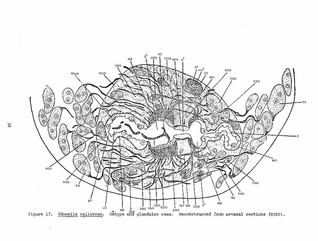
w co
Figure 17. Udonella caligorum. mass.
:,. •'j 1 SG
~ :.- ... -- .. , I.·. "':}!( I :X II:D!f.:.···.~ J 0
from se~eral sections (X350).
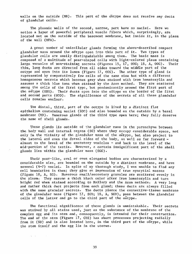
walls on the outside (MB). This part of the ootype does not receive any ducts of glandular cells.
The plasmic walls of the second, narrow, part have no nuclei. Here we notice a layer of powerful peripheral muscle fibers which, surprisingly, are located not on the outside of the basement membrane, but inside it, in the plasm of the wall (MFA).
A great number of unicellular glands forming the above-described compact glandular mass around the o6type open into this part of it. Two types of glandular cells are easily distinguishable among them. The basic mass is composed of a multitude of pear-shaped cells with light-colored plasm containing large vacuoles of non-staining secreta (Figures 15, 17, OOG; 18, A, OOG). Their thin, long ducts are directed from all sides toward the middle part of the ootype and open into its lumen (Figure 17, OOG). The other type of gland is represented by comparatively few cells of the same size but with a different homogeneous secreta which becomes grey when stained with iron hematoxylin and assumes a thick blue tone when stained by the Azan method. They are scattered among the cells of the first type, but predominantly around the first part of the oo"type (OSG). Their ducts open into the ootype on the border of the first and second parts (OOD). The significance of the secreta of all these glandular cells remains unclear.
The distal, third, part of the ootype is lined by a distinct flat epithelium containing nuclei (EOO) and also bounded on the outside by a basal membrane (MB). Numerous glands of the third type open here; they fully deserve the name of shell glands.
These glands lie outside of the glandular mass in the parenchyma between the body wall and internal organs (SG) where they occupy considerable space, not only in the vicinity of the glandular mass of the ootype, but also project to the lateral and even the dorsal sides of the body, as well as far forward -almost to the level of the excretory vesicles - and back to the level of the mid-portion of the testis. However, a certain insignificant part of the shell glands lies within the glandular mass (SGG).
Their pear-like, oval or even elongated bodies are characterized by a considerable size, are bounded on the outside by a distinct membrane, and have several (4-7) nuclei. In spite of my thorough study, I was unable to find any cell boundaries in them; they give an impression of true syncytial masses (Figure 18, A, SG). Numerous small/secretory granules are scattered evenly in the plasm. They assume a thick black color after iron hematoxylin and turn bright red when stained according to Mallory and the Azan methods. A very long and rather thick duct projects from each gland; these ducts are always filled with the same granular secreta. The ducts pierce the connective-tissue membrane of the glandular mass (Figure 17, SGD; 18, A, SGD), pass between the glandular cells of the latter and go to the third part of the ootype.
The functional significance of these glands is unmistakable. Their secreta was stained by all stains used, just as the substance of the membrane of the complex egg and its stem and, consequently, is intended for their construction. The end of the stem (Figure 17, CES) has short processes projecting radially from it (SD) and is also located here, in the third part of the ootype, while the stem itself and the egg lie in the uterus.
39
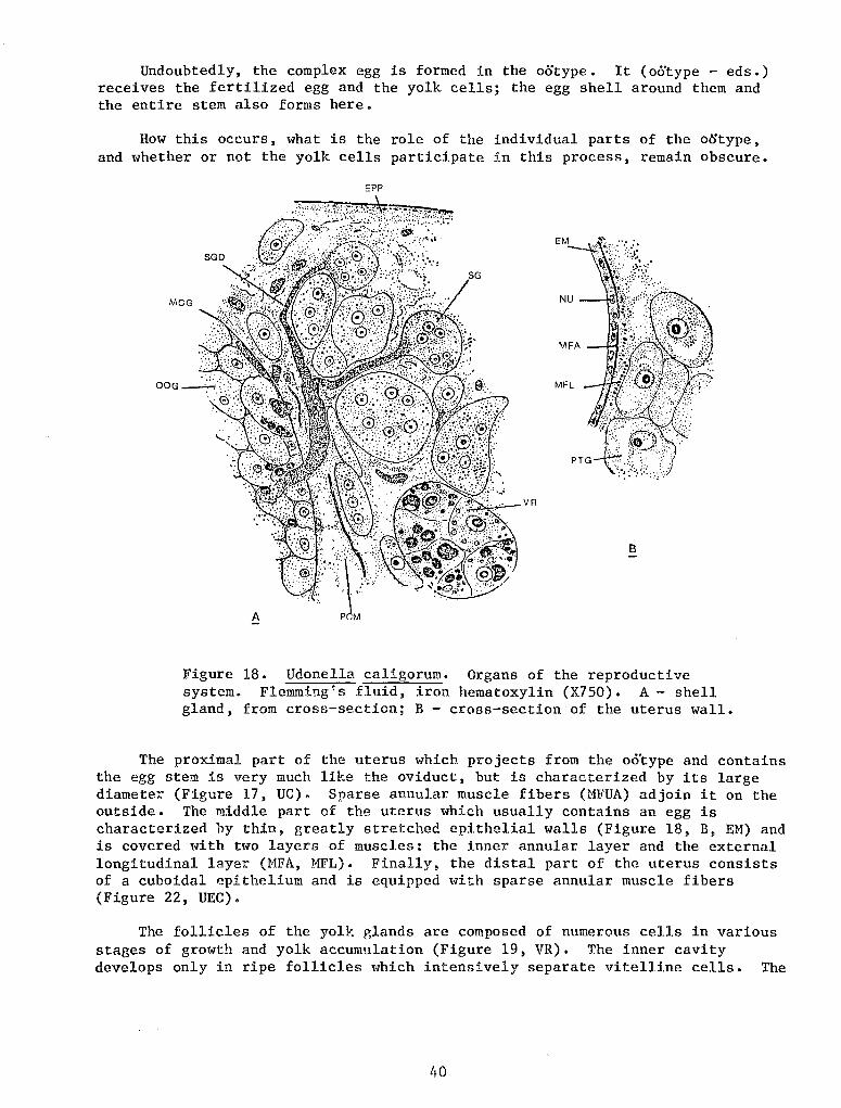
Undoubtedly, the complex egg is formed in the ootype. It ( o6'type - eds.) receives the fertilized egg and the yolk cells; the egg shell around them and the entire stem also forms here.
How this occurs, what is the role of the individual parts of· the ootype, and whether or not the yolk cells participate in this process, remain obscure.
OOG
EPP
B
Figure 18. Udonella caligorum. Organs of the reproductive system. Flemming's fluid, iron hematoxylin (X7SO). A- shell gland, from cross-section; B- cross-section of the uterus wall.
The proximal part of the uterus which projects from the oo'type and contains the egg stem is very much like the oviduct, but is characterized by its large diameter (Figure 17, UC). Sparse annular muscle fibers (MFUA) adjoin it on the outside. The middle part of the uterus which usually contains an egg is characterized by thin, greatly stretched epithelial walls (Figure 18, B, EM) and is covered with two layers of muscles: the inner annular layer and the external longitudinal layer (MFA, MFL). Finally, the distal part of the uterus consists of a cuboidal epithelium and is equipped with sparse annular muscle fibers (Figure 22, UEC).
The follicles of the yolk glands are composed of numerous cells in various stages of growth and yolk accumulation (Figure 19, VR). The inner cavity develops only in ripe follicles which intensively separate vitelline cells. The
40
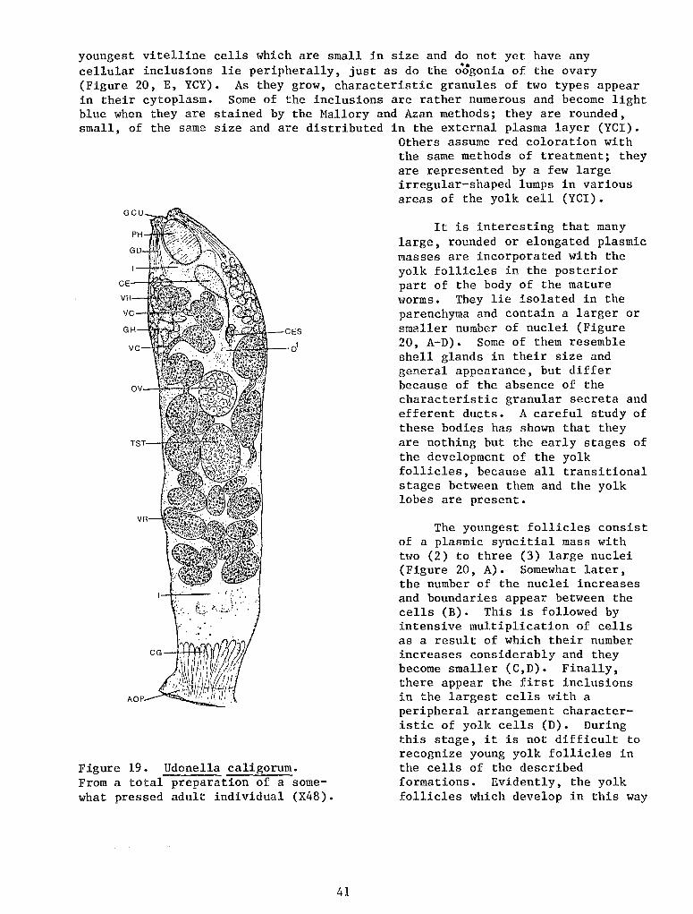
youngest vitelline cells which are small in size and do not yet have any cellular inclusions lie peripherally, just as do the o~gonia of the ovary (Figure 20, E, YCY). As they grow, characteristic granules of two types appear in their cytoplasm. Some of the inclusions are rather numerous and become light blue when they are stained by the Mallory and Azan methods; they are rounded, small, of the same size and are distributed in the external plasma layer (YCI).
Figure 19. Udonella caligorum. From a total preparation of a somewhat pressed adult individual (X48).
41
Others assume red coloration with the same methods of treatment; they are represented by a few large irregular-shaped lumps in various areas of the yolk cell (YCI).
It is interesting that many large, rounded or elongated plasmic masses are incorporated with the yolk follicles in the posterior part of the body of the mature worms. They lie isolated in the parenchyma and contain a larger or smaller number of nuclei (Figure 20, A-D). Some of them resemble shell glands in their size and general appearance, but differ because of the absence of the characteristic granular secreta and efferent ducts. A careful study of these bodies has shown that they are nothing but the early stages of the development of the yolk follicles, because all transitional stages between them and the yolk lobes are present.
The youngest follicles consist of a plasmic syncitial mass with two (2) to three (3) large nuclei (Figure 20, A). Somewhat later, the number of the nuclei increases and boundaries appear between the cells (B). This is followed by intensive multiplication of cells as a result of which their number increases considerably and they become smaller (C,D). Finally, there appear the first inclusions in the largest cells with a peripheral arrangement characteristic of yolk cells (D). During this stage, it is not difficult to recognize young yolk follicles in the cells of the described formations. Evidently, the yolk follicles which develop in this way
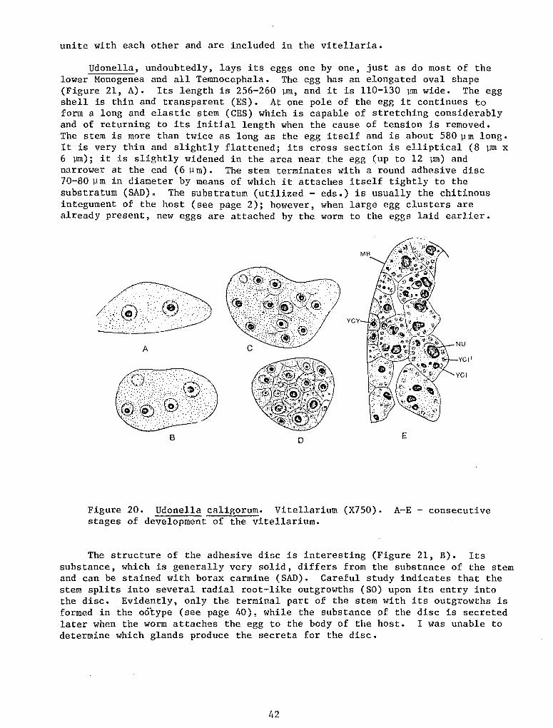
unite with each other and are included in the vitellaria.
Udonella, undoubtedly, lays its eggs one by one, just as do most of the lower Monogenea and all Temnocephala. The egg has an elongated oval shape (Figure 21, A). Its length is 256-260 vm, and it is 110-130 vm wide. The egg shell is thin and transparent (ES). At one pole of the egg it continues to form a long and elastic stem (CES) which is capable of stretching considerably and of returning to its initial length when the cause of tension is removed. The stem is more than twice as long as the egg itself and is about 580 ~m long. It is very thin and slightly flattened; its cross section is elliptical (8 ~ x 6 vm); it is slightly widened in the area near the egg (up to 12 lJI11) and narrower at the end (6 ~m). The stem terminates with a round adhesive disc 70-80 ~ m in diameter by means of which it attaches itself tightly to the substratum (SAD). The substratum (utilized- eds.) is usually the chitinous integument of the host (see page 2); however, when large egg clusters are already present, new eggs are attached by the worm to the eggs laid earlier.
(_:;.:':.®,/'::·.·® ... ··:/·: YCY
.···.:::.:· .. · .. ~"-·---
A
D E
Figure 20. Udonella caligorum. Vitellarium (X750). A-E- consecutive stages of development of the vitellarium.
The structure of the adhesive disc is interesting (Figure 21, B). Its substance, which is generally very solid, differs from the substance of the stem and can be stained with borax carmine (SAD). Careful study indicates that the stem splits into several radial root-like outgrowths (SO) upon its entry into the disc. Evidently, only the terminal part of the stem with its outgrowths is formed in the ootype (see page 40), while the substance of the disc is secreted later when the worm attaches the egg to the body of the host. I was unable to determine which glands produce the secreta for the disc.
42
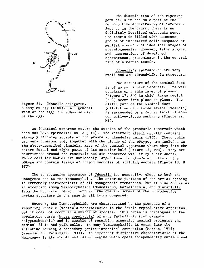
EM
CES
Figure 21. Udonella caligorum. A complex egg (X180). A- general view of the egg; B - adhesive disc of the egg.
The distribution of the ripening germ cells in the male part of the reproductive apparatus is of interest. Just as in the ovary, there is no definitely localized embryonic zone. The testis is filled with numerous groups of intermixed cells composed of genital elements of identical stages of spermiogenesis. However, later stages, and accumuations of developed spermatozoa, predominate in the central part of a mature testis.
Udonella's spermatozoa are very small and are thread-like in structure.
The structure of the seminal duct is of no particular interest. Its wall consists of a thin layer of plasma (Figure 17, SD) in which large nuclei (NU2) occur from place to place. The distal part of the seminal duct (dilatation of a false seminal vesicle) is surrounded by a rather thick fibrous connective-tissue membrane (Figure 22, MF).
An identical membrane covers the outside of the prostatic reservoir which does not have epithelial walls (PTR). The reservoir itself usually contains strongly staining secreta of the prostatic glandular cells (PTG). These cells are very numerous and, together with the glands of the ootype, are included in the above-described glandular mass of the genital apparatus where they form the entire dorsal and right parts of its anterior half (Figure 15, PTG). They are distributed around the reservoir and are connected with it by their ducts. Their cellular bodies are noticeably larger than the glandular cells of the ootype and contain irregular-shaped vacuoles of staining secreta (Figure 18, B, PTG).
The reproductive apparatus of Udonella is, generally, close to both the Monogenea and to the Temnocephala. The anterior position of the atrial opening is extremely characteristic of all monogenetic trematodes, but it also occurs as an exception among Temnocephalida (Monodiscus, Caridinicola, and Scutariella from the Scutariellidae). Further, the overall scheme of the reproductive system structure is the same in all forms compared.
However, the Temnocephalida are characterized by the presence of a resorbing vesicle (vesicula resorbiensis) in the female reproductive apparatus, but it does not occur in a number of species. This organ is homologous to the copulatory bursa (bursa copulatrix) of many Turbellaria (for example Kalyptorhynchia) and is capable of resorbing excessive genital products: the seminal fluid and yolk cells. In many Temnocephalida it opens into the intestine forming a secondary genito-intestinal connection (Merton, 1914; Bresslau and Reisinger, 1933). An important distinctive characteristic of the Monogenea is its simple and paired vagina which opens independently outside and
43
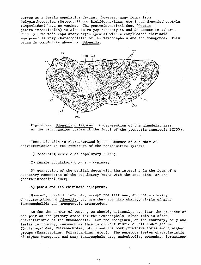
serves as a female copulative device. However, many forms from Polypisthocotylea (Octocotylidae, Diclidophoridae, etc.) and Monopisthocotyla (Capsalidae) have no vagina. The genitointestinal duct (ductus genito-intestinalis) is also in Polyopisthocotylea and is absent in others. Finally, the male copulatory organ (penis) with a complicated chitinoid equipment is very characteristic of the Temnocephala and the Monogenea. This organ is completely absent in Udonella.
PTG
PTG
Figure 22. Udonella caligorum. Cross-section of the glandular mass of the reproduction system at the level of the prostatic reservoir (X750).
Thus, Udonella is characterized by the absence of a number of characteristics in the structure of the reproductive system:
1) resorbing vesicle or copulatory bursa;
2) female copulatory organs - vaginae;
3) connection of the genital ducts with the intestine in the form of a secondary connection of the copulatory bursa with the intestine, or the genito-intestinal duct;
4) penis and its chitinoid equipment.
However, these differences, except the last one, are not exclusive characteristics of Udonella~ because they are also characteristic of many Temnocephalida and monogenetic trematodes.
As for the number of testes, we should, evidently, consider the presence of one pair as the primary state for the Temnocephala, since this is often characteristic of the Rhabdocoela. For the Monogenea, on the contrary, only one testis is primary, inasmuch as this is characteristic of all lower groups (Dactylogyridae, Tetraonchidae, etc.) and the most primitive forms among higher groups (Mazacreoides, Polystomoides, etc.). The numerous testes characteristic of higher Monogenea and many Temnocephala are, undoubtedly, secondary formations
44
through the splitting of one (1) male gonad in the first instance and two (2) male gonads in the second. A single unpaired testis never occurs in the Temnocephalida and its presence in Udonella, evidently, places Udonella closer to the lower Monogenea.
Among the common supplementary organs seen in Monogenea and Temnocephala and characteristic of the reproductive apparatus of Udonella are the seminal receptacle and the prostate glands. These organs are common accessories in monogenetic trematodes and Temnocephalida, where the prostate glands are often represented, just as in Udonella, by a complex of unicellular glands opening into a special reservoir. Thus, the reproductive system of Udonella fits well into the general scheme exhibited by the reproductive apparatus of the Monogenea and Temnocephala, and it is difficult to determine to which of these two groups our species is closer in this respect. However, with respect to the general distribution of the organs it resembles the reproductive apparatus of monogenetic trematodes. At the same time, it is clear that the reproductive system of all groups compared above has all of the main features of the Rhabdocoela, their ancestors.
There are certain pecularities which are characteristic exclusively of Udonella and, therefore, are of special interest to us. Except for the absence of the penis, which was mentioned above, these are: the combination of all supplementary glands of the male and female parts of the system into a sharply isolated glandular mass, formation of the ovary chamber and, finally, the distribution of the oogonia in the ovary.
The ovary chamber is a very unusual formation and we can see only a slight and very remote analogy of this chamber in the initial part of the oviduct in Benedenia melleni (Capsalidae). In this trematode, the oviduct begins deep in its spherical ovary near its center, i.e., its distal part is included in the ovary. Fertilization of the egg cellstakes place in the intraovarian part of the oviduct (Jahn and Kuhn, 1932). However, the resemblance of this structure to the ovary chamber of Udonella is very superficial. The inner section of the oviduct in Benedenia is not isolated into an independent chamber, but continues directly to the extra-ovarian part of the female duct. On the other hand, the inclusion of the seminal receptacle in the ovary indicates a process of an entirely different nature than that which resulted in the formation of the above peculiarities of Benedenia. Thus, the ovichamber of Udonella has no analog either among the Monogenea or among the Temnocephala.
Another interesting characteristic of Udonella is the absence of a localized zone of embryonic cells in the ovary. In the Monogenea and Temnocephala, just as in the Rhabdocoela, oogonia are located separately deep within the ovary, i.e., in a section which is the most removed from the beginning of the oviduct where the embryonic zone of gonads is situated.
Finally, it is also interesting to compare the structural characteristics of the shell of the complex egg. As is known, eggs of monogenetic trematodes are usually equipped with one (1) or two (2) rather long appendages which consist of the same substance as the shell. Unlike Udonella, whose eggs are attached to the substratum extremely tightly, whenever these worms attach their eggs to the gills or skin of the host, they are so loose that they are easily washed off with water. The only exceptions are Nitzschia, Epibdella, and a few other forms which attach their eggs tightly to the substratum on a short pedicle
45
formed by the substance of the egg shell. The eggs of Temnocephala are tightly attached to the body of the host, just as the eggs of our worm, by means of a stem. However, it is always very short and massive, and is not at all an outgrowth of the egg shell but is formed from the secretion of special cement glands which surround the genital opening (Baer, 1931; Bresslau and Reisinger, 1933). Evidently, this egg "stemlet" of the worm are similar to the eggs of monogenetic trematodes with respect to the development of a long outgrowth of the shell, and resemble the eggs of the Temnocephalida because of the presence of the cement substance; consequently, they are an unusual combination of both devices.
On the Position of the Udonella
Before embarking on the solution of the main problem of this work, namely, a general evaluation of Udonella's organization for the purpose of clarifying its systematic position among flatwoms, I cons·ider it advantageous to isolate those common features which are also equally characteristic of.the Temnocephala and the Honogenea and indicate their affinity to rhabdocoele Turbellaria. Such features are:
1) the structure of the dermomuscular tube and the histology of individual muscle fibers;
2) a pharynx of the pharynx doliiformis type;
3) the sac-like intestine;
4) the development of the head glands;
5) the overall type of the reproductive system.
We can also mention the development of paranephrocytes, with reservation, however, because they are not characteristic of the Monogenea and, possibly, do not occur in them at all. All these features, along \'lith many others which are common in all platyhelminths, such as the protonephridial type of excretory system, development of the parenchyma in the body cavity, absence of an anus, etc., contribute nothing to the problem in which we are interested.
In comparing them with other flatworms, we naturally turned to the Monogenea, because up to now almost no one doubted that Udonella belonged to this group and because it actually has much in common with them. It is also fully justifiable to compare it with the Temnocephala, inasmuch as the latter are, in general, very close to the Monogenea and at the same time resemble Udonella in their mode of life.
In evaluating the phylogenetic significance of certain characteristics, we should consider the possibility of convergent similarities caused by identical modes of life. Obviously, such similarities should not be given phylogenetic and, consequently, systematic significance. On the contrary, the organizational characteristics which cannot be considered as convergent devices are of greatest value to us. A detailed comparison with the Monogenea and Temnocephala indicates, first of all, that Udonella displays much more substantial similarities with the latter rather than with the former.
46
A. Comparison with the Monogenea.
It has the following characteristics in common with monogenetic trematodes:
1) overall body shape;
2) structure of the integuments;
3) overall type of the nervous system;
4) anterior position of the genital pore;
5) the presence of an unpaired testis;
6) overall arrangement of the organs of the reproductive system.
The elongated cylindrical shape of the body is characteristic of only a few lower Monogenea and is just as much uncharacteristic of them as of the Temnocephala. Similarity in the structure of the integuments is not a complete one. It has already been mentioned that the integument of Udonella is of the embedded epithelium type, although in a great majority of monogenetic trematodes no traces of the embedded cells are left. However, the mere fact that the integuments exhibit this type of embedded epithelium cannot serve as indisputable proof of a close affinity. The formation of the embedded epithelium is often a result of a parasitic or a commensal mode of life. Thus, for example, two groups of parasitic flatworms, Cestoda and Digenea, which are undoubtedly unrelated (not closely related- eds.), have embedded epithelium. According to Fedotov's data (1915), Protomyzostomum polynephris, a representative of an unusual oligomerous group of the Myzostomida, annelids parasitizing the bursae of the brittle star Gorgonocephalus, has a typical embedded epithelium, the development of which Fedotove correctly attributes to the parasitic mode of life. Further, a common feature in the structure of the nervous systems of Udonella and the Monogenea is the small number of posterior longitudinal nerve trunks. However, this characteristic should be approached with great caution because this similarity by itself cannot be considered decisive, since the Rhabdocoela, from which both the Monogenea and Udonella undoubtedly originated, has only two (2) to three (3) pairs of nerve trunks, among which one ventral pair is predominant. Thus, this characteristic is more likely a feature in common with rhabdocoele Turbellaria and indicates the closeness of the ancestors of the Monogenea and Udonella more than anythinbg else. As for the reproductive system, there is no doubt that features of Monogenea's reproductive system are clear in the overall arrangement of the gonads and the vitellaria, and particularly in the anterior position of the atrial opening and the unpaired testis of Udonella. Differences observed here do not weaken this impression.
In my opinion, the differences between Udonella and the Monogenea are much more basic. Among them, the following characteristics of Udonella are particularly important, and I shall discuss them individually:
1) absence of a ciliated larva in ontogenesis and, accordingly, direct development;
47
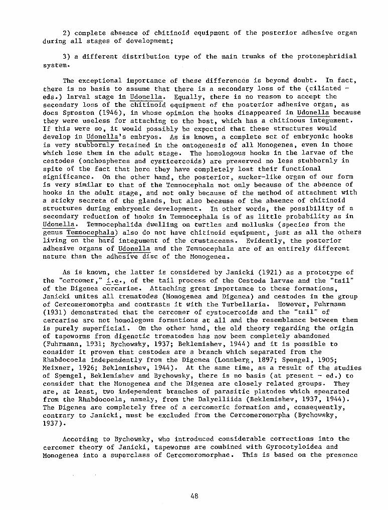
2) complete absence of chitinoid equipment of the posterior adhesive organ during all stages of development;
3) a different distribution type of the main trunks of the protonephridial system.
The exceptional importance of these differences is beyond doubt. In fact, there is no basis to assume that there is a secondary loss of the (ciliated -eds.) larval stage in Udonella. Equally, there is no reason to accept the secondary loss of the chitinoid equipment of the posterior adhesive organ, as does Sproston (1946), in whose opinion the hooks disappeared in Udonella because they were useless for attaching to the host, which has a chitinous integument. If this were so, it would possibly be expected that these structures would develop in Udonella's embryos. As is known, a complete set of embryonic hooks is very stubbornly retained in the ontogenesis of all Monogenea, even in those which lose them in the adult stage. The homologous hooks in the larvae of the cestodes (onchospheres and cysticercoids) are preserved no less stubbornly in spite of the fact that here they have completely lost their functional significance. On the other hand, the posterior, sucker-like organ of our form is very similar to that of the Temnocephala not only because of the absence of hooks in the adult stage, and not only because of the method of attachment with a sticky secreta of the glands, but also because of the absence of chitinoid structures during embryonic development. In other words, the possibility of a secondary reduction of hooks in Temnocephala is of as little probability as in Udonella. Temnocephalida dwelling on turtles and mollusks (species from the genus Temnocephala) also do not have chitinoid equipment, just as all the others living on the hard integument of the crustaceans. Evidently, the posterior adhesive organs of Udonella and the Temnocephala are of an entirely different nature than the adhesive disc of the Monogenea.
As is known, the latter is considered by Janicki (1921) as a prototype of the "cercomer," i.e., of the tail process of the Cestoda larvae and the "tail" of the Digenea cercariae. Attaching great importance to these formations, Janicki unites all trematodes (Monogenea and Digenea) and cestodes in the group of Cercomeromorpha and contrasts it with the Turbellaria. However, Fuhrmann (1931) demonstrated that the cercomer of cystocercoids and the "tail" of cercariae are not homologous formations at all and the resemblance between them is purely superficial. On the other hand, the old theory regarding the origin of tapeworms from digenetic trematodes has now been completely abandoned (Fuhrmann, 1931; Bychowsky, 1937; Beklemishev, 1944) and it is possible to consider it proven that cestodes are a branch which separated from the Rhabdocoela independently from the Digenea (Lonnberg, 1897; Spengel, 1905; Meixner, 1926; Beklemishev, 1944). At the same time, as a result of the studies of Spengel, Beklemishev and Bychowsky, there is no basis (at present - ed.) to consider that the Monogenea and the Digenea are closely related groups. They are, at least, two independent branches of parasitic platodes which spearated from the Rhabdocoela, namely, from the Dalyelliida (Beklemishev, 1937, 1944). The Digenea are completely free of a cercomeric formation and, consequently, contrary to Janicki, must be excluded from the Cercomeromorpha (Bychowsky, 1937).
According to Bychowsky, who introduced considerable corrections into the cercomer theory of Janicki, tapeworms are combined with Gyrocotyloidea and Monogenea into a superclass of Cercomeromorphae. This is based on the presence
48
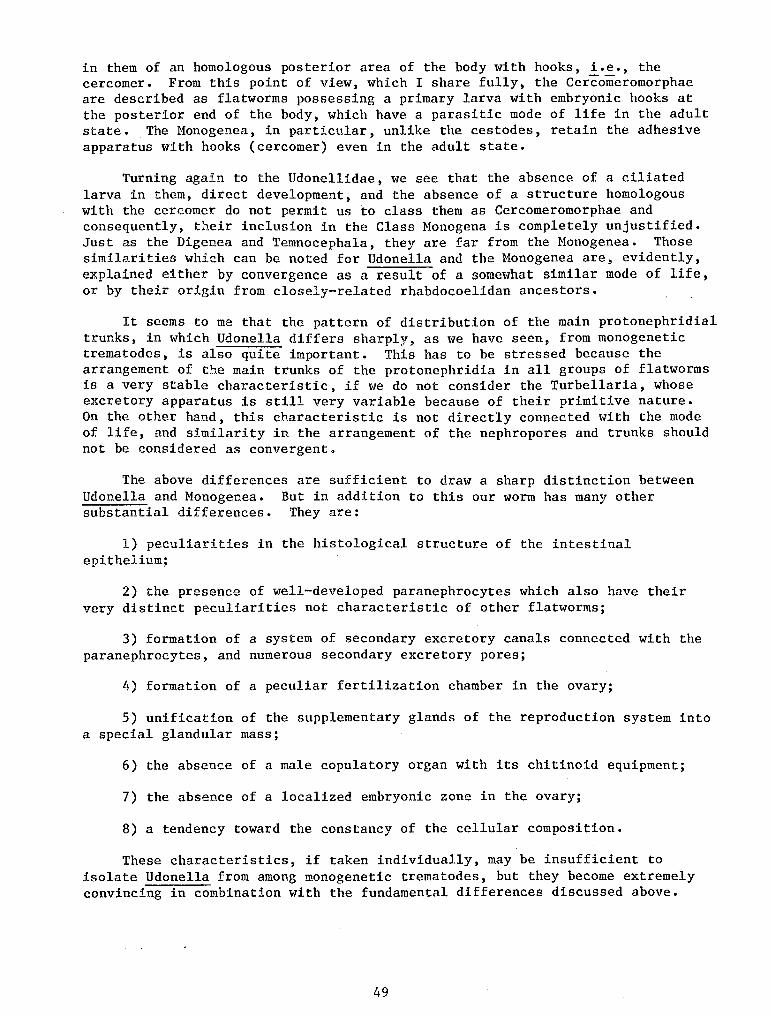
in them of an homologous posterior area of the body with hooks, i.e., the cercomer. From this point of view, which I share fully, the Cercomeromorphae are described as flatworms possessing a primary larva with embryonic hooks at the posterior end of the body, which have a parasitic mode of life in the adult state. The Monogenea, in particular, unlike the cestodes, retain the adhesive apparatus with hooks (cercomer) even in the adult state.
Turning again to the Udonellidae, we see that the absence of a ciliated larva in them, direct development, and the absence of a structure homologous with the cercomer do not permit us to class them as Cercomeromorphae and consequently, their inclusion in the Class Monogena is completely unjustified. Just as the Digenea and Temnocephala, they are far from the Monogenea. Those similarities which can be noted for Udonella and the Monogenea are, evidently, explained either by convergence as a result of a somewhat similar mode of life, or by their origin from closely-related rhabdocoelidan ancestors.
It seems to me that the pattern of distribution of the main protonephridial trunks, in which Udonella differs sharply, as we have seen, from monogenetic trematodes, is also quite important. This has to be stressed because the arrangement of the main trunks of the protonephridia in all groups of flatworms is a very stable characteristic, if we do not consider the Turbellaria, whose excretory apparatus is still very variable because of their primitive nature. On the other hand, this characteristic is not directly connected with the mode of life, and similarity in the arrangement of the nephropores and trunks should not be considered as convergent.
The above differences are sufficient to draw a sharp distinction between Udonella and Monogenea. But in addition to this our worm has many other substantial differences. They are:
1) peculiarities in the histological structure of the intestinal epithelium;
2) the presence of well-developed paranephrocytes which also have their very distinct peculiarities not characteristic of other flatworms;
3) formation of a system of secondary excretory canals connected with the paranephrocytes, and numerous secondary excretory pores;
4) formation of a peculiar fertilization chamber in the ovary;
5) unification of the supplementary glands of the reproduction system into a special glandular mass;
6) the absence of a male copulatory organ with its chitinoid equipment;
7) the absence of a localized embryonic zone in the ovary;
8) a tendency toward the constancy of the cellular composition.
These characteristics, if taken individually, may be insufficient to isolate Udonella from among monogenetic trematodes, but they become extremely convincing in combination with the fundamental differences discussed above.
49
B. Comparison with the Temnocephala.
If Udonella cannot be classed among monogenetic trematodes, can we not then include them in the class of the Temnocephala? The validity of this question follows not only from the above-mentioned common peculiarities of their organization, but also from a similar commensal mode of life on crustaceans. It is true, however, that the Temnocephala live on fresh-water hosts and Udonella on marine hosts.
The common characteristics of Udonella and the Temnocephala are:
1) the absence of cercomeric formations;
2) formation of a glandular adhesive organ of the posterior end of the body;
3) direct type of development;
4) a generally similar arrangement of the main excretory canals and nephropores;
5) the presence of paranephrocytes and, in addition, the same number of them;
6) attachment of the egg to the substratum with a hardening secreta different from the substance of the egg shell;
7) a definite tendency toward constancy of cellular composition and a small number of cells in many organs.
The last common feature is evident in various systems of the organs, namely: in the musculature, excretory system (two-celled excretory vesicles, paranephrocytes), in the reproductive system, and, possibly, also in the nervous system.
All these characteristics, which Udonella has in common with the Temnocephala, cannot be a result of a similar mode of life, but indicate a greater affinity to it than to the Honogenea. However, there are also very substantial differences. In fact, one of the Temnocephals's distinctive characteristics is a primitive structure of the integumentary elements which often retain their cilia and contain rhabdite glands. To the contrary, Udonella is characterized by an extremely specialized integument of the embedded type. The primitive nature of the nervous system should be considerd an equally important feature of the Temnocephala. It is characterized by an abundance of longitudinal nerve trunks and is, in essence, a true nerve orthagon which is characteristic of many Turbellaria. This peculiarity of the Temnocephala points to their origin from the Turbellaria with a rather primitive nerve orthagon, while this is not very probably with respect to Udonella.
Furthermore, very sharp differences are structure of the intestine, in the secondary apparatus, in the reproductive system, etc.
50
also expressed in the histological characteristics of the excretory All those differences from the
Temnocephalida may be summarized as follows:
1) integument of the embedded epithelium type;
2) small number of longitudinal nerve trunks;
3) histological structure of the intestines;
4) unusual development of the paranephrocytes dividing excretory canals into two physiologically independent systems;
5) secondary excretory canals and pores;
6) unpaired testes;
7) fertilization chamber of the ovary;
8) absence of male copulatory organ;
9) absence of resorbing vesicle;
10) absence of a localized embryonic zone in the ovary;
11) anterior position of the genital pore.
Thus, there is no adequate basis for the inclusion of Udonella in the Class Temnocephala. Similarity in the structure and functioning of the posterior adhesive organ in the forms being compared can be, apparently, explained by convergent adaptation to life on hosts which have hard chitinous integuments.
c. Position of Udonella in the System.
Many important features in the structure of Udonella proved to be very unusual, and characteristic of this form alone. This justifies its isolation into an independent Class for which I suggest the name Udonelloidea.
Diagnosis of the Class Udonelloidea A. Ivanov, 1952, Reports of the Academy of Sciences, USSR,
31(2):175-178
Commensal flatworms without a cercomer, but equipped with a posterior sucker-like organ with cement glands. Direct development without a larval stage.
Mouth subterminal. At the anterior end a pair of glandular depressions connected with the head glands. Integuments cuticularized, of the embedded epithelium type. Pharynx: pharynx doliiformis. Intestine sac-like, intestinal epithelium with many embedded cells. Excretory apparatus complicated by the development of huge metameric paranephrocytes dividing the canals into two physiologically independent excretory systems. One of them consists in part, of secondary vessels with their own numerous secondary pores. Reproductive system, with a common atrial opening in the anterior part of the body, without female copulatory organs and genitointestinal connection. Ovary, with fertilization
51
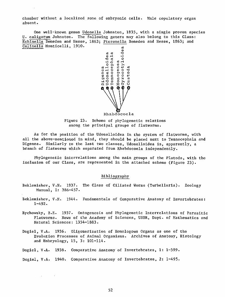
chamber without a localized zone of embryonic cells. Male copulatory organ absent.
One well-known genus Udonella Johnston, 1835, with a single proven species u. caligorum Johnston. The following genera may also belong to this Class: Echinella Beneden and Hesse, 1863; Pteronella Beneden and Hesse, 1863; and Calinella Monticelli, 1910.
Ill Ill a>r-i 'tl Ill •r-1 .!=: 0 OJ
Ill r-i a> a> r-i u I:; a> 0 a> I:; I:; tn 0 f:l
•r-l 'tl Q) OD8
Ill a> 'tl ·r-1 0
lllr-i a> :>.. J:: .jJ Ill a> 0 'tl tn u 0 0 0 .jJ J:: 1-r Ul 0 :>.. Q)
;:E: t!l u
Rhabdocoela
Figure 23. Scheme of phylogenetic relations among the principal groups of flatworms.
As for the position of the Udonelloidea in the system of flatworms, with all the above-mentioned in mind, they should be placed next to Temnocephala and Digenea. Similarly to the last two classes, Udonelloidea is, apparently, a branch of flatworms which separated from Rhabdocoela independently.
Phylogenetic interrelations among the main groups of the Platoda, with the inclusion of our Class, are represented in the attached scheme (Figure 23).
Bibliography
Beklemishev, V.N. 1937. The Class of Ciliated Worms (Turbellaria). Zoology Manual, I: 386-457.
Beklemishev, V.N. 1944. Fundamentals of Comparative Anatomy of Invertebrates: 1-492.
Bychowsky, B.E. 1937. Ontogenesis and Phylogenetic Interrelations of Parasitic Flatworms. News of the Academy of Sciences, USSR, Dept. of Mathematics and Natural Sciences: 1354-1883.
Dogie!, v.A. 1936. Oligomerization of Homologous Organs as one of the Evolution Processes of Animal Organisms. Archives of Anatomy, Histology and Embryology, 15, 3: 101-114.
Dogie!, V.A. 1938. Comparative Anatomy of Invertebrates, 1: 1-599.
Dogie!, V.A. 1940. Comparative Anatomy of Invertebrates, 2: 1-495.
52
Dogiel, v.A. 1947. The Phenomena of Polymerization and Oligomerization of Homologous Organs in the Animal Kingdom and their Evolutionary Significance. News of Academy of Sciences, USSR, Biology Series, 4: 471-486.
Pavlovsky, E.N. 1937. Supplement to the Turbellaria Class. Order Temnocephalida. Zoology Manual, 1: 457-460.
Fedotov, D. M. 1915. Protomyzostomum polynephris Fedotov and its relation to Gorgonocephalus eucnemis M. & Tr.: 1-240.
Baer, J.G. 1929. Contribution a l'etude des Temnocephales Dactylocephala madagascariensis (Vayssiere, 1982). Bull. bioi. France Belgique, 63: 539-561.
Baer, J.G. 1931. Etude monographique du groupe des Temnocephales. Bull. bioi. France Belgique, 65, 1:1-57.
Bettendorf, H. 1897. Uber Muskulatur und Sinneszellen der Trematoden. Zool. Jahrb., Abt. Anat. und Ontog., 10: 1-54.
Brandes, G. 1892. Zum feineren Bauder Trematoden. Ztschr. s. wiss. Zool., 53: 558-578.
Bresslau, E. 1928-1933. Turbellaria. Handbuch der Zoologie von Kukenthal und Krumbach, 2, 1: 54-293.
Bresslau, E. und E. Reisinger. 1933. 6 Ordung der Klasse Turbellaria: Temnocephalida. Handbuch der Zoologie van Kukenthal und Krumbach, 2, 1: 294-320.
Brinkmann, A. 1940. Contribution to our knowledge of the Monogenetic Trematodes. Bergens Mus. Arbok 1939/40, Naturvitenskapelig rekke, 1: 3-117.
Dawes, B. 1946. The Trematoda. Cambridge: 1-644.
Fuhrmann, 0. 1928. Trematoda. Handbuch der Zoologie von Kukenthal und . Krumbach, 2, 2: 1-140.
Fuhrmann, 0. 1931. Cestoidea. Handbuch der Zoologie von Kukenthal und Krumback, 2, 2: 141-416.
Goto, s. 1894. Studies on the ectoparasitic Trematodes of Japan. Journ. Coli. Sc. Imp. Univ. Tokyo, 8: 1-273.
Haswell, W.A. 1893. A Monograph of the Temnocephalae. Proc. Linn. Soc. New South Wales, Macleay memor., : 94-152.
Rein, w. 1904. Zur Epithelgrage der Trematoden. Ztschr. Wiss. Zool., 77: 346-385.
Janicki, D. 1921. Grundlinien einer "Cercomer" - Theorie zur Morphologie der Trematoden und Cestoden. Festschr. fur Zschokke, 30: 1-22.
53
•.
Jahn, T.L. and R. Kuhn. 1932. The life history of Epibdella melleni Maccallum, 1927, a monogenetic Trematode parasitic on marine fishes. Biol. Bull. Marine biolog. Labor., 62, 1: 89-111.
Johnston, G. 1835. Illustration in British Zoology. 44. Udonella caligorum. Mag. Nat. Hist., 8:496-498.
Kovalevsky, M. 1895. Studia helminthologiczne, 11. Rezpr. Ak. Krakow, 2, 9:372-390.
Lonnberg, E. 1897. Beitrage zur Phylogenie der parasitischer Plathelminthen. Centralblatt Bakteriol. Parasjtenk., Abt. 1, 22: 674-684, 725-731.
Meixner, J. 1926. Rhabdocoela.
Beitrag zur Morphologie und zum System der Turbellaria Ztschr. Wiss. Biolog., Abt. A., 5: 577-624.
Merton, H. 1914. Beitrage zur Anatomie und Histologie von Temnocephala. Abhandl. Senckenberg. Naturforsch. Gesell., 35, I: I-58.
Monticelli, F.S. 1889. Saggio di una morfologia dei Trematodi. Zbl. Bakt., 6:120-122.
Monticelli, F.S. 1893. Studii sui Trematodi endoparasiti. Jena: 1-229.
Monticelli, F.s. 1910. Calinella craneola, n. g., n. sp. Trematode nouveau de la famille des Udonellidae provenent des campagnes de s. A. s. le Prince de Honaco. Ann. Inst. Oceanogr. Monaco, 1: 1-9.
Pratt, H. 1909. The cuticula and subcuticula of Trematodes and Cestodes. Amer. Nat. New York., 43: 705-729.
Price, w. 1938. North American monogenetic trematodes. II. The families Monocotylidae, Microbothriidae, Acanthocotylidae and Udonellidae (Capsaloidea). Journ. Wash. Acad. Sci., 28, 4: 183-198.
Reisinger, E. 1922-1923. Untersuchungen uber Bau und Funktion des Exkretion-sapparates bei rhabdocoelen Turbellarien. Zool. Anz., 54: 200-209; 56: 205-224.
Russell, F.S. 1925. A new species of Caligus from Egypt, Caligus from Egypt, Caligus pageti n. sp. Ann. Hag, Nat. Hist., ser. 9, 15: 611-618.
Schnieder, A. 1873. Untersuchungen uber Plathelminthen. Giessen Oberhess. Gesell. Bericht, 14: 69-132.
Spengel, J .n. 1905. Die Monozootie der Cestoden. Ztschr. Wiss. Zool., 82: 252-287.
Sproston, N.G. 1946. A synopsis of the Monogenetic Trematodes. Transact. Zool. Soc. London, 25, 4: 185-600.
Van Beneden, P.J. et c.s. Hesse. 1863. Hirudinees et les Trematodes marins.
Recherches sur les bdellodes sur les Burxelles: 1-150.
Wright, R. and A. Maccallum. American helminthology.
1887. Sphyranura osleri- a contribution to Journ. Morpho!., 1(1): 1-48.
54
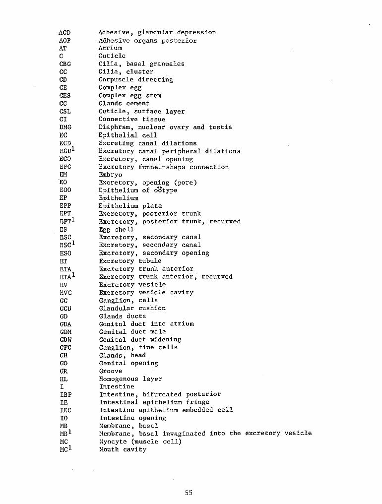
AGD AOP AT c CBG cc CD CE CES CG CSL CI DMG EC ECD Ecn1 ECO EFC EM EO EOO EP EPP EPT EPTl ES ESC Escl ESO ET ETA ETAl EV EVC GC GCU GD GDA GDM GDW GFC GH GO GR HL I IBP IE IEC IO ~1B MBl MC Mel
Adhesive, glandular depression Adhesive organs posterior Atrium Cuticle Cilia, basal granuales Cilia, cluster Corpuscle directing Complex egg Complex egg stem Glands cement Cuticle, surface layer Connective tissue Diaphram, nuclear ovary and testis Epithelia! cell Excreting canal dilations Excretory canal peripheral dilations Excretory, canal opening Excretory funnel-shape connection Embryo Excretory, opening (pore) Epithelium of ootype Epithelium Epithelium plate Excretory, posterior trunk Excretory, posterior trunk, recurved Egg shell Excretory, secondary canal Excretory, secondary canal Excretory, secondary opening Excretory tubule Excretory trunk anterior Excretory trunk anterio-r, recurved Excretory vesicle Excretory vesicle cavity Ganglion, cells Glandular cushion Glands ducts Genital duct into atrium Genital duct male Genital duct widening Ganglion, fine cells Glands, head Genital opening Groove Homogenous layer Intestine Intestine, bifurcated posterior Intestinal epithelium fringe Intestine epithelium embedded cell Intestine opening Membrane, basal Membrane, basal invaginated into the excretory vesicle Myocyte (muscle cell) Mouth cavity
55
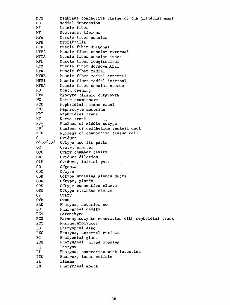
MCG MD MF MF MFA MFB t1FD MFEA MFIA MFL r1FM MFR MFRE MFRI MFUA MO MPO NC NCC NM NPT NT NUl NU2 NUC 0 ol,o2,o3 oc occ OD OIP 00 ooc OOD OOG oos OSG ov OVM PAE PC PCM PCN PCY PD PEC PG PGO PH PI PIC PL PM
Hembrane connective-tissue of the glandular mass Hedial depression Muscle fiber Membrane, fibrous Muscle fiber annular Myofibrilla Muscle fiber diagonal Muscle fiber annular external Muscle fiber annular inner Muscle fiber longitudinal Muscle fiber dorsoventral Huscle fiber radial Muscle fiber radial external Muscle fiber radial internal Huscle fiber annular uterus Mouth opening Myocyte plasmic outgrowth Nerve commissure Nephridial common canal Nephrocyte membrane Nephridial trunk Nerve trunk .. Nucleus of middle ootype Nucleus of epithelium seminal duct Nucleus of connective tissue cell Oviduct Ootype and its parts Ovary, chamber Ovary chamber cavity Oviduct dilation Oviduct, initial part Oogonia Oocyte Ootype staining glands ducts Ootype, glands Ootype connective sleeve Ootype staining glands Ovary Ovum Pharynx, anterior end Pharyngeal cavity Parenchyma Paranephrocycte connection with nephridial trunk Paranephrocyctes Pharyngeal disc Pharynx, external cuticle Pharyngeal gland Pharyngeal, gland opening Pharynx Pharynx, connection with intestine Pharynx, inner cuticle Plasma Pharyngeal mouth
56
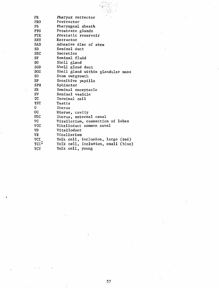
• < ~ •
PR Pharynx retractor PRO Protractor PS Pharyngeal sheath PTG Prostrate glands PTR Prostatic reservoir RET Retractor SAD Adhesive disc of stem SD Seminal duct SEC Secretion SF Seminal fluid SG Shell gland SGD Shell gland duct SGG Shell gland within glandular mass SO Stem outgrowth SP Sensitive papilla SPH Sphincter SR Seminal receptacle SV Seminal vesicle TC Terminal cell TST Testis U Uterus UC Uterus, cavity UEC Uterus, external canal VC Vitellarium, connection of lobes VCC Vitelloduct common canal VD Vitelloduct VR Vitellarium YCI Yolk cell, inclusion, large (red) YCil Yolk cell, inclusion, small (blue) YCY Yolk cell, young
57


