Embed Size (px)
Citation preview

IMMUNOLOGY AT MOUNT SINAI
Mononuclear phagocyte diversity in the intestine
Milena Bogunovic • Arthur Mortha •
Paul Andrew Muller • Miriam Merad
� Springer Science+Business Media, LLC 2012
Abstract Present in all organs, mononuclear phagocytes consist of a heteroge-
neous population of hematopoietic cells whose main role is to ensure tissue
homeostasis through their ability to scavenge cell debris, promote tissue repair and
maintain tolerance to self-antigens while simultaneously inducing innate and
adaptive immune responses against foreign antigens that breach the tissue. The
intestinal mucosa is particularly exposed to foreign antigen, through constant
exposure to high loads of commensal bacteria and dietary antigens as well as
providing a site of entry for viral and bacterial pathogens. The molecular mecha-
nisms that control the intestinal ability to distinguish between ‘‘innocuous’’ and
‘‘dangerous’’ antigens remains poorly understood although it is clear that mono-
nuclear phagocytes play a key role in this process. This review highlights recent
advances in our understanding of heterogeneous origin of the mononuclear
phagocytes that inhabit the intestinal mucosa and discusses how developmental
diversity allows for functional diversity to ensure intestinal integrity.
Keywords Mononuclear phagocytes (MPs) � Dendritic cells (DCs) �Macrophages � Intestine �Mucosa � Lamina propria �Muscularis
Introduction
Mononuclear phagocytes (MPs) represent a small popula-
tion of hematopoietic cells that populate most tissues and
share the ability to sample the extracellular milieu. In the
intestine, MPs are strategically located below the mucosal
epithelium allowing them to be in close contact with
luminal contents. They are also distributed throughout the
submucosa and the muscularis layer, which faces the
peritoneum. Tissue MPs consist of two main cell popula-
tions that include dendritic cells (DCs) and macrophages.
The central role of macrophages is to sustain tissue
homeostasis and repair, whereas the major function of DCs
is to capture and process antigenic material in the form of
peptide/major histocompatibility complex (MHC) com-
plexes that can be recognized by T lymphocytes. Upon
antigen uptake and maturation, DCs migrate from the
intestinal mucosa to the T cell areas of the mesenteric
lymph nodes (LNs) and promote the induction of adaptive
effector responses against pathogens that breach the
intestine or maintain T regulatory responses to commensals
M. Bogunovic (&) � A. Mortha � P. A. Muller � M. Merad (&)
The Immunology Institute, Mount Sinai School of Medicine,
One Gustave L. Levy Place, New York, NY 10029, USA
e-mail: [email protected]
M. Merad
e-mail: [email protected]
M. Bogunovic � A. Mortha � P. A. Muller � M. Merad
Department of Oncological Sciences, Mount Sinai School
of Medicine, One Gustave L. Levy Place, New York,
NY 10029, USA
Milena BogunovicMiriam Merad
123
Immunol Res
DOI 10.1007/s12026-012-8323-5

or self-antigens. This review summarizes recent advances
in our understanding of the diversity of the MP system
within the gut in the context of its developmental properties
and its role in gut immunity.
Architecture of the intestinal mononuclear phagocyte
system
Mucosal DCs
The intestinal mucosa consists of simple columnar epi-
thelium that provides an interface with the antigen-rich
luminal environment, the lamina propria and its lymphoid
structures also called gut-associated lymphoid tissue
(GALT) that include small lymphoid follicles scattered
throughout the intestinal mucosa as well as large organized
lymphoid structures called Peyer’s patches, and the mus-
cularis mucosae [1]. Similar to all DCs, mucosal DCs are
positive for the hematopoietic marker CD45 but lack
lymphocyte- and granulocyte-specific markers, and con-
stitutively express high MHC class II (MHCII) levels and
the integrin CD11c [2]. Mucosal DCs are distributed
throughout the intestinal tissue, forming an organized cel-
lular network in the lamina propria facing the lumen [3],
and are present in the GALT [4]. The phenotype of DCs in
PPs and mesenteric LNs that drain intestinal tissues have
been largely studied [5, 6], but the diversity of mucosal
DCs that accumulate in non-lymphoid tissues is only
starting to be unraveled.
Non-lymphoid tissue mucosal DCs accumulate mainly
in the lamina propria although some DCs can be found in
the epithelium. MHCIIhiCD11chi DCs can be further divi-
ded into two main populations best characterized as
CD103?CD11b?CX3CR1 (CX3C-chemokine receptor 1)-
and CD103-CD11b?CX3CR1? subsets. In addition, a
minor (5–10 % of MHCIIhiCD11chi cells) CD103?
CD11b-CD8a?CX3CR1- DC population found in muco-
sal cell suspensions is thought to reside in the isolated
lymphoid follicles scattered throughout the intestinal
mucosa and is a part of the GALT (Fig. 1, 2; Table 1)
[7–9]. DC subset distribution in the gut varies anatomi-
cally, with CD103?CD11b? DCs present at highest num-
bers in the duodenum and CD103-CD11b? DCs enriched
in the large bowel [10]. Recent studies further divide
CD103-CX3CR1? DC into CX3CR1hi and CX3CR1lo
subsets [11, 12], although it is not clear at this point
whether these cells are developmentally or functionally
distinct.
Although several phenotypically distinct DCs have been
described in the literature, we will discuss only DC subsets
with distinct phenotype, origin and function. This review
does not include plasmocytoid dendritic cells (pDCs), also
known as type I interferon-producing cells (IPCs). Though
these myeloid cells share a developmental pathway with
conventional DCs [13] and are abundant in the gut [14],
5
5
2 1030 10 104 105
0
2
3
4
10
10
10
10
CD11b
30
1D
C
2 1030 10 104 105
2
3
4
10
10
10
10
CD11b
30
1D
C
13.3
3.4
78.1
2 1030 10 104 105
0 0
2
3
4
10
10
10
10
CD11b
c1
1D
C
5
5
2 1030 10 104 105
0
2
3
4
10
10
10
10
CD11b
c1
1D
C
Mus
cula
risLP 5
2 1030 10 104 105
0
2
3
4
10
10
10
10
CD11b
c1
1D
C
25.3
3.5
6.9
84.1
Who
le S
B
5
2 1030 10 104 105
0
2
3
4
10
10
10
10
CD11b
30
1D
C
18.8
5.6
57.7
15.1
18.5
CD11c lo
CD11c hi
Gated on CD45+MHCIIhi
LP MPGated on CD45+MHCIIhi
Muscularis MP
CD103+CD11b+ LP DC
CD103_CD11b+ LP DC
CX3CR10 10 2 103 104 105
B
A
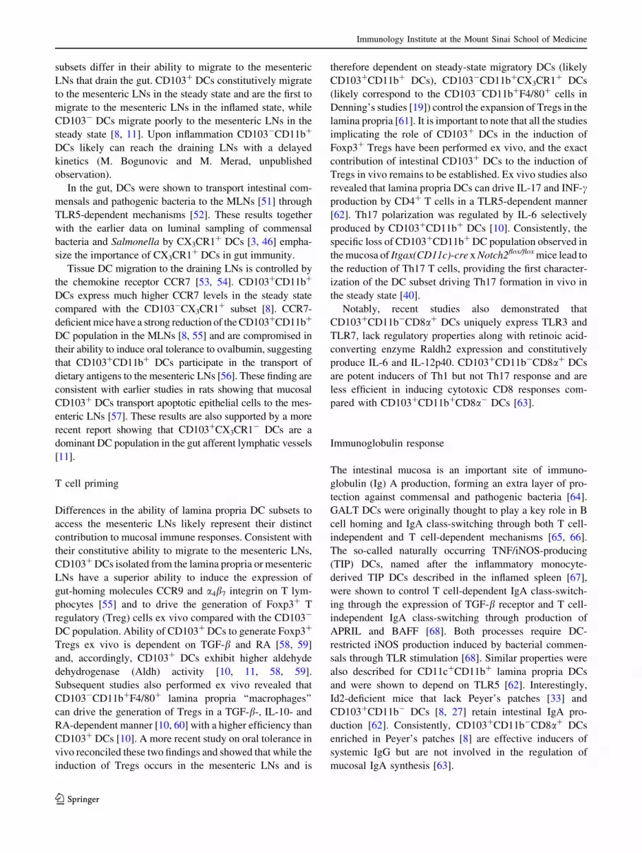
Fig. 1 Anatomical distribution of the intestinal mononuclear phago-
cyte (MP) subsets in the intestine. a Flow cytometry dot plots show
the phenotype and percentages of intestinal MPs in single cell
suspensions obtained from different anatomical compartments of the
small bowel (SB). The lamina propria (LP) and muscularis layers (leftpanel) were separated and single cell suspensions were analyzed by
seven-color flow cytometry. The results were compared to the whole
SB cell suspension devoid of Peyer’s patches (right panel). To define
CD11chi and CD11clo DCs, gates were set on viable
DAPI-CD45?MHCIIhi cells. CD11chi MPs were further divided into
CD103?CD11b-, CD103?CD11b? and CD103-CD11b? DCs.
CD11clo MPs form a homogenous population. b Flow cytometry
histograms represent the levels of CX3C-chemokine receptor 1
(CX3CR1) expression by lamina propria CD11chi CD103?CD11b?
and CD103-CD11b? DC subsets in transgenic CX3CR1-GFP
reporter (CX3CR1?/GFP) mice
Immunology Institute at the Mount Sinai School of Medicine
123

they are poor phagocytes [15] and are not considered to be
a part of the MP system.
Mucosal macrophages
The phenotype of intestinal macrophages is controversial.
Macrophages are classically defined as CD45?MHCIIlo
CD11cloF4/80hiCD11bhi cells. However, in the intestine,
the vast majority of CD45?MHCIIloCD11cloF4/80hi
CD11bhi cells appear to lack CX3CR1, express eosinophil-
specific marker SIGLEC-F and have eosinophil-like
morphology (Fig. 3) (M. Bogunovic and M. Merad,
unpublished observation). Recent studies suggest that
CD45?MHCII?CD11chiCD103-CD11b?CX3CR1? lamina
propria DCs, which also express F4/80 [8], are in fact
macrophages (discussed in [16]) based on their distinct cel-
lular morphology consisting of a large vacuolar cytoplasm,
selective expression of phagocytosis-associated genes and
enhanced phagocytic activity in vitro [8, 12, 17–19].
Muscularis MPs
Intestinal muscularis externa, comprised of longitudinal
and circular layers of smooth muscles, is positioned under
the mucosa and submucosa away from the antigen-rich
luminal environment and maintains continued peristalsis to
allow passage of digested material along the gut. A net-
work of MHCII? phagocytes exists in the intestinal mus-
cularis and was originally described as macrophages [20]
but later classified as DCs [21]. We have shown that the
intestinal muscularis contains a homogeneous population
of MHCIIhiCD11cloCD103-CD11b? cells that also express
CX3CR1 and F4/80 [8]. In a whole intestinal cell suspen-
sion, muscularis MPs can be distinguished from CD11chi
Fig. 2 Anatomical distribution of intestinal DC subsets in the steady
state. CD11chiCD103?CD11b? dendritic cells (DCs) (blue) and
CD103-CD11b? DCs (black) are distributed throughout the lamina
propria whereas CD11chiCD103?CD11b- DCs (red) are localized in
the isolated lymphoid follicles. CD103?CD11b? DCs migrate to the
mesenteric lymph nodes in a CCR7-dependent fashion. The intestinal
muscularis externa is a separated layer of the smooth muscle tissue
that harbors a unique population of CD11cloCD103-CD11b?
phagocytes
Immunology Institute at the Mount Sinai School of Medicine
123

Table 1 Developmental and functional properties of intestinal lamina propria DC subsets
Lamina propria
MP subsets
CD103? CD11b- CD103? CD11b? CD103- CD11b?
Surface marker
expression
MHCll? CD11chiCD103? CD11b-
CD8a? CX3CR1- F4/80-
CD172alo*CD64-*
MHCII? CD11chi CD103? CD11b?
CD8a- CX3CR1- F4/80lo CD172aint*
CD64-*
MHCll? CD11Chi CD103- CD11b?
CD8a- CX3CR1? F4/80?
CD172ahi* CD64?*
DC subset
deficient mice
Flt3-/-**, Batf3-/-, ld2-/-, irf8-/- Flt32/2, Csf2r2/2**, Notch2CD11c Csf1r2/2**
Developmental
origin
CDP, pre-DC CDP, pre-DC Monocytes
Anatomical
location in the
steady state
GALT and MLN Lamina propria, enriched in small bowel Lamina propria, enriched in colon
Cytokines that
control
development
FLT3L FLT3L, GM-CSF M-CSF
Function ex vivo:
T cell response Th1 induction Treg and Thl, Th17 induction Treg expansion
Phagocytosis N.D. Less efficient More efficient
Cytokines IL-6, IL-12 IL-6, TGFb, RA, IL-23 IL-10, TNF, IL-6, IL-12, IL-23, IL-27
T cell indep. IgA
CSR
Not involved Involved N.D.
Function in vivo N.D. Migrate to MLN, oral tolerance,
antimicrobial defense
Oral tolerance, inflammatory cytokine
secretion, colitis
Mononuclear phagocytes (MP); CD172a is also known as SIRPa; CD64 is also known as Fcc receptor I (FccRI); basic leucin zipper tran-
scriptional factor ATF-like 3 (Batf3); inhibitor of DNA-binding protein-2 (Id2); interferon-regulatory factor 8 (Irf8); neurogenic locus notch
homolog protein 2 (Notch2); macrophage colony-stimulating factor (M-CSF), its receptor is encoded by colony-stimulating factor 1 receptor
gene (Csf1r); granulocyte macrophage colony-stimulating factor (GM-CSF), its receptor is encoded by colony-stimulating factor 2 receptor gene
(Csf2r); FMS-related tyrosine kinase 3 ligand (FLT3L), its receptor is encoded by FMS-related tyrosine kinase 3 gene (Flt3); common dendritic
cell precursor (CDP); dendritic cells (DC); Gut-associated lymphoid tissue (GALT), Mesenteric lymph node (MLN); CD4? T helper cells (Th);
CD4? T regulatory cells (Treg); immunoglobulin (Ig); interleukin (IL); tumor necrosis factor (TNF); retinoic acid (RA); class switch recom-
bination (CSR); not determined (N.D.)
* Milena Bogunovic, Jennifer Miller and Miriam Merad, unpublished
** Incomplete depletion
0 102 103 104 1050
1000
2000
3000
4000
0 102 103 104 1050
20
40
60
80
100
0 102 103 104 1050
1000
2000
3000
4000
SS
C
SS
C
CX3CR1-GFP Gr-1
1(89%)
2 (10.1%)
1A
2
1B
1A
21B
A BGated on CD45+MHCII-
SIGLEC-F
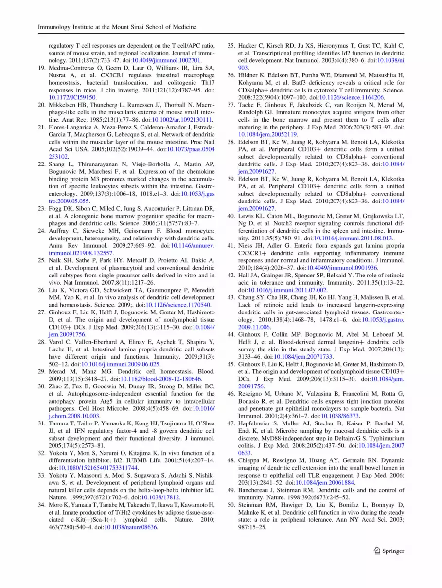
Fig. 3 Non-DC myeloid cell in the intestinal mucosa. a Dot plots
show the phenotypes of CD45?CD11b?MHCIIlo/- cell populations in
the SB intestinal mucosa. SSChiCX3CR1-Gr1loSIGLEC-F? cells (1a)
represent eosinophils; SSCintCX3CR1-Gr1hiSIGLEC-F?/- cells (1b)
are likely neutrophils; SSCloCX3CR1?Gr1hiSIGLEC-F- cells (2) are
likely undifferentiated monocytes. b Cytospin of purified
CD11b?MHClo/- cells demonstrates that the majority of this
population are polymorphonuclear eosinophils
Immunology Institute at the Mount Sinai School of Medicine
123

lamina propia DCs by the low expression of CD11c (Fig. 1,
2) [8]. However, most studies that use whole intestinal
digests unsuitably refer to the muscularis CD11clo MP
population as lamina propria macrophages [12, 17–19, 22].
Regulation of intestinal mononuclear phagocyte
development
Origin of intestinal MPs
Commitment to the MP lineage occurs at the level of
macrophage and DC precursor (MDP), a dedicated mye-
loid-derived bone marrow (BM) precursor that has lost
granulocytic, erythrocytic and megakaryocytic potential
[23]. MDP maintains a dual potential, differentiating into
either monocytes, which are thought to give rise to most
tissue macrophages [24] or to a DC-restricted progenitor
called the common DC precursor (CDP) that gives rise to
both classical and plasmacytoid dendritic cells [13, 25, 26].
In the BM, CDP differentiates into either plasmacytoid
DCs or so-called pre-DCs [26]. Pre-DCs migrate through
the blood to lymphoid organs where they differentiate into
classical CD8? and CD8- DCs [26]. Pre-DCs can also
migrate to the non-lymphoid tissues to give rise to classical
DCs [27].
In the intestine, CDPs and pre-DCs give rise only to
CD103? DC populations (both CD103?CD11b- and
CD103?CD11b?) but not to CD103-CD11b? DCs. In
contrast, monocytes differentiate into CD103-CD11b? but
not CD103? DCs [8, 28], indicating that intestinal CD103?
and CD103- DCs derive from two independent lineages
(Fig. 4). In addition, the distinct monocyte origin of
CD103-CD11b? DCs is in agreement with the fact that
these cells may indeed represent intestinal macrophages.
Regulation of MP homeostasis in the intestine
FMS-like tyrosine kinase 3 ligand (FLT3L) and its receptor
FLT3, granulocyte macrophage colony-stimulating factor
(GM-CSF) and its receptor GM-CSFR and macrophage
colony-stimulating factor (M-CSF) and its receptor
M-CSFR are key molecules involved in MP development
and homeostasis [29]. Flt3L controls the commitment and
differentiation of the DC lineage, and GM-CSF is essential
for the differentiation of DCs in vitro and is thought to
control the development of inflammatory DCs in vivo,
whereas M-CSF controls macrophage development and
homeostasis [30].
Recent studies established that these cytokines and their
receptors control intestinal MP development differently.
Flt3L and Flt3 control the differentiation of
CD103?CD11b- and CD103?CD11b? DCs [11, 28].
GM-CSF and GM-CSFR control the differentiation of
CD103?CD11b? DCs [8, 11], while M-CSFR controls
CD103-CD11b? lamina propria DC homeostasis (Fig. 4) [8].
Several transcription factors have recently been shown
to control DC development in vivo. Interferon-regulatory
factor (IRF) 8 belongs to the family of IRF proteins. Early
studies on the role of IRFs in DC development demon-
strated that in vitro development of BM-derived DCs
supplemented with Flt3L is preferentially controlled by
Irf8, while DCs generated in the presence of GM-CSF rely
on Irf4 [31]. The inhibitor of DNA-binding protein-2 (Id2)
controls formation of peripheral lymphoid organs including
GALT as well as the development of natural killer (NK)
cells, lymphoid tissue inducer (LTi) cells, natural helper 2
(NH2) cells [32–34] and epidermal Langerhans cells [35].
Recently, the transcription factor Batf3 (also known as Jun
dimerization protein p21SNFT) was shown to be highly
expressed in classical DCs [36]. Irf8, Id2 and Batf3 are
selectively required for the development of lymphoid tissue
CD8a? DCs [35–37] and lung and skin CD103? DCs [2,
27, 38]. Analysis of intestinal MPs in Id2, Irf8 or Batf3
deficient mice revealed a total absence of CD103?CD11b-
DCs, whereas CD103?CD11b? DCs remained intact in
these mice [8, 27, 39]. In contrast, the specific depletion of
Notch2 in CD11c? cells (Itgax(CD11c)-cre x Notch2flox/flox
mice) results in the selective depletion of CD103?CD11b?
lamina propria DCs but not CD103?CD11b- DCs [40].
Id2, IRF8, Batf3 and Notch2 do not play a role in the
development of CD103-CD11b? DCs.
Intestinal microbiota and mucosal MP development
Little is known about the influence of the intestinal mic-
robiota on MP diversity in the intestinal mucosa. A recent
study revealed that the total numbers of intestinal CD11c?
DCs are reduced in germ-free mice compared with specific
pathogen-free (SPF) animals [41]. Furthermore, this study
demonstrated that only CX3CR1? DC subset was signifi-
cantly reduced, whereas CD103? DCs developed normally
in the small and large bowel of germ-free animals, sug-
gesting the contribution of luminal microbiota to the
development of CX3CR1? but not CD103? DCs [41].
Diet and mucosal MP development
Retinoic acid (RA), a metabolite of the dietary component
vitamin A, plays a key role in the regulation of mucosal
immune responses [42]. Interestingly, mice deprived of the
dietary vitamin A accumulate high numbers of langerin?
DCs in the intestinal mucosa, GALT and mesenteric LNs
that control the induction of oral tolerance in these animals
[43]. DCs expressing the c-type lectin langerin accumulate
mostly in the skin and lung [44, 45] and are absent in the
Immunology Institute at the Mount Sinai School of Medicine
123

normal intestinal mucosa. These results indicate that die-
tary component likely influences DC differentiation and
function in the steady state.
Function of lamina propria mononuclear phagocytes
Sampling of luminal antigens
The epithelial cell monolayer covering the mucosal sur-
faces of the gut was originally thought to represent a
sealed barrier against non-invasive microbiota populating
the lumen. However, it now appears that subepithelial
DCs closely interact with the intestinal epithelium. They
can penetrate epithelial monolayers by projecting dendrite
extensions into the lumen allowing them to sample
luminal bacteria [3, 46]. In the terminal ileum, the process
of transepithelial dendrite formation and sampling is
dependent on CX3CR1, also known as fractalkine receptor
[3]. DC transepithelial protrusions were shown to be
dependent on the microbiota [41] but independent of
MyD88 [47]. CX3CR1 also controls the development of
the CD103-CD11b?CX3CR1? population likely through
the recruitment of its progenitors into the gut tissue, as
mice that lack CX3CR1 and its ligand CX3CL1 (also
known as fractalkine) have reduced numbers of the
intestinal CD103-CD11b?CX3CR1? DC subset [19].
Although the formation of transepithelial dendrites is
thought to be mainly restricted to the lamina propria
CD103-CD11b? DC subsets as it is the only DC subset
Fig. 4 DC-poiesis and cytokines regulating intestinal DC subsets.
Intestinal CD103?CD11b- DCs (red), CD103?CD11b? DCs (blue)
and CD103-CD11b? DCs (black) derive from bone marrow (BM)
progenitors. The granulocyte macrophage precursor (GMP) resides in
the BM and differentiates into the macrophages dendritic cell
precursor (MDP) that gives rise to monocytes or subsequently loses
macrophage potential by differentiating into the common dendritic
cell precursor (CDP). CDP develops into pre-DCs that exit the BM
and migrate into the tissue where they differentiate into FLT3-
dependent CD103? DCs (red and blue). CD103-CD11b? DCs
(black) arise from monocytes that leave the BM and circulate in the
blood. Once recruited into the tissue, circulating monocytes develop
into M-CSFR-dependent CD103-CD11b? DCs
Immunology Institute at the Mount Sinai School of Medicine
123

expressing CX3CR1 in the gut, subsequent studies have
suggested that chemokines other than CX3CL1 (such as
CCL20) and DC populations other than CX3CR1? DCs
can also contribute to transepithelial dendrite formation
[48]. Consistently, we have found that in contrast to
CD103-CX3CR1? DCs, which accumulate in the subep-
ithelial mucosal compartment, a significant number of
CD103? DCs are present in the epithelial layer [8], sig-
nifying that they too could potentially sample the luminal
antigens. Whether CD103? DCs sample through the for-
mation of transepithelial dendrites or other mechanisms
remains to be examined.
DC migration and antigen transport to the lymph nodes
The initiation of efficient adaptive immune response occurs
in the organized lymphoid structures [49]. Upon antigen
uptake and maturation, tissue DCs migrate to the afferent
lymphatics to present tissue antigens in the form of pep-
tide–MHC complexes to T lymphocytes [49]. Tissue DC
migration to the draining LNs in the steady state is thought
to control the induction of tolerance to self-antigens [50],
whereas DC migration during infection controls the
induction of effector immune responses against microbial
stimuli. Recent results revealed that lamina propria DC
Fig. 5 Functional implications of intestinal lamina propria DC
subsets in mucosal immunity. The cartoon demonstrates possible
scenarios that either lead to the maintenance of a tolerogenic state
(left panel), or the promotion of inflammation (right panel). In a
tolerogenic state (left), CD103?CD11b? DCs (blue) constantly
uptake bacterial and dietary antigens from the intestinal lumen and
transport them to the mesenteric lymph nodes (MLN). In the MLN
CD103?CD11b? DCs prime naı̈ve T cells to differentiate into
inducible T regulatory cells (iTregs). iTregs upregulate gut-homing
molecules CCR9 and a4b7 and home back to the lamina propria. In
the lamina propria, Treg expansion is controlled by IL-10 secreted by
CD103-CD11b? DCs to promote oral tolerance. Invasion of
pathogens in the mucosa results in inflammation followed by the
activation of both CD103?CD11b? DCs and CD103-CD11b? DCs.
CD103-CD11b? DCs adapt a pro-inflammatory phenotype and start
producing cytokines such as TNF, IL-12, IL-23 and IL-6.
CD103-CD11b? DCs might upregulate their migration to the MLN
and prime native T cells toward a Th17 phenotype. Furthermore
CD103?CD11b? DCs start to produce IL-23 which regulates the
expression of Th17- and innate lymphoid cell (ILC)-associated IL-22.
IL-22 regulates the production by epithelial cells of the antimicrobial
peptide RegIIIc Additionally, CD103?CD11b? DCs migrate to the
MLN and induce the differentiation of Th17 or Th1 phenotype that
promote local inflammation
Immunology Institute at the Mount Sinai School of Medicine
123
subsets differ in their ability to migrate to the mesenteric
LNs that drain the gut. CD103? DCs constitutively migrate
to the mesenteric LNs in the steady state and are the first to
migrate to the mesenteric LNs in the inflamed state, while
CD103- DCs migrate poorly to the mesenteric LNs in the
steady state [8, 11]. Upon inflammation CD103-CD11b?
DCs likely can reach the draining LNs with a delayed
kinetics (M. Bogunovic and M. Merad, unpublished
observation).
In the gut, DCs were shown to transport intestinal com-
mensals and pathogenic bacteria to the MLNs [51] through
TLR5-dependent mechanisms [52]. These results together
with the earlier data on luminal sampling of commensal
bacteria and Salmonella by CX3CR1? DCs [3, 46] empha-
size the importance of CX3CR1? DCs in gut immunity.
Tissue DC migration to the draining LNs is controlled by
the chemokine receptor CCR7 [53, 54]. CD103?CD11b?
DCs express much higher CCR7 levels in the steady state
compared with the CD103-CX3CR1? subset [8]. CCR7-
deficient mice have a strong reduction of the CD103?CD11b?
DC population in the MLNs [8, 55] and are compromised in
their ability to induce oral tolerance to ovalbumin, suggesting
that CD103?CD11b? DCs participate in the transport of
dietary antigens to the mesenteric LNs [56]. These finding are
consistent with earlier studies in rats showing that mucosal
CD103? DCs transport apoptotic epithelial cells to the mes-
enteric LNs [57]. These results are also supported by a more
recent report showing that CD103?CX3CR1- DCs are a
dominant DC population in the gut afferent lymphatic vessels
[11].
T cell priming
Differences in the ability of lamina propria DC subsets to
access the mesenteric LNs likely represent their distinct
contribution to mucosal immune responses. Consistent with
their constitutive ability to migrate to the mesenteric LNs,
CD103? DCs isolated from the lamina propria or mesenteric
LNs have a superior ability to induce the expression of
gut-homing molecules CCR9 and a4b7 integrin on T lym-
phocytes [55] and to drive the generation of Foxp3? T
regulatory (Treg) cells ex vivo compared with the CD103-
DC population. Ability of CD103? DCs to generate Foxp3?
Tregs ex vivo is dependent on TGF-b and RA [58, 59]
and, accordingly, CD103? DCs exhibit higher aldehyde
dehydrogenase (Aldh) activity [10, 11, 58, 59].
Subsequent studies also performed ex vivo revealed that
CD103-CD11b?F4/80? lamina propria ‘‘macrophages’’
can drive the generation of Tregs in a TGF-b-, IL-10- and
RA-dependent manner [10, 60] with a higher efficiency than
CD103? DCs [10]. A more recent study on oral tolerance in
vivo reconciled these two findings and showed that while the
induction of Tregs occurs in the mesenteric LNs and is
therefore dependent on steady-state migratory DCs (likely
CD103?CD11b? DCs), CD103-CD11b?CX3CR1? DCs
(likely correspond to the CD103-CD11b?F4/80? cells in
Denning’s studies [19]) control the expansion of Tregs in the
lamina propria [61]. It is important to note that all the studies
implicating the role of CD103? DCs in the induction of
Foxp3? Tregs have been performed ex vivo, and the exact
contribution of intestinal CD103? DCs to the induction of
Tregs in vivo remains to be established. Ex vivo studies also
revealed that lamina propria DCs can drive IL-17 and INF-cproduction by CD4? T cells in a TLR5-dependent manner
[62]. Th17 polarization was regulated by IL-6 selectively
produced by CD103?CD11b? DCs [10]. Consistently, the
specific loss of CD103?CD11b? DC population observed in
the mucosa of Itgax(CD11c)-cre x Notch2flox/flox mice lead to
the reduction of Th17 T cells, providing the first character-
ization of the DC subset driving Th17 formation in vivo in
the steady state [40].
Notably, recent studies also demonstrated that
CD103?CD11b-CD8a? DCs uniquely express TLR3 and
TLR7, lack regulatory properties along with retinoic acid-
converting enzyme Raldh2 expression and constitutively
produce IL-6 and IL-12p40. CD103?CD11b-CD8a? DCs
are potent inducers of Th1 but not Th17 response and are
less efficient in inducing cytotoxic CD8 responses com-
pared with CD103?CD11b?CD8a- DCs [63].
Immunoglobulin response
The intestinal mucosa is an important site of immuno-
globulin (Ig) A production, forming an extra layer of pro-
tection against commensal and pathogenic bacteria [64].
GALT DCs were originally thought to play a key role in B
cell homing and IgA class-switching through both T cell-
independent and T cell-dependent mechanisms [65, 66].
The so-called naturally occurring TNF/iNOS-producing
(TIP) DCs, named after the inflammatory monocyte-
derived TIP DCs described in the inflamed spleen [67],
were shown to control T cell-dependent IgA class-switch-
ing through the expression of TGF-b receptor and T cell-
independent IgA class-switching through production of
APRIL and BAFF [68]. Both processes require DC-
restricted iNOS production induced by bacterial commen-
sals through TLR stimulation [68]. Similar properties were
also described for CD11c?CD11b? lamina propria DCs
and were shown to depend on TLR5 [62]. Interestingly,
Id2-deficient mice that lack Peyer’s patches [33] and
CD103?CD11b- DCs [8, 27] retain intestinal IgA pro-
duction [62]. Consistently, CD103?CD11b-CD8a? DCs
enriched in Peyer’s patches [8] are effective inducers of
systemic IgG but are not involved in the regulation of
mucosal IgA synthesis [63].
Immunology Institute at the Mount Sinai School of Medicine
123

Control of intestinal homeostasis
Intestinal homeostasis depends on the delicate equilibrium
between inflammatory and anti-inflammatory factors. Each
intestinal DC population plays a unique role in regulating this
dynamic process. The lamina propria CD103-CD11b?
population provides a constitutive source of the regula-
tory cytokine IL-10 [10, 60]. IL-10 production by
CD103-CD11b? DCs is controlled by CX3CR1, and the
reduced levels of IL-10 in CX3CR1-deficient mice correlate
with impaired mucosal Treg expansion and inability to mount
oral tolerance [61]. Consistently, in a T cell transfer model of
colitis, IL-10 deficiency in myeloid cells prevents the sup-
pressive function of Tregs via the downregulation of Foxp3
[69]. The importance of the CD103-CD11b?CX3CR1?
population in maintaining intestinal integrity is further sup-
ported by two recent studies using naı̈ve T cell transfer and
DSS-induced colitis models [19, 41]. In both cases, CX3CR1
deficiency resulted in exacerbation of colitis. The strong
reduction of CD103-CD11b?CX3CR1? DCs/macrophages
in the lamina propria of CX3CR1-deficient mice is thought to
be responsible for the increased mucosal accumulation of
Th17 cells and disease aggravation [19].
CD103?CD11b? lamina propria DCs are also shown to
play an important role in intestinal homeostasis through the
enhancement of mucosal innate immune defenses. Sys-
temic administration of the TLR5 ligand flagellin leads to
IL-23 production, specifically by CD103?CD11b? DCs,
and IL-23 induced IL-22 upregulation by innate lymphoid
cells, followed by a rise in mucosal production of anti-
microbial peptide RegIIIc [70].
Several mouse models have demonstrated that develop-
mental or functional defects in the macrophage/DC com-
partment result in spontaneous inflammatory bowel disease
(IBD). For example, myeloid cell–specific depletion of
STAT3 in LysM-cre x Stat3flox/flox mice [71, 72], lack of
TLR5 [73] highly expressed by CD11b?CD11chi lamina
propria DCs [62], DC-specific loss of TGF-b activating
integrin avb8 in CD11c-cre x Itgb8flox/flox mice [74] and
constitutive ablation of DCs in CD11c-Cre/R-DTA mice that
express diphtheria toxin A chain (DTA) under the control of
CD11c promoter [75], all lead to spontaneous colitis. Addi-
tionally, deletion of Wnt-b-catenin signaling in intestinal
CD11c? cells (CD11c-cre x b-catflox/flox mice) exacerbated
DSS-induced colitis due to the reduced suppression and the
enhanced differentiation of Th1 and Th17 effector cells [76].
Induction of inflammatory response
DCs in inflamed tissue likely differ from those in the steady
state. Depending on the degree and phase of the inflam-
matory response, tissue DCs can be either reduced due to
their migration to the draining LNs or increased due to the
accumulation of newly recruited blood-derived cells.
Monocytes represent an important source of DCs in
inflamed tissue [67, 77], whereas the contribution of other
DC precursors to the DC pool in inflamed tissue is not yet
clear. In a T cell-dependent model of colitis, monocyte-
derived inflammatory CD103-CD11b?CX3CR1? DCs
express E-cadherin and produce pro-inflammatory cyto-
kines including IL-12p35, IL-12/23p40, IL-23p19, IL-6
and TNF [12, 78]. Reconstitution of DC-depleted mice
with monocyte-derived CD103-CD11b? DCs restores
mucosal inflammatory response after DSS treatment at
least in part via TNF production [28]. Consistently, exag-
gerated TNF production in TNFDARE mice results in severe
transmural intestinal inflammation [79–81]. Toxoplasma
infection also results in dramatic mucosal accumulation of
Gr1?F4/80? monocytes that express iNOS, TNF and IL-12
[82]. Interestingly, DCs isolated from the inflamed intes-
tinal mucosa of Toxoplasma gondii-infected mice express
p35 and p28 subunits of the IL-12 and IL-27 cytokines and
promote Treg conversion into CD4 effector cells [83].
These results suggest that monocyte-derived inflammatory
DCs could also contribute to intestinal inflammation
through their ability to revert Tregs into T effector cells.
Function of lamina propria DC subsets in mucosal
immune responses is summarized in Fig. 5 and Table 1.
Function of muscularis mononuclear phagocytes
Very little is known about the function of muscularis MPs.
They were originally described as phagocytic macrophage-
like cells [20]. More recently, muscularis MPs were shown
to express CD11c and induce antigen-specific CD4 and
CD8 T cell responses ex vivo, thus becoming classified as
DCs [21]. Interestingly, muscularis MPs are able to
respond to inflammatory changes in the intestinal mucosa
[21, 84, 85] by producing inflammatory chemokines [86]
and upregulating co-stimulatory molecules [21] likely to
provide a second line of defense against luminal threats.
They can also sense changes in the peritoneal environment
even in the absence of a pathogen and are implicated in the
development of postoperative ileus (POI) [87], an inflam-
matory condition of the gastrointestinal tract that results in
intestinal paralysis. In surgically manipulated areas of the
gut, the release of inflammatory mediators by muscularis
MPs is thought to directly impair smooth muscle contrac-
tility [87, 88]. However, it remained unclear how local
inflammation release can lead to a generalized intestinal
paralysis. Recent data suggest that abdominal surgery
promotes muscularis MPs to release IL-12 and activate
memory Th1 cells, which in turn migrate to intact areas of
the gut and spread inflammation [89, 90].
Immunology Institute at the Mount Sinai School of Medicine
123

Mononuclear phagocyte system in the human intestinal
mucosa
Very little is known about the phenotype and function of
MPs at the mucosal interface of the human gut. One study
demonstrated the cross-species preservation of the pheno-
typic and functional properties of CD103? DCs isolated
from the human mesenteric LNs [7]. CD103? DCs were
readily detected in the mesenteric LNs draining the normal
small intestine and displayed a more mature phenotype
compared with their CD103- counterparts. When incubated
with allogeneic CFSE-labeled peripheral blood lympho-
cytes (PBL), CD103? DCs upregulated the expression of
CCR9 on responding CD8? T cells to levels significantly
higher than their CD103- counterparts. Similar to mice,
induction of gut-homing molecules on T cells by human
CD103? DCs was dependent on RA receptor signaling. A
similar analysis was performed on mesenteric LNs from IBD
patients (Crohn’s disease) that drained the inflamed intes-
tine, but no differences were observed compared with
CD103? DCs from the normal mesenteric LNs [7].
A population of CD13? cells with morphologic features
of macrophages has been identified in the human intestinal
mucosa [91]. These cells do not produce proinflammatory
cytokines in response to inflammatory and bacterial stimuli
despite the expression of TLRs [91, 92] but have phagocytic
and bactericidal activity [91]. It is not clear whether these
cells are related to the CD103- mucosal DC population.
Summary
The intestinal MP system protects the mucosal interface
from potential threats and is a critical component of
mucosal immune responses. It plays an essential role in
initiating and maintaining immunity, while preserving
intestinal homeostasis during the steady state and inflam-
mation. Recent advances in the field have characterized the
complexity of MP system; however, our understanding of
the functional complexity and the underlying molecular
control of this cellular system remain in its early stages.
Acknowledgments We would like to thank M. C. Berin for her helpful
comments. M.B. is supported by a career development award from the
Crohn’s and Colitis Foundation of America and a primary caregiver
technical assistance supplement from the National Institute of Allergy and
Infectious Diseases. A.M. is supported by a fellowship from the German
Research Foundation (DFG), MO 2380/1-1:1. M.M. is supported by the
National Institutes of Health grants HL086899, AI095611 and CA154947.
References
1. Hooper LV, Macpherson AJ. Immune adaptations that maintain
homeostasis with the intestinal microbiota. Nat Rev Immunol.
2010;10(3):159–69. doi:10.1038/nri2710.
2. Helft J, Ginhoux F, Bogunovic M, Merad M. Origin and functional
heterogeneity of non-lymphoid tissue dendritic cells in mice.
Immunol Rev. 2010;234(1):55–75. doi:10.1111/j.0105-2896.2009.
00885.x.
3. Niess JH, Brand S, Gu X, Landsman L, Jung S, McCormick BA,
et al. CX3CR1-mediated dendritic cell access to the intestinal
lumen and bacterial clearance. Science. 2005;307(5707):254–8.
doi:10.1126/science.1102901.
4. Johansson C, Kelsall BL. Phenotype and function of intestinal
dendritic cells. Semin Immunol. 2005;17(4):284–94. doi:10.1016/
j.smim.2005.05.010.
5. Iwasaki A. Mucosal dendritic cells. Annu Rev Immunol. 2007;
25:381–418. doi:10.1146/annurev.immunol.25.022106.141634.
6. Kelsall BL, Leon F. Involvement of intestinal dendritic cells in
oral tolerance, immunity to pathogens, and inflammatory bowel
disease. Immunol Rev. 2005;206:132–48. doi:10.1111/j.0105-
2896.2005.00292.x.
7. Jaensson E, Uronen-Hansson H, Pabst O, Eksteen B, Tian J,
Coombes JL, et al. Small intestinal CD103? dendritic cells display
unique functional properties that are conserved between mice and
humans. J Exp Med. 2008;205(9):2139–49. doi:10.1084/jem.2008
0414.
8. Bogunovic M, Ginhoux F, Helft J, Shang L, Hashimoto D, Greter M,
et al. Origin of the lamina propria dendritic cell network. Immunity.
2009;31(3):513–25. doi:10.1016/j.immuni.2009.08.010.
9. McDonald KG, McDonough JS, Dieckgraefe BK, Newberry RD.
Dendritic cells produce CXCL13 and participate in the devel-
opment of murine small intestine lymphoid tissues. Am J Pathol.
2010;176(5):2367–77. doi:10.2353/ajpath.2010.090723.
10. Denning TL, Norris BA, Medina-Contreras O, Manicassamy S,
Geem D, Madan R, et al. Functional specializations of intestinal
dendritic cell and macrophage subsets that control Th17 and
regulatory T cell responses are dependent on the T cell/APC ratio,
source of mouse strain, and regional localization. J Immunol.
2011;187(2):733–47. doi:10.4049/jimmunol.1002701.
11. Schulz O, Jaensson E, Persson EK, Liu X, Worbs T, Agace WW,
et al. Intestinal CD103?, but not CX3CR1?, antigen sampling
cells migrate in lymph and serve classical dendritic cell functions.
J Exp Med. 2009;206(13):3101–14. doi:10.1084/jem.20091925.
12. Rivollier A, He J, Kole A, Valatas V, Kelsall BL. Inflammation
switches the differentiation program of Ly6Chi monocytes from
antiinflammatory macrophages to inflammatory dendritic cells in
the colon. J Exp Med. 2012;209(1):139–55. doi:10.1084/jem.20
101387.
13. Onai N, Obata-Onai A, Schmid MA, Ohteki T, Jarrossay D, Manz
MG. Identification of clonogenic common Flt3?M-CSFR?
plasmacytoid and conventional dendritic cell progenitors in
mouse bone marrow. Nat Immunol. 2007;8(11):1207–16. doi:
10.1038/ni1518.
14. Wendland M, Czeloth N, Mach N, Malissen B, Kremmer E, Pabst
O, et al. CCR9 is a homing receptor for plasmacytoid dendritic
cells to the small intestine. Proc Natl Acad Sci USA.
2007;104(15):6347–52. doi:10.1073/pnas.0609180104.
15. Villadangos JA, Young L. Antigen-presentation properties of
plasmacytoid dendritic cells. Immunity. 2008;29(3):352–61. doi:
10.1016/j.immuni.2008.09.002.
16. Varol C, Zigmond E, Jung S. Securing the immune tightrope:
mononuclear phagocytes in the intestinal lamina propria. Nat Rev
Immunol. 2010;10(6):415–26. doi:10.1038/nri2778.
17. Denning TL, Wang YC, Patel SR, Williams IR, Pulendran B.
Lamina propria macrophages and dendritic cells differentially
induce regulatory and interleukin 17-producing T cell responses.
Nat Immunol. 2007;8(10):1086–94. doi:10.1038/ni1511.
18. Denning TL, Norris BA, Medina-Contreras O, Manicassamy S,
Geem D, Madan R, et al. Functional specializations of intestinal
dendritic cell and macrophage subsets that control Th17 and
Immunology Institute at the Mount Sinai School of Medicine
123
regulatory T cell responses are dependent on the T cell/APC ratio,
source of mouse strain, and regional localization. Journal of immu-
nology. 2011;187(2):733–47. doi:10.4049/jimmunol.1002701.
19. Medina-Contreras O, Geem D, Laur O, Williams IR, Lira SA,
Nusrat A, et al. CX3CR1 regulates intestinal macrophage
homeostasis, bacterial translocation, and colitogenic Th17
responses in mice. J clin investig. 2011;121(12):4787–95. doi:
10.1172/JCI59150.
20. Mikkelsen HB, Thuneberg L, Rumessen JJ, Thorball N. Macro-
phage-like cells in the muscularis externa of mouse small intes-
tine. Anat Rec. 1985;213(1):77–86. doi:10.1002/ar.1092130111.
21. Flores-Langarica A, Meza-Perez S, Calderon-Amador J, Estrada-
Garcia T, Macpherson G, Lebecque S, et al. Network of dendritic
cells within the muscular layer of the mouse intestine. Proc Natl
Acad Sci USA. 2005;102(52):19039–44. doi:10.1073/pnas.0504
253102.
22. Shang L, Thirunarayanan N, Viejo-Borbolla A, Martin AP,
Bogunovic M, Marchesi F, et al. Expression of the chemokine
binding protein M3 promotes marked changes in the accumula-
tion of specific leukocytes subsets within the intestine. Gastro-
enterology. 2009;137(3):1006–18, 1018.e1–3. doi:10.1053/j.gas
tro.2009.05.055.
23. Fogg DK, Sibon C, Miled C, Jung S, Aucouturier P, Littman DR,
et al. A clonogenic bone marrow progenitor specific for macro-
phages and dendritic cells. Science. 2006;311(5757):83–7.
24. Auffray C, Sieweke MH, Geissmann F. Blood monocytes:
development, heterogeneity, and relationship with dendritic cells.
Annu Rev Immunol. 2009;27:669–92. doi:10.1146/annurev.
immunol.021908.132557.
25. Naik SH, Sathe P, Park HY, Metcalf D, Proietto AI, Dakic A,
et al. Development of plasmacytoid and conventional dendritic
cell subtypes from single precursor cells derived in vitro and in
vivo. Nat Immunol. 2007;8(11):1217–26.
26. Liu K, Victora GD, Schwickert TA, Guermonprez P, Meredith
MM, Yao K, et al. In vivo analysis of dendritic cell development
and homeostasis. Science. 2009;. doi:10.1126/science.1170540.
27. Ginhoux F, Liu K, Helft J, Bogunovic M, Greter M, Hashimoto
D, et al. The origin and development of nonlymphoid tissue
CD103? DCs. J Exp Med. 2009;206(13):3115–30. doi:10.1084/
jem.20091756.
28. Varol C, Vallon-Eberhard A, Elinav E, Aychek T, Shapira Y,
Luche H, et al. Intestinal lamina propria dendritic cell subsets
have different origin and functions. Immunity. 2009;31(3):
502–12. doi:10.1016/j.immuni.2009.06.025.
29. Merad M, Manz MG. Dendritic cell homeostasis. Blood.
2009;113(15):3418–27. doi:10.1182/blood-2008-12-180646.
30. Zhao Z, Fux B, Goodwin M, Dunay IR, Strong D, Miller BC,
et al. Autophagosome-independent essential function for the
autophagy protein Atg5 in cellular immunity to intracellular
pathogens. Cell Host Microbe. 2008;4(5):458–69. doi:10.1016/
j.chom.2008.10.003.
31. Tamura T, Tailor P, Yamaoka K, Kong HJ, Tsujimura H, O’Shea
JJ, et al. IFN regulatory factor-4 and -8 govern dendritic cell
subset development and their functional diversity. J immunol.
2005;174(5):2573–81.
32. Yokota Y, Mori S, Narumi O, Kitajima K. In vivo function of a
differentiation inhibitor, Id2. IUBMB Life. 2001;51(4):207–14.
doi:10.1080/152165401753311744.
33. Yokota Y, Mansouri A, Mori S, Sugawara S, Adachi S, Nishik-
awa S, et al. Development of peripheral lymphoid organs and
natural killer cells depends on the helix-loop-helix inhibitor Id2.
Nature. 1999;397(6721):702–6. doi:10.1038/17812.
34. Moro K, Yamada T, Tanabe M, Takeuchi T, Ikawa T, Kawamoto H,
et al. Innate production of T(H)2 cytokines by adipose tissue-asso-
ciated c-Kit(?)Sca-1(?) lymphoid cells. Nature. 2010;
463(7280):540–4. doi:10.1038/nature08636.
35. Hacker C, Kirsch RD, Ju XS, Hieronymus T, Gust TC, Kuhl C,
et al. Transcriptional profiling identifies Id2 function in dendritic
cell development. Nat Immunol. 2003;4(4):380–6. doi:10.1038/ni
903.
36. Hildner K, Edelson BT, Purtha WE, Diamond M, Matsushita H,
Kohyama M, et al. Batf3 deficiency reveals a critical role for
CD8alpha? dendritic cells in cytotoxic T cell immunity. Science.
2008;322(5904):1097–100. doi:10.1126/science.1164206.
37. Tacke F, Ginhoux F, Jakubzick C, van Rooijen N, Merad M,
Randolph GJ. Immature monocytes acquire antigens from other
cells in the bone marrow and present them to T cells after
maturing in the periphery. J Exp Med. 2006;203(3):583–97. doi:
10.1084/jem.20052119.
38. Edelson BT, Kc W, Juang R, Kohyama M, Benoit LA, Klekotka
PA, et al. Peripheral CD103? dendritic cells form a unified
subset developmentally related to CD8alpha? conventional
dendritic cells. J Exp Med. 2010;207(4):823–36. doi:10.1084/
jem.20091627.
39. Edelson BT, Kc W, Juang R, Kohyama M, Benoit LA, Klekotka
PA, et al. Peripheral CD103? dendritic cells form a unified
subset developmentally related to CD8alpha? conventional
dendritic cells. J Exp Med. 2010;207(4):823–36. doi:10.1084/
jem.20091627.
40. Lewis KL, Caton ML, Bogunovic M, Greter M, Grajkowska LT,
Ng D, et al. Notch2 receptor signaling controls functional dif-
ferentiation of dendritic cells in the spleen and intestine. Immu-
nity. 2011;35(5):780–91. doi:10.1016/j.immuni.2011.08.013.
41. Niess JH, Adler G. Enteric flora expands gut lamina propria
CX3CR1? dendritic cells supporting inflammatory immune
responses under normal and inflammatory conditions. J immunol.
2010;184(4):2026–37. doi:10.4049/jimmunol.0901936.
42. Hall JA, Grainger JR, Spencer SP, Belkaid Y. The role of retinoic
acid in tolerance and immunity. Immunity. 2011;35(1):13–22.
doi:10.1016/j.immuni.2011.07.002.
43. Chang SY, Cha HR, Chang JH, Ko HJ, Yang H, Malissen B, et al.
Lack of retinoic acid leads to increased langerin-expressing
dendritic cells in gut-associated lymphoid tissues. Gastroenter-
ology. 2010;138(4):1468–78, 1478.e1–6. doi:10.1053/j.gastro.
2009.11.006.
44. Ginhoux F, Collin MP, Bogunovic M, Abel M, Leboeuf M,
Helft J, et al. Blood-derived dermal langerin? dendritic cells
survey the skin in the steady state. J Exp Med. 2007;204(13):
3133–46. doi:10.1084/jem.20071733.
45. Ginhoux F, Liu K, Helft J, Bogunovic M, Greter M, Hashimoto D,
et al. The origin and development of nonlymphoid tissue CD103?
DCs. J Exp Med. 2009;206(13):3115–30. doi:10.1084/jem.
20091756.
46. Rescigno M, Urbano M, Valzasina B, Francolini M, Rotta G,
Bonasio R, et al. Dendritic cells express tight junction proteins
and penetrate gut epithelial monolayers to sample bacteria. Nat
Immunol. 2001;2(4):361–7. doi:10.1038/86373.
47. Hapfelmeier S, Muller AJ, Stecher B, Kaiser P, Barthel M,
Endt K, et al. Microbe sampling by mucosal dendritic cells is a
discrete, MyD88-independent step in DeltainvG S. Typhimurium
colitis. J Exp Med. 2008;205(2):437–50. doi:10.1084/jem.2007
0633.
48. Chieppa M, Rescigno M, Huang AY, Germain RN. Dynamic
imaging of dendritic cell extension into the small bowel lumen in
response to epithelial cell TLR engagement. J Exp Med. 2006;
203(13):2841–52. doi:10.1084/jem.20061884.
49. Banchereau J, Steinman RM. Dendritic cells and the control of
immunity. Nature. 1998;392(6673):245–52.
50. Steinman RM, Hawiger D, Liu K, Bonifaz L, Bonnyay D,
Mahnke K, et al. Dendritic cell function in vivo during the steady
state: a role in peripheral tolerance. Ann NY Acad Sci. 2003;
987:15–25.
Immunology Institute at the Mount Sinai School of Medicine
123

51. Macpherson AJ, Uhr T. Induction of protective IgA by intestinal
dendritic cells carrying commensal bacteria. Science. 2004;
303(5664):1662–5. doi:10.1126/science.1091334.
52. Uematsu S, Jang MH, Chevrier N, Guo Z, Kumagai Y,
Yamamoto M, et al. Detection of pathogenic intestinal bacteria
by Toll-like receptor 5 on intestinal CD11c? lamina propria
cells. Nat Immunol. 2006;7(8):868–74. doi:10.1038/ni1362.
53. Forster R, Schubel A, Breitfeld D, Kremmer E, Renner-Muller I,
Wolf E, et al. CCR7 coordinates the primary immune response by
establishing functional microenvironments in secondary lym-
phoid organs. Cell. 1999;99(1):23–33.
54. Ohl L, Mohaupt M, Czeloth N, Hintzen G, Kiafard Z, Zwirner J,
et al. CCR7 governs skin dendritic cell migration under inflam-
matory and steady-state conditions. Immunity. 2004;21(2):279–88.
55. Johansson-Lindbom B, Svensson M, Pabst O, Palmqvist C,
Marquez G, Forster R, et al. Functional specialization of gut
CD103? dendritic cells in the regulation of tissue-selective T cell
homing. J Exp Med. 2005;202(8):1063–73. doi:10.1084/jem.
20051100.
56. Worbs T, Bode U, Yan S, Hoffmann MW, Hintzen G, Bernhardt
G, et al. Oral tolerance originates in the intestinal immune system
and relies on antigen carriage by dendritic cells. J Exp Med.
2006;203(3):519–27. doi:10.1084/jem.20052016.
57. Huang FP, Platt N, Wykes M, Major JR, Powell TJ, Jenkins CD,
et al. A discrete subpopulation of dendritic cells transports
apoptotic intestinal epithelial cells to T cell areas of mesenteric
lymph nodes. J Exp Med. 2000;191(3):435–44.
58. Sun CM, Hall JA, Blank RB, Bouladoux N, Oukka M, Mora JR,
et al. Small intestine lamina propria dendritic cells promote de
novo generation of Foxp3 T reg cells via retinoic acid. J Exp
Med. 2007;204(8):1775–85. doi:10.1084/jem.20070602.
59. Coombes JL, Siddiqui KR, Arancibia-Carcamo CV, Hall J, Sun
CM, Belkaid Y, et al. A functionally specialized population of
mucosal CD103? DCs induces Foxp3? regulatory T cells via a
TGF-beta and retinoic acid-dependent mechanism. J Exp Med.
2007;204(8):1757–64. doi:10.1084/jem.20070590.
60. Denning TL, Wang YC, Patel SR, Williams IR, Pulendran B.
Lamina propria macrophages and dendritic cells differentially
induce regulatory and interleukin 17-producing T cell responses.
Nat Immunol. 2007;8(10):1086–94. doi:10.1038/ni1511.
61. Hadis U, Wahl B, Schulz O, Hardtke-Wolenski M, Schippers A,
Wagner N, et al. Intestinal tolerance requires gut homing and
expansion of FoxP3? regulatory T cells in the lamina propria.
Immunity. 2011;34(2):237–46. doi:10.1016/j.immuni.2011.01.016.
62. Uematsu S, Fujimoto K, Jang MH, Yang BG, Jung YJ, Nishiy-
ama M, et al. Regulation of humoral and cellular gut immunity by
lamina propria dendritic cells expressing Toll-like receptor 5. Nat
Immunol. 2008;9(7):769–76. doi:10.1038/ni.1622.
63. Fujimoto K, Karuppuchamy T, Takemura N, Shimohigoshi M,
Machida T, Haseda Y, et al. A new subset of CD103?CD8al-
pha? dendritic cells in the small intestine expresses TLR3,
TLR7, and TLR9 and induces Th1 response and CTL activity.
J Immunol. 2011;186(11):6287–95. doi:10.4049/jimmunol.1004
036.
64. Macpherson AJ, McCoy KD, Johansen FE, Brandtzaeg P. The
immune geography of IgA induction and function. Mucosal
Immunol. 2008;1(1):11–22. doi:10.1038/mi.2007.6.
65. Mora JR, Iwata M, Eksteen B, Song SY, Junt T, Senman B, et al.
Generation of gut-homing IgA-secreting B cells by intestinal
dendritic cells. Science. 2006;314(5802):1157–60. doi:10.1126/
science.1132742.
66. Massacand JC, Kaiser P, Ernst B, Tardivel A, Burki K, Schneider
P, et al. Intestinal bacteria condition dendritic cells to promote
IgA production. PLoS ONE. 2008;3(7):e2588. doi:10.1371/
journal.pone.0002588.
67. Serbina NV, Salazar-Mather TP, Biron CA, Kuziel WA, Pamer
EG. TNF/iNOS-producing dendritic cells mediate innate immune
defense against bacterial infection. Immunity. 2003;19(1):59–70.
doi:10.1016/S1074-7613(03)00171-7.
68. Tezuka H, Abe Y, Iwata M, Takeuchi H, Ishikawa H, Matsushita
M, et al. Regulation of IgA production by naturally occurring
TNF/iNOS-producing dendritic cells. Nature. 2007;448(7156):
929–33. doi:10.1038/nature06033.
69. Murai M, Turovskaya O, Kim G, Madan R, Karp CL, Cheroutre
H, et al. Interleukin 10 acts on regulatory T cells to maintain
expression of the transcription factor Foxp3 and suppressive
function in mice with colitis. Nat Immunol. 2009;10(11):
1178–84. doi:10.1038/ni.1791.
70. Jiao J, Sastre D, Fiel MI, Lee UE, Ghiassi-Nejad Z, Ginhoux F,
et al. Dendritic cell regulation of carbon tetrachloride-induced
murine liver fibrosis regression. Hepatology. 2012;55(1):244–55.
doi:10.1002/hep.24621.
71. Takeda K, Clausen BE, Kaisho T, Tsujimura T, Terada N, Forster
I, et al. Enhanced Th1 activity and development of chronic
enterocolitis in mice devoid of Stat3 in macrophages and neu-
trophils. Immunity. 1999;10(1):39–49. doi:10.1016/S1074-7613
(00)80005-9.
72. Kobayashi M, Kweon MN, Kuwata H, Schreiber RD, Kiyono H,
Takeda K, et al. Toll-like receptor-dependent production of
IL-12p40 causes chronic enterocolitis in myeloid cell-specific
Stat3-deficient mice. J Clin Invest. 2003;111(9):1297–308. doi:
10.1172/JCI17085.
73. Vijay-Kumar M, Sanders CJ, Taylor RT, Kumar A, Aitken JD,
Sitaraman SV, et al. Deletion of TLR5 results in spontaneous
colitis in mice. J Clin Invest. 2007;117(12):3909–21. doi:10.1172/
JCI33084.
74. Travis MA, Reizis B, Melton AC, Masteller E, Tang Q, Proctor
JM, et al. Loss of integrin alpha(v)beta8 on dendritic cells causes
autoimmunity and colitis in mice. Nature. 2007;449(7160):361–5.
doi:10.1038/nature06110.
75. Ohnmacht C, Pullner A, King SB, Drexler I, Meier S, Brocker T,
et al. Constitutive ablation of dendritic cells breaks self-tolerance
of CD4 T cells and results in spontaneous fatal autoimmunity.
J Exp Med. 2009;206(3):549–59. doi:10.1084/jem.20082394.
76. Manicassamy S, Reizis B, Ravindran R, Nakaya H, Salazar-
Gonzalez RM, Wang YC, et al. Activation of beta-catenin in
dendritic cells regulates immunity versus tolerance in the intes-
tine. Science. 2010;329(5993):849–53. doi:10.1126/science.1188
510.
77. Leon B, Lopez-Bravo M, Ardavin C. Monocyte-derived dendritic
cells. Semin Immunol. 2005;17(4):313–8. doi:10.1016/j.smim.
2005.05.013.
78. Siddiqui KR, Laffont S, Powrie F. E-cadherin marks a subset of
inflammatory dendritic cells that promote T cell-mediated colitis.
Immunity. 2010;32(4):557–67. doi:10.1016/j.immuni.2010.03.017.
79. Kontoyiannis D, Pasparakis M, Pizarro TT, Cominelli F, Kollias
G. Impaired on/off regulation of TNF biosynthesis in mice
lacking TNF AU-rich elements: implications for joint and gut-
associated immunopathologies. Immunity. 1999;10(3):387–98.
doi:10.1016/S1074-7613(00)80038-2.
80. Kontoyiannis D, Boulougouris G, Manoloukos M, Armaka M,
Apostolaki M, Pizarro T, et al. Genetic dissection of the cellular
pathways and signaling mechanisms in modeled tumor necrosis
factor-induced Crohn’s-like inflammatory bowel disease. J Exp
Med. 2002;196(12):1563–74.
81. Armaka M, Apostolaki M, Jacques P, Kontoyiannis DL, Elewaut
D, Kollias G. Mesenchymal cell targeting by TNF as a common
pathogenic principle in chronic inflammatory joint and intestinal
diseases. J Exp Med. 2008;205(2):331–7. doi:10.1084/jem.2007
0906.
Immunology Institute at the Mount Sinai School of Medicine
123

82. Dunay IR, Damatta RA, Fux B, Presti R, Greco S, Colonna M,
et al. Gr1(?) inflammatory monocytes are required for mucosal
resistance to the pathogen Toxoplasma gondii. Immunity.
2008;29(2):306–17. doi:10.1016/j.immuni.2008.05.019.
83. Oldenhove G, Bouladoux N, Wohlfert EA, Hall JA, Chou D,
Dos Santos L, et al. Decrease of Foxp3? Treg cell number and
acquisition of effector cell phenotype during lethal infection.
Immunity. 2009;31(5):772–86. doi:10.1016/j.immuni.2009.10.
001.
84. Kinoshita K, Horiguchi K, Fujisawa M, Kobirumaki F, Yamato S,
Hori M, et al. Possible involvement of muscularis resident
macrophages in impairment of interstitial cells of Cajal and
myenteric nerve systems in rat models of TNBS-induced colitis.
Histochem Cell Biol. 2007;127(1):41–53. doi:10.1007/s00418-
006-0223-0.
85. Zhao A, Urban JF Jr, Anthony RM, Sun R, Stiltz J, van Rooijen
N, et al. Th2 cytokine-induced alterations in intestinal smooth
muscle function depend on alternatively activated macrophages.
Gastroenterology. 2008;135(1):217–25, 225.e1. doi:10.1053/j.gas
tro.2008.03.077.
86. Hori M, Nobe H, Horiguchi K, Ozaki H. MCP-1 targeting inhibits
muscularis macrophage recruitment and intestinal smooth muscle
dysfunction in colonic inflammation. Am J Physiol Cell Physiol.
2008;294(2):C391–401. doi:10.1152/ajpcell.00056.2007.
87. Wehner S, Behrendt FF, Lyutenski BN, Lysson M, Bauer AJ,
Hirner A, et al. Inhibition of macrophage function prevents
intestinal inflammation and postoperative ileus in rodents. Gut.
2007;56(2):176–85. doi:10.1136/gut.2005.089615.
88. Boeckxstaens GE, de Jonge WJ. Neuroimmune mechanisms in
postoperative ileus. Gut. 2009;58(9):1300–11. doi:10.1136/gut.
2008.169250.
89. Engel DR, Koscielny A, Wehner S, Maurer J, Schiwon M,
Franken L, et al. T helper type 1 memory cells disseminate
postoperative ileus over the entire intestinal tract. Nat Med. 2010;
16(12):1407–13. doi:10.1038/nm.2255.
90. Koscielny A, Engel D, Maurer J, Hirner A, Kurts C, Kalff JC.
Impact of CCR7 on the gastrointestinal field effect. Am J Physiol
Gastrointest Liver Physiol. 2011;300(4):G665–75. doi:10.1152/
ajpgi.00224.2010.
91. Smythies LE, Sellers M, Clements RH, Mosteller-Barnum M,
Meng G, Benjamin WH, et al. Human intestinal macrophages
display profound inflammatory anergy despite avid phagocytic
and bacteriocidal activity. J Clin Invest. 2005;115(1):66–75. doi:
10.1172/JCI19229.
92. Smythies LE, Shen R, Bimczok D, Novak L, Clements RH,
Eckhoff DE, et al. Inflammation anergy in human intestinal
macrophages is due to Smad-induced IkappaBalpha expression
and NF-kappaB inactivation. J Biol Chem. 2010;285(25):
19593–604. doi:10.1074/jbc.M109.069955.
Immunology Institute at the Mount Sinai School of Medicine
123


