Embed Size (px)
Citation preview

. . . . . . . . . . . . . . . . . . . . . . . . . . . . . . . . . . . . . . . . . . . . . . . . . . . . . . . . . . . . . . . . . . . . . . . . . . . . . . . . . . . . . . . . . . . . . . . . . . . . . . . . . . . . . . . . . . . . . . . . . . . . . . . . . . . . . . . . . . . . . . . . . . . . . . . . . . . . . . . . . . . . . . . . . . . . . . . . . . . . .
. . . . . . . . . . . . . . . . . . . . . . . . . . . . . . . . . . . . . . . . . . . . . . . . . . . . . . . . . . . . . . . . . . . . . . . . . . . . . . . . . . . . . . . . . . . . . . . . . . . . . . . . . . . . . . . . . . . . . . . . . . . . . . . . . . . . . . . . . . . . . . . . . . . . . . . . . . . . . . . . . . . . . . . . . . . . . . . . . . . . .
BASIC SCIENCE
Mononuclear cell secretome protects fromexperimental autoimmune myocarditisKonrad Hoetzenecker1,2, Matthias Zimmermann1,2, Wolfram Hoetzenecker3,Thomas Schweiger1,2, Dagmar Kollmann4, Michael Mildner5, Balazs Hegedus1,Andreas Mitterbauer1,2, Stefan Hacker2,6, Peter Birner7, Christian Gabriel8,Mariann Gyongyosi9, Przemyslaw Blyszczuk10,11, Urs Eriksson10,11
and Hendrik Jan Ankersmit1,2*
1Department of Thoracic Surgery, Medical University Vienna, Vienna, Austria; 2Christian Doppler Laboratory for Cardiac and Thoracic Diagnosis and Regeneration, WahringerGurtel 18-20, 1090 Vienna, Austria; 3Harvard Skin Disease Research Center, Brigham and Women’s Hospital, Boston, MA, USA; 4Institute of Pathophysiology, Medical UniversityVienna, Vienna, Austria; 5Department of Dermatology, Medical University Vienna, Vienna, Austria; 6Department of Plastic and Reconstructive Surgery Medical University of Vienna,Vienna, Austria; 7Clinical Institute of Pathology, Medical University of Vienna, Vienna, Austria; 8Red Cross Transfusion Service for Upper Austria, Linz, Austria; 9Department ofCardiology, Medical University Vienna, Vienna, Austria; 10Division of Cardioimmunology, Cardiovascular Research and Zurich Center for Integrative Human Physiology, Institute ofPhysiology, University of Zurich, Zurich, Switzerland; and 11Department of Medicine, GZO, Zurich Regional Health Center, Wetzikon, Switzerland
Received 4 July 2012; revised 18 November 2012; accepted 5 December 2012
This paper was guest edited by Filippo Crea, Direttore, Dipartimento di Scienze Cardiovascolari, Universita Cattolica Rome, Rome, Italy
Aims Supernatants of serum-free cultured mononuclear cells (MNC) contain a mix of immunomodulating factors (secre-tome), which have been shown to attenuate detrimental inflammatory responses following myocardial ischaemia. In-flammatory dilated cardiomyopathy (iDCM) is a common cause of heart failure in young patients. Experimentalautoimmune myocarditis (EAM) is a CD4+ T cell-dependent model, which mirrors important pathogenic aspectsof iDCM. The aim of this study was to determine the influence of MNC secretome on myocardial inflammationin the EAM model.
Methodsand results
BALB/c mice were immunized twice with an alpha myosin heavy chain peptide together with Complete Freund ad-juvant. Supernatants from mouse mononuclear cells were collected, dialysed, and injected i.p. at Day 0, Day 7, or Day14, respectively. Myocarditis severity, T cell responses, and autoantibody formation were assessed at Day 21. Theimpact of MNC secretome on CD4+ T cell function and viability was evaluated using in vitro proliferation andcell viability assays. A single high-dose application of MNC secretome, injected at Day 14 after the first immunization,effectively attenuated myocardial inflammation. Mechanistically, MNC secretome induced caspase-8-dependent apop-tosis in autoreactive CD4+ T cells.
Conclusion MNC secretome abrogated myocardial inflammation in a CD4+ T cell-dependent animal model of autoimmunemyocarditis. This anti-inflammatory effect of MNC secretome suggests a novel and simple potential treatmentconcept for inflammatory heart diseases.
- - - - - - - - - - - - - - - - - - - - - - - - - - - - - - - - - - - - - - - - - - - - - - - - - - - - - - - - - - - - - - - - - - - - - - - - - - - - - - - - - - - - - - - - - - - - - - - - - - - - - - - - - - - - - - - - - - - - - - - - - - - - - - - - - - - - - - - - - - - - - - - - - - - - - - - - - - -Keywords Myocarditis † Conditioned medium † Secretome † Mononuclear cells
IntroductionMyocarditis denotes inflammation of the heart muscle. Clinical pre-sentations include subclinical disease to fatal courses with
progressive heart failure, arrhythmia, and sudden death.1,2 Thecause of myocarditis often remains unknown in the individualpatient, but virus-triggered autoimmunity is thought to play an im-portant role in disease development. Immunosuppressive regimens
* Corresponding author. Tel: +43 1 40400 6777, Fax: +43 1 40400 6977, Email: [email protected]
& The Author 2013. Published by Oxford University Press on behalf of the European Society of Cardiology.This is an Open Access article distributed under the terms of the Creative Commons Attribution License (http://creativecommons.org/licenses/by-nc/3.0/), which permits non-commercial use, distribution, and reproduction in any medium, provided that the original authorship is properly and fully attributed; the Journal, Learned Society and OxfordUniversity Press are attributed as the original place of publication with correct citation details given; if an article is subsequently reproduced or disseminated not in its entirety butonly in part or as a derivative work this must be clearly indicated. For commercial re-use, please contact [email protected]
European Heart Journaldoi:10.1093/eurheartj/ehs459
European Heart Journal Advance Access published January 14, 2013 by guest on A
pril 10, 2016http://eurheartj.oxfordjournals.org/
Dow
nloaded from

have failed to improve functional outcomes in large clinical trials ofacute myocarditis,3– 5 but are beneficial during chronic phases ofdisease in patients without evidence of viral genomes in heartmuscle biopsies.6
The idea of using conditioned medium as a therapeutic agentevolved in the field of stem cell research. Many of the regenerativeeffects seen after administration of stem cells were rathermediated via paracrine signalling than by direct cellular interac-tions.7 Conditioned culture medium containing the secretome ofmesenchymal stem cells is rich in angiogenic and chemotacticfactors.8 Besides, there is growing evidence that stem cell condi-tioned medium has immunomodulating features as well.9,10
We have recently shown that a high-dose application of thesecretome of peripheral blood mononuclear cells (PBMC) directlyinfluences the endogenous inflammatory response after acutemyocardial infarction (AMI). In a porcine closed-chest reperfusioninfarction model, an i.v. injection of PBMC secretome effectivelysuppressed inflammatory responses and tissue damage.11 –13
Moreover, we were able to show that PBMC secretome alsoattenuates microvascular obstruction, inhibits platelet aggregation,and causes vasodilation in a NOS-dependent manner.14 On thebasis of these observations, we specifically addressed immunomo-dulatory features of MNC secretome and tested its anti-inflammatory effects in a model of autoimmune myocarditis.
Experimental autoimmune myocarditis (EAM) can be induced insusceptible mouse strains by immunization with a heart musclemyosin-specific peptide (MyHC-a614 – 629) together with a strongadjuvant. The majority of immunized mice develops myocarditispeaking 21 days after the first immunization.15 Experimental auto-immune myocarditis represents a CD4+ cell-mediateddisease,16,17 accordingly, depletion of CD4+ cells effectively pre-vents disease development.18–20
Here, we provide for the first time evidence that high-dose applica-tion of MNC secretome attenuates EAM. Mechanistically, the secre-tome induces apoptosis of autoreactive CD4+ T cells.
Methods
Generation of murine and humanmononuclear cell secretomeSpleens from donor Balb/c mice were removed and homogenizedunder sterile conditions. Splenocytes were resuspended in UltraCul-ture serum-free medium (Cambrex Corp., North Brunswick, NJ,USA; 1 × 10⁶ cells/mL). After incubation for 24 h supernatants weredialysed against ammonium acetate (at a concentration of 50 mM,cut-off 3.5kD), sterile filtered, frozen, lyophilized, and kept frozen at2808C until further used. Mononuclear cell secretome pooled from10 different donor mice were used for further experiments. Forsome experiments, PBMC obtained from young healthy volunteers(ethics committee vote: 2010/034) were used for the production ofMNC secretome. The mononuclear cell fraction was separated fromvenous whole-blood samples by Ficoll density-gradient centrifugation.Mononuclear cell secretome was produced according to the protocoldescribed above. The content of mouse and human MNC secretome(obtained from 25 × 106 cells) was analysed using commercially avail-able cytokine arrays (Proteome Profiler Arrays obtained from R&D,MN, USA) following the manufacturer’s instructions.
Experimental autoimmune myocarditisinductionAnimal experiments were approved by the University of Vienna,Austria (GZ66.009/0055-II/10b/2010). Experimental autoimmunemyocarditis was induced in 6–8-week-old Balb/c mice by subcutane-ous injection of 150 mg of the MyHC-a (MyHC-a614 –629:Ac-SLKLMATLFSTYASAD) or ovalbumin emulsified 1:1 in PBS/CFA(1 mg/mL, H37Ra) with a 7-day interval between injections (on Day0 and Day 7, respectively).21 Supernatant of 4 × 10⁶ syngeneic,murine MNC cultures was i.p. injected at different time points(Day 0, Day 7, and Day 14). Injections of lyophilized culturemedium served as a negative control. Mice were sacrificed on Day21 (climax of inflammation) and hearts were evaluated for myocardialinfiltrates.
Histopathological evaluationHaematoxylin-eosin stained heart sections were scored according to asemi-quantitative scale (0, indicated no inflammatory infiltrates; 1, smallfoci of inflammatory cells between myocytes; 2, larger foci of .100inflammatory cells; 3, ,10% of a cross-section involved; 4, .30% ofa cross-section involved), as previously described.22
Enzyme-linked immunosorbent assaysTo characterize the impact of MNC secretome on the systemic inflam-matory state, enzyme-linked immunosorbent assays (ELISA) were per-formed. IL-1b, IL-6, TNF-a, IFN-g, IL-10, IL-17 and TGF-b1 wereanalysed in plasma samples obtained on Day 21 using commerciallyavailable kits (R&D, MN, USA). Formation of MyHC-a specific anti-bodies was determined by a solid phase ELISA, coating plates with5 mg/mL MyHC-a. Since the original peptide sequence is hydrophobic,four lysine residues were added to the N-terminus to make thepeptide water soluble (KKKKRSLKLMATLFSTYASADR). Plasma wasdiluted 1:10 for IgM, 1:50 for IgG1, 1:10 for IgG2a and IgG2b, and1:50 for IgG3 and bound antibodies were detected with monoclonalrat anti-mouse IgM, IgG1, IgG2a, IgG2b, and IgG3 antibodies (Pharmin-gen, CA, USA) diluted 1:1000 and a HRP-coupled goat anti-rat anti-serum (Amersham, Biosciences, UK) diluted 1:2000. The substratefor HRP was ABTS [60 mM/L citric acid, 77 mM/L Na2HPO4 ×2H2O, 1.7 mM/L ABTS (Sigma, MO, USA), 3 mM/L H2O2]. Thecontent of sFAS, sFASL, sCD40, and sCD40L in MNC preparationswas measured by commercially available ELISA kits (R&D Systems,Minneapolis, MN, USA).
Flow cytometryIsolated mouse splenocytes and human PBMC were analysed foramounts of CD4+ T cells, CD8+ T cells, B cells, and monocytes.Mouse spleens were dissected and passed through a 40 mm cell strain-er (BD Biosciences). Cells were washed with PBS and remaining ery-throcytes were lysed with a commercially available haemolysis buffer(Morphisto, Frankfurt am Main, Germany). Isolated splenocytes andhuman PBMC were washed and analysed using the followingfluorescence-labelled monoclonal antibodies: fluorescein isothiocyan-ate (FITC)-anti-CD4, phycoerythrin (PE)-anti-CD8 and PE-anti-CD19.All antibodies were obtained from Biozyme (Oldendorf, Germany). Ap-propriate isotype controls were included and gates were set accordingto isotype-matched controls. The content of monocytes was deter-mined by placing a gate in the forward/side scatter dot blot. Analysiswas performed on a FACSCalibur flow cytometer (BD Biosciences),and data were evaluated using the FlowJo software (Tree Star,Ashland, OR, USA). To test the CD4+/CD8+ cell ration in vivo, whole-
K. Hoetzenecker et al.Page 2 of 11
by guest on April 10, 2016
http://eurheartj.oxfordjournals.org/D
ownloaded from
blood samples were obtained from mice sacrificed 12 and 36 h afterMNC secretome or medium control treatment on Day 14. Erythro-cytes were lysed and cell pellets were stained with anti-CD4,anti-CD8 (both Acris, Herford, Germany) and 7-AminoactinomycinD (7-AAD; Beckman Coulter, CA, USA). Numbers of CD4+ andCD8+ cells, CD4+/CD8+ ratio, and amount of 7-AAD positiveCD4+ cells were determined by flow cytometry.
Proliferation assaysSpleens were homogenized and splenocytes (1 × 10⁵) were culturedfor 5 days with different concentrations of water-soluble MyHC-a.CD4+ cells were purified from spleens or human peripheral mono-nuclear cells obtained from healthy volunteers using the MACS beadsystem (Miltenyi Biotec, Bergisch Gladbach, Germany). 1×10⁵ cellsper well were either stimulated with phytohaemagglutinin (PHA,7 mg/mL, Sigma, MO, USA) or a monoclonal antibody to CD3(10mg/mL, Becton Dickinson, NJ, USA) in 96-well round-bottomplates. Human MNC secretome was added in different concentrations.Plates were incubated for 5 days and then pulsed for 18 h with3[H]-thymidine. Proliferation of splenocytes and CD4+ cells was mea-sured in a liquid scintillation counter.
Detection of apoptosisPurified human CD4+ T cells, JURKAT cells (ATCC, VA, USA), ormurine T cell lymphoma cells (CLS, Eppelheim, Germany) were incu-bated in a humidified atmosphere with or without human MNC secre-tome of 1.1 × 10⁶ cells. Cell viability was monitored by AnnexinV-fluorescein/propidium iodide (FITC/PI) co-staining (Becton Dickin-son, Franklin Lakes, NJ, USA) at different time points (0, 6, 12, 24 h)or by determination of released histones (18 h) using a commerciallyavailable kit (Roche Molecular Biochemicals, Penzberg, Germany). Al-ternatively, purified CD4+ cells were pre-incubated for 30 min with20 mM of different caspase inhibitors (Z-VAD, Z-DEDV, Z-IETD,Z-LEHD; purchased from R&D, MN, USA) before adding MNC secre-tome or lyophilized medium control. For antibody-blocking experi-ments, CD4+ cells were pre-incubated with antibodies directedagainst CD40L, FASL, VEGF, IL8, ENA78, MMP9, isotype control (allR&D, MN, USA), TRAIL1 or TRAIL2 (both Adipogen, Liestal, Switzer-land) for 30 min. Mononuclear cell secretome of 1.1 × 10⁶ cells wasadded and after 18 h of incubation, histone release was monitored.
Endocytosis and dendritic cellactivation assaysBlood was obtained from young healthy volunteers and monocyteswere purified by CD14 positive selection using MACS beads (MiltenyiBiotec, Bergisch Gladbach, Germany). Cells were incubated at concen-tration of 1 × 10⁶ cells/mL for 5 days with IL-4 (1000 U/mL) andGM-CSF (50 ng/mL; both Peprotech, NJ, USA). The phenotype ofnaive dendritic cells (DCs) was determined by flow cytometry usingCD14, CD1a, CD11c, CD80, CD83, CD86, and human leucocyteantigen (HLA)-DR specific antibodies (all Beckman Coulter, CA,USA). Endocytic activity was assessed by flow cytometry after incubat-ing cells for 1 h with either MNC secretome (obtained from 1.25 ×10⁶ cells) or medium control together with 1 mg/mL FITC-Dextran(Sigma, MO, USA). In an additional set of experiments, naive DCswere incubated for 24 h with MNC secretome (obtained from 1.25× 10⁶ cells) or control medium. Then, 1 mg/mL lipopolysaccharide(LPS; Sigma, MO, USA) was added and the expression of maturationmarkers (CD80, CD83, CD86, HLA-DR) was determined by flowcytometry.
Statistical analysisResults are depicted as means+ standard error of the mean and levelsof significances were determined by the two-sided student’s t-test,two-sided Mann–Whitney U test, or ANOVA adjusted by a Bonferronicorrection for multiple testings. Data analysis was performed withSPSS 18.0 (SPSS, Inc., USA) and GraphPad Prism 5 (GraphPad Soft-ware, Inc., CA, USA). A P-value ,0.05 was regarded as statistically sig-nificant (*P , 0.05; **P , 0.01; ***P , 0.001).
Results
Mononuclear cell secretome attenuatesexperimental autoimmune myocarditisMononuclear cell secretome has recently been shown to reducethe inflammatory response during AMI. We, therefore, tested itseffects in the EAM model, which mirrors important aspects ofhuman inflammatory dilated cardiomyopathy (iDCM). Myosinpeptide immunized mice were treated i.p. with MNC secretomeat different time points. Secretome treatment during the phaseof immunization (Day 0 or Day 7) had no impact on the extentof myocardial inflammation as expressed by the myocarditisscore at Day 21 (Day 0 injection: MNC secretome 2.8+0.6;control medium: 2.3+ 0.6; P ¼ 0.606 / Day 7 injection: MNCsecretome 3.1+0.3; control medium: 3.4+0.5; P ¼ 0.639). Incontrast, injection of MNC secretome on Day 14 almost com-pletely abrogated myocarditis at Day 21 (MNC secretome:0.1+ 0.1; control medium: 2.4+0.4; P ¼ 0.0089; Figure 1A–D).Hearts from MNC secretome-treated animals had only sparselymphocytic infiltrations and no areas of cardiomyocyte apoptosisand/or necrosis (Figure 1E), whereas hearts from mice treated withcontrol medium consistently showed dense inflammatory infil-trates (Supplementary material online, Figure S1).
Circulating levels of autoantibodies areonly marginally affected by mononuclearcell secretomeNext, we evaluated the effect of MNC secretome on the forma-tion of MyHC-a specific antibodies. Lower levels of circulatingIgM and IgG1 were measured in MNC-treated animals, however,no differences in IgG2a, IgG2b, and IgG3 levels were foundbetween the groups (Figure 2A).
Levels of inflammatory cytokines arereduced in mononuclear cellsecretome-treated animalsTo further characterize the anti-inflammatory effect of MNCsecretome, we analysed plasma for levels of IL-1b, IL-6, TNF-a,IFN-g, IL-10, IL-17, and TGF-b1. There were no detectableamounts of TNF-a, IFN-g, IL-10, IL-17 in the circulation. Therewas a trend of lower IL-1, IL-6, and TGF-b1 levels in the treatedgroup when compared with control animals, however, theseobservations did not reach significance (107.9+35.5 vs. 43.7+19.5 pg/mL; P ¼ 0.115 / 45.9+33.7 vs. 9.6+3.1 pg/mL; P ¼0.241 / 181.4+ 103.7 vs. 12.5+0.3 pg/mL; P ¼ 0.083, respective-ly; Figure 2B).
Mononuclear cell secretome in the treatment of myocarditis Page 3 of 11
by guest on April 10, 2016
http://eurheartj.oxfordjournals.org/D
ownloaded from
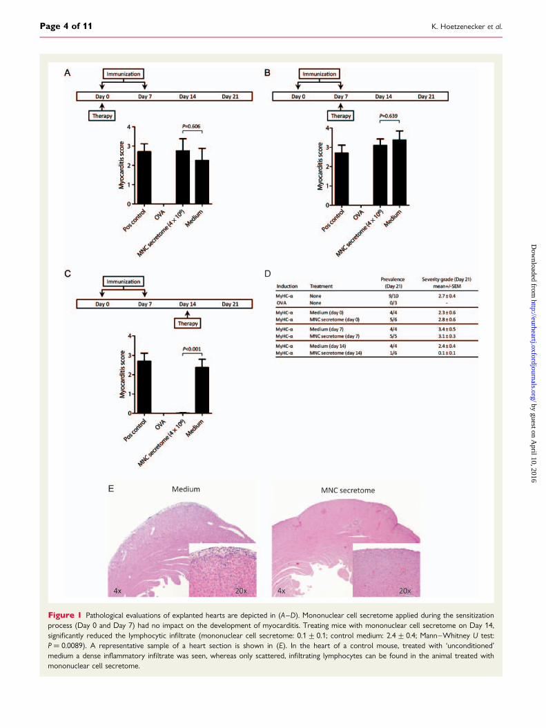
Figure 1 Pathological evaluations of explanted hearts are depicted in (A–D). Mononuclear cell secretome applied during the sensitizationprocess (Day 0 and Day 7) had no impact on the development of myocarditis. Treating mice with mononuclear cell secretome on Day 14,significantly reduced the lymphocytic infiltrate (mononuclear cell secretome: 0.1+0.1; control medium: 2.4+0.4; Mann–Whitney U test:P ¼ 0.0089). A representative sample of a heart section is shown in (E). In the heart of a control mouse, treated with ‘unconditioned’medium a dense inflammatory infiltrate was seen, whereas only scattered, infiltrating lymphocytes can be found in the animal treated withmononuclear cell secretome.
K. Hoetzenecker et al.Page 4 of 11
by guest on April 10, 2016
http://eurheartj.oxfordjournals.org/D
ownloaded from
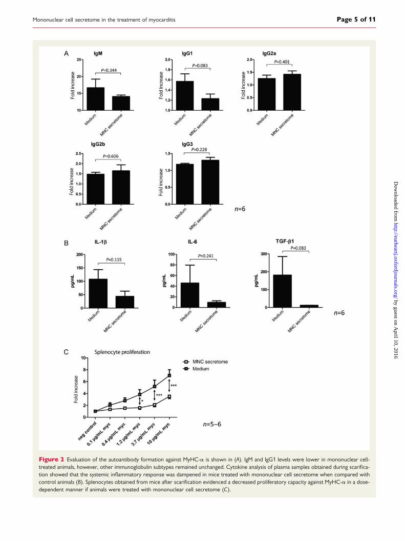
Figure 2 Evaluation of the autoantibody formation against MyHC-a is shown in (A). IgM and IgG1 levels were lower in mononuclear cell-treated animals, however, other immunoglobulin subtypes remained unchanged. Cytokine analysis of plasma samples obtained during scarifica-tion showed that the systemic inflammatory response was dampened in mice treated with mononuclear cell secretome when compared withcontrol animals (B). Splenocytes obtained from mice after scarification evidenced a decreased proliferatory capacity against MyHC-a in a dose-dependent manner if animals were treated with mononuclear cell secretome (C).
Mononuclear cell secretome in the treatment of myocarditis Page 5 of 11
by guest on April 10, 2016
http://eurheartj.oxfordjournals.org/D
ownloaded from

Splenocyte proliferation to MyHC-a614–629is strongly impaired in mononuclear cellsecretome-treated animalsPrevious studies have shown that proliferative responses to themyosin peptide in vitro are strongly linked to the development ofEAM.22,23 We, therefore, isolated splenocytes from immunizedmice, treated with either MNC secretome or control mediumon Day 21. Splenocytes were stimulated with different concentra-tions of MyHC-a and proliferation was assessed by measuring3[H]-thymidine uptake. As shown in Figure 2C, proliferation of sple-nocytes obtained from MNC secretome-treated animals was sig-nificantly impaired as calculated by ANOVA (Figure 2C).
Mononuclear cell secretome obtainedfrom mouse splenocytes is comparablewith mononuclear cell secretomeobtained from human peripheral bloodmononuclear cellsAs a proof of principle that MNC secretome produced frommouse splenocyte cultures is comparable with MNC secretomefrom human PBMC cultures, we evaluated the distribution ofCD4+ T cells, CD8+ T cells, B cells, and monocytes and per-formed cytokine arrays with both secretomes. Although spleno-cytes contained markedly more B cells, levels of secretedproteins were comparable in both preparation (Supplementarymaterial online, Figure S2A and B, Figure 3A). Both secretomes con-tained considerable amounts of IL-1Ra, IL-16, MCP-1, RANTES,and sICAM-1. All other tested cytokines and chemokines wereonly present in low concentrations.
Mononuclear cell secretome suppressesproliferation of CD41 T cells in vitro buthas no impact on dendritic cell functionExperimental autoimmune myocarditis is a CD4+ T cell-mediateddisease. We, therefore, investigated the effect of MNC secretomeon CD4+ cell proliferation in vitro. First, purified human CD4+cells were stimulated either with PHA or with a monoclonal anti-body to the T cell receptor. The addition of MNC secretome tothe stimulation assays significantly reduced the proliferative re-sponse in a dose-dependent manner (Figure 3B). Since MNC secre-tome treatment on Day 0 and Day 7 had no impact on thedevelopment of myocarditis and dendritic cells (DCs) are consid-ered pivotal during this sensitization process, we sought to furtherhighlight the role of MNC secretome on DC function. Althoughthe maturation of monocyte-derived dendritic cells seemed tobe slightly impaired by MNC secretome treatment, endocytosis ac-tivity was not influenced (Supplementary material online, FigureS3A and B).
Mononuclear cell secretome inducesapoptosis in CD41 T cells, JURKAT, andmurine T cell lymphoma cellsNext, we evaluated the effect of MNC secretome on cell viability.Co-incubation of unstimulated CD4+ T cells, a JURKAT cell line
or a mouse lymphoma cell line with MNC secretome resulted inan apoptosis induction as determined by Annexin V/PI co-stainingand by histone release assays (Figure 3C and E, Supplementary ma-terial online, Figure S2C ). To exclude direct cytotoxic effects ofMNC secretome, we pre-incubated cells with a pan-caspase inhibi-tor. Induction of apoptosis was inhibited by adding Z-VAD to theexperimental setting (MNC secretome: 1.56+0.11 O.D.; MNCsecretome + Z-VAD 20 mM: 0.09+0.07 O.D.; P ¼ 0.008; MNCsecretome + Z-VAD 100 mM: 0.01+0.01 O.D.; P ¼ 0.005;Figure 3D).
Caspase blocking experimentsTo define whether apoptosis is mediated through external ormitochondrial pathways, we selectively blocked caspase-9,caspase-8, and caspase-3. Pre-incubation of purified CD4+ Tcells with caspase-8 and caspase-3 but not caspase-9 inhibitorsresulted in a significantly reduced Annexin staining (Figure 3F).These observations indicate that the external pathway is involvedin MNC secretome-mediated apoptosis. Consequently, we evalu-ated known apoptosis-inducing factors in the secretome. As illu-strated in Figure 3G, TNF-a, sCD40L, sFASL, and sFAS were onlypresent in low concentrations in the MNC secretome. These find-ings suggest that the pro-apoptotic capacity is largely mediated bystill unknown factor(s) or by a not yet understood interplaybetween several specific factors within the secretome.
Blocking antibodies against knownpro-apoptotic are ineffective in reversingCD41 T cell apoptosisTo characterize the role of known pro-apoptotic factors in theapoptosis-inducing capacity of MNC secretome we co-incubatedpurified CD4+ T cells with blocking antibodies directed againstCD40L, TRAIL1, TRAIL2, and FASL. Antibodies directed againstdifferent chemokines without an apoptosis-inducing capacity(VEGF, IL8, ENA78, and MMP9) served as control. Histonerelease of MNC secretome-treated CD4+ cells was notreduced by blocking these factors, indicating that MNC secretomedisplay its cell death inducing feature aside commonly acceptedpathways (Figure 3H).
CD4/CD8 cell ratio is reduced inmononuclear cell secretome-treatedanimalsOn the basis of our in vitro findings, we measured CD4+ andCD8+ cells 12 and 36 h after treating EAM animals with MNCsecretome or control medium. The CD4/CD8 ratio was reducedin mice receiving the treatment when compared with controlanimals although this trend reached significance only at the 36 htimepoint (12 h: 2.1+ 0.3 vs. 1.7+0.4; P ¼ 0.441; 36 h: 2.9+0.2 vs. 2.0+ 0.1; P ¼ 0.007; Table 1). In parallel, the number of7-AAD positive circulating CD4+ cells was increased in MNCsecretome-treated animals when compared with the controlgroup.
K. Hoetzenecker et al.Page 6 of 11
by guest on April 10, 2016
http://eurheartj.oxfordjournals.org/D
ownloaded from
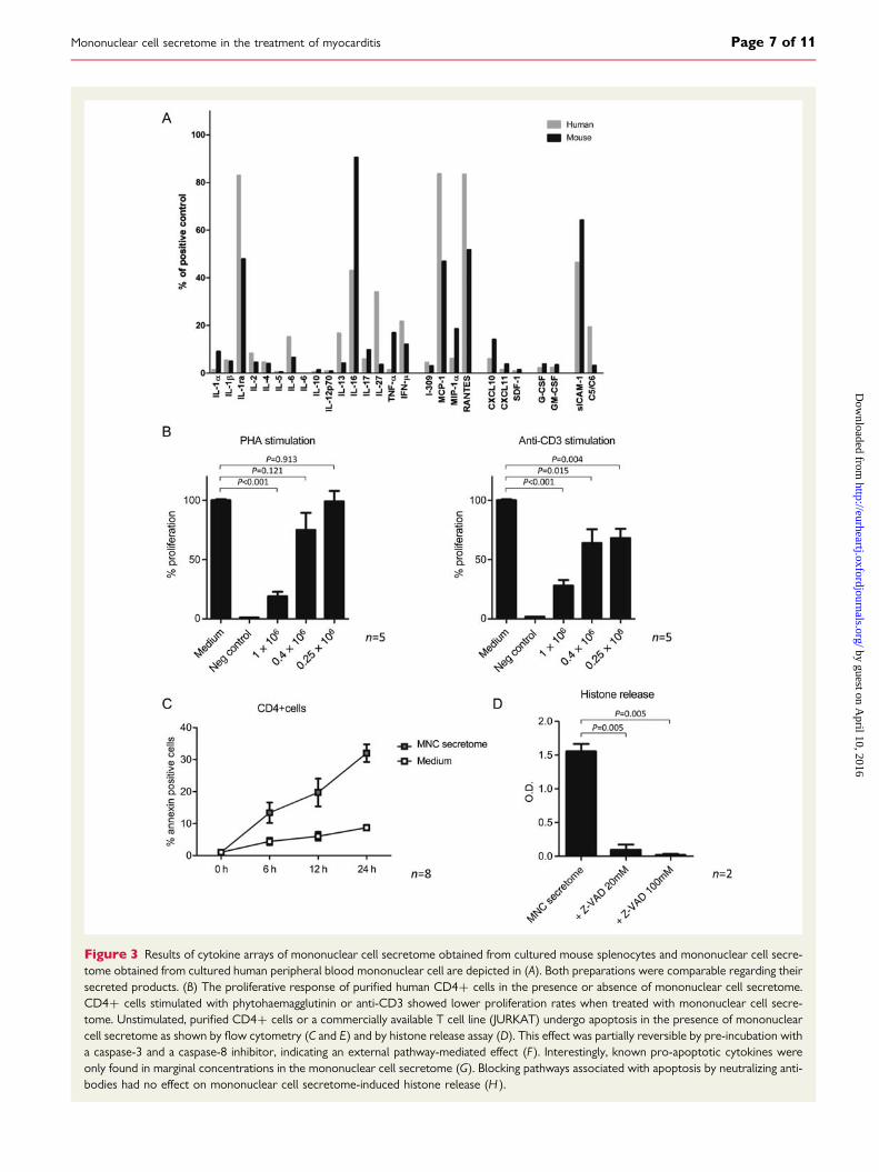
Figure 3 Results of cytokine arrays of mononuclear cell secretome obtained from cultured mouse splenocytes and mononuclear cell secre-tome obtained from cultured human peripheral blood mononuclear cell are depicted in (A). Both preparations were comparable regarding theirsecreted products. (B) The proliferative response of purified human CD4+ cells in the presence or absence of mononuclear cell secretome.CD4+ cells stimulated with phytohaemagglutinin or anti-CD3 showed lower proliferation rates when treated with mononuclear cell secre-tome. Unstimulated, purified CD4+ cells or a commercially available T cell line (JURKAT) undergo apoptosis in the presence of mononuclearcell secretome as shown by flow cytometry (C and E) and by histone release assay (D). This effect was partially reversible by pre-incubation witha caspase-3 and a caspase-8 inhibitor, indicating an external pathway-mediated effect (F ). Interestingly, known pro-apoptotic cytokines wereonly found in marginal concentrations in the mononuclear cell secretome (G). Blocking pathways associated with apoptosis by neutralizing anti-bodies had no effect on mononuclear cell secretome-induced histone release (H ).
Mononuclear cell secretome in the treatment of myocarditis Page 7 of 11
by guest on April 10, 2016
http://eurheartj.oxfordjournals.org/D
ownloaded from

DiscussionIn this study, we showed for the first time that a systemic, high-dose application of MNC secretome attenuates EAM. In vitro ana-lysis revealed an apoptosis-inducing effect of MNC secretome onCD4+ T cells. This observation was reversible by blocking the ex-ternal apoptosis pathway.
Myocarditis is one of the leading causes for iDCM. The patho-physiology underlying the disease is still not completely under-stood. Nevertheless, autoimmunity is considered a key factorpromoting ongoing inflammation, fibrosis, and pathological
remodelling. Accordingly, specific subgroups of affected patientsmay take advantage of immunosuppressive treatment. However,first clinical trials testing immunosuppression for acute myocarditisfailed. In a study by Parrillo et al.24 no advantage of immunosup-pressive treatment was found. The Myocarditis Treatment Trial,comparing a placebo group to two immunosuppressive regimens(prednisolone and azathioprine or prednisolone and cyclosporine)came up with similar results a few years later.4 Both trails suggestthat immunosuppression is not an option for the treatment ofacute, viral myocarditis and this view was followed in the guide-lines.25,26 The question of immunosuppression for myocarditis
Figure 3 Continued.
K. Hoetzenecker et al.Page 8 of 11
by guest on April 10, 2016
http://eurheartj.oxfordjournals.org/D
ownloaded from

was readdressed when knowledge on pathophysiological aspectsof myocarditis increased. Whereas in earlier studies patientswere recruited without excluding cases of an acute viral myocardi-tis, Wojnicz et al.27 treated patients with chronic inflammatoryheart disease and increased HLA expression on heart biopsies,with either prednisolone and azathioprine or placebo for 3months. LVEF improved significantly in the immunosuppressiongroup, even 2 years after treatment. These encouraging resultswere confirmed by Frustaci et al.6,28 on patients which fulfilled cri-teria for inflammatory heart disease but had no evidence of viralgenome in biopsy samples. Future clinical trials testing immunomo-dulatory or immunosuppressive drug regimens should carefully dis-tinguish between patients with acute viral myocarditis and chronicinflammatory heart disease where autoimmunity is the prevailingcause for ongoing disease after clearance of the virus. Tests meas-uring autoantibody load might help to better define forms of auto-immune myocarditis and could be valuable to monitor diseaseseverity in the future.29
The EAM model was first described by Neu et al.30 The experi-mental basis of the EAM model is an immunization with a cardiacspecific peptide—MyHC-a. Susceptible mouse strains such asBalb/c are immunized by a subcutaneously injection of an homolo-gous a-myosin fragment together with a strong adjuvant.15 TheEAM model is currently considered the best available model mim-icking autoimmune mechanisms of inflammatory heart disease. Itoffers the great advantage to study disease pathogenesis and treat-ment effects in vivo in the absence of an infective agent.31 However,despite the advantage in testing new and promising therapeutictargets, data from animal models should be estimated withcaution and must not uncritically be extrapolated to the humansystem.
We have found that treating mice with MNC secretome inhib-ited the development of an autoimmune myocarditis. Thisfinding, however, was restricted to an application of the compoundon Day 14, because treatment on Day 0 and Day 7 had no impacton disease severity. The reason for this might be a time-limitedeffect of MNC secretome on CD4+ cell suppression in vivo. Inthe EAM model, the injected myosin fragment persists at the de-position site and the injected MyHC-a/PBS/CFA suspension canbe still found at the time of scarification when opening the inguinalregion. On the other hand the half-life of the MNC secretome is
currently unknown, however, as the effective components aremost likely peptides/proteins a rapid decline in function can beconsidered within 24 h. Another explanation for the time-dependent efficacy of MNC secretome could be distinct immuno-logical processes at different stages of EAM. During the sensitiza-tion phase dendritic cell function is crucial. Dendritic cells takeup the injected myosin homologue, process it, and present it tonaıve CD4+ T cells. Interestingly, MNC secretome had only amarginal impact on DC function. Maturation to LPS stimulationwas only minimally impaired and endocytosis was unaltered inthe presence of MNC secretome.
A major limitation of this study is that mice were treated by asingle-dose protocol and the effect was only monitored on Day21. Data on long-term effects of MNC secretome treatment arestill missing. In addition, myocarditis is a chronic disease in the clin-ical scenario, therefore, repeated treatment for a longer timeperiod is necessary. We plan to address these two questions ina future study.
Stem cells have been shown to possess—besides their regenera-tive capacity—considerable immunomodulatory features, e.g. theycan effectively reduce lymphocyte proliferation in vitro.32 It hasbeen suggested that these anti-proliferative effects are mediated,at least partly, via paracrine mechanisms.10,33 The idea of using‘conditioned’ medium from stem cell cultures instead of stemcells has recently developed mainly supported by research in thefield of regenerative medicine. Several groups have so far reportedencouraging results of using the secretome of mesenchymal orbone marrow-derived stem cells in treating myocardial infarc-tion.7,8,34,35 We have recently expanded the concept of regenera-tory, stem cell-derived paracrine factors, by showing that thesecretome of PBMC also mediates myocyte protection followingmyocardial ischaemia.11– 13 Our findings were corroborated bythe work of Wollert and colleagues36 showing in a detailed analysisthat the secretome derived from stem cells only slightly differsfrom the secretome from peripheral blood leucocytes. A majoradvantage of using paracrine factors from PBMC instead of stemcells is that they are easily accessible. Our protocol of dialysisand lyophilization was developed for an off-the shelf scenario forfuture clinical applications. For the dialysis step, a cut-off of3.5 kD was used to avoid a loss of proteins. Neither dialysis norlyophilization had an impact on observed effects (unpublished
. . . . . . . . . . . . . . . . . . . . . . . . . . . . . . . . . . . . . . . . . . . . . . . . . . . . . . . . . . . . . . . . . . . . . . . . . . . . . . . . . . . . . . . . . . . . . . . . . . . . . . . . . . . . . . . . . . . . . . . . . . . . . . . . . . . . . . . . . . . . . . . . . . . . . . . . . . . . . . . . . . . . . . . . . . . . . . .
Table 1 The CD41 and CD81 cell counts in whole-blood samples obtained 12 and 36 h after mononuclear cellsecretome treatment (n 5 4–5)
CD41 (%) CD81 (%) CD4/CD8 ratio CD41/7-AAD pos
12 h
Medium 19.9+1.3 10.8+1.9 2.1+0.3 8.1+0.7
MNC secretome 13.3+1.0 9.3+2.0 1.7+0.4 8.8+2.8
36 h
Medium 23.9+1.6 7.4+1.1 2.9+0.2 5.4+0.5
MNC secretome 19.1+2.4 9.3+0.5 2.0+0.1 10.6+1.1
The CD4+/CD8+ cell ratio was reduced in treated animals when compared with controls. Additionally, CD4+/7-AAD positive cells were found more frequently.
Mononuclear cell secretome in the treatment of myocarditis Page 9 of 11
by guest on April 10, 2016
http://eurheartj.oxfordjournals.org/D
ownloaded from
data). In the clinical setting, MNC secretome could be produced inanalogy to other ‘biologicals’ (e.g. i.v. immunoglobulins) from blooddonations of healthy volunteers. However, strict regulatory prere-quisites (e.g. virus inactivation, potency assays, and mandated GMPfacilities) have to be met in order to reach human clinical trials.
The main mechanistic finding of this work is the capacity ofMNC secretome to induce apoptosis of CD4+ T cells in vitroand in vivo. We have thoroughly evaluated this observation inprimary CD4+ T cell cultures, a human JURKAT, and a mouseCD4+ T cell lymphoma cell line. Although the observed effectmight not be limited to T-helper cells, suppressing the CD4+cell function is substantial in regard to treating myocarditis. Previ-ous work has shown that treatment with anti-CD4 monoclonalantibody significantly improved cardiac functional parameters in arat myocarditis model. In addition, lymphocytes obtained fromtreated animals showed no proliferative response after in vitrostimulation with a myosin fragment.18,20 In a tedious work by Vala-perti et al.,16 the importance of CD4+ cell function in the EAMmodel was addressed by showing that treatment with CD11b+monocytes, suppressed the CD4+ dependent, MyHC-a-specificautoimmune response. These findings are supported by the clinicalobservation that T cell depletion is a possible rescue therapy forfulminant autoimmune myocarditis.37,38
Autoantibody formation is a well-described feature in the patho-genesis of myocarditis. Antibodies against a wide range of recep-tors, mitochondrial, and contractile proteins have been foundboth in human and in animal models.39 Myosin-specific antibodiesare detectable in 46% of sera from patients suffering from dilatedcardiomyopathy in western blot analysis.40 In contrast to this clin-ical observation, autoantibody formation is not directly involved inthe development of myocardial infiltrates in the EAM model, sinceB-cell deficient mice still develop a myocarditis.22 However, levelsof anti-myosin antibodies can be considered a surrogate marker tomonitor disease severity in the EAM model. In our study,decreased circulating anti-myosin IgG1 and IgM together withreduced levels of IL-1b, IL-6, and TGF-b underline the therapeuticeffect of MNC secretome.
One limitation of this study is that the MNC secretome by def-inition comprises of a myriad of proteins.36 Currently, although adetailed mapping of the protein content of MNC secretome hasbeen performed, we have only limited knowledge regarding thefactors mediating observed effects. Unfortunately, this is an un-solved problem for most of the work done in the field of secre-tome research. In some studies, potential target proteins wereinactivated by blocking antibodies, however, effects were uniformlyat most partially reversible.12,35,41 In this present study, we tried tocorrelate the apoptosis-inducing capacity of MNC secretome toknown apoptosis-relevant factors. TNF-a, sCD40L, and sFASwere only marginally present in MNC secretome, sFASL was notdetectable as determined by the ELISA technique. In addition,blocking different pathways associated with programmed celldeath by neutralizing antibodies did not affect histone release inCD4+ cell cultures. Interestingly, non-protein mediated mechan-isms of conditioned medium were put up for discussion recently.Timmers and collegues42 could show that exosomes consistingof cholesterol, sphingomyelin, and phosphatidylcholine-mediatedcardioprotective effect.
To the best of our knowledge, this is the first study evaluatingimmunosuppressive features of a high-dose application of MNCsecretome in the murine EAM model. Further studies are war-ranted to perpetuate the concept of using MNC secretome forthe treatment of myocarditis.
Supplementary materialSupplementary material is available at European Heart Journalonline.
AcknowledgementsWe are thankful to Barbara Bohle, Veronika Sexl, Barbara Neudertand Christoph Inci for intellectual and technical assistance. U.E. andP.B acknowledge support from the Swiss National Foundation.
FundingThis study was funded by the Christian Doppler Research Associationand the Medical University of Vienna. The Medical University of Viennahas claimed financial interest (patent number: EP2201954,WO2010070105-A1, filed 18 December 2008). Funding to pay theOpen Access publication charges for this article was provided by theChristian Doppler Laboratory for Cardiac and Thoracic Diagnosisand Regeneration.
Conflict of interest: H.J.A. is a shareholder of APOSIENCE AG,which owns the rights to commercialize MNC secretome for thera-peutic use.
References1. Feldman AM, McNamara D. Myocarditis. N Engl J Med 2000;343:1388–1398.2. Cooper LT Jr. Myocarditis. N Engl J Med 2009;360:1526–1538.3. Maisch B, Hufnagel G, Schonian U, Hengstenberg C. The European Study of Epi-
demiology and Treatment of Cardiac Inflammatory Disease (ESETCID). Eur HeartJ 1995;16(Suppl O):173–175.
4. Mason JW, O’Connell JB, Herskowitz A, Rose NR, McManus BM, Billingham ME,Moon TE. A clinical trial of immunosuppressive therapy for myocarditis. The Myo-carditis Treatment Trial Investigators. N Engl J Med 1995;333:269–275.
5. Hia CP, Yip WC, Tai BC, Quek SC. Immunosuppressive therapy in acute myocar-ditis: an 18 year systematic review. Arch Dis Child 2004;89:580–584.
6. Frustaci A, Russo MA, Chimenti C. Randomized study on the efficacy of immuno-suppressive therapy in patients with virus-negative inflammatory cardiomyopathy:the TIMIC study. Eur Heart J 2009;30:1995–2002.
7. Gnecchi M, He H, Liang OD, Melo LG, Morello F, Mu H, Noiseux N, Zhang L,Pratt RE, Ingwall JS, Dzau VJ. Paracrine action accounts for marked protectionof ischemic heart by Akt-modified mesenchymal stem cells. Nat Med 2005;11:367–368.
8. Angoulvant D, Ivanes F, Ferrera R, Matthews PG, Nataf S, Ovize M. Mesenchymalstem cell conditioned media attenuates in vitro and ex vivo myocardial reperfusioninjury. J Heart Lung Transplant 2011;30:95–102.
9. Horn AP, Frozza RL, Grudzinski PB, Gerhardt D, Hoppe JB, Bruno AN,Chagastelles P, Nardi NB, Lenz G, Salbego C. Conditioned medium from mesen-chymal stem cells induces cell death in organotypic cultures of rat hippocampusand aggravates lesion in a model of oxygen and glucose deprivation. Neurosci Res2009;63:35–41.
10. Kim SY, Cho HS, Yang SH, Shin JY, Kim JS, Lee ST, Chu K, Roh JK, Kim SU,Park CG. Soluble mediators from human neural stem cells play a critical role insuppression of T-cell activation and proliferation. J Neurosci Res 2009;87:2264–2272.
11. Ankersmit HJ, Hoetzenecker K, Dietl W, Soleiman A, Horvat R, Wolfsberger M,Gerner C, Hacker S, Mildner M, Moser B, Lichtenauer M, Podesser BK. Irradiatedcultured apoptotic peripheral blood mononuclear cells regenerate infarcted myo-cardium. Eur J Clin Invest 2009;39:445–456.
12. Lichtenauer M, Mildner M, Hoetzenecker K, Zimmermann M, Podesser BK,Sipos W, Berenyi E, Dworschak M, Tschachler E, Gyongyosi M, Ankersmit HJ.Secretome of apoptotic peripheral blood cells (APOSEC) confers cytoprotection
K. Hoetzenecker et al.Page 10 of 11
by guest on April 10, 2016
http://eurheartj.oxfordjournals.org/D
ownloaded from
to cardiomyocytes and inhibits tissue remodelling after acute myocardial infarc-tion: a preclinical study. Basic Res Cardiol 2011;106:1283–1297.
13. Lichtenauer M, Mildner M, Baumgartner A, Hasun M, Werba G, Beer L,Altmann P, Roth G, Gyongyosi M, Podesser BK, Ankersmit HJ. Intravenous andintramyocardial injection of apoptotic white blood cell suspensions prevents ven-tricular remodelling by increasing elastin expression in cardiac scar tissue aftermyocardial infarction. Basic Res Cardiol 2011;106:645–655.
14. Hoetzenecker K, Assinger A, Lichtenauer M, Mildner M, Schweiger T, Starlinger P,Jakab A, Berenyi E, Pavo N, Zimmermann M, Gabriel C, Plass C, Gyongyosi M,Volf I, Ankersmit HJ. Secretome of apoptotic peripheral blood cells (APOSEC)attenuates microvascular obstruction in a porcine closed chest reperfusedacute myocardial infarction model: role of platelet aggregation and vasodilation.Basic Res Cardiol 2012;107:292.
15. Pummerer CL, Luze K, Grassl G, Bachmaier K, Offner F, Burrell SK, Lenz DM,Zamborelli TJ, Penninger JM, Neu N. Identification of cardiac myosin peptidescapable of inducing autoimmune myocarditis in BALB/c mice. J Clin Invest 1996;97:2057–2062.
16. Valaperti A, Marty RR, Kania G, Germano D, Mauermann N, Dirnhofer S,Leimenstoll B, Blyszczuk P, Dong C, Mueller C, Hunziker L, Eriksson U.CD11b+ monocytes abrogate Th17 CD4+ T cell-mediated experimental auto-immune myocarditis. J Immunol 2008;180:2686–2695.
17. Smith SC, Allen PM. Myosin-induced acute myocarditis is a T cell-mediateddisease. J Immunol 1991;147:2141–2147.
18. Yuan HT, Liao YH, Wang Z, Dong JH, Cao LS, Wang ZH, Wang JP, Fu ML. Pre-vention of myosin-induced autoimmune myocarditis in mice by anti-L3T4 mono-clonal antibody. Can J Physiol Pharmacol 2003;81:84–88.
19. Inomata T, Watanabe T, Haga M, Hirahara H, Abo T, Okura Y, Hanawa H,Kodama M, Izumi T. Anti-CD2 monoclonal antibodies prevent the induction ofexperimental autoimmune myocarditis. Jpn Heart J 2000;41:507–517.
20. Wang QQ, Wang YL, Yuan HT, Liu FQ, Jin YP, Han B. Immune tolerance tocardiac myosin induced by anti-CD4 monoclonal antibody in autoimmune myo-carditis rats. J Clin Immunol 2006;26:213–221.
21. Eriksson U, Ricci R, Hunziker L, Kurrer MO, Oudit GY, Watts TH, Sonderegger I,Bachmaier K, Kopf M, Penninger JM. Dendritic cell-induced autoimmune heartfailure requires cooperation between adaptive and innate immunity. Nat Med2003;9:1484–1490.
22. Eriksson U, Kurrer MO, Schmitz N, Marsch SC, Fontana A, Eugster HP, Kopf M.Interleukin-6-deficient mice resist development of autoimmune myocarditis asso-ciated with impaired upregulation of complement C3. Circulation 2003;107:320–325.
23. Eriksson U, Kurrer MO, Sebald W, Brombacher F, Kopf M. Dual role of the IL-12/IFN-gamma axis in the development of autoimmune myocarditis: induction byIL-12 and protection by IFN-gamma. J Immunol 2001;167:5464–5469.
24. Parrillo JE, Cunnion RE, Epstein SE, Parker MM, Suffredini AF, Brenner M,Schaer GL, Palmeri ST, Cannon RO III, Alling D, Wittes J, Ferrans V,Rodriguez R, Fauci A. A prospective, randomized, controlled trial of prednisonefor dilated cardiomyopathy. N Engl J Med 1989;321:1061–1068.
25. Howlett JG, McKelvie RS, Arnold JM, Costigan J, Dorian P, Ducharme A,Estrella-Holder E, Ezekowitz JA, Giannetti N, Haddad H, Heckman GA,Herd AM, Isaac D, Jong P, Kouz S, Liu P, Mann E, Moe GW, Tsuyuki RT,Ross HJ, White M. Canadian Cardiovascular Society Consensus Conferenceguidelines on heart failure, update 2009: diagnosis and management of right-sidedheart failure, myocarditis, device therapy and recent important clinical trials. Can JCardiol 2009;25:85–105.
26. Guidelines for diagnosis and treatment of myocarditis (JCS 2009): digest version.Circ J 2011;75:734–743.
27. Wojnicz R, Nowalany-Kozielska E, Wojciechowska C, Glanowska G,Wilczewski P, Niklewski T, Zembala M, Polonski L, Rozek MM, Wodniecki J. Ran-domized, placebo-controlled study for immunosuppressive treatment of inflam-matory dilated cardiomyopathy: two-year follow-up results. Circulation 2001;104:39–45.
28. Frustaci A, Chimenti C, Calabrese F, Pieroni M, Thiene G, Maseri A. Immunosup-pressive therapy for active lymphocytic myocarditis: virological and immunologicprofile of responders versus nonresponders. Circulation 2003;107:857–863.
29. Caforio AL, Tona F, Bottaro S, Vinci A, Dequal G, Daliento L, Thiene G, Iliceto S.Clinical implications of anti-heart autoantibodies in myocarditis and dilated car-diomyopathy. Autoimmunity 2008;41:35–45.
30. Neu N, Rose NR, Beisel KW, Herskowitz A, Gurri-Glass G, Craig SW. Cardiacmyosin induces myocarditis in genetically predisposed mice. J Immunol 1987;139:3630–3636.
31. Kania G, Blyszczuk P, Eriksson U. Mechanisms of cardiac fibrosis in inflammatoryheart disease. Trends Cardiovasc Med 2009;19:247–252.
32. Le Blanc K, Tammik L, Sundberg B, Haynesworth SE, Ringden O. Mesenchymalstem cells inhibit and stimulate mixed lymphocyte cultures and mitogenicresponses independently of the major histocompatibility complex. Scand JImmunol 2003;57:11–20.
33. Rasmusson I, Ringden O, Sundberg B, Le Blanc K. Mesenchymal stem cells inhibitlymphocyte proliferation by mitogens and alloantigens by different mechanisms.Exp Cell Res 2005;305:33–41.
34. Fazel S, Cimini M, Chen L, Li S, Angoulvant D, Fedak P, Verma S, Weisel RD,Keating A, Li RK. Cardioprotective c-kit+ cells are from the bone marrow andregulate the myocardial balance of angiogenic cytokines. J Clin Invest 2006;116:1865–1877.
35. Di Santo S, Yang Z, Wyler von Ballmoos M, Voelzmann J, Diehm N,Baumgartner I, Kalka C. Novel cell-free strategy for therapeutic angiogenesis: invitro generated conditioned medium can replace progenitor cell transplantation.PLoS One 2009;4:e5643.
36. Korf-Klingebiel M, Kempf T, Sauer T, Brinkmann E, Fischer P, Meyer GP, Ganser A,Drexler H, Wollert KC. Bone marrow cells are a rich source of growth factorsand cytokines: implications for cell therapy trials after myocardial infarction. EurHeart J 2008;29:2851–2858.
37. Ankersmit HJ, Ullrich R, Moser B, Hoetzenecker K, Hacker S, German P, Krenn C,Horvat R, Grimm M, Wolner E, Zuckermann A. Recovery from giant cell myocar-ditis with ECMO support and utilisation of polyclonal antithymocyte globulin: acase report. Thorac Cardiovasc Surg 2006;54:278–280.
38. Cooper LT Jr, Hare JM, Tazelaar HD, Edwards WD, Starling RC, Deng MC,Menon S, Mullen GM, Jaski B, Bailey KR, Cunningham MW, Dec GW. Usefulnessof immunosuppression for giant cell myocarditis. Am J Cardiol 2008;102:1535–1539.
39. Kallwellis-Opara A, Dorner A, Poller WC, Noutsias M, Kuhl U, Schultheiss HP,Pauschinger M. Autoimmunological features in inflammatory cardiomyopathy.Clin Res Cardiol 2007;96:469–480.
40. Caforio AL, Grazzini M, Mann JM, Keeling PJ, Bottazzo GF, McKenna WJ,Schiaffino S. Identification of alpha- and beta-cardiac myosin heavy chain isoformsas major autoantigens in dilated cardiomyopathy. Circulation 1992;85:1734–1742.
41. Yang Z, von Ballmoos MW, Faessler D, Voelzmann J, Ortmann J, Diehm N,Kalka-Moll W, Baumgartner I, Di Santo S, Kalka C. Paracrine factors secretedby endothelial progenitor cells prevent oxidative stress-induced apoptosis ofmature endothelial cells. Atherosclerosis 2010;211:103–109.
42. Lai RC, Arslan F, Lee MM, Sze NS, Choo A, Chen TS, Salto-Tellez M, Timmers L,Lee CN, El Oakley RM, Pasterkamp G, de Kleijn DP, Lim SK. Exosome secretedby MSC reduces myocardial ischemia/reperfusion injury. Stem Cell Res 2010;4:214–222.
Mononuclear cell secretome in the treatment of myocarditis Page 11 of 11
by guest on April 10, 2016
http://eurheartj.oxfordjournals.org/D
ownloaded from


