Embed Size (px)
Citation preview
MOLECULAR VARIATION IN COCONUT
CaDANG-CADANG VTROID (cccvd)
MARIA JUDITH B RODRIGUEZ
BSAgChem (University of the Philippines at Los Banos, PHILIPPINES)
Department of CroP Protection
Waite Agricultural Research Institute
The UniversitY of Adelaide
South Australia
Thesis submitted to rhe University of Adelaide in fulfiltment of the requirement
for the degree of Doctor of Philosophy
March, 1993
TABLE OF CONTENTSPage
Table of Contents
Summary
Statement
Acknowledgements
Chapter 1 GENERAL INTRODUCTION
1.1 Viroids
1.T.2
1.1.3
r.1,.4
1.1.5
1.1.6
l.l .7
t.2
1.1.1 Some biological properties
1 . 1. 1a Symptomatology
1.1.1b Ecology and ePidemiologY
Classihcation according to sequence and structure
Structural domains
I n vivo mutations and recombinations
I n v itr o mutational an alysis of stmctureÆunction relationship s
Mechanism of replication
Control measures
VI
vl1l
ix
1
1
1
1
2
2
4
5
7
8
8
Coconut cadang-cadang viroid (CCCVd)
1.2.1 Cadang-cadang disease
l.2.Ia History,disributionandeconomicimportance
l.z.lb Symptoms and host range
\.2.1c EPidemiologY
1.2.2 Diagnostic methds
1.2.3 Physical properties
1.2.4 Va¡iation in nucleotide sequence
1.2.5 Correlation of CCCVd sructure with disease progress
L2.6 CCCVd as related to cTivd and the viroid-like sequences
in oil and coconut palms and other monocotyledons in the
south-west Pacific
9
9
9
10
11
11
t212
t3
13
Scope of this thesis t4
u
2.1
Chapter 2 MATERIALS AND METHODS
Materials
2.r.r2.r.2
2.r.32.r.42.t.5
CCCVd32P-labelled cRNA probe
Synthetic CCCVd-specific DNA oligonucleotide primers
Biochemicals and miscellaneous chemicals
Phenol reagents, polyacrylamide/agarose gels and bacterial
media
Isolation of low molecular weight nucleic acids from tissues
of plants other than coconut
Mini-preparation of low molecular weight nucleic acids from
coconut leaves
Large-scale preparation of low molecula¡ weight nucleic acids
from coconut leaves
Viroid purification
2.2.4a Two-dimensional (2-D) or bi-directional poly-
acrylamide gel elecnophoresis (PAGE)
2.2.4b Recovery of the viroids from polyacrylamide gel
PAGE assay
Molecular hybridization assay
2.2.6a Synthesisof32P-labelledsingle-stranded
complementary DNA (32P-ss cDNA) probes
2.2.6b Preparationofdot-bloVelectroblot
2.2.6c Hybridization
Amplification of viroids by polymerase chain reaction (PCR)
2.2.7 a Preparation of template
2.2.7b PCR reaction
Molecular cloning of PCR products
2.2.8a Purification of PCR products and preparation of
cloning vector
2.2.8b Ligation of ds cDNA to the T-vector
2.2.8c Preparation of competent cells
2.2.8d Transformation of the competent cells with the
recombinant plasmids
2.2.8e Selection for recombinants
Page
15
15
15
15
15
16
r6
16
16
t7
t7
18
18
18
19
t9
T9
20
2T
2T
2l22
23
2.2 Methods
2.2.r
2.2.2
2.2.3
2.2.4
2.2.5
2.2.6
2.2.7
2.2.8
23
23
24
24
25
2.2.9
2.2.r0
2.2.tr
2.2.r2
Mini-preparation of recombinant plasmids
Analysis of the sizes of inserts in recombinant plasmids
Fluorescent dye primer cycle sequencing
2.2.lla Preparation of dsDNA template
2.2.llb Cycle sequencing reaction
Sequence analysis and determination of secondary structure
Pase
lll
25
26
27
27
27
28
29
29
29
29
30
Chapter 3 DETECTION OF CCCVd-RELATED SEQTIENCES IN PLANTS OTT{ER
THAN PALM SPECIES
Introduction
Experimental
3.1 Collection of samples of plant species associated with coconut
plantations
3.2 Isolation of low molecula¡ weight nucleic acids
J.J Molecular hybridization analyses for CCCVd
3.3. 1 Dot-blot hybridization assays
3.3.2 Electroblot hybridization assays
3.3.2a 207o non-denaturing PAGE
3.3.2b Two-dimensional 7Vo PAGE
Discussion
Chapter 4 DETECIION OF ELECTROPHORETIC FORMS OF CCCVd
ASSOCIATED WITH SEVERE "BROOMING'' SYMPTOMS IN
COCONUT
Introduction
Experimental
Observation of severe "brooming" symptoms in CCCVd-inoculated
palms
30
31
32
32
32
33
34
34
34
34
4.r
4.2 Correlation of the brooming symptoms with distinct electrophoretic forms
of CCCVd
4.3 Variation in the viroid pattern during disease progression
Discussion
Chapter 5 PURIFICAION OF CCCVd-RELATED NUCLEIC ACIDS FOR
AMPLIFICATION BY POLYMERASE CHAIN REACTION (PCR)
AND THEIR SUBSEQUENT MOLECULAR CLONING
Introduction
Experimental
1V
Page
37
40
4l4l4T
42
42
43
36
3l
5.1
5.2
Purif,rcation
5.1.1
5.t.2CCCVd-related nucleic acids in plants other than palm species
Electrophoretic forms of CCCVd associated with the brooming
symptoms in coconut
38
38
39
39
39
40
Polymerase chain reaction (PCR)
5.2.1 Standardization of PCR
5.2.1a Design of primers and preparation of template
5.2.1b MgCl2concentration
5.2.2 Development of a one-tube, one manipulation reverse
transcription (RT) - PCR reaction method
5.2.3 PCR of CCCVd-related nucleic acids
5.3 Molecular cloning of PCR products
Discussion 44
v
Pase
Chapter 6 NUCLEOTIDE SEQUENCING OF TFIE SELECTED CLONES OF THE
ELECTROPHORETIC FORMS OF CCCVd ASSOCIATED WITH TI{E
SEVERE BROOMING SYMPTOMS IN COCONUT
Introduction
Experimental
6.1 Evaluation of the sequencing method
6.2 Sequence analysis and secondary structure determination
6.2.I Forms with elecrophoretic mobilities between the CCCVd
variants 247 and246
6.2.2 Forms with electrophoretic mobilities between and less
than the CCCVd variants 297 and296
Discussion
Chapter 7 GENERAL DISCUSSION
Appendix A
Appendix B
Appendix C
References
45
45
46
46
47
47
48
49
50
55
57
61
62
v1
SUMMARY
Molecular hybridization with 32P-labe[ed cRNA and cDNA probes in dot-blots and
electroblots and the use of two-dimensional polyacrylamide gel electrophoresis (2-D PAGE)
demonstrated the presence of multiple coconut cadang-cadang viroid (CCCVd)-like nucleic
acids in 11 understorey plant species in coconut plantations. These nucleic acids seemed to be
at a lower concentration in their hosts than CCCVd is in coconut. Of the multiple bands found
in each plant species, those which were approximately the same size as CCCVd usually had
high nucleotide sequence homology with the probe. In contrast, the bands outside the CCCVd
region had low homology with the probe. The use of the polymerase chain reaction (PCR)
with some of the isolates purified by 2-D PAGE failed to amplify CCCVd-specific nucleic acids
under the conditions developed for successful amplif,rcation of CCCVd.
The 11 plant species with CCCVd-like sequences are the monocotyledonous hosts
Maranta arundinacea, Alpinia sp., Zingiber fficinale, Canna flaccida, Commelina diffusa,
Commelina benghalensis, Paspalum conjugatum, Brachiaria distachya, Imperata cylindrica,
Bambusa blumeana and the dicotyledonous host Urena lobata. The samples were collected at
11 separate sites in five provinces of southern Luzon, Philippines. None showed symptoms
which could be associated with the presence of the viroid-like nucleic acids. The incidence in
these species was independent of the level of cadang-cadang disease in adjacent coconut
plantations, and it is suggested that these plants could act as a reservoir for CCCVd-related
nucleic acids in the Philippines.
An unusually severe type of symptom was observed in approximately I2Vo of
1,787 palms infected by artificial inoculation with preparations of CCCVd. The symptom was
first observed in palms 3-7 years after inoculation and is characterized by a loss of leaf lamina,
and is associated with severe stunting of the palm and occasional premature death. This
symptom has been termed "brooming". PAGE and molecular analyses showed that all palms
with brooming had patterns of CCCVd-related nucleic acids which differed from the patterns
observed with the common form of cadang-cadang disease. Thus, four bands representing
viroids wirh 246, 247, 296 and 297 nucleotides are observed in extracts from palms with
vll
common cadang-cadang disease. Palms with "brooming" show additional or replacement
bands which have electrophoretic mobilities distinct from the above forms of viroid. Bi-
directional PAGE was used to separate the brooming-associated nucleic acids from other host
nucleic acids. These nucleic acids were eluted as a mixture of the various electrophoretic
forms. Three distinct primers were used for reverse transcription of the nucleic acids. The
producrs were amplified by PCR with three pairs of primers. Products with the expected sizes
were inserted into the Bluescript plasmid vector by the use of single base A:T overlaps.
Twenty-eight full-length clones and 19 half-length clones were obtained,29 of which were
selected and sequenced by fluorescent dye primer cycle sequencing. A number of mutations
were observed in the clones, and they fell into the following classes: (i) substitution of C at
position 197 of the central conserved domain with either AU, UG, or UU; (ii) substitution of U
with A at position 216 in the P domain; (iii) addition of A at position 86 or 87 so that AA
became AAA at the boundary of the cenral conserved region and the V domain; (iv) mutants of
varying length resulting from partial sequence duplications of the V and T2 domains
commencing from varying positions in the right-hand end of the viroid molecule; and (v)
mutations at the boundaries of duplications.
Models were proposed showing optimal secondary structures. These showed that
the substitution of C at position 197 of the central conserved domain with either AU or UG
could lead to the replacement of a U-loop with a single base pair and that mutations at the
boundaries of partial sequence duplications could cause structural changes in the V and T2
domains. The other mutations appeared not to affect secondary structure.
As a mutation at position 197 in the central conserved domain has been previously
reported for CCCVd, it is concluded that this site may be important for the control of
pathogenicity of CCCVd.
A one-tube, reverse transcription-PCR reaction system was developed for use with
CCCVd.
vul
STATEMENT
This thesis contains no material which has been accepted for the award of any other
degree or diploma in any University and to the best of my knowledge contains no material
previously published or written by another person, except where due reference is made in the
text.
o
f¿- ò
I give consent to this copy of my thesis, when deposited in the university Library, being available
for loan and PhotocoPpng.
DATE: 4zSIGNED:
I
IX
ACKNOWLEDGEMENTS
I particularly wish to thank my supervisor, Dr JW Randles, for his encouragement,
interest and critical discussion throughout the course of this study. I also wish to thank Dr D
Hanold for her helpful discussion on diagnostic methods, Dr SW Ding for his valuable advice
on molecular cloning and Dr N Shirley for his assistance in the sequence analysis.
Thanks are also due to Mrs E Cabot for typing the manuscript, Ms J Groom for the
photography work, Mr G Doroja for the assistance in computer analysis and Ms C Cueto and
Mr G Baylon for their help in the collection of samples in the Philippines.
I gratefully acknowledge the Australian International Development Assistance
Bureau (AIDAB) for a scholarship and the Philippine Coconut Authority (PCA), my employer,
for granting me study leave. I thank, in particular, Mr C Carpio for encouraging me to study.
I also thank my parents, brothers, sisters, relatives and friends for the inspiration
and spiritual support.
WAlit' ii ¡';' ¡i iU]i:LIBRARY
CHAPTER 1
GENERAL TN{TRODUCTION
I.1 VIROIDS
Viroids are defined as unencapsidated, single-stranded, covalently closed circular,
low molecular weight RNAs with extensive regions of intramolecular complementarity (Diener,
lg79). The first viroid described was the causal agent for potato spindle tuber disease @iener,
1971; Gros s et al., 1978). Since then, additional plant diseases, most of which were formerly
believed to be caused by viruses, have been shown to be viroid-incited (Symons, 1981;Gross
et a\.,1982; Haseloff et a\.,1982; Ohno et a|.,1983; Sano et a|.,1984; Candresse ¿t al., 1987;
Keese et a1.,1988; Rezaian, 1990).
1. 1.1 Some biological properties
1. 1. 1a Symptomatology
In some hosts, viroids produce a range of symptoms similar to those observed in
viral diseases. Otherwise, they are frequently latent. It has been suggested that viroids and
viruses affect the same or similar metabolic pathways in infected cells (Diener, 1987).
The macroscopic symptoms include stunting, epinasty, veinal discoloration, leaf
distortions, vein clearing, localized chlorotic or necrotic spots, mottling of leaves, necrosis of
leaves and death of the whole plant. The cytopathic effects are chloroplast abnormalities,
distortion of cell walls and accumulation of electron-dense deposits. Although viroid infection
does not cause disturbances in the synthesis or degradation of host nucleic acids, it induces
disruption in the metabolism of growth substances and quantitative changes in host proteins'
However, no viroid-coded proteins have been identified in infected cells (Diener, 1987).
2
1.1. I b Ecology and epidemiology
Most viroids replicate best at relatively high temperatures (30-33'C). High
temperature and light intensity are also necessary for rapid symptom expression (Diener, 1987).
All known viroids are transmissible by mechanical means, either readily or with
some difficulty. For example, mechanical transmission by contact with farm implements is
mainly responsible for the spread of potato spindle tuber viroid (PSTVd) in nature. With other
viroids, contaminated budding knives and other tools have been implicated (Diener, 1987).
There have been numerous attempts to identify anthropod vectors of viroids but few
positive results have been obtained. An example is the transmission of tomato planta macho
viroid (TPMVd) by an aphid species, Myzus persicae (Galindo et al., 1989).
Some evidence for transmission of viroid through seed and pollen has been
reported. With PSTVd, both seed and pollen transmission have been demonstrated (Diener,
1981; Singh et al., 1992) while evidence of vertical transmission has been shown only with
avocado sunblotch viroid (ASBVd) (Diener, 1987).
1.1.2 Classification according to sequence and structure
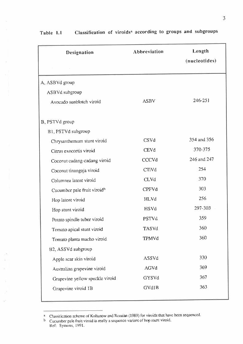
Sixteen distinct viroids which have been sequenced are listed in Table 1.1. Their
secondary structures have been predicted to give linear, rod-like structures. A classification
system has been established in which viroids are divided into two main groups based on their
conserved core sequences. ASBVd is so fa¡ the only member of one group, whereas the rest of
the viroids are further divided into two subgroups - B1, PSTVd subgroup and 82, ASSVd
subgroup. ASBVd does not contain either the PSTVd or the ASSVd core sequence and has a
higher A+IJ (62Vo) content than the other viroids. Furthermore, it has a self-cleavage site. 1¡¿
vitro, in the presence of divalent metal ions such as Mg+2 or Ca+2, multimers of ASBVd have
the ability to self-cleave into linear monomers. Members of the PSTVd and ASSVd subgroups
are easily distinguished by their conserved core sequence, with the PSTVd subgroup having a
U-bulged helix (Koltunow and Rezaian, 1989).
3
Table 1.1 Classification of viroidsa according to groups and subgroups
a Classilication scheme of Koltunow and Rezaian (1989) for viroids that have been sequenced.
b Cucumber pale fruit viroid is really a sequence variant of hop stunt viroid.Ref: Symons, 1991.
A, ASBVd goup
ASBVd subgroup
Avocado sunblotch viroid
B, PSTVd group
81, PSTVd subgroup
Chrysanthemum stunt viroid
Citrus exocortis viroid
Coconu t cadan g-cadang viroid
Coconut tinangaja viroid
Columnea latent viroid
Cucumber pale fruit viroidb
Hop latent viroid
Hop stunt viroid
Potato spindle tuber viroid
Tomato apical stunt viroid
Tomato planta macho viroid
82, ASSVd subgroup
Apple scar skin viroid
AusFalian grapevine viroid
Grapevine yellow speckle viroid
Grapevine viroid 1B
ASBV
CSVd
CEVd
CCCVd
CTiVd
CLVd
CPFVd
HLVd
HSVd
PSTVd
TASVd
TPMVd
ASSVd
AGVd
GYSVd
GVdIB
246-251
354 and356
370-375
246 and247
254
310
303
256
297-303
359
360
360
330
369
367
363
Designation Abbreviation Length
(nucleotides)
4
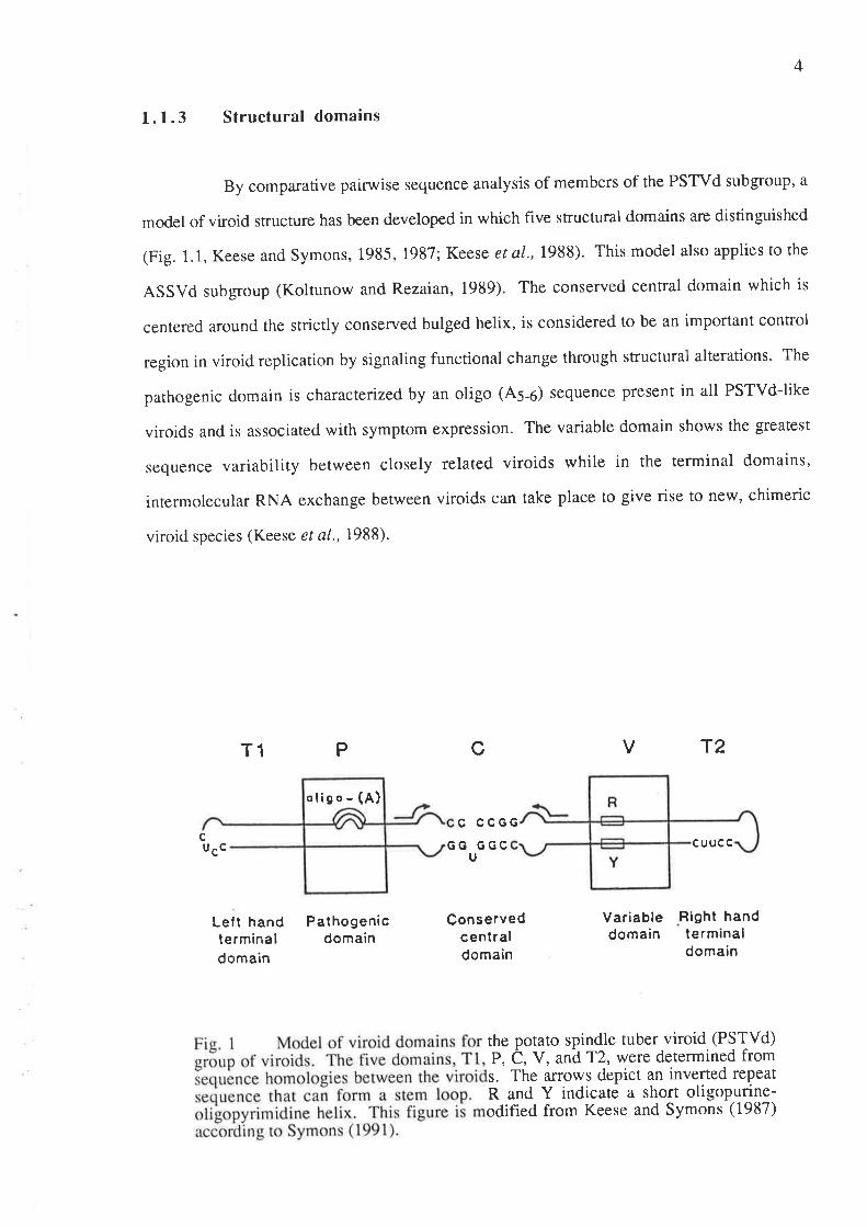
1.1.3 Structural domains
By comparative pairwise sequence analysis of members of the PSTVd subgroup, a
model of vi¡oid structure has been developed in which five stn¡ctural domains are distinguished
(Fig. 1.1, Keese and Symons, 1985, 1987; Keese et a\.,198S). This model also applies to tho
ASSVd subgroup (Koltunow and Rezaian, 1989). The conserved central domain which is
centered around the strictly conserved bulged helix, is considered to be an important control
region in viroid replication by signaling functional change through structural alterations. The
parhogenic domain is characterized by an oligo (As-o) sequence present in all PSTVd-like
viroids and is associated with symptom expression. The variable domain shows the greatest
sequence variability between closely related viroids while in the terminal domains,
intermolecular RNA exchange between viroids can take place to give rise to new, chimeric
viroìd species (Keese et a|.,1988).
T1 V T2
C CCGGc GG GGC
Ucuucc
cP
ucc
Lelt handterminaldomain
Pathogenicdomain
Conservedcentraldomain
Variabledomain
Right handterminaldomain
the potato spindle tuber viroid (PSTVd)P, C, V, and^12, were determined froms. The arrows depict an inverted repeatR and Y indicate a short oligopurine-odified from Keese and Symons (1987)
oligo - (A)

5
1. 1.4 In vivo mutations and recombinations
The natural occurrence of several viroids aS two or more variants has been very
helpful in defining the roles of the conserved and variable features of the viroid molecule' For
the 17 sequence variants of CEVd, mutations afe most frequently located in the P and V
domains. These variants form two classes of sequence which differ by a minimum of 26
nucleotides in a total 0f 370 to 37 5 residues. These two classes correlate with two biologically
distinct groups when propagated on tomato plants where one produces severe symptoms and
the other gives rise to mild symptoms (visvader and Symons, 1985).
Eight f,reld isolates of pSTVd of different virulence differ from each other only in a
few nucleotides in the p and v domains. The sequence variants are placed into four groups:
mild, intermediate, severe and lethal. The nucleotide changes aro compared to the mild
reference Sequence. The intermediate and severe isolates show changes at only three and one
sites, respectively, whereas two lethal isolates show changes at either six or seven sites'
Despite insertions and deletions, the chain length of 359 nucleotides is strictly conserved in the
eight isolates (Schnölzer et at., 1985). The sequence analysis of five new field isolates of
PSTVd revealed, however, that the tength of their RNA chain is not strictly conserved to 359
nucleotides and that it may vary, so fa¡, between 356 and 360 nucleotides' In comparison with
the previously sequenced and least virulent strain as a standard, the two new mild strains differ
from it by one or two nucleotides. The new intermediate and severe strains differ from the
standard by eight and nine nucleotides, Iespectively, whereas the new severe to lethal strain
differs in seven nucleotides. Most of these mutations are located within the P and v domains'
only two strains have been found in which a single mutation occurs in the right terminal
domain (Herold et al.,lgg2). A strain which was isolated from pepino (solanum muricatum)
plants has been found to exhibit major Sequence differences from those isolated from potato'
When compared to rhe standard mild strain, this new sffain which is 356 nucleotides long has
three insertions, six deletions and 14 nucleotide exchanges scattered mostly in the P and v
domains, with three of the exchanges in the T2 domain (Puchta et a1.,1990)' Also, in the two
mild strains of PSTVd isolated from wild solanum sp., one contains two nucleotide
6
subsrirurions wirhin the C domain while the other one is identical to the mild strain of PSTVd
used as a standard (Owens et al.,1992).
Although CPFVd is six residues longer than HSVd, it is considered to be a
sequence variant of HSVd because the two viroids have greater than 90% homology. The
nucleotide differences between HSVd and CPFVd are scattered throughout the molecule with
the greatest concentration in the P domain. Only two sequence variants of CSVd have been
sequenced and most of the sequence changes also occur in the P and V domains (Keese et al',
1988).
The domain model also led to the proposal that the evolution of viroids involved the
rearrangements of domains between viroids infecting the same cell followed by further
evolution (Keese and Symons, 1985). Experimental support of such a model is difficult to
obtain, but new viroid sequences that continue to appear provide indirect evidence (Symons,
1991). For example, in the CLVd sequence, the T1 andT2 domains show high sequence
homology ro rhe same domains in PSTVd and TASVd, respectively (Hammond et al.,1989).
The presence of subdomain lengths of sequences of TASVd in the P domains of PSTVd and of
HSVd in the C domain indicate that rearrangements can occur within a domain as well as at the
boundaries (Symons, 1991). In AGVd, nearly all the 370 nucleotides appear to be derived
from segments of CEVd, PSTVd, ASSVd and GYSVd (Rezaian, 1990). The citrus bent leaf
viroid (CBLVd) consisting of 318 nucleotides is constituted from a p¿ìrt of the C domain of the
ASSVd subgroup and part of the P and T1 domains of CEVd. It is the first member of the
ASSVd subgroup infecting citrus plants to be characterized (Ashulin et a1.,1991). The first
viroid to assume a branched conformation when it is folded in the model of lowest free energy
is the pear blister canker viroid (PBCVd) of 315 nucleotides. Although it has a conserved
sequence located in the T1 domain of the PSTVd subgroup, it is more closely related to the
ASSVd subgroup (Hernandez et al., 1992). The mechanism of these RNA rearrangements is
unknown. One probable mechanism is discontinuous transcription where an RNA polymerase
copying one viroid changes over to copy a juxtapositioned second template at some point, most
Iikely determined by the tertiary structures of the two templates (Keese and Symons, 1985,
1987).
7
1.f .5 In vítro mutational analysis of structure/function relationships
The success in preparing infectious cDNA clones or their respective RNA
transcripts (Cress et a\.,1983; Tabler and Sanger, 1984; Meshi et a|,,1984; Meshi et al.,1985;
Tabler and Sanger, 1985; Ishikawa et al., 1985; Candresse et al., 1990) permits the
investigation of structure/function relationships in viroids by site-directed mutagenesis of the
cDNAs, as well as construction of viroid chimeras, followed by bioassay. Recently, invitro
synthesis of an infectious unit length viroid without resorting to cloning procedures has been
demonsrrared. This will also allow routine application of oligonucleotide-directed mutagenesis
to the study of viroids (Ridgen and Rezaian,1992).
Mutagenesis has provided new insights into structural features responsible for
parhogenicity and host range (Owens and Hammond, 1990). Symptom production by novel
intraspecific and interspecific chimeras is conrolled by the source of the pathogenicity domain
(Visvader and Symons, 1986; Owens et al., 1990). In these experiments, intraspecific
recombinants were consructed by exchanging the left and right sides of two sequence variants
of CEVd (mild and severe strains) while interspecific chimeras were constructed by similar
manipulations involving TASVd and CEVd. Recently, speculation about the mechanism of
viroid pathogenesis has centred upon the thermodynamic stability of a virulence-modulating
region within the PSTVd pathogenicity domain (Schnölzer et al., 1985) and the ability of
nearby nucleotides to base-pair with the 5'-terminus of a 75 host RNA that may be involved in
protein translocation (Haas et a\.,1988). However, factors other than thermal stability of the
virulence-modulating region appear to be important for symptom expression (Visvader and
Symons, 1985; Owens and Hammond, 1990). Nonlethal alterations in the pathogenicity
domain appear to be clustered in areas which exhibit natural sequence variation (Owens and
Hammond, 1990). Sano ¿r al. (1992) have demonstrated that T1, P, V and T2 domains of
TASVd and CEVd contain three discrete regions of sequence and/or structural variability in
which variation is correlated with changes in viroid pathogenicity.
With respect to replication mechanisms, in vitro mutagenesis has highlighted the
importance of the upper portion of the cenral conserved region for cDNA infectivity (Visvader
et al., 1985; Owe ns et aL., 1986; Hammond and Owens , 1987; Candresse et al., 1990; Owens
8
et al., 1991). This result has been confirmed by studies of the conversion of multimeric
PSTVd RNA transcripts into circular monomers by nuclear extracts (Tsagris et al., 1987).
Mutagenesis has also been used to detect the presence of alternative sites for the in vivo
processing of multimeric viroid RNAs (Flammond et al., 1989).
l. 1.6 Mechanism of rePlication
The site of viroid synthesis in the cell is unknown. In PSTVd and CEVd, the
nucleolus is a major site of viroid accumulation but nothing is known about the actual site of
synthesis (Riesner, 1987). Since viroids do not function as mRNAs, they must be replicated
by pre-existing host enzymes (Diener, 1991). The presence of oligomeric viroid forms (usually
of opposite polarity) in infected plants suggests that replication occurs by a rolling circle-type
mechanism (Branch and Robertson, 1984; Symons et a\.,1985). In one of the two variations
of the rolling circle mechanism, the infections circular (+ sense) RNA is copied continuously by
an unidentified RNA-dependent RNA polym"rur.*ro form a concatameric (-) strand' Specific
cleavage of this strand produces monomers that are circularized by a host RNA ligase and then
copied by the same or a different RNA polymerase. Specific cleavage of the long linear (+)
strand produces monomers that a¡e circularized to give the progeny RNA. ASBVd most likely
follows rhis pathway (Symons, 1991). In the other variation, the linear (-) snand is not cleaved
bur copied directly to give a linear (+) strand that is cleaved to produce monomers and finally,
rhe circular progeny. Replication of most viroids probably follows this route (Symons, 1991).
1.1.7 Control measures
Most control measures used in agricultural practice are based on prevention rather
than cure. These include growing of crops from viroid-free seeds or planting stock and
measures to stop these pathogens from entering and spreading through crops (for example,
decontamination of rools with a hypochlorite solution). Separation of viroid-infected seeds or
propagation material from healthy ones requires diagnostic tests of adequate sensitivity,
specif,rcity and rapidity (Diener, 1987).
*or the DNA ddpendent RNA þolymerase tr ( Schindler & Muhlbach, Plant Sci. 84,221-229,1992)
9
So far, the approach of incorporating genetic resistance factors into the genomes of
commercially desirable cultivars has not been successful with viroid diseases. This is
apparently because of the absence of identif,rable resistance factors in vi¡oid host plants (Diener,
1987).
Interference in symptom expression (i.e. cross-protection) has been demonstrated
between srrains of PSTVd (Niblett et al., 1918; Branch et al., 1988; Khoury et al., 1988)'
However, it has not been shown in the other viroid systems. Furthermore, no mild strains
have been observed for some viroids.
I.2 COCONUT CADANG.CADANG VIROID (CCCVd)
Several reviews provide a detailed coverage of the nature of the coconut cadang-
cadang disease and its viroid agent (7r,lazny et a\.,1982; Randles, 1987; Randles et a|.,1988;
Hanold and Randles, 1991; Randles et al., 1992). The following is a brief summary'
1.2.1 Cadang-cadang disease
l.2.Ia History, distribution and economic importance
The term "cadang-cadang" which means dead or dying comes from the Bicol dialect
of the Philippines and is now used to refer to a premature decline and death of coconut palms in
the Philippines. The synonym "yellow mottle decline" is seldom used (Rillo and Rillo, 1981).
The first serious outbreak of cadang-cadang was reported in 1931 on San Miguel
Island in Albay Province (Ocfemia, 1937). This was followed by observation of the disease at
sites at increasing distances from the Bicol Peninsula, with intervening water or disease-free
growing areas (Price,IgTl). By the early 80's, it had spread widely in the central Philippines
and approximately 30 million coconut palms had been killed (Zelazny et al., 1982). The
disease continues to spread and at present, the most southernly site of occulrence is Homonhon
Island which poses a threat to the coconut plantations in Mindanao (Pacumbaba and Carpio,
1987).
10
Economic losses arise from the cessation of nut production on diseased palms, an
average of 5 years before they die from the disease. If palms are not replaced until they die,
and if it is assumed that replacement takes 5 to 8 years to reach full bearing, 10 to 13 years of
production may be lost from each diseased site (Randles et al., L992).
1.2.1b Symptoms and host range
A palm infected with cadang-cadang shows a series of symptoms staning with the
bearing of smaller, more spherical and scarified nuts. Then, yellow spots develop in the leaves
which become numerous as the disease progresses giving the lower two-thirds of the crown a
yellowish appearance. Inflorescences become necrotic, infertile and nut production eventually
ceases. Fibres remain attached to the bases of the fronds rather than breaking off as in a healthy
palm. Frond production and size gradually decline, leaflets become brittle and death of the
palm follow s (Zelazny et al., 1982). The time between the appearance of the first symptoms
and death of the palm ranges from three to more than 15 years and is, on the average, about 10
years. The early stage lasts an average of two years in 19-30 year-old palms but lasts up to
3.75 years in older palms. The medium stage lasts for an average of just over two years. The
late stage to death averagos about five years. Rarely, there are palms that become infected
before they commence bearing and never bear nuts even though they survive well beyond the
age of bearing (Zelazny and Niven, 1980).
Although root deterioration has been reported, studies of histological changes have
been concentrated on the leaf. Leaflets from infected palms are thinner and the palisade and
mesophyll tissues are disorganized. This hypolasia suggests that the disease induces changes at
the cell-differentiarion phase of leaf development. In the chlorotic leaf spots, chloroplasts
appear vesiculate, they accumulate sta¡ch and lamellae become disorganized. Some tannin body
accumulation has also been observed in vacuoles (Randles et a1.,1992).
The experimental and natural hosts of CCCVd identified so far, are all members of
the palm family. The mechanically and naturally infected hosts are Elaies guineensis (oil palm)
and. Corypha elata (buri palm). The other successfully inoculated palms are Areca catechu
(betel nut palm), Adonidia merillii (Manila palm), Chrysalidocarpus lutescens (palmera),
11
Oreodoxa regia (royal palm), Prycosperma macarthuri (Macarthur palm) and Phoenix
dactylifera (date palm). Mechanical inoculation is done using a high pressure injector.
Conventional methods of inoculation have not been effective (Imperial et al., 1985; Anon.,
1 986).
l.2.lc EpidemiologY
The cadang-cadang disease is rarely observed in young palms. The disease is
usually observed only after palms reach 10 years of age and incidence increases approximately
linearly for up to 40 years. Incidence then usually remains constant for older palms (Tnlazny
and pacumbaba, 19g2). The disease has a scattered and apparently random distribution. The
rate of spread is slow and gradual ranging from aboul O.I7o to 17o p.a. in low and high
incidence areas, respectively (Randles et a1.,1988). The outward advance of the disease is
about 500 m p.a. There is no evidence that it originated at one point (Zelazny,1979).
The natural mode of spread is unknown. The lack of a specific pattern of disease
increase does not allow the source of infection in plantation to be inferred and there are still
many questions to be answered before epidemiology is sufficiently well understood for control
measures to be developed (Randles et al-,1992).
1.2.2 Diagnostic methods
The detection of two small disease-associated RNAs by Randles in 1975 provided
the initial clue ro the etiology of cadang-cadang. Characterization of pure preparations by
elecrron microscopy (Randles and Hatta, lgTg), nucleotide sequencing (Haseloff et al', 1982)
and transmission experiments that demonstrated the infectivity of these RNAs (Randles et al',
1977; Moha med et al., 1985),finally proved that cadang-cadang is caused by a viroid'
Symptomatology is unreliable for disease diagnosis. Therefore, the polyacrylamide
gel electrophoresis (PAGE) technique has been used for routine detection of CCCVd. Several
modifications stârting from the extraction of nucleic acids to gel staining have been done to
simplify and miniaturize the procedure and at the same time improve its sensitivity (Impenal et
r2
al.,l98l; Imperial and Rodriguez, 1983). Two-dimensional and bi-directional electrophoresis,
which have been developed for general use in the detection of viroid-like RNAs (Schumacher er
al., 1983; Rivera-Bustamante et al., 1986; Tabler et al., 1989), are useful for identifying
CCCVd in mixtures when linear RNAs of similar mobility afe present.
CCCVd is detected by PAGE on tho basis of size. For a more sensitive and
specific assay based on its unique sequence, dot-blot and electroblot molecular hybridization
with labelled complementary RNA or DNA probe have been developed (Randles and
Palukaitis, 1979; Barker et a\.,1985; Impenal et a1.,1985; Hanold and Randles, 1991).
1.2.3 Physical properties
In the native state, CCCVd is partially sensitive to the single-strand-specific S1
nuclease (Randles et a\.,1976; Randles and Palukaitis, 1979) as expected for a partially base-
paired sÍucture. Thermodynamic studies indicate that the native molecules do not adopt
significant rertiary folding, but they exhibit highly cooperative melting of base-paired regions at
a Tm of 49'C in i0 mM NaCl. An intermediate structure, a hairpin comprising a single-
stranded loop of 14 bases and a stem of 9 base pairs, forms at temperatures just about the Tm.
The stem melts at about 58'C to produce a completely denatured circle. The sho¡t sequence that
is exposed as a single-stranded loop occurs in the upper central conserved region common to
most of the other viroids (Randles et a|.,1982; Riesner et al., 1983).
L.2.4 Variation in the nucleotide sequence
CCCVd with246 nucleotides is the smallest known viroid. In addition to this basic
form, at least three types of sequence variants have been recognized by sequencing isolates
from individual palms and from fronds of different age within a single palm. These include an
insertion of cytosine at position L91 of the C domain to give a247-ntcleotide sequence variant,
duplications of rhe V and T2 domains of 41,50,55 or 100 nucleotides and mutations of bases
adjacent to rhe boundaries of some of the partial duplications. Detected concurrently with the

13
monomers are their respective dimers which are covalently linked forms of the monomers,
incorporating the same sequence variations (Haseloff et a|.,1982; Keese et a|.,1988).
L.2.5 Correlation of CCCVd structure with disease progress
Variation in CCCVd is related to the stage of disease development. The small
2461241 varianrs (with their corresponding dimers) appear early in infection but, as symptoms
develop, the large 2871296129713011346 variants (also with their corresponding dimers) arise
and evenrually dominate the viroid population as the disease progresses (Imperial et a1.,1981;
Mohamed et a\.,1982). A systematic but more subtle progression has been observed for the
varianrs containing either one cytosine (246 or 296) or two cytosine (247 or 297) residues at
nucleotide position I97 . If infection starts with variant 246,247 appea-rs next followedby 296,
then 297. If variant 247 appears first, it is replaced only by 297 (Imperial and Rodriguez,
1983). All variants are infectious and in the field, the frequency of occurrence of CCCVd2a6 in
the early-stage palms at four sites was greater than that of CCCVd247 (Randles et a|.,1992).
The functional implications of the development of larger variants of CCCVd during
disease progression are not known. Presumably, it has some advantage in replication, perhaps
by providing increased competition for binding to some host component important for
replication, but which is in limited supply (Keese et a|.,1988).
t.2.6 CCCVd as related to CTiVd and the viroid-like sequences in oil and
coconut palms and other monocotyledons in the south-west Pacific
The symptoms of the tinangaja disease of coconuts in Guam are similar to those of
the cadang-cadang disease except for the effect on nut production. Whereas the cclconuts from
palms infected with cadang-cadang become more spherical and scarified, palms afflicted with
tinangaja produced mummified nuts with no kernel present (Boccardo et a|.,1981). The viroid
associated with this disease, CTiVd, is 254 nucleotides long and shares 647o ovetall sequence
homology with CCCVd (Keese et a|.,1988).
t4
The presence of small nucleic acids with nucleotide sequences similar to CCCVd
has been demonstrated in African oil palm, coconut palm and some other monocotyledonous
species in several areas of the south-west Pacific region. The oil palms have orange leaf spots
resembling those described for oil palm naturally infected with CCCVd in the Philippines, and
also characteristic of a condition known as "genetic orange spotting" (GOS). Preliminary
evidence has been provided that GOS is an infectious disorder caused by a viroid. The coconut
palms do not show symptoms typical of cadang-cadang disease, but sometimes are chlorotic,
stunted, or have a reduced yield. These findings suggest that viroids with nucleotide sequences
similar to CCCVd occur widely in palms and other monocotyledons outside the Philippines
(Hanold and Randles, 1991).
SCOPE OF THIS THESIS
In the central Philippines where the cadang-cadang disease has been in existence
since the 1930's, there is a greater possibility that alternate hosts and naturally occurring
variants of CCCVd will be found. Thus, in this thesis, a range of different plants other than
palm species which grow naturally in coconut plantations was screened. CCCVd-like
molecules were detected in some of the species tested and these were isolated, purified and
characterized. Then, in a signifrcant number of palms inoculated with CCCVd, a new type of
symptom appeared which was more severe than the typical cadang-cadang symptoms. These
palms were analyzed for their viroid pattern by polyacrylamide gel electrophoresis and the
viroids were also isolated, purified and further characterized by molecular cloning and
nucleotide seque ncing.
The detection of CCCVd-like sequences in plants other than palm species shows
that they could act as sources of inoculum for coconut palms in the field. Definition of the
CCCVd reservoir should lead to a better understanding of the epidemiology of the disease.
Furthermore, the identification of a herbaceous host would provide a system that will facilitate
studies on CCCVd. On the other hand, the detection of more variants of CCCVd would be
useful in identifying its conserved and variable features and their functional implications.
15
CHAPTER 2
MATBRIALS AND METHODS
2.T MATERIALS
2 .t.1. cccvd
Partially purified CCCVd used as standard marker was prepared according to
Section 2.2.3 from infected palms at different stages of the disease in order to obtain the same
amount of each of the 246,247 ,296 and 297 sequence variants with their respective dimers.
Pure CCCYdzqø used for the synthesis of 32P-labe[ed ss cDNA probe was
prepared according to Mohamed et al., (1985).
2.1.2 32P-labelled cRNA probe
The cRNA probe used in the early part of this study was prepared by Dr JL
Mclnnes of the Plant Science Department of the Waite Agricultural Research Institute (WARI).
Using the kit from BRESATEC (Adelaide), transcription was done from a pSP6a plasmid
containing a monomeric insert of the 246 nucleotide form of CCCVd at the BamHl site.
2.L.3 Synthetic cccvd-specific DNA oligonucleotide primers
The primers 5'-d(GGCCTCTCCTGCAGTGGTTTTTGGGGTGCCC)-3' and 5'-
d(CCACTGCAGGAGAGGCCGCTTGAGGGATCCCC)-3' were kindly provided by Dr W
Rohde of the Max Planck Institut für Züchtungsforschung, Köln, Germany.
The primers 5'-d(GAGTTGTATCCACCGGGTAGTCTCC)-3 "
5'd(GGTTTCCC
CGGGGATCCCTC)-3', 5' -d(AGGGATCCCCGGGGAAACCT)-3 and 5'd-(GGAGACTAC
CCGGTGGATACAACT)-3' were synthesized by Dr N Shirley of the Plant Science
Department, WARI, using the Applied Biosystems 3808 DNA synthesizer.
16
2.1.4 Biochemicals and miscellaneous chemicals
The main biochemicals and miscellaneous chemicals used in this study are listed in
Appendix A.
2.I.5 Phenol reagents, polyacrylamide/agarose gels and bacterial media
preparation of phenol reagents (for RNA and DNA extractions), polyacrylamide/
agarose gels and bacterial media are described in Appendix B.
2.2 METHODS
2.2.1 Isolation of low molecular weight nucleic acids from tissues of
plants other than coconut
Fresh or frozen tissues were homogenized with 1.5 vol (w/v) of buffer containing
0.5 M sodium acerare, 10 mM MgCl2, 207o ethanol and 3% SDS, and 1.5 vol (w/v) of water-
sarurared phenol (Laulhere and Rozier,Igl6). The homogenate was incubated with shaking at
37'C for 15 min and mixed vigorously for 10 min with 0.5 vol of chloroform. The aqueous
phase recovered by centrifugation at 10,000 rpm for 10 min was re-extracted with 0.5 vol of
phenol and 0.5 vol of chloroform (v/v) and again centrifuged as above. The nucleic acids were
recovered from the supernatant by precipitation wt¡h 0.33Vo CTAB for 30 min on ice and the
resulting pellet was washed twice with a 0.1 M sodium acetate solution in757o ethanol
(Imperial et a1.,19S5). The precipitate was dried briefly, resuspended in the minimum amount
of 0.25 M sodium acetate and extracted with 0.5 vol each of phenol and chloroform. After
centrifugation at 10,000 rpm for 10 min, the supernatant was recovered and an equal volume of
4 M LiCl was added. The mixture was incubated at 4'C for 15-18 hr and centrifuged at 10,000
rpm for l0 min at 4'C (Imperial et a\.,1981). The LiCl-soluble components were recovered
and precipitated with 2.5 vol of ethanol. The pellet was either kept in ethanol ar -20"C for long
t7
storage or dried and dissolved in 0.01 M sodium acetate solution with l07o sucrose for
immediate assay.
2.2.2 Mini-preparation of low molecular weight nucleic acids from coconut
lea ves
A 1-g leaf sample was placed in a plastic bag with I vol (w/v) of buffer containing
100 mM Na-K phosphate buffer, pH 7, 10 mM EDTA and 0.57o 2-mercaptoethanol. The
tissue was crushed with a pestle and the juice was squeezed out into a centrifuge tube
containing 0.5 vol each of phenol and chloroform. After vortexing for a few minutes, the
mixture was centrifuged at 10,000 rpm for 10 min. The nucleic acids in the aqueous phase
were precipitated with CTAB and fractionated with LiCl as in Section2.2.1' but omitting the
phenol/ chloroform extraction in between.
2.2.3 Large-scale preparation of low molecular weight nucleic acids from
coconut leaves
Bulk extraction of low molecular weight nucleic acids from coconut leaves was
done according to a previously described procedure (Mohamed et a|.,1985). Batches (250 g)
of chopped leaflets were blended in 750 ml of pre-cooled 0.1 M Na2S03. The slurry was
strained through cotton muslin and clarified by centrifugation at 10,000 rpm for 10 min at 4"C.
Supernaranrs from 5 kg of leaf material were pooled and87o PEG 6000 was added. After 2 hr
incubation at4"C, the resulting precipitate was collected by centrifugation as above' Nucleic
acids were exracted from the precipitate by dissolving it in 100 ml of l%o SDS, then adding 0'5
vol each of phenol and chloroform and shaking vigorously for t hr. The aqueous supernatant
phase was collected after centrifugation and re-extracted twice with 0.5 vol each of phenol and
chloroform for 30 min. Further purification steps with CTAB and LiCl were done as in Section
2.2.2.
18
2.2.4 Viroid purification
2.2.4a Two-dimensional (2-D) or bi-directional polyacrylamide gel
electrophoresis (PAGE)
Separation of viroids from contaminating host nucleic acids, either by two-
dimensional (2-D) or bi-directional PAGE, was done as described by Schumacher et al', (1983)
and Tabler et al., (1989). The first dimension gel was identical for both systems. Partially
purified nucleic acid extracts were electrophoresed on a non-denaturing 77o slab gel in TBE
buffer (90 mM Tris - 90 mM borate - 2 mM EDTA) (Peacock and Dingman, 1968). The gel
was stained with ethidium bromide (500 pglL) and the zones containing the viroid molecules
were cut our as required. Vertical lanes with different viroid samples wore cut out separately
for 2-D PAGE whereas for bi-directional system, the gel was horizontally cut across all lanes
containing the viroid position which was determined with reference to the marker viroid. Each
gel slice was placed either below or on top of the second dimension 77o gel with 8 M urea. The
circular and linear viroid forms were located by ethidium bromide staining (as above) and then
excised from the gel.
2.2.4b Recovery of the viroids from polyacrylamide gel
Where gels of less than I mm thickness were used, the viroids were efficiently
eluted by soaking the intact gel pieces for 14-16 hr at 37"C in 0.5 M ammonium acetate, I mM
EDTA, pH 8 and 0.17o SDS (Sambrook et a1.,1939). The elution buffer was removed,
extracted once with 0.5 vol each of phenol and chloroform and centrifuged at 10,000 rpm for
l0 min. The aqueous phase was recovered and 2.5 vol of ethanol was added to precipitate the
viroids.
Where the gel thickness was more than I mm, the viroids were electroeluted
(Maniatis et a1.,1982) by placing the excised band in a dialysis bag with 0.5 x TBE buffer.
The bag was closed and immersed in a tank filled with electrophoresis buffer. Current was
passed through the bag for 2-3 hr at 100 V to elute the viroids out of the gel and onto the inner
19
wall of the bag. The polarity of the current was reversedfor 2 min to release the viroids from
the bag wall. The gel buffer was removed from the bag and extracted once with phenol and
chloroform (as above). The viroids were precipitated with ethanol in the presence of 0.3 M
sodium acetate.
2.2.5 PAGE assay
Nucleic acid samples resuspended either in 0.01 M sodium acetate w\th l07o
sucrose or in 0.57o bromophenol blue/xylene cyanol in TBE with 507o glycerol (v/v) were
applied ro a non-denaturing polyacrylamide gel containing 20Vo acrylamide-bisacrylamide in the
rario of 99:1 (Imperial and Rodriguez, 1983). Electrophoresis was at 250 ot 125 V for 6 ot 12
hr, respectively, in TBE buffer. The gel was stained with silver nitrate according to Sammons
et aI. (1981) but with the following modifications: the gel was fixed in lOTo trichloroacetic acid
for l5 min (Imperi al et a\.,1985), followed by two brief rinses with glass-distilled water, then
soaked (with shaking) in 0.I87o silver nitrate for t hr. The gel was again washed two to three
times with water and soaked (with shaking) in reducing solution (87.5 mg/L of sodium
borohydride and 0.757o formaldehyde in 0.25 M NaOH) until the bands were visualized. The
bands were enhanced by incubating the gel in0.757o sodium carbonate with 57o acetic acid
added to prevent the gel from expanding.
2.2.6 Molecular hybridization assay
2.2.6a Synthesis of 32P-labelled single-stranded complementary DNA (32p-
ss cDNA) probes
Synthesis of 32P-ss cDNA probes was canied out in a reaction volume of 30 ¡rl
containing 15 mM Tris-HCl, pH 8.3, 40 mM KCl, 0.85 mM MgCl2,0.05Vo mercaptoethanol,
50 ¡rM each of dATp, dGTp and dTTP, 30 pCi of [cr-32p¡ dcTP (= 3000 cilmmol), 1 pg of
CCCVd-specific primer 5'-d(GAGTTGTATCCACCGGGTAGTCTCC)-3' or 5'-d(GGCCT
CTCCTGCAGTGGTTTT|GGGGTGCCC)-3', 1 pg of purihed CCCVdz¿o and 200 units of
20
M-MuLV reverse transcriptase. The template viroid and the primer with the buffer solution
were first incubated at 90'C for 3 min, then at 55'C for 3 min, after which the reverse
transcriptase and 32p-¿Ctp were added and the whole reaction mixture was incubated at 37'C
for 4-5 hr. The reaction was stopped by adding 20 mM EDTA. The mixture was then
chromarographed on a G50 (fine) - Sephadex column (1.5 ml vol in a2 ml Pasteur pipette) with
sterile TE buffer (10 mM Tris-HCl, pH 7.5, 0.1 mM EDTA). Approximately 400 pl of TE
buffer was added each time to collect at least 6 fractions. Usually, fractions 2 and 3 contained
the 32P-cDNA while the rest of the fractions contained the unincorporated 32p-dctp.
Radioactivity of the fractions was determined by Cerenkov-counting and those containing the
32P-cDNA were pooled and extracted once with 1 vol each of phenol and chloroform. After
centrifugation at 10,000 rpm for 2 min, the aqueous phase was recovered and an equal volume
of formamide was added. The mixture was heated at 100'C for 2-3 min, then chilled on ice
before use. The specific activity was ca. 2 x 10⁷ cpm/pg.
2.2.6b Preparation of dot-blot/electroblot
Nucleic acid samples of 1 pl were dotted directly onto a sheet of nylon membrane
(Zeta-probe, Biorad) which had been washed with distilled water and air dried. After
application of the samples, the membrane was baked for 2 hr at 80'C (Thomas, 1980)'
Nucleic acid samples and a CCCVd standard marker were fractionated by
electrophoresis in 207o polyacrylamide gel buffered with TBE. After cornpletion of the run, the
gel was equilibrated in TAE (9 mM Tris, 4 mM Na-acetate, 0.4 mM EDTA,p}ìT.4) for 15 min
and assembled in contact with the nylon membrane in an electroblot apparatus filled with TAE
buffer. Current was passed through the gel at 60 V, 0.6 A for 5 hr or at 30 V, 0.3 A for 16 hr
followed by 60 V, 0.6 A for I hr to transfer the nucleic acids to the nylon membrane (Hanold
and Randles 1991). The membrane was baked for 2 hr at 80'C.
2r
2.2.6c Hybridization
The nylon fitters were prehybridized for 15-20 hr at 42"C in a buffer made
according to Thomas (1980) but with some modifications recommended by the manufacturer of
the nylon membrane (Biorad): 507o deionized formamide; 5 X SSC (750 mM NaCl,75 mM
Na-citrate); 50 mM sodium phosphate buffer, pH 6.5; 5 mm EDTA; 0.27o SDS; 1 mg/ml
denatured carrier DNA and 0.27o each of bovine serum albumin (BSA), Ficoll 400 and
polyvinylpynolidone (PVP) of mol wt 40000.
The denatured cDNA or cRNA (Section 2.1.2) probe was added to the
hybridization mixture at approximately 106 cpm/ml. The hybridization mixture consisted of the
modified prehybridization buffer (except that 250 Lrlml denatured carrier DNA and0.027o each
of BSA, Ficoll 400 and PVP 40000 were used) and l}Vo dextran sulphate. Hybridization was
done at 42"C for 20-40 hr (Thomas, 1980). For washing at low stringency, the filters were
immersed in 0.5 X SSC,0.17o SDS for 5 min a¡.20C, then washed with agitation in 0.1 X
SSC, 0.17o SDS for 2 hr at 55'C and autoradiographed at -70"C using an intensifying screen.
For washing at high stringency, the filters were re-washed with agitation at 65'C for 2 hr in the
same buffer as above and again autoradiographed regulating the exposure times in order to
obtain signal intensities similar to those of the low stringency wash (Hanold and Randles,
199 I ).
2.2.7 Amplification of viroids by polymerase chain reaction (PCR)
2.2.7 a Preparation of template
First strand cDNA synthesis using M-MuLV reverse transcripras¿. This was done as in Section
2.2.6aexcept that dCTP was added at the same concenration as the other dNTPs and no 32P
was incorporated in the reaction mixture. The cDNA was precipitated from the reaction mixture
by adding 2.5 vol of ethanol and incubating the mixture at -20"C for at least 30 min. The
precipitate was recovered by centrifugation at 15,000 rpm for 20-30 min, dried briefly and
resuspended in deionized water.
22
First strand cDNA synthesis using AMV reverse transcriptase. This was done according to
Ding, SW (unpublished). The viroid RNA and the primer (1 pg each) resuspended in
deionized water were incubated at 80'C for 12 min and chilled on ice for 5 min. The
components (50 mM Tris-HCl, pH 8.3; 50 mM KCl, 10 mM MgClz,0.5 mM spermidine; 40
units of RNAsin; 10 mM DTT and 1 mM each of dATP, dCTP, dGTP and dTTP) required for
reverse transcription were added to a final volume of 25 pl. The reaction was started by adding
25 units of AMV reverse transcriptase and incubating the mixture at room temperature for 5
min, then at 55'C for 30 min. The mixture was extracted once with 1 vol each of phenol and
chloroform and cenrrifuged at 10,000 rpm for 5 min. The cDNA was recovered from the
aqueous phase by ethanol precipitation (2.5 vol) at -20"C for at least 30 min followed by
centrifugation ar 15,000 rpm for 20-30 min. The dried pellet was dissolved in deionized water
and stored at -20"C.
2.2.7b PCR reaction
The buffer solution (as provided in the PCR kit manufactured by Promega)
contained the following: 10 mM Tris-HCl, pH 8.3; 50 mM KCI; 1.5 mM MgCIZ; 0.0I7o gelatin
and,I.25 mM each of the four dNTPs. Approximately 250 ng each of the two primers and the
first strand cDNA were added to the buffer solution. The Taq polymerase (1.28 units) was
added to a final volume of 25 pl only after the initial denaturation of the above mixture at 96'C
for 3 min. The mixture was overlaid with mineral oil. Amplification was for 40 cycles on a
Hybaid thermal cycler under the following conditions: 93'C for 45 sec, 55'C for 45 sec and
72"Cfor 3 min. An exra elongation time at the end of the cycles was done at72"C for 15 min.
23
2.2.8 Molecular cloning of PCR products
2.2.8a Purification of PCR products and preparation of the cloning vector
The PCR mixtures, after termination of the reaction, were fractionated on aJVo
non-denaruring polyacrylamide gel in TBE buffer. The gel was stained with ethidium bromide
(500 pg/L) and the zones containing the ds cDNAs of the desired lengths as determined by
comparison with a DNA mol. wt. marker were cut out. The ds cDNAs were recovered from
the gel as described in Section2.2.4b and resuspended in deionized water.
The cloning vector was prepared according to Marchuk et al. (1991) and Holton
and Graham (1991). About I pg of Bluescript SK+ plasmid (Stratagene) was digested with
EcoRV (5-10 units/lQ pl vol) in the presence of the appropriate restriction buffer (provided by
the manufacturer, Boehringer) at37'C for 2 hr. The mixture was extracted once with 1 vol
each of phenol and chloroform, centrifuged at 10,000 rpm for 5 min and the blunt-ended vector
was recovered from the aqueous phase by ethanol precipitation (2.5 vol) at -20"C for at least 30
min. The pellet was dried briefly, dissolved in 10 ¡rl of deionized water and incubated for 2 hr
ar 70'C with Taq polymerase (5.5 units/pg plasmid/20 pl vol) in the presence of standard
buffer conditions (Section 2.2.7b) except that only 2 mM dTTP was added. The absence of
any other nucleotides in the reaction resulted in the addition of a single thymidine at the 3' end
of each fragment. The mixture was extracted once with 1 vol each of phenol and chloroform
and centrifuged at 10,000 rpm for 5 min. The T-vector was recovered from the supernatant by
ethanol precipitatio n (2.5 vol) at -20"C for at least 30 min. The dried pellet was dissolved in
deionized water.
2.2.8b Ligation of ds cDNA to the T-vector
The ligation reaction (Sambrook et a1.,1989; Mead et a1.,1991) was done in a 10
¡rl buffer solution containing 66 mM Tris-HCl, pH 7.5 at 20'C; 5 mM MgClz; 1 mM DTT; 1
mM ATP; 1 unit T4 DNA ligase (Weiss unit) and 20-25 ng of the T-vector. The ds cDNA was
added to maintain an approximate vector to insert ratio of 1: 1 and 1:3. This was achieved by
24
estimating the amount of insert DNA by gel electrophoresis in comparison with a standard of
known concentration. The ligation mixture was incubated overnight at 5-16'C'
2.2.8c Preparation of competent cells
E. coli JM109 competent cells were prepared according to Hanahan (1983) and
Sambrook et al. (1989). A single colony of E. coli JM109 was picked from a streak plate and
grown overnight in 3 ml of LB medium. Approximately 50 ml SOB medium containing 20
mM MgSO 4 in a 1 litre flask was inoculated with 500 ¡rl of the overnight culture. The cells
were grown (with shaking) at37"C until the ODOOo was about 0.45-0.55 (approximately after
2.5 to 3 hr incubation). For efficient transformation, it is essential that the number of viable
cells should not exceed 198 çslls/ml. Thus, to monitor the growth of cells, OD669 was
determined every 20-30 min. The culture was placed on ice for 10-15 min. The cells were
pelleted by centrifugarion at 2,500 rpm for 12 min at 4"C and resuspended in Il3 vol TFB (10
mM MES, pH 6.3; 45 mM MnCl2.4HzO; 10 mM CaClZ.2H2O;100 mM KCI and 3 mM
hexamine cobalt chloride) by gently vortexing or sucking up and down with a Pasteur pipette.
The suspension was placed on ice for 10-15 min and the cells were pelleted again at 2,500 rpm
for 12 min at 4"C. The pellet was resuspended in TFB at IOll25 of the original volume of the
cells. DMSO was added to3.57o, the mixture swirled and left on ice for 5 min. Then DTT was
added to 75 mM, rhe mixture swirled and left on ice for 10 min. DMSO was again added to
3.STo,the mixture swirled and left on ice for 5 min. Finally, the suspension was dispensed as
200 pl aliquots into chilled 1.5 ml tubes.
Z.Z.gd Transformation of the competent cells with the recombinant plasmids
Transformation of the competent cells was done according to Hanahan (1983). The
Iigation mixrure (Section 2.2.5b) containing the recombinant plasmids was added to 200 pl of
competenr cells in a chilled i.5 ml tube. The mixture was swirled, incubated on ice for 30 min,
heated a,r. 42"C for 2 min and immediately placed on ice for 2 min. Then, 800 ml of SOC
25
medium (1 ttl507o glucose/ml SOB) was added and mixed by inversion. The mixture was
transferred to a culture tube and incubated ar.37"C for 45 min to allow the cells to recover.
2.2.8e Selection for recombinants
As described by Sambrook et al. (1989) and Titus (1991), a200 ¡rl aliquot of the
transformation mixture was spread gently (using a bent Pasteur pipette) on an LB plate with 60
pglml of ampicillin, 0.5 mM IPTG and 40 Fglml X-gal: The rest of the mixture was
concentrated by centrifugation at 10,000 rpm for 1 min. The supernatant was poured off and
the cells resuspended in 200 ¡rl of LB medium were also plated as above. The plates were
incubated at3l"C for 14-16 hr to establish colonies. Recombinant colonies were white.
2.2.9 Mini-preparations of recombinant plasmids
Exfracrions of plasmid DNA were done by the alkaline lysis method (Sambrook er
al., 1989; Applied Biosystems User Bulletin 18, 1991).
White colonies were picked from the plate and each transferred into 3 ml LB
medium containing 60 ¡rglml of ampicillin. The cultures were incubated overnight at 37'C with
vigorous shaking. A 1.5 ml aliquot of each culture was pelleted in a microcentrifuge tube by
centrifugation at 10,000 rpm for 1 min at room temperature. The supernatant was removed by
aspiration and the bacterial pellet was resuspended (by pipetting up and down) in 200 pl of ice-
cold buffer solution containing 50 mM glucose, 25 mM Tris-HCl, pH 8.0 and 10 mM EDTA,
pH 8.0. About 300 pl of freshly prepared 0.2 NNaOH-17o SDS was added and the contents of
the tube were mixed by inversion before incubating on ice for 5 min. The use of a vortex mixer
was avoided so as to minimize shearing of the contaminating chromosomal DNAs. The
solution was neutralized by adding 300 ¡rl of ice-cold 3 M potassium acetate, pH 4.8. The
contents of the tube were mixed by inversion and then incubated on ice for 5 min. Cellular
debris was removed by cenfrifugation at 10,000 rpm for 10 min at room temperature. The
supernatant was transferred to a clean tube. RNase A (DNase-free) was added to a final
concentration of 20 pglml and the tube was incubated at37"C for 20 min. Extraction with 400
26
pl of chloroform was done twice by mixing the layers by hand for 30 sec after each extraction.
After centrifugation at 10,000 rpm for 5 min, the aqueous phase was rsmoved to a clean tube.
Total DNA was precipitated by adding an equal volume of l\OVo isopropanol and immediately
cenrrifuging the tube at 10,000 rpm for 10 min at room temperature. The DNA pellet was
washed with 500 ¡tl of 107o ethanol, dried under vacuum for 3 min, resuspended in 20 pl of
deionized water and stored at- -20"C.
2.2.10 Analysis of the sizes of inserts in recombinant plasmids by
restriction enzyme digestion and PAGE
The recombinant plasmids were analyzed for the presence of the correct size of
insert DNA by digestion with EcoRI and HindIII according to the manufacturer's directions
(Boehringer). The reaction mixtures were then fractionated on a 207o non-denaturing
polyacrylamide gel in TBE buffer using a 123 DNA ladder as the mol wt marker.
2.2.11 Fluorescent dye primer cycle sequencing
2.2.lla Preparation of dsDNA template
The template plasmid DNAs were prepared as in Section 2.2.9 and further purified
by pEG precipitation (Applied Biosystems User Bulletin 18, 1991). The plasmid pellet was
dissolved \n 32 ¡tl of deionized water. Then, 8 ¡rl of 4 M NaCl and 40 pl of sterile l37o PEG
8000 were added. After thorough mixing, the solution was incubated on ice for 20 min and the
plasmid DNA was pelleted by centrifugation at 10,000 rpm for 15 min at 4"C in a fixed-angle
roror, The supernatant was removed carefully and the pellet was rinsed with 500 ¡tl of 70Vo
ethanol. The pellet was dried under vacuum for 3 min, resuspended in 20 ml of deionized
water and stored at -20'C.
27
2.2.llb Cycle sequencing reaction
Cycle sequencing of the dsDNA was done using the Taq dye primer cycle
sequencing kit manufactured by Applied Biosystems Inc (ABI) and the recommended protocol
(ABI dye primer cycle sequencing/Part no.90I482/Rev. B).
The following reagents were aliquoted into four 0.5 ml tubes:
d/dd NTP mix
Dye primer (0.4 pmol/pl)
5X cycle seq. buffer
(400 mM Tris-HCl, pH 8.9 at RT;
100 mM (NII¿)z SO¿; 25 mM MgCl2)
DNA template (200-250 nglpl)
1 Lrl
1pl
1pl
lpl
1pl
lpl
1pl
1 Lrl
2 frl
2 ptl
2vl
2pr
2rú
2pr
2 ftl
2 ptl
Total volume 5pl 5pl 10Pl 10Pl
The d/ddNTP mixtures contained the following:
A: 1.5 mM ddATP; 62.5 ¡tMdATP; 250 pM dCTP, 375 pM CTdGTP and 250
pM dTTP
C: 0.75 mM ddCTP; 250 pM dATP; 62.5 ¡t"MdCTP; 375 uM C7¿Crp and 250
pM dTTP
G: 0.125 mM ddGTP; 250 pM dATP; 250 uM dCTP; 94 uM CTdGTP and250
¡tM dTTP
T: 1.25 mM ddTTP; 250 pM dATP; 250 pM dCTP; 375 uM CTdGTP and 62.5
pM dTTP
The diluted Taq was prepared by mixing 0.5 ¡rl of AmpliTaq@ DNA polymerase (8 U/ttl); 1.0
pl of 5X cycle sequencing buffer and 5.5 pl of deionized water.
28
The four reaction tubes above were overlaid with 30 ¡rl mineral oil and placed in a
thermal cycler (PTC-100TM Programmable Thermal Controller, MJ Research, Inc). The
cycling was started as follows: 95'C for 30 sec, 55'C for 30 sec and 70'C for 1 min for 15
cycles. Then it was continued for another 15 cycles at 95'C for 30 sec and 70'C for 1 min. At
the completion of the run, the temperature was rapidly dropped to 4'C. For best results, the
total time required for 30 cycles should be t hr and 45 min r 5 min.
The extension reaction mixtures from the four tubes were pipetted out and
combined into a 1.5 ml tube containing 80 ¡tl957o ethanol with 1.5 pl 3 M sodium acetate, pH
5.3. The tube was placed on wet ice for 10-15 min and centrifuged at 15,000 rpm for 15-30
min. The supernatant was carefully discarded. The pellet was rinsed with 250 ¡tI of 707o
erhanol, dned in vacuo for 1-3 min and resuspended in 6 pl of deionized formamide with 8.3
mM EDTA, pH 8.0. Insoluble materials were removed by brief centrifugation. The DNAs in
the supernatant were denatured by heating at 90"C for 2 min and loaded immediately on a pre-
elecrrophoresed 67o polyacrylamide gel with 8 M urea assembled in ABI DNA sequencer,
Model 3734 (operated by Dr N Shirley of the Department of Plant Science, WARI).
2.2.12 Sequence analysis and determination of secondary structure
The nucleotide sequences of the viroid forms were examined and aligned with
CCCVdZ¿6 for comparison using the Seq. Ed. computer program provided by ABI. The
secondary stn¡ctures representing the lowest free energy were determined by the program
developed by Zuker (1989).
29
CHAPTER 3
DETECTION OF CCCVd.LIKE SEQUENCES IN
PLANTS OTHER THAN PALM SPECIES
INTRODUCTION
The natural mode of spread of cadang-cadang is still unknown and several aspects
of disease epidemiology still have to be investigated (Randles er al., 1992). One important
question is whether alternative plant reservoirs of the viroid occur in the field. Natural and
experimental transmission of the viroid has been observed only in palm species (Randles et al.,
1980; Impenal et a\.,1985). Recently, preliminary evidence has been obtained suggesting that
orher plant species are natural hosts of CCCVd (Hanold, Rodriguez and Randles, 1989; Hanold
and Randles, 1991).
With the development of sensitive, diagnostic methods specific for CCCVd
(Schumacher et a|.,1983; Imperial et a\.,1985; Hanold and Randles, 1991), the search for
alternate hosts was pursued in this study by testing a large number of samples from a range of
different plant species that grow naturally in coconut plantations.
EXPERIMENTAL
3.1 COLLECTION OF SAMPLES OF PLANT SPECIES ASSOCIATED
WITH COCONUT PLANTATIONS
Sampling of the predominant plant species in coconut plantations was done at 11
separate sites (approximately 2 ha in area) in five provinces of southern Luzon, Philippines (see
Appendix C). Of these sites, eight had high cadang-cadang incidence, two had low incidence
and one had zero incidence. The aim was to collect, at random, at least five plants per species
in each site. Occasionally, however, only one plant per species was obtained, or none at all,
due to cultivation of the a¡ea under the coconuts to plant cash crops. The collected plants were
30
transplanted and maintained in a screenhouse for experimental use and observation of
symptoms. A sample of each species was dried and pressed for botanical identification at the
University of the Philippines at Los Banos. Some of the species were identified according to
Moody et al. (1984) and DahlgÍen et a/. (1985).
The selection of species was concentrated on monocots, although there was one
dicot species (Urena lobata) that was included because of its common occurrence in the sites
surveyed. Of the approximately 900 samples collected, none showed any specific stunting or
discoloration.
3.2 ISOLATION OF LOW MOLECULAR NUCLEIC ACIDS
Four extraction methods were compared for efficient isolation of low molecular
weight nucleic acids from the different plant species. These include the methods described in
Sections 2.2.I,2.2.2,2.2.3 and a method developed by Keese and Symons (1987) using a
buffer containing 37o SDS, 10 mM MgCl2 and 0.5 M NaCl. Method 2.2.1 was found to be
most effective in removing significant amounts of colored and other materials without
significant loss of the fraction containing the low molecula¡ weight nucleic acids. This method
was, therefore, used throughout. Moreover, large amounts of tissues (at least 20 g) had to be
extracted in order to detect unambiguously the presence of CCCVd-like sequences. Thus, in
most cases, the whole plant was extracted.
3.3 MOLECULAR HYBRIDIZATION ANALYSES FOR CCCVd
The plant extracrs were analyzed for the presence of CCCVd-like sequences by
molecular hybridization techniques using either 32P-cRNA (Section 2.1.2) or 32P-cDNA probe
(Section 2.2.6a) and partially purified CCCVd as a standard. These probes are composed of
single-stranded linear CCCVd RNAs or cDNAs of (-) polarity and therefore, could detect only
the infectious (+ sense) CCCVd molecules. The antisense RNA intermediates of the viroid
replication cycle which are usually present in vivo but in low concentration would not be
3l
detected. The use of a cRNA probe has the advantage in that RNA:RNA hybrids are
significantly more stable than DNA:RNA hybrids (Mclnnes and Symons, 1989).
For routine testing involving a large number of samples, the dot-blot procedure
(Section 2.2.6b and c) was used. Selected samples positive by dot-blot assay were further
analyzed by elecroblot hybridization (Secti on 2.2.6b and c) to determine the approximate sizes
of the nucleic acids hybridizing to the probe.
3 .3. I Dot-blot hybridization assays
Of the 21 plant species screened, 11 species (Fig. 3.1) that were common in each or
all of the sites surveyed gave positive results in the dot-blot assays. Five unidentified grass
species also gave positive results but were not commonly found in the sites with the result that
not enough materials were gathered for further analyses.
A total of 765 samples were collectedfrom the 11 plant species (Table 3'l). B.
blumeana had the highest number of positives out of the samples assayed (70.47o) while U'
lobata had the least positives (4.8Vo). The positive plants came from sites with high, low and
zero levels of cadang-cadang in the coconuts. Thus, it is possible that these infected plant
species could act as sources of inoculum for coconut palms in the field.
In Fig. 3.2, autoradiograms A and B (at low and high stringency wash,
respectively) show that the CCCVd probe bound strongly to some samples but was lost when a
high stringency wash was done suggesting that the nucleic acids had low sequence homology
with CCCVd. However, some samples gave very strong signals even after a high stringency
wash as shown in blots B and C indicating that these samples contained nucleic acids with high
sequence homology to CCCVd. The samples assayed in blots B and C came from sites with
high and zero cadang-cadang incidence, respectively. This is an example of the observation
that CCCVd-like sequences could be detected in some plants regardless of the presence of
cadang-cadang infected palms in the vicinity. In the area with zero incidence of cadang-cadang,
transmission of these CCCVd-like sequences from the understorey plants to the coconut palms
is possible.
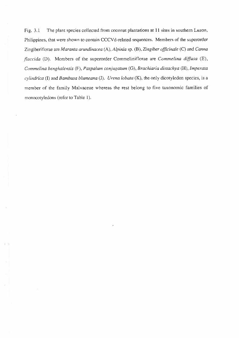
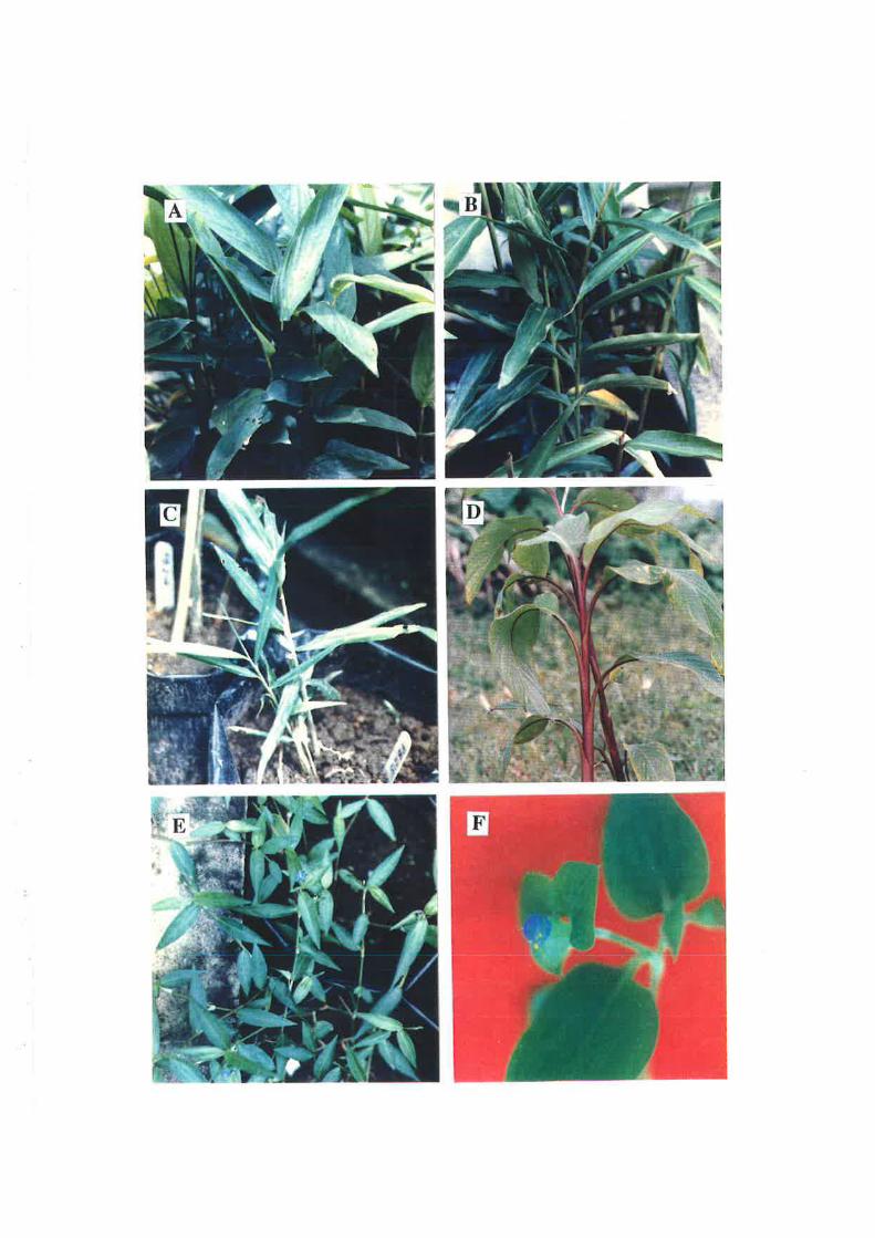
Fig. 3.1 The plant species collected from coconut plantations at I 1 sites in southern Luzon,
Philippines, that were shown to contain CCCVd-related sequences. Members of the superorder
Zingiberiflorae are Maranta arundinncea (A), Alpinia sp. (B), Zingiber fficinale (C) and Canna
flaccida (D). Members of the superorder Commeliniflorae are Commelina diffusa (E),
Commelina benghal.ensis (F), Paspalum conjugatum (G), Brachiaria distachya (H),lmperata
cylindrica (I) and Bambusa blumeana (I). Urena lobata (K), the only dicotyledon species, is a
member of the family Malvaceae whereas the rest belong to five taxonomic families of
monocotyledons (refer to Table 1).
:+
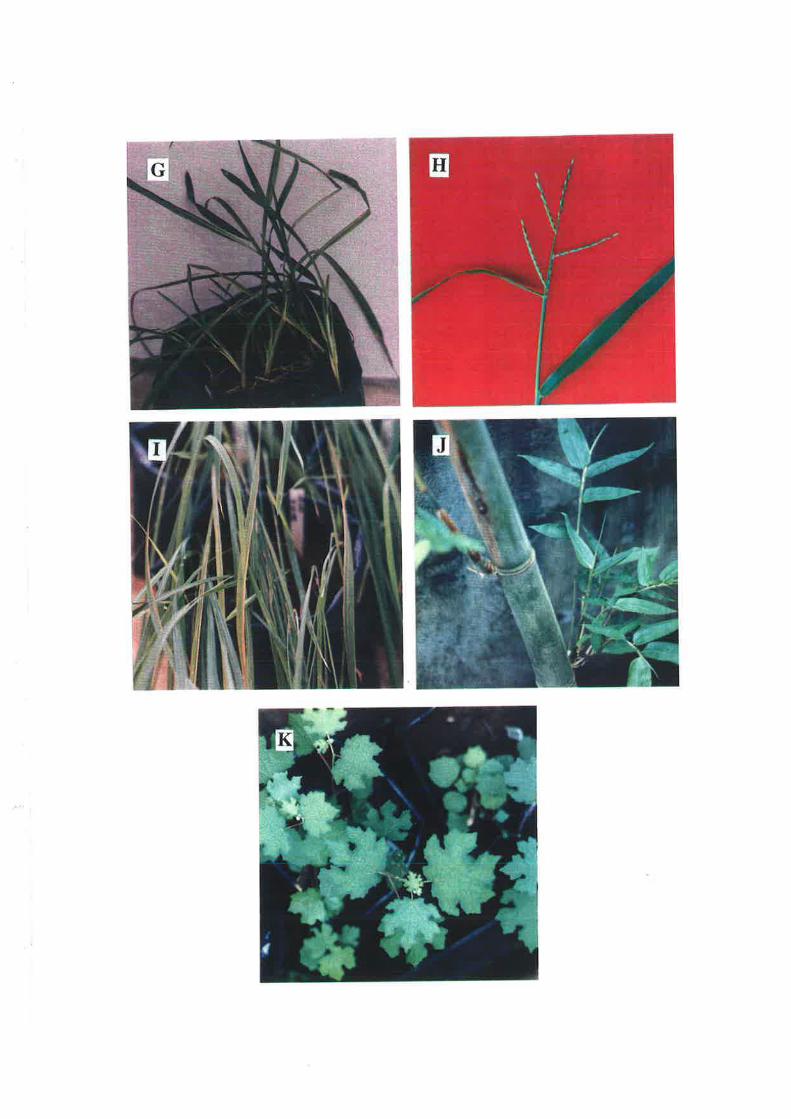
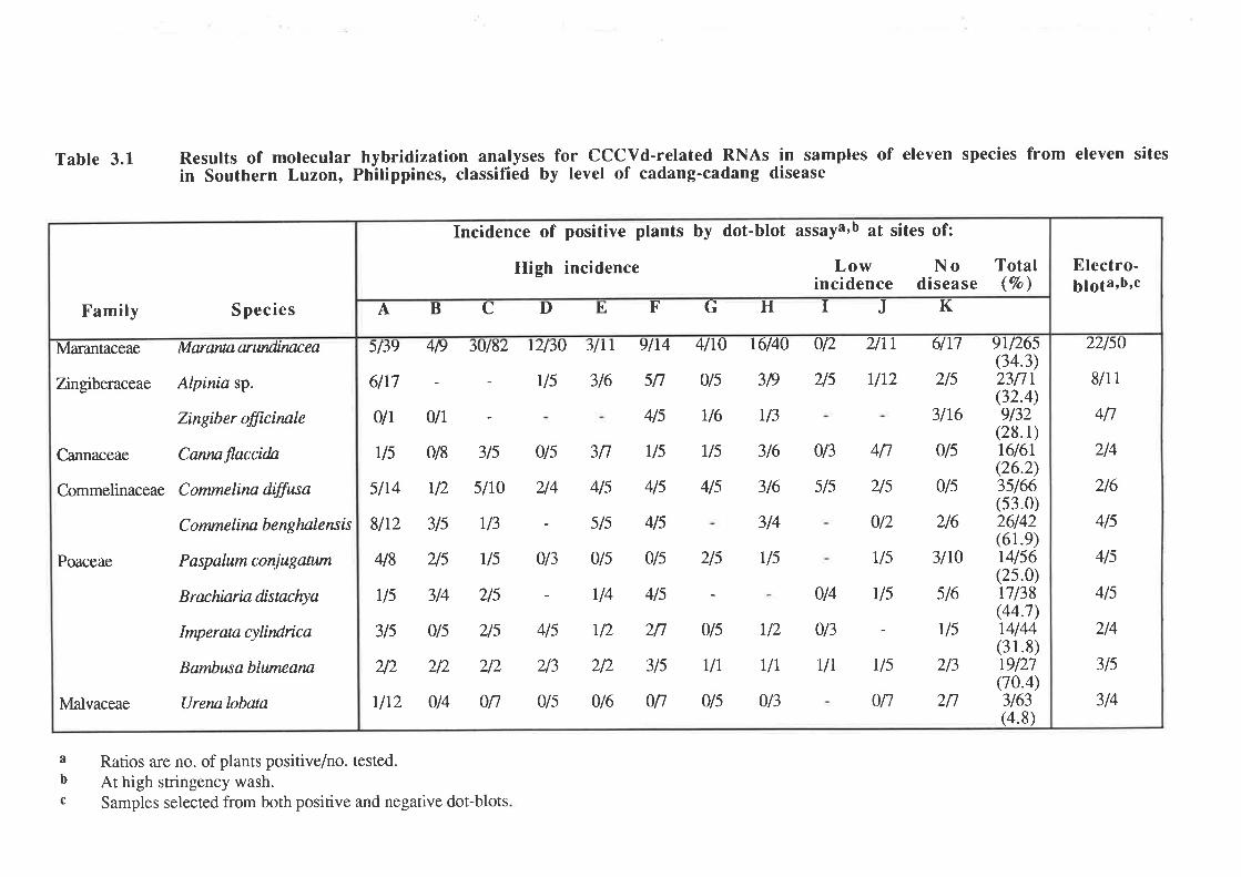
Table 3.1 Results of molecularin Southern Luzon,
hybridization analyses for CCCVd-related RNAs in samples of eleven species from eleven sitesPhilippines, classified by level of cadang-cadang disease
Marantaceae
Zngiberaceae
Cannaceae
Commelinaceae
Poaceae
Malvaceae
Marantaarwtdinncea
Alpinia sp.
Zingiber fficinøle
Cannøflnccida
Commelina diffusa
C orrne I i nn b e ng lnl e ns is
Paspalum conjugatwn
Brachiaria distaclrya
Imperata cylindrica
Bambusa blutneana
Urena lobata
Family S pecies
SILT
3/6
5lJ9
6lt7
0lr
rls
5l14
8/t2
418
U5
3ls
)t)
u12
4lr0
0/s
r/6
r/s
4/s
t6/40
3/9
U3
316
3t6
3/4
U5
012
2/s
013
s/s
014
0t3
UI
2lrr
UL2
6lt7
2/s
3lt6
015
0ls
216
3lt0
s16
vs
213
2n
419 'Juló',¿ r'¿l5U
U5
ylr
sn
4/s
us
4ls
4ls
3n
4ls
sls
4 9r/265(34.3)nnr(32.4)9/32
(28.1)t6l6r(26.2)3s166(53.0)26142(61.e)r4156(25.0)17138(44.7)14/44(31.8)19/27(70.4)3/63(4.8)
0lr
0/8 3ls
12 slt0
3ls r/3
215 tls
3/4 zls
Ùls 2/s
212 212
0t4 0n
0ts
2/4
- U4 4ls
4ls v2 2n Ùls
r/s
017
212 3ls rlr213
0ls
4tl
2ls
0/2
r/s
U5
013 0/s 015 2/s
0t6 0n 015
r12
UI
0t3
ABCDI, FGHIJK
Incidence of positive plants by dot-blot assaya 'b at sites of:
High incidence Low Noincidence disease
Total(vo)
22lso
8/1 I
4n
214
216
4/s
4/s
4ls
214
3ls
314
Electro-5¡g1a,b,c
a Ratios are no. of plants positive/no. tested.b At high stringency wash.c Samples selected from both positive and negative dot-blots
Fig.3.2 Dot-blot assays using the 32P-cDNA probe. A filter with samples from high
cadang-cadang incidence areas was probed, washed at low stringency and autoradiographed
(A), then re-washed at high stringency and again autoradiographed (B). M. arundinacea
samples are rows (a-e); C. benghalemsiJ, row (Ð; C. dffisa, rows (g-i); Alpinia sp., rows (-
Ð: Z. officinale, row (l); U. Iobata, row (m); C. flaccida, rows (n-o); B. blumeana, row (p);
unidentified grasses, row (q); B. distachya,row (n); P. conjugatum, rows (s-t); 1. cylindrica,
row (u) and unidentified grasses, rows (v-y). The CCCVd markers are (y-9) and (y-10). In
autoradiogram (C), the filter was washed at high-stringency and the samples were collected
from a site with zero level of cadang-cadang disease. The strong signals are Alpinia sp. (a-6),
B. distachya (b-6), M . arun"dinacea (b-4) and the CCCVd marker (f- 1).
Each dot is from a differentplant.
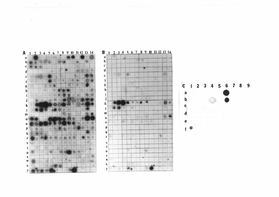
a
a. a
l}
a
l489r0ilr21314 B I 44561
¡r
t23
.lcQl rl-rr
Aa
b
c
d
c
fo'Þr
h
ijk
t
¡¡
m
no
p
q
f
s
tu
v$x
!
aa
-¡+i
I+, oI
t
a,
a
o
I
A
b
c
d
e
fob
h
¡
jkl
m
n
op
q
rS
tu
tw
xv
1rI'"il
2 3 4 5 6 7I Ioo
ca
b
ti
d
ea
ia
aÎ- Olrl
¡caqI
l¡
-]_1 olol
ì
a
I
!
aa
t
Iú
¡
{
o
a
I
t
o4:
a
t
t
Iì
Ia
¡
Io
tl
I)I
cI
a
llt
+
ll¡
a
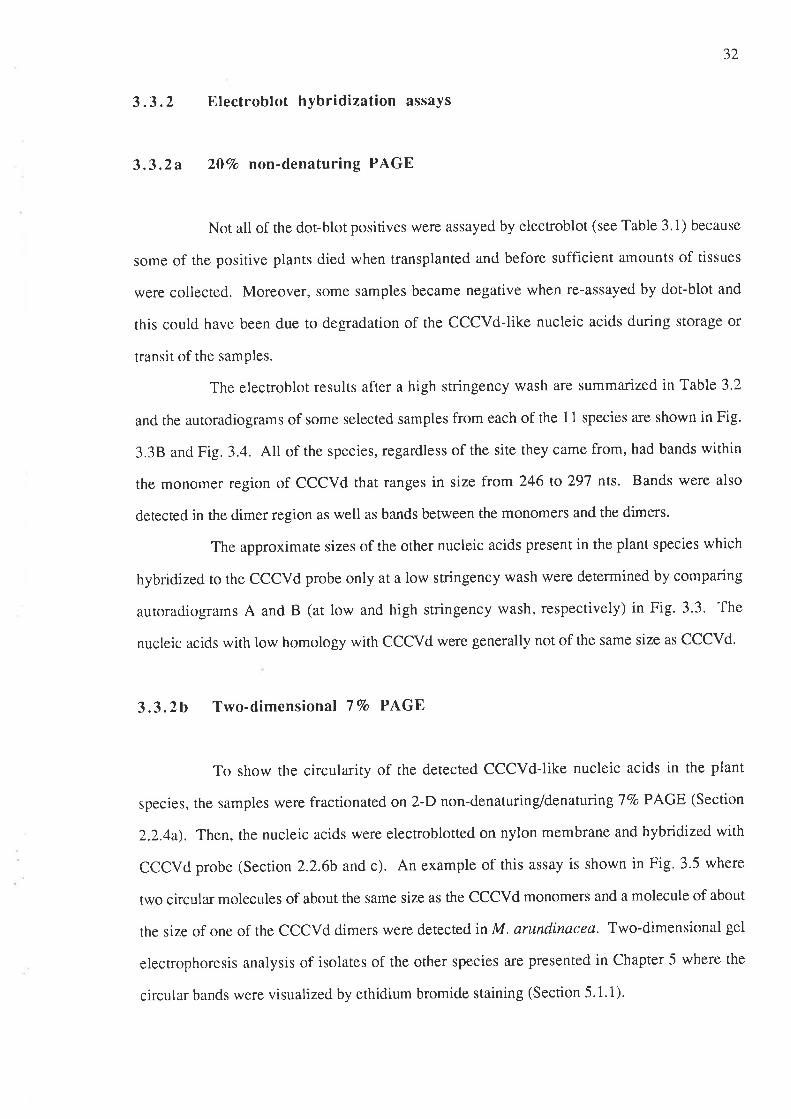
32
3.3.2 Electroblot hybridization assays
3.3.2a 2OVo non-denaturing PAGE
Not all of the dot-blot positives were assayed by elecroblot (see Table 3.1) because
some of the positive plants died when transplanted and before sufficient amounts of tissues
were collected. Moreover, some samples became negative when re-assayed by dot-blot and
this could have been due to degradation of the CCCVd-like nucleic acids during storage or
transit of the samples.
The electroblot results after a high stringency wash are summarized in Table 3.2
and the autoradiograms of some selected samples from each of the 11 species a¡e shown in Fig.
3.38 and Fig. 3.4. All of the species, regardless of the site they came from, had bands within
the monomer region of CCCVd that ranges in size from 246 to 297 nts. Bands were also
detected in the dimer region as well as bands between the monomers and the dimers.
The approximate sizes of the other nucleic acids present in the plant species which
hybridized ro rhe CCCVd probe only at a low stringency wash were determined by comparing
autoradiograms A and B (at low and high stringency wash, respectively) in Fig. 3.3. The
nucleic acids with low homology with CCCVd were generally not of the same size as CCCVd'
3.3.2b Two-dimensional 7Vo PA,GE
To show the circularity of the detected CCCVd-like nucleic acids in the plant
species, the samples were fractionated on 2-D non-denaturing/denatunngTVo PAGE (Section
2.2.4a). Then, the nucleic acids were electroblotted on nylon membrane and hybridized with
CCCVd probe (Section 2.2.6b and c). An example of this assay is shown in Fig. 3.5 where
two circular molecules of about the same size as the CCCVd monomers and a molecule of about
the size of one of the CCCVd dimers were detected in M. arundinacea. Two-dimensional gel
electrophoresis analysis of isolates of the other species are presented in Chapter 5 where the
circular bands were visualized by ethidium bromide staining (Section 5.1.1).
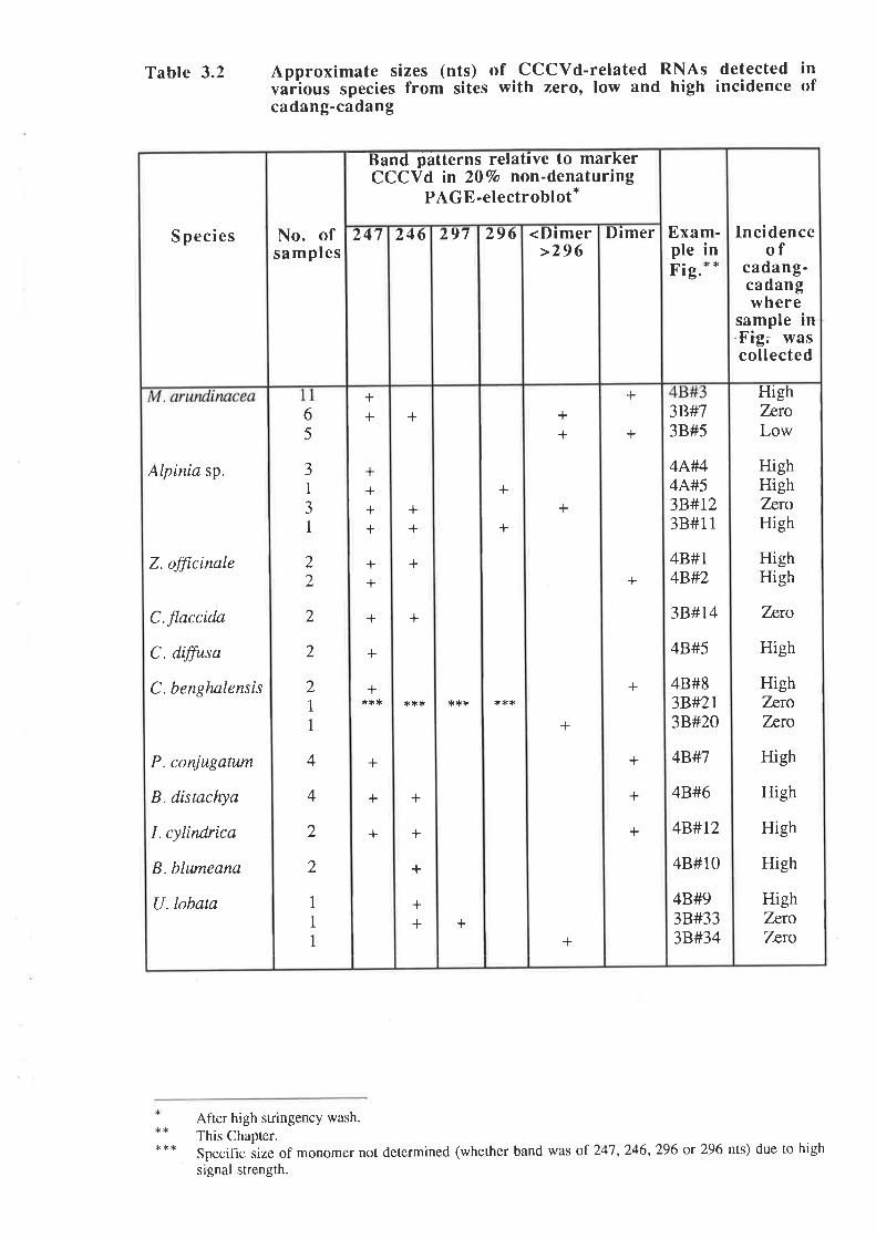
Table 3.2 Approximate sizes (nts) of cccvd-related RNAs detected invarÏous species from sites with zero, low and high incidence ofcadang-cadang
HighTnroLow
HighHighZnroHigh
HighHigh
Tnro
High
HighTrroTnro
High
High
High
High
HighTntoTnro
3B#73B#5
4A#44A#53B#1,238#1 1
48#14B'#2
3B#T4
48#5
48#83B#2r3B#20
4B#7
4B#6
4B#t2
48#10
4B#93B#333B#34
+
+
+
+
+
+
+
++
+
+
+
+
+
***t tr*
+
+
++
+
+
l.*i(
+
+
+
++
++
++++
++
+
+
+*ri.*
+
+
+
65
J1
JI
22
2
2
2I1
4
4
2
2
1
1
1
Alpinia sp.
Z. officinale
C.flaccidn
C. dffisa
C. benghnlensis
P. conjugatum
B. distachya
I. cylindrica
B. blwneana
U.lobata
Incidenceof
cadang-cadangwhere
sample in.Fig; wascollected
Exam-ple inr tg.
Dimer<Dimer>296
296297246247No. ofsamplcs
S peci es
-Band
patterns relative to markerCCCVd in 207o non-denaturing
PAGE-electroblot*
After high stringency wash.
This Chapter.Specific iize of monomer not determined (whether band was of 247 , 246,296 or 296 nts) due to high
signal strength.
**
Fig. 3.3 Hybridization assay of electroblots from 20Vo non-denaturing PAGE using 32p-
cDNA probe. Electroblots were washed first at low stringency, exposed (A), then re-washed at
high-stringency before exposure (B). M.arundinacea samples are lanes (1-8); Alpíniasp.,
lanes (9-12); C.flaccida,lanes (13-1a); C. dffisa,lanes (15-17); C. benghalensis, lanes (18-
2l); P. conjugatum,lanes (22-23): B. distachyc, lanes Qa-26): I. cylíndr\cd, lanes (27-28); B.
blumeann,lanes (29-31) and U. Iobata,lanes (32-34). M denotes CCCVd marker.
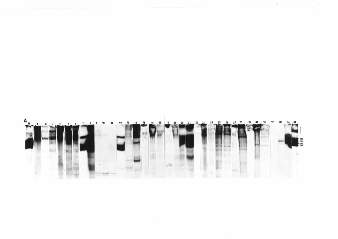
t?s2i3031323317
I
34*9101112
-
5ô123
a
I\r{
;;I¡
Ëùt"s
+
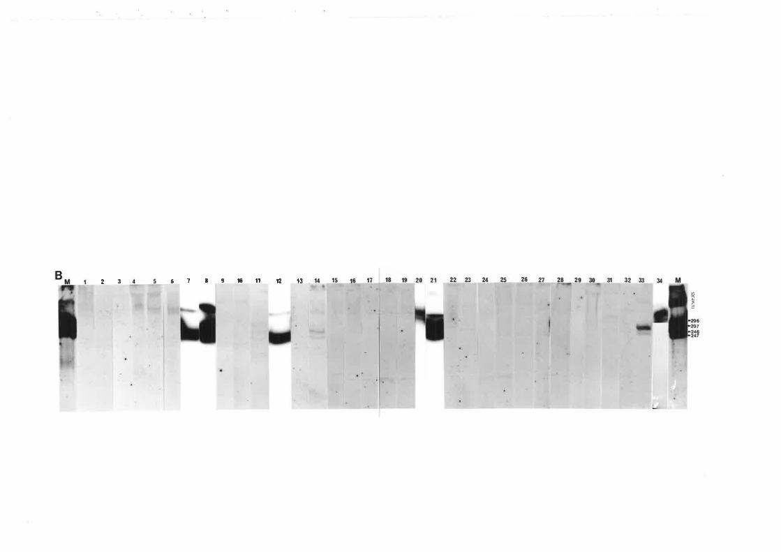
9l0l1t2t367823
,-t'h\ÐË
'1
I
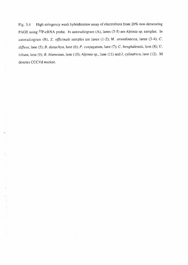
Fig. 3.4 High stringency wash hybridization assay of elecroblots from ZOVo non-denaturing
PAGE using 32P-cRNA probe. In autoradiogram (A), lanes (3-5) are Alpinia sp. samples. In
autoradiogram (B), Z. fficinale samples are lanes (I-2); M. arundinacea,lanes (3-4); C.
diffusa,lane (5); B. distachya,Iane (6); P. conjugatum. lane (7); C. benghalensis, lane (8); U.
lobata,lane (9); B. blumeana,lane (10); Alpinla sp.,lane (11) and I. cylindrica, lane (12). M
denotes CCCVd marker.
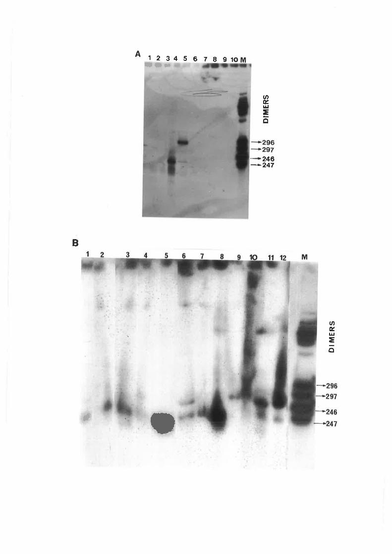
A 1234567891(,ltl¡
ØÉ¡¡¡.ô
246
-247
B
i!
ØÉ,u¡
=ô
*;
J
¿
8l)
I
-J
3
U
^4JJ
DISC USS IO N
CCCVd-like sequences have been detected in l1 plant species commonly found in
coconut plantations. One of these species is a dicot and the rest belong to five families of
monocotyledons from the superorders Commeliniflorae and Zingiberiflorae. As CCCVd-
related sequences could be detected in these plant species regardless ofthe incidence ofcadang-
cadang in coconuts, the infected understorey plants could act as sources of inoculum for
coconuts in the field.
Of the four extraction methods tested, the most effective procedure was blending of
the plant tissues with 0.5 M sodium acetate, 10 mM MgCl2, 2O7o ethanol,3Vo SDS and an
equal volume of water-saturated phenol followed by a series of phenol/chloroform extractions
and CTA B/LiCl p urification s.
The CCCVd-like nucleic acids were found to occur in very low concentrations in
the plant species. Thus, large amounts of tissues (at least 20 g) had to be extracted for these
nucleic acids to be detected by the diagnostic methods described in this study. No apparent
symptom could be observed in the positive plants suggesting that the infection was latent.
There is some degree of sequence and size heterogeneity between these CCCVd-
like nucleic acids and CCCVd, thus, nucleic sequencing is necessary to compare the different
isolates and to determine their degree of relatedness to CCCVd. Since they are present in low
concentrations in the plant tissues, amplification by the polymerase chain reaction (PCR) is
necessary to provide sufficient amount of nucleic acids either for direct sequencing or for
molecular cloning. fl, oft"n desirable, however, to construct clones so that diagnostic probes
can be made for subsequent studies such as viroid replication and epidemiology. Furthermore,
the infectivity of these CCCVd-like nucleic acids can be tested using the clones.
34
CHAPTER 4
DETECTION OF ELECTROPHORETIC FORMS OF CCCVd
ASSOCIATED WITH SEVERE BROOMING SYMPTOMS IN
COCONUT
INTRODUCTION
The naturally occurring sequence variants of CCCVd reported so far are discussed
in Section 1.2.3. Variants which arise during disease progression all contain the basic 246
nucleotides.
The difference between variants 246 and247 as well as between 296 and 297 is the
addirion of only one cytosine residue at position 197 (F{aseloff et al., 1982). This variation can
be detected by the non-denaturing polyacrylamide gel electrophoresis procedure described by
Imperial and Rodriguez (1983). Using this procedure for routine diagnosis of CCCVd in
coconut palms, minor bands which have not been described previously were observed in
positions between the four monomeric CCCVd forms in some cadang-cadang infected palms.
In this chapter, the relationship between the presence of these bands and the severity of
symptoms of the disease was investigated.
EXPERIMENTAL
4.I OBSERVATION OF SEVERE "BROOMING'' SYMPTOMS IN
CCCVd INOCULATED PALMS
At the Albay Research Center (ARC) of the Philippine Coconut Authority (PCA),
Philippines, inoculation of various coconut populations with CCCVd preparation is done with a
hand-primed high pressure injector to seek resistance or tolerance to viroid infections. Since
the small variants of CCCVd are more infectious than the larger variants (Mohamed et al.,
1985), the inoculum is prepared from a mixture of the 246 and247 variants as determined by
35
their mobility in the gel relative to the CCCVd standard marker. The inoculum is prepared in
batches from leaf materials obtained from various palms in local coconut plantations or at the
ARC. Preparations of inoculum are stored in ethanol at -20'C which remain infective for at
least three months (Randles et al., 1992). Approximately 1 ug of CCCVd is inoculated into
each test palm,
For the early inoculations shown in Table 4.1, yearly monitoring of infection by
PAGE assay and observation for symptoms revealed varied reactions to inoculation. The rate
of transmission, regardless of the coconut population, was higher when sprouts were
inoculated af l-7 days post emergence rather than when the sprouts were 2-3 mo-old. In
addition, the rate of symptom development varied (not shown in Table 4.1). An obvious new
symprom type appeared in a significant number of palms. It was characterized mainly by loss
of leaf lamina, giving the palm a "broomed" appearance associated with severe stunting of the
palm (Fig. 4.1). The basal fibres also remained attached to the fronds as in a typical cadang-
cadang infected palm. The observation of this severe symptom was not specific to a particular
coconut population (Table 4.1). In the 2-3 mo-old inoculated palms, the brooming symptom
appeared 4-7 years after inoculation while in the 1-7 d-old sprouts, the symptom first appeared
3-5 years after inoculation. The palms inoculated in 1981 and 1982 had a higher brooming
ìncidence (l8.8%o and22.7Vo, respectively) than the palms inoculated in 1983-1986 (4.4Vo ro
l2.9%o). Premature death was observed in some of the palms. Although there is no direct
evidence that this was caused by the severity of the disease, this observation is unusual even for
palms with the typical cadang-cadang symptoms.
Table 4.2 shows the results of an infectivity trial where the variants of CCCVd
determined by their mobility in the gel were isolated from each other and tested for their ability
to infect. Ten-fold dilution of each variant was done starting from 200 ng/test palm to 200
pgltest palm. The rate of rransmission was higher in palms inoculated with the 246 and 247
variants than in the palms inoculated with the 296 and297 vanants. Furthermore, brooming
incidence was essentially conf,rned to palms inoculated with the 246 or 247 variant, with only
one case of a brooming palm occurring among those inoculated with the 296 variant.
In all the trials conducted, a total of 1,787 successfully inoculated palms have been
monitored and l27o of these had brooming symptoms.
Table 4.1 Incidence of the brooming symptom in various coconut populations inoculated with CCVd isolated from palms with
246 and 247 forms onlY
Year Population inoculated
nocu at on assay ppearance ng
Years afterinoculation
Vo
broomingAge of testpa¡ms(p.e.)*
No. positive/no. inoculated
symptomYears after No. withinoculation brooming
mou30U30913019130ru30sl308/30
7
66
66
)
:
4
llr0lr419
4lt9OILI3lsl/8
BaguerBanga
6666666
55555
5
444
SpicataGatusanOrange Dwarf (OD) x BaYbaYCatigan x RennelTacunan x \üy'est African Tall (WAT) 18.8
1982 2mo Maìayan Yellow Dwarf (MYD)TambolilidCatiganMagtuodCameroon Red Dwarf (CRD)Kapatagan
UT71n331294ß02l18sl29
0lr3no13214
0120ts 22.1
1983 2mo mixed populationsL/[YD xV/ATOD x V/AT
3mns031243124
1 8/300ol30t3 5.9
Table 4.1 (Continued)
Post emergence
STB, STP, LT ¿rnd YD a¡e local Philippine selections
x55
55
;5
JJ
4
4
STB** x STP**CRD X CRDSTP x STPYD** x LT**OD x V/ATSTB X STBCRD X LTODxLT
44444444
z2222
I
r4124tsl22211242U24Lsl2418124t3/2319124
4/142lrssl2r4l2r0lts0/18IIT33l19 t2.9
1985 L-l d TambolilidCatiganODI/[YDTambolilid
286lssÙr04l2t513s1280tt4l210901n 4
r71286U1040lr3s0/tr4r4190
1986 l-l d Tambolilid 2421450 171242
4.4
t.0
assay
No. positive/no. inoculated
Year Population inoculated
ocu onat ppearance roomrng
Years afterinoculation
Vo
broomingAge of test
palms(p.e.)*
symptomYears after No. withinoculation brooming
Fig. 4.1 The unusual, severe brooming symptoms observed in coconuts inoculated with
CCCVd va¡iants 246 and247.
A. The palm is severely stunted with a "broomed" appearance due to the
reduction of the lamina. The basal fibres remain attached to the fronds as in a typical cadang-
cadang infected palm.
B. The frond is shorter (left) compared to a healthy frond (righÐ.
C. Leaflets have reduced laminae (left) as compared to cadang-cadang infected
leaflets (middle) and healthy leaflets (righÐ.
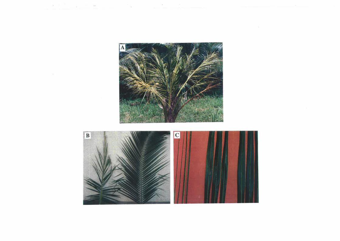
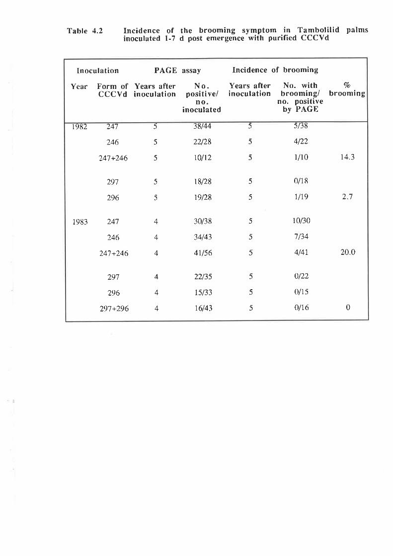
Table 4.2 Incidence of the brooming symptom in Tambolilid palmsinoculated L-7 d post emergence with purified CCCVd
1982 247
246
241+246
291
296
247
246
247+246
297
296
297+296
5
5
5
38144
22128
r0l12
5
5
5
sl38
4122
rlrc t4.3
5
5
18128
19128
5
5
0/18
u19 2.7
1983 4
4
4
30/38
34143
4U56
5
5
5
10/30
7/34
4/4r 20.o
4
4
4
2213s
rsl33
16143
5
5
5
0/22
olt5
0/16 0
Inoculation PAGE assay Incidence of brooming
Years after No. withinoculation brooming/
no. positiveby PAGE
Year Form of Years after N o.CCCVd inoculation positive/
no.inoculated
Vo
brooming
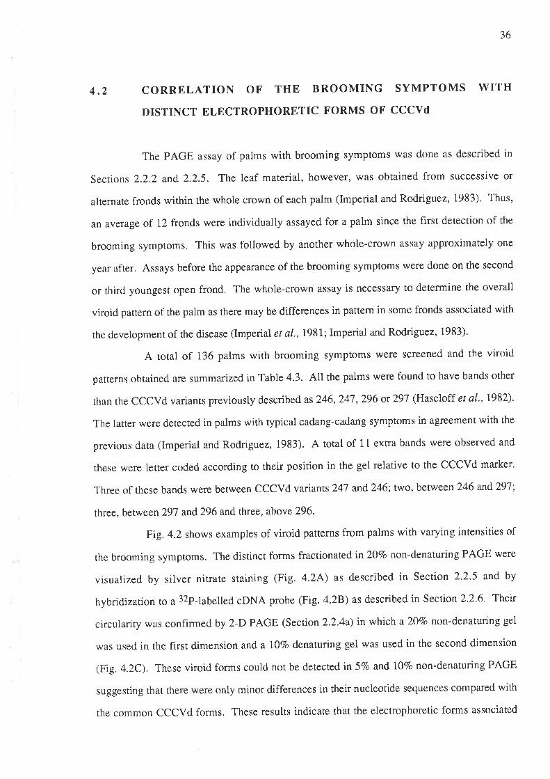
36
4.2 CORRELATION OF THE BROOMING SYMPTOMS WITH
DISTINCT ELECTROPHORETIC FORMS OF CCCVd
The PAGE assay of palms with brooming symptoms was done as described in
Sections 2.2.2 and 2.2.5. The leaf material, however, was obtained from successive or
alternate fronds within the whole crown of each palm (Imperial and Rodriguez, 1983). Thus,
an average of 12 fronds were individually assayed for a palm since the hrst detection of the
brooming symptoms. This was followed by another whole-crown assay approximately one
year after. Assays before the appearance of the brooming symptoms were done on the second
or third youngest open frond. The whole-crown assay is necessary to determine the overall
viroid pattern of the palm as there may be differences in pattern in some fronds associated with
the development of the disease (Imperial et a\.,1981; Imperial and Rodriguez, 1983).
A total of 136 palms with brooming symptoms were screened and the viroid
parterns obtained are summarized in Table 4.3. All the palms were found to have bands other
than the CCCVd variants previously described as 246,247 ,296 ot 297 (Haseloff et al., 1982).
The latter were detected in palms with typical cadang-cadang symptoms in agreement with the
previous data (Imperial and Rodriguez, 1983). A total of 11 extra bands were observed and
these were letter coded according to rheir position in the gel relative to the CCCVd marker.
Three of these bands were between CCCVd variants 247 and246; two, between 246 and29l;
three, between 297 and296and three, above296'
Fig. 4.2 shows examples of viroid patterns from palms with varying intensities of
the brooming symptoms. The distinct forms fractionatedin20To non-denaturing PAGE were
visualized by silver niffate staining (Fig. 4.24) as described in Section 2.2.5 and by
hybridization ro a 32P-labelled cDNA probe (Fig. a.2B) as described in Section 2.2'6. Their
circularity was confirmed by 2-D PAGE (Section 2.2.4a) in which a 207o non-denaturing gel
was used in the f,rrst dimension and a I\Vo denaturing gel was used in the second dimension
(Fig. a.2C). These viroid forms could not be detected in 5Vo and l07o non-denaturing PAGE
suggesting that there were only minor differences in their nucleotide sequences compared with
the common CCCVd forms. These results indicate that the electrophoretic forms associated
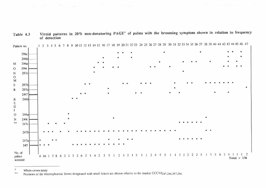
Table 4.3
29&,
296a
296
297
246a
246
24',1c
24'la
247
No. ofpalmsscreened
Viroid patterns inof detection
20q,o non-denaturing PAGE* of palms with the brooming symptom shown in relation to frequency
Pattern no t 2 3 4 5 6 1 8 9 10 Lrt2r3 1415 16 17 18 19 20212223 242526272829 303132333435363'1 383940414243444546 47
a a a a
a
a a a aa a
aa aa o aa
a
aa a aaa a aa aaaaaaa
a
aa aa a
a
a
a a aaaaaa
a a aaaaaaaaaaaaaa"".." aaaaa
aa a aaaaaa
aaa aaaaaaaaaaaa
616 I 28 6 2l 3 2 6 2 5 4 2 3 3 r 2 | 2l 3 I I 2 4 2 3 I I 2 2 2 2 2 s I 3 3 8
a
a
a
a
a
a
aa
a
aa
M
oNoME
R
REGIoN
a
aaa
a
a
o
a
a
a
aa
3lllTotal =
2
Vy'hole-crown assaypositions of rhe elecr¡ophoretic forms designated wi¡h small leners are shown relative to the ma¡ker CCCYd2al ,246,29'l ,296.
l3ó
Fig. 4.2 Assay of some palms with varying intensities of the brooming symptoms.
A. 20Vo non-denaturing PAGE assay of extracts from palms, nos. 1-5 (see Table
4.4) and a mixture of their isolates in lane no. 6. The bands were visualized by silver nitrate
staining.
B. Hybridization assay of an electroblot from a 2O7o non-denaturing PAGE of
the samples in A above.
C. Two-dimensional PAGE of a mixed isolate (lane no. 6 in A and B) wíth207o
non-denaturing gel in the first dimension and 107o denaturing gel in the second dimension. The
affows show the circular bands.
A
úf¡l¿a
+296+297
-->246
+247
B M 1 z 3 4 s 6M
-T.:'J=¡{ H-
ú,lJl
H
aa
+296
-+297-+2464247
c
ì
IIt
*
L
It
r l
37
with the severe brooming symptoms differed in primary and secondary structure from CCCVd,
and are probably mutants of CCCVd.
VARIATION IN THE VIROID PATTERNS DURING DISEASE
DEVELOPMENT
4.3
Twelve palms were analyzed for their viroid patterns from the time the viroids were
fi¡st detected until the brooming symptom appeared (Table 4.4). The hnal PAGE assays of five
of these palms are shown in Fig. 4.2. lt was observed that infection started almost always with
variant 247, although the inoculum was a mixture of variants 246 and 247. Then,
approximately a year before brooming appeared, additional bands with varying gel mobility
were detected. As the disease developed, more variations were observed with the size of the
molecules increasing. However, there were at least two palms (with viroid patterns 2 and3,
Table 4.3) that died before the larger molecula¡ variants could be detected. But again, there was
no direct proof that the death was caused by infection with the observed electrophoretic forms
associated with brooming symptoms.
DISC USSIO N
A severe "brooming" symptom was observed in approximalely I27o of the
CCCVd-inoculated palms screened. The symptom is characterized by the loss of leaf lamina,
hence, the brooming appearance associated with severe stunting of the palm, and occasional
premature death. Observed 3-7 years after inoculation, it was essentially confined to palms
incrculated with either 246 or 247 vaiants of CCCVd.
PAGE and hybridizarion analyses have shown that all brooming palms have distinct
electrophoretic forms of CCCVd. The next step is to sequence these forms to determine
whether the severity of the disease is caused by mutations in CCCVd.
Table 4.4 Time-course analysis of viroid pattern prior to the appearance of the brooming symptom
STB x STP I
ODxLTI
STP x STP 1
STP x STP 2
Tambolilid no. I
Tambolilid no.2
Tambolilid no. 3
Tambolilid no. 4
Tambolilid no. 5
Tambolilid no. 6
Tambolilid no. 7
Palm sample
1982
1983
1983
1983
1983
1985
1985
1985
1985
1985
1985
1986
Yearinoc-ulated
1981
1988
1988
1988
1988
r988
1989
1989
1989
1989
1989
1990
Yearbrooming
wasobserved
a a,t
NA*
247
247
NA
241
247
291,297a
247,246b
241 ,247b
241,246b
247,24'7b,296,296a,296b
247,241b,29Jc,296
24J,24Ja,247b,296,296b,296c
247b,246,246a
NA
247a,247b
NA
247,24lb
241 ,241b
+DB
NA 247,246 NA
247 NA 247 ,241b NA
247,246
247
NA
247
NA
241
+DB
NA
+DB**
NA
247
241
24la,24lb
241a,247b
241b
241,241b
24tb
241b 247 247b
1984 1985 r986Viroid pattern
1987 1988 1989 1990
t7 I
9 2
l6
t4
t9
8
4
4
6
2
6
2
J
4
5
Patternno. inTable4.3
Isolateno. inFig.4.2
Not assayed.
Positive in dot-blot assay
38
CHAPTER 5
PURIFICATION OF CCCVd.RELATED NUCLEIC ACIDS FOR
AMPLIFICATION BY POLYMERASE CHAIN REACTION (PCR)
AND THEIR SUBSEQUENT MOLECULAR CLONING
INTRODUCTION
A commonly used approach for purification of viroids has been to prepile a low
molecular weight nucleic acid extract of infected plant tissue and then to separate the circular
viroids from the contaminating cellular linear RNAs (Schumachet et al., 1983; Rivera-
Bustamante et al., 1936). In a denatured stato, the circular viroids have much lower
electrophoretic mobility than linear RNAs of the same size (Schumacher et a|.,1983). Viroids
isolated in this way have proved to be pure enough for many purposes such as direct RNA
sequencing or invitro synthesis of viroid cDNA for cloning (Keese and Symons, 1987).
Some viroids are present in plant tissues in small amounts so that their isolation,
purification and subsequent molecular analysis is difficult. However, with the introduction of
the polymerase chain reaction (PCR) technology, viroid amplification via its cDNA overcomes
this problem (puchta and Sanger, 1989). The PCR is a rapid procedure for in viÛo enzymatic
amplification of a specific segmenr of DNA (White et a1.,1989). The thermostable DNA
polymerase isolated from Thermus aquaticus (Taq polymerase) (Chien et al., 1976), has
allowed simplification of the procedure by enabling the amplification reaction to be performed at
higher remperatures. This significantly improves the speed, specificity, yield and sensitivity of
the procedure as well as the length of products that can be amplified (Saiki et al', 1988). A
three-step cycling process is involved: (1) denaturation of double-stranded (ds) DNA; (2)
annealing of primers to complemenrary sequences of each single-stranded (ss) DNA template;
and (3) primer extension by Taq DNA polymerase along both template strands in opposite,
overlapping directions. This can lead to the many million-fold amplification of the target DNA
fragment over rhe course of 20 to 30 cycles (White et a1.,1989). However, optimization of the
pCR technique is somewhat empirical. Individual reaction components and time/temperature
39
pilameters must be adjusted for efhcient amplification of specific targets. The optimization and
interaction of these factors and general guidelines for achieving the best results are discussed by
several authors (Williams, 1989; Sambrook ¿r a1.,1989; Kwok and Higuchi, 1989; Krawetz et
al., 1989; Sarkar and Sommer, 1990; Lowe et al., 1990; Ruano et al., l99l; Ausubel et al.,
1991).
For certain applications PCR can obviate the need for cloning, as amplification
products can be analyzed directly. However, many strategies generate complex mixtures of
products, necessitating molecular cloning for further analyses of specific target molecules
(Mead et a|.,1991).
Difficulties are frequently encountered when cloning PCR products. A simple
method, however, has been shown to be efficient and is based on the observatiorlthalTaq
polymerase can add a single non template-directed deoxyadenosine (A) residue to the 3' end of
duplex PCR products (Clark, 1938). This template-independent activity of Taq polymerase has
been exploited to create a cloning scheme which has the efficiency of sticky-end cloning, but
requires no additional enzymatic modification of the PCR product (Holton and Graham,l99L;
Marchuk et a|.,1991).
This chapter describes the purification of CCCVd-related nucleic acids for
amplification by PCR and molecular cloning of the PCR products.
EXPERIMENTAL
5.1 PURIFICATION
5. 1. 1 CCCVd-related nucleic acids in plants other than palm species
Preparations of CCCVd-related nucleic acids in plants other than palm species were
subjected to2-D gel electrophoresis (Section 2.2.4a). This method, although laborious as only
one or two samples can be analyzed on a single gel, is efficient in determining the gel positions
of unknown viroids varying in size and sequence.
40
Fig. 5.1 shows the results of 2-D gel analyses of partially purified extracts of some
of rhe plant species compared with the pattern for CCCVd. The circular molecules detected in
each species were numbered in the order of increasing elecrrophoretic mobility. Six circular
bands were detected in the CCCVd isolate, hve in M. arundinacea, five in Alpinia sp., four in
C. flaccida, four in C. diffusa and three in B. blumeana. The zones containing these molecules
were separately excised and eluted from the gels as described in Section 2.2.4b.
5.1.2 Electrophoretic forms of CCCVd associated with the brooming
symptoms in coconut
Listed in Table 5.1 are the isolates extracted as described in Section2.2.3 ftom
palms with varying intensities of the brooming symptoms. These partially purified isolates
showed viroid patterns that are more or less representative of the total brooming-associated
electrophoretic forms detected in the 136 palms screened (Table 4.3). Either singly or mixed,
the isolates were purified further by bi-directional PAGE (Section 2.2.4a). V/ith the bi-
directional system, large amounts of sample can be applied on a single gel since the position of
the electrophoretic forms in the native gel relative to the CCCVd marker is known (an example
is shown in Fig. 5.2). The purihed isolates were again analyzed for thei¡ viroid pattern on2O7o
non-denaruring PAGE (Fig. 5.3). Since most of the isolates had more than one viroid form,
attempts were made to separate each form from the others in a long first dimension non-
denaturing gel of the bi-directional PAGE. The separation was not successful particularly for
those forms very close to each other as shown in Fig. 5.3, Bl and 82.
5.2 POLYMERASE CHAIN REACTION (PCR)
In this study, CCCVd variant 246 and three sets of CCCVd-specific DNA primers
were used to determine the parameters suitable for PCR with CCCVd.
Fig.5.1 Two-dimensional (2-D) TVoPAGF. where a lane with one sample in the first
dimension non-denaturing gel was cut vertically and placed horizontally below the second
dimension denaturing gel. The samples are: pooled extracts from coconut palms at different
stages of the cadang-cadang disease (A); M. arundinace¿ (B); Alpinia sp. (C); C. flaccida (D);
C. dffisa (E) and B. blumeana (F). The circular molecules which migrate behind the diagonal
front of the linear nucleic acids are numbered in the order of increasing electrophoretic mobility.
The gels were stained with ethidium bromide.
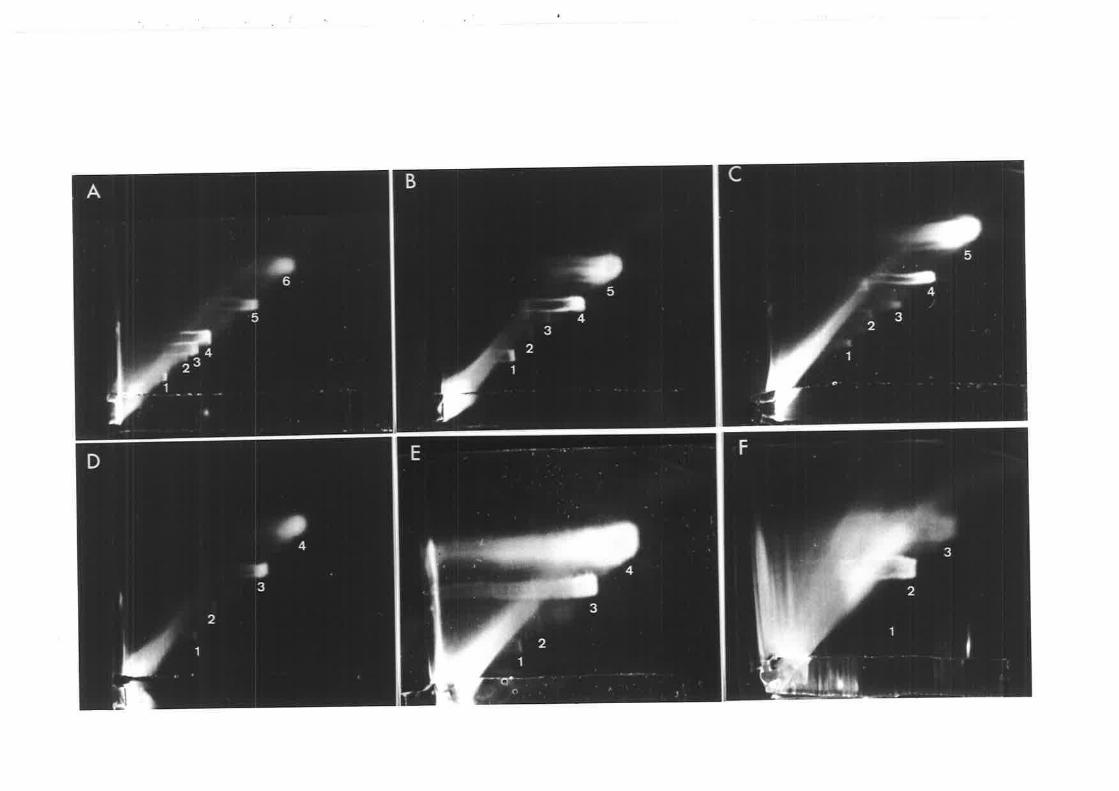
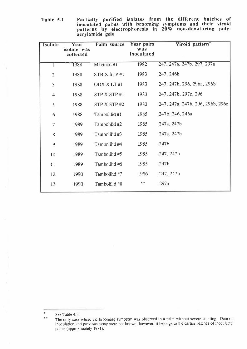
Table 5.1 Partially purified isolates frominoculated palms with broomingpatterns by electrophoresis inacrylamide gels
the different batches ofsym20Vo
ptoms and theirnon-denaturing
viroidpoly-
o¿ +t
2 1988 STB X STP#1
3 1988 ODX X LT #1
4 1988 STP X STP #1
5 1988 STP X STP #2
6 1988 Tambolilid #1
7 1989 Tambolilid #2
8 1989 Tambolilid #3
9 1989 Tambolilid #4
10 1989 Tambolilid #5
Tambolilid #6
Tambolilid #7
Tambolilid #8
11
t2
13
1989
1990
1990
1982
1983
1983
1983
1983
1985
1985
1985
1985
1985
1985
1986
**
247 ,247a, 247b,297, 29'7a
247,246b
247 , 24'7b, 296, 296a,296b
247 ,247b, 291c, 296
247 ,247 a, 247b, 296, 296b, 296c
247b,246,246a
247a,247b
241a,247b
241b
241,247b
24tb
247 ,247b
297a
Isolate Yearisolate was
collected
Palm source Year Pa lm Viroid pattern'was
inoculated
See Table 4.3.
The only case where the brooming symptom was observed in a palm without severe stunting. Date ofinoculation and previous assay were not known, however, it belongs to the earlier batches of inoculatedpalms (approximately l98l).
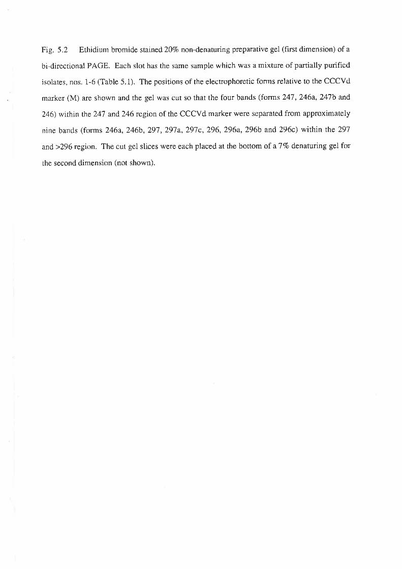
Fig. 5.2 Ethidium bromide stained 20Vo non-denaturing prep¿ìrative gel (first dimension) of a
bi-directional PAGE. Each slot has the same sample which was a mixture of partially purified
isolares, nos. 1-6 (Table 5.1). The positions of the electrophoretic forms relative to the CCCVd
marker (M) are shown and the gel was cut so that the four bands (forms 247,246a,247b and
246) within ¡he 247 and246 region of the CCCVd marker were separated from approximately
nine bands (forms 246a,246b,297,297a,297c,296,296a,296b and 296c) within the 297
and>296 region. The cut gel slices were each placed at the bottom of a77o denaturing gel for
the second dimension (not shown).
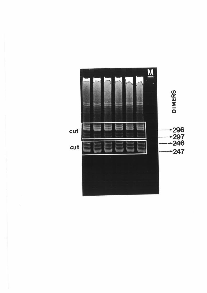
MFFrF!FIrFqH
ØÉ,l¡¡
=o
cut
cut
2967
246247
Fig. 5.3 Analysis on2o%o non-denaturing polyacrylamide gels (ethidium bromide stained) of
the brooming isolates (Table 5.1) purifred by bi-directional PAGE. M denotes CCCVd ma¡ker.
A-1 Isolate no.12 from a single palm with electrophoretic forms 247 and247b.
2 Mixed isolate (nos. 7-11) with electrophoretic forms 247,24'7aand247b.
3 Mixed isolate (nos. 1-6) with electrophoretic forms 247,24Ja,247b and246
(see Fig. 5.2).
4 Mixed isolate (nos. 1-6) with electrophoretic forms 246a,246b,297,297a,
297c,296,296a,296b and 296c (see Fig. 5.2).
B- 1 and 2. Separation of the electrophoretic forms in mixed isolate (A-2) above.
3 Isolate no. 13 from a single palm with electrophoretic form 297a.
A
296<297a
246<247<
B
>296>297>246>247
123
t-ll-l-,
4l
5.2.1 Standardization of PCR
5.2.\a Design of primers and preparation of template
Since the starting material is an RNA, the first factor to consider is the design of a
CCCVd-specific DNA primer that will eff,rciently prime the synthesis of the first strand cDNA.
For this cDNA to be amplified by PCR, the same primer is used again while a second primer
which is complementary to the 3'-end of the ss cDNA simultaneously primes the second strand
synthesis resulting in a ds cDNA as the PCR product.
To carry our rhe above reactions, three sets of primers (Table 5.2) derived from the
sequence of CCCVd variant 246 (Haseloff et al., 1982) were used. With primer sets I and II,
the left and right half-lengths of the total CCCVd sequence, respectively, were amplified only
when the synthesis of the first strand cDNAs was done using the AMV reverse transcriptase
merhod (Section 2.2.7a). On the other hand, full-length CCCVd was amplified using primer
ser III and the M-MuLV reverse transcriptase method (Section 2.2.7a). The priming sites for
primer sets I and II are at the highly conserved central region of CCCVd where there is more
secondary srructure and more G-C base pairs than in the priming site for primer set III. Thus,
it is concluded that the AMV reverse transcriptase method must have provided the right
conditions for the melting of the viroid and annealing of the primer.
5.2.1b MgCl2 concentration
The concentration of MgCl2 in the buffer is a variable that can greatly influence the
outcome of PCR. Thus, the optimum concentration of this component has to be determined.
Although each component of PCR can affect its outcome, most do not lend themselves as
readily to optimization as does MgCl2 concentration (Ausubel et a|.,1991).
According to Puchta and Sanger (1989), the MgCl2-concentration for optimal
amplification of HSVd cDNA ranges between 0.7-1.0 mM. An experiment was, therefore,
done to determine the MgCl2 concentration for optimal amplification of CCCVd. The results
showed (Fig. 5.4, A) that there was minimal difference between the rates of amplif,rcation for
Table 5.2 PropertiesCCCVdz¿r
of the DNA primers used for the reverse transcription (RT) and polymerase chain reaction (PCR) of
Primer Set
I
II
ilI
equence
5' -d (GGAGACTACCCGGTGGATACAACT)-3',
5'-d (GAGTTGTATCCACCGGGTAGTCTCC)-3',
5' -d (AGGGATCCCCGGGGAAACCT)-3'
5' -d (GGCCTCTCCTGCAGTGGTTTTTGGGGTG CCC)-3'
5', -d (CCACTGCAGGAGAGGCCGCTTGAGGGATCCCC)- 3',
-s A pnmer
Polaritya
+
+
+
Binding site atCCCVd2a6b(nucleotides
5'-+3')
10-5t
L7t-194
t95-r7l
52-11
46-16
30-61
Reaction
RT + PCR
PCR
RT + PCR
PCR
RT + PCR
PCR
Amplified CCC vdcDNA fragmentb'c(product length)
50-U246-195(102 nts)
r10-12(99 nts)
rs-u246-62(200 nts)
a The numbering of rhe CCCVd nucleotides is adopted from Haseloff et al. (1982).
b The infecrious viroid RNA is (+) RNA.c rhe nucleoúde posirions indicate rhe lengrhs of the non-prmer cDNA sequcnce rhat comprise rhe toral cccvd sequence except lor posiúons 5l -61 but these are part of the primer
sequence derived from the highly conserved central region of cccVd.
Fig. 5.4 Ethidium bromide stained I.27o asarose gels of PCR products from reactions using
primer ser III (Table 5.2) and Mu-MLV reverse transcribed cDNAs of pure CCCVdz¿O as
template. M denotes DNA MW ma¡ker III'
A. PCR described in Section 2.2.7b where the template cDNAs were purified by
chromatography on a Sephadex column but with the following MgCl2 concentrations:
1. 0.70 mM
2. 0.85 mM
3. 1.00 mM
4. 1.50 mM
5. Same as no. 4 except that reverse transcription buffer was used.
6. Same as no. 4 except that cDNA was purified by ethanol precipitation.
7. same as no. 4.
B. One-tube assay (RT-PCR) using reverse transcriptase buffer with the
following purified viroid template concentrations :
1. 200 ng.
2. 100 ng.
3. 10 ng.
4. 1 ng.
5. 0.1 ng.
6. Sample as in A-4 as connol.
g
9Zr-
799-
V
+
a
Ii
42
MgCl2 concentrations of 0.7 mM and 1.5 mM. Therefore, in the succeeding experiments, 1.5
mM MgCl2 was used in the PCR method as described in Section2.2.7b.
5 .2.2 Devetopment of a one-tube, one manipulation reverse transcription
(RT) - PCR reaction method
Krawetz et al. (1989) discovered that substituting reverse transcriptase buffer for
the PCR buffer markedly increases the efficiency of the PCR reaction. This was done for
CCCVd and the resulrs showed that there was minimal difference in the PCR yield between the
reaction using reverse transcriptase buffer (Section 2.2.7a) and that using the recommended
pCR buffer (Section 2.2.1b) (Fig.5.4, A). However, there was an increase in the PCR yield
when the f,rrst strand DNA was purified simply by ethanol precipitation before PCR rather than
doing a more laborious Sephadex column purification as described in Section 2-2.6a- With
these results, it was then possible to do a one-tube, RT-PCR reaction wherein reverse
transcription was done first aÍ.37"C, immediately followed by PCR in a thermal cyclþfter,^
adding the two primers andTaq polymerase. By varying the concentration of the CCCVdz¿O
template, it was shown that the minimum concentration of viroid for the PCR to work under the
above conditions was 100 ng (Fig. 5.4, B). This one-tube method did not work when PCR
buffer was used and it was not tried on the CCCVd-related nucleic acids.
5.2.3 PCR of CCCVd-related nucleic acids
Using the standardized reverse transcription and PCR procedures done separately
as described in Sectio n 2.2.7 and the primer set I shown in Table 5.2, the circular molecules
purified from the marker CCCVd exract, M. arundinacea and Alpinia sp. were tested (Fig. 5.1,
A, B and C, respectively). Not one of the circular molecules from the non-palm species was
amplified. Only the circular molecules 1-4 of the CCCVd extract had PCR products. The
circular molecules 4 and 3 correspond to the small (2461247 nts) and larye (2961291 nts)
CCCVd monomers, respectively, while the circular molecules 2 and 1 are their respective
dimers. The two smallest circular molecules (5 and 6) were not amplihed. Molecules of about
43
the same size as these were also observed in the plant extracts from the non-palm species (Fig.
s. 1).
When the purified isolates of the electrophoretic forms of CCCVd associated with
the brooming symptoms as discussed in Section 5.1.2 were amplified, PCR products were
obtained with the expected sizes as predicted from the three primer sets used (Table 5.2)'
5 .3 MOLECULAR CLONING OF PCR PRODUCTS
The PCR products with the electrophoretic forms of CCCVd as templates were
purified by gel electrophoresis (Section 2.2.8a) to avoid the cloning of spurious bands.
polyacrylamide gel was preferred to agarose gel because it gives higher resolution. In addition,
agarose is frequently contaminated with substances that are potent inhibitors of many enzymes
including ligases (Maniatis et a|.,1982).
The Bluescript plasmid was chosen as the cloning vector because it carries
lacZAMl5 and laclQ genes on an F' episome which provide cr-complementation of the p-
galactosidase gene (Sambrook et al., 1939). This allows blue/white color selection of
recombinant colonies on plates supplemented with IPTG and X-gal.
The plasmid was prepared for ligation as described in Section2.2.8a so that the
plasmid vector and the PCR product insert had complementary single base 3' overhangs (T:A)'
Vector self-ligation events were prohibited by the 3' thymidine (T) overhang whereas
concatamerization of the insert was prohibited by the unphosphorylated 5' end contributed by
the oligonucleotide primer, as well as the 3' adenosine (A) overhang added by Taq polymerase
during the pCR reaction (Marchuk et a\.,1991). Ligation of the PCR product to the vector was
done as described in Section 2.2.8b.
One of rhe recommended E. coli strains for the Bluescript plasmid is JM109.
Competent cells of this strain were prepared (Section 2.2.8c) and transformed with the
recombinant plasmid (Section 2.2.8d). Since a small percentage of vector molecules could
escape thymidine (T) addition, the recombinants were selected by employing the blue/white
44
color screening (Section 2.2.8e). Recombinant colonies were white. The average
transformation efficiency obtained was 5.5 x 10⁶ transformants/ug.
Mini-preparations (Section 2.2.9) of the recombinant plasmids were analyzed for
the sizes of inserts by digestion with restriction enzymes and gel electrophoresis (Section
2.2.10). The results showed that 28 clones each had a full-length viroid insert and that 19 each
had a half-length viroid insert. Fig. 5.5 shows examples of the products from restriction
digests of the recombinant plasmids'
The rapid one-step-cloning procedure reported by Puchta and Sanger (1988) was
tried unsuccessfully.
DIS C USSION
The polymerase chain reaction (PCR) used in this study was standardized using
CCCVdz¿O as rhe template and three sets of CCCVd-specific DNA primers. The CCCVd-like
circular molecules purified by 2-D PAGE from M. arundinacea and Alpinia sp., however, were
not amplified using this standardized procedure and the primer set derived from the highly
conserved central region of CCCVd. It is possible that this was due to either of the following:
(1) the extracrs were not sufficiently pure so that a plant substance could have inhibited either
the reverse transcription or the PCR reaction; (2) the circular molecules have a central domain
different from CCCVd but have common sequences with CCCVd in the other domains that will
explain their reaction ro the CCCVd probe. No further investigations were carried out due to
the limited availability of plant materials.
With the brooming-associated electrophoretic forms purified by bi-directional
PAGE, amplification using the standardized PCR procedure and the three sets of primers was
successful. The subsequent cloning of the PCR products using the procedures described in this
chapter were also efficient. The analysis of the sizes of inserts on the recombinant plasmids by
resrricrion enzyme digestion and PAGE revealed a total of 28 full-length clones and 19 half-
length clones.
Fig. 5.5 Analysis of the sizes of the viroid inserts in the recombinant plasmids by restriction
enzyme digestion and 20vo non-denaturing PAGE. The gels were stained with ethidium
bromide. M denotes a 123 DNA ladder'
A. Half-length viroid inserts (> 123 nts) generated by the different primer sets
(Table 5.2).
Primer set I using the following templates:
l-2. Mixed isolate, nos' 7-11 (Fig' 5'3' B-1)'
3. Mixed isolate, nos' 7-11 (Fig' 5'3'B-2)'
4. Isolate no. 13 (Fig' 5'3' B-3)'
Primer set II using the following templates:
6. Mixed isolate, nos' 7-11 (Fig' 5'3' B-1)'
1 . Mixed isolate, nos' 7-11 (Fig' 5'3'B-2)'
12. Isolate no. 13 (Fig' 5'3, B-3)'
Full-length viroid inserts (> 246 nts) generated by primer set III using theB
following templates:
1_3.
5-11
Mixed isolate, nos. 7-11 (Fig. 5.3, A-2)'
Mixed isolate, nos. 1-6 Fig. 5.3, A-3).
Þ@
I= Il- .fl * llt'
lto lfl'
[ [.to ID- fl- I
fl.o
Ifta
[Il= I=
t t I I I t T I t t
t IIt
t
[[ -
[[- [[' [[o Iu'
[[o [ [-
,.
[ [i- [[o [[o [[= rlis
--l=
- I L t
L t t t t ( L
tt
LI
I N G)
45
CHAPTER 6
NUCLEOTIDE SEQUENCING OF THE SELECTED CLON{ES OF
THE ELECTROPHORETIC FORMS OF CCCVd ASSOCIATED
WITH THE SEVERE BROOMING SYMPTOMS IN COCONUT
INTRODUCTION
There are two widely used methods for DNA sequencing: the chemical method of
Maxam and Gilbert (Igii) and the enzymaric method of Sanger et al. (1917 ). The latter is the
method of choice of most investigators as it is simpler and more convenient and it has been
improved to reliably yield unambiguous results from double-stranded DNA (dsDNA) templates
(Sambrook et al., 1989).
In the enzymatic method, four chain-termination reactions are set up, each of which
contains the template, a short oligonucleotide primer, DNA polymerase, four deoxyribo-
nucleoside triphosphates (dNTPs) and one dideoxyribonucleoside rriphosphate (ddNTP). The
primer is annealed to the template and is extended by the DNA polymerase which incorporates
dNTps in a 5'->3' direction. The DNA polymerase can also incorporate a ddNTP molecule but
since it lacks a 3'-OH group on the deoxyribose moiety, the ddNTP can not form a
phosphodiester bond with the next dNTP in the chain. Therefore, elongation is terminated at
this position. The rario of the ddNTP ro rhe corresponding dNTP is adjusted such that there is
only partial incorporation of the ddNTP. The four sequencing reactions, therefore, yield four
sets of products of different lengths, each of which has a fixed 5' end (the primer) and
terminates at either A, C, G or T. Generally, the extension reaction proceeds in the presence of
an cr-32p-dNTp. This enables the nucleotide sequence of the template to be determined after
the radiolabelled products have been fractionated on a denaturing gel and subjected to
autoradiography. The denaturing gel can resolve oligonucleotides which differ in length by
only one nucleotide. Since all possible oligonucleotides are contained within the four sets' the
complete DNA sequence can be read by loading the four sets on adjacent lanes (Sanger et al.,
1977; Smith, 1980).
46
Several different enzymes are used for dideoxy-mediated sequencing' These
include the Klenow fragment of E. coli DNA polymerase I, reverse rranscriptase, bacteriophage
T7 DNA polymerases that have been modified to eliminate 3'->5'exonuclease activity
(Sequenase and Sequenase Version 2.0) and the thermostable Taq DNA polymerase isolated
fromThermus aquaticus. The properties of these DNA polymerases differ geatly in ways that
can considerably affect the quantity and quality of the DNA sequence obtained from chain-
termination reactions (Sambrook et al., 1989)'
Recently, automated or fluorescent DNA cycle sequencing has been developed to
provide speed, accuracy and reliability. It is a variation of the Sanger method in which
fluorescent labels are covalently attached to the reaction products which are then distinguished
from each other by their fluorescence emission spectra collected during the polyacrylamide gel
electrophoresis. This new method of sequencing can be divided into two categories: "dye
primer" sequencing in which the fluorescent dyes are attached to the 5' end of the primer and
"dye terminator" sequencing in which the fluorescent dyes are attached to the ddNTPs (Cíota et
al.,I99l;l*,e et at,,1992). By using Tøq DNA polymerase, the enzymatic extension reactions
are performed on a DNA thermal cycler (Applied Biosystems, 1991).
The fluorescent dye primer cycle sequencing method was used in the nucleotide
analysis of the electrophoretic forms of CCCVd associated with the severe brooming
symptoms.
EXPERIMENTAL
6. I EVALUATION OF THE SEQUENCTNG METHOD
Sequencing of a CCCVd246 clone was first done manually using the T7 Super-Base
sequencing kit manufactured by Bresatec. Apart from the hazards in handling the radiolabelled
dNTps, the enzymatic extension reactions and gel autoradiography were time-consuming and
labor-intensive. The problem with "full stops", however, was the foremost drawback
encountered.
47
With the fluorescent dye primer cycle sequencing (Section 2.2.1I), "full stops"
were much less frequent due to high annealing and polymerization temperatures. Furthermore,
less template was required and preliminary denaturation and annealing steps were not
necessary. However, both strand sequencing of a large number of clones had to be done in
order to rule out PCR artefacts. The sequencing primers used based on the map of the
Bluescript plasmid were T7 and M13 reverse. Although expensive, fluorescent dye primer
cycle sequencing is ideal for large-scale sequencing analysis and does not have the haza¡ds
associated with radiolabelled chemicals.
6.2 SEQUENCE ANALYSIS AND SECONDARY STRUCTURE
DETERMINATION
6.2.L Forms with electrophoretic mobilities between the CCCVd variants
247 and 246
Sequence analysis (Section 2.2.12) of both strands of 22 full-length clones and
seven half-length clones proved that the electrophoretic forms of CCCVd associated with the
severe brooming symptoms were mutants of CCCVd. The clones generated by the mixed
isolate with electrophoretic forms 247,247a,247b and246 (seeFig.5.2) also consisted of
four sequence variants: the basic CCCVdz¿o and three variants with241 nts. The variants had
a substitution and addition at position 197 of the central conserved domain of CCCVdz46 where
c (Fig.6.1, A) was replaced by either AU (Fig. 6.1, B), UG (Fig.6.1, C) orUU (Fig' 6.1'
D). The determination of their secondary structures (Section 2.2.12) with a minimum
computed folding energy of approximately -100 kcal/mole showed that with the C+AU or
C-+UG exchange at position 197 in the central conserved domain, a U-loop was replaced with
a base pair. The C-+UU exchange, on the other hand, seemed to have a structural effect
similar ro thar of the C-+CC addition in the same 197 position of the previously described
CCCVd varianr 241 (Haseloff et al., 1982). From the mixed isolate containing elecrophoretic
forms 247,24'7a and247b (Fig. 5.3, A-2), only two types of variants were cloned. One
varianr had,247 nts with a U+A substitution at position216 in the P domain (Fig. 6.1, E)
A. cccvd 245 Energy: -100.9 kcal/mole
10CU AAÄU
ææ CUACA
CCCC GÀUGU
-U240
20 30 40 50 60
-CCAAAA-UA_U--GGÀCU AC GCAæ GAG GCCGC UGAG AUCC CCGGG
UGA UG UGUCC UUC CGGCG ACUC UAGG GGCCC
AÀAÀAÀÀUUAUCAACAU220 2LO 20a l-90 180
**
70 80 90
--GAÀÀUAAUÀACC CAAGCG UC GGGA GGG GCG
GG GUUCGC AG UCCU CCC CGC
AUCAGA--CC-170 160
100 110 r20UA-U-UUG
CUGGG CG UCG GC GCG GGAG A
æCCC GC AGC CG UGC CCUC G
UUAUUA140 130
æG CACC
CCC GUGG
UUU230
UAC
--c150
B- 241 nts Energy: -101.1 kcal/mole
20
CU AAAU
c- 241 nts Energ'y: -100-5 kcal/mole
l0 20 30 40 50
CUAAAU--CCAÀÀA-UA-GGGG CUACA GGG CÀCC ACU AC GCAGG GAG GCCGCU UGAG
CCCC GAUGU CCC GUGG UGA UG UGUCC UUC CæCGG ACUC
-UUUUAAA.AAÀAUUAUU240 230 220 210 200
**
D 241 rlLs Enerqy: -100.4 kcal/mole
r00 110 ]-20UA-U-UUG
CUGæ CG UCG GC GCG GGAG A
GGCCC GC AGC CG UGC CCUC G
UUAUUAr40 130
70 80 90 100 ll0 r20
-_GAÀA U AA U A A UAC U A -U - UU G
CC CAAGCG UC GGGA GGG GCG CUGGG CG UCG GC GCG GGAG A
GG GUUCGC AG UCCU CCC CGC GGCCC GC AGC CG UGC CCUC G
AUCAGA--CC----CUUAUUA170 160 150 140 130
?0 80 90 100 110 r2a
--GA.AA U AA U A A UAC U A -U - UU G
CC CAÀGCG UC GGGA GGG GCG CUGGG CG UCG GC GCG GGAG A
GG GUUCGC AG UCCU CCC CGC GGCCC GC AGC CG UGC CCUC G
AUCAGA-_CC___-CUUAUUA170 160 150 140 130
10 30 40 50 60 70 80 90
-CCAAAA-UA----GG---GÀÀAUAAUAAUACACU AC GCAGG GAG GCCGC UUGAG AUCC CCGGG CC CAAC'CG UC GGGA GGG GCG
UGA UG UGUCC UUC CGGCG ÀACUC UAGG CGCCC GG GUUCGC AG UCCU CCC CGC
AÀAAÀÀAUUAUUAACAUÀUCÀGA--CC----C220 2LO 200 l-90 180 1?0 160 150
æGGcccc
-U
CU
GGC'G
cccc-U
CUACA
GAUGU
240
GGG CACC
CCC GUGG
UUU230
60
AUCC CCGGG
UAGG GGCCC
AACA U
190 180
10AAAU
CUACA
GAUGU
240
20
GGG CACC
CCC GUGG
UUU230
30 40 50 60
-CCAAÀA-UÀ--U--GGACU AC GCAæ GÀG GCCGC UGAG AUCC CCGGG
UGA UG UGUCC UUC CGGCG ACUC UAGG GGCCC
AAÀÀAÄAUUAUIruAACAU220 2ro 20 0 190 1 80
CU
E. 241 nLs EnergrY: -98-9 kcal/mole
20 30 40 50
--CCAÀÀA-UA-UGGG CACC ACU AC GCAGG GAG GCCGC UGAG
_tìCCC GUGG UGA UG UGUCC UUC CGGCG ACUC
UUUAÀAÀAÀAAUAUC230 220 2lo 200
*240
F. 248 nls Enerqy: -98.4 kcal/mole
70 80 90 100 110
--GAAAUAAUAÀAUACUA-UCC CAÀGCG UC GGGA GGG GCG CUGGG CG UCG
GG GUUCGC AG UCCU CCC CGC GGCCC GC AGC
AUCAGA - -c c -- - --c uu
170 160 r50 140
10AAAU
60
--GGAUCC CCGGG
UAGG C'GCCC
AACA U
190 180
120-UUG
GC æG GGÀG A
CG UGC CCUC G
AUUA130
GGGG
ccccCUÀCA
GAUGU
-CCAÀAA
30 40
-UA_ACU AC GCAGG GAG GCCGC
UGA UG UGUCC UUC CGGCG
AAÀÀÀAÀUUAU220 210 200
UGAG AUCC CCGGG
ACUC UAC'G GGCCC
I,U AACA U
190 180
l0 80
--GAAAUAAUAÀCC CAÀGCG UC GGGA
GG GUUCGC AG UCCU
AUCAGA--CC_-170 160
90 100 110 r20AUACUA-U-UUG
GGG GCG CUGGG CG UCG GC GCG GGAG A
CCC CGC GGCCC GC AGC CG LIGC CCUC G
---CUUAUUA150 140 130
605010
CU AAÀU
GGGG CUACA
CCCC GAUGU
-U244
20
GGG CACC
CCC GUGG
UUU230
-U --GG
c- 248 nt.s Enerqy: -99.1 kcal/mole
10
CU AÀAU
GGGG CUACA
CCCC GAUGU
-U240
20 30 40 50 60
-CCAÀAA-UA----GGACU AC GCAGG GAG GCCGC UUGAG AUCC CCCCG
UGA UG UGUCC UUC CGGCG ÀACUC UAGG GGCCC
AAÀÄAAAUUAUUAACAU220 2ro 200 l-90 180
**
70 80 90 100 110
--GAAAUÀAUAÀAUACUA-UCC CAAGCG UC C.GGA GGG GCG CUGGG CG UCG
GG GUUCGC AG UCCU CCC CGC GGCCC GC AGC
AUCAGA - -c c -- - --c tlu
170 160 150 140
GGG CACC
CCC GUGG
UUU230
1.20
-UUGGC ':CG GGAG A
CG JGC CCUC G
AUUA130
48
while the other varianr had,248 nts with a C-+UU exchange at position 197 in the cenrral
conserved domain (Fig.6.1, F). Both variants had an A insertion at position 86 or 87
(AA-+AAA) which is at the boundary of the central conserved and V domains. Two variants
which were both 248 nts long, corresponding to the 247 and 247b elec¡rophoretic forms
detected in isolate no.12 from a single palm (Fig. 5.3, A-1), again both showed the A insertion
at position 86 or 87 and either the C-+UU (Fig. 6.1, F) or C-+AU (Fig. 6.1, G) exchange at
position 197.
6.2.2 Forms with electrophoretic mobilities between and less than the
CCCVd variants 297 and 296
As in a typical cadang-cadang infected palm (Haseloff et al., 1982), partial
sequence duplications in the V and T2 domains also occurred in the electrophoretic viroid forms
as the brooming synd,rome developed. However, the starting position and the length of the
duplicated sequences varied and mutations of bases adjacent to the boundaries were detected.
These pafiial sequence duplications and mutations at the boundaries were different from those
described by Keese et al. (1988).
In Fig. 6.2, only the right-hand structures where the duplications were found in the
three large electrophoretic forms cloned, are shown. Fig. 6.2(A) is the right-hand structure of
the variant with294 nts isolated singly from a brooming palm with mild stunting (isolate no.
i 3, Table 5.1). This variant had a U+A substitution at position 216 in the P domain of the
left-hand srrucrure (not shown). Nucleotides 98+14S (51 nts) were duplicated and inserted
between positions 122 and 126 as nucleotides 123,1.24 and 125 were deleted. Only two types
of variants were obtained form a mixed isolate with elecrophoretic forms within and above the
297 and, 296 CCCVd marker region (Fig. 5.2). This was due to the decline of the
transformation efficiency of the competent cells used. The left-hand structures of these variants
did not have any of the mutations described above and in Section 6.2.1. The variant with 303
nts (Fig. 6.2,8) had an addition of nucleotides CGUC followed by a duplication of nucleotides
100-152 (a total of 57 nts) inserted between positions 124 and L25 intheT2 domain. The
varianr with 304 nts (Fig. 6.2,C) had a duplication of nucleotides 97-153 followed by a G
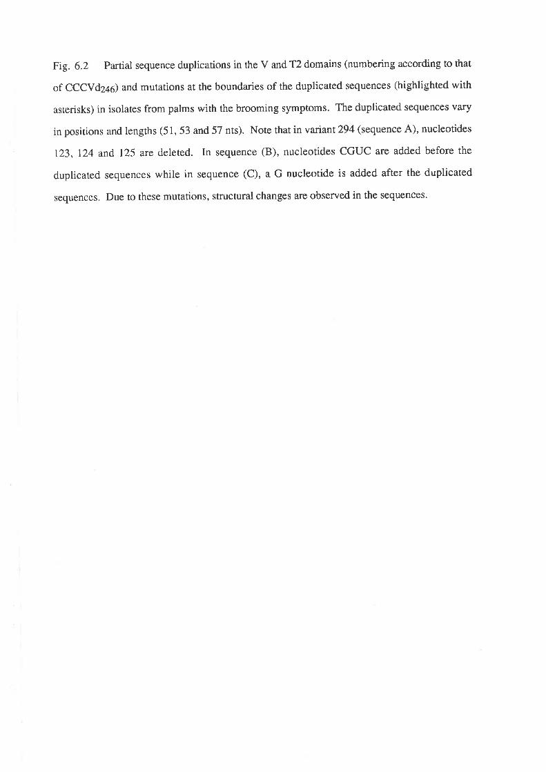
Fig. 6.2 Partial sequence duplications in the V and T2 domains (numbering according to that
of CCCVd2a) and mutations at the boundaries of the duplicated sequences (highlighted with
asterisks) in isolates from palms with the brooming symptoms. The duplicated sequences vary
in positions and lengths (51, 53 and 57 nts). Note that in variant 294 (sequence A), nucleotides
123, 124 and, I25 are deleted. In sequence (B), nucleotides CGUC are added before the
duplicated sequences while in sequence (C), a G nucleotide is added after the duplicated
sequences. Due to these mutations, structural changes are observed in the sequencos.
12.A92
I
. 294 nts98
uACl
CGC GGCCI --ct153 1Æ
UA-U UU
¡98
i1"èc
G
GGAUA-U
GGG CG UCG
UU
UUGGC GCG GGAG ACG UGC CCUC G
AUUAGCG CUGGG CG UCG GC GCG
CG UGC
UUAUUC GC AGC CCU CG CCC GC AGC
B. 303 nts
92 1(x)
CGC GGCCC GC AGC
124 1(X)I ** ** Il_lrluA-u
UGGAGGAè CG UCÖGG CG UCG
GCUUCCUC GC GGCCC GC AGCal c uuit|l152
't25
UUGGC GCG GGAG A
CG UGC CCUC G
AUUA
1G
UAC ICG CUGG
UA-UG CG UCG
UU
GCGUGC
CG__AU
c. 392
fuGCGCGC
0497+
AC
nts
UACUGGG CG UCGGGCCC GC AGC
_U UUGC GCG GGAGCG UGC CCUC
UUAUU
UUA-UGGG CG UCG
UUGGGAG ACCUC G
UUA
97
GC GCG
CG UGCA
I AGCGC
l.I153--c
125 153
CGG CCC GC AGCUU
49
addition (a total of 58 nts) inserted also between positions 124 and 125 in the'f2 domain. The
mutations at the boundaries in variants 303 and 304 had caused some changes in their
secondary structures.
DIS C US SIO N
Fluorescent dye primer cycle sequencing proved efficient and rapid in sequencing a
large number of samples. In addition, the hazards involved in handling radiolabelled chemicals
are avoided as well as the tedium in the film analysis task as data collection and analysis are
automated. However, it has the disadvantage of high capital cost.
Using the above sequencing method, five types of mutations in CCCVd were
discovered to be associated with the severe brooming symptoms in coconut. Three of these
rypes are variations of those previously described by Haseloff et al. (1982) and Keese et al-
(1988). The five types of mutations detected are: (1) exchange of C at position 197 of the
cenrral conserved domain by either AU, UG or UU; (2) U-+A substitution at position 216 in
the p domain; (3) AA-+AAA addition at position 86 or 87 which is at the boundary of the
central conserved and V domains; (4) partial sequence duplications of the V and T2 domains at
varying positions and lengths of the duplicated sequences; and (5) mutations at the boundaries
of duplications which caused srructural changes on the viroid molecule'
As a mutation at position 197 in the central conserved domain has been previously
reported for CCCVd, rhat is, a C-+CC addirion to give the CCCVd variant 247 Qlaseloff et al.,
1982), this site may be important for rhe control of pathogenicity of CCCVd. Mutations in this
site may have an effect on the switching event predicted to occur during replication in which the
native structure is converted into a stem loop srmcture (Keese et a|.,1988). To determine the
functional implications of the new mutations in this site as well as the other types of mutations
described above, infectivity trials using the mutant clones need to be carried out.
50
CHAPTER 7
GENBRAL DISCUSSION
This thesis shows that some non-palm species in the Philippines have nucleic acids
similar to the coconut cadang-cadang viroid (cccvd) (section 3.3' 1)' The nucleic acids were
identified by molecular hybridization with 32P-labelled cRNA and cDNA probes in dot-blots
and electroblots and by the use of two-dimensional polyacrylamide gel electrophoresis (2-D
PAGE). Multiple bands were found in each plant species' Those which were approximately
rhe same size as cccvd usua[y had high homology with the probe while the bands outside the
cccvd region had low homology with the probe (Sections 3.3.1 and 3'3'2a)' The probable
circularity of these bands was demonsrrared 6y 2-D PAGE (Sections 3.3.2b and 5.1.1). Thus,
it is concluded that the nucleic acids with CCCVd-like sequences are viroid-like, although their
identity as RNA has not been confirmed by tests of nuclease sensitivity' No such sequences in
non-palm species have been reported previously except by Hanold and Randles (1991) who
conducted a parallel study in the south-west Pacific. However, sample collection in this thesis
was more intensive and concentrated on plant species closely associated with coconut palms in
the area where cadang-cadang disease occurs'
None of the non-palm species showed symptoms which could be associated with
the presence of the viroid-like nucleic acids. The incidence of the viroid-like nucleic acids in
these plant species was independent of the level of cadang-cadang disease in adjacent coconut
plantations, and it is suggested that these plants could act as a reservoir for cccvd-related
nucleic acids in the Philippines. This would allow experiments to be done to determine whether
the disease in coconut parms can be controlled by the eradication of such potential alternate
sources.
Nucleotide sequencing would be expected to resolve whether the CCCVd-like
nucleic acids can be classed as viroids. Since they seemed to be at a lower concentfation in
their hosts than cccvd is in coconut (Section 3.2), attempts were made to amplify some of the
purified isolates (Section 5.1.1) by the polymerase chain reaction (PCR) via their cDNAs'
However, the conditions developed for PCR with CCCVd2a6 were not successful for the
WAliii iil'i;jítriÊI,IBRAR Y
CCCVd-like nucleic acids from the non-palm species. This could be due to either: (i) e
extracts not being pure so that a plant substance could have inhibited either the reverse
ranscription or the pCR reaction, or (ii) the CCCVd-like nucleic acids having a central domain
different from CCCVd but having common sequences with CCCVd in the other domains so as
to explain their reaction to the CCCVd probe. This study was not continued because of the
limited availability of plant materials. Further work should include experiments on: (i) the
eff,rcient isolation and purification of the CCCVd-like nucleic acids; (ii) the use of PCR primers
derived from other parrs of the CCCVd molecule; (iii) further optimization of the PCR; (iv) the
use of the one-tube, reverse transcription-PCR method developed for CCCVd246 so as to
minimize losses of cDNA after reverse transcription; and (v) increasing the amounts of plant
tissue to allow direct RNA sequencing.
The continuation of the study above may also lead to the possibility of finding
naturally occurring variants of CCCVd in plant species other than coconut which would be
useful in cros s-protection studies.
The variants of CCCVd that have been reported previously a¡e associated with
disease progression but not with unusual types of symptom (Sections l'2'4 and l'2'5)' All of
these va¡iants contain the basic 246 nucleotides. In Chapter 4, the appearance of a new
unusually severe brooming symptom in palms infected by artificial inoculation with
preparations of cccvd is discussed. "Brooming" is a severe form of cadang-cadang disease
characterized by a loss of leaf lamina, severe stunting and occasional premature death'
Approxima tely l\Loof 1,787 CCCVd-inoculated palms screened showed brooming' observed
3-7 years after inoculation, it was essentially confined to palms with either CCCVd vaiant246
or 247.
pAGE and molecular hybridization analyses of 136 palms with "brooming" showed
that all of them have electrophoretic forms distinct from the previously reported CCCVd
variants (Section 4.2). Whereas four bands fepresenting viroids with 246,247 ,296 and297
nts are observed in extracts from palms with common cadang-cadang disease, different palms
with brooming symptoms showed a total of 11 additional or replacement bands' On 207o
pAGE, three of these bands had electrophoretic mobilities between CCCVd variant 247 and
246; two,between 246 and297; three, between 291 and296, and three' less than 296' To
52
determine whether these electrophoretic forms are mutants of CCCVd, some isolates
representing most of the above additional or replacement bands were cloned and sequenced'
cloning had to be done since most of the isolates contained more than one electrophoretic form
making it difficult to relate a particular form to a particulil symptom as well as to analyze data
generated by direct sequencing.
As described in Chapter 5, isolates with a mixturo of brooming-associated
electrophoretic forms were first purified by bi-directional PAGE (Section 5'l'2) and then
amplified by PCR via their cDNAs (Section 5.2.3). Attempts to separate each form from the
others in rhe bi-directional PAGE failed. The design of primers in the synthesis of cDNAs and
their subsequent amplification of PCR is an important factor to consider' In Section 5'2'la'
three sets of primers were used to polymerase almost the whole cccvd sequence so as not to
miss any sites of mutation: (i) primer set I to produce the left half-length of the total cccvd
sequence, (ii) primer set II to produce the right half-length, and (iii) primer set III to generate
full-length fragments. With primer sets I and II, amplification was successful only when the
synthesis of the first strand cDNA was done using the AMV reverse transcription method' on
the other hand, fuu_lengrh cccvd was amplified using primer ser III and the M-MuLV reverse
transcriptase merhod. The priming sites for primer sets I and II are at the highly conserved
central region of cccvd where there is more secondary structure and more G-C base pairs
than in the priming site for primer set III which is at the upper left portion of the molecule'
Thus, it is concluded that AMV reverse transcriptase methd must have provided the right
conditions for the melting of the viroid and annealing of the primers'
pCR products wirh the expected sizes were cloned by inserting them into the
Bluescript plasmid vector with the use of single base A:T overlaps (Section 5'3)' a method that
has the efficiency of a sticky-end cloning. A total of 28 full-length clones and 19 half-length
clones were obtained. only 27 wete selected for sequence analysis.
Sequencing of the selected clones was done by fluorescent dye primer cycle
sequencing (Sections 6.1 and 6.2). This method proved efficient and rapid in sequencing a
large number of samples. Moreover, the hazards involved in handling radiolabelled chemicals
were avoided as well as the tedium in the final analysis task as data collection and analysis were
53
automated. However, sequencing of both strands of a large number of clones had to be done in
order to rule out PCR artefacts.
The sequencing data revealed the following types of mutation observed in the
clones in addition ro rhe basic CCCYI246: (i) substitution of C at position 197 of the central
conserved domain with either AU, UG or UU; (ii) substitution of U with A at position 216 in
the p domain; (iii) addition of A at position 86 or 87 so that AA became AAA at the boundary of
the central conserved region and the V domain; (iv) mutants of varying length resulting from
partial sequence duplications of the v and T2 domains commencing from varying positions in
the right-hand end of the viroid molecule; and (v) mutations at the boundaries of duplications.
Murarions (i), (iv) and (v) are variations of those previously described by Haseloff et al. (1982)
and Keese et al. (1988).
Determination of optimal secondary structures showed that with the C-+AU or
C+UG substitution at position 197 in the central conserved domain, a U-loop was replaced
wìth a base pair. Also, mutations at the boundaries of panial sequence duplications could cause
structural changes in the V and T2 domains. The other mutations appeared not to affect
secondary srructure. Previous observation, however, has been reported that although a C-+CC
addition at position 197 of the central conserved region appeals not to affect secondary
srrucrure, ir causes the CCCVd molecule to migate with markedly different electrophoretic
mobiliry from the basic CCCydz¿ein which there is no added cytosine residue (Haseloff et al.,
19g2; Imperial and Rodriguez, 1983). Thus, the new variant discovered in this thesis with the
c+uu substitution at position 197, could have the same structural effect as that of the above
CCCVd variant with 247 nts.
As position 197 of the central conserved region seemed to be vulnerable to
murations with the resulting variants having different mobilities in PAGE, this site may be
important for the conrrol of pathogenicity of CCCVd. Mutations in this site may have an effect
on the switching event predicted to occur during replication in which the native structure is
converted into a stem loop structure (Keese et al',1988)'
In two of the sequence variants with partial sequence duplications of the V and T2
domains, no mutation was observed in the left hand-portion of the native molecules. This was
despite the presence of small monomers with mutations in the said portion. Haseloff et al'
54
(1982) and Imperial and Rodriguez (1983) have both reported that occasionally, when additions
of the duplicated molecular segment occur, only CCCVdZqO (without cytosine) appears despite
the presence of CCCYdZa,l (with cytosine). However, the third large mutant which was
isolated singly from a palm with "brooming" but with mild stunting, had a U+A substitution at
position 216 in the P domain of the left-hand structure aside from partial sequence duplication
of the V and T2 domains at the right-hand portion of the molecule.
It will only be possible to determine whether there is a direct causal relationship
between the mutations and "brooming" by inoculating the mutant clones to coconut palms.
This will determine what particular type or combination of mutations causes the severe
symptoms. This may identify sites in the CCCVd molecule where invitro mutagenesis can be
induced to modify the pathogenicity of the viroid.
Sequencing of the other electrophoretic forms associated with "brooming" as
described in Section 4.2 could reveal more novel types of mutation in CCCVd'
The use of molecular biology techniques in this study has demonsnated that
CCCVd-related sequences are widespread in a range of non-palm plant species. The possibility
arises that these plants could act as sources of infection for ccrconuts in the field. Furthermore,
elucidation of the molecular basis of the unusually severe "brooming" symptoms observed in
coconuts has been made possible. This will be useful in re-evaluating the distribution of
functions between the conserved and variable features of the CCCVd molecule in comparison
with previously described models for other viroids (Keese et al., 1987). These molecular
studies will assist in developing appropriate control measures for CCCVd.
55
APPENDIX A
BIOCHEMICAL
dATP
dGTP
dCTP
dTTP
EcoRV
T+DNA ligase
RNAse A (DNAse-free)
DNA molecular weight marker III
EcoRI
Hind III
BamHI
RNAsin
a-32s-dATP
T7 Super-Base sequencing kit
a-32p-dcrP
M-MuLV reverse transcriPtase
AMV reverse transcriPtase
Taq DNA polymerase PCR kit
123 DNA ladder
Taq dye primer cycle sequencing kit
SOURCE
Boehringer, Mannheim, FRG
Boehringer, Mannheim, FRG
Boehringer, Mannheim, FRG
Boehringer, Mannheim, FRG
Boehringer, Mannheim, FRG
Boehringer, Mannheim, FRG
Boehringer, Mannheim, FRG
Boehringer, Mannheim, FRG
Boehringer, Mannheim, FRG
Boehringer, Mannheim, FRG
Boehringer, Mannheim, FRG
Bresatec, Australia
Bresatec, Ausralia
Bresatec, Australia
Amersham Intemational, England
Bethesda Research Laboratories, USA
Pharmacia LKB Biotechnology, Sweden
Promega
GIBCO, BRL
Applied BiosYstems, Inc.
56
CHEMICAL SOURCE
Cetythrimethylammonium bromide (CTAB) Sigma' USA
Polyethylene glycol (PEG) 6000 Sigma' USA
Sodium clodecyl sulphate (SDS) Sigma' USA
Mineral oil for PCR Sigma' USA
Dimethyl sulfoxide @MSO) Sigma' USA
Ethidium bromide Sigma' USA
Agarose Type II: medium EEO Sigma' USA
Acrylamide Bio-Rad Laboratories' USA
N, N'-Methylene-bis-acrylamide Bio-Rad Laboratories' USA
N,N,N,-N,-Tetramethylethylenediamine(TEMED) Bio-RadLaboratories,USA
G 50 (fine) - Sephadex Pharmacia' Sweden
Ficoll400 Pharmacia' Sweden
Polyvinylpyrrolidone (PVP) Sigma' USA
Dextran sulphate Pharmacia' Sweden
Isopropyl-b-D-thiogalactopyranoside (IPTG) Boehringer' Mannheim
5 -Bromo-4-chloro-3-indolyl-b-D- galactopyranoside (X-gal) Boehringer, Mannheim
Silver nitrate (AgNQ) BDH Chemicals' Australia
Sodium borohydride (NaBH¿) Sigma' USA
Formardehyde Ajax chemicals Ltd., Ausnalia
Formamide Sigma' USA
Lithium chloride (LiCl) Sigma' USA
2-(N-Morpholino) ethanesulfonic acid (NßS) Sigma' USA
Purified agar Oxoid Ltd'' England
Bacto-tryptone Oxoid Ltd'' England
Bacto-yeast extract oxoid Ltd" England
IlfordrapidfixerllfordPty.Ltd',Australia
Kodak developer Kodak PtY' Ltd'
Phenol BDH Chemicals' Australia
PHENOL REAGENTS
For RNA exEaction:
For DNA extraction:
POLYACRYLAMIDE GELS
77o non-denaturing gel
40Vo acrYlamide (37o bis) =
lOXTBE =
deionized water =
TEMED =
107o arnmonium PersulPhate (AP) =
7 7o denaturing gel (8 M urea)
40Vo acrYlurtide (37o bis)
10 X TBE
deionized water
TEMED
lÙVo AP
Urea
57
APPENDIX B
907o phenol in deionized water wirh Ù'lVo 8-
hydroxyquinoline
Liquified phenol equilibrated in equal volume of TE
buffer (10 mM Tris-HCl, pH 8; 1 mM EDTA)
20 ml solution
3.5 ml
2.0
13.892
0.008
0.6
20 ml solution
= 3.5 ml
= 2.0
= 7.O
= 0.008
= 0.6
= g.6g
58
20Vo non'denaturing gel
40Vo acrYlamide (37o bis)
10 X TBE
deionized water
TEMED
lj%o AP
20 ml solution
10.0 ml
2.O
7.192
0.008
0.8
AGAROSE GEL
1.27o agafose (Type II: medium EEO) in TAE buffer (40 mM Tris-acetate; 1 mM
EDTA) with i PPm ethidium bromide'
BACTERIAL MEDIA
LB medium
Per liter:
To 950 ml of deionized water, add:
bacto-tryptone
bacto-yeast extract
NaCl
1og
5g
log
Shake until the solutes have dissolved. Adjust the pH to 7'0 with 5 N
NaOH. Adjust the volume of the solution to 1 liter with deionized
warer. Sterilize by autoclaving for 20 min at 15 lb/sq. in. on liquid
cycle.
59
SOB medium
Per liter:
To 950 ml of deionized water, add:
bacto-tryptone
bacto-yeast extract
NaCl
SOC medium
2oe
5a
0.5 e
Shake until the solutes have dissolved. Add 10 ml of a 250 mM
solution of KCI (this solution is made by dissolving 1.86 g of KCI in
100 ml of deionized water). Adjust the pH to 7.0 with 5 N NaOH.
Adjust the volume of the solution to 1 liter with deionized water'
Sterilize by autoclaving for 20 min at 15 lb/sq. in. on liquid cycle.
Just before use, add 5 ml of a sterile solution of 2 M MgCl2. This
solution is made by dissolving 19 g of MgCl2 in 90 ml deionized water'
Adjust the volume of the solution to 100 ml with deionized water and
sterilize by autoclaving as above.
SOC medium is identical to SOB medium, except that it contains 0'357o
glucose. After the SOB medium has been autoclaved, allow it to cool
to 60'C or less and then add 7 ml of a sterile 507o solution of glucose
(this solution is made by dissolving 50 g of glucose in 90 ml of
deionized water). After the sugar has dissolved, adjust the volume of
the solution to 100 ml with deionized water and sterilize by filtration
through a 0.22-miuon filter).
60
Storage of bacterial culture containing glycerol
To 0.85 ml of bacterial culture, add 0.15 ml of sterile gylcerol. Vortex
the culture to ensure that the glycerol is evenly dispersed and transfer to
a labelled storage tube equipped with a screw cap. Freeze the culture in
liquid nitrogen and then transfer the tube to -70"C for long-term
storage.
RIZAL ê0
ûê(v
útv
Fl
Þ
l?qo
"f''{il".N.ï
tCo"
160 -LUZON
Polillo l3
ê
- 14" 140-
xttufL tl
^ru¡13 ¡ a?u
Þ¿
BuTlUg N
Ì"ñ
T rlll
\R
lzo 0 o e l2Lû a. ill'10-a ou^¡Frñ r '
ü
D
ß ¡t^it^ô
ll
ð oP (3rororrxox
Er I l¡V I SAYAS
s ls
lo" ¡
t- DEL }TORTE
C M¡NDANAO
40'o a
oÐo
0o3
('o)Jo
oC)oúl¡z{
l¡ll-
l¡JJ
G
EUI
ì
fr
BOHOL
{DC LOILO
CAPIZ
TARLAC
PANGASI N
RI
oISABELA
ECIJAU EVA
NUEVAIZCAYA
I
1200
61
APPENDIX C
The provinces (shaded areas) in the central Philippines with cadang-cadang infected coconut
plantations. The site codes listed in Table 3.1 are: (A) Pili, Bacacay, Albay; (B) Albay
Research Center; (C) Pili, Camarines Sur; (D) Langa, Camarines Sur; (E) Sangay, Camarines
Sur; (F) Atulayan, Camarines Sur; (G) Infanta, Quezon; (H) Calabanga, Camarines Sur; (I)
Tublejon, Sorsogon; (J) Cogon, Sorsogon; and (K) Lipa, Batangas'
62
REFERENCES
Anonymous (19g6) Annual Report 1986 of the Philippine Coconut Authority, Agricultural
Resea¡ch and Development Branch'
Applied Biosystems (1991) Dye Primer cycle sequencing Part No. 901482/Rev. B.
Applied Biosystems (1991) User Bulletinfor DNA Sequencer' Model 373ê^ No' 18'
Ashulin, L, Lachman, o, Hadas, R and Bar-Joseph, M (1991) Nucleotide sequence of a new
viroid species, citrus bent leaf viroid (CBLVd) isolated from grapefruit in Israel.
Nucl. Acids R¿s. 19(17)z 476'7 '
Ausubel, FM, Brent, R, Kingston, RE, MOOre, DD, Seidman, JG, Smith, JA and struhl, K
(1991) The polymerase chain reaction. Pages 15.0.3-15.4.5Ln Current Protocols
in Molecular Biology. John Wiley and Sons, USA'
Barker, JM, Mclnnes, JL, Murphy, PJ and Symons, RH (1935) Dot-blot procedure with
[32p] UNR probes for the sensitive detection of avocado sunblotch and other
viroids in plants. J. Virol. Merhods 10: 87-98'
Boccardo, G, Beaver, RG, Randles, JW and Imperial, JS (1981) Tinangaja and bristle top,
coconut diseases of uncertain etiology in Guam and their relationship to cadang-
cadang disease of coconut in the Philippines . Phytopathology lIz ll04'
Branch, AD and Robertson, HD (1984) A replication cycle for viroids and other small
infectious RNAs. Science 223: 450-455'
Branch, AD, Benenfeld, BJ, Franck, ER, Shaw, JF, Varban, ML, WilliS, KK, ROSen, DL and
Roberrson, HD (1988) Inrerference between coinoculated viroids. Virology 163"
538-546.
candresse, T, Diener, TO and owens, RA (1990) The role of the viroid central conserved
region in cDNA infectivity' Virology lr75z 232-237 '
candresse, T, Smith, D and Diener, TO (1987) Nucleotide sequence of a full-length infectious
clone of the Indonesian strain of romato apical stunt viroid (TASV). Nucl. Acids
Res. 15(24): 10597'
chien, A, Edgar, DB and Trela, JM (1976) Deoxyribonucleic acid polymerase from the
exreme thermophil e Thermus aquaticus. J . Bacteriolosy 127(3): 1550-1557'
63
Ciora, T, Denéfle, P and Mayaux, JF (1991) Rapid one-step automated sequencing reactlons
for 16 DNA samples usingTaq polymerase and fluorescent primers' Nucl' Acids
R¿s. 19(1): 188.
clark, JM (1ggg) Novel non-templated nucleotide addition reactions catalyzed by procaryotic
and eucaryotic DNA polymerases. NucI. Acids Res.16(20)z 9677-9686'
cress, DE, Kiefer, MC and owens, RA (1983) Constmction of infectious potato spindle tuber
viroid cDNA clones. Nucl. Acids R¿s. 11(19): 6821-6835.
Dahlgren, RMT, Clifford, HT and Yeo, PF (19S5) The Famities of the Monocoryledons:
Structure, Evolution andTaxonomy' 520 pp. Springer-Verlag, Berlin.
Diener, TO (1971) Potato spindle tuber "virus". IV. A replicating, low molecular weight
RNA. VirologY 452 4ll-428'
Diener, TO (1979) Viroids andviroid Diseases. John wiley and Sons, New York'
Diener, TO (1987) Biological properties. Pages 9-29 in The Viroids' TO Diener' ed' Plenum
Press, New York.
Diener, TO (1991) Subviral pathogens of plants: viroids and viroid-like satellite RNAs'
FASEB /' 5: 2808-2813'
Galindo, J,Lopez,C and Aguilar, T (1989) Discovery of the transmitting agent of tomato
ptanta macho viroid. Revista Mexicana de FitopatologiaTz 61-65'
Gross, HJ, Domdey, H, Lossow, C, Jank, P, Raba' M' Alberty' H and Sanger' HL (1978)
Nucleotide Sequence and secondary structure of potato spindle tuber viroid ' Narure
(London) 2732 203.
Gross,HJ,Krupp,G,Domdey,H,Raba,M,Jank'P'Lossow'C'Alberty'H'Ramm'Kand
Sanger, HL (1982) Nucleotide sequence and secondary structure of citrus
exocoftis and chrysanthemum stunt viroid . Eur. J ' Biochem' l2lz 249'
Haas, B, Klanner, A, Ramm, K and sanger, HL (1988) The 7s RNA from tomato leaf tissue
resembles a signal recognition particle RNA and exhibits a remarkable sequence
complementaritytoviroids.EMB)J.T(|3)24063.4074.
Hammond, R, Smith, DR and Diener, TO (1989) Nucleotide sequence and proposed
secondary stmcture of columnea latent viroid: A natural mosaic of viroid sequences'
Nucl' Acids Res' 17t 10083-10094'
64
Hammond, RW and Owens, RA (1987) Mutational analysis of PSTVd reveals complex
relationships between structure and infectivity. Proc. Natl. Acad Sci. USA 84:
3967-397r.
Hammond, RW, Diener, TO and Owens, RA (1989) Infectivity of chimeric viroid transcripts
reveals the presence of alternative processing sites in PSTVd. Virology 17Ú 486-
495.
Hanahan, D (1983) Studies on rransformation of Escherichia coli with plasmids. ,I' Mol' Biol.
166: 557-580.
Hanold, D and Randles, Iw (1991) Coconut cadang-cadang disease and its viroid agent. Plant
Disease 7 5(4): 330-335.
Hanold, D and Randles, Iw (1gg1) Detection of the cadang-cadang viroid-like sequences in oil
and coconut palm and other monocotyledons in the south-west Pacific. Ann. appl.
Biol. ll8: 139-151'
Hanold, D, Rodriguez, MJB and Randles, JW (1989) Detection of sequences related to
coconut cadang-cadang viroid in new species and in new locations' In
proceedings, 7th Ausrralasian Plant Pathology Society Conference, Brisbane, p'
73.
Haseloff, J, Mohamed, NA and Symons, RH (1932) Viroid RNAs of cadang-cadang disease
of coconuts. Nature 299:316-321'
Hernandez, C, Elena, SF, Moya, A and Flores, R (1992) Pear blister canker viroid is a
member of the apple scar skin subgroup (apscaviroids) and also has sequence
homology with viroids from other subgtoups. J. gen. Virol.73: 2503-2507 .
Herold, T, Haas, B, Singh, RP, Boucher, A and Sanger, HL (1992) Sequence analysis of five
new field isolates demonsrrates that the chain length of potato spindle tuber viroid
(pSTVd) is not strictly conserved but as variable as in other viroids. Plant Mol.
Biol.Ig; 329-333-
Holton, TA and Graham, MW (1991) A simple and efficient method for direct cloning of PCR
products using ddT-tailed vectors. Nucl. Acids R¿s. 19: 1156.
65
Imperial, JS and Rodriguez, MJB (1983) Variation in the coconut cadang-cadang viroid:
Evidence for single base addition with disease progress. Philipp. J' Crop Sci'
8(2):87-91.
Imperial, JS, Bautista, RM and Randles, JW (1985) Transmission of the coconut cadang-
cadang viroid to six species of palm by inoculation with nucleic acid extracts. Plant
PathologY 342 391-401'
Imperial, JS, Rodriguez, MJB and Randles, rw (1981) Variation in the viroid-like RNA
associated with cadang-cadang disease: Evidence for an increase in molecular
weight with disease progress' J ' gen' Virol' 562 7l -85 '
Ishikawa, M, Meshi, T, Okada, Y, Sano, T and Shikata, E (1935) In vitro mutagenesis of
infectious viroid cDNA clone. J. Biochem. 9s(6): 1615-1620.
Keese, P and Symons, RH (1935) Domains in viroids: Evidence of intermolecular RNA
managements and their contribution to viroid evolution. Proc. Natl. Acad' Sci'
USA 822 4582-4586.
Keese, P and Symons, RH (1987) The structure of viroids and virusoids' Pages l-4'7 in:
Viroids and Viroid-like Pathogens
FL.
JS Semancik, ed. CRC Press, Boca Raton,
Keese, P, Osorio-Keese, ME and Symons, RH (19SS) Coconut tinangaja viroid: Sequence
homology with coconut cadang-cadang viroid and other potato spindle tuber viroid
related RNAs. Virology 162: 508-510'
Keese, P, Visvader, JE and Symons, RH (1983) Sequence variability in plant viroid RNAs'
Pages 71-98 in: RNA Genetics, variabiliry of RNA Genomes, Vol' III' E'
Domingo, JJ Holland and P Ahlquist, eds. cRc Press, Boca Raton, FL'
Khoury, J, Singh, RP, Boucher, A and Coombs, DH (1983) Concentration and distribution of
mild and severe strains of potato spindle tuber viroid in cross-protected tomato
plants. Phytopathotogy 78(10): 1331-1336'
Kolrunow, AM and Rezaian, MA (1989) A scheme for viroid classification. Intervirology 302
194-201.
Krawetz, SA, Pon, RT and Dixon, GH (1939) Increased efficiency of the Taq polymerase
catalyzed polymerase chain reaction. Nucl. Acids Res.17(2)z 819'
66
Kwok, S and Higuchi, R (1989) Avoiding false positives with PCR. N¿rare 339:237-238'
Laulhere, J and Rozier, c (1976) One-step extraction of plant nucleic acids' Plant science
Leners 6: 231-242'
Lee, LG, Commell, CR, Woo, SL, Cheng, RD, McArdle' BF' Fuller' CW' Halloran' ND and
Wilson, RK (1992) DNA sequencing with dye-labelled terminations and fi DNA
polymerase: Effect of dyes and dNTPs on incorporation of dye-terminators and
probability analysis of termination fragments. Nucl. Acids R¿s.20(10)z 2471'
2483.
Lowe, T, Sharefkin, J, yang, SQ and Dieffenbach, CW (1990) A computer program for
selection of oligonucleotide primers for polymerase chain reaction. Nucl. Acids
R¿s. 18(7) z 1757-1761'
Maniatis, T, Fritsch, EF and Sambrook, J (1932) Molecular Cloning, A Laboratory Manual'
Cold Spring Harbor Laboratory Press, USA'
Marchuk, D, Drumm, M, Sauline, A and Collins, FS (1991) Construction of T-vectors, a
rapid and general system for direct cloning of unmodified PcR products' Nucl'
Acids R¿s' 19(5): 1 154'
Maxam, AM and Gilben, W (1977) A new method for sequencing DNA' Proc' Natl' Acad'
Sci.74: 560.
Maxam, AM and Gilbert, w (1980) rn Methods in Enzymology e5(1): 499-560' L
GrossmanandKMoldave,eds'Academichess'NewYork'
Mclnnes, JL and Symons, RH (1989) Nucleic acid probes in the diagnosis of plant viruses
andviroids.Pagesl13-138in:NucleicAcidProbes.RHsymons'ed'cRC
Press, Inc., Boca Raton, Florida'
Mead, DA, Pey, NK, Herrnstadt, c, Marcil, RA and Smith, LM (1991) A universal method
for the direct cloning of PCR amplified nucleic acid. BiolTechnology 9z 657 -663'
Meshi, T, Ishikawa, M, Ohno, T, Okada, Y, Sano, T, Ueda, I and shikata, E (1984) Double-
stranded cDNAs of hop srunr viroid are infectious. J. Biochem. 95(5): l52l-
1524.
67
Meshi, T, Ishikawa, M, Watanabe, Y, Yamaya, J' Okada' Y' Sano' T and Shikata' E (1985)
The sequence necessary for the infectivity of hop stunt viroid cDNA clones' Mol'
Gen' Gener. 200: 199-206'
Mohamed, NA, Bautista, R, Buenaflor, G and Imperial, JS (1985) Purification and infectivity
of the coconut cadang-cadang viroid. Phytopathology 75(|)z 79-83,
Mohamed, NA, Bautista, R, Buenaflor, G and Imperial, JS (1985) Purification and infectivity
of the coconut cadang-cadang viroid. Phytopatholosy 75(l)z 79-83'
Mohamed, NA, Haseloff, J, Imperial, Js and Symons, RH (1982) Characterization of the
different electrophoretic forms of the cadang-cadang viroid' J' gen' Virol' 632
18 1- 188.
Moody, K, Munroe, cE, Lubigan, RT and Paller, EC (1984) Major weeds of the Philippines'
Weed Science Sociery of the Philippines' UPLB'
Niblett, cL, Dickson, E, Fernow, KH, Horst, RK and zaítlin, M (1973) Cross protection
among four viroids. Virology 91: 198-203'
ocfemia, Go (1g37) The probably nature of cadang-cadang disease of coconuts. Philippine
Agri.26z 338-340.
Ohno, T, Takamatsu, N, Meshi, T and Okada, Y (1983) Hop stunt viroid: Molecular cloning
and nucleotide sequence of the complete cDNA copy' Nucl' Acids R¿s' 11: 6185'
owens, RA and Hammond, Rw (1990) Mutarional analysis of viroid pathogenicity and
movement. Pages 165-l'76 in: Viral genes and plant pathogenesis' TP Pirone and
TG Shaw, eds. Springer-Verlag' New York'
owens, RA and Hammond, RV/ (1990) Mutational analysis'of viroid' seminars invirology
l: 101-106.
Owens, RA, Candresse, T and Diener, TO (1990) Construction of novel viroid chimeras
containing porrions of tomato apical stunt and citrus exocortis viroids. Virology
l7 5: 238-246.
Owens, RA, Hammond, RW, Gardner, RC, Kiefer' MC' Thompson' SM and Cress' DE
(19g6) Site-specific mutagenesis of poraro spindle tuber viroid CDNA: Alterations
within premelting region 2 thatabolish infectivity. Plant Mol. Biol. 6: 179-192'
68
owens, RA, Khurana, SMP, Smith, DR, Singh, MN and Garg, ID (1992) A new mild strain
of potato spindle tuber viroid isolated from wild Solanum spp. in India. Plant
Disease 76(5): 527 -529.
owens, RA, Thompson, sM and steger, G (1991) Effects of random mutagenesis upon
poraro spindle tuber viroid replication and symptom expression- Virology 185: 18-
31'
Pacumbaba, EP and Carpio, CB (1937) Report on cadang-cadang surveys in western and
eastern Sama¡. Philippine Coconut Authority, Albay Resea¡ch Center'
Peacock, AC and Dingman, CW (1968) Molecular weight estimation and separation of
ribonucleic acid by electrophoresis in agarose-acrylamide gels' Biochemistry 7:
668-614.
price, wc (1971) Cadang-cadang of coconut - A review. Plant science 3: 1-13.
Puchta, H and Sanger, HL (19SS) An improved procedure for the rapid one-step-cloning of
full-length viroid cDNA. Arch'Virol' 101: 137-140'
puchra, H and sanger, HL (1989) Sequence analysis of minute amounts of viroid RNA using
the polymerase chain reaction (PCR). Arch. virol. 106: 335-340.
Puchta, H, Herold, T, VerhOeven, K, RoenhOrst' A, Ramm, K, Schmidt-Puchta' W and
Sanger, HL (1990) A new strain of potato spindle tuber viroid (PSTVd-N) exhibits
major sequence differences as compared to all other PSTVd strains sequenced so
far. Planr Mol. Biol. 15: 509-51 I '
Puchta, H, Ramm, K and Sanger, HL (1988) The molecular structure of hop latent viroid
(HLVd), a new viroid occurring worldwide in hops. Nucl. Acids R¿s' 16(10):
4197-4216.
Randles, JS, Boccardo, G and Imperial, JS (1930) Detection of the cadang-cadang RNA in
African oil palm and buri palm. Phytopathology 70:185-189.
Randles, JV/ (1975) Association of two ribonucleic acid species with cadang-cadang disease of
coconut palm. Phytopathology 652 163-167 '
Randles, Jw (1987) Coconut cadang-cadang. Pages 265-277 in: The viroids ' To Diener, ed'
Plenum Press, New York'
69
Randles, JW and Hatra, T (1979) Circularity of the ribonucleic acids associated with cadang-
cadang dise ase. Virology 96: 47-53.
Randles, JW and Palukaitis, P (1979) In vitro synthesis and characterization of DNA
complementary to cadang-cadang-associated RNA. J . ge n. V irol. 43:. 649-662.
Randles, JW, Boccardo, G, Retuerma, ML and Rillo, ES (1977) Transmission of the RNA
species associated with cadang-cadang of coconut palm, and insensitivity of the
disease to antibiotics. Phytopathology 67:. L2ll-1276.
Randles, JW, Hanold, D, Pacumbaba, EP and Rodriguez, MJB (1992) Cadang-cadang
disease of coconut palm. Pages 277-295 in: Plant Diseases of International
Importance. AN Mukhopadhyay, J Kumar, HS Chaube and US Singh, eds.
Prentice Hall Inc., New JerseY.
Randles, JW, Rillo, EP and Diener, TO (1976) The viroid-like stmcture and cellular location
of anomalous RNA associated with the cadang-cadang disease. Virology 74: 128-
r39.
Randles, JW, Rodriguez, MJB and Imperial, JS (1988) Cadang-cadang disease of coconut
palm. Microbiological Scienc¿s 5(1): 18-22.
Randles, JW, Steger, G and Riesner, D (1982) Structural transitions in viroid-like RNAs
associated with cadang-cadang disease, velvet tobacco mottle virus, and Solanum
nodiflorum mottle virus. Nucl. Acids R¿s. 10(18): 5569-5585'
Rezaian, MA (1990) Australian grapevine viroid - evidence for extensive recombination
between viroids. Nucl. Acids Res' 18: 1813-1818.
Riesner, D (1987) Subcellular location and in sir¿ association with cellular components. Pages
99-116 in: The viroids. To Diener, ed. Plenum Press, New York.
Riesner, D, Steger, G, Schumacher, J, Gross, HJ, Randles, JW and Sänger, HL (1983)
Strucrure and function of viroids. Biophys. struct, Mech.9z I45.
Rigden, JE and Rezaian, MA (1992) In vitro synthesis of an infectious viroid: Analysis of the
infectivity of monomeric Iinear cEVd. virology l86t 20I-206.
Rillo, EP and Rillo, AR (19S1) Abstracts on the cadang-cadang disease of coconut (1937-
19S0). Philippine Coconut Authority.
70
Rivera-Bustamante, RF, Gin, R and Semancik, JS (1986) Enhanced resolution of circular and
linear molecular forms of viroid and viroid-like RNA by electrophoresis in a
discontinuous-pH sysrem. Analytical Biochemistry 156: 91-95.
Ruano, G, Brash, DE and Kidd, KK (1991) PCR: The first few cycles. Perkin Elmer Ceîus'
Amplifications, A Forumfor PCR Users. lssue 7: l-4.
Saiki, RK, Gelfand, DH, Stoffel, S, Scharf, SJ, Higuchi, R, Horn, GT, Mullis, KB and
Erlich, HA (1988) Primer-directed enzymatic amplification of DNA with a
thermostable DNA polymerase . Science 2392 487 -491.
Sambrook, J, Fritsch, EF and Maniatis, T (1989) DNA sequencing. Pages 13.3-13.95 in:
Molecular Cloning, A Laboratory Manual, Cold Spring Harbor Laboratory Press,
USA.
Sambrook, J, Frirsch, EF and Maniatis, T (1989) In vitro'amplification of DNA by the
polymerase chain reaction. Pages 14.2-I4.35 in Molecular Cloning, A Laboratory
Manual. Cold Spring Harbor Laboratory Press, USA.
Sambrook, J, Fritsch, EF and Maniatis, T (1989) Molecular Cloning, A Laboratory Manual.
Cold Spring Harbor Laboratory Press, New York'
Sammons, DW, Adams, LD and Nishazawa, EE (1981) Ultrasensitive silver-based color
staining of polypeptides in polyacrylamide gels. Electrophoresis 2: 135-141.
Sanger, F, Nicklen, FS and Coulson, AR (1977) DNA sequencing with chain-terminating
inhibitors. Proc. Natl. Acad. Sci' 74: 5463'
Sano, T, Candresse, T, Hammond, RW, Diener, TO and Owens, RA (1992) Identification of
mu¡iple srructural domains regulating viroid pathogenicity. Proc. Natl. Acad. Sci.
USA 89: 10104-10108.
Sano, T, Uyeda, I, Shikata, E, Ohno, T and Okada, Y (1934) Nucleotide sequence of
cucumber pale fruit viroid: Homology to hop stunt viroid . NucI. Acids Res. L2z
2437.
Sarkar, G and Sommer, SS (1990) Shedding light on PCR contamination. Nature 3432 27.
Schnölzer, M, Haas, B, Ramm, K, Hofmann, H and Sanger, HL (1985) Correlation between
strucrure and pathogenicity of potato spindle tuber viroid (PSTV). EMBO J. 4z
2r8t-2r99.
7l
Schumacher, J, Randles, JW and Riesner, D (1983) A two-dimensional electrophoretic
technique for the detection of circular viroids and virusoids. Analytical
B iochemistry 135: 288-295.
Singh, RP, Boucher, A and Somerville ,TH (1992) Detection of potato spindle tuber viroid in
the pollen and various parts of potato plant pollinated with viroid-infected pollen.
Planr Disease 76(9): 951-953.
Smith, AJH (19S0) DNA sequence analysis by primed synthesis. Methods Enzymol. 65:
560,
Symons, RH (1981) Avocado sunblotch viroid: Primary sequence and proposed secondary
structure. Nucl. Acids Res.9:6527.
Symons, RH (1991) The intriguing viroids and virusoids: What is their information content
and how did they evolve? Molecular PIant-Miuobe Interactions 4(2)z llL-121.
Symons, RH, Haseloff, J, Visvader, JE, Keese, P, Murphy, PJ, Gill, DS, Gordon, KFII and
Bruening, G (1985) On the mechanism of replication of viroids, virusoids and
satellite RNAs. Pages 235-263 \n: Subviral Pathogens of Plants and Animals:
Viroids and Prions. K Maramorosch and JJ McKelvey, eds. Academic Press,
New York.
Tabler, M and Sanger, HL (1984) Cloned single- and double-stranded DNA copies of potato
spindle tuber viroid (PSTV) RNA and co-inoculated subgenomic DNA fragments
are infectious. EMBO "r. 3: 3055-3062.
Tabler, M and Sanger, HL (1985) Infectivity studies on different potato spindle tuber viroid
(PSTV) RNAs synthesized in vitro with the SPC transcription system. EMBO J.
4(9):219r-2199.
Tabler, M, Günther, I, Kern, R and Sanger, HL (1989) A microscale procedure for isolating
and sequencing the viroid RNA present in one gram of infected leaf tissue. J.
Virol. Methods 232 lll-126.
Thomas, P (1980) Hybridization of denatured RNA and small DNA fragments transferred to
nitrocellulose. Proc. Natl. Acad.Sci., USA 77: 5201-5205.
Titus, DE, ed. (1991) Promega Protocols and Applications Guide.
72
Tsagris, M, Tabler, M, Mühlbach, HP and Sanger, HL (1987) Linear oligomeric potato
spindle tuber viroid (PSTVd) RNAs are accurately processed in vitro to the
monomeric circular viroid proper when incubated with a nuclear extract from
healthy potato cells. EMBO J.6:2173-2183.
Visvader, JE and Symons, RH (1985) Eleven new sequence variants of citrus exoconis viroid
and the correlation of sequence with pathogenicity. Nucl. Acids R¿s. 13: 2907-
2920.
Visvader, JE and Symons, RH (1986) Replication of in vito constructed viroid mutants:
Location of the pathogenicity-modulating domain of citrus exocortis viroid. EMBO
Y. 5(9): 2051-2055.
Visvader, JE, Forster, AC and Symons, RH (1985) Infectivity and in vitro mvtagenesis of
monomeric cDNA clones of citrus exocortis viroid indicates the site of processing
of viroid precursors. Nucl. Acids R¿s. 13(16): 5843-5856'
White, TJ, Arnheim, N and Erlich, HA (1989) The polymerase chain reaction. Trends in
Genetics 5: 185-189.
Williams, JF (1989) Optimization strategies for the polymerase chain reaction. BioTechniques
7 (7): 7 62-7 6t .
Zelazny, B (1979) Distribution and spread of the cadang-cadang disease of coconut palm.
Acta Phyropath. Acad. Sci. Hung.l4z 115-126.
Zelazny, B and Niven, BS (1980) Duration of the stages of cadang-cadang disease of coconut
palm. Plant Disease 64:841-842.
Zelazny, B and Pacumbaba, E (1982) Incidence of cadang-cadang of coconut palm in the
Philippines . Plant Dis. 662 547 -549.
Zelazny, B, Randles, J'W, Boccardo, G and Imperial, JS (1982) The viroid nature of the
cadang-cadang disease of coconut palm. Scientia Filipinns 2: 46-63.
Zuker, M (1989) On finding all suboptimal foldings of an RNA molecule. Scíence 244: 48-
52.


