Embed Size (px)
Citation preview

Molecular immune signatures of HIV-1 vaccines in humanPBMCs
Alessandro Monacoab, Francesco M. Marincolaa, Marianna Sabatinoa, Zoltan Posa, MariaLina Torneselloc, David F. Stronceka, Ena Wanga, George K. Lewisd, Franco M.Buonaguroc, and Luigi Buonagurocd
aInfectious Disease and Immunogenetics Section (IDIS), Department of Transfusion Medicine,Clinical Center, National Institutes of Health Bethesda, MD, USAbIRCCS Istituto Nazionale Tumori “Giovanni Paolo II”, Bari, ItalycLaboratory of Molecular Biology and Viral Oncogenesis & AIDS Reference Center, IstitutoNazionale Tumori “Fondazione G. Pascale”, Naples, ItalydInstitute of Human Virology, University of Maryland School of Medicine, Baltimore, MD, USA
AbstractThe global transcriptional profile of peripheral blood mononuclear cells (PBMCs) stimulated withHIV candidate vaccine (virus-like particles, VLPs) has been evaluated in HIV-infected patientswith low/high viral load compared to healthy volunteers. Baseline activation of chemokineproduction was observed in PBMC from HIV-infected patients and innate immune stimulationwith HIV-VLPs was not blunted. The immune profile among HIV-infected patients was found tobe qualitatively similar but quantitatively extremely variable. This diversity was independent ofviral load and it might be dependent on individual immunogenetic traits or concurrentimmunological status.
This ex vivo screening strategy represents an efficient tool for guiding modifications/optimizationsof vaccination strategies and understanding failures in individuals enrolled in clinical trials.
KeywordsImmunogenomics; Vaccine; HIV-1; Peripheral blood mononuclear cell
1. IntroductionHIV-1 Pr55gag virus-like particles (VLPs) [1], [2], [3] and [4] have been extensivelycharacterized by our group for the ability to induce maturation and activation of in vitrocultured monocyte-derived dendritic cells (MDDCs) partly through Toll-like receptor(TLR)-3 and -9 signaling [5]. We observed that the transcriptional profile of VLP-stimulatedMDDCs indicates the reduced transcription of genes associated with phagocytosis andpathogen recognition and the activation of genes with function associated with antigenpresentation and MDDCs migration [6]. Similar information can be observed also in CD14+uncultured peripheral blood mononuclear cells (PBMCs) [7].
Recently, we have demonstrated that HIV-1 seropositivity status does not significantlyimpair the immune activation status and the responsiveness of circulating monocyte CD14+cell populations to an immunogenic stimulus [8]. Moreover, Th-1 and Th-2 cytokineproduction was compared among healthy volunteers, HIV patients with low and high viralloads demonstrating higher basal levels of Th2 interleukin (IL)-6 [8].
NIH Public AccessAuthor ManuscriptFEBS Lett. Author manuscript; available in PMC 2012 August 14.
Published in final edited form as:FEBS Lett. 2009 September 17; 583(18): 3004–3008. doi:10.1016/j.febslet.2009.07.060.
NIH
-PA Author Manuscript
NIH
-PA Author Manuscript
NIH
-PA Author Manuscript

To better dissect the potential differences in the response of circulating PBMCs from healthyvolunteers as well as low and high viremia HIV-positive subjects to VLPs, in the presentstudy we applied transcriptional profiling to three representative samples from each of thethree categories and tested baseline transcriptional patterns as well as those after stimulationwith VLPs or lipopolysaccharide (LPS) [9].
2. Methods2.1. Cell culture medium
PBMC culture medium consisted of RPMI 1640 medium (Life Technologies, Carlsbad, CA)supplemented with 2 mM l-glutamine (Sigma), 1% nonessential amino acids (LifeTechnologies), 1% sodium pyruvate (Life Technologies), 50 µM 2-mercaptoethanol(Sigma), 50 µg of gentamicin (Life Technologies) per ml, and 10% fetal calf serum (LifeTechnologies).
2.2. Patient and donor cohorts, PBMC preparation and treatmentHuman specimens were obtained under informed consent, as approved by the University ofMaryland Baltimore Institutional Review Board. Fresh PBMCs were isolated by Ficoll-Hypaque density gradient centrifugation and plated in 6-well plates at a concentration of 1 ×10⁷/well in 3 ml/well volume. PBMCs were stimulated with 6 µg/ml of HIV-VLPs or 1 µg/ml of LPS. Control PBMCs were added with PBS. The residual endotoxin activity possiblyleft over by the VLP preparation was inhibited by pre-incubation with Polymixin B Sulfate(Sigma) at a concentration of 10 µg/ml. After 4 and 8 h the cells were harvested andprocessed for total RNA extraction.
2.3. RNA preparation and microarray hybridizationPBMCs were harvested, total RNA and RNA for hybridization were obtained as previouslydescribed [10] and [11]. Six micrograms of amplified test samples aRNA were labeled withCy5 (Amersham) while the same amount of reference sample (pooled normal donorPBMCs) was labeled with Cy3. Test-reference sample pairs were mixed and co-hybridizedto a custom-made 37 K oligo-based microarray platform encompassing the whole humangenome. Array data consistency was assessed based on the principle of referenceconcordance as previously described [12].
2.4. Microarrays and statistical analysesHybridized arrays were scanned at 10-µm resolution on a GenePix 4000 scanner (AxonInstruments). Resulting jpeg and data files were deposited at microarray database (mAdb)(http://nciarray.nci.nih.gov) and retrieved after median centered, filtering of intensity (>300)and spot elimination (bad and no signal). Data were further analyzed using the NCI/BRBsoftware [13]. Hierarchical cluster analysis was conducted on according to Eisen et al. [14];differential expressed genes were visualized by Treeview and displayed according to thecentral method of normalization [15].
Class comparison was performed using paired or unpaired Student’s t test as appropriate.Multivariate analysis was performed on multiple groups based on the F test with a P-valuecutoff of 0.001. Assessment of random identification of significance was based on univariateand multivariate permutation test as previously described [16]. Boolean comparisons werebased on the BRBArray Software. Fisher’s exact test or χ2-test was used to assess thesignificance of sample distribution among self-organizing classes of samples as appropriate.
Monacoa et al. Page 2
FEBS Lett. Author manuscript; available in PMC 2012 August 14.
NIH
-PA Author Manuscript
NIH
-PA Author Manuscript
NIH
-PA Author Manuscript

3. Results3.1. Clinical parameters of the subjects included in the analysis
Eleven subjects were enrolled in the study. Eight were HIV-1-seropositive, of whom fourshowed a low HIV-1 viremia (<2.6 log RNA copies/ml) and four a high viremia (>4.69 logRNA copies/ml) (SI Table 1). The first group was under ART therapy, the latter enrolled attheir first visit and naïve for ART treatment. Three healthy seronegative volunteers wereenrolled as controls [8].
3.2. LPS- and VLP-induced activation of PBMC in healthy volunteersThe effect of VLPs and LPS on the activation of PBMCs obtained from healthy volunteersidentified 560 genes down-regulated and 596 up-regulated by either treatment (multivariatepermutation test < 0.001), with a broad overlap among genes modulated by LPS and VLPs(Fig. 1).
3.3. Baseline transcriptional patterns of PBMCsNine hundred and seventy eight up-regulated genes and 960 down-regulated genes werefound differentially expressed in unstimulated PBMCs from HIV-infected individualscompared to healthy volunteers. PBMCs of healthy volunteers demonstrated a highlyconsistent pattern while PBMCs from HIV-infected individuals displayed a heterogeneoustranscriptional patterns and samples from low- and high-viremia group do not neatlysegregate into two independent clusters (Fig. 2A). Patients 05 and 12 were excluded fromthis global analysis, considering that they have been found anergic according to functionalanalyses described in our recent paper [8]. However, patient 12 (a high-viremia sample)segregated in between experimental cohorts (red arrow, Fig. 2A), while patient 05 (a low-viremia sample) segregated with the other samples from low-viral load patients butdisplayed a relatively reduced induction of about 50% of HIV-specific genes (yellow arrow,Fig. 2A).
Unstimulated PBMCs from HIV-infected individuals largely reflected in baseline conditionsthe transcriptional profile of PBMCs from healthy volunteers stimulated ex vivo with VLPs.No transcripts specifically induced by LPS in vitro in healthy volunteers were found to beconstitutively expressed in vivo by HIV-infected patients (Fig. 2B).
3.4. Transcription pattern of stimulated PBMCsThe transcriptional profile of PBMCs stimulated with LPS from healthy volunteers showssignificant differences with those from HIV-infected patients, independent of viral load (Fig.3A). Boolean comparison of genes differentially expressed in each cohort identifiedpredominantly overlapping transcriptional patterns consistent among the three groups withthe least overlap between PBMCs from healthy volunteers and those from HIV-infected,low-viral load patients (Fig. 3B) [17]. The ISGs modulated by LPS in the differentexperimental are shown in SI Table 2.
Similarly, a spontaneous segregation is observed between the transcriptional profile ofPBMCs from healthy volunteers and HIV-infected individuals stimulated with VLPs (Fig.3C). The transcriptional patterns of individuals with HIV shows a close overlap incorrelative distance (Spearman’s correlation; 1 − R2 values) among patients stimulated withLPS (Fig. 3A) or VLPs (Fig. 3C). Boolean comparison of genes differentially expressed inresponse to VLPs identified overlapping transcriptional patterns among the three cohortswith the least overlap between PBMCs from normal individuals and those from HIV-infected, low-viral load patients (Fig. 3D).
Monacoa et al. Page 3
FEBS Lett. Author manuscript; available in PMC 2012 August 14.
NIH
-PA Author Manuscript
NIH
-PA Author Manuscript
NIH
-PA Author Manuscript

In particular, considering specifically the ISG genes, a multiple dimensional scaling basedon IFNa2b-induced 453 ISGs genes identified by our group (Pos et al., manuscript inpreparation) clearly shows a distinct baseline pattern in unstimulated PBMCs from healthyseronegative and HIV-seropositive subjects as well as a distinct pattern in VLP-inducedPBMCs from the three different groups (SI Fig. 1). The genes, including ISGs, modulatedby VLPs in the different experimental are shown in SI Table 3.
3.5. Comparison of genes induced by LPS and VLPsThe expression of distinct ISGs genes was stimulated by LPS and VLPs, while the downregulation of some interferon regulated transcripts occurred in common with that induced byVLPs (see SI Table 4). On the contrary, the expression of several genes with immunefunction was significantly affected by LPS and VLP stimulation and are listed according toexperimental cohorts (see SI Table 5).
In addition, self-organizing maps based on immune genes differentially expressed upon LPSand VLP stimulation by the three groups identified a cluster of transcripts that included aspecific sub-group of ISGs (Fig. 4Aorange dashed line a). Reshuffling of patients’ anddonors’ sample based on this specific ISG cluster clearly predicted a class enriched of LPS-stimulated samples (10 of 11 samples) compared with VLP-stimulated samples (3 of 11samples) or unstimulated samples (2 of 11 samples; χ2P-value < 0.001, SI Fig. 2).
3.6. Patterns of gene expressionThe expression of several genes with immune function was significantly affected by LPSand VLP and five distinct patterns of gene expression were identified. (1) Genes like IL-6were not differentially expressed in baseline conditions among experimental cohorts, buttheir response to either VLP or LPS was strongly affected by HIV-status (Fig. 5A). (2)Genes like IL-8 that were very strongly differentially expressed between HIV patients (up-regulated) compared to healthy volunteers in baseline conditions and whose post-stimulationlevels were partially affected by the HIV-status (Fig. 5B). (3) Genes like CCL3 whoseexpression was significantly higher in baseline conditions in HIV patients compared tohealthy volunteers and experienced strong up-regulation in response to VLP, more than toLPS, in HIV-infected patients more than in healthy volunteers (Fig. 5C). (4) Genes likeCCL22 whose expression was not altered by LPS or VLP stimulation in healthy volunteersbut it was strongly induced by LPS and VLPs in HIV-infected patients independent of viralload (Fig. 5D). (5) Genes like IRF-7, whose expression was only induced by LPS and it wascomparable among all experimental cohorts independent of HIV infection status (Fig. 5E).
3.7. Analysis of range of gene inductionWe finally investigated the dynamic range of gene induction in response to VLP-stimulationindependent of HIV infections status. This analysis identified 133 transcripts whosedynamic range in response to VLPs differed significantly between HIV-infected patients andhealthy volunteers. A selection of lymphokines among these genes is shown in SI Fig. 3.Although all patients and donors experienced increases in the stimulated values of eachcytokine, the dynamic range was quite different and was strongly affected by the HIV statusindependent of viral load. This was particularly evident for IL-1β and the IL-2R α-chainwhich were more strongly up-regulated in HIV-infected patients while the dynamic rangewas reversed for several lymphokines. In spite of this significance difference in Δ-VLPbetween healthy donors and HIV-infected individuals, the cumulative Cy5/Cy3 values inresponse to VLP stimulation were consistently higher in HIV-infected patients due to theirhigher baseline expression.
Monacoa et al. Page 4
FEBS Lett. Author manuscript; available in PMC 2012 August 14.
NIH
-PA Author Manuscript
NIH
-PA Author Manuscript
NIH
-PA Author Manuscript

4. DiscussionThe relevance of innate immunity in the natural history of HIV infection is becomingincreasingly recognized [18], [19], [20], [21], [22] and [20]. To address the baseline profileof innate immunity and its modulation, we studied the ex vivo responsiveness of PBMCs tothe HIV-VLP vaccine molecule and the lipopolysaccharide (LPS), as example of pathogenassociated molecular pattern (PAMP), comparing HIV-infected individuals to normal,healthy volunteers.
According to previous observations [6], the transcriptional activation of PBMCs in baselineconditions or in response to stimulation with VLPs or LPS recapitulated that observed inMDDCs; this has important practical implications because ex vivo stimulation provides asimplified tool for the analysis of individual patient’s responsiveness to VLP that does notrequire extensive in vitro manipulation. This may be of particular importance consideringthe current lack of clinical immunological/biological correlates of responsiveness of HIV-infected individuals, which may be extremely relevant within the conduction of vaccinationprotocols. Transcriptional patterns of PBMC from HIV-infected patients clearly differedfrom those of healthy volunteers and signatures consistent with profound activation ofchemokine production were observed. Interestingly, the RNA expression levels did notalways correlate with those protein expression, reported in our recent paper [8], which aredependent also on external metabolic pathways affecting either their half life or theircompartmentalized production.
Unexpectedly, in spite of extensive heterogeneity among individual patients, severalconsistent immunological patterns could be identified distinguishing HIV-infectedindividuals from healthy volunteers that may shed important insights about the immunebiology of HIV infection.
An unexpected observation was the finding that the IFN-independent innate immuneresponse of HIV patients to LPS and VLPs is not hampered but rather enhanced. Inparticular, stimulation of PBMCs from HIV-infected patients with VLPs further enhancedthe innate immune response already ongoing in HIV-infected individuals. A distinct patternof ISGs induced by VLPs and LPS in PBMCs from HIV seronegative as well as seropositivesubjects may suggest the need to combine the VLPs with adjuvanting “LPS-like” moleculesfor an optimal transient induction of the innate immunity and, downstream, of the adaptiveanti-HIV-1 responses aiming to an effective viral clearance.
Furthermore, the immune profile among HIV-infected patients was found to be qualitativelysimilar but quantitatively extremely variable and this diversity was independent of viral loadsuggesting that such differences may be more likely related either to immunogenetic traits,as recently discussed by Singh et al. [23], or to concurrent immunological status [24]. Thisstriking variation should be taken into account when interpreting correlative studies relatedto the natural history of the disease or its responsiveness to therapy.
In summary, this study indicates that this screening approach is able to identify specificgenomic signatures induced by two distinct immune activators (HIV-VLPs and LPS) andprovides a road map for the study of HIV-infected patients based on HIV-specificsignatures, their heterogeneity among patients, their response to exogenous LPS and VLPs.
Supplementary MaterialRefer to Web version on PubMed Central for supplementary material.
Monacoa et al. Page 5
FEBS Lett. Author manuscript; available in PMC 2012 August 14.
NIH
-PA Author Manuscript
NIH
-PA Author Manuscript
NIH
-PA Author Manuscript
AcknowledgmentsThis study was supported by grants from the Ministero Italiano Università e Ricerca (MIUR, 2004), the MinisteroItaliano della Sanità (Ricerca Corrente and Progetto Finalizzato AIDS 2006), and the Institute of Human Virologyand the NIH (NIH grants to G.K.L.).
References1. Buonaguro L, Buonaguro FM, Tornesello ML, Mantas D, Beth-Giraldo E, Wagner R, Michelsonr S,
Prevost M-C, Wolf H, Giraldo G. High efficient production of Pr55gag Virus-like Particlesexpressing multiple HIV-1 epitopes, including a gp120 protein derived from an Ugandan HIV-1isolate of subtype A. Antiviral Res. 2001; 49:35–47. [PubMed: 11166859]
2. Buonaguro L, Racioppi L, Tornesello ML, Arra C, Visciano ML, Biryahwaho B, Sempala SDK,Giraldo G, Buonaguro FM. Induction of neutralizing antibodies and CTLs in Balb/c miceimmunized with Virus-like Particles presenting a gp120 molecule from a HIV-1 isolate of clade A(HIV-VLPAs). Antiviral Res. 2002; 54:189–201. [PubMed: 12062391]
3. Buonaguro L, Visciano ML, Tornesello ML, Tagliamonte M, Biryahwaho B, Buonaguro FM.Induction of systemic and mucosal cross-clade neutralizing antibodies in BALB/c mice immunizedwith human immunodeficiency virus type 1 clade A virus-like particles administered by differentroutes of inoculation. J. Virol. 2005; 79:7059–7067. [PubMed: 15890945]
4. Buonaguro L, Devito C, Tornesello ML, Schroder U, Wahren B, Hinkula J, Buonaguro FM. DNA-VLP prime-boost intra-nasal immunization induces cellular and humoral anti-HIV-1 systemic andmucosal immunity with cross-clade neutralizing activity. Vaccine. 2007; 25:5968–5977. [PubMed:17629365]
5. Buonaguro L, Tornesello ML, Tagliamonte M, Gallo RC, Wang LX, Kamin-Lewis R, AbdelwahabS, Lewis GK, Buonaguro FM. Baculovirus-derivedhumanimmunodeficiencyvirustype1virus-likeparticlesactivatedendriticcellsandinduceexvivoT-cellresponses. JVirol. 2006; 80:9134–9143.[PubMed: 16940524]
6. Aricò E, Wang E, Tornesello ML, Tagliamonte M, Lewis GK, Marincola FM, Buonaguro FM,Buonaguro L. Immature monocyte derived dendritic cells gene expression profile in response toVirus-Like Particles stimulation. J. Transl. Med. 2005; 3:45. [PubMed: 16384534]
7. Buonaguro L, Monaco A, Arico E, Wang E, Tornesello ML, Lewis GK, Marincola FM, BuonaguroFM. Gene expression profile of peripheral blood mononuclear cells in response to HIV-VLPsstimulation. BMC Bioinformatics. 2008; 9(Suppl. 2):S5. [PubMed: 18387207]
8. Buonaguro L, Tornesello ML, Gallo RC, Marincola FM, Lewis GK, Buonaguro FM. Th2polarization in peripheral blood mononuclear cells from human immunodeficiency virus (HIV)infected subjects, as activated by HIV Virus-Like Particles. J. Virol. 2009; 83:304–313. [PubMed:18945779]
9. Nagorsen D, Deola S, Smith K, Wang E, Monsurro V, Zanovello P, Marincola FM, Panelli MC.Polarized monocyte response to cytokine stimulation. Genome Biol. 2005; 6:R15. [PubMed:15693944]
10. Wang E, Miller LD, Ohnmacht GA, Liu ET, Marincola FM. High-fidelitymRNAamplificationforgeneprofiling. Nat. Biotechnol. 2000; 18:457–459. [PubMed:10748532]
11. Wang E. RNA amplification for successful gene profiling analysis. J. Transl. Med. 2005; 3:28.[PubMed: 16042807]
12. Jin P, Zhao Y, Ngalame Y, Panelli MC, Nagorsen D, Monsurro V, Smith K, Hu N, Su H, TaylorPR, Marincola FM, Wang E. Selection and validation of endogenous reference genes using a highthroughput approach. BMC Genomics. 2004; 5:55. [PubMed: 15310404]
13. Xu X, Zhao Y, Simon R. Gene set expression comparison kit for BRB-ArrayTools. Bioinformatics.2008; 24:137–139. [PubMed: 18006549]
14. Eisen MB, Spellman PT, Brown PO, Botstein D. Cluster analysis and display of genome-wideexpression patterns. Proc. Natl. Acad. Sci. USA. 1998; 95:14863–14868. [PubMed: 9843981]
15. Ross DT, Scherf U, Eisen MB, Perou CM, Rees C, Spellman PT, Iyer V, Jeffrey SS, Van de RijnM, Waltham M, Pergamenschikov A, Lee JC, Lashkari D, Shalon D, Myers TG, Weinstein JN,
Monacoa et al. Page 6
FEBS Lett. Author manuscript; available in PMC 2012 August 14.
NIH
-PA Author Manuscript
NIH
-PA Author Manuscript
NIH
-PA Author Manuscript

Botstein D, Brown PO. Systematic variation in gene expression patterns in human cancer celllines. Nat. Genetics. 2000; 24:227–235. [PubMed: 10700174]
16. Wang E, Miller LD, Ohnmacht GA, Mocellin S, Perez-Diez A, Petersen D, Zhao Y, Simon R,Powell JI, Alexander H, Duray PH, Herlyn M, Restifo NP, Liu ET, Rosenberg SA, Marincola FM.Prospective moleculare profiling of melanoma metastases suggests classifiers of immuneresponsiveness. Cancer Res. 2005; 62:3581–3586. [PubMed: 12097256]
17. Jin P, Wang E, Provenzano M, Deola S, Selleri S, Ren J, Voiculescu S, Stroncek D, Panelli MC,Marincola FM. Molecular signatures induced by interleukin-2 on peripheral blood mononuclearcells and T cell subsets. J. Transl. Med. 2006; 4:26. [PubMed: 16805915]
18. Doherty PC, Turner SJ. The challenge of viral immunity. Immunity. 2007; 27:363–365. [PubMed:17892844]
19. Iqbal SM, Kaul R. Mucosal innate immunity as a determinant of HIV susceptibility. Am. J.Reprod. Immunol. 2008; 59:44–54. [PubMed: 18154595]
20. Lehner T, Wang Y, Pido-Lopez J, Whittall T, Bergmeier LA, Babaahmady K. The emerging roleof innate immunity in protection against HIV-1 infection. Vaccine. 2008; 26:2997–3001.[PubMed: 18180080]
21. Lobo PI, Schlegel KH, Yuan W, Townsend GC, White JA. Inhibition of HIV-1 infectivity throughan innate mechanism involving naturally occurring IgM anti-leukocyte autoantibodies. J.Immunol. 2008; 180:1769–1779. [PubMed: 18209074]
22. Shattock RJ, Haynes BF, Pulendran B, Flores J, Esparza J. Improving defences at the portal of HIVentry: mucosal and innate immunity. PLoS Med. 2008; 5:e81. [PubMed: 18384232]
23. Singh P, Kaur G, Sharma G, Mehra NK. Immunogenetic basis of HIV-1 infection, transmissionand disease progression. Vaccine. 2008; 26:2966–2980. [PubMed: 18321617]
24. Borkow G, Bentwich Z. HIV and helminth co-infection: is deworming necessary? ParasiteImmunol. 2006; 28:605–612. [PubMed: 17042932]
Monacoa et al. Page 7
FEBS Lett. Author manuscript; available in PMC 2012 August 14.
NIH
-PA Author Manuscript
NIH
-PA Author Manuscript
NIH
-PA Author Manuscript
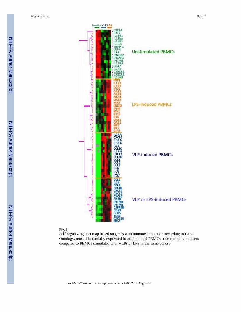
Fig. 1.Self-organizing heat map based on genes with immune annotation according to GeneOntology, most differentially expressed in unstimulated PBMCs from normal volunteerscompared to PBMCs stimulated with VLPs or LPS in the same cohort.
Monacoa et al. Page 8
FEBS Lett. Author manuscript; available in PMC 2012 August 14.
NIH
-PA Author Manuscript
NIH
-PA Author Manuscript
NIH
-PA Author Manuscript

Fig. 2.(A) Self-organizing heat map representing 1097 genes differentially expressed in PBMC.The red and yellow arrows points to subjects #12 and #5, respectively, who were foundanergic to VLP-stimulation among the high and low-viremia patients. (B) Immune genesselected, among those identified according to the criteria in panel A, according the list inFig. 1.
Monacoa et al. Page 9
FEBS Lett. Author manuscript; available in PMC 2012 August 14.
NIH
-PA Author Manuscript
NIH
-PA Author Manuscript
NIH
-PA Author Manuscript

Fig. 3.Unsupervised hierarchical clustering of PBMC samples stimulated with LPS (A) or VLPs(B). High stringent filter was applied resulting in the inclusion of 4,529 cDNA clones. VennDiagram comparing the overlap of genes differentially expressed in PBMCs uponstimulation with LPS (B) or VLPs (D).
Monacoa et al. Page 10
FEBS Lett. Author manuscript; available in PMC 2012 August 14.
NIH
-PA Author Manuscript
NIH
-PA Author Manuscript
NIH
-PA Author Manuscript

Fig. 4.(A) Self-organizing heat map based on 156 genes with immune annotations according togene ontology modulated at high degree of significance by stimulation with either VLPs orLPS. The orange dashed line emphasizes the ISGs cluster (a); the blue dashed lineemphasizes the lymphokine pattern (b). (B) The yellow and red arrows point to patient 05and 12, respectively.
Monacoa et al. Page 11
FEBS Lett. Author manuscript; available in PMC 2012 August 14.
NIH
-PA Author Manuscript
NIH
-PA Author Manuscript
NIH
-PA Author Manuscript

Fig. 5.(A–E) Relative Cy5/Cy3 values of IL-6, IL-8, CCL3, CCL22 and IRF-7 in different patientcohorts and type of stimulation. Data represent cumulative Cy5/Cy3 values for eachexperimental category. Red, yellow and green boxes indicate high–low-viral load andhealthy normal controls, respectively. (*, **, *** = P-value <0.05, <01 and <0.001,respectively) [15].
Monacoa et al. Page 12
FEBS Lett. Author manuscript; available in PMC 2012 August 14.
NIH
-PA Author Manuscript
NIH
-PA Author Manuscript
NIH
-PA Author Manuscript


