Embed Size (px)
Citation preview
Acta Veterinaria Hungarica 58 (1), pp. 39–46 (2010) DOI: 10.1556/AVet.58.2010.1.4
0236-6290/$ 20.00 © 2010 Akadémiai Kiadó, Budapest
MOLECULAR EVIDENCE OF NATURAL INFECTION WITH BABESIA CANIS CANIS IN CROATIA
Mirna BRKLJAČIĆ1*, Vesna MATIJATKO1, Ivana KIŠ1, Nada KUČER1, Jadranka FORŠEK1, Renata Barić RAFAJ2, Darko GRDEN1, Marin TORTI1, Iva MAYER1 and Vladimir MRLJAK1
1Clinic for Internal Diseases and 2Department of Chemistry and Biochemistry, Faculty of Veterinary Medicine, University of Zagreb,
Heinzelova 55, 10 000 Zagreb, Croatia
(Received 30 October 2008; accepted 23 March 2009)
The aim of the present study was to detect and characterise the species and subspecies of Babesia spp. that cause canine babesiosis in Croatia. Twenty-eight dogs with typical signs of babesiosis (lethargy, anorexia, fever, dark urine and thrombocytopenia) were included in this study. Their blood smears showed the presence of Babesia canis. The results showed the detection of one subspecies, namely Babesia canis canis using PCR, and subsequent sequence analysis dem-onstrated portions of the nss rRNA gene in 27 out of 28 samples. Sequence analy-sis of the isolates showed 100% identity in 11 samples, 99.7% identity (one nu-cleotide difference) in 11 samples and 99.4% identity (two nucleotides difference) in 5 samples with B. canis canis. The results of this study confirm the presence of B. canis canis in infected dogs in Croatia and demonstrate a slightly new genetic variant of Babesia subspecies.
Key words: Babesiosis, dog, DNA, sequence analysis
Babesia species are tick-borne intraerythrocytic apicomplexan parasites found in a variety of domestic and wild animals and in humans (Kuttler, 1988). Babesiosis is a common tick-borne disease among the dogs in Croatia, mostly confined to the area of the capital city, Zagreb, according to the geographical dis-tribution of the vector, Dermacentor reticulatus (personal communication from Relja Beck, DVM, PhD, Department for Parasitology, Faculty of Veterinary Medicine, University of Zagreb, Croatia). The highest notified incidence is dur-
*Corresponding author; E-mail: [email protected]; Phone: 00385 (1) 239-0343; Fax: 00385 (1) 492-3520
40 BRKLJAČIĆ et al.
Acta Veterinaria Hungarica 58, 2010
ing the spring and autumn, probably due to the vector’s seasonal activity (Föld-vári et al., 2007). The criteria commonly used for the identification of Babesia species are host specificity and morphology, particularly size (Levine, 1988).
There are two main species of canine piroplasms: Babesia canis, the large piroplasm (4–5 µm), and Babesia gibsoni, the small piroplasm (1–2.5 µm). On the basis of differences in geographical distribution, vector specificity and anti-genic properties, Babesia canis has been divided into three different subspecies, known as B. canis canis, transmitted by Dermacentor reticulatus (Europe); B. canis vogeli, transmitted by Rhipicephalus sanguineus (tropical and subtropical regions); and B. canis rossi, transmitted by Haemaphysalis leachi (South Africa) (Hauschild et al., 1995; Schetters et al., 1997). Variations in pathological manifes-tations are known to exist among the species and subspecies of canine piro-plasms. Therefore, accurate detection and identification are imperative for the di-agnosis of an individual infection (Jefferies et al., 2007). Parasitaemia is often low, especially in dogs infected with B. canis canis, and also in chronically in-fected dogs and asymptomatic carries (Scurrell, 2006). The morphological simi-larity among the species and subspecies of canine piroplasms has led to much confusion over accurate diagnosis using light microscopy (Kjemtrup et al., 2000). Serology, including ELISA and immunofluorescent antibody tests, is the most commonly used confirmatory diagnostic method for suspected babesiosis infections, although this may have limitations in very young dogs (Maia et al., 2007). Furthermore, dogs suffering from acute disease may initially be seronega-tive, and in such cases diagnosis may require repeated serum sampling (Taboada and Merchant, 1991). Serological testing has difficulty differentiating clinically important infection from prior exposure and problems with species cross-reactivity (Shaw et al., 2001). Babesia canis and Babesia gibsoni cross-react serologically and can only be distinguished by PCR (Scurrell, 2006).
Therefore, the most reliable method of distinguishing these subspecies is genetic analysis (Földvári et al., 2005). To date, Croatian dogs suffering from babesiosis have only been diagnosed on the basis of clinical examination, his-tory, microscopic examination of the thin blood smear, and response to therapy (i.e. diagnosis ex juvantibus) with imidocarb dipropionate (Imizol®, Schering-Plough), which could be suitable methods for diagnosis but are inadequate for determining the subspecies. The only genetic research data on Croatian babesio-sis was provided by Cacciò et al. (2002), who molecularly characterised natural Babesia infection in 11 dogs from Europe, including Croatian dogs, with results showing the occurrence of two subspecies, B. canis canis and B. canis vogeli. The aim of this study was to detect and characterise the Babesia species occur-ring in Croatian dogs, and to clarify the occurrence of the Babesia subspecies found.
MOLECULAR EVIDENCE OF NATURAL INFECTION WITH BABESIA CANIS CANIS 41
Acta Veterinaria Hungarica 58, 2010
Materials and methods
Sample collection
Blood samples for analysis were collected from the cephalic vein of 28 dogs brought to the Clinic for Internal Diseases of the Faculty of Veterinary Medicine in Zagreb during 2004 and 2005 with typical signs of babesiosis (leth-argy, anorexia, vomiting, fever, diarrhoea, dark urine). The samples were placed into tubes with EDTA for haematological analysis and tubes with no anticoagu-lant, which were centrifuged at 3000 rpm for 10 min. Sixteen of the tested dogs were male and twelve were female, ranging from 2 months to 13 years in age. All of the dogs selected for the study lived in Croatia and had no history of trav-elling abroad. The patients represented various canine breeds (Table 1). All the dogs were examined clinically, their haematological profile was determined, and for most of them additional serum biochemical parameters and urinalysis were also performed. Haematological values were determined using a Serono Baker 9120 automatic haematology analyser (Serono-Baker Diagnostics, Allentown, PA, USA), while biochemical values were determined with an Olympus AU 600 automatic analyser (Olympus Diagnostica GmbH, O’Callaghan Mills, Co. Clare, Ireland). Urinalysis was done by Bayer Clinitek Status (Bayer HealthCare AG, Leverkusen, Germany). Basic history, including breed, gender, age, tick infesta-tion and travel history, was provided by the owners.
Blood smears
Thin blood smears were prepared from all the dogs, air-dried, stained with Romanowski (Giemsa-May-Grünwald) and examined under light microscopy at × 1200 magnification.
DNA isolation
DNA was extracted from 200 µl of EDTA-anti-coagulated whole venous blood from each dog, using a NUCLEOSPIN blood kit (Macherey-Nagel, Düren, Germany) according to the manufacturer’s instructions. The concentrations ob-tained for each sample are shown in Table 1.
PCR and sequencing
The DNA samples were tested with forward PIRO-A and reverse PIRO-B primers, designed to amplify the 407-, 408- and 435-bp (base pair) fragments of the nuclear small subunit ribosomal RNA (nss rRNA) gene of B. odocoilei, B. divergens and B. microti, respectively (Olmeda et al., 1997; Armstrong et al., 1998). The alignment of the complete sequences of the nss rRNA from various species of Babesia deposited in the GenBank revealed that the 407-bp fragment
42 BRKLJAČIĆ et al.
Acta Veterinaria Hungarica 58, 2010
of B. canis nss rRNA could also be amplified with this primer set (Duh et al., 2004). The amplification protocol was slightly changed regarding the annealing temperature reaching up to 60 °C. The reaction consisted of an initial 1-min de-naturation step at 94 °C, denaturation at 94 °C for 45 sec, annealing at 60 °C for 45 sec, and extension at 72 °C for 45 sec. The final extension was performed at 72 °C for 5 min (hold step at 4 °C). The amplification was carried out in 40 cy-cles (denaturation, annealing and extension).
Sequencing on both strands was carried out in an automated sequencer us-ing a BigDye Terminator Cycle Sequencing Ready Reaction Kit (PE Applied Biosystems, Foster City, California, USA). The obtained sequences were analysed with the computer programmes of the Lasergene 5.0 software package (DNASTAR, Madison, Wisconsin, USA) and submitted to the GenBank for the determination of accession numbers.
Results
The blood smears from 28 tested dogs showing clinical signs of babesiosis were confirmed positive for Babesia infection. Microscopic evaluation of the blood smears revealed the presence of large (4–5 µm) single or paired intraeryth-rocytic, pyriform trophozoites of Babesia canis.
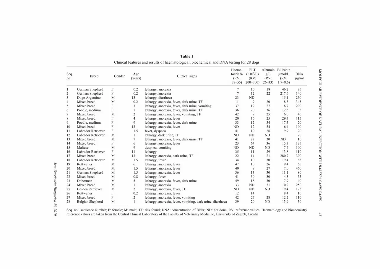
The most common clinical signs found in canine babesiosis were lethargy, anorexia, fever and dark urine (Table 1). Diarrhoea, vomiting and dyspnoea were rarely observed. Seven out of 28 dogs had ticks attached to the skin at the time of clinical presentation. The primary haematological abnormalities found in the af-fected dogs were mild to severe anaemia and thrombocytopenia. Alterations in the biochemical parameters varied depending on the severity of the case. The most commonly reported biochemical changes were hypoalbuminaemia (6 dogs) and hyperbilirubinaemia (17 dogs). Urinalysis showed haemoglobinuria in 10 cases. Seventeen of nineteen patients X-rayed had splenomegaly.
Sequencing of the ssrRNA gene was performed on 28 amplicons sized 407 bp. Twenty-seven out of the 28 blood samples tested were positive for Babesia-specific PCR (using PIRO-A and PIRO-B primers). An identity search (BLAST) and alignment (Lasergene, DNASTAR) of the sequence of the babesial 18S rRNA gene from the tested dogs revealed that all of them were infected with B. canis canis (accession number AY072926). However, three genetic groups (variations) were present: Group 3 (SEQ 3, 4, 5, 13, 20, 21, 22, 24, 26, 27 and 28), with GenBank ac-cession number FJ913769, included 11 samples matching B. canis canis in 100%, Group 1 (SEQ 1, 6, 7, 8, 10, 11, 12, 16, 18, 19 and 25), accession number FJ913767, included another 11 samples with one-nucleotide difference, and the remaining 5 samples belonged to Group 2 (SEQ 2, 9, 15, 17 and 23), accession number FJ913768 with a two-nucleotide difference determined. The one- or two-base differences found in the sequences could be attributed to minor mutations or even sequencing errors.
Acta VeterinariaH
ungarica 58, 2010
M
OLEC
ULA
R EV
IDEN
CE O
F NATU
RA
L INFEC
TION
WITH
BABESIA CAN
IS CAN
IS 43
Table 1 Clinical features and results of haematological, biochemical and DNA testing for 28 dogs
Seq. no. Breed Gender Age
(years) Clinical signs
Haema-tocrit %
(RV: 37–55)
PLT (×109/L)
(RV: 200–700)
Albumin g/L
(RV: 26–33)
Bilirubin µmol/L
(RV: 1.7–8.6)
DNA µg/ml
1 German Shepherd F 0.2 lethargy, anorexia 7 10 18 46.2 85 2 German Shepherd F 0.2 lethargy, anorexia 7 12 22 217.6 140 3 Dogo Argentino M 13 lethargy, diarrhoea 23 ND 15.1 250 4 Mixed breed M 0.2 lethargy, anorexia, fever, dark urine, TF 11 9 20 8.3 345 5 Mixed breed F 3 lethargy, anorexia, fever, dark urine, vomiting 37 19 27 6.7 290 6 Poodle, medium F 7 lethargy, anorexia, fever, dark urine, TF 36 20 36 12.5 35 7 Mixed breed M 2 lethargy, anorexia, fever, vomiting, TF 42 9 25 6.0 40 8 Mixed breed F 4 lethargy, anorexia, fever 20 16 25 29.3 115 9 Poodle, medium F 9 lethargy, anorexia, fever, dark urine 33 12 34 17.5 20 10 Mixed breed F 13 lethargy, anorexia, fever ND 12 34 6.4 100 11 Labrador Retriever F 1.5 fever, dyspnea 41 10 26 9.9 20 12 Labrador Retriever M 1 lethargy, dark urine, TF ND ND ND 70 13 Mixed breed M 7 lethargy, anorexia, fever, dark urine, TF 41 27 30 ND 10 14 Mixed breed F 6 lethargy, anorexia, fever 23 64 36 15.3 135 15 Maltese M 9 dyspnea, vomiting ND ND ND 7.7 100 16 Labrador Retriever F 11 lethargy 35 11 29 13.8 110 17 Mixed breed M 13 lethargy, anorexia, dark urine, TF 22 14 21 280.7 390 18 Labrador Retriever M 1.5 lethargy, fever 34 10 30 19.4 85 19 Rottweiler M 6 lethargy, anorexia, fever 47 10 26 9.4 65 20 Mixed breed M 1.5 lethargy, anorexia, fever 40 8 27 7.0 460 21 German Shepherd M 1.5 lethargy, anorexia, fever 36 13 30 11.1 80 22 Mixed breed M 0.8 lethargy, fever 41 30 30 4.3 55 23 Doberman M 5 lethargy, anorexia, fever, dark urine 49 18 30 7.9 40 24 Mixed breed M 1 lethargy, anorexia 33 ND 31 10.2 250 25 Golden Retriever M 2 lethargy, anorexia, fever, TF ND ND ND 19.4 125 26 Rottweiler F 0.2 lethargy, anorexia, fever 12 14 8.4 10 27 Mixed breed F 2 lethargy, anorexia, fever, vomiting 42 27 28 12.2 110 28 Belgian Shepherd M 1 lethargy, anorexia, fever, vomiting, dark urine, diarrhoea 39 20 ND 13.9 30
Seq. no.: sequence number; F: female; M: male; TF: tick found; DNA: concentration of DNA; ND: not done; RV: reference values. Haematology and biochemistry reference values are taken from the Central Clinical Laboratory of the Faculty of Veterinary Medicine, University of Zagreb, Croatia
44 BRKLJACIC et al.
Acta Veterinaria Hungarica 58, 2010
Discussion
Canine babesiosis is one of the most important tick-borne infectious dis-eases among dogs with highly variable clinical manifestations that interfere with clinical, therapeutic and even microscopic diagnoses (Matijatko et al., 2007). Variations in the pathological manifestations are known to exist among the species and subspecies of canine piroplasms and, therefore, accurate detection and identifi-cation are imperative for the diagnosis of individual cases (Jefferies et al., 2007).
The diagnosis of infections with these protozoa is usually based on the de-tection of pathogens in the peripheral blood under a microscope. The approxi-mate limit of detectable parasitaemia by light microscope is 0.001%. Molecular techniques, including PCR and sequence analysis, have recently been used in the diagnosis and epidemiological studies of canine Babesia because of their higher sensitivity and specificity for the detection of the target pathogens in the periph-eral blood, but also in finding unexpected new hosts for piroplasms such as Babesia canis, Theileria annae and Th. equi (Cacciò et al., 2002; Criado-Fornelio et al., 2003; Duh et al., 2004; Földvári et al., 2005; Sobczyk et al., 2005). However, there are marked differences in the distribution, vector specific-ity, virulence, prognosis and treatment response among the various recognised Babesia species and subtypes that infect dogs (Zahler et al., 1998). The piro-plasmids present in canids in southern Europe are B. canis vogeli, B. canis canis, Th. annae and Th. equi (Criado-Fornelio et al., 2003).
Infection with B. canis canis is distributed in Europe and Asia and is ca-pable of causing a wide range of clinical signs such as lethargy, anorexia, fever, jaundice, anaemia and thrombocytopenia (Taboada and Merchant, 1991; Barić Rafaj et al., 2005; Barić Rafaj et al., 2009).
Twenty-eight dogs naturally infected with Babesia sp. were identified by combining clinical findings, history data (tick infestation, place of residence or travelling through endemic areas) and light microscopic evidence of B. canis in-fection. The positive response to antibabesial therapy (improved clinical profile 24 hours after the application of Imizol®) also contributed to establishing the di-agnosis, but molecular analysis was necessary to identify the Babesia subspecies. The first such research was performed during the fall of 2000 and the spring of 2001 by Cacciò et al. (2002). That study, together with dogs from other countries (France, Italy and Poland), included eight Croatian dogs suffering from babesio-sis. Analysis of the complete sequence of the nss rRNA genes was performed and the results showed that the tested dogs were infected either with B. canis canis or B. canis vogeli. Since Cacciò et al. (2002) reported that the sequence analysis of the PCR products from all 8 dogs from Croatia revealed the presence of just one nss rRNA sequence (1714 bp) with no differences, we wanted to in-vestigate whether this would be confirmed with a much greater sample number. An identity search (BLAST) and alignment (Lasergene, DNASTAR) of the se-
MOLECULAR EVIDENCE OF NATURAL INFECTION WITH BABESIA CANIS CANIS 45
Acta Veterinaria Hungarica 58, 2010
quence of the babesial 18S rRNA gene from the tested dogs revealed that all of them were infected with B. canis canis.
Földvári et al. (2005) chose five samples for sequencing and demonstrated 99.8–100% similarity with the B. canis canis sequence deposited in the GenBank by Cacciò et al. (2002). Duh et al. (2004) showed the presence of B. canis canis and B. canis vogeli affecting dogs in Slovenia, based on clinical, microscopic and molecular investigations. They noted three groups of genetic variation within B. canis canis isolates. Three isolates were 100% identical with B. canis canis. Two isolates displayed one transition mutation and were thus 99.7% identical with B. canis canis, and six isolates displayed two transition mutations (99.5% identity with B. canis canis of accession numbers AY072926). Based on sequence analy-sis of the studied isolates, Sobczyk et al. (2005) concluded that canine babesiosis in the Warsaw area is caused by B. canis canis. Molecular analysis of PCR prod-ucts from an Italian dog that acquired babesiosis during its stay in Poland con-firmed this result (Cacciò et al., 2002). Criado-Fornelio et al. (2003) compared sequences obtained from piroplasmids infecting Spanish dogs with GenBank en-tries by BLAST: B. canis canis isolates from Spain were 100% identical with a Croatian isolate. This confirms that B. canis canis is a very common parasite in European dogs (Cacciò et al., 2002).
The results of this study confirm the presence of B. canis canis in infected dogs in Croatia. We did not find B. canis vogeli in our dogs, yet the expanding international mobility of pet dogs and the presence of competent tick vectors may result in the introduction of this parasite into Croatia.
Acknowledgements
We would like to thank the clinicians and the owners of the dogs for their co-operation. Thanks also go to Darja Duh of the Institute of Microbiology and Immunol-ogy, Medical Faculty, Ljubljana, Slovenia, for technical expertise in DNA analysis. We would also like to express our thanks to the laboratory of the Clinical Hospital Centre ‘Zagreb’, Croatia for assistance with DNA extraction and blood sample processing. This research was supported by the Ministry of Science, Education and Sports of the Republic of Croatia (Project No. 053-0532266-2220).
References
Armstrong, P. M., Katavolos, P., Caporale, D. A., Smith, R. P., Spielman, A. and Telford, S. R. (1998): Diversity of Babesia infecting deer ticks (Ixodes dammini). Am. J. Trop. Med. Hyg. 58, 739–742.
Barić Rafaj, R., Mrljak, V., Guelfi, J. F., Marinculić, A., Potočnjak, D., Žvorc, Z. and Kučer, N. (2005): Nombre de plaquettes et volume moyen plaquettaire dans la babesiose du chien. Re-vue Med. Vet. 156, 95–98.
46 BRKLJACIC et al.
Acta Veterinaria Hungarica 58, 2010
Barić Rafaj, R., Matijatko, V., Kiš, I., Kučer, N., Živičnjak, T., Lemo, N., Žvorc, Z., Brkljačić, M. and Mrljak, V. (2009): Alterations in some blood coagulation parameters in naturally occurring cases of canine babesiosis. Acta Vet. Hung. 57, 295–304.
Cacciò, S. M., Antunovic, B., Moretti, A., Mangili, V., Marinculic, A., Rafaj Baric, R., Slemenda, S. B. and Pieniazek, N. J. (2002): Molecular characterisation of Babesia canis canis and Babesia canis vogeli from naturally infected European dogs. Vet. Parasitol. 106, 285–292.
Criado-Fornelio, A., Martinez-Marcos, A., Buling-Sarana, A. and Barba-Carretero, J. C. (2003): Mo-lecular studies on Babesia, Theileria and Hepatozoon in southern Europe. Part I. Epizo-otiological aspects. Vet. Parasitol. 113, 189–201.
Duh, D., Tozon, N., Petrovec, M., Strašek, K. and Avšić-Županc, T. (2004): Canine babesiosis in Slo-venia: Molecular evidence of Babesia canis canis and Babesia canis vogeli. Vet. Res. 35, 363–368.
Földvári, G., Hell, É. and Farkas, R. (2005): Babesia canis canis in dogs from Hungary: detection by PCR and sequencing. Vet. Parasitol. 127, 221–226.
Földvári, G., Márialigeti, M., Solymosi, N., Lukács, Z., Majoros, G., Kósa, P. J. and Farkas, R. (2007): Hard ticks infesting dogs in Hungary and their infection with Babesia and Borrelia species. Parasitol. Res. 101 (Suppl 1.), 25–34.
Hauschild, S., Shayan, P. and Schein, E. (1995): Characterization and comparison of merozoite anti-gens of different Babesia canis isolates by serological and immunological investigations. Parasitol. Res. 81, 638–642.
Jefferies, R., Ryan, U. M. and Irwin, P. J. (2007): PCR-RFLP for the detection and differentiation of the canine piroplasm species and its use with filter paper-based technologies. Vet. Parasitol. 144, 20–27.
Kjemtrup, A. M., Kocan, A. A., Whitworth, L., Meinkoth, J., Birkenheuer, A. J., Cummings, J., Boudreaux, M. K., Stockham, S. L., Irizarry-Rovira, A. and Conrad, P. A. (2000): There are at least three genetically distinct small piroplasms from dogs. Int. J. Parasitol. 30, 1501–1505.
Kuttler, K. L. (1988): World-wide impact of babesiosis. In: Ristic, M. (ed.) Babesiosis of Domestic Animals and Man. CRC Press, Boca Raton. pp. 1–22.
Levine, N. D. (1988): The protozoan phylum Apicomplexa. Vol. II. CRC Press, Boca Raton. pp. 35–41. Maia, M. G., Costa, R. T., Haddad, J. P. A., Passos, L. M. F. and Ribeiro, M. F. B. (2007): Epidemi-
ological aspects of canine babesiosis in the semiarid area of the state of Minas Gerais, Brazil. Prev. Vet. Med. 79, 155–162.
Matijatko, V., Mrljak, V., Kiš, I., Kučer, N., Foršek, J., Živičnjak, T., Romić, Ž., Šimec, Z. and Ceron, J. J. (2007): Evidence of an acute phase response in dogs naturally infected with Babesia canis. Vet. Parasitol. 144, 242–250.
Olmeda, A. S., Armstrong, P. M., Rosenthal, B. M., Valladares, B., del Castillo, A., de Armas, F., Miguelez, M., Gonzalez, A., Rodriguez, J. A., Spielman, A. and Telford, S. R. (1997): A sub-tropical case of human babesiosis. Acta Trop. 67, 229–234.
Schetters, T. P. M., Moubri, K., Precigout, E., Kleuskens, J., Scholtes, N. C. and Gorenflot, A. (1997): Different Babesia canis isolates, different diseases. Parasitology 115, 485–493.
Scurrell, E. (2006): What is your diagnosis? J. Small Anim. Pract. 47, 226–227. Shaw, E. S., Day, M. J., Birtles, R. J. and Breitschwerdt, E. B. (2001): Tick-borne infectious diseases
of dogs. Trends Parasitol. 17, 74–80. Sobczyk, A. S., Kotomski, G., Gorski, P. and Wedrychowicz, H. (2005): Usefulness of touch-down
PCR assay for the diagnosis of atypical cases of Babesia canis canis infections in dogs. Bull. Vet. Inst. Puławy 49, 407–410.
Taboada, J. and Merchant, S. (1991): Babesiosis of companion animals and man. Vet. Clin. North Am. Small Anim. Pract. 21, 103–124.
Zahler, M., Schein, E., Rinder, H. and Gothe, R. (1998): Characteristic genotypes discriminate be-tween Babesia canis isolates of differing vector specificity and pathogenicity to dogs. Parasi-tol. Res. 84, 544–548.


