Embed Size (px)
Citation preview
Ministry of Health of Ukraine
Danylo Halytsky Lviv National Medical University
Department of Surgical Dentistry and Maxillofacial Surgery
METHODICAL GIDE
(for the English-Medium students of 3rd
course of Dental faculty)
from surgical dentistry
Second level of higher education (Master's Degree)
Sphere of Knowledge 22 «Healthcare»
Specialty 221 «Dentistry»
Faculty, Year: Dentistry, III
Content module 1: Propedeutics of Surgical Dentistry
(part 2)
Lviv – 2019
2
Recommended by the by the profile methodical commission for dentistry
(Protocol No. __ of _______201_)
Methodological guide was prepared by: Head of the of the Department of Surgical Dentistry and
Maxillofacial Surgery Professor Ya. E. Vares, Associate Professor of the Department of Surgical
Dentistry and Maxillofacial Surgery Yu. O. Medvid, Assistant Professor of the Department of
Surgical Dentistry and Maxillofacial Surgery A. V. Filipskyi, Phd student N. V. Shtybel, PhD
stydent Ya. S. Gudzan
Reviewers:
Head of the Department of Ortodontics of the Danylo Halytsky Lviv National Medical University
Associate Professor N. L. Chukhray,
Professor of the Department of Prosthetic Dentistry of the Danylo Halytsky Lviv National Medical
University A. Yu. Kordiyak.
Head of Department of the Latin and Foreign languages of the Danylo Halytsky Lviv National
Medical University, Assoc. Prof. Sodomora P. А.
A person in charge of the publication: Head of the Department of Surgical Dentistry and
Maxillofacial Surgery, Professor Ya. E. Vares.
3
INTRODUCTION
Curriculum for Surgical Dentistry
(in accordance with the Standard of Higher Education for the second level of higher education
(Master's Degree)
Sphere of Knowledge 22 "Healthcare"
Specialty 221 "Dentistry"
Master's Degree Program in Dentistry
Description of the discipline (abstract). The discipline involves the study of surgical
dentistry in its main sections: "Propedeutics of surgical dentistry and MFD", "Inflammatory
diseases of MFA", "Oncology of MFA", "Traumatology of MFA", "Reconstructive-Restorative
Surgery of MFA", with emphasis on the study of etiology, pathogenesis, clinics, diagnostics,
emergency treatment and prophylaxis of the main and most widespread diseases of MFA.
The main focus of the program is on the formation and development of students' skills in
collecting anamnesis, conducting the examination and differential diagnosis of diseases of the MFA
with a variety of clinical course and their complications. In the course of this program, modern
approaches to the diagnosis are taught in practice, the principles of treatment and prophylaxis are
studied on the basis of evidence-based medicine. Furthermore, students are introduced to the range
of urgent states in practical surgical dentistry. Students are also involved into the diagnostic and
treatment process of in- and out- patients under the guidance of assistants and associate professors
of the department. Students also look into a wide scope of therapeutic and prophylactic measures,
which are most often applied in dental surgical practice.
The study of surgical dentistry in theory and practice, contributes to the formation of a
holistic view of the structure and functioning of organs of MFA, deepening of theoretical and
practical training, acquisition of professional practical skills for further independent medical
activity.
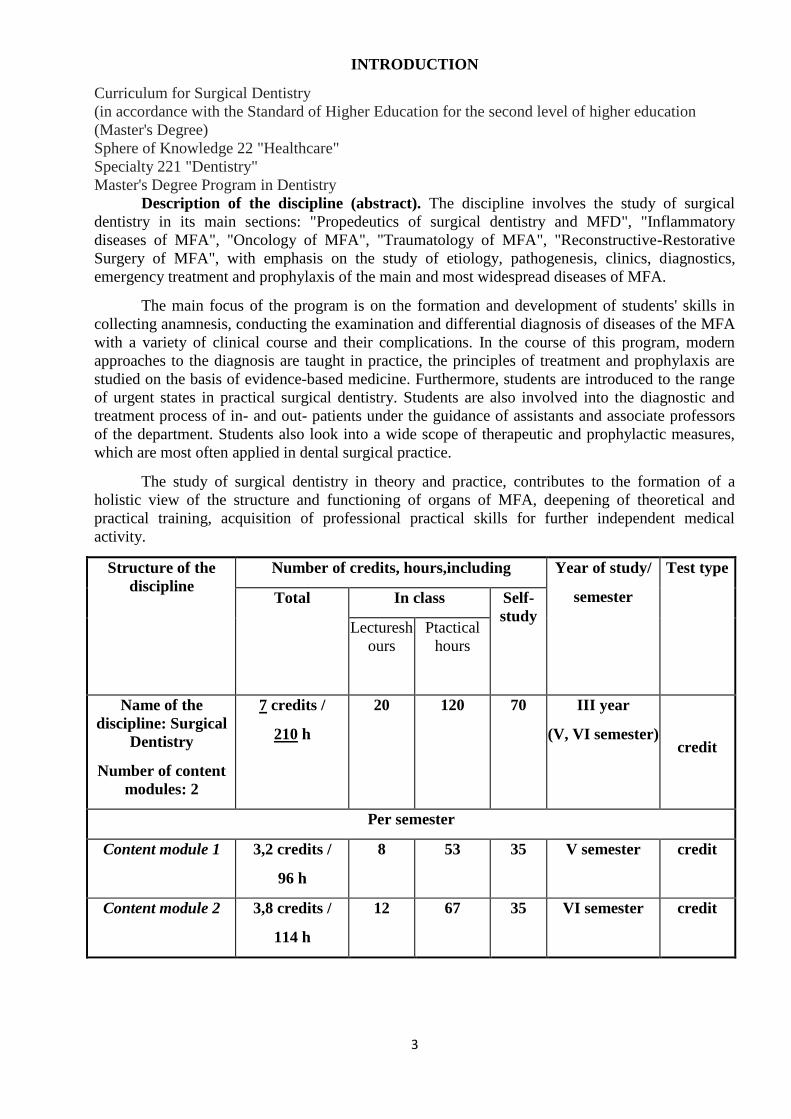
Structure of the
discipline
Number of credits, hours,including Year of study/
semester
Test type
Total In class Self-
study Lecturesh
ours
Ptactical
hours
Name of the
discipline: Surgical
Dentistry
Number of content
modules: 2
7 credits /
210 h
20 120 70 III year
(V, VI semester) credit
Per semester
Content module 1 3,2 credits /
96 h
8 53 35 V semester credit
Content module 2 3,8 credits /
114 h
12 67 35 VI semester credit
4
The subject of the study of the discipline is the pathological processes of MFA, which
relate to the sphere of competence of surgical dentistry and maxillofacial surgery, their clinical
course, the main diagnostic and therapeutic manipulations used in the practice of a surgical dentist.
Interdisciplinary connections: therapeutic dentistry, paediatric dentistry, orthopedic
stomatology, normal anatomy, histology, normal physiology, pathologic physiology, topographical
anatomy and operative surgery, microbiology, biochemistry, pharmacology, internal diseases,
endocrinology, skin and venereal, nervous diseases, otorhinolaryngology, ophthalmology, medicine
of extreme conditions.
1. Purpose and tasks of the discipline
1.1. The purpose of teaching the discipline (surgical dentistry) is to provide a
comprehensive and highly-specialized training of a dentist, which involves mastering the theory and
practice of all sections of surgical dentistry and basics of MFD, from organization of surgical
department of dental clinic and maxillofacial hospital to the ability of providing urgent care in
extreme conditions and qualified surgical dental and reconstructive-restoration assistance in MFD.
1.2. The main tasks of the study of surgical dentistry are to educate a professional surgical
dentist who is able to provide a thorough examination of the patient, diagnose the main symptoms
and syndromes of MFA pathologies, to substantiate and formulate the preliminary diagnosis; to
analyze the results of the examination and conduct differential diagnosis, to formulate a clinical
diagnosis of major diseases, to identify the manifestations of somatic diseases in the oral cavity, to
define the principles of integrated treatment in the clinic of surgical dentistry, to identify various
clinical variants and complications of the most common diseases of the MFA, to be aware of the
measures of primary and secondary prevention the most common surgical dental diseases.
Content module #1:
Explain and interpret the principles of deontology and medical ethics in surgical dentistry
and MFD, the method of examination of MFD patients, involvement of adjacent specialists in the
examination.
Analyze the indications and contraindications, especially the application of the basic methods
of general and local anesthesia, sedation in the practice of a surgical dentist.
Make a plan and conduct a patient's examination with MFA pathology, refer to an
additional research (if needed) and be able to interpret their results, plan for comprehensive
examination and treatment of AIDS patients.
Collect anamnesis and examination results of the patient with the specified MFA pathology,
fill in the relevant medical documentation; carry out cardiopulmonary resuscitation.
Collect the material for additional research (microbiological, cytological, histological);
preventive measures and emergency care.
Assign an individual scheme of premedication, depending on the psycho-somatic state of the
patient, the nature and extent of surgical intervention, medical therapy in the postoperative period,
to provide appropriate recommendations.
Demonstrate the techniques of preoperative preparation of the surgeon's hands by modern
techniques, the technique of antiseptic treatment of the surgical site, techniques of local anesthesia
on the upper and lower jaws; operations for the removal of individual groups of teeth on the upper
and lower jaw, pericoronarectomy, atypical tooth extraction
5
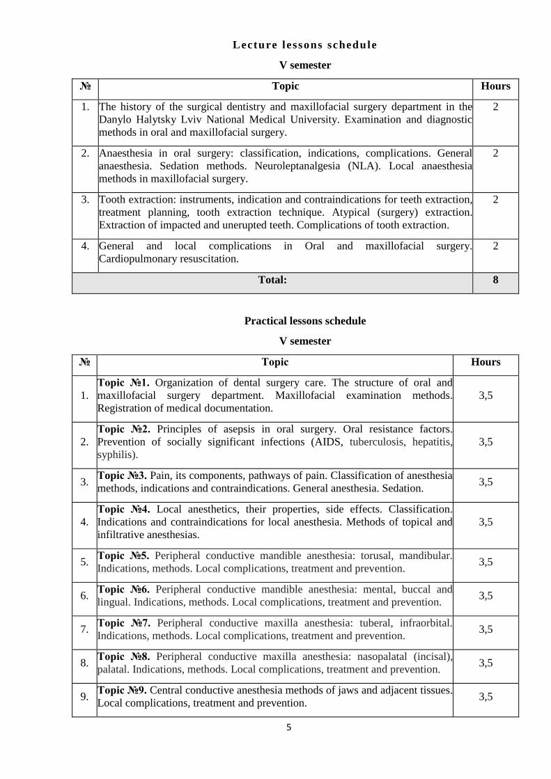
Lecture lessons schedule
V semester
№ Topic Hоurs
1. The history of the surgical dentistry and maxillofacial surgery department in the
Danylo Halytsky Lviv National Medical University. Examination and diagnostic
methods in oral and maxillofacial surgery.
2
2. Anaesthesia in oral surgery: classification, indications, complications. General
anaesthesia. Sedation methods. Neuroleptanalgesia (NLA). Local anaesthesia
methods in maxillofacial surgery.
2
3. Tooth extraction: instruments, indication and contraindications for teeth extraction,
treatment planning, tooth extraction technique. Atypical (surgery) extraction.
Extraction of impacted and unerupted teeth. Complications of tooth extraction.
2
4. General and local complications in Oral and maxillofacial surgery.
Cardiopulmonary resuscitation.
2
Total: 8
Practical lessons schedule
V semester
№ Тopic Hours
1.
Тopic №1. Organization of dental surgery care. The structure of oral and
maxillofacial surgery department. Maxillofacial examination methods.
Registration of medical documentation.
3,5
2.
Тopic №2. Principles of asepsis in oral surgery. Oral resistance factors.
Prevention of socially significant infections (AIDS, tuberculosis, hepatitis,
syphilis).
3,5
3. Тopic №3. Pain, its components, pathways of pain. Classification of anesthesia
methods, indications and contraindications. General anesthesia. Sedation. 3,5
4.
Topic №4. Local anesthetics, their properties, side effects. Classification.
Indications and contraindications for local anesthesia. Methods of topical and
infiltrative anesthesias.
3,5
5. Тopic №5. Peripheral conductive mandible anesthesia: torusal, mandibular.
Indications, methods. Local complications, treatment and prevention. 3,5
6. Тopic №6. Peripheral conductive mandible anesthesia: mental, buccal and
lingual. Indications, methods. Local complications, treatment and prevention. 3,5
7. Тopic №7. Peripheral conductive maxilla anesthesia: tuberal, infraorbital.
Indications, methods. Local complications, treatment and prevention. 3,5
8. Тopic №8. Peripheral conductive maxilla anesthesia: nasopalatal (incisal),
palatal. Indications, methods. Local complications, treatment and prevention. 3,5
9. Тopic №9. Central conductive anesthesia methods of jaws and adjacent tissues.
Local complications, treatment and prevention. 3,5
6
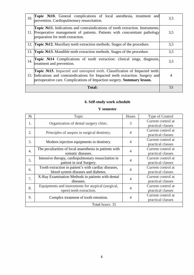
10. Тopic №10. General complications of local anesthesia, treatment and
prevention. Cardiopulmonary resuscitation. 3,5
11.
Тopic №11. Indications and contraindications of tooth extraction. Instruments.
Preoperative management of patients. Patients with concomitant pathology
preparation for teeth extraction.
3,5
12. Тopic №12. Maxillary teeth extraction methods. Stages of the procedure. 3,5
13. Тopic №13. Mandible teeth extraction methods. Stages of the procedure. 3,5
14. Тopic №14 Complications of tooth extraction: clinical sings, diagnosis,
treatment and prevention. 3,5
15.
Тopic №15. Impacted and unerupted teeth. Classification of Impacted teeth.
Indications and contraindications for Impacted teeth extraction. Surgery and
perioperative care. Complications of Impaction surgery. Summary lesson.
4
Total: 53
6. Self-study work schedule
V semester
№ Тopic Hours Type of Control
1. Organization of dental surgery clinic. 3 Current control at
practical classes
2. Principles of asepsis in surgical dentistry. 4 Current control at
practical classes
3. Modern injection equipments in dentistry. 4 Current control at
practical classes
4. The peculiarities of local anaesthesia in patients with
somatic diseases. 4
Current control at
practical classes
5. Intensive therapy, cardiopulmonary resuscitation in
patient in oral Surgery. 4
Current control at
practical classes
6. Tooth extraction in patient’s with cardiac diseases,
blood system diseases and diabetes. 4
Current control at
practical classes
7. X-Ray Examination Methods in patients with dental
diseases. 4
Current control at
practical classes
8. Equipments and instruments for atypical (surgical,
open) teeth extraction. 4
Current control at
practical classes
9. Complex treatment of tooth retention. 4 Current control at
practical classes
Total hours: 35
7
Ministry of Health of Ukraine
Danylo Halytsky Lviv National Medical University
“Approwed”
on the meeting of the Department
of Surgical Dentistry
and Maxillofacial Surgery
Head of the Department:
professor Ya. E. Vares
METHODICAL GIUDE FOR PRACTICAL LESSONS
Educational discipline SURGICAL DENTISTRY
Topic of the lesson
Topic №9. Central conductive methods
of anesthesia of jaws and surrounding tissues.
Local complications, their treatment.
Course 3rd
Faculty Dental
Lviv – 2019
8
Actuality of the topic: for pathological processes that are localized in the mouth and in the oral
cavity and do not allow the blockage of the peripheral branches of the trigeminal nerve, as well as
in case of need for anesthesia of the entire half of the jaw with major surgery, shows the
introduction of anesthetic to the main trunk - the second or third branch at the point of their exit
from the cavity of the skull. This requires a deep knowledge of topographic anatomy and mastering
techniques of central conduction anesthesia on the upper and lower jaws.
Aim of the lesson: to teach students the techniques of conducting central conduction anesthesia on
the upper and lower jaws. To practice on the phantoms the technique of blockade of the second and
third branches of the trigeminal nerve near the round and oval openings.
Learning objectives:
Professional competence:
1. Collection of medical information on the patient's condition.
2. Evaluation of the results of laboratory and instrumental research.
3. Establishment of a clinical diagnosis of dental disease.
4. Planning and conducting preventive measures for dental diseases.
5. Execution of medical and dental manipulations.
6. Organization and conducting of dental medical examination of persons subject to dispensary
supervision.
7. Assessment of the environmental impact on the health of the population (individual, family,
population).
8. Maintaining medical records.
9. Processing of state, social and medical information.
General competence:
1. The ability to abstract thinking, analysis and synthesis; the ability to learn and be trained
today.
2. Knowledge and understanding of the subject area and understanding of the profession.
3. Ability to apply knowledge in practical situations.
4. Ability to communicate in the state language both verbally and in writing; Ability to
communicate in a second language.
5. Skills in the use of information and communication technologies.
6. Ability to search, process and analyze information from various sources.
7. Ability to adapt and act in a new situation; ability to work autonomously.
8. Ability to identify, put and solve problems.
9. Ability to choose a communication strategy.
10. Ability to work in a team.
11. Interpersonal skills.
12. Ability to act on the basis of ethical considerations (motives).
13. Ability to act in a socially responsible and civic conscious manner.
Methods of training:
Preparatory stage - Frontal oral interview.
The main stage - practical training, role-playing game.
The final stage is brainstorming.
Interdisciplinary integration
Disciplines Student should know Student should be able to
Normal anatomy
Know the anatomical and
physiological features of the
maxillofacial area:
To be able to explain the
structure of systems and organs
of maxillo-facial area (MFA)
9
Normal physiology
- structure of the upper and lower
jaws;
- innervation and vascularization of
these sites;
- structure of the lymphatic system
of the head and neck;
- structure of the muscles of the head
and neck;
- structure of the head and neck
areas.
Topographical anatomy To know the topography of the
organs of MFA
To be able to explain the
topography of the organs of
MFA
Hystology
To know histological structure of
soft and hard tissues of MFA
To be able to explain the stages
of manufacturing of
cytological, histological
preparations
Pharmacology. To know pharmacological features
of preparations used for local
analgesia in the maxillofacial area
To be able to characterize the
anesthetics of different
pharmacological groups,
vasoconstrictor drugs.
Intradisciplinary integration
Topic 4. (Module 1) Local
anesthetics, their
properties, side effects...
To know the pharmacological
features of local anesthetics,
indications and contraindications to
use.
To be able to explain the
pharmacological features of
local anesthetics.
Plan and organizational structure of practical lesson of the discipline
Duration of practical lesson is 3.5 academic hours – 2 hours. 40 minutes including 10 minutes for a
break.
№ The main stages of
the lesson, their
functions and
content
Time
period
Methods of eductation
and control
Materials of methodical support
1. Preparatory stage 30 min.
1.1 Organizational
measures
5 min.
1.2 Setting up of
educational goals
and motivation.
5 min.
1.3 Control of the initial
level of knowledge
(standardized control
methods).
20 min. Individual theoretical
evaluation. Solving
typical tasks.Test
control.Written
interwiev.
Question for an
individual oral
and written
evaluation.
Typical
situational tasks
and tests.
Tables,
phantoms,
collapsible jaws,
textbooks,
manuals,
reference books,
atlas, methodical
recommendation
s, video films
2. Main Stage 90 min.
Formation of Formation of Patients with pathology of
10
professional skills
and abilities:
1. To work out
on phantom
technique of
conducting of the
central conductive
anesthesia to a round
foramen by a
submalar-pterygoid
way.
2. To work out
on phantom
technique of
carrying out of the
central conductive
anesthesia to a round
foramen by a tuberal
way.
3. To work out
on phantom
technique of
carrying out of the
central conductive
anesthesia to a round
foramen by a palatal
way.
4. To work out
on phantom
technique of
carrying out of the
central conductive
anesthesia to the
oval foramen by the
submalar-pterygoid
way.
5. To work out
on a phantom
technique of
carrying out of the
central conducting
anesthesia to the
oval foramen by
submalar way.
6. To work out on
phantom technique
of carrying out of
the central
conducting
anesthesia to the
oval foramen in a
supramalar way
professional skills:
Work with patients
with pathology of
maxillofacial area.
Work out the results of
additional methods of
examination of patients
with diseases of the
maxillofacial area.
Solving typical
situational tasks. Oral
and written evaluation
on standardized list of
issues.Work with
phantoms, view
thematic videos.
maxillofacial area. The history of
the disease.Selection of results of
additional survey methods.
Situational
tasks.Algorithms.Phantoms,
surgical instruments.Thematic
videos.
11
3. Final stage 30 min.
3.1 Control and
correction of the
level of professional
skills and abilities
Individual skills
control.Control of skills
by solving non-typical
situational problems
with illustrative
material.
Phantoms, surgical
instruments.The history of the
disease.Selection of results of
additional methods of examination
of thematic patients.Unusual
situational tasks.
3.2 Control and
correction of the
level of professional
skills and abilities.
Final evaluation of the
students
3.3 Homework.
Informing students
about the topic of the
next lesson.
Recommended literature
Methodology of organization of educational process in practical lesson.
STRUCTURE OF PRACTICAL LESSON
Preparation stage (30 min.)
To substantiate the significance of the subject for further study of the discipline and
professional activity of the doctor in order to formulate motivation and purposeful educational
activity. Get acquainted with students with specific goals and lesson plans.Conduct standardized
control of the initial level of student training, discussion and student answers.
• Organizational part of the lesson: presence check, evaluation of the uniform.
• Informing about of the topic and the purpose of the lesson.
Topic of the lesson: «Central conductive methods of anesthesia of jaws and surrounding
tissues. Local complications, their treatment.»
Aim of the lesson: to teach students the techniques of conducting central conduction anesthesia
on the upper and lower jaws. To practice on the phantoms the technique of blockade of the second
and third branches of the trigeminal nerve near the round and oval openings.
Motivation of educational activity.
For pathological processes that are localized in the mouth and in the oral cavity and do not
allow the blockage of the peripheral branches of the trigeminal nerve, as well as in case of need for
anesthesia of the entire half of the jaw with major surgery, shows the introduction of anesthetic to
the main trunk - the second or third branch at the point of their exit from the cavity of the skull. This
requires a deep knowledge of topographic anatomy and mastering techniques of central conduction
anesthesia on the upper and lower jaws.
Materials of methodical support of the preparatory stage of the lesson:
Questions to frontal survey:
1. Anatomical structure of branches of the trigeminal nerve;
2. Anatomical landmarks, zone of innervation of branches of the trigeminal nerve;
3. Anesthetics used for injectable analgesia, their concentration and properties;
4. 4. The instrumentation is required for central conduction anesthesia.
The main stage: the formation of professional skills (90 min)
Conducting professional training.
Materials of methodical support of the main stage of the lesson:
Patients with pathology of maxillofacial area. The history of the disease.Selection of results of
additional survey methods. Situational tasks.Algorithms.Phantoms, surgical instruments.Thematic
videos.
Anaesthesia of the barrel of the third branch of the trigeminal nerve is done in the cases of
impossibility of its conducting by the intraoral method (there is an inflammatory process in the area
12
of the comer of the lower jaw, reflex contraction of jaws etc).
Submaxillary - wing-shaped way is developed by S. N. Weisblat. In the middle of the trago-
orbital line do a prick by a long needle (6-7 cm) and move it towards the rest of the external plate of
the wing-shaped appendix of the sphenoid bone. The depth of penetration is fixed by a finger. A
needle is pulled out to the hypoderm, not moving a finger which fixes the depth of bedding of the
wing-shaped appendix. Return the tag of the needle back under a corner not less than 20° and again
dip a needle into the soft tissues at the depth marked before. Reach to the target point of anaesthesia
- the oval opening.
Supramalar way of anaesthesia was suggested by S. N. Weisblat in 1955. Prick of the needle is
done in the middle of the trago-orbital line above a malar arc with an insignificant inclination, that
enables to get to the external plate of the sphenoid bone. Implementation of this way of conductig
anaes- thetization in the following steps does not differ from the submaxillary one.
Submalar way was applied by S. N. Weisblat in 1937. A syringe with a needle, not less than 8
cm long, is used. The distance from the place of prick to the lower edge of a malar arc is mark in the
needle by a finger or sterile elastic.
The place of prick is typical for extraoral submaxillary anaesthesia. Get into the internal surface
of the branch of the lower jaw. Having passed 0,5- 0,75 cm of the marked way, take the end of the
needle from a bone wall inward. For this purpose, take a syringe outside under the same corner
under which we turned it inside while the previous measuring of distance from the place of prick to
the lower edge of the malar arc.
Orbital way was suggested by S. N. Weisblat in 1956. A place of prick is near the lower edge of
the eye socket nearby its lower external comer. The needle in the distance of 2-2,5 cm penetrates
through a wide lateral part of the lower orbital crack into a submaxillary fossula, and then, moving
up in a contact with the lower wall of the eye socket, trick into the oval opening.
Complication. While conducting block anaesthesia of the third branch because of violation of
technique of anaesthetizing, there is a possibility of damages of the middle artery of the brain
membrane, internal jaw artery,wing-shaped venous plexus, otosalpinx.
Also complications arising during local anaesthesia could be the general and become such as
intoxication or syncope, collapse or anaphylactic shock.
Algorithms for the formation of professional skills.
1. To collect anamnesis and to conduct a review of the patient with the pathology of the
maxillofacial area.
2. To identify and justify the indications and contraindications to central conduction anesthesia;
3. To be able to select instruments for anesthesia;
4. To be able to choose a local anesthetic, and determine the dose of the injection;
5. To be able to determine, by means of anatomical guidelines, the location of the anesthesia target
point;
6. To work out on phantom technique of conducting of the central conductive anesthesia to a
round foramen by a submalar-pterygoid way.
7. To work out on phantom technique of carrying out of the central conductive anesthesia to a
round foramen by a tuberal way.
8. To work out on phantom technique of carrying out of the central conductive anesthesia to a
round foramen by a palatal way.
9. To work out on phantom technique of carrying out of the central conductive anesthesia to the
oval foramen by the submalar-pterygoid way.
10. To work out on a phantom technique of carrying out of the central conducting anesthesia to the
oval foramen by submalar way.
11. To work out on phantom technique of carrying out of the central conducting anesthesia to the
oval foramen in a supramalar way.
13
Practical tasks (typical, atypical, unpredictable situations). Individual tasks:
Task №1.
A pain conductivity of which nerve is blocked during central anesthesia to the round opening?
A. Ocular
B. Occipital
C. Small palatine
D. Maxillary
E. The mandibular
Tasks for independent work and work in small groups (interactive teaching methods).
Patient 43, appealed to the dentist with complaints of pain in the 16th tooth, the severity of the
right half of the face. X-ray examination revealed a vein of the maxillary sinus at 2/3 of the volume.
In the area of the tips of the roots of the 16 tooth there is a destruction of bone tissue with a
diameter of 5 mm with penetration into the lumen of the maxillary sinus. The patient is shown a
radical sinus surgery with removal of 16 tooth and plastic conjugation through the hole of the
removed tooth. Which method of anesthesia is optimal in this case? Make your choice, describe and
demonstrate your anesthesia technique.
Final stage (30 min.)
Summing up of the lesson
Materials of methodological support of the final stage of the lesson:
Brain storm.Students demonstrate an exhaustive description of the unusual clinical situation
and offer to offer the most rational diagnostic methods.After recording all the proposed
diagnostic methods during the discussion, students choose the most rational.
Tasks for self-employment.To work on phantoms the technique of central conductive
anaesthezia to f.rotundum and f.ovale under conditions of phantom class.
Evaluation.
Conduct standardized final control using individual test tasks and questions (20 min.), Work
check (10 min.). Evaluate the student's current activities during the classroom, taking into account
standardized final control, analyze the student's progress, announce the evaluation of each student's
activity, and display it in the student attendance and student log book.An adult group at the same
time makes assessments in the record of the record of success and attendance of classes by students,
the teacher certifies them with his signature.
Brief informing the students about the topic of the next lesson and the methodical measures for
preparing for it.
Basic knowledge level:
1. Topographic anatomy of the upper and lower jaws.
2. Innervation and blood supply to the upper and lower jaws.
3. Pharmacological preparations used for local analgesia in the maxillofacial area.
List of questions to be studied by the student:
1. Classification of central conductive anesthesia.
2. Submalar-pterygoid way of anesthesia to the round foramen: the place of the injection of
the needle, the direction and depth of needle insertion, the target point of anesthesia, the amount of
injected anesthetic. Clinical effect of anesthesia. Zone of anesthesia.
3. Tuberal way of anesthesia to the round foramen: the place of the injection of the needle,
the direction and depth of needle insertion, the target point of anesthesia, the amount of anesthetic
administered. Clinical effect of anesthesia. Zone of anesthesia.
4. Palatine way: needle position, direction and depth of needle insertion, target point of
anesthesia, the amount of injected anesthetic. Clinical effect of anesthesia. Zone of anesthesia.
5. Inframalar-pterygoid way of anesthesia to the oval foramen: place of the injection of the
needle direction and depth of needle insertion, the target point of anesthesia, the amount of injected
anesthetic. Clinical effect of anesthesia. Zone of anesthesia.
14
6. Inframalar way of anesthesia to the oval foramen: the place of the injection of the needle,
the direction and depth of needle insertion, the target point of anesthesia, the amount of injected
anesthetic. Clinical effect of anesthesia. Zone of anesthesia.
7. Supramalar way of anesthesia to the oval foramen: the place of the injection of the needle,
direction and depth of needle insertion, the target point of anesthesia, the amount of injected
anesthetic. Clinical effect of anesthesia. Zone of anesthesia.
8. Local complications when conducting central conductive anesthesia causes of their
occurrence. Clinical manifestations.
9. Treatment of the patient in the case of complications.
The list of practical skills to be learned by the student:
1. To work out on phantom technique of conducting of the central conductive anesthesia to a
round foramen by a submalar-pterygoid way.
2. To work out on phantom technique of carrying out of the central conductive anesthesia to a
round foramen by a tuberal way.
3. To work out on phantom technique of carrying out of the central conductive anesthesia to a
round foramen by a palatal way.
4. To work out on phantom technique of carrying out of the central conductive anesthesia to
the oval foramen by the submalar-pterygoid way.
5. To work out on a phantom technique of carrying out of the central conducting anesthesia to
the oval foramen by submalar way.
6. To work out on phantom technique of carrying out of the central conducting anesthesia to
the oval foramen in a supramalar way
Situational tasks and questions on the topic of the lesson:
1. The patient for the blockade of the mandibular nerve is shown trunkular anesthesia. Which
opening should the anesthetic be drawn to?
A. To the foramen ovale.
B. To the foramen rotundum.
C. To the foramen jugulare.
D. To foramen caroticum.
E. To the foramen spinosum.
2. Which anesthetic route does not apply to anesthesia to the oval opening?
A. Submalar-pterigoid.
B. Malar.
C. Mandibular.
D. Palatinal.
3. When undergoing central conduction anesthesia by tuberal route, the needle should be advanced
to the depth of:
A. Up to 1 - 1.5 cm.
B. Up to 2 - 2.5 cm.
C. Up to 3 - 3.5 cm.
D. Up to 4 - 4.5 cm.
4. Determine the injection site of the needle during central conduction anesthesia with the submalar-
pterigoid path:
A. 2 cm to the front of the goat ear.
B. The middle of the trace orbital line.
C. The outer third of the trace orbital line.
D. 1 cm to the front of the goat ear.
E. 0.5 cm to the front of the goat ear.
15
5. A man 35 years old, went to the dentist for removal of 26 teeth. During central conduction
anesthesia, a tubular route showed a rapid increase in tissue edema and a restriction on the opening
of the oral cavity. What causes this condition?
A. Vascular injury.
B. Muscle trauma during anesthesia.
C. Injury to nerve trunks.
D. Anesthetics for patients with intolerance.
E. Anaphylactic shock.
Literature:
Basic:
1. Oral and Maxillofacial Surgery: Textbook, Part 1, 2 / V.O. Malanchuk. – Vinnytsia: Nova
Knyha Publishers, 2011. – 453p.
2. Principles of Dental Local Anaesthesia and Teeth Removal / Ya. E. Vares, R. Z. Ogonovsky,
Ch. R. Pohranychna – LNMU, 2007. – 63p.
3. Atlas of Human Anatomy / F. Netter – 2nd
ed. – New Jersey: ICON Learning Systems. – 592 p.
Additional:
1. Contemporary Oral and Maxillofacial Surgery / L. J. Peterson, E. Ellis, J. R. Hupp, M.R. Tucker
– 3rd ed. – St. Louis: Mosby – Year Book, Inc. – 1998. – 1477 p.
16
Ministry of Health of Ukraine
Danylo Halytsky Lviv National Medical University
“Approwed”
on the meeting of the Department
of Surgical Dentistry
and Maxillofacial Surgery
Head of the Department:
professor Ya. E. Vares
METHODICAL GIUDE FOR PRACTICAL LESSONS
Educational discipline SURGICAL DENTISTRY
Topic of the lesson
Topic №10. General complications of
local anesthesia, their prevention and treatment.
Cardiopulmonary resuscitation.
Course 3rd
Faculty Dental
Lviv – 2019
17
Actuality of the topic: great amount of the dental interventions are performed under local
anesthesia. Anamnesis collection, individual anesthetic selection and dose determination, careful
adherence to local anesthesia techniques allow the safe use of local anesthesia in practice. However,
even with strict adherence to the rules and algorithms of local anesthesia, there is a risk of
complications of a general nature. The dentist is obliged to quickly diagnose the patient's condition
and immediately provide him with urgent help, and in cardiac conditions to perform
cardiopulmonary resuscitation.
Aim of the lesson: to acquaint students with the possible general complications arising from local
anesthesia of the tissues of the maxillofacial area, their treatment and prevention. To develop
students with systemic integrated clinical thinking and professional clinical skills, to be able to
differentiate the complications of local anesthesia and to provide emergency medical care if
necessary.
Learning objectives:
Professional competence:
1. Collection of medical information on the patient's condition.
2. Evaluation of the results of laboratory and instrumental research.
3. Establishment of a clinical diagnosis of dental disease.
4. Planning and conducting preventive measures for dental diseases.
5. Execution of medical and dental manipulations.
6. Organization and conducting of dental medical examination of persons subject to dispensary
supervision.
7. Assessment of the environmental impact on the health of the population (individual, family,
population).
8. Maintaining medical records.
9. Processing of state, social and medical information.
General competence:
1. The ability to abstract thinking, analysis and synthesis; the ability to learn and be trained
today.
2. Knowledge and understanding of the subject area and understanding of the profession.
3. Ability to apply knowledge in practical situations.
4. Ability to communicate in the state language both verbally and in writing; Ability to
communicate in a second language.
5. Skills in the use of information and communication technologies.
6. Ability to search, process and analyze information from various sources.
7. Ability to adapt and act in a new situation; ability to work autonomously.
8. Ability to identify, put and solve problems.
9. Ability to choose a communication strategy.
10. Ability to work in a team.
11. Interpersonal skills.
12. Ability to act on the basis of ethical considerations (motives).
13. Ability to act in a socially responsible and civic conscious manner.
Methods of training:
Preparatory stage - Frontal oral interview.
The main stage - practical training, role-playing game.
The final stage is brainstorming.
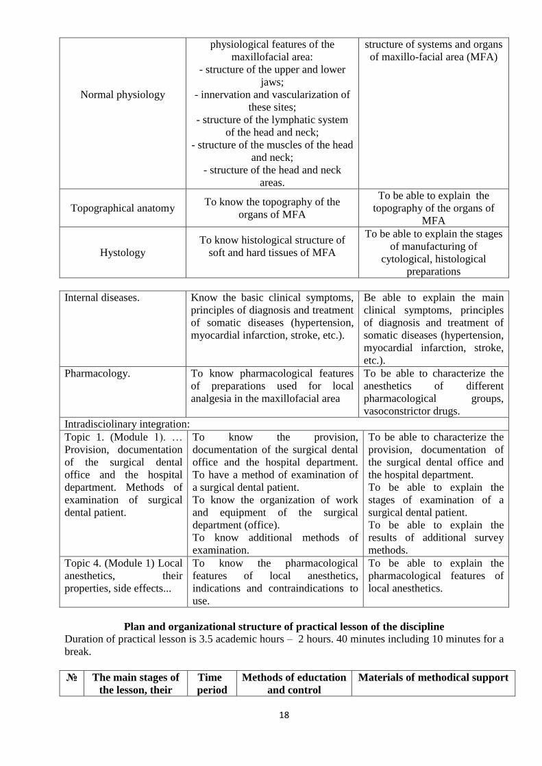
Interdisciplinary integration
Disciplines Student should know Student should be able to
Normal anatomy Know the anatomical and To be able to explain the
18
Normal physiology
physiological features of the
maxillofacial area:
- structure of the upper and lower
jaws;
- innervation and vascularization of
these sites;
- structure of the lymphatic system
of the head and neck;
- structure of the muscles of the head
and neck;
- structure of the head and neck
areas.
structure of systems and organs
of maxillo-facial area (MFA)
Topographical anatomy To know the topography of the
organs of MFA
To be able to explain the
topography of the organs of
MFA
Hystology
To know histological structure of
soft and hard tissues of MFA
To be able to explain the stages
of manufacturing of
cytological, histological
preparations
Internal diseases. Know the basic clinical symptoms,
principles of diagnosis and treatment
of somatic diseases (hypertension,
myocardial infarction, stroke, etc.).
Be able to explain the main
clinical symptoms, principles
of diagnosis and treatment of
somatic diseases (hypertension,
myocardial infarction, stroke,
etc.).
Pharmacology. To know pharmacological features
of preparations used for local
analgesia in the maxillofacial area
To be able to characterize the
anesthetics of different
pharmacological groups,
vasoconstrictor drugs.
Intradisciolinary integration:
Topic 1. (Module 1). …
Provision, documentation
of the surgical dental
office and the hospital
department. Methods of
examination of surgical
dental patient.
To know the provision,
documentation of the surgical dental
office and the hospital department.
To have a method of examination of
a surgical dental patient.
To know the organization of work
and equipment of the surgical
department (office).
To know additional methods of
examination.
To be able to characterize the
provision, documentation of
the surgical dental office and
the hospital department.
To be able to explain the
stages of examination of a
surgical dental patient.
To be able to explain the
results of additional survey
methods.
Topic 4. (Module 1) Local
anesthetics, their
properties, side effects...
To know the pharmacological
features of local anesthetics,
indications and contraindications to
use.
To be able to explain the
pharmacological features of
local anesthetics.
Plan and organizational structure of practical lesson of the discipline
Duration of practical lesson is 3.5 academic hours – 2 hours. 40 minutes including 10 minutes for a
break.
№ The main stages of
the lesson, their
Time
period
Methods of eductation
and control
Materials of methodical support
19
functions and
content
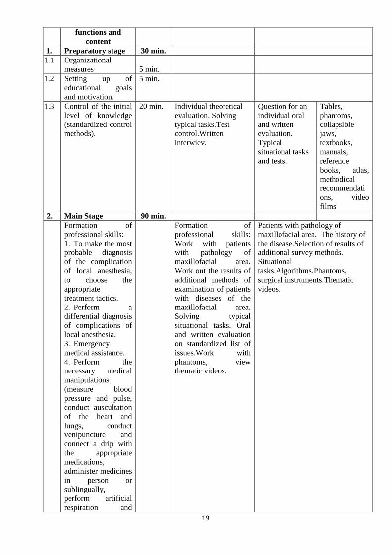
1. Preparatory stage 30 min.
1.1 Organizational
measures
5 min.
1.2 Setting up of
educational goals
and motivation.
5 min.
1.3 Control of the initial
level of knowledge
(standardized control
methods).
20 min. Individual theoretical
evaluation. Solving
typical tasks.Test
control.Written
interwiev.
Question for an
individual oral
and written
evaluation.
Typical
situational tasks
and tests.
Tables,
phantoms,
collapsible
jaws,
textbooks,
manuals,
reference
books, atlas,
methodical
recommendati
ons, video
films
2. Main Stage 90 min.
Formation of
professional skills:
1. To make the most
probable diagnosis
of the complication
of local anesthesia,
to choose the
appropriate
treatment tactics.
2. Perform a
differential diagnosis
of complications of
local anesthesia.
3. Emergency
medical assistance.
4. Perform the
necessary medical
manipulations
(measure blood
pressure and pulse,
conduct auscultation
of the heart and
lungs, conduct
venipuncture and
connect a drip with
the appropriate
medications,
administer medicines
in person or
sublingually,
perform artificial
respiration and
Formation of
professional skills:
Work with patients
with pathology of
maxillofacial area.
Work out the results of
additional methods of
examination of patients
with diseases of the
maxillofacial area.
Solving typical
situational tasks. Oral
and written evaluation
on standardized list of
issues.Work with
phantoms, view
thematic videos.
Patients with pathology of
maxillofacial area. The history of
the disease.Selection of results of
additional survey methods.
Situational
tasks.Algorithms.Phantoms,
surgical instruments.Thematic
videos.
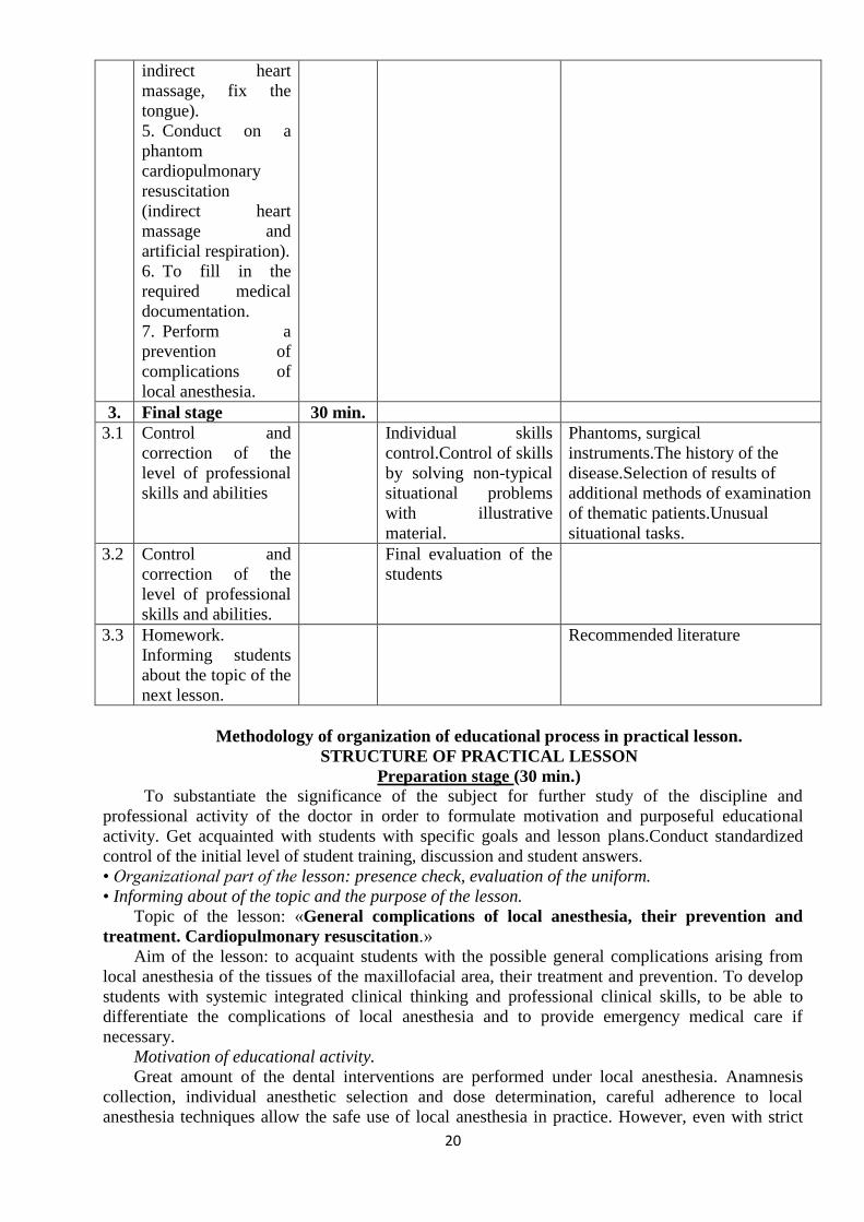
20
indirect heart
massage, fix the
tongue).
5. Conduct on a
phantom
cardiopulmonary
resuscitation
(indirect heart
massage and
artificial respiration).
6. To fill in the
required medical
documentation.
7. Perform a
prevention of
complications of
local anesthesia.
3. Final stage 30 min.
3.1 Control and
correction of the
level of professional
skills and abilities
Individual skills
control.Control of skills
by solving non-typical
situational problems
with illustrative
material.
Phantoms, surgical
instruments.The history of the
disease.Selection of results of
additional methods of examination
of thematic patients.Unusual
situational tasks.
3.2 Control and
correction of the
level of professional
skills and abilities.
Final evaluation of the
students
3.3 Homework.
Informing students
about the topic of the
next lesson.
Recommended literature
Methodology of organization of educational process in practical lesson.
STRUCTURE OF PRACTICAL LESSON
Preparation stage (30 min.)
To substantiate the significance of the subject for further study of the discipline and
professional activity of the doctor in order to formulate motivation and purposeful educational
activity. Get acquainted with students with specific goals and lesson plans.Conduct standardized
control of the initial level of student training, discussion and student answers.
• Organizational part of the lesson: presence check, evaluation of the uniform.
• Informing about of the topic and the purpose of the lesson.
Topic of the lesson: «General complications of local anesthesia, their prevention and
treatment. Cardiopulmonary resuscitation.»
Aim of the lesson: to acquaint students with the possible general complications arising from
local anesthesia of the tissues of the maxillofacial area, their treatment and prevention. To develop
students with systemic integrated clinical thinking and professional clinical skills, to be able to
differentiate the complications of local anesthesia and to provide emergency medical care if
necessary.
Motivation of educational activity.
Great amount of the dental interventions are performed under local anesthesia. Anamnesis
collection, individual anesthetic selection and dose determination, careful adherence to local
anesthesia techniques allow the safe use of local anesthesia in practice. However, even with strict
21
adherence to the rules and algorithms of local anesthesia, there is a risk of complications of a
general nature. The dentist is obliged to quickly diagnose the patient's condition and immediately
provide him with urgent help, and in cardiac conditions to perform cardiopulmonary resuscitation.
Materials of methodical support of the preparatory stage of the lesson:
Questions to frontal survey:
1. Classification of complications of local anesthesia of the maxillofacial area (general and
local, directly during and after some time after anesthesia).
2. Loss of consciousness: causes, clinic, diagnosis, treatment and prevention.
3. Collapse: causes, clinic, diagnosis, treatment and prevention.
4. Anaphylactic shock: causes, clinic, diagnosis, treatment and prevention.
5. Anesthetic and vasoconstrictor intoxication: causes, clinic, diagnosis, treatment and
prevention.
6. Idiosyncrazy: causes, clinic, diagnosis, treatment and prevention.
7. Principles of cardio-pulmonary resuscitation in the practice of a dental surgeon.
The main stage: the formation of professional skills (90 min)
Conducting professional training.
Materials of methodical support of the main stage of the lesson:
Patients with pathology of maxillofacial area. The history of the disease.Selection of results of
additional survey methods. Situational tasks.Algorithms.Phantoms, surgical instruments.Thematic
videos.
There are essential drugs and items of equipment that every dental practitioner should have
available for use in an emergency. Some of these are based on providing simple and uncomplicated
treatments while others necessitate providing early definitive treatment. Acute asthma and
anaphylaxis are two examples of emergencies where simple first aid measures are inadequate and
definitive treatment should be started by the dentist while waiting for the ambulance service to
transfer the patient to an accident and emergency (A&E) department. This essential treatment is
described as first-line treatment in the following protocols. Some drugs are available in preloaded
syringes for fast preparation.
Syncope
Signs and symptoms: may be preceded by nausea and closing in of visual fields; pallor and
sweating; heart rate below60 beats/min (bradycardia) during attack. First line treatment including:
• Lay flat
• Give oxygen
• Expect prompt recovery.
The main causes of faint could be pain or anxiety.
Principles of treatment:
• Need to encourage oxygenated blood flow to brain as rapidly as possible
• May need to block vagal activity with atropine and allow heart rate to increase.
Hyperventilation
Signs and symptoms: light-headed; tingling in the extremities; muscle spa cm may lead to
characteristic finger position (carpo-pedal spasm).
First line: reassure; ask patient to re-breathe from cupped hands or reservoir bag of inhalational
sedation or general anesthetic apparatus.
Causes - anxiety.
Principles of treatment: reduce anxiety; over-breathing has blown off carbon dioxide, resulting in
brain blood vessel vasoconstriction. Return carbon dioxide levels in blood to normal.
Postural hypotension
Signs and symptoms: light-headed; dizzy; loss of consciousness on returning to upright or
standing position from supine position.
Treatment:
• Lay the patient flat and give oxygen
22
• Sit the patient up very slowly.
More likely to occur if the patient is taking beta-blockers which reduce the capacity to
compensate for normal cardiovascular postural changes.
Principles of treatment: encourage oxygenated blood flow to brain.
Diabetic emergencies: hypoglycaemia
Signs and symptoms: shaking and trembling; sweating; hunger; headache and confusion.
• If the patient is conscious, give three sugar lumps or glucose and a little water or glucose oral
gel; repeated if necessary in 10 minutes
Cause: usually known diabetic; the patient may have taken medication as normal but not eaten
before a dental visit.
The main principles of treatment is to return blood glucose level to normal by giving glucose or
by converting the patient’s own glycogen to glucose by giving glucagon.
Further management
• Transfer the patient to A&E
• Give up to 50 ml 20% glucose i.v. infusion followed by 0.9% saline flush as the glucose
damages the vein
• Expect prompt recovery.
Anaphylactic shock
Signs and symptoms:
• Paresthesia, flushing and swelling of face, especially eyelids and lips
• Generalized urticaria, especially the hands and feet
• Wheezing and difficulty in breathing
• Rapid weak pulse.
These may develop over 15 to 30 minutes following the oral administration of a drug or rapidly
over a few minutes or seconds following i.v. drug administration.
• Lay the patient flat and raise his feet
• Give oxygen
• Give 0.5 ml epinephrine (adrenaline) 1 mg/ml (1 in 1000) intramuscular - 0.25 ml for 6-12
years; - 0.12 ml (or 6 months to 6 years)
- Repeated every 10 min until improvement.
Principles of treatment
Requires prompt energetic treatment of
• laryngeal oedema
• bronchospa cm
• hypotension.
Further management
• Transfer to A&V.
• Chlorphenamine (chlorpheniramine) 10 mg in 1 ml intramuscular or slow i.v. injection
• Hydrocortisone sodium succinate 200 mg by slow i.v. injection: valuable as action persists
after that of adrenaline has worn off
• Fluids i.v. (colloids) infused rapidly if shock not responding quickly to adrenaline.
Stroke
Stroke results from either cerebral haemorrhage or cerebral ischaemia. Signs and symptoms
• Confusion followed by signs and symptoms of focal brain damage
• Hemiplegia or quadriplegia
• Sensory loss
• Dysphasia
• Locked-in syndrome (aware, but unable to respond).
First line: reassure; transfer to A&E.
Principles of treatment
Maintain and transfer for further investigation.
Early basic life support for cardiac arrest
The following instructions are based on the UK Resuscitation Council guidelines for basic life
23
support. The essential features are remembered by ABC: airway, breathing and circulation.
Risks to the rescuer
• Before starting a resuscitation attempt, the rescuer must rapidly assess the risks: traffic, falling
masonry, toxic fumes and other potential hazards relevant to the environment
• Mucous membrane exposure to hepatitis В virus (HBVJ and human immunodeficiency virus
(HIV) is less of a risk than needle stick exposure, strongly suggesting that the chance of
infection from mouth-to- mouth ventilation is negligible. However, the US Centers for Disease
Control and Prevention advises universal precautions.
Basic life support
• Initial patient asses cment, airway maintenance, expired air ventilation and chest compression
constitute basic life support (BLS) or cardiopulmonary resuscitation.
• BLS is a “holding operation” maintaining ventilation and circulation until treatment of the
underlying cause can be instigated.
• BLS implies that no equipment is used. Where a simple airway or face mask is used, this is
described as ‘basic life support with airway adjunct’.
Theory of chest compression
• The ‘thoracic pump’ theory proposes that chest compression, an increasing intrathoracic
pressure, propels blood out of the thorax, forward flow occurring because veins at the thoracic
inlet collapse while the arteries remain patent.
• Even when performed optimally, chest compressions do not achieve more than 30% of the
normal cerebral perfusion.
Basic airway management
• Jaw thrust rather than chin lift is method of choice for the trauma victim .
• An oropharyngeal airway such as a Guedel or nasopharyngeal airway, may be used
• A face mask used for ventilation allows oxygen enrichment. Sequence of actions
1. Ensure safety of rescuer and victim (referred to below as he, for simplicity).
2. Check whether casualty is responsive.
3. If he responds by answering or moving
• leave him in the position in which you find him (providing he is not in further danger), check his
condition and get help if needed
• reassess him regularly. If he does not respond
• shout for help
• open the airway by tilting the head and lifting the chin
4. Keeping the airway open; look, listen and feel for breathing.
5. If he is breathing
• turn him into the recovery position;
• check for continued breathing
• send someone for help.
If he is not breathing
• send someone for help or, if you are on your own, leave the victim and go or help
• turn victim unto his back
• remove visible obstruction from the victim’s mouth
• give 2 breaths.
6. Assess the victim for signs of a circulation
• check the carotid pulse
• take no more than 10 seconds to do this.
7. If you are confident that you can detect signs of a circulation
• continue rescue breathing
• check circulation about every minute
• if the victim starts to breathe on his own but remains unconscious, turn him into the recovery
position.
If (there are no signs of a circulation or you are at all unsure
• start chest compression
24
• combine rescue breathing and compression in the ratio of 2:15.
• Continue until successful, help arrives, you become exhausted.
Algorithms for the formation of professional skills.
1. To collect anamnesis and to conduct a review of the patient with the pathology of the
maxillofacial area.
2. Learn how to identify and justify indications and contraindications to local anesthesia.
3. To be able to choose a local anesthetic, and determine the dose of the injection.
4. To make the most probable diagnosis of the complication of local anesthesia, to choose the
appropriate treatment tactics.
5. Perform a differential diagnosis of complications of local anesthesia. Emergency medical
assistance.
6. Perform the necessary medical manipulations (measure blood pressure and pulse, conduct
auscultation of the heart and lungs, conduct venipuncture and connect a drip with the appropriate
medications, administer medicines in person or sublingually, perform artificial respiration and
indirect heart massage, fix the tongue)
7. Conduct on a phantom cardiopulmonary resuscitation (indirect heart massage and artificial
respiration).
8. Learn to fill in the required medical documentation.
9. Perform a prevention of complications of local anesthesia
Practical tasks (typical, atypical, unpredictable situations).Individual tasks:
Task №1.
During local anesthesia, a patient, 34 years of age, experienced dizziness, spastic cough,
shortness of breath, fainting, a sharp decrease in blood pressure. What is the complication?
A. Anaphylactic shock.
B. Collapse.
C. Dizziness.
D. Edema of Quincke.
E. Swelling of the larynx.
Task №2.
The patient, 55 years, after anesthesia felt a sharp weakness, a sharp pain behind the sternum,
which radiates to the left arm, shoulder blade, heartbeat. Objectively: the patient is tired,
sluggish, his forehead is covered with cold sweat, the paleness of the skin is defined, the blood
pressure is 180/100 mmHg, the tone of the heart is deaf, the pulse is filamentous arrhythmic.
What condition has developed in the patient?
A. Hypertensive crisis.
B. Cardiogenic form of anaphylactic shock.
C. Angina attack.
D. Cardiac pain.
E. Myocardial infarction.
Tasks for independent work and work in small groups (interactive teaching methods).
In 1 minute after a torus anesthesia with 2% solution of novocaine - 4 ml, there were complaints of
the patient with difficulty breathing. Upper and lower lip, larynx mucosa and mouth are swollen,
sharply hyperemic. What complication did the patient experience? What additional survey methods
should be performed? What is the help algorithm? What drugs should be used, in what sequence
and doses? Justify your answers.
Final stage (30 min.)
Summing up of the lesson
Materials of methodological support of the final stage of the lesson:
25
Brain storm.Students demonstrate an exhaustive description of the unusual clinical situation
and offer to offer the most rational diagnostic methods.After recording all the proposed
diagnostic methods during the discussion, students choose the most rational.
Tasks for self-employment.To work on phantoms the technique of CPR.
Evaluation.
Conduct standardized final control using individual test tasks and questions (20 min.), Work
check (10 min.). Evaluate the student's current activities during the classroom, taking into account
standardized final control, analyze the student's progress, announce the evaluation of each student's
activity, and display it in the student attendance and student log book.An adult group at the same
time makes assessments in the record of the record of success and attendance of classes by students,
the teacher certifies them with his signature.
Brief informing the students about the topic of the next lesson and the methodical measures for
preparing for it.
Basic knowledge level:
1. Anatomical and physiological features of the structure of the maxillofacial area.
2. Innervation and blood supply to hard and soft facial tissues.
3. Clinic, diagnosis, treatment of somatic pathology (hypertension, myocardial infarction,
stroke, epilepsy, bronchial asthma).
4. Pharmacological properties of drugs used for the treatment and prevention of complications
of local anesthesia.
List of questions to be studied by the student:
1. Classification of complications of local anesthesia of the maxillofacial area (general and
local, directly during and after some time after anesthesia).
2. Loss of consciousness: causes, clinic, diagnosis, treatment and prevention.
3. Collapse: causes, clinic, diagnosis, treatment and prevention.
4. Anaphylactic shock: causes, clinic, diagnosis, treatment and prevention.
5. Anesthetic and vasoconstrictor intoxication: causes, clinic, diagnosis, treatment and
prevention.
6. Idiosyncrazy: causes, clinic, diagnosis, treatment and prevention.
7. Principles of cardio-pulmonary resuscitation in the practice of a dental surgeon.
The list of practical skills to be learned by the student:
1. To make the most probable diagnosis of the complication of local anesthesia, to choose the
appropriate treatment tactics.
2. Perform a differential diagnosis of complications of local anesthesia.
3. Emergency medical assistance.
4. Perform the necessary medical manipulations (measure blood pressure and pulse, conduct
auscultation of the heart and lungs, conduct venipuncture and connect a drip with the
appropriate medications, administer medicines in person or sublingually, perform artificial
respiration and indirect heart massage, fix the tongue).
5. Conduct on a phantom cardiopulmonary resuscitation (indirect heart massage and artificial
respiration).
6. To fill in the required medical documentation.
7. Perform a prevention of complications of local anesthesia.
Situational tasks and questions on the topic of the lesson:
1. During local anesthesia, a patient, 34 years of age, experienced dizziness, spastic cough,
shortness of breath, fainting, a sharp decrease in blood pressure. What is the complication?
A. Anaphylactic shock.
V. Collapse.
C. Dizziness.
26
D. Edema of Quincke.
E. Swelling of the larynx.
2. The patient, 55 years, after anesthesia felt a sharp weakness, a sharp pain behind the sternum,
which radiates to the left arm, shoulder blade, heartbeat. Objectively: the patient with
consciousness, sluggish, forehead covered with cold sweat, is determined by the pallor of the skin,
blood pressure 180/100 mm Hg, heart sounds are deaf, the pulse is filamentous arrhythmic. What
condition has developed in the patient?
A. Hypertensive crisis.
B. Cardiogenic form of anaphylactic shock.
C. Angina attack.
D. Cardiac pain.
E. Myocardial infarction.
3. In 1 minute after a torus anesthesia with 2% solution of novocaine - 4 ml, there were complaints
of the patient with difficulty breathing. Upper and lower lip, larynx mucosa and mouth are swollen,
sharply hyperemic. What complication did the patient experience?
A. Anaphylactic shock.
B. Edema of Quincke.
S. Collapse.
D. Anesthetic intoxication.
E. Acute pulmonary insufficiency
4. After anesthesia, after 2 minutes, the patient lost consciousness. The red border of the lips and the
skin are pale, sweat drops on the surface of the skin, palms moist. What condition did the patient
have?
A. Collapse.
B. Fainting.
C. Acute myocardial infarction.
D. Anesthetic intoxication.
E. Anaphylactic shock.
5. During the removal of 46 teeth in a patient of 34 years, a brief eclipse of consciousness, pallor of
the mucous membranes and skin appeared. Pulse is weak and frequent, blood pressure is 90/60
mmHg. What first aid should be given to the patient?
A. To give the patient the position of Trendelenburg.
C. Introduce doses of 0.5 ml. 0.1% of adrenaline.
C. Introduce 1ml. 1% solution of dimedrol.
D. Enter 2ml. the district of dexamethasone.
E. Introduce 2ml. the district of cordyamine.
Literature:
Basic:
1. Oral and Maxillofacial Surgery: Textbook, Part 1, 2 / V.O. Malanchuk. – Vinnytsia: Nova
Knyha Publishers, 2011. – 453p.
2. Principles of Dental Local Anaesthesia and Teeth Removal / Ya. E. Vares, R. Z. Ogonovsky,
Ch. R. Pohranychna – LNMU, 2007. – 63p.
3. Atlas of Human Anatomy / F. Netter – 2nd
ed. – New Jersey: ICON Learning Systems. – 592
p.
Additional:
1. Contemporary Oral and Maxillofacial Surgery / L. J. Peterson, E. Ellis, J. R. Hupp, M.R.
Tucker – 3rd ed. – St. Louis: Mosby – Year Book, Inc. – 1998. – 1477 p.
27
Ministry of Health of Ukraine
Danylo Halytsky Lviv National Medical University
“Approwed”
on the meeting of the Department
of Surgical Dentistry
and Maxillofacial Surgery
Head of the Department:
professor Ya. E. Vares
METHODICAL GIUDE FOR PRACTICAL LESSONS
Educational discipline SURGICAL DENTISTRY
Topic of the lesson
Topic №11. Indications and
contraindications to the operation of tooth
extraction. Instruments for tooth extraction.
Preparation of patients with concomitant
pathology for tooth extraction.
Course 3rd
Faculty Dental
Lviv – 2019
28
Actuality of the topic: One of the main tasks of dentistry is the fight for the preservation of teeth. In
light of the latest advances in modern dentistry, tooth extraction is being attempted not so often.
Nonetheless, tooth extraction is still one of the most common surgical interventions in polyclinic
dental practice; it is a radical measure, but there are situations in which it is impossible to avoid
removal. After tooth extraction, certain changes occur not only in the area of the alveolar process
where the tooth was placed, but also in adjacent areas and anatomical structures. The loss of a large
number of teeth negatively affects the functioning of the whole body. All this gives reason to
believe that the operation of tooth extraction should be performed only on narrow indications,
weighing all the positive and negative consequences of this intervention.
Aim of the lesson: to teach students clear indications and contraindications to tooth extraction. To
familiarize with surgical tools for tooth extraction; principles of preparation of patients with
concomitant pathology.
Learning objectives:
Professional competence:
1. Collection of medical information on the patient's condition.
2. Evaluation of the results of laboratory and instrumental research.
3. Establishment of a clinical diagnosis of dental disease.
4. Planning and conducting preventive measures for dental diseases.
5. Execution of medical and dental manipulations.
6. Organization and conducting of dental medical examination of persons subject to dispensary
supervision.
7. Assessment of the environmental impact on the health of the population (individual, family,
population).
8. Maintaining medical records.
9. Processing of state, social and medical information.
General competence:
1. The ability to abstract thinking, analysis and synthesis; the ability to learn and be trained
today.
2. Knowledge and understanding of the subject area and understanding of the profession.
3. Ability to apply knowledge in practical situations.
4. Ability to communicate in the state language both verbally and in writing; Ability to
communicate in a second language.
5. Skills in the use of information and communication technologies.
6. Ability to search, process and analyze information from various sources.
7. Ability to adapt and act in a new situation; ability to work autonomously.
8. Ability to identify, put and solve problems.
9. Ability to choose a communication strategy.
10. Ability to work in a team.
11. Interpersonal skills.
12. Ability to act on the basis of ethical considerations (motives).
13. Ability to act in a socially responsible and civic conscious manner.
Methods of training:
Preparatory stage - Frontal oral interview.
The main stage - practical training, role-playing game.
The final stage is brainstorming.
Interdisciplinary integration
Disciplines Student should know Student should be able to
Normal anatomy Know the anatomical and To be able to explain the
29
Normal physiology
physiological features of the
maxillofacial area:
- structure of the upper and lower
jaws;
- innervation and vascularization of
these sites;
- structure of the lymphatic system
of the head and neck;
- structure of the muscles of the head
and neck;
- structure of the head and neck
areas.
structure of systems and organs
of maxillo-facial area (MFA)
Pathologic anatomy
Pathologic physiology
To know the appearance and flow of
the pathological processes in the
tissues and organd of MFA
To be able to explain the
appearance and flow of the
pathological processes in the
tissues and organd of MFA
Topographical anatomy To know the topography of the
organs of MFA
To be able to explain the
topography of the organs of
MFA
Hystology
To know histological structure of
soft and hard tissues of MFA
To be able to explain the stages
of manufacturing of
cytological, histological
preparations
Mycrobiology To know the species identification of
microorganisms in the oral cavity
To be able to explain the stages
of manufacturing of
microbiological preparations
and the essence of
bacteriological examination
Radiation diagnostics. To know the methods of radiological
examination used in dental practice
To be able to explain the
principles on which these or
other methods are based (X-
ray, CT, MRI, ultrasound)
Therapeutic dentistry
Pediatric dentistry
To know diseases of hard tissues of
tooth and pulp.To know additional
methods of examination, which are
used in dental practice.
Be able to carry out an intra-
oral examination of individual
teeth.Be able to diagnose
caries, pulpitis of temporary
and permanent teeth.
Plan and organizational structure of practical lesson of the discipline
Duration of practical lesson is 3.5 academic hours – 2 hours. 40 minutes including 10 minutes for a
break.
№ The main stages of
the lesson, their
functions and
content
Time
period
Methods of eductation
and control
Materials of methodical support
1. Preparatory stage 30 min.
1.1 Organizational
measures
5 min.
1.2 Setting up of
educational goals
and motivation.
5 min.
30
1.3 Control of the initial
level of knowledge
(standardized control
methods).
20 min. Individual theoretical
evaluation. Solving
typical tasks.Test
control.Written
interwiev.
Question for an
individual oral
and written
evaluation.
Typical
situational tasks
and tests.
Tables,
phantoms,
collapsible jaws,
textbooks,
manuals,
reference books,
atlas, methodical
recommendation
s, video films
2. Main Stage 90 min.
Formation of
professional skills
and abilities:
1. Be able to
determine general
and local immediate
(absolute)
indications before
tooth extraction.
2. Be able to
determine planned
(relative) indications
before tooth
extraction:
- sanation;
- prosthetic;
- esthetic.
3. Be able to identify
general and local
contraindications to
tooth extraction.
4. Learn to
distinguish tools for
tooth extraction
(forceps, elevators)
on different grounds.
5. Select instruments
to extract different
groups of teeth on
the upper and lower
jaw
Formation of
professional skills:
Work with patients
with pathology of
maxillofacial area.
Work out the results of
additional methods of
examination of patients
with diseases of the
maxillofacial area.
Solving typical
situational tasks. Oral
and written evaluation
on standardized list of
issues.Work with
phantoms, view
thematic videos.
Patients with pathology of
maxillofacial area. The history of
the disease.Selection of results of
additional survey methods.
Situational
tasks.Algorithms.Phantoms,
surgical instruments.Thematic
videos.
3. Final stage 30 min.
3.1 Control and
correction of the
level of professional
skills and abilities
Individual skills
control.Control of skills
by solving non-typical
situational problems
with illustrative
material.
Phantoms, surgical
instruments.The history of the
disease.Selection of results of
additional methods of examination
of thematic patients.Unusual
situational tasks.
3.2 Control and
correction of the
level of professional
skills and abilities.
Final evaluation of the
students
3.3 Homework. Recommended literature
31
Informing students
about the topic of the
next lesson.
Methodology of organization of educational process in practical lesson.
STRUCTURE OF PRACTICAL LESSON
Preparation stage (30 min.)
To substantiate the significance of the subject for further study of the discipline and
professional activity of the doctor in order to formulate motivation and purposeful educational
activity. Get acquainted with students with specific goals and lesson plans.Conduct standardized
control of the initial level of student training, discussion and student answers.
• Organizational part of the lesson: presence check, evaluation of the uniform.
• Informing about of the topic and the purpose of the lesson.
Topic of the lesson: «Indications and contraindications to the operation of tooth extraction.
Instruments for tooth extraction. Preparation of patients with concomitant pathology for
tooth extraction».
Aim of the lesson: to teach students clear indications and contraindications to tooth extraction. To
familiarize with surgical tools for tooth extraction; principles of preparation of patients with
concomitant pathology.
Motivation of educational activity.
One of the main tasks of dentistry is the fight for the preservation of teeth. In light of the latest
advances in modern dentistry, tooth extraction is being attempted not so often. Nonetheless, tooth
extraction is still one of the most common surgical interventions in polyclinic dental practice; it is a
radical measure, but there are situations in which it is impossible to avoid removal. After tooth
extraction, certain changes occur not only in the area of the alveolar process where the tooth was
placed, but also in adjacent areas and anatomical structures. The loss of a large number of teeth
negatively affects the functioning of the whole body. All this gives reason to believe that the
operation of tooth extraction should be performed only on narrow indications, weighing all the
positive and negative consequences of this intervention.
Materials of methodical support of the preparatory stage of the lesson:
Questions to frontal survey:
1. Basic indications before tooth extraction.
2. The main urgent (absolute) indications for the teeth extraction.
3. Relative indications to tooth extraction.
4. Prosthetic, aesthetic, sanation indications for the teeth extraction.
5. The main general and local contraindications for the teeth extraction.
6.Classification of instrumentsfor tooth extraction on the lower jaw, specifity of forceps and
elevators.
7.Classification of instrumentsfor tooth extraction on the upper jaw, specifity of forceps and
elevators.
The main stage: the formation of professional skills (90 min)
Conducting professional training.
Materials of methodical support of the main stage of the lesson:
Exodontia means extraction or removal of teeth. Exodondia is a procedure that incorporates the
principles of surgery and many principles from phisics and mechanics. When these principles are
applied correctly, a tooth must probably be removed intact from the alveolar process without
untoward sequelae. This operation does not require a large amount of brute force and rather can be
accomplished with finesse and controlled force in such a manner that the tooth is not pulled from
the bone but instead is lifted gently from the alveolar process.
32
During the preoperative period the degree of difficulty that is anticipated for removing a
particular tooth is assessed. If the preoperative assessment leads the surgeon to believe that the
degree of difficulty will be high and the initial attempts at tooth removal confirm this, a deliberate
surgical approach should be taken. Excessive force may injure local tissues and destroy surrounding
bone and teeth. Moreover, excessive force heightens the intraoperative discomfort and anxiety of
the patient. The most efficient way to remove a tooth is slowly.
While the great majority of extractions can be safely done in the dental office, some patients require
hospitalization for this surgery because of systemic conditions which make them poor surgical risks.
Teeth may require removal for many reasons including the following:
I. GENERAL INDICATIONS (morbid conditions caused by decayed teeth as a source of
infection): odontogenous chroniosepsis and chronic odontogenous intoxication, odontogenous
disease of any organ (in the background of chroniosepsis).
II. LOCAL INDICATIONS is divided into 6 groups:
Sanation; Functional; Prosthetic; Orthodontic; Esthetic; Combination of above mentioned
I. Sanation
• Chronic granulating or granulomatous periodontitis in case of curved or obstructed root
canals
• Acute purulent periodontitis, if there is no possibility to evacuate pus through root canal.
• Acute odontogenous osteomyelitis.
• Presence of pathological process around semi-impacted tooth (retro- molar periostitis).
• Decidui teeth, in presence of inflammatory process.
• Decidui teeth, which cause acute septic conditions.
II. Functional - indications which concerns cases of injury of soft tissues by impacted or semi-
impacted tooth (buccal mucosa, pterygomandibular fossa); supernumerary teeth; in case of birth
with erupted teeth, and there is no possibility to cover it with a protective plate.
III. Prosthetic including the following:
Single teeth, which compromises denture stability; “gangrenous” roots, which cannot be filled
and used as support for denture; teeth extended from dentition (Godon-Popov phenomena), which
interferes with forming of occlusal arch significantly decayed permanent 1st molars in children
(making room for correct eruption of 7 and 8 teeth).
IV. Orthodontic: 4th permanent teeth, which interferes orthodontic moving of anterior teeth;
teeth extraction in case of space deficiency in orthodontic treatment.
V. Esthetic: supernumerary teeth, malposed and teratic teeth; teeth, that spoil appearance,
especially while smiling
VI. Combination of local indications
Complications associated with treatment of teeth (perforation of the tooth cavity floor, root);
impacted teeth in uneffective orthodontic treatment; diseases or conditions when it is impossible to
perform tooth treatment; the patient insists on tooth extraction.
There are no absolute contraindications for tooth extraction but there are some diseases and
conditions which require operation suspending or, if possible, preoperative medication.
Relative contraindications:
1. Heart disease (Angina, arythmia, reumatism, endocarditis in acute course, heart failure).
2. Kidney disease (acute glomerulonephritis, renal failure).
3. Infectious diseases (diphtheria, hepatitis)
4. Blood disease (haemophilia, leukosis, trombocytopenia).
5. CNS diseases (meningitis, encephalitis).
6. Mental diseases in acute condition (epilepsy, schizophrenia).
7. Teeth placed in malignant tumor or hemangioma.
8. Acute diseases of the respiratory tract (ARD, flu, bronchitis)
9. Pregnancy.
The information from the history and examination is used to formulate the best plan for the
patient. This will include measures for adequate preparation for the procedure and also the selection
of anaesthesia: whether local anaesthesia, conscious sedation with local anaesthesia or general
33
anaesthesia. Anticipated difficulties are better discussed with the patient before treatment rather
than during treatment, when they may be perceived as excuses for inadequate planning or
experience.
Presurgical assessment of the patient includes evaluation of the level of anxiety,
determination of health status and any necessary modifications of routine procedures, evaluation of
the clinical presentation of the tooth to be removed, and radiographic evaluation of the tooth root
and bone. All four of these major factors must be weighed when estimating the difficulty of the
extraction. If any factor or combination of factors presents a level of difficulty that seems too great,
the dentist should refer the patient to an oral and maxillofacial surgeon.
Surgeons must prevent inadvertent injury or transmission of infection to their patients or to
themselves. The concept of universal precautions states that all patients must be viewed as having
blood-borne diseases that can be transmitted to the surgical team. To prevent this trans-mission,
surgical gloves, surgical mask, and eyewear with side shields are required. Additionally most
authorities recommend that the surgical team wear long- sleeved gowns that can be changed when
they become visibly soiled.
Surgeon has to use protective eyeglasses, mask, and gloves. Surgeons should have short or
pinned-back hair and should wear long-sleeved smocks that are changed daily or sooner if they
become soiled. The patient should have full, waterproof drape. If the surgeon has long hair, it is
essential that the hair be held in position with barrettes or other holding devices or be covered with
a surgical cap. It is a major breach in aseptic technique to allow the surgeon’s hair to hang over the
patient’s face and mouth. Before the patient undergoes the surgical procedure, a minimal amount of
draping is necessary. A sterile drape should be put across the patient’s chest to decrease the risk of
contamination.
Before the extraction, patients should vigorously rinse their mouths with an antiseptic mouth
rinse, such as chlorhexidine 0.5 mg. This reduces the gross bacterial contamination in the patient’s
mouth, which helps to reduce the incidence of postoperative infection.
To prevent teeth or fragments of teeth from falling into the mouth and potentially being swallowed
or aspirated into the lungs, many surgeons prefer to place the 5 x 5 cm gauze loosely into the back
of the mouth. This oral partition serves as a barrier so that, should a tooth slip from the forceps or
shatter under the pressure of the forceps, it will be caught in the gauze rather than be swallowed or
aspirated. The surgeon must take care that the gauze is not positioned so far posterior that it makes
the patient gag. The surgeon should explain the purpose of the partition to gain the patient’s
acceptance and cooperation for allowing the gauze to be placed.
Algorithms for the formation of professional skills.
1. Be able to determine general and local immediate (absolute) indications before tooth extraction.
2. Be able to determine planned (relative) indications before tooth extraction:
- sanation;
- prosthetic;
- esthetic.
3. Be able to identify general and local contraindications to tooth extraction.
4. Learn to distinguish tools for tooth extraction (forceps, elevators) on different grounds.
5. Select instruments to extract different groups of teeth on the upper and lower jaw.
Practical tasks (typical, atypical, unpredictable situations).Individual tasks:
Task №1.
A 45-year-old patient complains of lower jaw pain after removal of 36. Objectively, the cell is
covered with a blood clot. The radiograph shows no removed distal root 36. What tools are
necessary to remove this root?
A. Elevator at right angle
B. Elevator at an angle, left
C. The straight elevator
34
D. S-shaped forceps
E. Bayonet forceps
Task №2.
A 70-year-old female patient applied for an appointment with a dental surgeon to remove central
maxillary incisors with III stage mobility. Which tool should I use?
A. Straight tongs
B. Direct elevator
C. Beak-shaped forceps
D. Bayonet forceps
E. S-shaped forceps
Tasks for independent work and work in small groups (interactive teaching methods).
The patient of 30 years went to the doctor for removal of 26 teeth about the exacerbation of chronic
periodontitis. Objectively: Crown 26 is destroyed 1/3. What additional examination methods are
needed to diagnose it? Which method of analgesia will you choose? What tools can be used to
remove this tooth? What are the stages of tooth extraction? Justify your choice.
Final stage (30 min.)
Summing up of the lesson
Materials of methodological support of the final stage of the lesson:
Brain storm.Students demonstrate an exhaustive description of the unusual clinical situation
and offer to offer the most rational diagnostic methods.After recording all the proposed
diagnostic methods during the discussion, students choose the most rational.
Tasks for self-employment.To work on phantoms the technique of examination and
palpation of maxillofacial area, oral cavity under conditions of phantom class.
Evaluation.
Conduct standardized final control using individual test tasks and questions (20 min.), Work
check (10 min.). Evaluate the student's current activities during the classroom, taking into account
standardized final control, analyze the student's progress, announce the evaluation of each student's
activity, and display it in the student attendance and student log book.An adult group at the same
time makes assessments in the record of the record of success and attendance of classes by students,
the teacher certifies them with his signature.
Brief informing the students about the topic of the next lesson and the methodical measures
for preparing for it.
Basic knowledge level.
1. Anatomical and physiological features of the structure of the jaw bones and teeth.
2. Innervation and blood supply to hard and soft facial tissues.
3. Diseases of hard tissues of tooth, pulp, periodontium.
List of questions to be studied by the student:
1. Basic indications before tooth extraction.
2. The main urgent (absolute) indications for the teeth extraction.
3. Relative indications to tooth extraction.
4. Prosthetic, aesthetic, sanation indications for the teeth extraction.
5. The main general and local contraindications for the teeth extraction.
6.Classification of instrumentsfor tooth extraction on the lower jaw, specifity of forceps and
elevators.
7.Classification of instrumentsfor tooth extraction on the upper jaw, specifity of forceps and
elevators.
The list of practical skills to be learned by the student:
1. Be able to determine general and local immediate (absolute) indications before tooth extraction.
35
2. Be able to determine planned (relative) indications before tooth extraction:
- sanation;
- prosthetic;
- esthetic.
3. Be able to identify general and local contraindications to tooth extraction.
4. Learn to distinguish tools for tooth extraction (forceps, elevators) on different grounds.
5. Select instruments to extract different groups of teeth on the upper and lower jaw
Situational tasks and questions on the topic of the lesson:
1. Patient H. 38 years old, went to the surgical office of the dental clinic to remove 36 teeth.
Complaints of intermittent lower jaw pain on the left. Objectively: the crown of the 36 tooth is
destroyed by the carious process to the level of the gum margin. Tooth percussion is painless. On a
radiograph: rarefaction of rounded bone tissue with clear contours in the area of the apex of the root
of a 36 tooth with a diameter of about 1 cm. What is the indication for the removal of this tooth?
A. Local relative
B. Absolute local
C. Absolute common
D. There are no indications
E. Local Absolute
2. A 35-year-old man is sent to a surgery room to remove 24 teeth due to exacerbation of chronic
periodontitis. The tooth crown is preserved. Which tool should I remove?
A. S-shaped forceps without spikes on cheeks
B. Beak-shaped forceps, with cheekbones converging
C. Straight forceps
D. Beak-shaped forceps, with cheeks that do not converge
E. S-shaped forceps with spikes on cheeks
3. The student is directed by a dentist to remove 21 teeth for purulent periodontitis. Which tool
should I use in this case?
A. Straight forceps
B. Straight elevator
C. Beak-shaped forceps
D. Forceps curved in plane
E. Angular elevator
4. In a patient of 37 years, the medial root 36 was removed and the distal root fractured in the
middle third. Which tool will you use to remove the root?
A. Elevator at right angle
B. Elevator at an angle, left
C. Straight elevator
D. Beak-shaped forceps with non-converging cheeks
E. Beak-shaped forceps with converging cheeks
5. A patient consulted an oral surgeon about maxillary pain on the left, which arose three days ago.
After examination, the patient was diagnosed with exacerbation of chronic periodontitis of the
17 tooth. The 17 tooth is indicated for extraction. What nerves should be blocked for a
painless extraction of the 17 tooth?
A. Posterior alveolar nerves and greater palatine nerve
B. Greater palatine nerve
C. Anterior alveolar nerves and incisive nerve
D. Middle alveolar nerves and greater palatine nerve
36
E. Nasopalatine nerve
Literature:
Basic:
1. Oral and Maxillofacial Surgery: Textbook, Part 1, 2 / V.O. Malanchuk. – Vinnytsia: Nova
Knyha Publishers, 2011. – 453p.
2. Principles of Dental Local Anaesthesia and Teeth Removal / Ya. E. Vares, R. Z. Ogonovsky,
Ch. R. Pohranychna – LNMU, 2007. – 63p.
3. Atlas of Human Anatomy / F. Netter – 2nd
ed. – New Jersey: ICON Learning Systems. – 592 p.
Additional:
1. Contemporary Oral and Maxillofacial Surgery / L. J. Peterson, E. Ellis, J. R. Hupp, M.R.
Tucker – 3rd ed. – St. Louis: Mosby – Year Book, Inc. – 1998. – 1477 p.
37
Ministry of Health of Ukraine
Danylo Halytsky Lviv National Medical University
“Approwed”
on the meeting of the Department
of Surgical Dentistry
and Maxillofacial Surgery
Head of the Department:
professor Ya. E. Vares
METHODICAL GIUDE FOR PRACTICAL LESSONS
Educational discipline SURGICAL DENTISTRY
Topic of the lesson
Topic №12. Methods of the teeth
extraction on the upper jaw. Stages of operation.
Course 3rd
Faculty Dental
Lviv – 2019
38
Actuality of the topic: Tooth extraction should be as atraumatic as possible, regardless of the
complexity (i.e. with minimal damage to the surrounding tissues). This is necessary for faster
healing of the surgical wound and prevention of complications during the rehabilitation period, as
well as for reducing the size of the bone defect at the site of the removed tooth. The latter is
especially true with subsequent implantation. The dentist-surgeon should strictly adhere to the stage
of surgery and take into account the features of removal of different groups of teeth on the upper
jaw.
Aim of the lesson: to acquaint students with the tools and stages of the operation of tooth extraction
in the upper jaw. Perform the steps of removing different groups of teeth on the phantoms.
Learning objectives:
Professional competence:
1. Collection of medical information on the patient's condition.
2. Evaluation of the results of laboratory and instrumental research.
3. Establishment of a clinical diagnosis of dental disease.
4. Planning and conducting preventive measures for dental diseases.
5. Execution of medical and dental manipulations.
6. Organization and conducting of dental medical examination of persons subject to dispensary
supervision.
7. Assessment of the environmental impact on the health of the population (individual, family,
population).
8. Maintaining medical records.
9. Processing of state, social and medical information.
10. Knowladge of jaws anatomy.
General competence:
1. The ability to abstract thinking, analysis and synthesis; the ability to learn and be trained
today.
2. Knowledge and understanding of the subject area and understanding of the profession.
3. Ability to apply knowledge in practical situations.
4. Ability to communicate in the state language both verbally and in writing; Ability to
communicate in a second language.
5. Skills in the use of information and communication technologies.
6. Ability to search, process and analyze information from various sources.
7. Ability to adapt and act in a new situation; ability to work autonomously.
8. Ability to identify, put and solve problems.
9. Ability to choose a communication strategy.
10. Ability to work in a team.
11. Interpersonal skills.
12. Ability to act on the basis of ethical considerations (motives).
13. Ability to act in a socially responsible and civic conscious manner.
Methods of training:
Preparatory stage - Frontal oral interview.
The main stage - practical training, role-playing game.
The final stage is brainstorming.
Interdisciplinary integration
Disciplines Student should know Student should be able to
Normal anatomy
Know the anatomical and
physiological features of the
maxillofacial area:
To be able to explain the
structure of systems and organs
of maxillo-facial area (MFA)
39
Normal physiology
- structure of the upper and lower
jaws;
- innervation and vascularization of
these sites;
- structure of the lymphatic system
of the head and neck;
- structure of the muscles of the head
and neck;
- structure of the head and neck
areas.
Pathologic anatomy
Pathologic physiology
To know the appearance and flow of
the pathological processes in the
tissues and organd of MFA
To be able to explain the
appearance and flow of the
pathological processes in the
tissues and organd of MFA
Topographical anatomy To know the topography of the
organs of MFA
To be able to explain the
topography of the organs of
MFA
Hystology
To know histological structure of
soft and hard tissues of MFA
To be able to explain the stages
of manufacturing of
cytological, histological
preparations
Mycrobiology To know the species identification of
microorganisms in the oral cavity
To be able to explain the stages
of manufacturing of
microbiological preparations
and the essence of
bacteriological examination
Radiation diagnostics. To know the methods of radiological
examination used in dental practice
To be able to explain the
principles on which these or
other methods are based (X-
ray, CT, MRI, ultrasound)
Therapeutic dentistry
Pediatric dentistry
To know diseases of hard tissues of
tooth and pulp.To know additional
methods of examination, which are
used in dental practice.
Be able to carry out an intra-
oral examination of individual
teeth.Be able to diagnose
caries, pulpitis of temporary
and permanent teeth.
Intradisciplinary integration
Topic 1. (Module 1). …
Provision, documentation
of the surgical dental
office and the hospital
department. Methods of
examination of surgical
dental patient.
To know the provision,
documentation of the surgical dental
office and the hospital department.
To have a method of examination of
a surgical dental patient.
To know the organization of work
and equipment of the surgical
department (office).
To know additional methods of
examination.
To be able to characterize the
provision, documentation of
the surgical dental office and
the hospital department.
To be able to explain the
stages of examination of a
surgical dental patient.
To be able to explain the
results of additional survey
methods.
Topic 7, 8. (Module 1)
Conductive anesthesia on
the upper jaw: indications,
methods of conducting. …
To know the techniques of
conducting anesthesia on the upper
jaw.
To know the clinical effect of
anesthesia, the area of anesthesia.
To be able to explain the
techniques of conducting
anesthesia on the upper jaw.
To be able to characterize the
clinical effect of anesthesia,
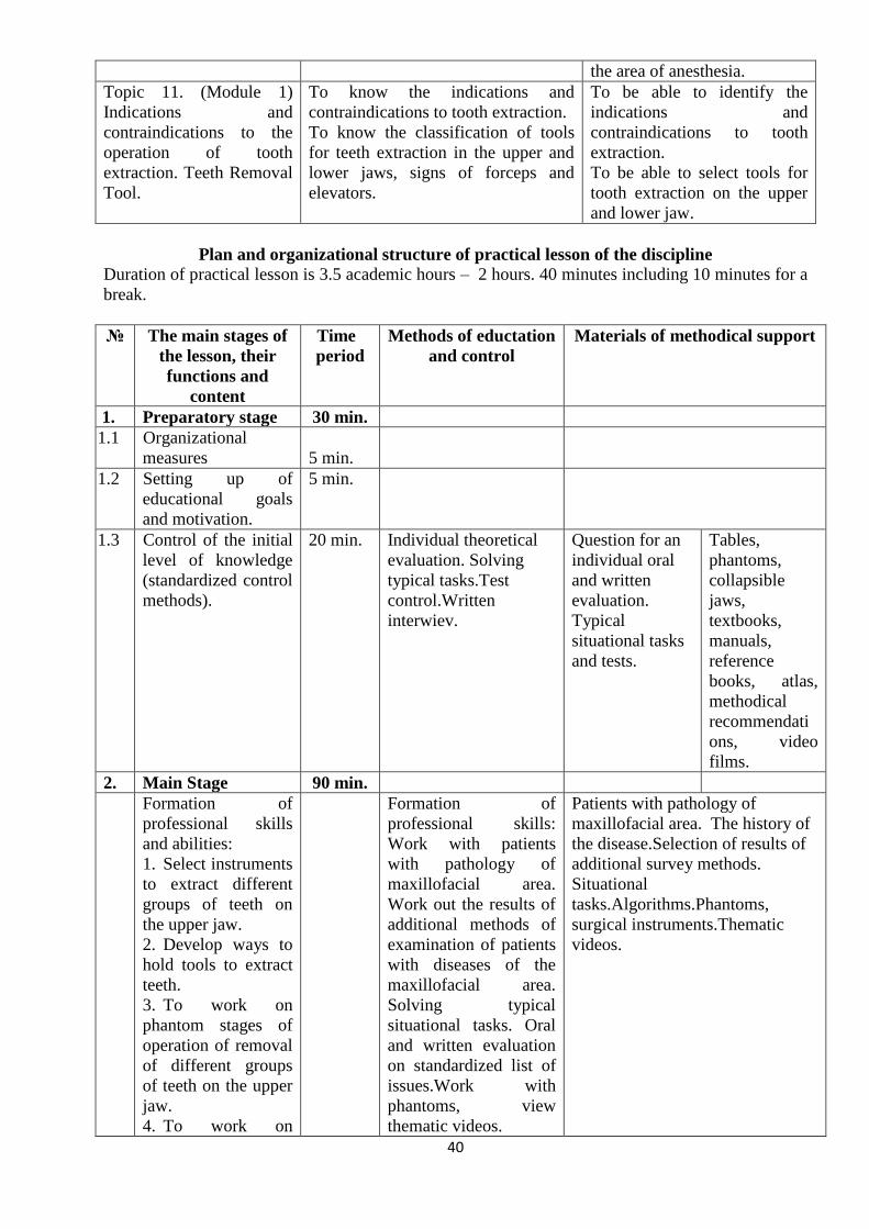
40
the area of anesthesia.
Topic 11. (Module 1)
Indications and
contraindications to the
operation of tooth
extraction. Teeth Removal
Tool.
To know the indications and
contraindications to tooth extraction.
To know the classification of tools
for teeth extraction in the upper and
lower jaws, signs of forceps and
elevators.
To be able to identify the
indications and
contraindications to tooth
extraction.
To be able to select tools for
tooth extraction on the upper
and lower jaw.
Plan and organizational structure of practical lesson of the discipline
Duration of practical lesson is 3.5 academic hours – 2 hours. 40 minutes including 10 minutes for a
break.
№ The main stages of
the lesson, their
functions and
content
Time
period
Methods of eductation
and control
Materials of methodical support
1. Preparatory stage 30 min.
1.1 Organizational
measures
5 min.
1.2 Setting up of
educational goals
and motivation.
5 min.
1.3 Control of the initial
level of knowledge
(standardized control
methods).
20 min. Individual theoretical
evaluation. Solving
typical tasks.Test
control.Written
interwiev.
Question for an
individual oral
and written
evaluation.
Typical
situational tasks
and tests.
Tables,
phantoms,
collapsible
jaws,
textbooks,
manuals,
reference
books, atlas,
methodical
recommendati
ons, video
films.
2. Main Stage 90 min.
Formation of
professional skills
and abilities:
1. Select instruments
to extract different
groups of teeth on
the upper jaw.
2. Develop ways to
hold tools to extract
teeth.
3. To work on
phantom stages of
operation of removal
of different groups
of teeth on the upper
jaw.
4. To work on
Formation of
professional skills:
Work with patients
with pathology of
maxillofacial area.
Work out the results of
additional methods of
examination of patients
with diseases of the
maxillofacial area.
Solving typical
situational tasks. Oral
and written evaluation
on standardized list of
issues.Work with
phantoms, view
thematic videos.
Patients with pathology of
maxillofacial area. The history of
the disease.Selection of results of
additional survey methods.
Situational
tasks.Algorithms.Phantoms,
surgical instruments.Thematic
videos.
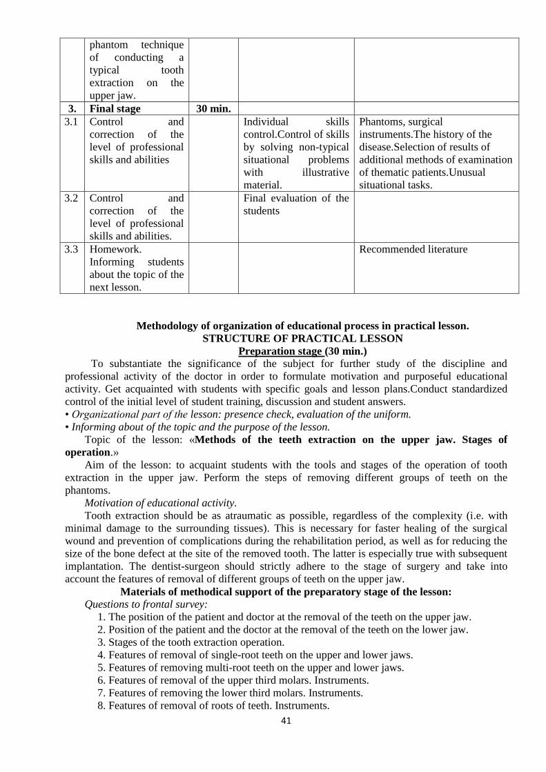
41
phantom technique
of conducting a
typical tooth
extraction on the
upper jaw.
3. Final stage 30 min.
3.1 Control and
correction of the
level of professional
skills and abilities
Individual skills
control.Control of skills
by solving non-typical
situational problems
with illustrative
material.
Phantoms, surgical
instruments.The history of the
disease.Selection of results of
additional methods of examination
of thematic patients.Unusual
situational tasks.
3.2 Control and
correction of the
level of professional
skills and abilities.
Final evaluation of the
students
3.3 Homework.
Informing students
about the topic of the
next lesson.
Recommended literature
Methodology of organization of educational process in practical lesson.
STRUCTURE OF PRACTICAL LESSON
Preparation stage (30 min.)
To substantiate the significance of the subject for further study of the discipline and
professional activity of the doctor in order to formulate motivation and purposeful educational
activity. Get acquainted with students with specific goals and lesson plans.Conduct standardized
control of the initial level of student training, discussion and student answers.
• Organizational part of the lesson: presence check, evaluation of the uniform.
• Informing about of the topic and the purpose of the lesson.
Topic of the lesson: «Methods of the teeth extraction on the upper jaw. Stages of
operation.»
Aim of the lesson: to acquaint students with the tools and stages of the operation of tooth
extraction in the upper jaw. Perform the steps of removing different groups of teeth on the
phantoms.
Motivation of educational activity.
Tooth extraction should be as atraumatic as possible, regardless of the complexity (i.e. with
minimal damage to the surrounding tissues). This is necessary for faster healing of the surgical
wound and prevention of complications during the rehabilitation period, as well as for reducing the
size of the bone defect at the site of the removed tooth. The latter is especially true with subsequent
implantation. The dentist-surgeon should strictly adhere to the stage of surgery and take into
account the features of removal of different groups of teeth on the upper jaw.
Materials of methodical support of the preparatory stage of the lesson:
Questions to frontal survey:
1. The position of the patient and doctor at the removal of the teeth on the upper jaw.
2. Position of the patient and the doctor at the removal of the teeth on the lower jaw.
3. Stages of the tooth extraction operation.
4. Features of removal of single-root teeth on the upper and lower jaws.
5. Features of removing multi-root teeth on the upper and lower jaws.
6. Features of removal of the upper third molars. Instruments.
7. Features of removing the lower third molars. Instruments.
8. Features of removal of roots of teeth. Instruments.
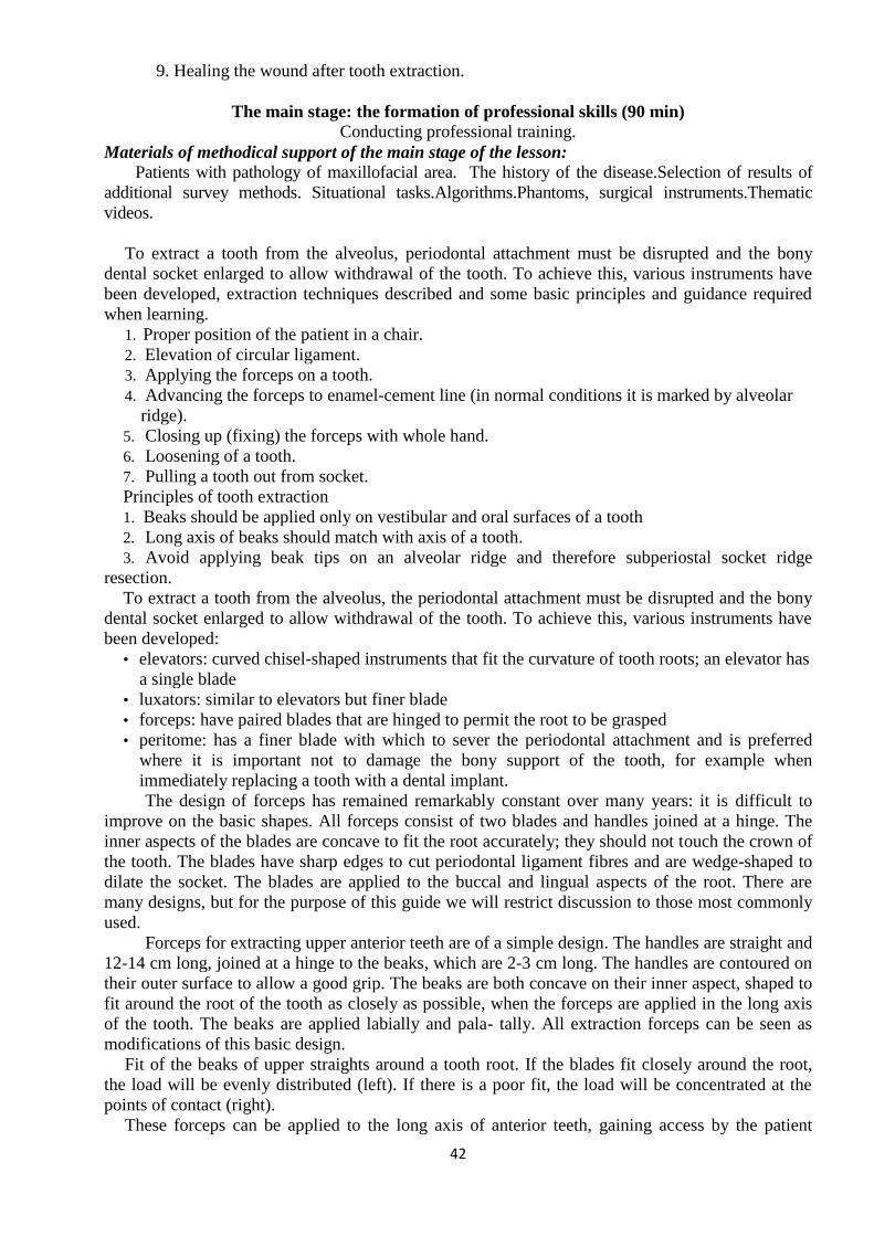
42
9. Healing the wound after tooth extraction.
The main stage: the formation of professional skills (90 min)
Conducting professional training.
Materials of methodical support of the main stage of the lesson:
Patients with pathology of maxillofacial area. The history of the disease.Selection of results of
additional survey methods. Situational tasks.Algorithms.Phantoms, surgical instruments.Thematic
videos.
To extract a tooth from the alveolus, periodontal attachment must be disrupted and the bony
dental socket enlarged to allow withdrawal of the tooth. To achieve this, various instruments have
been developed, extraction techniques described and some basic principles and guidance required
when learning.
1. Proper position of the patient in a chair.
2. Elevation of circular ligament.
3. Applying the forceps on a tooth.
4. Advancing the forceps to enamel-cement line (in normal conditions it is marked by alveolar
ridge).
5. Closing up (fixing) the forceps with whole hand.
6. Loosening of a tooth.
7. Pulling a tooth out from socket.
Principles of tooth extraction
1. Beaks should be applied only on vestibular and oral surfaces of a tooth
2. Long axis of beaks should match with axis of a tooth.
3. Avoid applying beak tips on an alveolar ridge and therefore subperiostal socket ridge
resection.
To extract a tooth from the alveolus, the periodontal attachment must be disrupted and the bony
dental socket enlarged to allow withdrawal of the tooth. To achieve this, various instruments have
been developed:
• elevators: curved chisel-shaped instruments that fit the curvature of tooth roots; an elevator has
a single blade
• luxators: similar to elevators but finer blade
• forceps: have paired blades that are hinged to permit the root to be grasped
• peritome: has a finer blade with which to sever the periodontal attachment and is preferred
where it is important not to damage the bony support of the tooth, for example when
immediately replacing a tooth with a dental implant.
The design of forceps has remained remarkably constant over many years: it is difficult to
improve on the basic shapes. All forceps consist of two blades and handles joined at a hinge. The
inner aspects of the blades are concave to fit the root accurately; they should not touch the crown of
the tooth. The blades have sharp edges to cut periodontal ligament fibres and are wedge-shaped to
dilate the socket. The blades are applied to the buccal and lingual aspects of the root. There are
many designs, but for the purpose of this guide we will restrict discussion to those most commonly
used.
Forceps for extracting upper anterior teeth are of a simple design. The handles are straight and
12-14 cm long, joined at a hinge to the beaks, which are 2-3 cm long. The handles are contoured on
their outer surface to allow a good grip. The beaks are both concave on their inner aspect, shaped to
fit around the root of the tooth as closely as possible, when the forceps are applied in the long axis
of the tooth. The beaks are applied labially and pala- tally. All extraction forceps can be seen as
modifications of this basic design.
Fit of the beaks of upper straights around a tooth root. If the blades fit closely around the root,
the load will be evenly distributed (left). If there is a poor fit, the load will be concentrated at the
points of contact (right).
These forceps can be applied to the long axis of anterior teeth, gaining access by the patient
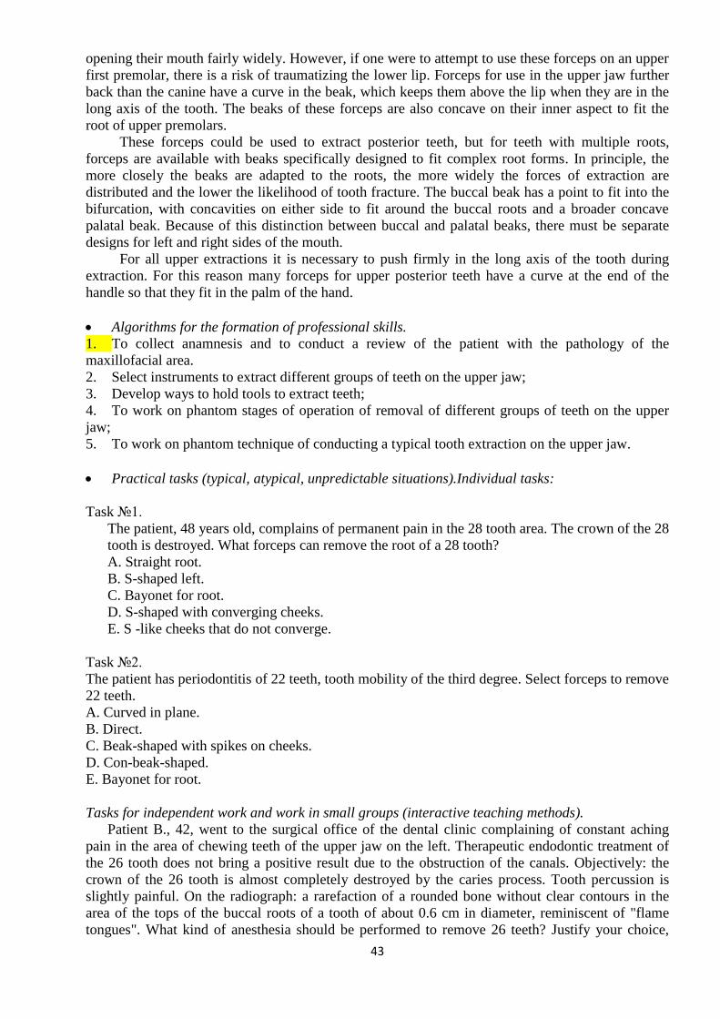
43
opening their mouth fairly widely. However, if one were to attempt to use these forceps on an upper
first premolar, there is a risk of traumatizing the lower lip. Forceps for use in the upper jaw further
back than the canine have a curve in the beak, which keeps them above the lip when they are in the
long axis of the tooth. The beaks of these forceps are also concave on their inner aspect to fit the
root of upper premolars.
These forceps could be used to extract posterior teeth, but for teeth with multiple roots,
forceps are available with beaks specifically designed to fit complex root forms. In principle, the
more closely the beaks are adapted to the roots, the more widely the forces of extraction are
distributed and the lower the likelihood of tooth fracture. The buccal beak has a point to fit into the
bifurcation, with concavities on either side to fit around the buccal roots and a broader concave
palatal beak. Because of this distinction between buccal and palatal beaks, there must be separate
designs for left and right sides of the mouth.
For all upper extractions it is necessary to push firmly in the long axis of the tooth during
extraction. For this reason many forceps for upper posterior teeth have a curve at the end of the
handle so that they fit in the palm of the hand.
Algorithms for the formation of professional skills.
1. To collect anamnesis and to conduct a review of the patient with the pathology of the
maxillofacial area.
2. Select instruments to extract different groups of teeth on the upper jaw;
3. Develop ways to hold tools to extract teeth;
4. To work on phantom stages of operation of removal of different groups of teeth on the upper
jaw;
5. To work on phantom technique of conducting a typical tooth extraction on the upper jaw.
Practical tasks (typical, atypical, unpredictable situations).Individual tasks:
Task №1.
The patient, 48 years old, complains of permanent pain in the 28 tooth area. The crown of the 28
tooth is destroyed. What forceps can remove the root of a 28 tooth?
A. Straight root.
B. S-shaped left.
C. Bayonet for root.
D. S-shaped with converging cheeks.
E. S -like cheeks that do not converge.
Task №2.
The patient has periodontitis of 22 teeth, tooth mobility of the third degree. Select forceps to remove
22 teeth.
A. Curved in plane.
B. Direct.
C. Beak-shaped with spikes on cheeks.
D. Con-beak-shaped.
E. Bayonet for root.
Tasks for independent work and work in small groups (interactive teaching methods).
Patient B., 42, went to the surgical office of the dental clinic complaining of constant aching
pain in the area of chewing teeth of the upper jaw on the left. Therapeutic endodontic treatment of
the 26 tooth does not bring a positive result due to the obstruction of the canals. Objectively: the
crown of the 26 tooth is almost completely destroyed by the caries process. Tooth percussion is
slightly painful. On the radiograph: a rarefaction of a rounded bone without clear contours in the
area of the tops of the buccal roots of a tooth of about 0.6 cm in diameter, reminiscent of "flame
tongues". What kind of anesthesia should be performed to remove 26 teeth? Justify your choice,
44
describe and demonstrate your anesthesia technique. Which tool should you choose to remove the
root of the tooth? What is the technique of working with the chosen toolkit? What complications are
possible when removing the teeth of the upper jaw?
Final stage (30 min.)
Summing up of the lesson
Materials of methodological support of the final stage of the lesson:
Brain storm.Students demonstrate an exhaustive description of the unusual clinical situation
and offer to offer the most rational diagnostic methods.After recording all the proposed
diagnostic methods during the discussion, students choose the most rational.
Tasks for self-employment.To work on phantoms the technique of tooth extraction on upper
jaw.
Evaluation.
Conduct standardized final control using individual test tasks and questions (20 min.), Work
check (10 min.). Evaluate the student's current activities during the classroom, taking into account
standardized final control, analyze the student's progress, announce the evaluation of each student's
activity, and display it in the student attendance and student log book.An adult group at the same
time makes assessments in the record of the record of success and attendance of classes by students,
the teacher certifies them with his signature.
Brief informing the students about the topic of the next lesson and the methodical measures for
preparing for it.
Basic knowledge level:
1. Anatomical and physiological features of the structure of the jaw bones and teeth.
2. Innervation and blood supply to hard and soft facial tissues.
3. Local conductive anaesthesia on the upper jaw.
4. Classification of tools for removal of teeth on the upper jaw, signs of forceps and elevators.
List of questions to be studied by the student:
1. The position of the patient and doctor at the removal of the teeth on the upper jaw.
2. Position of the patient and the doctor at the removal of the teeth on the lower jaw.
3. Stages of the tooth extraction operation.
4. Features of removal of single-root teeth on the upper and lower jaws.
5. Features of removing multi-root teeth on the upper and lower jaws.
6. Features of removal of the upper third molars. Instruments.
7. Features of removing the lower third molars. Instruments.
8. Features of removal of roots of teeth. Instruments.
9. Healing the wound after tooth extraction.
The list of practical skills to be learned by the student:
1. Select instruments to extract different groups of teeth on the upper jaw.
2. Develop ways to hold tools to extract teeth.
3. To work on phantom stages of operation of removal of different groups of teeth on the upper
jaw.
4. To work on phantom technique of conducting a typical tooth extraction on the upper jaw.
Situational tasks and questions on the topic of the lesson:
1. A patient of 70 years, complains about the mobility of 21 teeth. The tooth has not been treated
before. Objectively: 21 have grade II motility, the root is exposed by 1/2. Which tool should be used
for tooth extraction?
A. S-shaped forceps
B. Streight elevator
45
C. Beak-shaped forceps
D. Straight forceps
E. Bognet-like forceps
2. Patient 35 years old, applied for the removal of 14 teeth due to exacerbation of chronic
periodontitis after unsuccessful therapeutic treatment. What is your choice of removal tools?
A. S-shaped right forceps
B. Bognet-shaped crown forceps
C. Bognetic root forceps
D. Straight forseps
E. S-shaped forceps
3. A 56-year-old woman went to a dentist to remove 17 teeth. Which removal tool should I choose?
A. Bognetic root forceps
B. Bognet-shaped crown forceps
C. S-shaped right forceps
D. S-shaped left forceps
E. Straight forceps
4. A 49-year-old patient consulted a dental surgeon about the oral cavity sanation. He has an
indication for the extraction of the 16 tooth. History: the tooth hasn’t been treated before, decayed
within the last 4 years. Objectively: the 16 tooth’s crown is destroyed by over 2/3, the mucosa
exhibits no pathological changes. Which tool is required for the tooth extraction?
A. Bayonet root forceps
B. S-shaped forceps (right)
C. Straight elevator
D. S-shaped closed-beak forceps
E. Crown bayonet forceps
5. Patient 30 years old, went to the doctor to remove 26 teeth about exacerbation of chronic
periodontitis. Objectively: crown 26 is destroyed by 1/3. What forceps can be used to remove this
tooth?
A. S-shaped with a spike on the left cheek
B. Streight elevator
C. S-shaped without spikes
D. S-shaped with a spike on the right cheek
E. Straight forceps.
Literature:
Basic:
1. Oral and Maxillofacial Surgery: Textbook, Part 1, 2 / V.O. Malanchuk. – Vinnytsia: Nova
Knyha Publishers, 2011. – 453p.
2. Principles of Dental Local Anaesthesia and Teeth Removal / Ya. E. Vares, R. Z. Ogonovsky,
Ch. R. Pohranychna – LNMU, 2007. – 63p.
3. Atlas of Human Anatomy / F. Netter – 2nd
ed. – New Jersey: ICON Learning Systems. – 592
p.
Additional:
1. Contemporary Oral and Maxillofacial Surgery / L. J. Peterson, E. Ellis, J. R. Hupp, M.R.
Tucker – 3rd ed. – St. Louis: Mosby – Year Book, Inc. – 1998. – 1477 p.
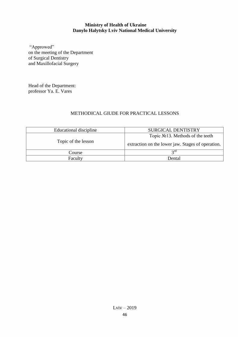
46
Ministry of Health of Ukraine
Danylo Halytsky Lviv National Medical University
“Approwed”
on the meeting of the Department
of Surgical Dentistry
and Maxillofacial Surgery
Head of the Department:
professor Ya. E. Vares
METHODICAL GIUDE FOR PRACTICAL LESSONS
Educational discipline SURGICAL DENTISTRY
Topic of the lesson
Topic №13. Methods of the teeth
extraction on the lower jaw. Stages of operation.
Course 3rd
Faculty Dental
Lviv – 2019
47
Actuality of the topic: Tooth extraction should be as atraumatic as possible, regardless of the
complexity (i.e. with minimal damage to the surrounding tissues). This is necessary for faster
healing of the surgical wound and prevention of complications during the rehabilitation period, as
well as for reducing the size of the bone defect at the site of the removed tooth. The latter is
especially true with subsequent implantation. The dentist-surgeon should strictly adhere to the stage
of surgery and take into account the features of removal of different groups of teeth on the lower
jaws.
Aim of the lesson: to acquaint students with the tools and stages of the operation of tooth extraction
in the lower jaw. Perform the steps of removing different groups of teeth on the phantoms.
Learning objectives:
Professional competence:
1. Collection of medical information on the patient's condition.
2. Evaluation of the results of laboratory and instrumental research.
3. Establishment of a clinical diagnosis of dental disease.
4. Planning and conducting preventive measures for dental diseases.
5. Execution of medical and dental manipulations.
6. Organization and conducting of dental medical examination of persons subject to dispensary
supervision.
7. Assessment of the environmental impact on the health of the population (individual, family,
population).
8. Maintaining medical records.
9. Processing of state, social and medical information.
General competence:
1. The ability to abstract thinking, analysis and synthesis; the ability to learn and be trained
today.
2. Knowledge and understanding of the subject area and understanding of the profession.
3. Ability to apply knowledge in practical situations.
4. Ability to communicate in the state language both verbally and in writing; Ability to
communicate in a second language.
5. Skills in the use of information and communication technologies.
6. Ability to search, process and analyze information from various sources.
7. Ability to adapt and act in a new situation; ability to work autonomously.
8. Ability to identify, put and solve problems.
9. Ability to choose a communication strategy.
10. Ability to work in a team.
11. Interpersonal skills.
12. Ability to act on the basis of ethical considerations (motives).
13. Ability to act in a socially responsible and civic conscious manner.
Methods of training:
Preparatory stage - Frontal oral interview.
The main stage - practical training, role-playing game.
The final stage is brainstorming.
Interdisciplinary integration
Disciplines Student should know Student should be able to
Normal anatomy
Know the anatomical and
physiological features of the
maxillofacial area:
- structure of the upper and lower
To be able to explain the
structure of systems and organs
of maxillo-facial area (MFA)
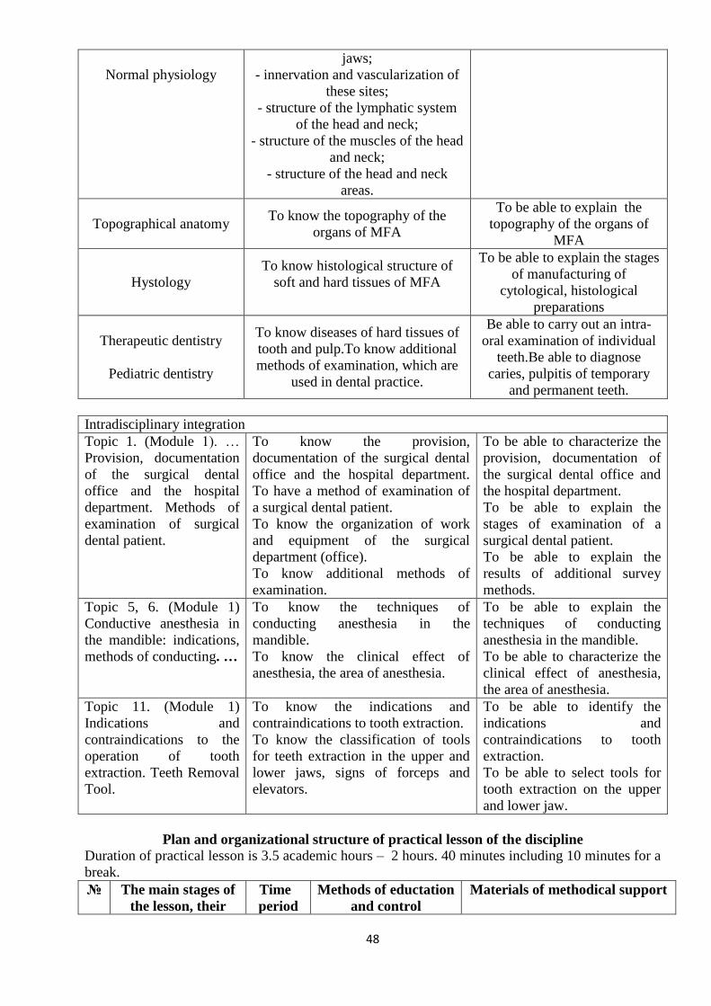
48
Normal physiology
jaws;
- innervation and vascularization of
these sites;
- structure of the lymphatic system
of the head and neck;
- structure of the muscles of the head
and neck;
- structure of the head and neck
areas.
Topographical anatomy To know the topography of the
organs of MFA
To be able to explain the
topography of the organs of
MFA
Hystology
To know histological structure of
soft and hard tissues of MFA
To be able to explain the stages
of manufacturing of
cytological, histological
preparations
Therapeutic dentistry
Pediatric dentistry
To know diseases of hard tissues of
tooth and pulp.To know additional
methods of examination, which are
used in dental practice.
Be able to carry out an intra-
oral examination of individual
teeth.Be able to diagnose
caries, pulpitis of temporary
and permanent teeth.
Intradisciplinary integration
Topic 1. (Module 1). …
Provision, documentation
of the surgical dental
office and the hospital
department. Methods of
examination of surgical
dental patient.
To know the provision,
documentation of the surgical dental
office and the hospital department.
To have a method of examination of
a surgical dental patient.
To know the organization of work
and equipment of the surgical
department (office).
To know additional methods of
examination.
To be able to characterize the
provision, documentation of
the surgical dental office and
the hospital department.
To be able to explain the
stages of examination of a
surgical dental patient.
To be able to explain the
results of additional survey
methods.
Topic 5, 6. (Module 1)
Conductive anesthesia in
the mandible: indications,
methods of conducting. …
To know the techniques of
conducting anesthesia in the
mandible.
To know the clinical effect of
anesthesia, the area of anesthesia.
To be able to explain the
techniques of conducting
anesthesia in the mandible.
To be able to characterize the
clinical effect of anesthesia,
the area of anesthesia.
Topic 11. (Module 1)
Indications and
contraindications to the
operation of tooth
extraction. Teeth Removal
Tool.
To know the indications and
contraindications to tooth extraction.
To know the classification of tools
for teeth extraction in the upper and
lower jaws, signs of forceps and
elevators.
To be able to identify the
indications and
contraindications to tooth
extraction.
To be able to select tools for
tooth extraction on the upper
and lower jaw.
Plan and organizational structure of practical lesson of the discipline
Duration of practical lesson is 3.5 academic hours – 2 hours. 40 minutes including 10 minutes for a
break.
№ The main stages of
the lesson, their
Time
period
Methods of eductation
and control
Materials of methodical support
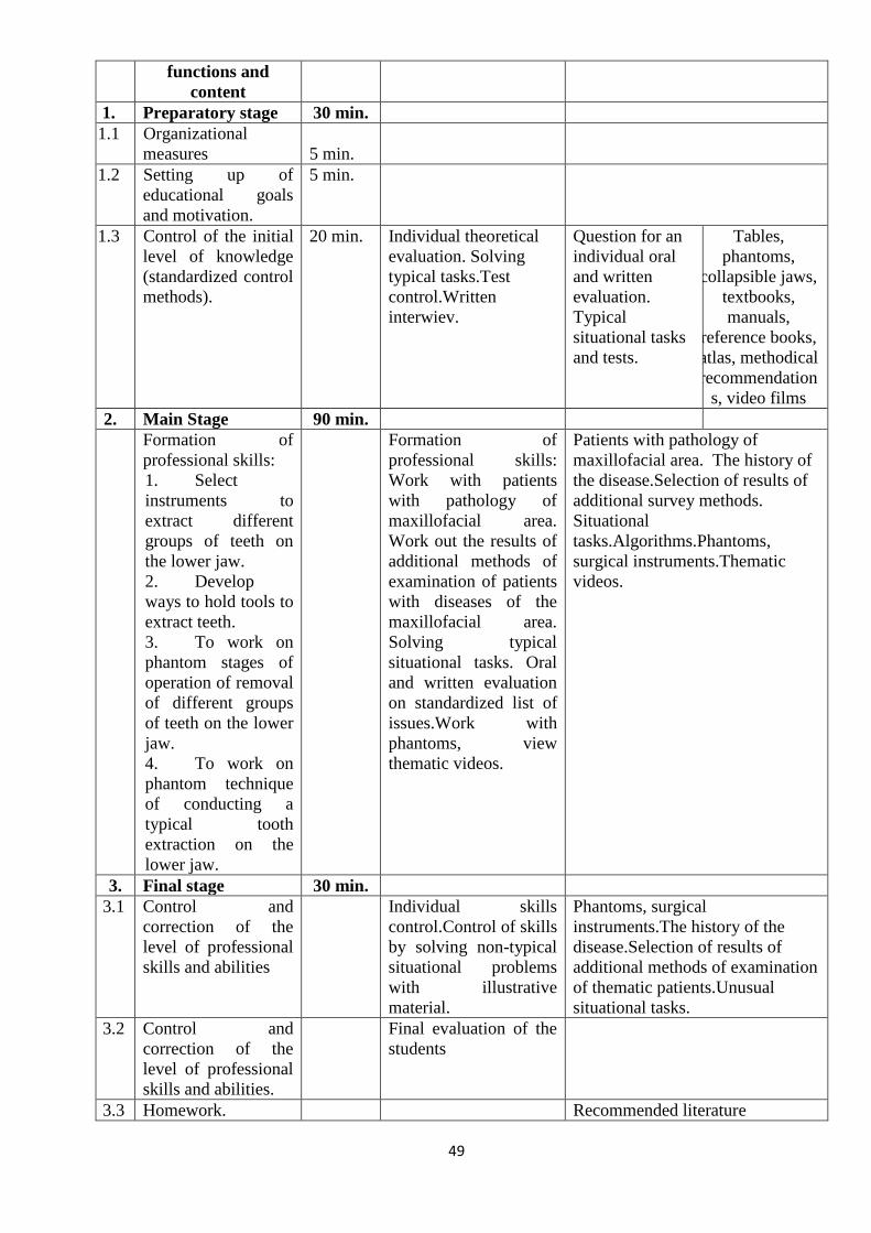
49
functions and
content
1. Preparatory stage 30 min.
1.1 Organizational
measures
5 min.
1.2 Setting up of
educational goals
and motivation.
5 min.
1.3 Control of the initial
level of knowledge
(standardized control
methods).
20 min. Individual theoretical
evaluation. Solving
typical tasks.Test
control.Written
interwiev.
Question for an
individual oral
and written
evaluation.
Typical
situational tasks
and tests.
Tables,
phantoms,
collapsible jaws,
textbooks,
manuals,
reference books,
atlas, methodical
recommendation
s, video films
2. Main Stage 90 min.
Formation of
professional skills:
1. Select
instruments to
extract different
groups of teeth on
the lower jaw.
2. Develop
ways to hold tools to
extract teeth.
3. To work on
phantom stages of
operation of removal
of different groups
of teeth on the lower
jaw.
4. To work on
phantom technique
of conducting a
typical tooth
extraction on the
lower jaw.
Formation of
professional skills:
Work with patients
with pathology of
maxillofacial area.
Work out the results of
additional methods of
examination of patients
with diseases of the
maxillofacial area.
Solving typical
situational tasks. Oral
and written evaluation
on standardized list of
issues.Work with
phantoms, view
thematic videos.
Patients with pathology of
maxillofacial area. The history of
the disease.Selection of results of
additional survey methods.
Situational
tasks.Algorithms.Phantoms,
surgical instruments.Thematic
videos.
3. Final stage 30 min.
3.1 Control and
correction of the
level of professional
skills and abilities
Individual skills
control.Control of skills
by solving non-typical
situational problems
with illustrative
material.
Phantoms, surgical
instruments.The history of the
disease.Selection of results of
additional methods of examination
of thematic patients.Unusual
situational tasks.
3.2 Control and
correction of the
level of professional
skills and abilities.
Final evaluation of the
students
3.3 Homework. Recommended literature
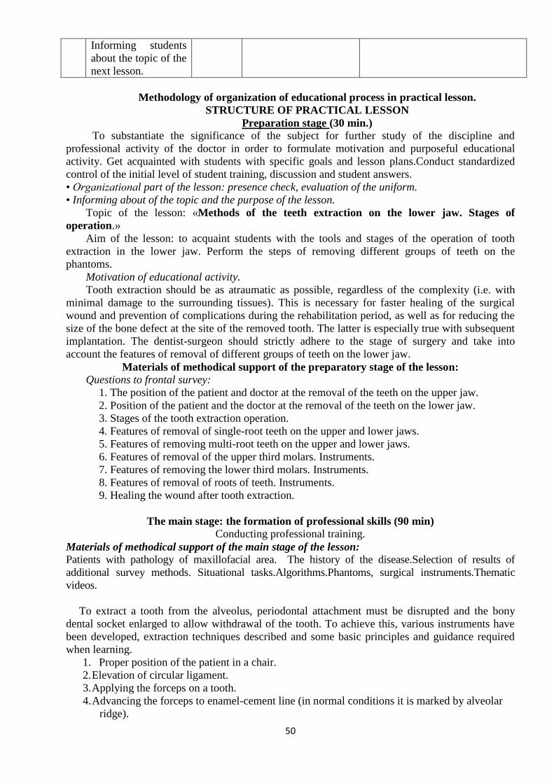
50
Informing students
about the topic of the
next lesson.
Methodology of organization of educational process in practical lesson.
STRUCTURE OF PRACTICAL LESSON
Preparation stage (30 min.)
To substantiate the significance of the subject for further study of the discipline and
professional activity of the doctor in order to formulate motivation and purposeful educational
activity. Get acquainted with students with specific goals and lesson plans.Conduct standardized
control of the initial level of student training, discussion and student answers.
• Organizational part of the lesson: presence check, evaluation of the uniform.
• Informing about of the topic and the purpose of the lesson.
Topic of the lesson: «Methods of the teeth extraction on the lower jaw. Stages of
operation.»
Aim of the lesson: to acquaint students with the tools and stages of the operation of tooth
extraction in the lower jaw. Perform the steps of removing different groups of teeth on the
phantoms.
Motivation of educational activity.
Tooth extraction should be as atraumatic as possible, regardless of the complexity (i.e. with
minimal damage to the surrounding tissues). This is necessary for faster healing of the surgical
wound and prevention of complications during the rehabilitation period, as well as for reducing the
size of the bone defect at the site of the removed tooth. The latter is especially true with subsequent
implantation. The dentist-surgeon should strictly adhere to the stage of surgery and take into
account the features of removal of different groups of teeth on the lower jaw.
Materials of methodical support of the preparatory stage of the lesson:
Questions to frontal survey:
1. The position of the patient and doctor at the removal of the teeth on the upper jaw.
2. Position of the patient and the doctor at the removal of the teeth on the lower jaw.
3. Stages of the tooth extraction operation.
4. Features of removal of single-root teeth on the upper and lower jaws.
5. Features of removing multi-root teeth on the upper and lower jaws.
6. Features of removal of the upper third molars. Instruments.
7. Features of removing the lower third molars. Instruments.
8. Features of removal of roots of teeth. Instruments.
9. Healing the wound after tooth extraction.
The main stage: the formation of professional skills (90 min)
Conducting professional training.
Materials of methodical support of the main stage of the lesson:
Patients with pathology of maxillofacial area. The history of the disease.Selection of results of
additional survey methods. Situational tasks.Algorithms.Phantoms, surgical instruments.Thematic
videos.
To extract a tooth from the alveolus, periodontal attachment must be disrupted and the bony
dental socket enlarged to allow withdrawal of the tooth. To achieve this, various instruments have
been developed, extraction techniques described and some basic principles and guidance required
when learning.
1. Proper position of the patient in a chair.
2. Elevation of circular ligament.
3. Applying the forceps on a tooth.
4. Advancing the forceps to enamel-cement line (in normal conditions it is marked by alveolar
ridge).
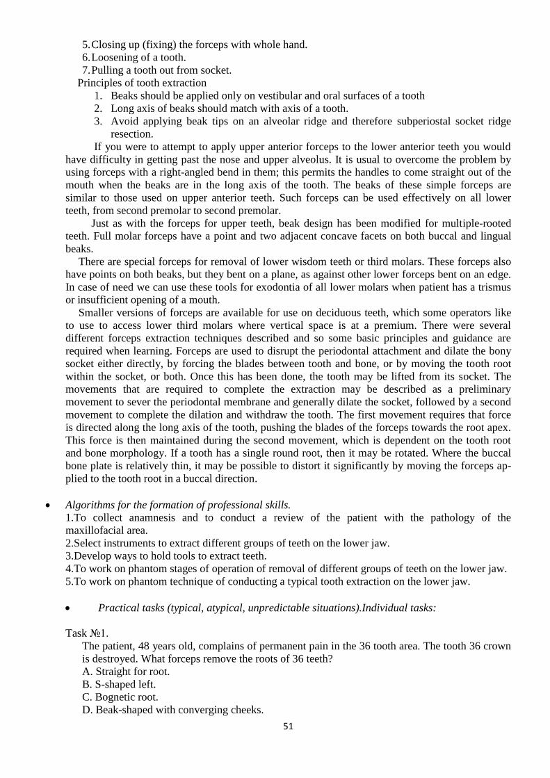
51
5. Closing up (fixing) the forceps with whole hand.
6. Loosening of a tooth.
7. Pulling a tooth out from socket.
Principles of tooth extraction
1. Beaks should be applied only on vestibular and oral surfaces of a tooth
2. Long axis of beaks should match with axis of a tooth.
3. Avoid applying beak tips on an alveolar ridge and therefore subperiostal socket ridge
resection.
If you were to attempt to apply upper anterior forceps to the lower anterior teeth you would
have difficulty in getting past the nose and upper alveolus. It is usual to overcome the problem by
using forceps with a right-angled bend in them; this permits the handles to come straight out of the
mouth when the beaks are in the long axis of the tooth. The beaks of these simple forceps are
similar to those used on upper anterior teeth. Such forceps can be used effectively on all lower
teeth, from second premolar to second premolar.
Just as with the forceps for upper teeth, beak design has been modified for multiple-rooted
teeth. Full molar forceps have a point and two adjacent concave facets on both buccal and lingual
beaks.
There are special forceps for removal of lower wisdom teeth or third molars. These forceps also
have points on both beaks, but they bent on a plane, as against other lower forceps bent on an edge.
In case of need we can use these tools for exodontia of all lower molars when patient has a trismus
or insufficient opening of a mouth.
Smaller versions of forceps are available for use on deciduous teeth, which some operators like
to use to access lower third molars where vertical space is at a premium. There were several
different forceps extraction techniques described and so some basic principles and guidance are
required when learning. Forceps are used to disrupt the periodontal attachment and dilate the bony
socket either directly, by forcing the blades between tooth and bone, or by moving the tooth root
within the socket, or both. Once this has been done, the tooth may be lifted from its socket. The
movements that are required to complete the extraction may be described as a preliminary
movement to sever the periodontal membrane and generally dilate the socket, followed by a second
movement to complete the dilation and withdraw the tooth. The first movement requires that force
is directed along the long axis of the tooth, pushing the blades of the forceps towards the root apex.
This force is then maintained during the second movement, which is dependent on the tooth root
and bone morphology. If a tooth has a single round root, then it may be rotated. Where the buccal
bone plate is relatively thin, it may be possible to distort it significantly by moving the forceps ap-
plied to the tooth root in a buccal direction.
Algorithms for the formation of professional skills.
1.To collect anamnesis and to conduct a review of the patient with the pathology of the
maxillofacial area.
2.Select instruments to extract different groups of teeth on the lower jaw.
3.Develop ways to hold tools to extract teeth.
4.To work on phantom stages of operation of removal of different groups of teeth on the lower jaw.
5.To work on phantom technique of conducting a typical tooth extraction on the lower jaw.
Practical tasks (typical, atypical, unpredictable situations).Individual tasks:
Task №1.
The patient, 48 years old, complains of permanent pain in the 36 tooth area. The tooth 36 crown
is destroyed. What forceps remove the roots of 36 teeth?
A. Straight for root.
B. S-shaped left.
C. Bognetic root.
D. Beak-shaped with converging cheeks.
52
E. Beak-shaped with non-converging cheeks.
Task №2.
The patient has periodontitis of 32 teeth, III degree. Select forceps to remove 32 teeth.
A. Curved in plane.
B. Direct.
C. Beak-shaped with spikes on cheeks.
D. Beak-shaped with non-converging cheeks.
E. Bayonet for roots.
Tasks for independent work and work in small groups (interactive teaching methods).
Patient V., 42, went to the surgical office of the dental clinic, complaining of constant aching
pain in the lower jaw body on the left. Therapeutic endodontic treatment of 36 tooth positive does
not bring about because of the obstruction of the canals. Objectively: the crown of the 36 tooth is
destroyed by a carious process by one fourth of the volume of the crown. Tooth percussion is
slightly painful. On the radiograph: a rarefaction of a rounded bone without clear contours in the
area of the apexes of the buccal roots of 36 teeth with a diameter of about 0.6 cm, resembling
“flame tongues”. What type of anesthesia should be performed to remove a 36 tooth? Justify your
choice, describe and demonstrate your anesthesia technique. Which tool should you choose to
remove the root of the tooth? What is the technique of working with the chosen toolkit? What
complications are possible when removing the teeth of the upper jaw?
Final stage (30 min.)
Summing up of the lesson
Materials of methodological support of the final stage of the lesson:
Brain storm.Students demonstrate an exhaustive description of the unusual clinical situation
and offer to offer the most rational diagnostic methods.After recording all the proposed
diagnostic methods during the discussion, students choose the most rational.
Tasks for self-employment.To work on phantoms the technique of tooth extraction on lower
jaw.
Evaluation.
Conduct standardized final control using individual test tasks and questions (20 min.), Work
check (10 min.). Evaluate the student's current activities during the classroom, taking into account
standardized final control, analyze the student's progress, announce the evaluation of each student's
activity, and display it in the student attendance and student log book.An adult group at the same
time makes assessments in the record of the record of success and attendance of classes by students,
the teacher certifies them with his signature.
Brief informing the students about the topic of the next lesson and the methodical measures for
preparing for it.
Basic knowledge level:
1. Anatomical and physiological features of the structure of the jaw bones and teeth.
2. Innervation and blood supply to hard and soft facial tissues.
3. Local conductive anaesthesia on the lower jaw.
4. Classification of tools for removal of teeth on the lower jaw, signs of forceps and elevators.
List of questions to be studied by the student:
1. The position of the patient and doctor at the removal of the teeth on the upper jaw.
2. Position of the patient and the doctor at the removal of the teeth on the lower jaw.
3. Stages of the tooth extraction operation.
4. Features of removal of single-root teeth on the upper and lower jaws.
5. Features of removing multi-root teeth on the upper and lower jaws.
6. Features of removal of the upper third molars. Instruments.
53
7. Features of removing the lower third molars. Instruments.
8. Features of removal of roots of teeth. Instruments.
9. Healing the wound after tooth extraction.
The list of practical skills to be learned by the student:
1. Select instruments to extract different groups of teeth on the lower jaw.
2. Develop ways to hold tools to extract teeth.
3. To work on phantom stages of operation of removal of different groups of teeth on the lower
jaw.
4. To work on phantom technique of conducting a typical tooth extraction on the lower jaw.
Situational tasks and questions on the topic of the lesson:
1. During removal of the 47 teeth, the distal root remained in the cell. Select the tool to complete the
uninstallation.
A. Angular left-hand elevator
B. Angular right-hand elevator
C. Streight elevator
D. Universal forceps-bayonet
E. Beak-shaped forceps for root removal
2. In the clinic of surgical dentistry, the patient is shown removal of the root of the 36 tooth, whose
crown is missing, the medial root is removed. The distal root is deep in the hole. Select a tool to
remove the distal root of tooth 36:
A. The Lecluse Elevator
B. Left-Angled elevator
C. Beak-shaped root forceps
D. Streight elevator
E. Right-Angled elevator
3.During extraction of the 47 tooth its distal root was broken halfway along its length. What tool
should be chosen for extraction of the residual root fragments?
A. Left angled elevator
B. Broad-beaked forceps
C. Close-beaked forceps
D. Right angled elevator
E. Straight elevator
4. In the surgical dental office the patient complained of the presence of the roots of the destroyed
37 tooth. What tools do you need to use to remove the root of the 37 tooth?
A. Beak-shaped forceps, root, angular elevators
B. Plane-curved forceps, corner elevators
C. Bognet-like forceps, straight elevator
D. S-shaped forceps, straight and angular elevators
E. Beak-shaped forceps, crown, straight elevator
5. A 27-year-old male patient underwent extraction of the medial root of the 36 tooth, but the
distal tooth root was broken in its middle third. What tools should be applied for root extraction?
A. Left angled elevator
B. Right angled elevator
C. Straight elevator
D. Broad-beaked forceps
E. Close-beaked forceps

54
Literature:
Basic:
1. Oral and Maxillofacial Surgery: Textbook, Part 1, 2 / V.O. Malanchuk. – Vinnytsia: Nova
Knyha Publishers, 2011. – 453p.
2. Principles of Dental Local Anaesthesia and Teeth Removal / Ya. E. Vares, R. Z. Ogonovsky,
Ch. R. Pohranychna – LNMU, 2007. – 63p.
3. Atlas of Human Anatomy / F. Netter – 2nd
ed. – New Jersey: ICON Learning Systems. – 592
p.
Additional:
1. Contemporary Oral and Maxillofacial Surgery / L. J. Peterson, E. Ellis, J. R. Hupp, M.R.
Tucker – 3rd ed. – St. Louis: Mosby – Year Book, Inc. – 1998. – 1477 p.
55
Ministry of Health of Ukraine
Danylo Halytsky Lviv National Medical University
“Approwed”
on the meeting of the Department
of Surgical Dentistry
and Maxillofacial Surgery
Head of the Department:
professor Ya. E. Vares
METHODICAL GIUDE FOR PRACTICAL LESSONS
Educational discipline SURGICAL DENTISTRY
Topic of the lesson
Topic №14. Complications during tooth
extraction: clinical picture, diagnosis, treatment
and prevention.
Course 3rd
Faculty Dental
Lviv – 2019
56
Actuality of the topic: During the operation of tooth extraction there can be a number of general
and local complications related to the peculiarities of the anatomical structure of the dental-
maxillofacial system, significant pathological changes of the tissues of the maxillofacial area, the
presence of concomitant general somatic pathology, as well as iatrogenic nature (associated with
errors in diagnosis, non-observance of surgery, lack of clinical experience, etc.). Anamnesis
collection, individual approach to the patient, careful adherence to surgery techniques, as a rule,
allow to avoid negative consequences. However, in the event of a complication, the dentist is
obliged to quickly diagnose it, provide the patient with urgent care and plan a follow-up treatment.
Aim of the lesson: to acquaint students with the methods of diagnosis and treatment of
complications that occur during tooth extraction and the postoperative period. Work out algorithms
for helping patients.
Learning objectives:
Professional competence:
1. Collection of medical information on the patient's condition.
2. Evaluation of the results of laboratory and instrumental research.
3. Establishment of a clinical diagnosis of dental disease.
4. Planning and conducting preventive measures for dental diseases.
5. Execution of medical and dental manipulations.
6. Organization and conducting of dental medical examination of persons subject to dispensary
supervision.
7. Assessment of the environmental impact on the health of the population (individual, family,
population).
8. Maintaining medical records.
9. Processing of state, social and medical information.
General competence:
1. The ability to abstract thinking, analysis and synthesis; the ability to learn and be trained
today.
2. Knowledge and understanding of the subject area and understanding of the profession.
3. Ability to apply knowledge in practical situations.
4. Ability to communicate in the state language both verbally and in writing; Ability to
communicate in a second language.
5. Skills in the use of information and communication technologies.
6. Ability to search, process and analyze information from various sources.
7. Ability to adapt and act in a new situation; ability to work autonomously.
8. Ability to identify, put and solve problems.
9. Ability to choose a communication strategy.
10. Ability to work in a team.
11. Interpersonal skills.
12. Ability to act on the basis of ethical considerations (motives).
13. Ability to act in a socially responsible and civic conscious manner.
Methods of training:
Preparatory stage - Frontal oral interview.
The main stage - practical training, role-playing game.
The final stage is brainstorming.
Interdisciplinary integration
Disciplines Student should know Student should be able to
Normal anatomy
Know the anatomical and
physiological features of the
To be able to explain the
structure of systems and organs
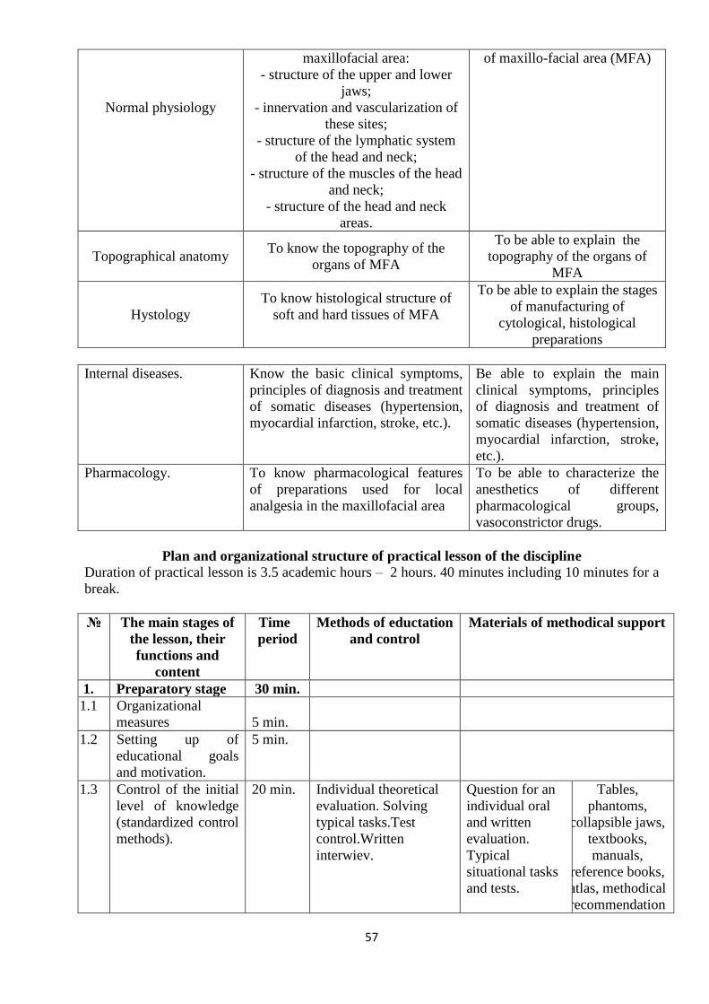
57
Normal physiology
maxillofacial area:
- structure of the upper and lower
jaws;
- innervation and vascularization of
these sites;
- structure of the lymphatic system
of the head and neck;
- structure of the muscles of the head
and neck;
- structure of the head and neck
areas.
of maxillo-facial area (MFA)
Topographical anatomy To know the topography of the
organs of MFA
To be able to explain the
topography of the organs of
MFA
Hystology
To know histological structure of
soft and hard tissues of MFA
To be able to explain the stages
of manufacturing of
cytological, histological
preparations
Internal diseases. Know the basic clinical symptoms,
principles of diagnosis and treatment
of somatic diseases (hypertension,
myocardial infarction, stroke, etc.).
Be able to explain the main
clinical symptoms, principles
of diagnosis and treatment of
somatic diseases (hypertension,
myocardial infarction, stroke,
etc.).
Pharmacology. To know pharmacological features
of preparations used for local
analgesia in the maxillofacial area
To be able to characterize the
anesthetics of different
pharmacological groups,
vasoconstrictor drugs.
Plan and organizational structure of practical lesson of the discipline
Duration of practical lesson is 3.5 academic hours – 2 hours. 40 minutes including 10 minutes for a
break.
№ The main stages of
the lesson, their
functions and
content
Time
period
Methods of eductation
and control
Materials of methodical support
1. Preparatory stage 30 min.
1.1 Organizational
measures
5 min.
1.2 Setting up of
educational goals
and motivation.
5 min.
1.3 Control of the initial
level of knowledge
(standardized control
methods).
20 min. Individual theoretical
evaluation. Solving
typical tasks.Test
control.Written
interwiev.
Question for an
individual oral
and written
evaluation.
Typical
situational tasks
and tests.
Tables,
phantoms,
collapsible jaws,
textbooks,
manuals,
reference books,
atlas, methodical
recommendation
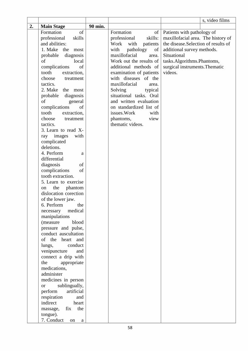
58
s, video films
2. Main Stage 90 min.
Formation of
professional skills
and abilities:
1. Make the most
probable diagnosis
of local
complications of
tooth extraction,
choose treatment
tactics.
2. Make the most
probable diagnosis
of general
complications of
tooth extraction,
choose treatment
tactics.
3. Learn to read X-
ray images with
complicated
deletions.
4. Perform a
differential
diagnosis of
complications of
tooth extraction.
5. Learn to exercise
on the phantom
dislocation corection
of the lower jaw.
6. Perform the
necessary medical
manipulations
(measure blood
pressure and pulse,
conduct auscultation
of the heart and
lungs, conduct
venipuncture and
connect a drip with
the appropriate
medications,
administer
medicines in person
or sublingually,
perform artificial
respiration and
indirect heart
massage, fix the
tongue).
7. Conduct on a
Formation of
professional skills:
Work with patients
with pathology of
maxillofacial area.
Work out the results of
additional methods of
examination of patients
with diseases of the
maxillofacial area.
Solving typical
situational tasks. Oral
and written evaluation
on standardized list of
issues.Work with
phantoms, view
thematic videos.
Patients with pathology of
maxillofacial area. The history of
the disease.Selection of results of
additional survey methods.
Situational
tasks.Algorithms.Phantoms,
surgical instruments.Thematic
videos.
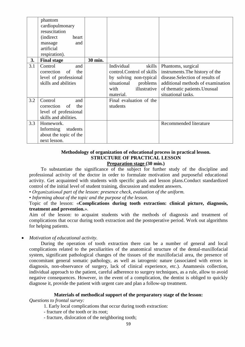
59
phantom
cardiopulmonary
resuscitation
(indirect heart
massage and
artificial
respiration).
3. Final stage 30 min.
3.1 Control and
correction of the
level of professional
skills and abilities
Individual skills
control.Control of skills
by solving non-typical
situational problems
with illustrative
material.
Phantoms, surgical
instruments.The history of the
disease.Selection of results of
additional methods of examination
of thematic patients.Unusual
situational tasks.
3.2 Control and
correction of the
level of professional
skills and abilities.
Final evaluation of the
students
3.3 Homework.
Informing students
about the topic of the
next lesson.
Recommended literature
Methodology of organization of educational process in practical lesson.
STRUCTURE OF PRACTICAL LESSON
Preparation stage (30 min.)
To substantiate the significance of the subject for further study of the discipline and
professional activity of the doctor in order to formulate motivation and purposeful educational
activity. Get acquainted with students with specific goals and lesson plans.Conduct standardized
control of the initial level of student training, discussion and student answers.
• Organizational part of the lesson: presence check, evaluation of the uniform.
• Informing about of the topic and the purpose of the lesson.
Topic of the lesson: «Complications during tooth extraction: clinical picture, diagnosis,
treatment and prevention.».
Aim of the lesson: to acquaint students with the methods of diagnosis and treatment of
complications that occur during tooth extraction and the postoperative period. Work out algorithms
for helping patients.
Motivation of educational activity.
During the operation of tooth extraction there can be a number of general and local
complications related to the peculiarities of the anatomical structure of the dental-maxillofacial
system, significant pathological changes of the tissues of the maxillofacial area, the presence of
concomitant general somatic pathology, as well as iatrogenic nature (associated with errors in
diagnosis, non-observance of surgery, lack of clinical experience, etc.). Anamnesis collection,
individual approach to the patient, careful adherence to surgery techniques, as a rule, allow to avoid
negative consequences. However, in the event of a complication, the dentist is obliged to quickly
diagnose it, provide the patient with urgent care and plan a follow-up treatment.
Materials of methodical support of the preparatory stage of the lesson:
Questions to frontal survey:
1. Early local complications that occur during tooth extraction:
- fracture of the tooth or its root;
- fracture, dislocation of the neighboring tooth;
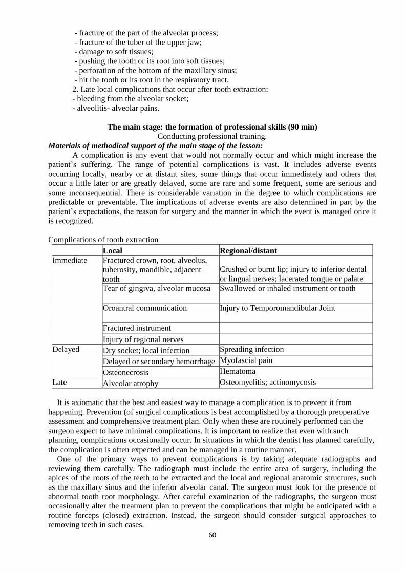
60
- fracture of the part of the alveolar process;
- fracture of the tuber of the upper jaw;
- damage to soft tissues;
- pushing the tooth or its root into soft tissues;
- perforation of the bottom of the maxillary sinus;
- hit the tooth or its root in the respiratory tract.
2. Late local complications that occur after tooth extraction:
- bleeding from the alveolar socket;
- alveolitis- alveolar pains.
The main stage: the formation of professional skills (90 min)
Conducting professional training.
Materials of methodical support of the main stage of the lesson:
A complication is any event that would not normally occur and which might increase the
patient’s suffering. The range of potential complications is vast. It includes adverse events
occurring locally, nearby or at distant sites, some things that occur immediately and others that
occur a little later or are greatly delayed, some are rare and some frequent, some are serious and
some inconsequential. There is considerable variation in the degree to which complications are
predictable or preventable. The implications of adverse events are also determined in part by the
patient’s expectations, the reason for surgery and the manner in which the event is managed once it
is recognized.
Complications of tooth extraction
Local Regional/distant
Immediate Fractured crown, root, alveolus,
tuberosity, mandible, adjacent
tooth
Crushed or burnt lip; injury to inferior dental
or lingual nerves; lacerated tongue or palate
Tear of gingiva, alveolar mucosa Swallowed or inhaled instrument or tooth
Oroantral communication Injury to Temporomandibular Joint
Fractured instrument
Injury of regional nerves
Delayed Dry socket; local infection Spreading infection
Delayed or secondary hemorrhage Myofascial pain
Osteonecrosis Hematoma
Late Alveolar atrophy Osteomyelitis; actinomycosis
It is axiomatic that the best and easiest way to manage a complication is to prevent it from
happening. Prevention (of surgical complications is best accomplished by a thorough preoperative
assessment and comprehensive treatment plan. Only when these are routinely performed can the
surgeon expect to have minimal complications. It is important to realize that even with such
planning, complications occasionally occur. In situations in which the dentist has planned carefully,
the complication is often expected and can be managed in a routine manner.
One of the primary ways to prevent complications is by taking adequate radiographs and
reviewing them carefully. The radiograph must include the entire area of surgery, including the
apices of the roots of the teeth to be extracted and the local and regional anatomic structures, such
as the maxillary sinus and the inferior alveolar canal. The surgeon must look for the presence of
abnormal tooth root morphology. After careful examination of the radiographs, the surgeon must
occasionally alter the treatment plan to prevent the complications that might be anticipated with a
routine forceps (closed) extraction. Instead, the surgeon should consider surgical approaches to
removing teeth in such cases.
61
After an adequate medical history has been taken and the radiographs have been analyzed, the
surgeon must do the preoperative planning. This is not simply a preparation of a detailed surgical
plan but is also a plan for managing patient anxiety and pain and postoperative recovery (instruc-
tions and modifications of normal activity for the patient). Thorough preoperative instructions and
explanations for the patient are essential in preventing the majority of complications that occur in
the postoperative period. If the instructions are not thoroughly explained or their importance made
clear, the patient is less likely to follow them.
Finally, to keep complications at a minimum, the surgeon must always follow the basic surgical
principles. There should always be clear visualization and access to the operative field, which
requires adequate light, adequate soft tissue reflection (including lips, cheeks, tongue, and soft tis-
sue flaps), and adequate suction. The teeth to be removed must have an unimpeded pathway for
removal. Occasionally, bone must be removed and teeth must be sectioned to achieve this goal.
Controlled force is of paramount importance; this means “finesse,” not “force.” The surgeon must
follow the principles of asepsis, atraumatic handling of tissues, hemostasis, and thorough
debridement of the wound after the surgical procedure.
Violation of these principles leads to an increased incidence and severity of surgical complications.
Injuries to the soft tissue of the oral cavity are almost always the result of the surgeon’s lack of
adequate attention to the delicate nature of the mucosa and the use of excessive and uncontrolled
force. The surgeon must pay careful attention to the soft tissue while working primarily on the bone
and tooth structure, develop adequate-sized flaps and use minimal force for retraction of the soft
tissue.
The second soft tissue injury that occurs with some frequency is inadvertent puncturing of the
soft tissue. Instruments, such as a straight elevator or periosteal elevator, may slip from the surgical
field and puncture or tear into the adjacent soft tissue.
Once again, this injury is the result of using uncontrolled force instead of finesse and is best
prevented by the use of controlled force, with special attention given to the supporting fingers or
support from the opposite hand in anticipation of slippage. If the instrument slips from the tooth or
bone, the fingers thus catch the hand before injury occurs. When a puncture wound does occur, the
treatment is aimed primarily at preventing infection and allowing healing to occur, usually by
secondary intention. If the wound bleeds excessively, it should be controlled by direct pressure on
the soft tissue. Once hemostasis is achieved, the wound is usually left open and not sutured, so that
if a small infection were to occur, there would be an adequate pathway for drainage.
Abrasions or burns of the lips and corners of the mouth are usually the result of the rotating
shank of the bur rubbing on the soft tissue. When the surgeon is focused on the cutting end of the
bur, the assistant should be aware of the location of the shank of the bur in relation to the cheeks
and lips. If such an abrasion does develop, the dentist should advise the patient to keep it covered
with Vaseline or an antibiotic ointment. It is important that the patient keeps the ointment only on
the abraded area and not spread onto intact skin, because it is quite likely to result in a rash. These
abrasions usually take 5 to 10 days to heal. The patient should keep the area moist with the ointment
during the entire healing period to prevent eschar formation, scarring, and delayed healing, as well
as to keep the area reasonably comfortable.
The most common complication associated with the tooth being extracted is fracture of its roots.
Long, curved, divergent roots that he in dense bone are most likely to be fractured. The main
method of preventing fracture of roots is to perform an open extraction technique and to remove
bone to decrease the amount of force necessary to remove the tooth.
Extraction of wrong teeth
A complication that every dentist believes can never happen - but happens surprisingly often is
extraction of the wrong tooth. This should never occur if appropriate attention is given to the
planning and execution of the surgical procedure.
This problem may be the result of inadequate attention to the preoperative assessment. If the tooth
to be extracted is grossly carious, it is less likely that the wrong tooth will be removed. The wrong
tooth is most commonly extracted when the dentist is asked to remove teeth for orthodontic
purposes, especially from patients who are in mixed dentition stages and whose orthodontists have
62
asked for unusual extractions. Careful preoperative planning and clinical assessment of which tooth
is to be removed before the forceps is applied is the main method of preventing this complication.
Fractured alveolus
Most extractions result in delivery of the tooth, intact, with no other tissue apart from some
periodontal ligament. However, bone can fracture in a number of circumstances. A small piece of
buccal plate of bone is sometimes removed with the tooth. Provided that it is small, and that the
mucosa is not tom in removing it, this is of little consequence. Sharp edges of bone beneath mucosa
may need to be smoothed.
A maxillary molar standing alone, or associated with a large maxillary antrum, is particularly
prone to fracture of the tuberosity. The tuberosity is also vulnerable to a fully empted maxillary
third molar being elevated backwards (see comments about effects of large elevators). In such
circumstances, if the bone fragment is small and the antrum will not be left exposed, it may be best
to dissect the bone out with the tooth then close the wound. For large fragments, however, the
extraction should be abandoned and the fractured piece should be splinted (other temporary means
of pain control may be required) and the tooth surgically removed about 4 weeks later, once the
bone has healed.
Fractures may also occur if an instrument is forced between teeth or between roots that do not
displace out of their sockets, or if undue force is used in any extraction. It is difficult to measure
exactly what constitutes excessive force other than by its adverse effects. However, it is very
difficult for most dentists extracting most teeth to apply sufficient force in the correct direction to
actually do damage. The human body is remarkably resistant to steadily applied force. It is
important to be prepared to stop if it appears that reasonable force is not achieving the goal.
Fracture of maxillary tuberosity
Fracture of a large section of bone in the maxillary tuberosity area is a situation of special concern.
The maxillary tuberosity is especially important for the construction of a stable retentive maxillary
denture. If a large portion of this tuberosity is removed along with the maxillary tooth, denture
stability may be compromised. The fracture of the maxillary tuberosity most commonly results from
extraction of an erupted maxillary third molar or from a second molar if it happens to be the last
tooth in the arch.
Injury to regional nerves
The branches of the fifth cranial nerve, which provide innervation to the mucosa and skin, are the
structures most likely to be injured during extraction. The most frequently involved specific
branches are the mental nerve, the lingual nerve, the buccal nerve, and the nasopalatine nerve. The
nasopalatine and buccal nerves are frequently sectioned during the creation of flaps for removal of
impacted teeth. The area of sensory innervation of these two nerves is relatively small, and
reinnervation of the affected area usually occurs rapidly. Therefore the nasopalatine and long buccal
nerves can be surgically sectioned without sequelae or complications.
Surgical removal of mandibular premolar roots or impacted mandibular premolars and periapical
surgery in the area of the mental nerve and mental foramen must be performed with great care. If
the mental nerve is injured, the patient will have an anesthesia or paresthesia of the lip and chin. If
the injury is the result of flap reflection or simple manipulation, the altered sensation usually
disappears in a few weeks to a few months. If the mental nerve is sectioned at its exit from the
mental foramen or tom along its course, it is likely that mental nerve function will not return, and
the patient will have a permanent state of anesthesia. If surgery is to be performed in the area of the
mental nerve or the mental foramen, it is imperative that surgeons have a keen awareness of the
potential morbidity from injury to this nerve. If surgeons have any question concerning their ability
to perform the indicated surgical procedure, they should refer the patient to an oral and
maxillofacial surgeon. If a three-comer flap is to be used in the area of the mental nerve, the vertical
releasing incision must be placed far enough anterior to avoid severing any portion of the mental
nerve. Rarely is it advisable to make the vertical releasing incision at the interdental papilla between
the canine and first premolar.
The lingual nerve is anatomically located directly against the lingual aspect of the mandible in
the retromolar pad region. The lingual nerve rarely regenerates if it is severely traumatized.
63
Incisions made in the retromolar pad region of the mandible should be placed to avoid severing this
nerve. Therefore incisions made for surgical exposure of impacted third molars or of bony areas in
the posterior molar region should be made well to the buccal aspect of the mandible. Prevention of
injury to the lingual nerve is of paramount importance for controlling this difficult complication.
Finally, the inferior alveolar nerve may be traumatized along the course of its intrabony canal. The
most common place of injury is the area of the mandibular third molar. Removal of impacted third
molars may crash or sharply injure the nerve in its canal. This complication is common enough
during the extraction of third molars that it is important to inform patients on a routine basis that it
is a possibility. The surgeon must then take every precaution possible to avoid injuring the nerve
during the extraction.
Oroantral communications
Removal of maxillary molars occasionally results in communication between the oral cavity and
the maxillary sinus. If the maxillary sinus is large, if no bone exists between the roots of the teeth
and the maxillary sinus, and if the roots of the tooth are widely divergent, then it is increasingly
probable that a portion of the bony floor of the sinus will be removed with the tooth. If this
complication occurs, appropriate measures are necessary to prevent a variety of sequelae. The two
sequelae of most concern are postoperative maxillary sinusitis and formation of a chronic oroantral
fistula. The probability that either of these two sequelae will occur is related to the size of the
oroantral communication and the management of the exposure.
Diagnosis of the oroantral communication can be made in several ways. The first is to examine
the tooth once it is removed. If a section of bone is adhered to the root ends of the tooth, the surgeon
can be relatively certain that a communication between the sinus and mouth exists. If a small
amount of bone or no bone adheres to the molars, a communication may exist anyway. To confirm
the presence of a communication, the best technique is to use the nose-blowing test. Pinching the
nostrils together occludes the patient’s nose, and the patient is asked to blow gently through the
nose while the surgeon observes the area of the tooth extraction. If a communication exists, there
will be passage of air through the tooth socket and bubbling of blood in the socket area.
After the diagnosis of oroantral communication has been established, the surgeon must determine
the approximate size of the communication, because the treatment will depend on the size of the
opening. If the communication is small (2 mm in diameter or less), no additional surgical treatment
is necessary. The surgeon should take measures to ensure the formation of a high-quality blood clot
in the socket and then advise the patient to take sinus precautions to prevent dislocation of the blood
clot.
Sinus precautions are aimed at preventing increases or decreases in the maxillary sinus air
pressure that would dislodge the clot. Patients should be advised to avoid blowing the nose, violent
sneezing, sucking on straws, and smoking. Patients who smoke and who cannot stop (even
temporarily) should be advised to smoke in small puffs, not in deep drags, to avoid pressure
changes.
The surgeon must not probe through the socket into the sinus with a periapical curette or a root
tip pick. It is possible that the bone of the sinus has been removed without perforation of the sinus
lining. To probe the socket with an instrument might unnecessarily lacerate the membrane. Probing
of the communication may also introduce foreign material, including bacteria, into the sinus and
thereby further complicate the situation. Probing of the communication is therefore absolutely
contraindicated.
If the opening between the mouth and sinus is of moderate size (2 to 6 mm), additional measures
should be taken. To help ensure the maintenance of the blood clot in the area, a Fig.ure-of-eight
suture should be placed over the tooth socket. The patient should also be told to follow sinus
precautions. Finally, the patient should be prescribed several medications to help lessen the
possibility that maxillary sinusitis will occur. Antibiotics, usually amoxicillin or clindamycin,
should be prescribed for 5 days. In addition, a decongestant nasal spray should be prescribed to
shrink the nasal mucosa to keep the ostium of the sinus patent. As long as the ostium is patent and
normal sinus drainage can occur, sinusitis and sinus infection are less likely. An oral decongestant
is also sometimes recommended.
64
If the sinus opening is large (7 mm or larger), the dentist should consider closing the sinus
communication with a flap procedure. This usually requires that the patient be referred to an oral
and maxillofacial surgeon, because flap development and closure of a sinus opening are somewhat
complex procedures that require skill and experience.
The most commonly used flap is a buccal flap. This technique mobilizes buccal soft tissue to
cover the opening and provide for a primary closure. This technique should be performed as soon as
possible, preferably on the same day in which the opening occurred. The same sinus precautions
and medications are usually required.
Postextraction haemorrhage
After a routine extraction it is expected that bleeding will cease after no more than 10 minutes. If
bleeding continues, the area should be inspected for signs of mucosal tearing or other evident cause
for continued hemorrhage. In the absence of any such sign, a further period of 10 minutes with firm
pressure on the wound should be tried. Every effort should be made to determine whether the
bleeding is arising from mucosa or bone.
If tears in the mucosa are found they should be sutured. They can be remarkably difficult to find if
they have occurred posteriorly, especially lingual to a lower third molar or posteriorly to a maxillary
third molar. Suturing can be effectively performed with a variety of materials, but black wattled
suture permits the knot to be precisely tensioned. If bleeding continues, the next action is to suture
the mucosa over the socket, not in an attempt to approximate the mucosal edges but to pull the
gingival margin tightly down onto the bone edge of the socket. This restricts the blood supply to the
gingiva, from which the bleeding frequently originates. A suture taking bites of tissue from all four
comers of the socket, pulled together as a figure of eight, works well.
If suturing alone is not successful, a resorbable haemostatic agent (e.g. oxidized cellulose, fibrin
foam, gelatin foam, collagen granules, alginate fibres) is placed in the socket and the wound
resutured. Usually a bleeding socket responds to these measures. However, if bleeding continues,
consideration should be given to the possibility of factors other than a purely local minor problem.
Bleeding, of almost any cause, can be arrested by packing the socket with gauze soaked in an
antiseptic varnish. This material slowly sets to a firm consistency over hours to days and is well
tolerated against bone. It does not become foul for a matter of weeks, so can be left in place for
some time if necessary. Because of its consistency, the dressing can be compressed into the socket
and secured readily with sutures. However, it should be removed, normally at about one week.
In transalveolar (surgical) removal of teeth vigorous bleeding can occur from a defined point in
the bone. Such bleeds are often best dealt with by pushing a small quantity of bone wax into the
bleeding point. This material is not resorbable and tends to become infected if used in large
quantities; it should thus be used sparingly. Alternatively, adjacent bone may be crushed into the
bleeding point with artery forceps. Significant arterial or venous bleeds from soft tissue are best
dealt with by ligation. A pair of artery forceps is clipped over the bleeding point, leaving the tips
showing, then a suture (such as polyglycolic acid) is tied around the forceps and slipped over the
end and tightened. Smaller bleeding points can be dealt with by diathermy or even by clipping the
vessel temporarily without a suture. If bleeding continues despite reasonable attempts to stop it, or
if it restarts within 3 days, bleeding and clotting function tests should be performed. These include
the international normalized ratio (INR) (for the extrinsic part of the coagulation cascade), activated
partial thromboplastin time (APTT) (for the intrinsic part) and a platelet count, which may be done
as part of a full blood count that will also include hemoglobin estimation. Any further investigations
that might be indicated as a result of these tests may be better performed in a hematology unit.
It is not usual to ask patients about to have extractions to stop taking drugs they are taking to
reduce platelet activity such as aspirin. The incidence of abnormal bleeding in such individuals is
low. Patients on warfarin therapy, however, require very careful management. It is now
recommended that provided the INR is 4.0 or less on the day of extraction, treatment may proceed
but the sockets should be dressed with a resorbable haemostatic agent such as oxidized cellulose
and sutured. Clinical trials have shown few serious bleeding complications with this approach.
Patients with a known blood-clotting disorder will be under the care of the regional hemophilia
centre. Such units are generally extremely helpful in preparation and after care of the patient for
65
dental purposes, but they must be consulted early in the planning process. Patients may require both
factor replacement and antifibrinolytic medication.
Algorithms for the formation of professional skills.
1. Make the most probable diagnosis of local complications of tooth extraction, choose
treatment tactics.
2. Make the most probable diagnosis of general complications of tooth extraction, choose
treatment tactics.
3. Learn to read X-ray images with complicated deletions.
4. Perform a differential diagnosis of complications of tooth extraction.
5. Learn to exercise on the phantom dislocation corection of the lower jaw.
6. Perform the necessary medical manipulations (measure blood pressure and pulse,
conduct auscultation of the heart and lungs, conduct venipuncture and connect a drip
with the appropriate medications, administer medicines in person or sublingually,
perform artificial respiration and indirect heart massage, fix the tongue).
7. Conduct on a phantom cardiopulmonary resuscitation (indirect heart massage and
artificial respiration).
Practical tasks (typical, atypical, unpredictable situations).
Individual tasks:
Task №1.
The doctor pushed the root into the jawbone soft tissues. The root is located under the mucous
membrane of the alveolar part of the jaw and is clearly palpated by the finger. What is the doctor's
tactics?
A. Cut the soft tissue over the root and remove it
B. Remove the root through the tooth socket
C. Remove root through submandibular operative access
D. Make a radiological examination of the patient to determine further tactics
E. Leave root in soft tissues
Task №2.
A 27-year-old patient turned to a dentist for an exacerbation of chronic periodontitis 17. During the
tooth extraction surgery, the palatal root got into the maxillary sinus. What is the doctor's tactics in
this situation?
A. Perform a radiological examination and send the patient to the hospital
B. Fill the socket with iodine gauze
C. Remove the root through the perforation formed
D. Perform a sinus surgery
E. Suture the socket of the removed tooth
Tasks for independent work and work in small groups (interactive teaching methods).
Patient 32 years of age, was directed to remove the root of the 35 tooth due to exacerbation of
chronic periodontitis. 2-3 minutes after the mandibular anesthesia, the patient began complaining of
a feeling of compression in the throat, voice became loose, difficulty in breathing, there was
swelling of the tongue, lower lip, tissues of the bottom of the mouth. What complication can this
be? What is the doctor's tactics in this situation? Justify your choice.
Final stage (30 min.)
Summing up of the lesson
Materials of methodological support of the final stage of the lesson:

66
Brain storm.Students demonstrate an exhaustive description of the unusual clinical situation
and offer to offer the most rational diagnostic methods.After recording all the proposed
diagnostic methods during the discussion, students choose the most rational.
Tasks for self-employment.To work on phantoms the technique of examination and
palpation of maxillofacial area, oral cavity under conditions of phantom class.
Evaluation.
Conduct standardized final control using individual test tasks and questions (20 min.), Work
check (10 min.). Evaluate the student's current activities during the classroom, taking into account
standardized final control, analyze the student's progress, announce the evaluation of each student's
activity, and display it in the student attendance and student log book.An adult group at the same
time makes assessments in the record of the record of success and attendance of classes by students,
the teacher certifies them with his signature.
Brief informing the students about the topic of the next lesson and the methodical measures
for preparing for it.
Basic level of knowledge:
1. Anatomical and physiological features of the structure of the jaw bones and teeth.
2. Innervation and blood supply to hard and soft facial tissues.
3. Methods of teeth extraction on the upper and lower jaws. Stages of operation.
4. Methods of atypical tooth extraction of the upper and lower jaws.
5. Principles of performing first aid and cardiopulmonary resuscitation in the practice of dental
surgeon.
List of questions to be studied by the student:
1. Early local complications that occur during tooth extraction:
- fracture of the tooth or its root;
- fracture, dislocation of the neighboring tooth;
- fracture of the part of the alveolar process;
- fracture of the tuber of the upper jaw;
- damage to soft tissues;
- pushing the tooth or its root into soft tissues;
- perforation of the bottom of the maxillary sinus;
- hit the tooth or its root in the respiratory tract.
2. Late local complications that occur after tooth extraction:
- bleeding from the alveolar socket;
- alveolitis- alveolar pains.
The list of practical skills to be learned by the student:
1. Make the most probable diagnosis of local complications of tooth extraction, choose
treatment tactics.
2. Make the most probable diagnosis of general complications of tooth extraction, choose
treatment tactics.
3. Learn to read X-ray images with complicated deletions.
4. Perform a differential diagnosis of complications of tooth extraction.
5. Learn to exercise on the phantom dislocation corection of the lower jaw.
6. Perform the necessary medical manipulations (measure blood pressure and pulse,
conduct auscultation of the heart and lungs, conduct venipuncture and connect a drip
with the appropriate medications, administer medicines in person or sublingually,
perform artificial respiration and indirect heart massage, fix the tongue).
7. Conduct on a phantom cardiopulmonary resuscitation (indirect heart massage and
artificial respiration).
67
Situational tasks and questions on the topic of the lesson:
1. During the removal of 28 teeth, the patient had a fracture of the tuber of the upper jaw. What care
should be given to the patient?
A. To remove the tuber, to mobilize the wound edges and to tighten the wound tightly.
B. Perform radical sinustomy with plastic closure of the oro-antral conjunction.
C. Remove the tuber and pad the wound with an iodine swab.
D. Perform osteosynthesis of the upper jaw.
E. Remove the tuber and drain the wound with tubular drainage.
2. The man went to the clinic for the removal of 17 teeth. During the tooth extraction surgery, one
of the roots was pushed into the maxillary sinus. What is the doctor's tactics in this situation?
A. Carry out a radiological examination and send the patient to the hospital.
B. Do not take any action.
C. Remove root through perforated hole.
D. Perform a sinus surgery.
E. Suture the socket of the removed tooth.
3. The patient at the age of 44 years at the time of tooth extraction fractured his crown part. During
further manipulation with the elevator, the forceps could not remove the root of the tooth. What
kind of manipulation should the surgeon perform to successfully remove the root of the tooth:
A. Separate the roots with a drill and fissure bur.
B. Apply an angled elevator.
C. Send the patient to the hospital.
D. Finish deleting during another visit.
E. Apply the Lecluse elevator.
4. In the process of root extraction of the 38th tooth with Lecluse root elevator a dentist has
pushed the root through into the perimandi-bular soft tissues. The root is situated under the alveolar
arch mucosa and can be clearly palpated. What approach should the doctor choose in the given
case?
A. Dissection of soft tissues covering the root followed by root extraction
B. To try and extract the root through the tooth socket
C. Surgical root extraction through extraoral submandibular way of access
D. Further actions can be determined only after X-ray examination
E. To leave the root in the soft tissues
5. The dentist surgeon applied S-shaped forceps to the crown of the 25 tooth and after the luxation
movements performed the tooth traction. Thus there was a complication - a rupture of a mucous
membrane of a cheek. How to prevent this complication:
A. Perform the stage of destruction of the circular ligament of the tooth.
B. Use a spatula to protect the alveolar process from the palatal side.
C. Use a spatula to protect the alveolar process from the sacral side.
D. With the fingers of the left hand to fix the alveolar process on both sides.
E. To perform rotational movements.
Literature:
Basic:
1. Oral and Maxillofacial Surgery: Textbook, Part 1, 2 / V.O. Malanchuk. – Vinnytsia: Nova
Knyha Publishers, 2011. – 453p.
2. Principles of Dental Local Anaesthesia and Teeth Removal / Ya. E. Vares, R. Z. Ogonovsky,
Ch. R. Pohranychna – LNMU, 2007. – 63p.

68
3. Atlas of Human Anatomy / F. Netter – 2nd
ed. – New Jersey: ICON Learning Systems. – 592
p.
Additional:
1. Contemporary Oral and Maxillofacial Surgery / L. J. Peterson, E. Ellis, J. R. Hupp, M.R.
Tucker – 3rd ed. – St. Louis: Mosby – Year Book, Inc. – 1998. – 1477 p.
69
Ministry of Health of Ukraine
Danylo Halytsky Lviv National Medical University
“Approwed”
on the meeting of the Department
of Surgical Dentistry
and Maxillofacial Surgery
Head of the Department:
professor Ya. E. Vares
METHODICAL GIUDE FOR PRACTICAL LESSONS
Educational discipline SURGICAL DENTISTRY
Topic of the lesson
Topic №15. Retention and dystopia.
Atypical tooth extraction. Stages of operation.
Indications, instruments, techniques.
Postoperative wound care.
Final lesson " Oral Surgery Propedeutics ".
Course 3rd
Faculty Dental
Lviv – 2019
70
Actuality of the topic: dental surgeon's clinical practice often has situations where it is impossible
to perform or complete tooth or root removal with forceps and elevators. Due to the peculiarities of
the placement of the tooth (eg retentive, semi-retentive, dystopic) or root (remnant of the apex of
the root in the depth of the hole, features of the anatomical structure of the root, significant
hypercementosis, etc.), which is radiological confirmed, atypical removal surgery proceed without
making previous attempts at removal using other, less sophisticated methods. The surgery involves
the removal (alveolectomy) or trepanation (alveolotomy) of the wall or jaw bone and the extraction
of the exposed tooth (root).
Aim of the lesson: to teach students the stages of surgery atypical tooth extraction. To practice the
technique of performing atypical tooth extraction on phantoms.
Learning objectives:
Professional competence:
1. Collection of medical information on the patient's condition.
2. Evaluation of the results of laboratory and instrumental research.
3. Establishment of a clinical diagnosis of dental disease.
4. Planning and conducting preventive measures for dental diseases.
5. Execution of medical and dental manipulations.
6. Organization and conducting of dental medical examination of persons subject to dispensary
supervision.
7. Assessment of the environmental impact on the health of the population (individual, family,
population).
8. Maintaining medical records.
9. Processing of state, social and medical information.
General competence:
1. The ability to abstract thinking, analysis and synthesis; the ability to learn and be trained
today.
2. Knowledge and understanding of the subject area and understanding of the profession.
3. Ability to apply knowledge in practical situations.
4. Ability to communicate in the state language both verbally and in writing; Ability to
communicate in a second language.
5. Skills in the use of information and communication technologies.
6. Ability to search, process and analyze information from various sources.
7. Ability to adapt and act in a new situation; ability to work autonomously.
8. Ability to identify, put and solve problems.
9. Ability to choose a communication strategy.
10. Ability to work in a team.
11. Interpersonal skills.
12. Ability to act on the basis of ethical considerations (motives).
13. Ability to act in a socially responsible and civic conscious manner.
Methods of training:
Preparatory stage - Frontal oral interview.
The main stage - practical training, role-playing game.
The final stage is brainstorming.
Interdisciplinary integration
Disciplines Student should know Student should be able to
Normal anatomy
Know the anatomical and
physiological features of the
To be able to explain the
structure of systems and organs
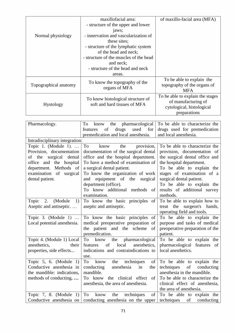
71
Normal physiology
maxillofacial area:
- structure of the upper and lower
jaws;
- innervation and vascularization of
these sites;
- structure of the lymphatic system
of the head and neck;
- structure of the muscles of the head
and neck;
- structure of the head and neck
areas.
of maxillo-facial area (MFA)
Topographical anatomy To know the topography of the
organs of MFA
To be able to explain the
topography of the organs of
MFA
Hystology
To know histological structure of
soft and hard tissues of MFA
To be able to explain the stages
of manufacturing of
cytological, histological
preparations
Pharmacology. To know the pharmacological
features of drugs used for
premedication and local anesthesia.
To be able to characterize the
drugs used for premedication
and local anesthesia.
Intradisciplinary integration:
Topic 1. (Module 1). …
Provision, documentation
of the surgical dental
office and the hospital
department. Methods of
examination of surgical
dental patient.
To know the provision,
documentation of the surgical dental
office and the hospital department.
To have a method of examination of
a surgical dental patient.
To know the organization of work
and equipment of the surgical
department (office).
To know additional methods of
examination.
To be able to characterize the
provision, documentation of
the surgical dental office and
the hospital department.
To be able to explain the
stages of examination of a
surgical dental patient.
To be able to explain the
results of additional survey
methods.
Topic 2. (Module 1)
Aseptic and antiseptic. …
To know the basic principles of
aseptic and antiseptic.
To be able to explain how to
treat the surgeon's hands,
operating field and tools.
Topic 3. (Module 1) …
Local potential anesthesia.
To know the basic principles of
medical preoperative preparation of
the patient and the scheme of
premedication.
To be able to explain the
purpose and tasks of medical
preoperative preparation of the
patient.
Topic 4. (Module 1) Local
anesthetics, their
properties, side effects...
To know the pharmacological
features of local anesthetics,
indications and contraindications to
use.
To be able to explain the
pharmacological features of
local anesthetics.
Topic 5, 6. (Module 1)
Conductive anesthesia in
the mandible: indications,
methods of conducting. …
To know the techniques of
conducting anesthesia in the
mandible.
To know the clinical effect of
anesthesia, the area of anesthesia.
To be able to explain the
techniques of conducting
anesthesia in the mandible.
To be able to characterize the
clinical effect of anesthesia,
the area of anesthesia.
Topic 7, 8. (Module 1)
Conductive anesthesia on
To know the techniques of
conducting anesthesia on the upper
To be able to explain the
techniques of conducting
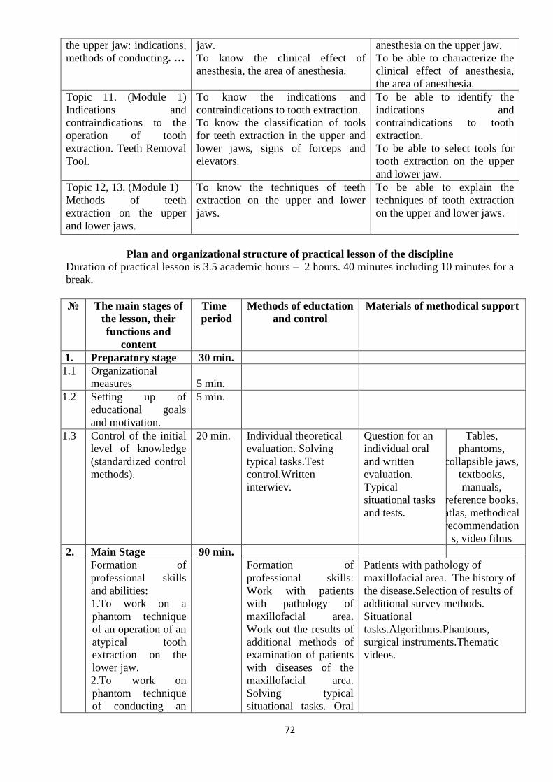
72
the upper jaw: indications,
methods of conducting. …
jaw.
To know the clinical effect of
anesthesia, the area of anesthesia.
anesthesia on the upper jaw.
To be able to characterize the
clinical effect of anesthesia,
the area of anesthesia.
Topic 11. (Module 1)
Indications and
contraindications to the
operation of tooth
extraction. Teeth Removal
Tool.
To know the indications and
contraindications to tooth extraction.
To know the classification of tools
for teeth extraction in the upper and
lower jaws, signs of forceps and
elevators.
To be able to identify the
indications and
contraindications to tooth
extraction.
To be able to select tools for
tooth extraction on the upper
and lower jaw.
Topic 12, 13. (Module 1)
Methods of teeth
extraction on the upper
and lower jaws.
To know the techniques of teeth
extraction on the upper and lower
jaws.
To be able to explain the
techniques of tooth extraction
on the upper and lower jaws.
Plan and organizational structure of practical lesson of the discipline
Duration of practical lesson is 3.5 academic hours – 2 hours. 40 minutes including 10 minutes for a
break.
№ The main stages of
the lesson, their
functions and
content
Time
period
Methods of eductation
and control
Materials of methodical support
1. Preparatory stage 30 min.
1.1 Organizational
measures
5 min.
1.2 Setting up of
educational goals
and motivation.
5 min.
1.3 Control of the initial
level of knowledge
(standardized control
methods).
20 min. Individual theoretical
evaluation. Solving
typical tasks.Test
control.Written
interwiev.
Question for an
individual oral
and written
evaluation.
Typical
situational tasks
and tests.
Tables,
phantoms,
collapsible jaws,
textbooks,
manuals,
reference books,
atlas, methodical
recommendation
s, video films
2. Main Stage 90 min.
Formation of
professional skills
and abilities:
1.To work on a
phantom technique
of an operation of an
atypical tooth
extraction on the
lower jaw.
2.To work on
phantom technique
of conducting an
Formation of
professional skills:
Work with patients
with pathology of
maxillofacial area.
Work out the results of
additional methods of
examination of patients
with diseases of the
maxillofacial area.
Solving typical
situational tasks. Oral
Patients with pathology of
maxillofacial area. The history of
the disease.Selection of results of
additional survey methods.
Situational
tasks.Algorithms.Phantoms,
surgical instruments.Thematic
videos.
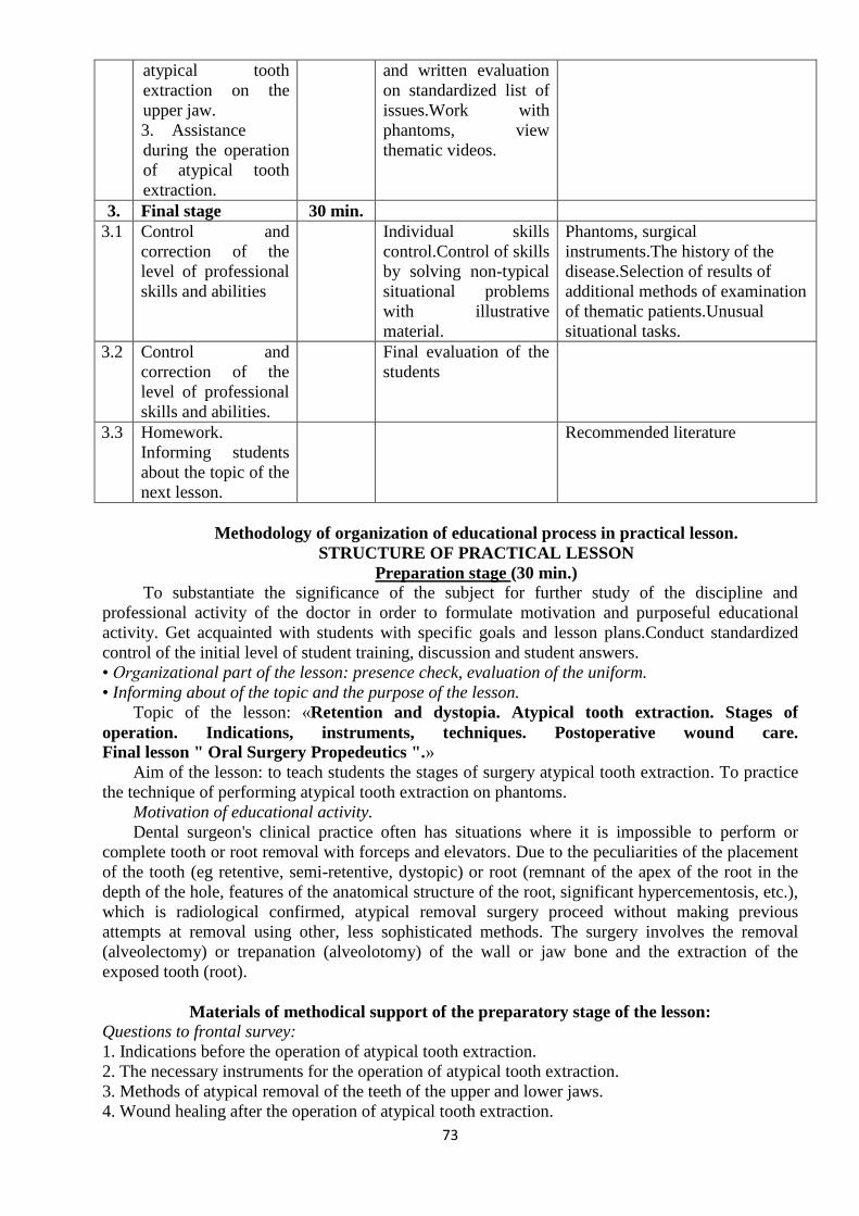
73
atypical tooth
extraction on the
upper jaw.
3. Assistance
during the operation
of atypical tooth
extraction.
and written evaluation
on standardized list of
issues.Work with
phantoms, view
thematic videos.
3. Final stage 30 min.
3.1 Control and
correction of the
level of professional
skills and abilities
Individual skills
control.Control of skills
by solving non-typical
situational problems
with illustrative
material.
Phantoms, surgical
instruments.The history of the
disease.Selection of results of
additional methods of examination
of thematic patients.Unusual
situational tasks.
3.2 Control and
correction of the
level of professional
skills and abilities.
Final evaluation of the
students
3.3 Homework.
Informing students
about the topic of the
next lesson.
Recommended literature
Methodology of organization of educational process in practical lesson.
STRUCTURE OF PRACTICAL LESSON
Preparation stage (30 min.)
To substantiate the significance of the subject for further study of the discipline and
professional activity of the doctor in order to formulate motivation and purposeful educational
activity. Get acquainted with students with specific goals and lesson plans.Conduct standardized
control of the initial level of student training, discussion and student answers.
• Organizational part of the lesson: presence check, evaluation of the uniform.
• Informing about of the topic and the purpose of the lesson.
Topic of the lesson: «Retention and dystopia. Atypical tooth extraction. Stages of
operation. Indications, instruments, techniques. Postoperative wound care.
Final lesson " Oral Surgery Propedeutics ".»
Aim of the lesson: to teach students the stages of surgery atypical tooth extraction. To practice
the technique of performing atypical tooth extraction on phantoms.
Motivation of educational activity.
Dental surgeon's clinical practice often has situations where it is impossible to perform or
complete tooth or root removal with forceps and elevators. Due to the peculiarities of the placement
of the tooth (eg retentive, semi-retentive, dystopic) or root (remnant of the apex of the root in the
depth of the hole, features of the anatomical structure of the root, significant hypercementosis, etc.),
which is radiological confirmed, atypical removal surgery proceed without making previous
attempts at removal using other, less sophisticated methods. The surgery involves the removal
(alveolectomy) or trepanation (alveolotomy) of the wall or jaw bone and the extraction of the
exposed tooth (root).
Materials of methodical support of the preparatory stage of the lesson:
Questions to frontal survey:
1. Indications before the operation of atypical tooth extraction.
2. The necessary instruments for the operation of atypical tooth extraction.
3. Methods of atypical removal of the teeth of the upper and lower jaws.
4. Wound healing after the operation of atypical tooth extraction.
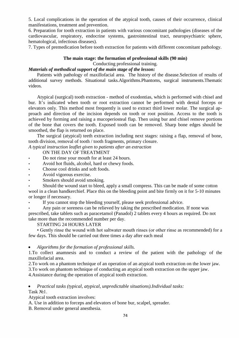
74
5. Local complications in the operation of the atypical tooth, causes of their occurrence, clinical
manifestations, treatment and prevention.
6. Preparation for tooth extraction in patients with various concomitant pathologies (diseases of the
cardiovascular, respiratory, endocrine systems, gastrointestinal tract, neuropsychiatric sphere,
hematological, infectious diseases).
7. Types of premedication before tooth extraction for patients with different concomitant pathology.
The main stage: the formation of professional skills (90 min)
Conducting professional training.
Materials of methodical support of the main stage of the lesson:
Patients with pathology of maxillofacial area. The history of the disease.Selection of results of
additional survey methods. Situational tasks.Algorithms.Phantoms, surgical instruments.Thematic
videos.
Atypical (surgical) tooth extraction - method of exodontias, which is performed with chisel and
bur. It’s indicated when tooth or root extraction cannot be performed with dental forceps or
elevators only. This method most frequently is used to extract third lower molar. The surgical ap-
proach and direction of the incision depends on tooth or root position. Access to the tooth is
achieved by forming and raising a mucoperiosteal flap. Then using bur and chisel remove portions
of the bone that covers the tooth. Exposed tooth can be removed. Sharp bone edges should be
smoothed, the flap is returned on place.
The surgical (atypical) teeth extraction including next stages: raising a flap, removal of bone,
tooth division, removal of tooth / tooth fragments, primary closure.
A typical instruction leaflet given to patients after an extraction
ON THE DAY OF TREATMENT
• Do not rinse your mouth for at least 24 hours.
• Avoid hot fluids, alcohol, hard or chewy foods.
• Choose cool drinks and soft foods.
• Avoid vigorous exercise.
• Smokers should avoid smoking.
• Should the wound start to bleed, apply a small compress. This can be made of some cotton
wool in a clean handkerchief. Place this on the bleeding point and bite firmly on it for 5-10 minutes
or longer if necessary.
• If you cannot stop the bleeding yourself, please seek professional advice.
• Any pain or soreness can be relieved by taking the prescribed medication. If none was
prescribed, take tablets such as paracetamol (Panadol) 2 tablets every 4 hours as required. Do not
take more than the recommended number per day.
STARTING 24 HOURS LATER
• Gently rinse the wound with hot saltwater mouth rinses (or other rinse as recommended) for a
few days. This should be carried out three times a day after each meal
Algorithms for the formation of professional skills.
1.To collect anamnesis and to conduct a review of the patient with the pathology of the
maxillofacial area.
2.To work on a phantom technique of an operation of an atypical tooth extraction on the lower jaw.
3.To work on phantom technique of conducting an atypical tooth extraction on the upper jaw.
4.Assistance during the operation of atypical tooth extraction.
Practical tasks (typical, atypical, unpredictable situations).Individual tasks:
Task №1.
Atypical tooth extraction involves:
A. Use in addition to forceps and elevators of bone bur, scalpel, spreader.
B. Removal under general anesthesia.
75
C. Single-implantation.
D. Removal of teeth with atypical structure.
E. All of the above.
Task №2.
Indications for atypical tooth extraction:
A. Removal of the retented tooth.
B. Removal of dystopic and retented tooth.
C. Removal of a semi-retented tooth.
D. When tooth extraction is not possible with forceps and elevators.
E. All options are correct.
Tasks for independent work and work in small groups (interactive teaching methods).
Patient O., 23, was referred to a dental surgeon to remove 48 teeth according to orthodontic
indications. Objectively, the face is symmetrical, proportional. Regional lymph nodes are not
palpated. During the examination of the oral cavity, only the distal humps of the 48 tooth are
visualized, the medial humps are covered with a pale pink mucosa, and on the radiograph, the roots
of the 48 teeth are formed. The tooth is tilted on the axis by 45 °, the medial humps rests on the root
of the 47 tooth. What type of anesthesia should be performed for atypical removal of 48 teeth?
Justify your choice, describe and demonstrate your anesthesia technique. Which tool should you
choose for atypical tooth extraction? What are the stages of the operation? What is the technique of
working with the chosen toolkit? What is the follow-up medication for patients after atypical
removal? What are the possible complications in the postoperative period?
Final stage (30 min.)
Summing up of the lesson
Materials of methodological support of the final stage of the lesson:
Brain storm.Students demonstrate an exhaustive description of the unusual clinical situation
and offer to offer the most rational diagnostic methods.After recording all the proposed
diagnostic methods during the discussion, students choose the most rational.
Tasks for self-employment. To work on phantoms the technique of atypical tooth extraction
on mandible and maxilla.
Evaluation.
Conduct standardized final control using individual test tasks and questions (20 min.), Work
check (10 min.). Evaluate the student's current activities during the classroom, taking into account
standardized final control, analyze the student's progress, announce the evaluation of each student's
activity, and display it in the student attendance and student log book.An adult group at the same
time makes assessments in the record of the record of success and attendance of classes by students,
the teacher certifies them with his signature.
Brief informing the students about the topic of the next lesson and the methodical measures for
preparing for it.
Basic knowledge level:
1. Anatomical and physiological features of the structure of the jaw bones and teeth.
2. Innervation and blood supply to hard and soft facial tissues.
3. Local conductive anesthesia on the upper and lower jaws.
4. Pharmacological features of preparations for local anesthesia and premedication.
5. Classification of tools for tooth extraction on the upper and lower jaws, signs of forceps and
elevators. Classification of surgical instruments.
List of questions to be studied by the student:
1. Indications before the operation of atypical tooth extraction.
2. The necessary instruments for the operation of atypical tooth extraction.
3. Methods of atypical removal of the teeth of the upper and lower jaws.
76
4. Wound healing after the operation of atypical tooth extraction.
5. Local complications in the operation of the atypical tooth, causes of their occurrence, clinical
manifestations, treatment and prevention.
6. Preparation for tooth extraction in patients with various concomitant pathologies (diseases of the
cardiovascular, respiratory, endocrine systems, gastrointestinal tract, neuropsychiatric sphere,
hematological, infectious diseases).
7. Types of premedication before tooth extraction for patients with different concomitant pathology.
The list of practical skills to be learned by the student:
1. To work on a phantom technique of an operation of an atypical tooth extraction on the lower
jaw.
2. To work on phantom technique of conducting an atypical tooth extraction on the upper jaw.
3. Assistance during the operation of atypical tooth extraction.
Situational tasks and questions on the topic of the lesson:
1. During the removal of 28 teeth, the patient had a fracture of the tuber of the upper jaw. What
care should be given to the patient?
A. To remove the tuber, to mobilize the wound edges and to tighten the wound tightly.
B. Perform radical sinustomy with plastic closure of the oro-antral conjunction.
C. Remove the tuber and pad the wound with an iodine swab.
D. Perform osteosynthesis of the upper jaw.
E. Remove the tuber and drain the wound with tubular drainage.
2. The man went to the clinic for the removal of 17 teeth. During the tooth extraction surgery, one
of the roots was pushed into the maxillary sinus. What is the doctor's tactics in this situation?
A. Carry out a radiological examination and send the patient to the hospital.
B. Do not take any action.
C. Remove root through perforated hole.
D. Perform a sinus surgery.
E. Suture the socket of the removed tooth.
3. The patient at the age of 44 years at the time of tooth extraction fractured his crown part. During
further manipulation with the elevator, the forceps could not remove the root of the tooth. What
kind of manipulation should the surgeon perform to successfully remove the root of the tooth:
A. Separate the roots with a drill and fissure bur.
B. Apply an angled elevator.
C. Send the patient to the hospital.
D. Finish deleting during another visit.
E. Apply the Lecluse elevator.
4. In the process of root extraction of the 38th tooth with Lecluse root elevator a dentist has
pushed the root through into the perimandi-bular soft tissues. The root is situated under the alveolar
arch mucosa and can be clearly palpated. What approach should the doctor choose in the given
case?
A. Dissection of soft tissues covering the root followed by root extraction
B. To try and extract the root through the tooth socket
C. Surgical root extraction through extraoral submandibular way of access
D. Further actions can be determined only after X-ray examination
E. To leave the root in the soft tissues
77
5. The dentist surgeon applied S-shaped forceps to the crown of the 25 tooth and after the luxation
movements performed the tooth traction. Thus there was a complication - a rupture of a mucous
membrane of a cheek. How to prevent this complication:
A. Perform the stage of destruction of the circular ligament of the tooth.
B. Use a spatula to protect the alveolar process from the palatal side.
C. Use a spatula to protect the alveolar process from the sacral side.
D. With the fingers of the left hand to fix the alveolar process on both sides.
E. To perform rotational movements.
Literature:
Basic:
1. Oral and Maxillofacial Surgery: Textbook, Part 1, 2 / V.O. Malanchuk. – Vinnytsia: Nova
Knyha Publishers, 2011. – 453p.
2. Principles of Dental Local Anaesthesia and Teeth Removal / Ya. E. Vares, R. Z. Ogonovsky,
Ch. R. Pohranychna – LNMU, 2007. – 63p.
3. Atlas of Human Anatomy / F. Netter – 2nd
ed. – New Jersey: ICON Learning Systems. – 592
p.
Additional:
1. Contemporary Oral and Maxillofacial Surgery / L. J. Peterson, E. Ellis, J. R. Hupp, M.R.
Tucker – 3rd ed. – St. Louis: Mosby – Year Book, Inc. – 1998. – 1477 p.
78
Control methods.
Control measures are a necessary element of feedback in the learning process. They
determine the correspondence of the level of knowledge acquired by students with the requirements
of the normative documents on higher education.
Control methods and assessment system are developed in accordance with the requirements
of "Criteria, rules and procedures for evaluating the results of students' educational activities at the
Danylo Halytsky LNMU, approved by the Academic Council of LNMU of 02/21/2018, protocol
No. 1.
Control measures in the study of "Surgical dentistry" include current control and final
control, which is called a semester test.
Before studying a new course, students have to take 'entrance control' test in order to
determine the level of preparation of students to the discipline (based on the fundamentals of
previous studies). Entrance control is carried out on the first lesson and is based on the assignments
corresponding to the program of relevant discipline studied before. The results of the entrance
control are analyzed at the department (chair) meetings and also involve the representatives of
methodology committee as well as the teachers of the relevant discipline. According to the results
of the entrance control test, students may be provided with individual assistance or with some
measures of adjusting the educational process.
Current control is carried out on every practical lesson according to the specific goals of
each topic. It is based on the comprehensive evaluation of the student's activity, which takes into
account the entrance control test, the quality of practical work, the level of theoretical training, the
level of performance in individual assignments according to the thematic plan and the results of the
final control.
In the course of assessing the educational activity of students, the preference is given to
standardized methods of control: test tasks, situational tasks, control questions, oral questioning,
structured written work, structured control of practical skills in conditions that are close to real ones
(algorithm -based).
Final control, which is a semester credit, is a form of summarizing control, which assesses
the level of student solely on the basis of the results of certain types of work in practical classes. It
is conducted in accordance with the curriculum in terms set by the schedule of the educational
process and in the amount of educational material determined by the program of academic
discipline.
Assessment procedure for the discipline of "Surgical Dentistry", presented by the two
content modules, is rating-based and is made up of the sum of evaluation points of the current
educational activity, which can be gained for theoretical knowledge and practical skills in
accordance with the lists determined by the discipline program.
Current control is carried out during the training sessions and aims at checking students'
acquisition of the material, the level of theoretical and practical training. Current control can be
presented in the form of testing, solving situational problems, solving clinical situational problem,
demonstration of practical skills or abilities, answers to standardized theoretical questions. Forms of
assessment of the current educational activity are standardized and correspond to the standards of
answers.
Assessment of current educational activity. Evaluation of current student's progress made on
each practical lessons on the 4- point scale and recorded in the register of academic success.
Knowledge of students are evaluated both theoretical and practical training by the following
criteria:
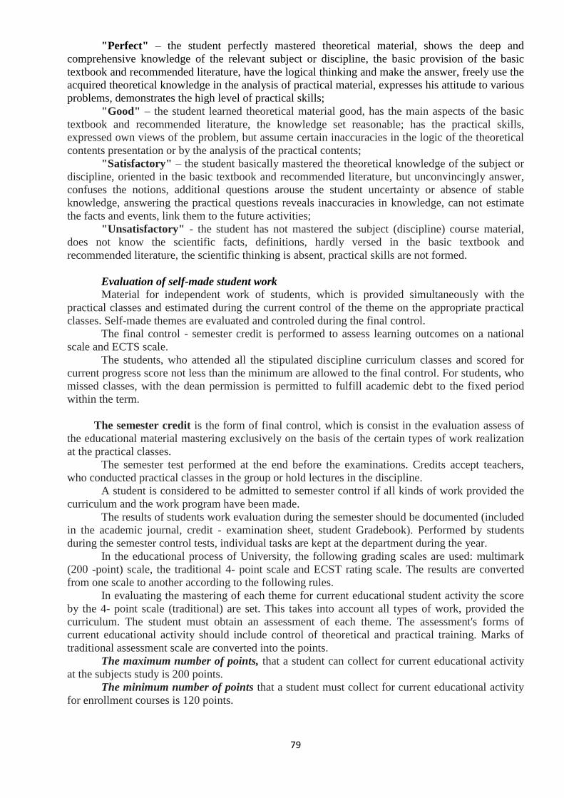
79
"Perfect" – the student perfectly mastered theoretical material, shows the deep and
comprehensive knowledge of the relevant subject or discipline, the basic provision of the basic
textbook and recommended literature, have the logical thinking and make the answer, freely use the
acquired theoretical knowledge in the analysis of practical material, expresses his attitude to various
problems, demonstrates the high level of practical skills;
"Good" – the student learned theoretical material good, has the main aspects of the basic
textbook and recommended literature, the knowledge set reasonable; has the practical skills,
expressed own views of the problem, but assume certain inaccuracies in the logic of the theoretical
contents presentation or by the analysis of the practical contents;
"Satisfactory" – the student basically mastered the theoretical knowledge of the subject or
discipline, oriented in the basic textbook and recommended literature, but unconvincingly answer,
confuses the notions, additional questions arouse the student uncertainty or absence of stable
knowledge, answering the practical questions reveals inaccuracies in knowledge, can not estimate
the facts and events, link them to the future activities;
"Unsatisfactory" - the student has not mastered the subject (discipline) course material,
does not know the scientific facts, definitions, hardly versed in the basic textbook and
recommended literature, the scientific thinking is absent, practical skills are not formed.
Evaluation of self-made student work
Material for independent work of students, which is provided simultaneously with the
practical classes and estimated during the current control of the theme on the appropriate practical
classes. Self-made themes are evaluated and controled during the final control.
The final control - semester credit is performed to assess learning outcomes on a national
scale and ECTS scale.
The students, who attended all the stipulated discipline curriculum classes and scored for
current progress score not less than the minimum are allowed to the final control. For students, who
missed classes, with the dean permission is permitted to fulfill academic debt to the fixed period
within the term.
The semester credit is the form of final control, which is consist in the evaluation assess of
the educational material mastering exclusively on the basis of the certain types of work realization
at the practical classes.
The semester test performed at the end before the examinations. Credits accept teachers,
who conducted practical classes in the group or hold lectures in the discipline.
A student is considered to be admitted to semester control if all kinds of work provided the
curriculum and the work program have been made.
The results of students work evaluation during the semester should be documented (included
in the academic journal, credit - examination sheet, student Gradebook). Performed by students
during the semester control tests, individual tasks are kept at the department during the year.
In the educational process of University, the following grading scales are used: multimark
(200 -point) scale, the traditional 4- point scale and ECST rating scale. The results are converted
from one scale to another according to the following rules.
In evaluating the mastering of each theme for current educational student activity the score
by the 4- point scale (traditional) are set. This takes into account all types of work, provided the
curriculum. The student must obtain an assessment of each theme. The assessment's forms of
current educational activity should include control of theoretical and practical training. Marks of
traditional assessment scale are converted into the points.
The maximum number of points, that a student can collect for current educational activity
at the subjects study is 200 points.
The minimum number of points that a student must collect for current educational activity
for enrollment courses is 120 points.
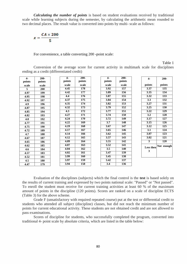
80
Calculating the number of points is based on student evaluations received by traditional
scale while learning subjects during the semester, by calculating the arithmetic mean rounded to
two decimal places. The result value is converted into points by multi- scale as follows:
For convenience, a table converting 200 -point scale:
Table 1
Conversion of the average score for current activity in multimark scale for disciplines
ending as a credit (differentiated credit)
Evaluation of the disciplines (subjects) which the final control is the test is based solely on
the results of current training and expressed by two points national scale: "Passed" or "Not passed".
To enroll the student must receive for current training activities at least 60 % of the maximum
amount of points in the discipline (120 points). Scores are ranked on a scale of discipline ECTS
(Table 3) for the above scheme.
Grade F (unsatisfactory with required repeated course) put at the test or differential credit to
students who attended all subject (discipline) classes, but did not reach the minimum number of
points for current educational activity. These students are not obtained credit and are not allowed to
pass examinations.
Scores of discipline for students, who successfully completed the program, converted into
traditional 4- point scale by absolute criteria, which are listed in the table below:
4- 200- points points
scale scale
5 200
4.97 199
4.95 198
4.92 197
4.9 196
4.87 195
4.85 194
4.82 193
4.8 192
4.77 191
4.75 190
4.72 189
4.7 188
4.67 187
4.65 186
4.62 185
4.6 184
4.57 183
4.52 181
4.5 180
4.47 179
4- 200- points points
scale scale
4.45 178
4.42 177
4.4 176
4.37 175
4.35 174
4.32 173
4.3 172
4.27 171
4.24 170
4.22 169
4.19 168
4.17 167
4.14 166
4.12 165
4.09 164
4.07 163
4.04 162
4.02 161
3.99 160
3.97 159
3.94 158
4- 200- points points
scale scale
3.92 157
3.89 156
3.87 155
3.84 154
3.82 153
3.79 152
3.77 151
3.74 150
3.72 149
3.7 148
3.67 147
3.65 146
3.62 145
3.57 143
3.55 142
3.52 141
3.5 140
3.47 139
3.45 138
3.42 137
3.4 136
4-
points
scale
200-
points
scale 3.37 135
3.35 134
3.32 133
3.3 132
3.27 131
3.25 130
3.22 129
3.2 128
3.17 127
3.15 126
3.12 125
3.1 124
3.07 123
3.02 121
3 120
Less then
3
Not enough
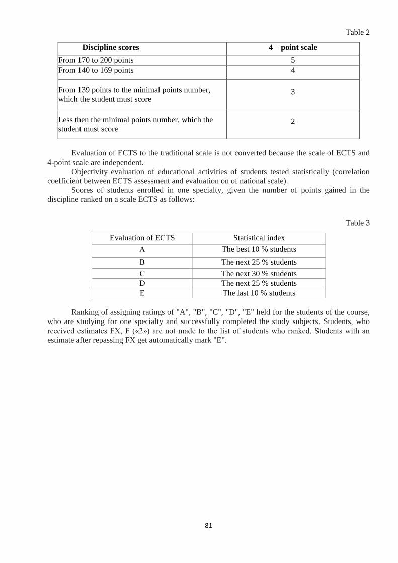
81
Table 2
Evaluation of ECTS to the traditional scale is not converted because the scale of ECTS and
4-point scale are independent.
Objectivity evaluation of educational activities of students tested statistically (correlation
coefficient between ECTS assessment and evaluation on of national scale).
Scores of students enrolled in one specialty, given the number of points gained in the
discipline ranked on a scale ECTS as follows:
Table 3
Evaluation of ECTS Statistical index
А The best 10 % students
В The next 25 % students
С The next 30 % students
D The next 25 % students
E The last 10 % students
Ranking of assigning ratings of "A", "B", "C", "D", "E" held for the students of the course,
who are studying for one specialty and successfully completed the study subjects. Students, who
received estimates FX, F («2») are not made to the list of students who ranked. Students with an
estimate after repassing FX get automatically mark "E".
Discipline scores 4 – point scale
From 170 to 200 points 5
From 140 to 169 points 4
From 139 points to the minimal points number,
which the student must score 3
Less then the minimal points number, which the
student must score 2