Embed Size (px)
Citation preview

ORIGINAL RESEARCH ARTICLEpublished: 24 September 2013
doi: 10.3389/fnhum.2013.00582
Mindfulness-induced selflessness: a MEGneurophenomenological studyYair Dor-Ziderman1*, Aviva Berkovich-Ohana2, Joseph Glicksohn1,3 and Abraham Goldstein1,4
1 The Leslie and Susan Gonda (Goldschmied) Multidisciplinary Brain Research Center, Bar-Ilan University, Ramat Gan, Israel2 Department of Neurobiology, Weizmann Institute of Science, Rehovot, Israel3 Department of Criminology, Bar-Ilan University, Ramat Gan, Israel4 Department of Psychology, Bar-Ilan University, Ramat Gan, Israel
Edited by:
Wendy Hasenkamp, Mind and LifeInstitute, USA
Reviewed by:
Brenton W. McMenamin, Universityof Maryland, USANorman Farb, Baycrest, Canada
*Correspondence:
Yair Dor-Ziderman, ElectromagneticBrain Imaging Unit, The Leslie andSusan Gonda (Goldschmied)Multidisciplinary Brain ResearchCenter, Bar-Ilan University, Buildingnumber 901, Ramat Gan 52900,Israele-mail: [email protected]
Contemporary philosophical and neurocognitive studies of the self have dissociated twodistinct types of self-awareness: a “narrative” self-awareness (NS) weaving togetherepisodic memory, future planning and self-evaluation into a coherent self-narrativeand identity, and a “minimal” self-awareness (MS) focused on present momentaryexperience and closely tied to the sense of agency and ownership. Long-term Buddhistmeditation practice aims at realization of a “selfless” mode of awareness (SL),where identification with a static sense of self is replaced by identification with thephenomenon of experiencing itself. NS-mediating mechanisms have been explored byneuroimaging, mainly fMRI, implicating prefrontal midline structures, but MS processesare not well characterized and SL even less so. To this end we tested 12 long-termmindfulness meditators using a neurophenomenological study design, incorporatingboth magnetoencephalogram (MEG) recordings and first person descriptions. We foundthat (1) NS attenuation involves extensive frontal, and medial prefrontal gamma band(60–80 Hz) power decreases, consistent with fMRI and intracranial EEG findings; (2)MS attenuation is related to beta-band (13–25 Hz) power decreases in a networkthat includes ventral medial prefrontal, medial posterior and lateral parietal regions;and (3) the experience of selflessness is linked to attenuation of beta-band activityin the right inferior parietal lobule. These results highlight the role of dissociablefrequency-dependent networks in supporting different modes of self-processing, andthe utility of combining phenomenology, mindfulness training and electrophysiologicalneuroimaging for characterizing self-awareness.
Keywords: self-awareness, minimal self, narrative self, MEG, mindfulness meditation, neurophenomenology, beta
frequency band, right inferior parietal lobule
INTRODUCTIONAn unremitting companion of human experience is the sense ofself. Amidst the ocean of coming-and-going waves of percep-tions, cognitions and emotions, an absolute certainty regardingthe identity of the present-moment experiencer—“self as I”—remains unwavering (James, 1890). On the other hand, the threadof a constant, static, unchanging self—the “self as Me”—stretchesback to childhood years, and extends as far into the future asone can imagine. The protagonist of both scenarios is experi-enced as one-and-the-same, even though the respective (imag-ined/remembered) bodies, mental capacities, as well as externalcontexts have completely changed. These phenomenally distinctaspects of self-awareness are being re-conceptualized by contem-porary philosophers, psychologists and neurobiologists, aiming ata fruitful exchange between philosophy of mind, phenomenology,and the cognitive sciences. One such influential conceptualiza-tion has been offered by Gallagher (2000) as “minimal” and“narrative” forms of self-awareness.
The “minimal” self (MS) is defined as a consciousnessof oneself as the immediate subject of experience. It is
pre-reflective, present-centered, experiential in nature, andimportantly, involves a sense of “ownership” and “agency”: thesense that it is I who is undergoing an experience (Gallagher,2000). MS, or “core self” in Damasio’s (1999, 2010) terms, isunderstood to be intermittent. Damasio describes it as “. . . atransient entity, ceaselessly recreated for each and every objectwith which the brain interacts” (Damasio, 1999, p. 17), in thisway implementing a self/non-self distinction and thus speci-fying the self in perception, cognition, emotion, and action(Christoff et al., 2011). The “narrative” self (NS), on the otherhand, refers to a self extended in time, heavily reliant onlanguage, episodic/autobiographical memory and imagination(planned/expected future), and corresponding to identity andpersonhood. The notion of NS has appeared in the literatureunder other names such as the “extended” self and “concep-tual” self (Neisser, 1988), the “autonoetic” self (Gardiner, 2001)and the “autobiographical” self (Damasio, 1999, 2010), and hasbeen shown to be closely tied to a neurophysiological baseline(Gusnard et al., 2001; Buckner et al., 2008), the so-called default-mode network (DMN, Gusnard and Raichle, 2001; Raichle et al.,
Frontiers in Human Neuroscience www.frontiersin.org September 2013 | Volume 7 | Article 582 | 1
HUMAN NEUROSCIENCE
Dor-Ziderman et al. Mindfulness-induced selflessness
2001) and to mind-wandering (Mason et al., 2007; Christoff et al.,2009; Hasenkamp et al., 2012). It is important to note that NSand MS are processes which may operate concurrently. Like otherconscious mental content produced by the brain, NS represen-tations, perceived as thoughts and feelings, are stamped with thesubjective signature of being our thoughts and feelings. Thus, self-specifying processes are at play also during NS (Gallagher, 2000;Damasio, 2010).
Eastern philosophy, and in particular Buddhist philosophy,exhibits a radically different view of the self and personal iden-tity, advocating a “selfless” mode of processing phenomena (SL).At the core of Buddhist psychology lies the teaching of therebeing no such thing as a permanent, unchanging self (Dreyfusand Thompson, 2007; Olendzki, 2010). The self is understoodto be illusory in the sense of being no more than a men-tal process—and identification with it is understood to be atthe very root of suffering. Thus, a primary target of Buddhistpractice is the realization of the illusory nature of the self andcultivation of a selfless, boundless mode of experience whereidentification with a static sense of self is replaced by iden-tification with the phenomenon of experiencing itself (Hart,1987; Dalai Lama, 1991; Austin, 2000; Ekman et al., 2005;Wallace, 2006; Nydahl, 2008). The notion of relinquishing thesense of owning and directing experience, may seem to theWestern mind as nothing short of pathological (for a relateddiscussion, see Engler, 2003). And indeed, pathological brainssuch as of schizophrenic patients experiencing “thought inser-tion” (Frith, 1992; Gallagher, 2004) or patients who have suf-fered lesions (Damasio et al., 2012; Philippi et al., 2012) com-promising specific self functions, have largely contributed toDamasio’s and Gallagher’s understanding of the minimal/coreself-concept. In a similar vein, as has been previously suggested(Lutz et al., 2007; Tagini and Raffone, 2010), long-term mind-fulness meditators can provide parallel information regardingthe self through diminishing the agentive/ownership aspects ofpresent-moment experience. Such information, however, has theadvantage of being volitionally produced and in non-diseasedbrains.
The neurophysiology of NS is relatively well established. Awealth of recent large-scale meta-analyses of mainly fMRI stud-ies investigating self-referential processing through a variety ofparadigms have consistently shown it to be modulated by a sub-set of the DMN, namely the central midline structures, and inparticular the medial prefrontal cortex (mPFC) (Northoff et al.,2006, 2011; Buckner et al., 2008; Andrews-Hanna et al., 2010;Qin and Northoff, 2011; Whitfield-Gabrieli et al., 2011; Kim,2012). Translating these findings into electrophysiological terms,there is accumulating evidence that the fMRI’s hemodynamicresponse signal attributed to DMN and self-referential process-ing is correlated with neuronal activity in the gamma band (EEGstudies—Mantini et al., 2007; Berkovich-Ohana et al., 2012), andin particular high-gamma band (intracranial EEG studies—Niret al., 2007; Jerbi et al., 2010; Ossandón et al., 2011; Ramot et al.,2012). It should be noted that whether self-referential paradigmscan reveal neural activity specific to the self is a matter of cur-rent debate, as these tasks involve, and are thus confounded by,higher-order cognitive functions such as evaluation, judgment
and reflective thought (Legrand and Ruby, 2009; Christoff et al.,2011; Northoff et al., 2011).
The neural correlates of MS are less well established, withapproaches aiming at identifying self-specifying pre/non–reflective processes including merely perceiving, withoutjudgment or evaluation, self-specific vs. non-specific stimuli(Schneider et al., 2008; Northoff et al., 2009), or improvingtime resolution using event-related EEG (Esslen et al., 2008) orMEG (Walla et al., 2007). Other approaches are informed byphenomenology (Gallagher and Sørensen, 2006) and target MSvia one of its core attributes—the sense of agency and ownership(for reviews see David et al., 2008; Sperduti et al., 2011). Keyregions here include the inferior parietal lobule (IPL) and theinsula. Importantly, the literature does not supply informationregarding the oscillatory signature of these mostly fMRI findings.Oscillatory power increases/decreases that occur in specific fre-quency bands and within different cortical areas have been shownto be functionally relevant in the brain (Singh, 2012). In partic-ular, the different modes of self-awareness might not only involvedifferent brain topographies, but perhaps also different oscilla-tory signatures. In this regard, MEG is an appealing research toolas it has both an excellent temporal (and thus spectral) resolution,and it allows for a reliable source reconstruction (relative to EEG)with a reasonable spatial resolution (Hansen et al., 2010).
In line with the advent of producing SL experiences in thelab, the participants employed in the present study are long-termmindfulness meditation practitioners. Mindfulness is defined andpracticed as a non-judgmental awareness of bodily or mentalexperiences arising in the present moment. Regardless of howpleasant or unpleasant the arising experiences are, the medita-tor trains not to cling to, nor to push them away, but ratherto treat them with acceptance, openness and curiosity, watchingthem arise, play in the theater of the mind and finally dissolveback into the space of the mind (Kabat-Zinn, 1990). Mindfulnessis a current and widespread form of Buddhist practice (Williamsand Kabat-Zinn, 2011), and has been shown to enhance cognitivefunctions such as attentional abilities, emotional regulation, exec-utive functions and memory (Chiesa and Serretti, 2010; Chiesaet al., 2011), altering the brain circuits and neuropsychologicalmechanisms underlying these functions (Cahn and Polich, 2006;Davidson and Lutz, 2008; Lutz et al., 2008; Hölzel et al., 2011b),and even altering brain structure in regions typically activatedduring mindfulness meditation (Lazar et al., 2005; Hölzel et al.,2008, 2011a).
One of mindfulness’s mechanisms of action is an altered senseof self (Hölzel et al., 2011b). Mindful awareness induces a sharpersense of the normally perceived subjective sense of self (Lutzet al., 2008), but treats it as an object of meditation. This culti-vated shift to an “observer perspective” (Kerr et al., 2011) inducesa change in the perspective of self and first-person experience.Indeed, a recent integrative theoretical framework and systems-based neurobiological model suggests understanding mindfulnessby focusing on self-processing and the neural networks underly-ing self-awareness, self-regulation, and self-transcendence (Vagoand Silbersweig, 2012). A growing number of studies show thatmindfulness alters DMN and self-related activity and connectiv-ity (EEG: Berkovich-Ohana et al., 2012; Lehmann et al., 2012;
Frontiers in Human Neuroscience www.frontiersin.org September 2013 | Volume 7 | Article 582 | 2

Dor-Ziderman et al. Mindfulness-induced selflessness
fMRI: Pagnoni et al., 2008; Brewer et al., 2011; Ives-Deliperiet al., 2011; Froeliger et al., 2012; Hasenkamp and Barsalou, 2012;Taylor et al., 2013). In particular, Farb et al. (2007) used fMRIand a mindfulness-based stress reduction (MBSR, Kabat-Zinn,1982; Kabat-Zinn et al., 1992) intervention to dissociate narra-tive from experiential modes of processing. The present studycontinues Farb et al. (2007) in using mindfulness meditators forrevealing the neural correlates of momentary (parallel to MS)and across-time (parallel to NS) self processing, but goes fur-ther in exploring SL: momentary phenomenal experience freeof the sense of agency and ownership. Figure 1 illustrates theencapsulated relationship between NS, MS and SL.
The working basis for the present study’s design is that long-term mindfulness meditators: (1) are adept in keeping theirattention for extended time periods on an object of choice, beit a physical object, the breath, or a produced state of self; and(2) develop through practice their goal of dissolving the expe-rience of a fixed subjective core comprising their self-identity.Thus, such participants were recruited and requested to mentallyproject states representing NS, MS, and SL, while their brain activ-ity was recorded by MEG. The purpose of the study is to map thedifferential neural activity related to NS and MS, as well as char-acterize SL, a present-centered conscious experience devoid ofan experiencing subjective self. The study’s aims and hypothesesare to:
1. Map the neural correlates of NS. Given that MS is at play alsoduring NS, contrasting the two conditions is expected to reflectNS attenuation, and is hypothesized to result in a reduction ofmPFC high-gamma oscillatory activity. This part of the studyis expected to bridge results from the prevalent fMRI imagingliterature and the present MEG methodology.
2. Map the neural correlates of MS. Given that both MS andSL share a present-centered experiential aspect, differing only
FIGURE 1 | Working model of self-awareness modes. NS, MS and SL asencapsulated processing modes.
in terms of the experiencer—the agency/ownership aspectsaccompanying experience, contrasting MS and SL is expectedto reflect the neural correlates of MS attenuation. Here predic-tions are less clear, nevertheless, the IPL and insula are likelyto play roles. Localizing this differential activation within thefrequency domain will be a novel contribution of the presentstudy.
3. Use first-person reports for grouping the MEG data and iden-tifying the neural correlates of the subjective aspects of theBuddhist-described “selfless” experience.
MATERIALS AND METHODSPARTICIPANTSSixteen experienced meditation practitioners were recruited forthe research project. Two participants’ data were discarded earlyon in the experiment. The first due to complaints of tiredness andlack of focus, and the second due to back pain (to the point ofstopping the MEG recording). In addition, in order to establisha common frame of reference for the first-person descriptions,as well as control for confounds that might result from differ-ent sources of training, only participants practicing very similarforms of meditation were included in the study. Thus, the dataof two further participants (Zen and non-dual practitioners, seeLutz et al., 2007 for details on these forms of meditation) were notanalyzed. The remaining 12 practitioners, all mindfulness med-itators practicing similar forms of Vipassana, either originatingfrom or inspired by the Buddhist Theravada tradition, comprisethe participants of the present study. All are right-handed (9males and 3 females, mean age 45.2, SD = 11.3, ranging from31 to 66) with no history of mental or neurological disease. Allof the participants are long-term practitioners with an average of16.5 (SD = 7.9, ranging from 9 to 34) years of meditation prac-tice, and an average of 11,225 (SD = 9909, ranging from 1290to 29,293) total hours of meditation practice. All the performedprocedures are in compliance with the Code of Ethics of theWorld Medical Association (Declaration of Helsinki), and wereapproved by the Research Ethics Board of Bar-Ilan University.The participants gave their written consent and were financiallycompensated for their time.
PRE-RECORDING PROCEDURESThe participants were welcomed and introduced to the experi-ment and the research facility. They then filled out forms notingtheir agreement to participate in the experiment, their personaldetails, and a form estimating their formal meditation experience.Pre-task procedures included an average of 45 min of clarifyingthe study’s setup, tasks and stimuli using a PowerPoint presenta-tion and allowing time for questions and discussion. In this way,misunderstanding, alteration, or rejection of the scripts providedby the researchers, were minimized (Roepstorff, 2001).
TASKSThe experiment included seven experimental sessions. Of these,only the “self” session (fifth in order) is reported here. Each ses-sion consisted of performing tasks during which the participant’sbrain activity was recorded. This was followed by an interviewconducted via the intercom system, during which brain activity
Frontiers in Human Neuroscience www.frontiersin.org September 2013 | Volume 7 | Article 582 | 3
Dor-Ziderman et al. Mindfulness-induced selflessness
was not recorded. The participants were encouraged to stretchtheir limbs and relax during the interview, but were requestednot to move and to keep their eyes closed while performing thetasks. To correct for head and body movements during the inter-view session, head-shapes were re-registered at the beginningof each session. A 20-min break was suggested to the partici-pants after completing the 5th session of the experiment, duringwhich refreshments were offered. Total time in the MEG rangedfrom 2 to 3 h. The participants’ condition was closely monitoredthroughout the experiment via the intercom (during the inter-view sessions) and the closed-circuit TV camera (at all times). Inaddition, participants were asked a number of times throughoutthe experiment if they were tired and needed an additional break.
The task relevant to the present study is the “self task.”Participants were requested to mentally project themselves intocertain self-related states, which had been described and discussedduring the PowerPoint presentation. The session included 3 con-ditions, each repeated 3 times in succession for 30 s. This moreecologically-valid design (in contrast to the commonly employedevent-related designs) was chosen due to mediators’ heightenedcapacities in directing and sustaining attention (Brefczynski-Lewis et al., 2007; Lutz et al., 2008). The first 4 s of each 30-sepoch were omitted so as to allow participants sufficient time toenter the states (SL in particular). This decision was made afterconsulting a well-known, very-long-term, meditation teacher,concerning the study design, and after 2 pilot runs (with twoof the authors, Aviva Berkovich-Ohana and Yair Dor-Ziderman,who are also long-term meditators). A recording with instruc-tions for each condition was sounded, after which the participantperformed the requested task. At the end of the 30 s, a sound washeard indicating to the participant to stop and rate task perfor-mance success (on a 1–3 scale). This measure was incorporated inorder to allow post-hoc identification of bad trials (button pressof 1). However, as none of the participants in any of the condi-tions reported here indicated such bad trials, this measure wasnot further used. After pressing the corresponding button, thenext instruction was delivered. The session was followed by astructured interview conducted via the intercom system.
The exact instructions for each self-projected condition were:
i “narrative” condition (NS)—“Try to think what characterizesyou.”
ii “minimal” condition (MS)—“Try to experience what is hap-pening to you at the present moment.”
iii “selfless” condition (SL)—“Try to experience what is happeningat the present moment, when you are not in the center.”
DATA ACQUISITIONMEGMEG recordings were conducted with a whole-head, 248-channelmagnetometer array (4-D Neuroimaging, Magnes 3600 WH) ina magnetically-shielded room. Reference coils located approxi-mately 30 cm above the head oriented by the x, y, and z axes wereused to remove environmental noise. Head position was indi-cated by attaching 5 coils to the scalp and determining, to a 1 mmresolution, their position relative to the sensor array before andafter measurement. Head localization was performed before and
after each set of tasks to determine degree of head movement.Head shape and coil position were digitized using a PollhemusFASTTRAK digitizer. Brain signals were recorded with a samplerate of 1017.25 Hz and an analog online 0.1–400 Hz band-passfilter. The instructions for each condition were presented usingE-prime 1.0 and delivered via a STAX SRS-005 amplifier andSR-003 push-pull electrostatic ear speakers coupled by a vinyltube to silicon earpieces to prevent magnetic noise within theshielded room. Task performance ratings were collected using aLUMItouch photon control response box.
Subjective reportsRetrospective reports. Participants were asked to provide retro-spective reports regarding their perceived (relative to past experi-ences) success and stability (on a 1–10 scale, with 1 denoting “verylow” and 10 denoting “very high”) in performing the tasks, as wellas report on the emotional content of their experiences during thedifferent tasks.
Introspective reports. Participants were asked to describe theirSL experience freely and in their own words, without reflectionor judgment (Jack and Roepstorff, 2002; Schooler, 2002; Lutzand Thompson, 2003). In addition, the descriptions were col-lected immediately after they were produced in order to minimizereliance on episodic recall (Jack and Roepstorff, 2002).
MEG DATA ANALYSISCleaning and preprocessingData processing and analysis was performed using Matlab®R2009b and FieldTrip toolbox for MEG analysis (Open SourceSoftware for Advanced Analysis of MEG, Oostenveld et al., 2011).Data were cleaned for line frequency (by recording on an addi-tional channel the 50 Hz from the power outlet, and subtractingthe average power-line response from every MEG sensor), and24 Hz building vibration (measured in x, y, and z directions using3 Bruel and Kjaer accelerometers) artifacts (Tal and Abeles, 2013).The data from the 3 “self” tasks were then segmented into non-overlapping 2-s epochs. Each epoch was visually examined formuscle and jump (in the MEG sensors) artifacts. Contaminatedepochs were discarded. No malfunctioning MEG sensors wereidentified. To ensure the removal of all heartbeat, eye, and muscleartifact, an independent component analysis (ICA) was per-formed on the data (Jung et al., 2000). Segmented data weredown-sampled to 339 (1017/3) Hz to speed up data decompo-sition. The data were then decomposed into a set of independentcomponents (248, as the number of sensors) ordered by degreeof their explained variance. Components indicating heartbeatsor eye movements were determined from a visual inspection ofthe 2D scalp maps and time course of each component. 2.6 ±1.2 components were taken out on average, and the remainingcomponents were then used to reconstruct the pre down-sampleddata.
Sensor-level analysesIn order to level the number of trials for all participants andconditions, the first 32 of the remaining epochs were markedas the data for further sensor-level analyses. The segmented 2-s
Frontiers in Human Neuroscience www.frontiersin.org September 2013 | Volume 7 | Article 582 | 4

Dor-Ziderman et al. Mindfulness-induced selflessness
epochs were multiplied by a Hanning taper, and subjected to aFast Fourier Transformation (FFT) for the frequencies rangingfrom 0.5 to 100 Hz. This resulted in a power spectrum with afrequency resolution of 0.5 Hz for each epoch. The power spec-tra were then averaged across the epochs of each condition, thusobtaining the mean power for each condition and participant.The next step involved calculating, for each frequency of each sen-sor of each participant, a power percent-in-signal-change (PSC)metric, for estimating power differences in NS vs. MS and MS vs.SL. PSC was computed in the following manner: for sensor S, fre-quency f, and power values of conditions A and B, PSC[S(f )] =[(A/B) − 1] if A >= B, and [1-(B/A)] if B > A. This manipula-tion yields a balanced PSC distribution centered on 0. Each par-ticipant’s PSC values for the two comparisons were then collapsedacross all sensors, and averaged across the delta (0.5–3.5 Hz),theta (4–7.5 Hz), alpha (8–12.5 Hz), beta (13–25 Hz), gamma(25.5–59.5 Hz), high-gamma (60–80 Hz), and very-high-gamma(80.5–100 Hz) frequency bands. To reduce dimensionality priorto localization, 1-sample t-tests were performed for each fre-quency band and for each comparison against the null hypothesisthat the PSC measures came from a continuous, normal distribu-tion with a zero mean. Results were then Bonferroni-corrected.Finally, 2D scalp topographies of the mean PSC in the significantfrequency bands and comparisons were created.
Source-space projectionLocalization was performed for the frequency bands which evi-denced significant PSC in the sensor-level data. Sources were esti-mated using Synthetic Aperture Magnetometry (SAM, Robinsonand Vrba, 1999). SAM is an adaptive nonlinear minimum-variance beamformer algorithm. It calculates the signal covari-ance from the MEG sensor data and uses it in conjunction with aforward solution for the dipoles at each 3D brain voxel (of a speci-fied size) to construct optimum spatial filters. The spatial filteringsuppresses interference of unwanted signals from other locations.
For source estimation, the pre-ICA data were used. A numberof works have shown that interfering biomagnetic sources suchas cardiac, respiratory, and eye movements are effectively sup-pressed by beamforming (e.g., Sekihara et al., 2006; Brookes et al.,2008, 2011). Data were band filtered (using the SAM default IIRfilter) for each participant and condition in the frequency bandsspecified through the sensor-level analysis. Covariance matrices,and subsequently SAM weights, were computed for each 5 cubic-mm voxel using the data from the two conditions participating ineach signal change calculation, for each frequency-band-filteredtime-series data. For each voxel, the data were multiplied by theweights, thus creating “virtual sensor” time-series, which werethen transformed via FFT to the frequency domain, thus deriv-ing power values. Finally, PSC values (same metric as the onedescribed in the sensor-level analysis section, pseudo-F in SAM)were computed for each comparison, participant and each andevery voxel.
To facilitate group analysis, head models were constructedby co-registering each participant’s SAM volume to a previouslyobtained MRI scan (T1-weighted anatomical images acquiredwith high-resolution 1-mm slice thickness, obtained by one ofthe authors (Aviva Berkovich-Ohana) by means of a 3T Trio
Magnetom Siemens scanner located at the Weizmann Institute ofScience, Rehovot, Israel) based on the position of the fiduciarymarkers established during the digitization phase. Each partici-pant’s MRI image and its co-registered SAM volume were thentransposed into a common anatomical space (Talairach coordi-nates, Talairach and Tournoux, 1988). Voxel-level group statistics,for each comparison and frequency band, were conducted usinga non-parametric permutation analysis procedure (2000 per-mutations, Nichols and Holmes, 2001; Singh et al., 2003), andcorrected for multiple comparisons based on a Monte Carlo sim-ulation of random noise distribution (using AFNI’s 3dClustSimmodule, Forman et al., 1995).
NEUROPHENOMENOLOGICAL ANALYSISSubjective reportsSuccess and stability. For assessing whether participants’ ratingsfor perceived success and stability were different for the differenttasks, repeated-measures ANOVA was conducted for success andstability as dependent variables.
Emotional content. Participant reports of emotional contentduring each task were collected and arranged in 4 categories:neutral—here participants either reported no emotional contentor explicitly stated a neutral state; positive—here participantsreported only positive emotions (such as enjoyment, comfort,quiet, pleasant, rest, and lightness); negative—here participantsreported only negative emotions (such as pride, fear, anxiety,confusion, insecurity, and dislike), and mixed—which includedreports of both positive and negative emotions. In addition, anumber of participants spontaneously reported low level of emo-tions (NS—4 participants, 2 in the negative category and 2 in themixed category; SL—1 participant in the mixed category). A noteregarding the categorization of “pride” as a negative emotion:this is in alignment with the Buddhist context (Goleman, 1995;Chambers et al., 2009), given that the participants are long-termpractitioners of Buddhist traditions.
Meditation experience. Meditation experience was gauged usinga normalized measure incorporating both total number of yearsand hours of meditation. The maximum values of meditationyear and hour estimates were extracted, and all other values weredivided by them, resulting in values between 0 and 1. The twometrics were then averaged, giving equal weight to meditationyears and hours.
First-person SL descriptions. A careful reading by the authors ofthe participants’ first-person descriptions of their SL experiencesindicated three rather broad but distinct types of experiences.Age was ruled out as a confounding factor [ANOVA, F(1, 11) =3.76, ns]. The suggested categorization was further validated bypresenting the raw participant descriptions and category expla-nations (as presented below but without the examples) to 12naïve referees (graduate students and postdoctoral researchers),and asking them to categorize the descriptions according to thesuggested scheme. Descriptions which were categorized differ-ently than the suggested scheme by more than one referee wereexcluded from the analysis. Two descriptions were thus removed
Frontiers in Human Neuroscience www.frontiersin.org September 2013 | Volume 7 | Article 582 | 5

Dor-Ziderman et al. Mindfulness-induced selflessness
(sub14 and sub16’s, 4 referees categorized each of them differ-ently), resulting in 10 SL descriptions. The participants’ descrip-tions (including those finally excluded from the analysis) andtheir categorization are presented below in Table 1. The suggestedcategories are:
a. Lack-of-ownership (LO): The 4 participants in this categoryreported experiencing what was happening, only with thesense of agency/ownership absent. As an example, participantsub12 described: “It was emptiness, as if the self fell out of thepicture. There was an experience but it had no address, it was notattached to a center or subject . . . ”
b. Altered-experience (AE): The 4 participants in this categoryreported an altered experience of their bodies/senses/spatial-context. For example, sub11 reported: “On the level of feelingand sensing—as if I took a step back and am looking at myselffrom the back. I see myself but am also aware of what ishappening around . . . ”
c. Less-happening (LH): The grouping of this category wassomewhat looser than the other categories. The 4 partici-pants in this category reported a quieting or general relaxationof body, reflectivity, cognition, or experience in general. Forexample, participant sub6 reported: “Very pleasant and relaxedand quiet. It was the most devoid of effort relative to the previousone . . . ”
MEG source estimatesTo identify within the MS vs. SL beta-band (the only frequencyband relevant for characterizing SL—see results section) networkregions specifically correlated with the phenomenological cate-gories, MEG source estimates of each phenomenological category(vs. the other two categories) were derived in a manner similarto the one described above. Group statistics, limited to the net-work of interest (significant MS vs. SL beta band regions), werecomputed on the Talairach-transformed individual images usingnon-parametric random permutations with a 2-sample t-teststatistic.
RESULTSSUBJECTIVE REPORTSThe first-person reports indicated that the participants were ableto successfully produce the different self-states. The means for tasksuccess were high (on a 1–10 scale with 1 denoting “very low” and10 denoting “very high”) for the NS, MS, and SL tasks (8.6 ± 0.9,7.8 ± 1.4, and 8 ± 1.2, respectively). In addition, participantsreported high measures of task stability: 8.2 ± 1.6, 8 ± 1.4, and7.6 ± 1.5 for NS, MS, and SL, respectively. These indicate that theparticipants managed—in their subjective experience—to pro-duce and maintain the requested self-states in a stable mannerfor the task duration. The ANOVA indicated no significant dif-ferences between the states for both task success [F(2, 11) = 2.2,
Table 1 | SL descriptions and their phenomenological categorization.
Phenomenal categories during SL
Lack-of-ownership (LO) Altered-experience (AE) Less-happening (LH)
Sub4: “The question was ‘what washappening?’ As a sort of subjectivity focus,which was softened and dissolved. And all thatwas left was what was happening not to me.”
Sub5: “Like in a dream. Like I’m not awakenow but dreaming. Sensations of all kinds ofthings flickering. . . A sort of meditativephenomena and flickering of light anddarkness—difficult to describe in words. Likecolored dots on black. Wide and open,something was liberated.”
Sub1: “. . . felt that less was happening, or thatnot much was happening.”
Sub9: “. . . I understood that it was just asensation, it was not the hand itself, and thesensation was liberated, and so on in otherareas. There were jumps of liberation; therewas a deep thought that all this was not mine.”
Sub8: “Floating above the entrance door,between the room and the lab. . . ”
Sub2: “. . . less judgmental element; lessnaming of the experience, less verbally.Technique was something like ‘what ishappening to you right now’ task, but morerelaxed.”
Sub12: “It was emptiness, as if the self fell outof the picture. There was an experience but ithad no address, it was not attached to a centeror subject. It was not 100%, but there was nosense of an object there running the show.Emptiness is the best word.”
Sub11: “. . . as if I took a step back and amlooking at myself from the back. I see myselfbut am also aware of what is happeningaround. . . ”
Sub6: “Very pleasant and relaxed and quiet. Itwas the most devoid of effort relative to theprevious ones. Resting within the experienceand presence, easy and pleasant. . . ”
Sub14: “It was to be aware of the body, thesensations, pulse, location of limbs, soundsand sights—to be only a witness to all this.”
EXCLUDED FROM THE ANALYSISSub16: “I rested within the body, and from theperspective of how I perceive myself as NA Ikind of lost that. It was like being within aspace that is a space in distinction tosomething with sides and a center.”
EXCLUDED FROM THE ANALYSISSub14: “. . . There was a feeling of a shift inalertness, a cessation of reflectivity. A differentkind of quiet. The language changed.”
Participants’ first-person SL experiences descriptions arranged by the suggested categorization scheme. Left column = LO category; middle column = AE category;
right column = LH category. Descriptions excluded from the analysis due to the validation process are so noted.
Frontiers in Human Neuroscience www.frontiersin.org September 2013 | Volume 7 | Article 582 | 6

Dor-Ziderman et al. Mindfulness-induced selflessness
MSE = 1.58, ns] and task stability [F(2, 11) = 1.08, MSE = 1.39,ns]. These results help rule out attribution of between-conditionsdifferences to task difficulty.
The emotional content reported by the participants did dif-fer between conditions. The emotional profiles during each ofthe tasks are depicted in Figure 2. A marked difference can beobserved between the NS and the other two self-tasks. While inthe NS condition 10 participants reported negative (5) or mixed(5) emotions, in the MS and SL conditions only one participantreported negative or mixed emotions, and on the other hand,8 (SL) and 9 (MS) reported a neutral affective state, while theremaining 3 and 2 (respectively) participants reported positiveemotions.
SENSOR-LEVEL RESULTSOf the frequency bands tested (delta, theta, alpha, beta,gamma, high-gamma, very-high-gamma), the sensor level resultsindicated a significant decrease in global (all 248 sensors)PSC between the NS and MS conditions only in the highgamma 60–80 Hz band (mean PSC = −0.052 ± 0.0472; p <
0.02, 1-sample t-test, Bonferroni-corrected). In contrast, the onlyfrequency band evidencing a significant PSC when contrastingthe MS and SL conditions was the 13–25 Hz beta band (meanPSC = −0.103 ± 0.1107; p < 0.05, 1-sample t-test, Bonferroni-corrected). The other frequency bands evidenced no significantpower PSC differences—even prior to the Bonferroni correction.Figure 3 provides 2D topographic representations of the sensorlevel power PSC in these two significant frequency bands. Notethe different topography for the two comparisons, with the high-gamma NS vs. MS decreases in power occurring predominantlyover frontal-left electrodes and the beta MS vs. SL decreases beingmore central and right lateralized.
SOURCE LOCALIZATION ESTIMATESSAM beamforming source estimates are reported for the60–80 Hz high-gamma band for NS vs. MS, and in the 13–25 Hz
FIGURE 2 | Emotional content during NS, MS, and SL. Distribution ofemotional content among participants (x-axis) during NS, MS, and SL(y -axis). Note the marked difference between NS and other 2 conditionsregarding negative and mixed vs. neutral emotions.
beta band for MS vs. SL—as indicated by the sensor-level data.As a comparative measure, the complementing high-gamma (forMS vs. SL) and beta (for NS vs. MS) localization solutions are alsoreported.
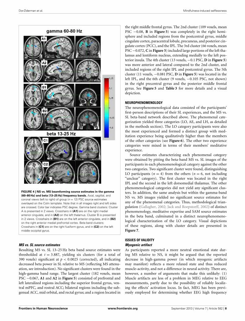
NS vs. MS source estimatesResulting NS vs. MS 60–80 Hz high-gamma band images werethresholded at the maximum t-value possible for a non-parametric random permutation analysis with 2000 permuta-tions (t = 4.863, see methods section for details), yielding 2robust (p < 0.0005, corrected) rather large clusters (314 and264 voxels) spanning almost exclusively frontal regions, and allindicating decreases in gamma power in MS relative to NS(reflecting NS attenuation, see introduction). The larger cluster(mean PSC −0.083, A1 and A2 in Figure 4) was more poste-rior, including right and left precentral gyrus, middle cingulatecortex, middle frontal gyrus and thalamic regions. In the righthemisphere, the cluster included the posterior part of the infe-rior frontal gyrus and operculum, lentiform nucleus and caudatebody. Two thirds of the cluster was in the right hemisphere; how-ever, the left hemisphere PSC was stronger. The second cluster(264 voxels, mean PSC −0.086, B1 and B2 in Figure 4), was moreanterior (prefrontal) and mostly left-lateralized (76%). The clus-ter spanned bilateral dorsal and anterior regions of the medialfrontal gyrus, superior frontal gyrus, and dorsal ACC. Moreventrally, left-lateralized regions included subgenual ACC, midorbital gyrus, middle frontal gyrus and middle cingulate cortex.See Figure 4 and Table 2 for more details and a visual depiction.
In the beta band (13–25 Hz), the NS vs. MS contrast alsoresulted in significant PSC results, albeit markedly less robustcompared to the gamma band results and solely in posteriorregions. These images were thresholded at t = 3.887, yielding3 significant clusters at the p < 0.0025 (corrected) level. Right-hemisphere regions included mainly the fusiform and middletemporal gyrus (48 voxels, mean PSC −0.148, C1 in Figure 4),and a small cluster in the right cerebellum (8 voxels, meanPSC -0.141, not shown). Left-hemisphere regions encompassedmainly the middle occipital and lingual gyrus (36 voxels, meanPSC −0.099, C2 in Figure 4). See Figure 4 and Table 2 for moredetail and visual depiction.
FIGURE 3 | 2D scalp maps of frequency bands with significant power
PSC. 2D topographic representations of significant sensor-level power PSCfor the NS vs. MS high-gamma 60–80 Hz (left), and MS vs. SL beta13–25 Hz (right). Dots on the map represent sensors; color bar scaleindicates PSC from 0.2 (dark red) to −0.2 (dark blue).
Frontiers in Human Neuroscience www.frontiersin.org September 2013 | Volume 7 | Article 582 | 7
Dor-Ziderman et al. Mindfulness-induced selflessness
FIGURE 4 | NS vs. MS beamforming source estimates in the gamma
(60–80 Hz) and beta (13–25 Hz) frequency bands. Axial, sagittal, andcoronal views (left to right) of group (n = 12) PSC source estimatesoverlayed on the Colin template. Note that in all images right and left sidesare crossed. Color bar indicates PSC degree. Gamma band clusters: ClusterA is presented in 2 views. Crosshairs in (A1) are on the right medialanterior cingulate, and in (A2) on the left thalamus. Cluster B is presentedin 2 views. Crosshairs in (B1) are on the left anterior cingulate, and in (B2)
on the right anterior medial prefrontal cortex. Beta band clusters:Crosshairs in (C1) are on the right fusiform gyrus, and in (C2) on the leftmiddle occipital gyrus.
MS vs. SL source estimatesResulting MS vs. SL 13–25 Hz beta band source estimates werethresholded at t = 3.887, yielding six clusters (for a total of390 voxels) significant at p < 0.0025 (corrected), all indicatingdecreased beta power in SL relative to MS (reflecting MS attenu-ation, see introduction). No significant clusters were found in thehigh-gamma band range. The largest cluster (182 voxels, meanPSC −0.067, A1 and A2 in Figure 5) consisted of prefrontal andleft lateralized regions including the superior frontal gyrus, ven-tral mPFC, and rostral ACC; bilateral regions including the sub-genual ACC, mid orbital, and rectal gyrus; and a region located in
the right middle frontal gyrus. The 2nd cluster (109 voxels, meanPSC −0.08, B in Figure 5) was completely in the right hemi-sphere and included regions from the postcentral gyrus, middlecingulate cortex, paracentral lobule, precuneus, and posterior cin-gulate cortex (PCC), and the IPL. The 3rd cluster (66 voxels, meanPSC −0.072, C in Figure 5) included large portions of the left tha-lamus and lentiform nucleus, extending medially to the left pos-terior insula. The 4th cluster (13 voxels, −0.1 PSC, D in Figure 5)was more anterior and lateral compared to the 2nd cluster, andincluded regions of the right IPL and postcentral gyrus. The 5thcluster (11 voxels, −0.081 PSC, D in Figure 5) was located in theleft IPL, and the 6th cluster (9 voxels, −0.105 PSC, not shown)in the right precentral gyrus and the posterior middle frontalgyrus. See Figure 5 and Table 3 for more details and a visualdepiction.
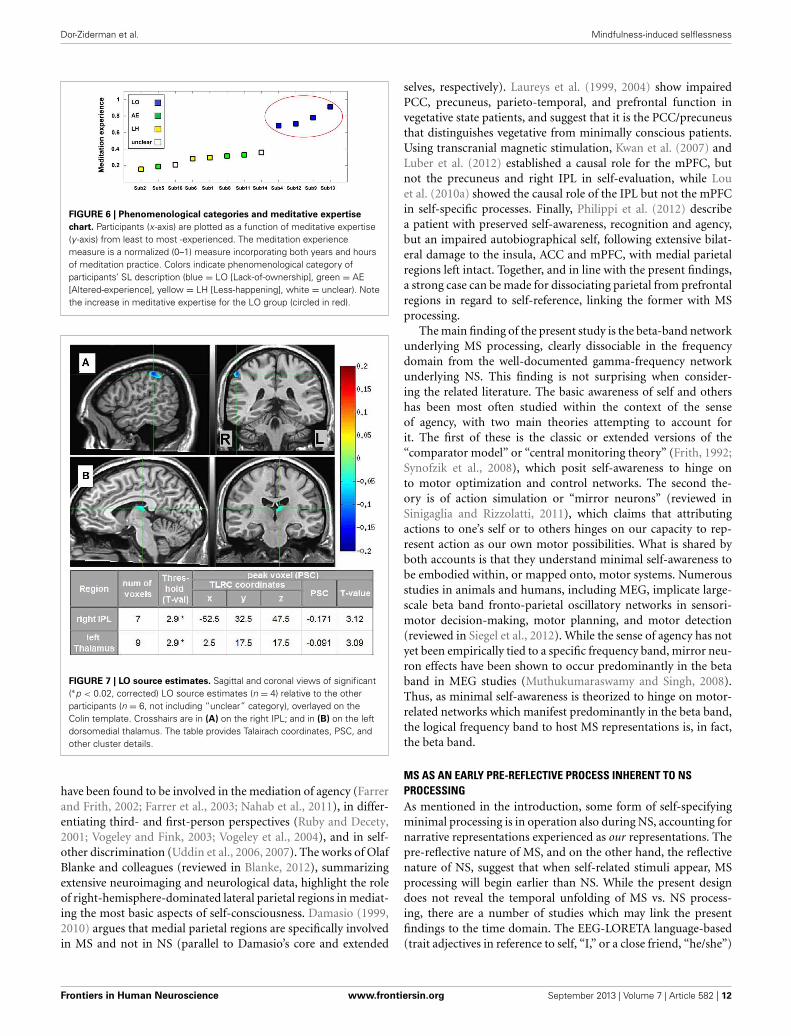
NEUROPHENOMENOLOGYThe neurophenomenological data consisted of the participants’first-person descriptions of their SL experiences, and the MS vs.SL beta-band network described above. The phenomenal cate-gorization yielded three categories (LO, AE, and LH, as detailedin the methods section). The LO category participants were alsothe most experienced and formed a distinct group with med-itation experience being qualitatively higher than the membersof the other categories (see Figure 6). The other two experiencecategories were mixed in terms of their members’ meditativeexperience.
Source estimates characterizing each phenomenal categorywere obtained by pitting the beta-band MS vs. SL images of theparticipants in each phenomenological category against the othertwo categories. Two significant cluster were found, distinguishingLO participants (n = 4) from the others (n = 6, not including“unclear” category). The first cluster was located in the rightIPL and the second in the left dorsomedial thalamus. The otherphenomenological categories did not yield any significant clus-ters. In addition, the same analysis but within the gamma-bandNS vs. MS images yielded no significant source estimates forany of the phenomenal categories. Thus, methodological trian-gulation (Gallagher, 2002; Jack and Roepstorff, 2002) of distinctphenomenology, meditative expertise and SAM source estimatesin the beta band, culminated in a distinct neurophenomeno-logical characterization of the LO category. Visual depictionsof these regions, along with cluster details are presented inFigure 7.
ISSUES OF VALIDITYMyogenic artifactAs participants reported a more neutral emotional state dur-ing MS relative to NS, it might be argued that the reporteddecrease in high-gamma power (in which myogenic artifactsmay manifest) reflects a more relaxed state and thus reducedmuscle-activity, and not a difference in neural activity. There are,however, a number of arguments that make this unlikely: (1)Muscle artifacts are less of a problem in MEG relative to EEGmeasurements, partly due to the possibility of reliably localiz-ing the effects’ activation locus. In fact, MEG has been previ-ously employed for determining whether EEG high frequency
Frontiers in Human Neuroscience www.frontiersin.org September 2013 | Volume 7 | Article 582 | 8

Dor-Ziderman et al. Mindfulness-induced selflessness
Table 2 | NS vs. MS beamforming solutions for gamma (60–80 Hz) and beta (13–25 Hz).
Condition Cluster #
(number of
voxels)
Threshold
(t-value)
Peak voxel (sig change) Hemisphere
overlap
Regions included in cluster
(atlas TT_Daemon)
TLRC coordinates (mm, RAI) PSC t-value
x y z Left (%) Right (%) Name Overlap (%)
NS
vs.M
S(6
0–80
Hz)
1 (314) 4.86** 37.5 7.5 37.5 −0.148 4.86 32.40 65.10 L/R Cingulate gyrus 22.0
L/R Precentral gyrus 20.0
L/R Middle frontal gyrus 11.3
L/R Thalamus 7.5
R Inferior frontal gyrus 3.7
R Lentiform nucleus 2.8
R Postcentral gyrus 1.4
R Caudate 1.2
R Insula 0.9
2 (262) 4.86** 2.5 −42.5 −2.5 −0.108 4.86 76.00 23.1 L/R Medial frontal gyrus 36.5
L/R Anterior cingulate 21.3
L/R Superior frontal gyrus 15.1
L/R Cingulate gyrus 5.6
L Middle frontal gyrus 1.0
NS
vs.M
S(1
3–25
Hz)
1 (48) 3.89* −42.5 57.5 −12.5 −0.189 4.02 0.00 99.60 R Fusiform gyrus 31.0
R Middle temporal gyrus 13.9
R Inferior temporal gyrus 6.0
R Declive 4.8
R Parahippocampal gyrus 3.7
2 (46) 3.89* 32.5 67.5 −2.5 −0.122 4.61 100.00 0.00 L Middle occipital gyrus 26.2
L Lingual gyrus 7.1
L Middle temporal gyrus 5.4
L Posterior cingulate 3.9
L Inferior temporal gyrus 2.7
L Inferior occipital gyrus 2.2
3 (8) 3.89* −32.5 72.5 −32.5 −0.146 3.95 0.00 100.00 R Pyramis 62.4
R Tuber 28.5
Information supplied includes number of voxels in each cluster, thresholds, peak voxel characteristics, hemispheric overlap, and brain regions included in the cluster.
The Afni supplied TT Daemon atlas was used. Due to poor resolution and signal leakage to non-brain regions, overlap percentages do not always add up to 100%.*p < 0.0025; **p < 0.0005 (corrected).
components may have a muscular origin (Zimmermann andScharein, 2004; Claus et al., 2012). (2) Pre-emptive measureswere taken to counter muscle artifacts during both data collec-tion (supine positioning, eyes closed) and data cleaning (visualinspection of all the data). (3) The high-gamma effect foundhere is dissimilar to typical myogenic artifacts (described inMuthukumaraswamy, 2013) in several ways. The effect, on thesensor level is: (a) highly lateralized (and it is unlikely that forall the participants the artifact was confined to one hemisphere);(b) does not extend to the montage borders (which is the nor-mal case for muscle artifacts); and (c) the activity is narrowlyconfined to the 60–80 Hz band (while myogenic artifacts tend tobe “patchy” and to command a wider spectrum). (4) Finally, thereported regions are consistent with a well-established body ofliterature.
In addition, in order to empirically test the link betweenincreased emotionality, the myogenic artifact and increasedhigh-gamma PSC, a further analysis was conducted. The valuesof the peak activation voxels of each of the two NS vs. MS clus-ters (reported in Table 2), were extracted for each participant (bytransposing the group Talairach coordinates back to each partici-pant’s MRI image and its co-registered SAM volume). Then, thesevalues (for each voxel) were sorted into two groups: participantswho spontaneously reported little or no emotions (4 and 1 partic-ipants, respectively) during NS, and participants who did not (7participants). There were no significant differences between thegroups for both tested voxels (2-sample t-test, ns), with the PSCmeans of the decreased emotionality group being actually higherthan the normal group (contrary to what could be expected if thetested hypothesis was correct).
Frontiers in Human Neuroscience www.frontiersin.org September 2013 | Volume 7 | Article 582 | 9

Dor-Ziderman et al. Mindfulness-induced selflessness
FIGURE 5 | MS vs. SL beamforming source estimates in the beta
(13–25 Hz) band. Axial, sagittal, and coronal views (left to right) ofgroup (N = 12) SAM pseudo-F source estimates overlayed on theColin template. Note that in all images right and left sides arecrossed. Color bar indicates PSC degree. Cluster A reveals prefrontaldeactivations in two views: the crosshairs in (A1) are on the left
anterior medial prefrontal gyrus, and in (A2) on the right subgenualanterior cingulate. Cluster (B) shows deactivation in the posteriormedial cortex, with the crosshairs pinpointing the right precuneus.Cluster (C) shows deactivation in the left thalamus; and clusters (D)
and (E) deactivations in the right and left inferior parietal lobules,respectively.
Attentional demandsTo rule out confounds resulting from attentional demands beingdifferent for the different tasks, we ran some additional analyses(on top of the subjective ratings of task success which evidencedno significant differences). The bulk of the meditation-relatedliterature (see Cahn and Polich, 2006 for an extensive review)reports changes in anterior and posterior alpha and/or mid-linetheta oscillatory activity as measures gauging the concentrativeattention-related aspects of meditation. Thus, we checked (usinga robust, cluster-based non-parametric permutations approach,Maris and Oostenveld, 2007) whether any significant clusters inthese frequency bands could be identified. None where found.In addition, activity in the dorsolateral PFC has been specificallyfound to reflect task difficulty, in particular regarding long-term meditators (Brefczynski-Lewis et al., 2007). The fact thatthe present results showed no sign of dorsolateral PFC activitychanges between the conditions can be taken as yet another indi-cation that attentional demands do not account for the reportedresults.
DISCUSSIONIn summarizing the findings of the present study, three mainpoints emerge: (1) NS attenuation is characterized by decreasesin high-gamma (60–80 Hz) oscillatory activity. These are left-hemisphere-dominated and manifest in frontal, thalamic andextensive dorsal and ventral mPFC regions, in line with therelated fMRI literature; (2) MS attenuation is characterized bydecreases in beta-band (13–25 Hz) oscillatory activity in bothoverlapping (with the gamma network) regions including theleft ventral mPFC and thalamus, and a right pre-motor region,
and non-overlapping regions including the right PCC and pre-cuneus medially, and bilateral but right-hemisphere dominatedIPL. While these regions have been previously tied to MS pro-cessing, the frequency band hosting these deactivations—the betaband—is a novel finding of the present study; (3) Phenomenalcharacterization of participants’ descriptions of their SL experi-ences yielded three distinct categories of experience. In particular,the LO group, whose experiences indicated a sharp attenuationof the sense of agency/ownership, and who were also distinct interms of their greater meditative expertise, also evidenced a dis-tinct neural signature characterized by a further attenuation ofthe right IPL and left dorsomedial thalamus in the beta band.The implications of these results are discussed in the followingparagraphs.
NS ATTENUATION IS LINKED TO DECREASED mPFC CORTICALACTIVITY AND DECREASED NEGATIVE EMOTIONSAs predicted, frontal, and especially medial prefrontal, high-gamma-band decreases in oscillatory activity resulted from atten-uating the narrative mode of processing toward a minimalexperiential mode (NS vs. MS, Figure 4). The link betweenNS attenuation and reduced mPFC activity, is, as noted, sup-ported by virtually all fMRI research and review studies regardingself-referential processing (Gusnard et al., 2001; D’Argembeauet al., 2005; Northoff et al., 2006; Christoff et al., 2011; Qin andNorthoff, 2011; Whitfield-Gabrieli et al., 2011; Kim, 2012). Also,as mentioned, intracranial EEG studies (Nir et al., 2007; Jerbiet al., 2010; Ossandón et al., 2011; Ramot et al., 2012) correlateself-referential and DMN blood-oxygenation-level-dependent(BOLD) reductions to suppressed high gamma-band oscillatory
Frontiers in Human Neuroscience www.frontiersin.org September 2013 | Volume 7 | Article 582 | 10

Dor-Ziderman et al. Mindfulness-induced selflessness
Table 3 | MS vs. SL beamforming solutions for gamma (60–80 Hz) and beta (13–25 Hz).
Condition Cluster #
(number of
voxels)
Threshold
(t-Value)
Peak voxel (PSC) Hemisphere
overlap
Regions included in cluster
(atlas TT_Daemon)
TLRC coordinates (mm, RAI) PSC t-value
x y z Left (%) Right (%) Name Overlap (%)
MS
vs.S
L(1
3–25
Hz)
1 (182) 3.89* 2.5 −47.5 2.5 −0.0087 4.1 61.30 36.90 L/R Medial frontal gyrus 35.8
L/R Anterior cingulate 22.8
L Sup frontal gyrus 17.0
R Caudate 2.2
R Middle frontal gyrus 0.8
2 (109) 3.89* −37.5 37.5 47.5 −0.139 3.96 0.00 100.00 R Cingulate gyrus 24.9
R Precuneus 19.8
R Paracentral lobule 14.1
R Inf parietal lobule 5.2
R Postcentral gyrus 4.4
R Supramarginal gyrus 1.7
R Angular gyrus 0.5
3 (66) 3.89* 22.5 27.5 12.5 −0.093 4.1 94.00 2.00 L Thalamus 37.0
L Insula 6.7
L Lentiform nucleus 5.8
L Claustrum 2.4
4 (13) 3.89* −47.5 32.5 47.5 −0.14 4.19 0.00 98.50 R Inf parietal lobule 77.0
R Postcentral gyrus 21.7
5 (11) 3.89* 37.5 32.5 37.5 −0.089 4.02 100.00 0.00 L Inf parietal lobule 40.5
6 (9) 3.89* −42.5 2.5 47.5 −0.122 3.96 0.00 97.40 R Precentral gyrus 66.1
R middle frontal gyrus 33.9
MS
vs.S
L(6
0–80
Hz) No significant clusters
Information supplied includes number of voxels in each cluster, thresholds, peak voxel characteristics, hemispheric overlap, and brain regions included in the cluster.
The Afni supplied TT Daemon atlas was used. Due to poor resolution and signal leakage to non-brain regions, overlap percentages do not always add up to 100%.*p < 0.0025 (corrected).
activity. As existing MEG studies of the self are either event-related studies (Walla et al., 2007) or connectivity studies (Louet al., 2010b), the present study is the first to directly bridge fMRIBOLD and frequency-dependent MEG power results in the con-text of self-referential processing. The robust and extensive mPFCdecreased gamma-power in MS relative to NS provide furtherevidence regarding the neural underpinning of the BOLD fMRIresults, but also, importantly, anchor the results acquired throughMEG to the main fMRI body of literature.
In addition to the reduced mPFC gamma oscillations, NSattenuation was also marked by a dramatic reduction of negativeand mixed (both positive and negative) emotions: from 10 par-ticipants reporting such emotions in NS to only 1 in MS and SL,respectively. These are in alignment with findings directly asso-ciating increased midline activity in DMN regions to self-relatedemotionality (Northoff et al., 2009; Wiebking et al., 2011). As thelink between increased self-focus, mPFC activity, and mood and
anxiety disorders has been previously established (for reviews seeRessler and Mayberg, 2007; Lemogne et al., 2012), the presentfindings supports the notion that approaching self-experiencethrough a more present-centered focus may be critical to humanwell-being (Davidson, 2004). A similar conclusion was reached byKillingsworth and Gilbert (2010) who showed, based on a large-scale web-based experience sampling survey, that a wanderingmind (dominating over 46% of waking experience) is less happythan a mind focused on what it is doing—regardless of the valenceof the activity being engaged.
MEDIAL AND LATERAL PARIETAL BETA-BAND OSCILLATORY ACTIVITYMEDIATE MS PROCESSINGAs mentioned, the MS network evidencing beta-band powerattenuation (MS vs. SL) included posterior medial and lateralparietal regions (Figure 5), which were not part of the NS net-work (in both beta and gamma). The IPL and right precuneus
Frontiers in Human Neuroscience www.frontiersin.org September 2013 | Volume 7 | Article 582 | 11
Dor-Ziderman et al. Mindfulness-induced selflessness
FIGURE 6 | Phenomenological categories and meditative expertise
chart. Participants (x-axis) are plotted as a function of meditative expertise(y -axis) from least to most -experienced. The meditation experiencemeasure is a normalized (0–1) measure incorporating both years and hoursof meditation practice. Colors indicate phenomenological category ofparticipants’ SL description (blue = LO [Lack-of-ownership], green = AE[Altered-experience], yellow = LH [Less-happening], white = unclear). Notethe increase in meditative expertise for the LO group (circled in red).
FIGURE 7 | LO source estimates. Sagittal and coronal views of significant(∗p < 0.02, corrected) LO source estimates (n = 4) relative to the otherparticipants (n = 6, not including “unclear” category), overlayed on theColin template. Crosshairs are in (A) on the right IPL; and in (B) on the leftdorsomedial thalamus. The table provides Talairach coordinates, PSC, andother cluster details.
have been found to be involved in the mediation of agency (Farrerand Frith, 2002; Farrer et al., 2003; Nahab et al., 2011), in differ-entiating third- and first-person perspectives (Ruby and Decety,2001; Vogeley and Fink, 2003; Vogeley et al., 2004), and in self-other discrimination (Uddin et al., 2006, 2007). The works of OlafBlanke and colleagues (reviewed in Blanke, 2012), summarizingextensive neuroimaging and neurological data, highlight the roleof right-hemisphere-dominated lateral parietal regions in mediat-ing the most basic aspects of self-consciousness. Damasio (1999,2010) argues that medial parietal regions are specifically involvedin MS and not in NS (parallel to Damasio’s core and extended
selves, respectively). Laureys et al. (1999, 2004) show impairedPCC, precuneus, parieto-temporal, and prefrontal function invegetative state patients, and suggest that it is the PCC/precuneusthat distinguishes vegetative from minimally conscious patients.Using transcranial magnetic stimulation, Kwan et al. (2007) andLuber et al. (2012) established a causal role for the mPFC, butnot the precuneus and right IPL in self-evaluation, while Louet al. (2010a) showed the causal role of the IPL but not the mPFCin self-specific processes. Finally, Philippi et al. (2012) describea patient with preserved self-awareness, recognition and agency,but an impaired autobiographical self, following extensive bilat-eral damage to the insula, ACC and mPFC, with medial parietalregions left intact. Together, and in line with the present findings,a strong case can be made for dissociating parietal from prefrontalregions in regard to self-reference, linking the former with MSprocessing.
The main finding of the present study is the beta-band networkunderlying MS processing, clearly dissociable in the frequencydomain from the well-documented gamma-frequency networkunderlying NS. This finding is not surprising when consider-ing the related literature. The basic awareness of self and othershas been most often studied within the context of the senseof agency, with two main theories attempting to account forit. The first of these is the classic or extended versions of the“comparator model” or “central monitoring theory” (Frith, 1992;Synofzik et al., 2008), which posit self-awareness to hinge onto motor optimization and control networks. The second the-ory is of action simulation or “mirror neurons” (reviewed inSinigaglia and Rizzolatti, 2011), which claims that attributingactions to one’s self or to others hinges on our capacity to rep-resent action as our own motor possibilities. What is shared byboth accounts is that they understand minimal self-awareness tobe embodied within, or mapped onto, motor systems. Numerousstudies in animals and humans, including MEG, implicate large-scale beta band fronto-parietal oscillatory networks in sensori-motor decision-making, motor planning, and motor detection(reviewed in Siegel et al., 2012). While the sense of agency has notyet been empirically tied to a specific frequency band, mirror neu-ron effects have been shown to occur predominantly in the betaband in MEG studies (Muthukumaraswamy and Singh, 2008).Thus, as minimal self-awareness is theorized to hinge on motor-related networks which manifest predominantly in the beta band,the logical frequency band to host MS representations is, in fact,the beta band.
MS AS AN EARLY PRE-REFLECTIVE PROCESS INHERENT TO NSPROCESSINGAs mentioned in the introduction, some form of self-specifyingminimal processing is in operation also during NS, accounting fornarrative representations experienced as our representations. Thepre-reflective nature of MS, and on the other hand, the reflectivenature of NS, suggest that when self-related stimuli appear, MSprocessing will begin earlier than NS. While the present designdoes not reveal the temporal unfolding of MS vs. NS process-ing, there are a number of studies which may link the presentfindings to the time domain. The EEG-LORETA language-based(trait adjectives in reference to self, “I,” or a close friend, “he/she”)
Frontiers in Human Neuroscience www.frontiersin.org September 2013 | Volume 7 | Article 582 | 12
Dor-Ziderman et al. Mindfulness-induced selflessness
event-related study (Esslen et al., 2008) is particularly interest-ing, as it not only distinguishes, in line with the present and otherfindings (Zysset et al., 2003; Northoff et al., 2006; Schneider et al.,2008), the dorsal and ventral mPFC as differentially involved inreflective vs. pre-reflective self processing, but also determinestheir activation time-course. In the pre-reflective self condition,both the ventral mPFC and the insula were activated as early as134–170 ms post stimulus, while differential dorsal mPFC acti-vation in self vs. other -reference was only found when averagingover the whole time-course (700 ms). These temporal distinctionsare upheld by a single MEG sensor-level study of self-awarenessfound in the literature (Walla et al., 2007). This event-relatedstudy, also language-based, examined encoding effects of theGerman language equivalents of “a,” “his” and “mine,” assumed toreflect different levels of self representation. The results indicatedearly (200–400 ms) and late (500–800 ms) time window effects.The 2D topography of the early time window reveals differentialactivity in posterior central electrodes (and in a few prefrontalones), very similar to the 13–25 Hz beta-band 2D scalp map(Figure 3). Walla et al. interpret the early window effect as indi-cating a stage when the perceptual object, here a word, has notyet been branded as self/non-self, or in other words, a pre-MSstage. In contrast, the 2D representation of the late time windowbears striking similarity to the 60–80 Hz high-gamma 2D scalpmap presented in Figure 3 (frontal left activity). The similaritybetween the 2D cortical maps of MS and the early window, andNS and the later time window, together with the ventral vs. dorsalmPFC differential activation which holds both in terms of tempo-rality (early vs. late time windows) and in terms of self processingmode (MS vs. NS), argue in favor of MS reflecting an early processinherent to the cognition of NS processing.
THE NEUROPHENOMENOLOGY OF MINDFULNESS-INDUCEDSELFLESSNESSPhenomenology played a double role in the present study, guid-ing both its design as well as data analysis. Regarding design,this study was inspired by “front-loading phenomenologicalinsights into experimental design” (Gallagher and Sørensen,2006). Specifically, and like other studies (Hasenkamp et al.,2012), mindfulness was employed in the spirit expressed by Varelaet al. (1991) as a “. . . disciplined perspective on human experiencethat can enlarge the domain of cognitive science to include directexperience . . . ” (p. 33). Requesting long-term mindfulness prac-titioners to produce in laboratory settings the state of SL alloweda unique view of the neural correlates specific to the “mini-mal” aspect of momentary experience, rendering these aspects ofhuman experience scientifically tractable (Lutz et al., 2007).
Regarding data analysis, collecting first-person descriptions ofthe SL experience allowed grouping the data into three distinctphenomenological categories (Table 1): AE descriptions indi-cated an altered spatial/sensual perspective of self experience,while LH descriptions indicated an attenuation of experience/r.LO descriptions, on the other hand, produced by participants cul-tured by a qualitatively greater meditation experience (Figure 6),indicated an attenuation of the agentive/ownership aspectsaccompanying experience. Despite the different phenomenolog-ical descriptions, all of the participants reported similar high
rates of success and stability in all the tasks (including SL) rela-tive to their past experiences (see section 3.1). This discrepancycan be interpreted as indicating a diminished MS experience forall participants, but diminished through different strategies andaccompanied by distinct phenomenological experiences. In par-ticular, the more experienced LO group is interesting as theirdescriptions indicate a specific subtraction of agency/ownershipfrom momentary experience. This distinct phenomenology wasthen tied to a distinct neural signature: a further attenuation ofbeta-band power (relative to the AE and LH groups) in the leftdorsomedial thalamus and right IPL (Figure 7).
Subcortical regions have only recently begun to be incorpo-rated into theories of self-awareness (see Northoff and Panksepp,2008; Damasio, 2010 and Christoff et al., 2011). The reportedsuppressed beta power in the dorsomedial thalamus support theseresearchers’ hypotheses regarding the crucial involvement of sub-cortical circuits in the mediation of primal mammalian coreprocesses tagging phenomena as self/not-self, which then feedinto higher MS cortical representations. On the cortical level, theright inferior parietal sulcus has been highlighted as a region inte-grating multisensory bodily signals and reflecting the consciousexperience of being an “I,” a spatially localized entity correspond-ing to first-person perspective and identity (Ionta et al., 2011;Blanke, 2012). The IPL has also been hypothesized as a key regionresponsible for the sense of agency and subjective sense of con-trol (e.g., Farrer et al., 2008; Nahab et al., 2011; Haggard andChambon, 2012). Along with these studies, the present findingssupport the role of this region in reflecting one of the most aston-ishing features of the human mind, the subjective “self as I” aspectof conscious experience, and put forth the hypothesis—to beexamined by subsequent research—that it is mediated specificallywithin the beta band.
STUDY LIMITATIONSOne limitation of the study concerns its unique participants. Weacknowledge a potential lack of generalizability to non-vipassanaand non-meditator general populations, in particular regardingthe state of SL, which is an experience cultured by meditationpractice and comprehensible from a Buddhist, but perhaps notWestern, point of view (but see Metzinger, 2003). Another lim-itation regards the small sample of participants, especially inthe neurophenomenological analysis, which yielded very smallgroups. Thus, the results reported here warrant replication ina larger group and in other meditative traditions. In addition,the reported phenomenological analysis is rudimentary in nature.This is partly due to the experimental conditions of interview-ing participants via intercom between tasks, but partly also to theexploratory nature of the advent of translating phenomenologicalinsights of long-established contemplative traditions into currentneurocognitive terms. Future studies can build on these prelim-inary results and develop more sophisticated phenomenologicalcharacterizations of self and selfless modes of awareness usingmore rigorous qualitative/phenomenological analysis methods.
CONCLUDING REMARKSThe present study highlighted the role of frequency-dependentnetworks, dissociable in the frequency domain but partially
Frontiers in Human Neuroscience www.frontiersin.org September 2013 | Volume 7 | Article 582 | 13

Dor-Ziderman et al. Mindfulness-induced selflessness
overlapping in brain topography, in supporting different modesof self-processing. These results emphasize the unique contri-bution of MEG to the neuroimaging self-awareness literature.In addition, the present study illustrated the utility of combin-ing first-person reports, neuroimaging, and Buddhist-inspiredmind training for scientifically characterizing selflessness. Indeed,a non-trivial outcome of the present study is that long-termmindfulness meditators are actually able, under experimentalconditions, to successfully produce and steadily hold a self-less mode of awareness. This state of mind, which is aliento normal non-pathological conscious experience and whichhas not been previously scientifically documented and neu-rocognitively mapped, allows a unique glance at the neural
underpinnings of the more subtle and basic processes of self-awareness.
ACKNOWLEDGMENTSWe thank Prof. Rafael Malach for the anatomical data fromhis lab, and Dr. Stephen Fulder for his assistance in designingthe experiment. This research was supported by the Mind andLife Institute, Francisco J. Varela Research Award 2012-Varela-Berkovich, and by a grant from the Bial Foundation (27/10).This paper is based on a thesis written by the first author andsupervised by two of the authors (Joseph Glicksohn and AbrahamGoldstein), submitted to Bar-Ilan University in partial fulfillmentof the requirements toward an M.Sc. degree.
REFERENCESAndrews-Hanna, J. R., Reidler, J. S.,
Sepulcre, J., Poulin, R., and Buckner,R. L. (2010). Functional-anatomicfractionation of the brain’s defaultnetwork. Neuron 65, 550–562. doi:10.1016/j.neuron.2010.02.005
Austin, J. H. (2000). Consciousnessevolves when the self dissolves.J. Conscious. Stud. 7, 209–230.
Berkovich-Ohana, A., Glicksohn,J., and Goldstein, A. (2012).Mindfulness-induced changes ingamma band activity - implicationsfor the default mode network,self-reference and attention. Clin.Neurophysiol. 123, 700–710. doi:10.1016/j.clinph.2011.07.048
Blanke, O. (2012). Multisensorybrain mechanisms of bodily self-consciousness. Nat. Neurosci. 13,556–571. doi: 10.1038/nrn3292
Brefczynski-Lewis, J. A., Lutz, A.,Schaefer, H. S., Levinson, D. B.,and Davidson, R. J. (2007). Neuralcorrelates of attentional expertisein long-term meditation prac-titioners. Proc. Natl. Acad. Sci.U.S.A. 104, 11483–11488. doi:10.1073/pnas.0606552104
Brewer, J. A., Worhunsky, P. D., Gray,J. R., Tang, Y.-Y., Weber, J., andKober, H. (2011). Meditation expe-rience is associated with differencesin default mode network activityand connectivity. Proc. Natl. Acad.Sci. U.S.A. 108, 20254–20259. doi:10.1073/pnas.1112029108
Brookes, M. J., Hale, J. R., Zumer,J. M., Stevenson, C. M., Francis,S. T., Barnes, G. R., et al. (2011).Measuring functional connec-tivity using MEG: methodologyand comparison with fcMRI.Neuroimage 56, 1082–1104. doi:10.1016/j.neuroimage.2011.02.054
Brookes, M. J., Vrba, J., Robinson,S. E., Stevenson, C. M., Peters, A.M., Barnes, G. R., et al. (2008).Optimising experimental designfor MEG beamformer imaging.
Neuroimage 39, 1788–1802. doi:10.1016/j.neuroimage.2007.09.050
Buckner, R. L., Andrews-Hanna, J.R., and Schacter, D. L. (2008).The brain’s default network. Ann.N.Y. Acad. Sci. 1124: 1–38. doi:10.1196/annals.1440.011
Cahn, B. R., and Polich, J. (2006).Meditation states and traits: EEG,ERP, and neuroimaging studies.Psychol. Bull. 132, 180–211. doi:10.1037/0033-2909.132.2.180
Chambers, R., Gullone, E., and Allen,N. B. (2009). Mindful emo-tion regulation: an integrativereview. Clin. Psychol. Rev. 29,560–572. doi: 10.1016/j.cpr.2009.06.005
Chiesa, A., and Serretti, A. (2010).A systematic review of neuro-biological and clinical featuresof mindfulness meditations.Psychol. Med. 40, 1239–1252. doi:10.1017/S0033291709991747
Chiesa, A., Calati, R., and Serretti,A. (2011). Does mindfulnesstraining improve cognitive abil-ities? A systematic review ofneuropsychological findings. Clin.Psychol. Rev. 31, 449–464. doi:10.1016/j.cpr.2010.11.003
Christoff, K., Cosmelli, D., Legrand,D., and Thompson, E. (2011).Specifying the self for cog-nitive neuroscience. TrendsCogn. Sci. 15, 104–112. doi:10.1016/j.tics.2011.01.001
Christoff, K., Gordon, A. M.,Smallwood, J., Smith, R., andSchooler, J. W. (2009). Experiencesampling during fMRI revealsdefault network and executivesystem contributions to mindwandering. Proc. Natl. Acad.Sci. U.S.A. 106, 8719–8724. doi:10.1073/pnas.0900234106
Claus, S., Velis, D., Lopes da Silva, F.H., Viergever, M. A., and Kalitzin,S. (2012). High frequency spectralcomponents after secobarbital:the contribution of muscular
origin–a study with MEG/EEG.Epilepsy Res. 100, 132–141. doi:10.1016/j.eplepsyres.2012.02.002
Dalai Lama, XIV. (1991). Path to Bliss.Ithaca, NY: Snow Lion.
Damasio, A., Damasio, H., andTranel, D. (2012). Persistenceof feelings and sentience afterbilateral damage of the insula.Cereb. Cortex 23, 833–846. doi:10.1093/cercor/bhs077
Damasio, A. R. (1999). The Feeling ofWhat Happens: Body and Emotionin the Making of Consciousness. NewYork, NY: Harcourt Inc.
Damasio, A. R. (2010). When SelfComes to Mind. New York, NY:Pantheon Books.
D’Argembeau, A., Collette, F., Van derLinden, M., Laureys, S., Del Fiore,G., Deguelrdre, C., et al. (2005).Self-referential reflective activityand its relationship with rest: a PETstudy. Neuroimage 25, 616–624. doi:10.1016/j.neuroimage.2004.11.048
David, N., Newen, A., and Vogeley, K.(2008). The “sense of agency”and its underlying cogni-tive and neural mechanisms.Conscious. Cogn. 17, 523–534. doi:10.1016/j.concog.2008.03.004
Davidson, R. J. (2004). Well-being andaffective style: neural substrates andbiobehavioural correlates. Philos.Trans. R. Soc. Lond. B Biol. Sci. 359,1395–1411. doi: 10.1098/rstb.2004.1510
Davidson, R. J., and Lutz, A. (2008).Buddha’s brain: neuroplastic-ity and meditation. IEEE SignalProcess. Mag. 25, 171–176. doi:10.1109/MSP.2008.4431873
Dreyfus, G., and Thompson, E.(2007). “Asian perspectives:Indian theories of mind,” inThe Cambridge Handbook ofConsciousness, eds E. Thompson,P. D. Zelazo, and M. Moscovitch(Cambridge: Cambridge UniversityPress), 89–114. doi: 10.1017/CBO9780511816789.006
Ekman, P., Davidson, R. J., Ricard, M.,and Wallace, A. (2005). Buddhistand psychological perspectives onemotions and well-being. Curr.Dir. Psychol. Sci. 14, 59–63. doi:10.1111/j.0963-7214.2005.00335.x
Engler, J. (2003). “Being somebodyand being nobody: a reexamina-tion of the understanding of self inpsychoanalysis and Buddhism,” inPsychoanalysis and Buddhism, ed J.D. Safran (Somerville, MA: WisdomPublications), 35–100.
Esslen, E., Metzler, S., Pascual-Marqui, R., and Jancke, L. (2008).Pre-reflective and reflective self-reference: a spatiotemporal EEGanalysis. Neuroimage 42, 437–449.doi: 10.1016/j.neuroimage.2008.01.060
Farb, N. A. S., Segal, Z. V., Mayberg,H., Bean, J., McKeon, D., Fatima,Z., et al. (2007). Attending tothe present: mindfulness med-itation reveals distinct neuralmodes of self-reference. Soc. Cogn.Affect. Neurosci. 2, 313–322. doi:10.1093/scan/nsm030
Farrer, C., Franck, N., Georgieff,N., Frith, C. D., Decety, J., andJeannerod, M. (2003). Modulatingthe experience of agency: a positronemission tomography study.Neuroimage 18, 324–333. doi:10.1016/S1053-8119(02)00041-1
Farrer, C., Frey, S. H., Van Horn, J. D.,Tunik, E., Turk, D., Inati, S., et al.(2008). The angular gyrus com-putes action awareness representa-tions. Cereb. Cortex 18, 254–261.doi: 10.1093/cercor/bhm050
Farrer, C., and Frith, C. D. (2002).Experiencing oneself vs anotherperson as being the cause ofan action: the neural correlatesof the experience of agency.Neuroimage 15, 596–603. doi:10.1006/nimg.2001.1009
Forman, S. D., Cohen, J. D., Fitzgerald,M., Eddy, W. F., Mintun, M. A.,and Noll, D. C. (1995). Improved
Frontiers in Human Neuroscience www.frontiersin.org September 2013 | Volume 7 | Article 582 | 14
Dor-Ziderman et al. Mindfulness-induced selflessness
assessment of significant acti-vation in functional magneticresonance imaging (fMRI): useof a cluster-size threshold. Magn.Reson. Med. 33, 636–647. doi:10.1002/mrm.1910330508
Frith, C. D. (1992). The CognitiveNeuropsychology of Schizophrenia.Hillsdale, NJ: Erlbaum.
Froeliger, B., Garland, E. L., Kozink,R. V., Modlin, L. A., Chen, N.-K.,McClernon, F. J., et al. (2012).Meditation-state functional con-nectivity (msfc): strengthening ofthe dorsal attention network andbeyond. Evid. Based Complement.Alternat. Med. 2012, 680407. doi:10.1155/2012/680407
Gallagher, S. (2000). Philosophical con-ceptions of the self: implicationsfor cognitive science. Trends Cogn.Sci. 4, 14–21. doi: 10.1016/S1364-6613(99)01417-5
Gallagher, S. (2002). Experimentingwith introspection. Trends Cogn.Sci. 6, 374–375. doi: 10.1016/S1364-6613(02)01979-4
Gallagher, S. (2004). Neurocognitivemodels of schizophrenia: a neu-rophenomenological critique.Psychopathology 37, 8–19. doi:10.1159/000077014
Gallagher, S., and Sørensen, J. B.(2006). Experimenting with phe-nomenology. Conscious. Cogn. 15,119–134. doi: 10.1016/j.concog.2005.03.002
Gardiner, J. M. (2001). Episodicmemory and autonoetic con-sciousness: a first-person approach.Philos. Trans. R. Soc. Lond. BBiol. Sci. 356, 1351–1361. doi:10.1098/rstb.2001.0955
Goleman, D. (1995). EmotionalIntelligence. New York, NY: BantamBooks.
Gusnard, D. A., Akbudak, E., Shulman,G. L., and Raichle, M. E. (2001).Medial prefrontal cortex andself-referential mental activity:relation to a default mode ofbrain function. Proc. Natl. Acad.Sci. U.S.A. 98, 4259–4264. doi:10.1073/pnas.071043098
Gusnard, D. A., and Raichle, M. E.(2001). Searching for a baseline:functional imaging and the restinghuman brain. Nat. Rev. Neurosci. 2,685–694. doi: 10.1038/35094500
Haggard, P., and Chambon, V. (2012).Sense of agency. Curr. Biol. 22,390–392. doi: 10.1016/j.cub.2012.02.040
Hansen, P., Kringelbach, M., andSalmelin, R. (eds.). (2010). MEG:An Introduction to Methods. NewYork, NY: Oxford UniversityPress. doi: 10.1093/acprof:oso/9780195307238.001.0001
Hart, W. (1987). The Art of Living:Vipassana Meditation. SanFrancisco, CA: HarperOne.
Hasenkamp, W., and Barsalou, L.W. (2012). Effects of meditationexperience on functional connec-tivity of distributed brain networks.Front. Hum. Neurosci. 6:38. doi:10.3389/fnhum.2012.00038
Hasenkamp, W., Wilson-Mendenhall,C. D., Duncan, E., and Barsalou,L. W. (2012). Mind wanderingand attention during focused med-itation: a fine-grained temporalanalysis of fluctuating cognitivestates. Neuroimage 59, 750–760. doi:10.1016/j.neuroimage.2011.07.008
Hölzel, B. K., Carmody, J., Vangel, M.,Congleton, C., Yerramsetti, S. M.,Gard, T., et al. (2011a). Mindfulnesspractice leads to increases inregional brain gray matter density.Psychiatry Res.191, 36–43. doi:10.1016/j.pscychresns.2010.08.006
Hölzel, B. K., Lazar, S. W., Gard,T., Schuman-Olivier, Z., Vago,D. R., and Ott, U. (2011b). Howdoes mindfulness meditationwork? Proposing mechanismsof action from a conceptualand neural perspective. Perspect.Psychol. Sci. 6, 537–559. doi:10.1177/1745691611419671
Hölzel, B. K., Ott, U., Gard, T., Hempel,H., Weygandt, M., Morgen, K.,et al. (2008). Investigation of mind-fulness meditation practitionerswith voxel-based morphome-try. Soc. Cogn. Affect. Neurosci.3, 55–61. doi: 10.1093/scan/nsm038
Ionta, S., Heydrich, L., Lenggenhager,B., Mouthon, M., Fornari, E.,Chapuis, D., et al. (2011).Multisensory mechanisms intemporo-parietal cortex supportself-location and first-person per-spective. Neuron 70, 363–374. doi:10.1016/j.neuron.2011.03.009
Ives-Deliperi, V. L., Solms, M., andMeintjes, E. M. (2011). Theneural substrates of mindful-ness: an fMRI investigation.Soc. Neurosci. 6, 231–242. doi:10.1080/17470919.2010.513495
Jack, A. I., and Roepstorff, A. (2002).Introspection and cognitive brainmapping: from stimulus–responseto script–report. Trends Cogn. Sci.6, 333–339. doi: 10.1016/S1364-6613(02)01941-1
James, W. (1890). The Principlesof Psychology. New York, NY:Henry-Holt and Company. doi:10.1037/11059-000
Jerbi, K., Vidal, J. R., Ossandón, T.,Dalal, S. S., Jung, J., Hoffmann, D.,et al. (2010). Exploring the elec-trophysiological correlates of the
default-mode network with intrac-erebral EEG. Front. Syst. Neurosci.4:27. doi: 10.3389/fnsys.2010.00027
Jung, T. P., Makeig, S., Westerfield, M.,Townsend, J., Courchesne, E., andSejnowski, T. J. (2000). Removalof eye activity artifacts from visualevent-related potentials in nor-mal and clinical subjects. Clin.Neurophysiol. 111, 1745–1758. doi:10.1016/S1388-2457(00)00386-2
Kabat-Zinn, J. (1982). An outpatientprogram in behavioral medicinefor chronic pain patients basedon the practice of mindfulnessmeditation: theoretical considera-tions and preliminary results. Gen.Hosp. Psychiatry 4, 33–47. doi:10.1016/0163-8343(82)90026-3
Kabat-Zinn, J. (1990). Full CatastropheLiving: Using the Wisdom of YourBody and Mind to Face Stress, Pain,and Illness. New York, NY: Delta.
Kabat-Zinn, J., Massion, A. O.,Kristeller, J., Peterson, L. G.,Fletcher, K. E., Pbert, L.,et al. (1992). Effectiveness of ameditation-based stress reductionprogram in the treatment of anxietydisorders. Am. J. Psychiatry 149,936–943.
Kerr, C. E., Josyula, K., and Littenberg,R. (2011). Developing an observingattitude: an analysis of meditationdiaries in an MBSR clinical trial.Clin. Psychol. Psychother. 18, 80–93.doi: 10.1002/cpp.700
Killingsworth, M. A., and Gilbert, D.T. (2010). A wandering mind is anunhappy mind. Science 330, 932.doi: 10.1126/science.1192439
Kim, H. (2012). A dual-subsystemmodel of the brain’s default net-work: self-referential processing,memory retrieval processes, andautobiographical memory retrieval.Neuroimage 61, 966–977. doi:10.1016/j.neuroimage.2012.03.025
Kwan, V. S., Barrios, V., Ganis, G.,Gorman, J., Lange, C., Kumar, M.,et al. (2007). Assessing the neuralcorrelates of self-enhancement bias:a transcranial magnetic stimulationstudy. Exp. Brain Res. 182, 379–385.doi: 10.1007/s00221-007-0992-2
Laureys, S., Goldman, S., Phillips, C.,Van Bogaert, P., Aerts, J., andLuxen, A. (1999). Impaired effec-tive cortical connectivity in vegeta-tive state. Neuroimage 9, 377–382.doi: 10.1006/nimg.1998.0414
Laureys, S., Owen, A. M., and Schiff, N.D. (2004). Brain function in coma,vegetative state, and related disor-ders. Lancet Neurol. 3, 537–546. doi:10.1016/S1474-4422(04)00852-X
Lazar, S. W., Kerr, C. E., Wasserman,R. H., Gray, J. R., Greve, D. N.,Treadway, M. T., et al. (2005).
Meditation experience is associatedwith increased cortical thick-ness. Neuroreport 16, 1893–1897.doi: 10.1097/01.wnr.0000186598.66243.19
Legrand, D., and Ruby, P. (2009). Whatis self-specific? Theoretical investi-gation and critical review of neu-roimaging results. Psychol. Rev. 116,252–282. doi: 10.1037/a0014172
Lehmann, D., Faber, P. L., Tei, S.,Pascual-Marqui, R. D., Milz, P.,and Kochi, K. (2012). Reducedfunctional connectivity betweencortical sources in five meditationtraditions detected with laggedcoherence using EEG tomography.Neuroimage 60, 1574–1586. doi:10.1016/j.neuroimage.2012.01.042
Lemogne, C., Delaveau, P., Freton,M., Guionnet, S., and Fossati, P.(2012). Medial prefrontal cortexand the self in major depression.J. Affect. Disord. 136, e1–e11. doi:10.1016/j.jad.2010.11.034
Lou, H. C., Gross, J., Biermann-Ruben,K., Kjaer, T. W., and Schnitzler, A.(2010b). Coherence in conscious-ness: paralimbic gamma synchronyof self-reference links consciousexperiences. Hum. Brain Mapp. 31,185–192. doi: 10.1002/hbm.20855
Lou, H. C., Luber, B., Stanford, A.,and Lisanby, S. H. (2010a). Self-specific processing in the defaultnetwork: a single-pulse TMS study.Exp. Brain Res. 207, 27–38. doi:10.1007/s00221-010-2425-x
Luber, B., Lou, H. C., Keenan, J. P.,and Lisanby, S. H. (2012). Self-enhancement processing in thedefault network: a single-pulseTMS study. Exp. Brain Res. 223,177–187. doi: 10.1007/s00221-012-3249-7
Lutz, A., Dunne, J. D., and Davidson,R. J. (2007). “Meditation andthe neuroscience of conscious-ness,” in Cambridge Handbook ofConsciousness, eds P. Zelazo, M.Moscovitch, and E. Thompson(New York, NY: CambridgeUniversity Press), 499–555. doi:10.1017/CBO9780511816789.020
Lutz, A., Slagter, H. A., Dunne, J.D., and Davidson, R. J. (2008).Attention regulation and mon-itoring in meditation. TrendsCogn. Sci. 12, 163–169. doi:10.1016/j.tics.2008.01.005
Lutz, A., and Thompson, E. (2003).Neurophenomenology: integratingsubjective experience and braindynamics in the neuroscience ofconsciousness. J. Conscious. Stud.10, 31–52.
Mantini, D., Perrucci, M. G.,Del Gratta, C., Romani, G.L., and Corbetta, M. (2007).
Frontiers in Human Neuroscience www.frontiersin.org September 2013 | Volume 7 | Article 582 | 15

Dor-Ziderman et al. Mindfulness-induced selflessness
Electrophysiological signaturesof resting state networks in thehuman brain. Proc. Natl. Acad.Sci. U.S.A. 104, 13170–13175. doi:10.1073/pnas.0700668104
Maris, E., and Oostenveld, R. (2007).Nonparametric statistical testing ofEEG- and MEG-data. J. Neurosci.Methods 164, 177–190. doi:10.1016/j.jneumeth.2007.03.024
Mason, M. F., Norton, M. I., VanHorn, J. D., Wegner, D. M., Grafton,S. T., and Macrae, C. N. (2007).Wandering minds: the defaultnetwork and stimulus-independentthought. Science 315, 393–395. doi:10.1126/science.1131295
Metzinger, T. (2003). Being No One. TheSelf-Model Theory of Subjectivity.Cambridge: MIT Press.
Muthukumaraswamy, S. D. (2013).High-frequency brain activity andmuscle artifacts in MEG/EEG:a review and recommendations.Front. Hum. Neurosci. 7:138. doi:10.3389/fnhum.2013.00138
Muthukumaraswamy, S. D., andSingh, K. D. (2008). Modulationof the human mirror neuronsystem during cognitive activity.Psychophysiology 45, 896–905. doi:10.1111/j.1469-8986.2008.00711.x
Nahab, F. B., Kundu, P., Gallea, C.,Kakareka, J., Pursley, R., Pohida,T., et al. (2011). The neural pro-cesses underlying self-agency. Cereb.Cortex 21, 48–55. doi: 10.1093/cer-cor/bhq059
Neisser, U. (1988). Five kinds of self-knowledge. Philos. Psychol. 1, 35–59.doi: 10.1080/09515088808572924
Nichols, T. E., and Holmes, A. P.(2001). Nonparametric per-mutation tests for functionalneuroimaging: a primer with exam-ples. Hum. Brain Mapp. 15, 1–25.doi: 10.1002/hbm.1058
Nir, Y., Fisch, L., Mukamel, R., Gelbard-Sagiv, H., Arieli, A., Fried, I., et al.(2007). Coupling between neuronalfiring rate, gamma LFP, and BOLDfMRI is related to interneuronal cor-relations. Curr. Biol. 17, 1275–1285.doi: 10.1016/j.cub.2007.06.066
Northoff, G., Heinzel, A., de Greck, M.,Bermpohl, F., Dobrowolny, H., andPanksepp, J. (2006). Self-referentialprocessing in our brain–a meta-analysis of imaging studies on theself. Neuroimage 31, 440–457. doi:10.1016/j.neuroimage.2005.12.002
Northoff, G., and Panksepp, J.(2008). The trans-species conceptof self and the subcortical-cortical midline system. TrendsCogn. Sci. 12, 259–264. doi:10.1016/j.tics.2008.04.007
Northoff, G., Qin, P., and Feinberg,T. E. (2011). Brain imaging of
the self–conceptual, anatomi-cal and methodological issues.Conscious. Cogn. 20, 52–63. doi:10.1016/j.concog.2010.09.011
Northoff, G., Schneider, F., Rotte,M., Matthiae, C., Tempelmann,C., and Wiebking, C. (2009).Differential parametric modulationof self-relatedness and emotionsin different brain regions. Hum.Brain Mapp. 30, 369–382. doi:10.1002/hbm.20510
Nydahl, O. (2008). The Way Things Are:A Living Approach to Buddhism. UK:O Books.
Olendzki, A. (2010). UnlimitingMind: The Radically ExperientialPsychology of Buddhism. Somerville,MA: Wisdom Publications.
Oostenveld, R., Fries, P., Maris, E., andSchoffelen, J.-M. (2011). FieldTrip:open source software for advancedanalysis of MEG, EEG, and invasiveelectrophysiological data. Comput.Intell. Neurosci. 2011, 156869. doi:10.1155/2011/156869
Ossandón, T., Jerbi, K., Vidal, J. R.,Bayle, D. J., Henaff, M. A., Jung,J., et al. (2011). Transient sup-pression of broadband gammapower in the default-mode networkis correlated with task complex-ity and subject performance.J. Neurosci. 31, 14521–14530. doi:10.1523/JNEUROSCI.2483-11.2011
Pagnoni, G., Cekic, M., and Guo,Y. (2008). “Thinking about Not-Thinking”: neural correlates of con-ceptual processing during zen med-itation. PLoS ONE 3:e3083. doi:10.1371/journal.pone.0003083
Philippi, C. L., Feinstein, J. S., Khalsa,S. S., Damasio, A., Tranel, D.,Landini, G., et al. (2012). Preservedself-awareness following exten-sive bilateral brain damage tothe insula, anterior cingulate,and medial prefrontal cor-tices. PLoS ONE 7:e38413. doi:10.1371/journal.pone.0038413
Qin, P., and Northoff, G. (2011). Howis our self related to midline regionsand the default-mode network?Neuroimage 57, 1221–1233. doi:10.1016/j.neuroimage.2011.05.028
Raichle, M. E., MacLeod, A. M.,Snyder, A. Z., Powers, W. J.,Gusnard, D. A., and Shulman,G. L. (2001). A default mode ofbrain function. Proc. Natl. Acad.Sci. U.S.A. 98, 676–682. doi:10.1073/pnas.98.2.676
Ramot, M., Fisch, L., Harel, M.,Kipervasser, S., Andelman, F.,Neufeld, M. Y., et al. (2012).A widely distributed spectralsignature of task-negative electro-corticography responses revealedduring a visuomotor task in
the human cortex. J. Neurosci.32, 10458–10469. doi: 10.1523/JNEUROSCI.0877-12.2012
Ressler, K. J., and Mayberg, H. S.(2007). Targeting abnormal neuralcircuits in mood and anxiety dis-orders: from the laboratory to theclinic. Nat. Neurosci. 10, 1116–1124.doi: 10.1038/nn1944
Robinson, S. E., and Vrba, J. (1999).“Functional neuroimaging bysynthetic aperture magnetom-etry,” in Recent Advances inBiomagnetism: Proceedings From the11th International Conference onBiomagnetism, eds T. Yoshimine,M. Kotani, S. Kuriki, H. Karibe,and N. Nakasato (Sendai: TokokuUniversity Press), 302–305.
Roepstorff, A. (2001). Brains in scan-ners: an Umwelt of cognitive neuro-science. Semiotica 134, 747–765.
Ruby, P., and Decety, J. (2001). Effect ofsubjective perspective taking duringsimulation of action: a PET inves-tigation of agency. Nat. Neurosci. 4,546–550. doi: 10.1038/87510
Schneider, F., Bermpohl, F., Heinzel, A.,Rotte, M., Walter, M., Tempelmann,C., et al. (2008). The restingbrain and our self: self-relatednessmodulates resting state neu-ral activity in cortical midlinestructures. Neuroscience 157,120–131. doi: 10.1016/j.neuroscience.2008.08.014
Schooler, J. W. (2002). Re-representingconsciousness: dissociationsbetween experience and meta-consciousness. Trends Cogn. Sci. 6,339–344. doi: 10.1016/S1364-6613(02)01949-6
Sekihara, K., Hild, K. E., and Nagarajan,S. S. (2006). A novel adaptivebeamformer for MEG sourcereconstruction effective whenlarge background brain activitiesexist. IEEE Trans. Biomed. Eng. 53,1755–1764. doi: 10.1109/TBME.2006.878119
Siegel, M., Donner, T. H., and Engel,A. K. (2012). Spectral fingerprintsof large-scale neuronal interactions.Nat. Rev. Neurosci. 13, 121–134. doi:10.1038/nrn3137
Singh, K. D. (2012). Which “neuralactivity” do you mean? fMRI, MEG,oscillations and neurotransmitters.Neuroimage 62, 1121–1130. doi:10.1016/j.neuroimage.2012.01.028
Singh, K. D., Barnes, G. R., andHillebrand, A. (2003). Groupimaging of task-related changesin cortical synchronisation usingnonparametric permutation testing.Neuroimage 19, 1589–1601. doi:10.1016/S1053-8119(03)00249-0
Sinigaglia, C., and Rizzolatti, G. (2011).Through the looking glass: self
and others. Conscious. Cogn. 20,64–74. doi: 10.1016/j.concog.2010.11.012
Sperduti, M., Delaveau, P., Fossati,P., and Nadel, J. (2011). Differentbrain structures related to self-and external- agency attribution:a brief review and meta-analysis.Brain Struct. Funct. 216, 151–157.doi: 10.1007/s00429-010-0298-1
Synofzik, M., Vosgerau, G., andNewen, A. (2008). Beyond thecomparator model: a multifacto-rial two-step account of agency.Conscious. Cogn. 17, 219–239. doi:10.1016/j.concog.2007.03.010
Tagini, A., and Raffone, A. (2010).The ‘I’ and the ‘Me’ in self-referential awareness: a neurocog-nitive hypothesis. Cogn. Process.11, 9–20. doi: 10.1007/s10339-009-0336-1
Tal, I., and Abeles, M. (2013). CleaningMEG artifacts by external cues.J. Neurosci. Methods 217, 31–38. doi:10.1016/j.jneumeth.2013.04.002
Talairach, J., and Tournoux, P. (1988).Co-planar Stereotaxic Atlas of theHuman Brain: 3-DimensionalProportional System – An Approachto Cerebral Imaging. New York, NY:Thieme Medical Publishing.
Taylor, V. A., Daneault, V., Grant, J.,Scavone, G., Breton, E., Roffe-Vidal,S., et al. (2013). Impact of medita-tion training on the default modenetwork during a restful state. Soc.Cogn. Affect. Neurosci. 8, 4–14. doi:10.1093/scan/nsr087
Uddin, L. Q., Iacoboni, M., Lange,C., and Keenan, J. P. (2007). Theself and social cognition: therole of cortical midline struc-tures and mirror neurons. TrendsCogn. Sci. 11, 153–157. doi:10.1016/j.tics.2007.01.001
Uddin, L. Q., Molnar-Szakacs, I.,Zaidel, E., and Iacoboni, M. (2006).rTMS to the right inferior parietallobule disrupts self-other discrimi-nation. Soc. Cogn. Affect. Neurosci.1, 65–71. doi: 10.1093/scan/nsl003
Vago, R., and Silbersweig, D. A.(2012). Self-awareness, self-regulation, and self-transcendence(S-ART): a framework for under-standing the neurobiologicalmechanisms of mindfulness.Front. Hum. Neurosci. 6:296. doi:10.3389/fnhum.2012.00296
Varela, F. J., Thompson, E. T., andRosch, E. (1991). The EmbodiedMind: Cognitive Science and HumanExperience. Boston, MA: MIT Press.
Vogeley, K., and Fink, G. R.(2003). Neural correlates ofthe first-person-perspective.Trends Cogn. Sci. 7, 38–42. doi:10.1016/S1364-6613(02)00003-7
Frontiers in Human Neuroscience www.frontiersin.org September 2013 | Volume 7 | Article 582 | 16

Dor-Ziderman et al. Mindfulness-induced selflessness
Vogeley, K., May, M., Ritzl, A., Falkai,P., Zilles, K., and Fink, G. R.(2004). Neural correlates of first-person perspective as one con-stituent of human self conscious-ness. J. Cogn. Neurosci. 16, 817–827.doi: 10.1162/089892904970799
Walla, P., Greinerb, K., Dureggerb, C.,Deecke, L., and Thurner, S. (2007).Self-awareness and the subcon-scious effect of personal pronounson word encoding: a magne-toencephalography (MEG) study.Neuropsychologia 45, 796–809.doi: 10.1016/j.neuropsychologia.2006.08.017
Wallace, B. A. (2006). The AttentionRevolution: Unlocking the Power ofthe Focused Mind. Boston, MA:Wisdom Publications.
Whitfield-Gabrieli, S., Moran, J. M.,Nieto-Castañón, A., Triantafyllou,C., Saxe, R., and Gabrieli, J. D.
E. (2011). Associations and dis-sociations between default andself-reference networks in thehuman brain. Neuroimage 55,225–232. doi: 10.1016/j.neuroimage.2010.11.048
Wiebking, C., de Greck, M., Duncan,N. W., Heinzel, A., Tempelmann,C., and Northoff, G. (2011). Areemotions associated with activ-ity during rest or interoception?An exploratory fMRI study inhealthy subjects. Neurosci. Lett. 491,87–92. doi: 10.1016/j.neulet.2011.01.012.
Williams, J. M. G., and Kabat-Zinn,J. (2011). Mindfulness: diverseperspectives on its meaning, ori-gins, and multiple applicationsat the intersection of science anddharma. Contemp. Buddhism 12,1–18. doi: 10.1080/14639947.2011.564811
Zimmermann, R., and Scharein, E.(2004). MEG and EEG show differ-ent sensitivity to myogenic artifacts.Neurol. Clin. Neurophysiol. 2004, 78.
Zysset, S., Huber, O., Samson, A., Fersti,E. C., and von Cramon, D. Y. (2003).Functional specialization within theanterior medial prefrontal cortex:a functional magnetic resonanceimaging study with human subjects.Neurosci. Lett. 335, 183–186. doi:10.1016/S0304-3940(02)01196-5
Conflict of Interest Statement: Theauthors declare that the researchwas conducted in the absence of anycommercial or financial relationshipsthat could be construed as a potentialconflict of interest.
Received: 17 April 2013; accepted:29 August 2013; published online: 24September 2013.
Citation: Dor-Ziderman Y, Berkovich-Ohana A, Glicksohn J and GoldsteinA (2013) Mindfulness-induced selfless-ness: a MEG neurophenomenologicalstudy. Front. Hum. Neurosci. 7:582. doi:10.3389/fnhum.2013.00582This article was submitted to the journalFrontiers in Human Neuroscience.Copyright © 2013 Dor-Ziderman,Berkovich-Ohana, Glicksohn andGoldstein. This is an open-accessarticle distributed under the terms ofthe Creative Commons AttributionLicense (CC BY). The use, distribu-tion or reproduction in other forumsis permitted, provided the originalauthor(s) or licensor are credited andthat the original publication in thisjournal is cited, in accordance withaccepted academic practice. No use,distribution or reproduction is per-mitted which does not comply withthese terms.
Frontiers in Human Neuroscience www.frontiersin.org September 2013 | Volume 7 | Article 582 | 17


