Embed Size (px)
Citation preview

1
Mindfulness and Emotion Regulation – an fMRI Study
Jacqueline Lutz1, Uwe Herwig
1,2, Sarah Opialla
1, Anna Hittmeyer
1, Lutz Jäncke
3, Michael
Rufer4, Martin Grosse Holtforth
5, Annette B. Brühl
1
1 Department for Psychiatry, Psychotherapy and Psychosomatics, University Hospital of
Psychiatry Zürich, Zürich, Switzerland
2 Department of Psychiatry and Psychotherapy III, University of Ulm, Ulm, Germany
3 Department of Neuropsychology, University of Zürich, Zürich, Switzerland
4 Department of Psychiatry and Psychotherapy, University Hospital Zürich, Zürich,
Switzerland
5 Department of Psychology, Psychotherapy of Affective Disorders, University of Zürich,
Zürich, Switzerland
Corresponding author:
Dr. Annette Beatrix Brühl, M.D.
University Hospital of Psychiatry Zürich
Department for Psychiatry, Psychotherapy and Psychosomatics
Lenggstrasse 32 Phone: +41 44 384 23 57
P.O.-Box 1931 Fax: +41 44 384 25 06
CH – 8032 Zürich, Switzerland
E-Mail: [email protected]
Key words: Mindfulness, Emotion Regulation, fMRI, insula, amygdala, prefrontal cortex
Running title: Mindfulness and Emotion Regulation
© The Author (2013). Published by Oxford University Press. For Permissions, please email: [email protected]
Social Cognitive and Affective Neuroscience Advance Access published April 5, 2013 at U
niversitaet Zuerich on A
pril 9, 2013http://scan.oxfordjournals.org/
Dow
nloaded from

2
Abstract
Mindfulness – an attentive, non-judgmental focus on present experiences – is increasingly
incorporated in psychotherapeutic treatments as a skill fostering emotion regulation.
Neurobiological mechanisms of actively induced emotion regulation are associated with
prefrontally mediated down-regulation of, for instance, the amygdala. We were interested
in neurobiological correlates of a short mindfulness instruction during emotional arousal.
Using functional magnetic resonance imaging, we investigated effects of a short
mindfulness intervention during the cued expectation and perception of negative and
potentially negative pictures (50% probability) in 24 healthy individuals compared to 22
controls.
The mindfulness intervention was associated with increased activations in prefrontal
regions during the expectation of negative and potentially negative pictures compared to
controls. During the perception of negative stimuli, reduced activation was identified in
regions involved in emotion processing (amygdala, parahippocampal gyrus). Prefrontal
and right insular activations when expecting negative pictures correlated negatively with
trait mindfulness, suggesting that more mindful individuals required less regulatory
resources to attenuate emotional arousal.
Our findings suggest emotion regulatory effects of a short mindfulness intervention on a
neurobiological level.
at Universitaet Z
uerich on April 9, 2013
http://scan.oxfordjournals.org/D
ownloaded from

3
Introduction
Emotional dysregulation and maladaptive emotion regulation are major deficits in many
psychiatric disorders, such as anxiety disorders or depression (Gross and Muñoz, 1995).
Cognitive control strategies, such as the reappraisal of emotional situations or reality
checking, are applied in psychotherapy to compensate these deficits (Beck, 2005;
Ochsner and Gross, 2005; Disner et al., 2011). On the neurobiological level, cognitive
emotion regulation studies find regulatory influences of prefrontal top–down inhibitory
structures, such as the dorsolateral and ventrolateral prefrontal cortex (DLPFC, VLPFC)
and the dorsomedial prefrontal cortex (DMPFC) on bottom-up emotion propagating
structures, such as the amygdala (recent reviews: Kalisch, 2009; Kanske et al., 2011;
Diekhof, 2011; selected studies: Beauregard et al., 2001; Banks et al., 2007; Urry et al.,
2006; Herwig et al., 2007b; Ochsner et al., 2002). The notion of top-down regulatory
influences of prefrontal regions on amygdala is further supported by tracing and
stimulation studies in animals (Ghashghaei et al., 2007; Quirk, 2007).
Another strategy to deal with emotions is the practice of mindfulness, which can be
described as an attentive, non-judgmental focus on experiences in the here and now
(Kabat-Zinn, 1994). While mindfulness, rooted in ancient Eastern tradition, is typically
related to meditation techniques, it is also increasingly implemented in western
psychotherapy (Hofmann et al., 2010). Programs such as Mindfulness-Based Stress
Reduction (MBSR; Kabat-Zinn, 1982) have shown beneficial effects on well-being in
general (Brown and Ryan, 2003; Baer, 2003; Grossman et al., 2004) and on symptoms of
various mental disorders (Chiesa et al., 2011; Hofmann et al., 2010). At the same time,
the underlying neurobiological mechanisms of mindfulness remain elusive. Most studies
have investigated extended mindful states in meditation practitioners (Cahn, 2006; Hölzel
et al., 2011), where meditation was mostly associated with activation in attention and
at Universitaet Z
uerich on April 9, 2013
http://scan.oxfordjournals.org/D
ownloaded from
4
emotion regulating areas, such as prefrontal cortex (PFC) and anterior cingulate cortex
(ACC) (Chiesa and Serretti, 2009), whereas the emotion generating amygdala region
showed decreased activation (Hölzel et al., 2011; Farb et al., 2007). Similarly, meditators
showed structural brain changes in PFC areas as well as in viscerosensory and
somatosensory areas – reflecting the focus on perception and interoception in meditation
(Ott et al., 2011; Luders et al., 2009).
Beyond the context of meditation, mindfulness can be seen more generally as an attitude
to face emotional situations with a focus on the current experience (Bishop et al., 2004).
From this perspective, mindfulness could have an emotion regulating effect in every-day
emotional situations, similar to its implementation for example in psychotherapy for
Borderline personality disorder (Linehan, 1993). However, few studies have looked at the
induction of short states of mindfulness, which might reflect an aspect of such a general
attitude.
Studies on affect labeling as a measure to increase experiential awareness found reduced
activity in the amygdala and related structures and increases in prefrontal activity when
labeling of negative facial stimuli (compared to gender labeling) (Lieberman et al., 2007).
Further, subjects with higher trait mindfulness scores showed greater prefrontal and less
amygdala activity during affect labeling (Creswell et al., 2007). Another study used the
mindfulness-related construct ‘level of emotional awareness’ and found similar positive
correlations between level of emotional awareness and activation in response to
emotional stimuli in prefrontal, cingulate and insular cortex (McRae et al., 2008).
It is important to note that the above mentioned studies contained a behavioral
component, which could have interfered with generating a mindful state. The effects of a
‘pure’ mindfulness intervention during emotional stimulation have not been investigated
so far. In a previous study, we investigated the mere direction of attention towards the
current experience of emotions and bodily sensations without explicit behavioral control
at Universitaet Z
uerich on April 9, 2013
http://scan.oxfordjournals.org/D
ownloaded from

5
in comparison with cognitive self-reflection (Herwig et al., 2010). The experiential focus
reduced amygdala activity and increased activation in frontal areas such as DMPFC and
ACC and in brain regions related to somato- and viscerosensation (right insula). These
findings point to a regulatory effect of mindfulness on brain regions related to emotion
processing (Ochsner and Gross, 2005; Diekhof, 2011). However, in this paradigm,
subjects did not face actual emotional stimuli.
To study the neural correlates of a short mindful state during expectation and perception
of negative or potentially negative stimuli without a behavioral component, we used
functional magnetic resonance imaging (fMRI) in a group of healthy subjects who were
compared with a matched group not applying any emotion regulation strategy (see
Herwig et al., 2007a). Regions of interest (ROI), in particular the amygdala, and whole
brain activations were analyzed. We hypothesized that when expecting and perceiving
negative or potentially negative pictures, mindfulness would be associated with increased
activations in regulatory structures (DLPFC, DMPFC) and with decreased activations in
regions associated with emotional arousal, such as amygdala and insula.
Methods
Subjects
Fourty-nine healthy subjects (ages 20–57, Mage = 29.98, SD = 7.96, 32 female, all right-
handed according to a handedness questionnaire (Annett, 1970)) were included in the
study. As assessed with semistructured interviews and checklists performed by an
experienced psychiatrist (ABB), the exclusion criteria were prior and current neurological
and psychiatric illnesses; pregnancy; intake of any medication (except for oral
contraceptives) or psychotropic drugs including excessive consumption of alcohol
(regular intake of > 7 units/week), cigarettes (> 2 packs/day) and caffeine (> 10 cups/day)
at Universitaet Z
uerich on April 9, 2013
http://scan.oxfordjournals.org/D
ownloaded from

6
and general contraindications against MRI examinations. Participants were recruited via
mailing lists of the university of Zurich and personal contacts. The study was approved by
the ethics committee of the canton of Zürich and conducted in compliance with the
Declaration of Helsinki (2000). All participants gave written informed consent and
received a financial compensation.
Twenty-three subjects participated in the trial without any emotion regulation strategy
(‘basic group’) and twenty-six subjects were given a mindfulness instruction
(‘mindfulness group’). Two subjects of the mindfulness and one of the basic group were
excluded from further analysis due to excessive head movements (> 3 mm in at least one
direction) or reported drowsiness in the scanner, resulting in 24 analyzed subjects in the
mindfulness group and 22 in the basic group (see details in Supplementary Table S1). The
groups were assigned pseudo-randomly (matched for age and gender).
Meditation experience was assessed only in the mindfulness group. Experience with
meditation or other mindfulness techniques was neither an inclusion nor an exclusion
criterion for the mindfulness group. The goal was to obtain a naturalistic variation in this
variable and to study overarching effects of a basic mindfulness instruction (overview of
meditation experience: see Supplementary table S2).
Task
During fMRI scanning, the subjects performed an emotional expectation paradigm
(described in detail in Herwig et al., 2007a, Figure 1), in which they expected and
perceived cued emotional pictures of ‘known’ or ‘unknown’ valence. In ‘known’ trials, a
cue was presented (duration 1000 ms), depicting either a ‘positive’ ‘∪‘, a ‘negative’ ‘∩‘,
or a ‘neutral’ symbol ‘−’ announcing the emotional valence of the picture after the
expectation period. In ‘unknown’ trials, the symbol ‘|’ announced an emotional picture of
either pleasant or unpleasant content (50% probability). ‘Unknown’ therefore refers to an
expectation period in which the valence of the subsequent picture is ambiguous.
at Universitaet Z
uerich on April 9, 2013
http://scan.oxfordjournals.org/D
ownloaded from
7
Expectation periods (6920 ms) followed the cue showing a blank screen with a small
fixation point. Thereafter, the respective picture of positive, negative or neutral content
was presented for 7920 ms (4 repetition times (TR)). Trials ended with a baseline period
of 15840 ms (8 TR) allowing the BOLD signal to level off between trials.
The task comprised 1 run consisting of 56 randomized trials, 14 for each condition:
known positive (ps), known negative (ng), known neutral (nt) and ‘unknown’ (uk). The
task was programmed with PresentationTM
(Neurobehavioral Systems, USA) and
presented via digital video goggles (Resonance Technologies, Northridge, CA). The
symbols were intuitively understandable and required little cognitive resources to grasp
their meaning. The task did not involve any motor reaction that could have interfered with
the task.
Group Instructions
Unlike most mindfulness studies that induce continuous mindful states, we restricted the
mindfulness intervention to certain conditions: Subjects in the mindfulness group were
instructed to apply mindful awareness only during unpleasant and ‘unknown’ trials. We
chose this focus, because in the therapeutic setting mindfulness strategies are commonly
applied to deal with unpleasant emotional events. Positive conditions served primarily to
balance the overall emotional valence of the stimuli and avoid negative mood induction.
The written instructions explicitly mentioned neither regulation/emotion regulation nor
mindfulness but described common aspects of mindfulness definitions – i.e. non-
judgmental awareness of the present moment, and openness to experience (Brown and
Ryan, 2003; Kabat-Zinn, 1994): “Try to consciously be aware of yourself, of what
happens to you at this moment. Do this while expecting the picture and while looking at
it. Do not judge; remain conscious and attentive to your present state. You may focus on
thoughts, on emotions or on bodily sensations.”
at Universitaet Z
uerich on April 9, 2013
http://scan.oxfordjournals.org/D
ownloaded from

8
The attentional focus was formulated openly as it is common to various mindfulness
techniques. Also, we wanted to study general effects of mindfulness, independently of
attentional focus.
The basic group was instructed to expect and perceive the emotional stimuli.
Prior to scanning, all participants completed a supervised training session to get
accustomed to task and instruction. Training pictures did not re-appear in the main task.
In a structured interview after scanning, participants were asked about their general
experience of being in the scanner, of the task and of the respective instructions
(subjective performance).
Stimuli
Pictures were taken from the International Affective Picture System (IAPS; Peter Lang,
Miami, USA; Lang, 1995). They were matched for valence difference from neutral,
complexity, contents, and as far as possible for arousal (compare discussion in Herwig et
al., 2007b). After scanning, subjects rated the emotional valence of the presented pictures
(on printouts) on a nine-point Likert scale (1 ̶ very negative, 9 ̶ very positive).
FMRI Acquisition
Imaging was performed with a 3.0 T GE SignaTM
HD Scanner (GE Medical Systems,
Milwaukee, USA, 8-channel head coil). Echo-planar imaging was performed for fMRI
(TR/TE 1980/32 ms, 22 sequential axial slices, whole-brain, slice thickness 3.5 mm, 1
mm gap, resulting voxel size 3.125×3.125×4.5 mm, matrix 64×64, flip angle 70°). Nine
hundred-eight volumes were obtained per subject, 16 per trial. The first 4 volumes were
discarded to allow for T2* equilibration effects. High-resolution 3D T1 weighted
anatomical volumes were acquired (TR/TE 9.9/2.9 ms; matrix size 256×256; 1×1×1 mm3
resolution, axial orientation) for co-registration with the functional data. T2-weighted
at Universitaet Z
uerich on April 9, 2013
http://scan.oxfordjournals.org/D
ownloaded from

9
images in parallel to the EPI sequence were acquired to exclude T2-sensitive
abnormalities
FMRI Data Analysis and Statistics
FMRI data were analyzed using BrainVoyager QX 2.3 (Brain Innovation, The
Netherlands, Goebel et al., 2006). Preprocessing of functional scans comprised motion
correction, slice scan time correction, high frequency temporal filtering, and removal of
linear trends. Functional and 3-D structural measurements were then co-registered and
transformed into Talairach space (Talairach and Tournoux, 1988). The resulting data sets
(voxel size 3×3×3 mm3) were spatially smoothed with an 8 mm
3 full width at half-
maximum Gaussian kernel for group analysis. Single trials with fMRI signal artefacts of
more than threefold the mean signal change resulting in outliers of beta weights (e.g. due
to head movements) were eliminated manually.
The model for statistical analysis contained eight predictors representing four expectation
conditions (ng, ps, nt, uk), the respective presentation conditions and the factor group,
resulting in totally nine predictors. The conditions were modeled as epochs using a two-
gamma hemodynamic response function adapted to the applied period duration provided
by BrainVoyager.
FMRI data analysis, based on the general linear model (GLM), comprised the following
steps: On the single-subject level, fixed effects analyses were calculated for the
expectation phase contrasts negative versus neutral (eng > ent) and ‘unknown’ versus
neutral (euk > ent) and for the perception phase contrast negative versus neutral (png >
pnt). The neutral condition was subtracted in order to study emotion processing without
general effects of expectation and perception of visual stimuli. Resulting summary images
were subjected to second level group analyses within predefined cubic ROIs: in our main
ROI, the amygdala (Figure 2), and additionally in the DMPFC, DLPFC and anterior
insula. ROIs were defined according to activation coordinates in fMRI studies on emotion
at Universitaet Z
uerich on April 9, 2013
http://scan.oxfordjournals.org/D
ownloaded from
10
regulation (amygdala; Ochsner et al., 2002; Ochsner et al., 2004; Herwig et al., 2010;
Goldin et al., 2008) and studies on mindfulness (anterior insula, DMPFC, DLPFC;
Herwig et al., 2010; Creswell et al., 2007) and anatomically validated using the Talairach
client (Lancaster et al., 2000). MNI-coordinates were non-linearly transformed into TAL
space with Matlab’s mni2tal-function (Brett, 1999). Resulting ROI coordinates and sizes
are given in Table 1. In each ROI, mean beta weights for our contrasts of interest (eng >
ent, euk > ent, png > pnt) were compared between groups using student’s t-tests, effect
sizes were calculated using Cohen’s d (Cohen, 1998). In the mindfulness group, mean
beta-weights were also correlated with trait mindfulness scores.
Furthermore, for the abovementioned contrasts (eng > ent, euk > ent, png > pnt) we
performed whole-brain random effects group comparisons. Results are reported on a
voxel-wise threshold of p < .005 and a cluster threshold of 5 voxels (135 mm3), as
suggested by Lieberman and Cunningham (2009), to avoid too many false negatives.
Additionally, to correct for multiple comparisons, maps with a voxel-wise threshold of p
< .005 were submitted to a Monte Carlo simulation (Goebel et al., 2006) for estimating
cluster-level false-positive rates, yielding a corrected cluster-level of p < .05.
To verify the results of our main contrasts of interest we also analysed the contrast
negative versus positive (ng > ps, Tables S4 and S5). We hypothesized activations similar
to ng > nt since mindfulness was applied in the negative, but not in the positive condition,
although here valence constitutes a confounding factor.
To control for general differences between groups in emotion processing, we compared
the contrast perception positive versus neutral. These conditions were not included in the
mindfulness instruction, therefore no group difference was hypothesized.
Finally, to control for general perceptual and attentional differences individually and
between groups, we inspected activity in the primary visual cortex during stimulus
at Universitaet Z
uerich on April 9, 2013
http://scan.oxfordjournals.org/D
ownloaded from

11
perception; since closed eyes or diverted gaze would have resulted in decreased activity
in V1 (ROI analysis, size = 729 mm3, x, y, z = 5/-5, -86, -3, data not shown).
Questionnaires and Correlation Statistics
Subjects completed German versions of questionnaires to assess levels of depression
(Self-Rating Depression Scale, SDS, Zung, 2005), anxiety (State-Trait Anxiety-
Inventory, STAI, Laux et al., 1981), as well as neuroticism and extraversion (Eysenck
Personality Inventory, EPI, Eysenck and Eysenck, 1964).
The mindfulness group additionally completed one-dimensional trait mindfulness self-
report questionnaires: The Freiburg Mindfulness Inventory (FMI, Walach et al., 2006)
and the Mindful Attention and Awareness Scale (MAAS; Brown and Ryan, 2003). For
more information consult Supplementary material (Table S3).
Results
Participant’s Characteristics
The 46 subjects included in the analysis (ages 20-57, Mage = 29.87, SD = 8.18, 15 female)
– 22 in the basic, 24 in the mindfulness group – did not differ between groups in terms of
age (t(44) = -.28, p = .78) or gender distribution (Χ2(1, 46) = .12, p = .93). For an
overview of demographic data see Supplementary material; Table S1.
Psychometric Characteristics
There was no significant group difference regarding levels of depression, anxiety,
neuroticism, extraversion, or trait mindfulness (see Supplementary Table S3).
The sample did not show clinically relevant degrees of depression or anxiety.
The mindfulness measures (MAAS/FMI) were highly intercorrelated (r(22) = .52, p <
.01).
at Universitaet Z
uerich on April 9, 2013
http://scan.oxfordjournals.org/D
ownloaded from

12
Interview and Valence Ratings after the Scan
All subjects confirmed that they had been able to perform the general task. Subjects in the
mindfulness group reported sufficient capability of following the mindfulness instruction
in the scanner and experienced no abnormal emotional or meditative states
(Supplementary material, Figure S1A). Subjects’ main focus of attention was distributed
evenly over bodily sensations, thoughts and feelings (Chi square tests for categorical
variables: p = .75, ns., see Supplementary material, Figure S1B).
The valence ratings of the pictures had good internal consistencies (Cronbach’s alpha α >
.8), and groups did not differ in their ratings (tng(43) = .06, p = .95; tps(43) = .68, p = .50;
tnt(43) = -.55, p = .59).
FMRI Analysis
ROI Analysis and Correlation Results (Table 1)
Our main ROI, the amygdala, showed decreased activation in the mindfulness group
(right amygdala, d = .71, medium effect, Figure 3) during the perception of negative
stimuli.
Expecting negative stimuli was associated with increased activity in the mindfulness
group in the left and right DMPFC (drDMPFC = .68, medium effect; dlDMPFC = .81, large
effect), the left anterior insula (d = .70, medium effect), and the left DLPFC (d = .85,
large effect) compared to the basic group. Expecting ‘unknown’ stimuli revealed similar
group differences; except for the group difference in the left DLPFC that did not reach
significance. The amygdala was not differentially activated in any expectation phase.
In the expectation phase, negative correlations between beta weights of the ROIs and trait
mindfulness (MAAS) were found in the right DMPFC (eng > ent: r(22) = -.51, p = .01;
euk > ent: r(22) = -.51, p = .01) and more strongly in the left DMPFC (eng > ent: r(22) =
-.59, p = .002; euk > ent: r(22) = -.55, p = .005). Further, the MAAS correlated negatively
at Universitaet Z
uerich on April 9, 2013
http://scan.oxfordjournals.org/D
ownloaded from

13
with activity in the right anterior insula (eng > ent: r(22) = -.50, p = .01; euk > ent: r(22)
= -.49, p = .02, Figure 5).
Whole-brain Group Comparison ‘Mindfulness’ versus ‘No Emotion
Regulation’ (Tables 2/3)
In the whole-brain analysis during the perception of negative stimuli (Table 3) the group
comparison between mindfulness and basic group yielded no significant results after
applying the cluster-wise corrected threshold according to the Monte Carlo simulation.
When applying the empirical threshold of 135 mm3, we found activation clusters in left
middle frontal gyrus and reduced activations in right hippocampus and left posterior
insula.
The expectation of negative stimuli (Table 2) revealed increased left-sided prefrontal
activations (inferior DLPFC, middle and inferior frontal gyrus) in the mindfulness group
compared to the basic group. Another left prefrontal activation located in the superior
medial frontal gyrus (DMPFC) extended into the ACC. Two clusters in middle temporal
gyrus were more active in the mindfulness group.
Similar left frontal activations were found in the mindfulness group during the
expectation of ‘unknown’ stimuli (DLPFC, DMPFC, extending into left ACC, Figure 4).
Further, the mindfulness group showed stronger activations in bilateral anterior insula,
right inferior parietal lobulus and subcortically in the left caudate.
The contrast expectation negative versus expectation positive revealed similar increased
mindfulness related activations in left prefrontal areas (Supplementary Table S5). Group
comparisons for the ‘positive versus neutral’ contrasts revealed no significant differential
brain activity (data not shown).
at Universitaet Z
uerich on April 9, 2013
http://scan.oxfordjournals.org/D
ownloaded from

14
Discussion
In the present study, a brief mindfulness intervention showed evidence of emotion-
regulating effects on the neural level during an emotional expectation task. We found,
increased activation in brain regions associated with emotion regulation, along with
reduced activation in brain regions involved in the processing of emotional valence and
arousal.
Specifically, during the expectation of potentially negative (‘unknown’) or certainly
negative stimuli, the mindfulness group showed increased activation in DMPFC and other
prefrontal regions as compared to the basic group – both in the ROI and the whole-brain
analysis. Further, the mindfulness instruction reduced activity in regions involved in
emotion processing during the perception of negative stimuli in the right amygdala,
parahippocampal and insular regions.
During the expectation of negative or potentially negative pictures, activity in DMPFC
and insula correlated negatively with trait mindfulness.
Activations in Structures involved in Interoceptive Processing and
Attention
During the perception of negative stimuli, the posterior insula showed less activation in
the mindfulness group in comparison with the basic group. A comparable deactivation
has been reported in a study on verbal affect labeling (Lieberman et al., 2007). Arousal
has been found to activate the insula (Lewis et al., 2007). Therefore, the mindful
instruction could have resulted in less arousal/autonomic activation during negative-
picture viewing compared to the basic group. Also, the negative correlation between right
insula activation with trait mindfulness in the expectation phase could indicate that more
mindful individuals experienced less emotional arousal.
at Universitaet Z
uerich on April 9, 2013
http://scan.oxfordjournals.org/D
ownloaded from

15
In the ROI analysis, in the expectation phase activity in the left anterior insula was
increased in the mindfulness group. The insula is also a key structure for interoceptive
awareness (Critchley et al., 2004) and the awareness of emotions (Craig, 2009), and it has
been found to undergo structural changes in meditators (Lazar et al., 2005; Hölzel et al.,
2010). Increased activity in this region in the mindfulness group could therefore reflect
the instructed focus on bodily sensations and emotions. There was no group difference in
the ROI analysis in the right anterior insula in the expectation phase. However in our
design, insula activation remains difficult to interpret because reduced arousal and focus
on body awareness may have opposite effects on this region. Additionally, depending on
the size of these two effects and on the specific area within the insula group difference
will become apparent or not. Besides the involvement of the insula in the processing of
internal information, particularly emotional arousal, the insula is activated in regulatory
processes (e.g. Diekhof et al., 2011). Again, this adds complexity to the interpretation of
our findings regarding the insula.
Increased Frontal Activations in the Mindfulness Group
Increased DMPFC activation during the expectation of ‘unknown’ and negative stimuli in
the mindfulness group is in line with findings on cognitive emotion regulation (Herwig et
al., 2007b; Ochsner et al., 2002, meta-analysis: Diekhof, 2011; Kalisch, 2009). Further,
the DMPFC was activated in self-experiential states such as affect labeling (Creswell et
al., 2007; Taylor et al., 2003; Lieberman et al., 2007) and mindful self-awareness without
any external stimulation (Herwig et al., 2010). Further, this cortical structure has been
found to be thicker in meditators (Lazar et al., 2005) and to be active during meditation
(Hölzel et al., 2007; Brewer et al., 2011; Ott et al., 2011). However in comparison to
meditation novices, experienced meditators showed decreased DMPFC activations in a
mindful state (Farb et al., 2007) or when viewing emotional pictures during meditation
at Universitaet Z
uerich on April 9, 2013
http://scan.oxfordjournals.org/D
ownloaded from

16
(Taylor et al., 2011). These findings parallel the negative correlation between DMPFC
activity in the expectation of negative stimuli and trait mindfulness (MAAS). The
correlation indicates that more mindful individuals allocated less frontal resources,
possibly reflecting a more efficient use of these structures (Brefczynski-Lewis et al.,
2007), or a less cognitive evaluation of emotional situations (Farb et al. 2007; Taylor et
al., 2011).
Differential activation of the DLPFC between groups in the expectation phase was
identified in the ROI and whole-brain analyses. The DLPFC is a core structure for
executive functions (Smith and Jonides, 1999) that has been associated with reappraisal
of negative stimuli (Ochsner et al., 2002; Herwig et al., 2010) and with state-mindfulness
in meditation-naïve subjects (Farb et al., 2007; Creswell et al., 2007). Further, a meta-
analysis identified less (left) DLPFC activation in depression (Fitzgerald et al., 2008).
With regards to our instruction, the activation could also reflect the subject’s attempt to
hold experiences of the ‘present’ in working memory (Farb et al., 2007).
The whole-brain analysis revealed mindfulness-related prefrontal activation in bilateral
inferior frontal gyrus (IFG) extending into the anterior insula. This region was activated
in previous studies on cognitive control (Herwig et al., 2007b; Beauregard et al., 2001;
Diekhof, 2011), as well as in in self-awareness tasks (Morin and Michaud, 2007).
Taken together, we interpret the prefrontal activation in the mindfulness group as
activation of regulatory structures due to the mindfulness instruction, although regulation
was not explicitly mentioned to the subjects. These structures may also be activated due
to other conscious regulation strategies (i.e. reappraisal). However, due to the rather low
level of stress induced by the task we have no reason to believe that participants in the
mindfulness group or low in trait mindfulness used such conscious regulation. Also, all
participants reported in the structured interview that they had been able to follow the
instructions, and none mentioned the use of other regulatory strategies.
at Universitaet Z
uerich on April 9, 2013
http://scan.oxfordjournals.org/D
ownloaded from

17
Did Mindfulness Attenuate Negative Emotions?
The mindfulness group showed reduced activation in the right amygdala ROI during the
perception of negative pictures, compared to the basic group. The amygdala –initially
related to fear processing (e.g. Phan et al., 2004) – has been found to be activated in
expectation of ‘unknown’ or negative pictures (Phelps et al., 2001; Herwig et al., 2007a;
Bermpohl et al., 2006). Rather than valence-specific processing, amygdala activation
supposedely reflects more general emotional arousal or salience (Fusar-Poli et al., 2009;
Anderson and Phelps, 2001; Morrison and Salzman, 2010). The mindfulness group also
displayed decreased activity in the parahippocampal area and insula during the perception
of negative stimuli, and more mindful subjects had a reduced insula activity in the
expectation phase. This reduced activity in brain regions associated with emotional
arousal supports the interpretation that our short mindfulness intervention had emotion
regulatory effects on the neural level.
Only an Effect of Attention?
Frontal activations in the mindfulness group could reflect general networks for task
execution and attention (Hölzel et al., 2011). Attention influences the processing of
emotional stimuli (Pessoa et al., 2005), dampens emotional reactivity, and decreases
amygdala activation (Lutz et al., 2008; McRae et al., 2010). Thus, one could conclude
that a main effect of the mindfulness instruction lies in a modified attentional focus.
However, some reduction of attention supposedly is a general aspect of emotion
regulation, be it reappraisal (additional cognitive processes added to the perception) or
distraction (decreasing attention to the stimuli), which resulted in comparable activation
patterns (Kanske et al., 2011; McRae et al., 2010). Furthermore, mindful breathing in
novices similarly involved neural structures of attention (Dickenson et al., in press).
However, the influence of regulatory strategies on attention does not weaken their
at Universitaet Z
uerich on April 9, 2013
http://scan.oxfordjournals.org/D
ownloaded from
18
usefulness for therapeutic purposes. And in contrast to other emotion regulation
strategies, mindfulness deliberately draws the attention to the present moment experience
and to feelings, thus not away from but to the trigger of arousal.
If our results were only an effect of attention withdrawal, this would have resulted in
reduced general activation of visual areas in comparison to the basic group (Pessoa et al.,
2002). However, activations differed neither on the whole-brain level (even at low
statistical thresholds (p = .05)) nor in an ROI analysis of V1. Considering activations in
the expectation and perception phases, we rather suggest that increased awareness of
bodily sensations, thoughts and emotions before the emotional event (reflected in PFC
activations in the expectation phase (compare Barrett et al., 2007)), influenced emotion
generation such that emotional reactivity was dampened when the emotional stimulus
appeared. Possibly this represents the core mechanism of mindfulness and the way it
allows for a detached, metacognitive experience of emotions (Arch and Craske, 2006).
Therapeutic Implications and Future Research
In our study, trait mindfulness correlated negatively with neuroticism, depression and
anxiety, concurring with the repeatedly reported positive relation between mindfulness
and well-being (Giluk, 2009; Brown & Ryan, 2003).
Focusing on present sensations, feelings and thoughts – as induced in this task – is a
minimal, readily applicable aspect of mindfulness, which has not yet been extensively
studied in the context of mental health. The current study indicates that already such a
short and simple mindfulness instruction holds the potential of regulating emotion
processing. The identified neural correlates of increased top-down prefrontal control over
bottom-up emotion generating processes are disrupted in psychiatric diseases such as
depression (DeRubeis et al., 2008; Drevets et al., 2008). Thus fostering mindfulness skills
might hold the potential for strengthening emotion regulation and add support to the use
at Universitaet Z
uerich on April 9, 2013
http://scan.oxfordjournals.org/D
ownloaded from

19
of integrative approaches in psychotherapy that incorporate mindfulness practice (Hayes
et al., 2005; Segal et al., 2002). We tentatively interpret our results in this manner.
However, further studies investigating short mindful states in healthy subjects and
psychiatric populations will provide deeper insights into these mechanisms. Also, studies
on the effect of mindfulness on positive emotional events are desirable. This would
broaden our understanding of mindfulness and could offer clinically relevant implications
for disturbed positive emotion regulation in disorders such as manic episodes or
depressive anhedonia. On the neural level, future studies could clarify the interaction of
brain regions in short mindful states and further elucidate the role of the insula in emotion
regulation.
Limitations
The subjects’ experience with meditation and trait mindfulness were assessed only in the
mindfulness group, and the prior meditation experience was diverse in this sample. This
naturalistic approach was chosen to study neural correlates of the initialization of
mindfulness in general, which is considered a small, but fundamental part of the complex
process of mindfulness and meditative states. However, the heterogeneity of the sample
with regard to this factor might have blurred some mindfulness-related effects on the
neural level.
The expectation of negative or possibly negative pictures in the whole-brain analysis
revealed no group differences in the amygdala. Possibly, the threat of upcoming negative
pictures might have been too weak to elicit prominent arousal in the sample of healthy
individuals in the basic group, which showed no amygdala activation on the whole-brain
level in the expectation phase. Therefore, the present data cannot comprehensively
answer the question of emotion regulation through mindfulness in the expectation phase.
Furthermore, mindful regulation and the certainly and possibly negative conditions are
at Universitaet Z
uerich on April 9, 2013
http://scan.oxfordjournals.org/D
ownloaded from

20
completely overlapping. Therefore, the study cannot separate effects of trait mindfulness
on emotion processing from the implementation of the mindful regulation instruction. In
this study, as in comparable previous studies (e.g. Herwig et al., 2007b), we deliberately
decided against a behavioral control task, which would induce preparatory and executive
processes and may cause distraction from the mental task and emotional involvement.
However, attentional presence was systematically inquired in post-scanning interviews
and by monitoring individual brain activation in visual areas. Pictures in this study were
not rated with respect to arousal. Valence ratings after the task showed no group
differences. A previous study on cognitive control showed the same effect (Herwig et al.,
2007b). However, this measurement after scanning cannot give exact evidence of the
emotional experience in the scanner.
Conclusion
The present study examined neural correlates of a short and simple mindfulness induction
when expecting and facing negative or potentially negative emotional events.
Mindfulness was associated with marked recruitment of brain structures involved in top-
down emotion regulation, mainly in the expectation of negative or potentially negative
stimuli. During the perception of the negative stimuli, mindfulness attenuated activations
in brain regions associated with emotion processing. These results are reminiscent of
findings of cognitive control instructions (Ochsner et al., 2002), of mindfulness without
emotional stimulation (Herwig et al., 2010), and of attention and emotion regulation
network activations in meditators (Chiesa and Serretti, 2009). It seems that at least some
components of mindful states that may have an attenuating effect on emotional arousal
can be elicited without intensive training and their neural correlates become visible.
Further studies are desirable to clarify the neurobiological mechanisms of short mindful
states and mindful emotion regulation.
at Universitaet Z
uerich on April 9, 2013
http://scan.oxfordjournals.org/D
ownloaded from

21
Acknowledgments
This work was supported by Swiss National Science Funds (SNF) grant No. 3200B0
12120.
at Universitaet Z
uerich on April 9, 2013
http://scan.oxfordjournals.org/D
ownloaded from

22
References
Anderson, A.K., & Phelps, E.A. (2001) Lesions of the human amygdala impair enhanced
perception of emotionally salient events. Nature, 411, 305-309.
Annett, M. (1970) A classification of hand preference by association analysis. Br J
Psychol, 61, 303-321.
Arch, J.J., & Craske, M.G. (2006) Mechanisms of mindfulness: Emotion regulation
following a focused breathing induction. Behaviour Research and Therapy, 44, 1849-
1858.
Baer, R.A. (2003) Mindfulness training as a clinical intervention: a conceptual and
empirical review. Clinical Psychology: Science and Practice, 10, 125-143.
Banks, S.J., Eddy, K.T., Angstadt, M., Nathan, P.J., & Phan, K.L. (2007) Amygdala–
frontal connectivity during emotion regulation. Social Cognitive and Affective
Neuroscience, 2, 303-312.
Barrett, L.F., Mesquita, B., Ochsner, K.N., & Gross, J.J. (2007) The Experience of
Emotion. Annual Review of Psychology, 58, 373-403.
Beauregard, M., Levesque, J., & Bourgouin, P. (2001) Neural correlates of conscious
self-regulation of emotion. Journal of Neuroscience, 6993-7000.
Bermpohl, F., Pascual-Leone, A., Amedi, A., Merabet, L.B., Fregni, F., Gaab, N., et al.
(2006) Dissociable networks for the expectancy and perception of emotional stimuli in
the human brain. NeuroImage, 30, 588-600.
Bishop, S.R., Lau, M., Shapiro, S., Carlson, L., Anderson, N.D., Carmody, J., et al.
(2004) Mindfulness: A proposed operational definition. Clinical Psychology: Science and
Practice, 11, 230-241.
Brefczynski-Lewis, J.A., Lutz, A., Schaefer, H.S., Levinson, D.B., & Davidson, R.J.
(2007) Neural correlates of attentional expertise in long-term meditation practitioners.
Proceedings of the National Academy of Sciences, 104, 11483-11488.
Brett, M. (1999) The MNI brain and the Talairach atlas. CBU Imaging Wiki.
at Universitaet Z
uerich on April 9, 2013
http://scan.oxfordjournals.org/D
ownloaded from
23
Brewer, J.A., Worhunsky, P.D., Gray, J.R., Tang, Y.-Y., Weber, J., & Kober, H. (2011)
Meditation experience is associated with differences in default mode network activity and
connectivity. Proceedings of the National Academy of Sciences, 108, 20254-20259.
Brown, K.W., & Ryan, R.M. (2003) The benefits of being present: Mindfulness and its
role in psychological well-being. Journal of Personality and Social Psychology, 84, 822-
848.
Cahn, B.R. (2006) Meditation states and traits: EEG, ERP, and neuroimaging studies.
Psychological Bulletin, 132, 180-211.
Chiesa, A., & Serretti, A. (2009) A systematic review of neurobiological and clinical
features of mindfulness meditations. Psychological Medicine, 1-14.
Chiesa, A., Brambilla, P., & Serretti, A. (2011) Neuro-imaging of mindfulness
meditations: implications for clinical practice. Epidemiology and Psychiatric Sciences,
20, 205-210.
Cohen, J. (1998) Statistical power analysis for the behavioral sciences, 2 ed. Hillsdale,
NJ: Lawrence Earlbaum Associates.
Craig, A.D. (2009) How do you feel - now? The anterior insula and human awareness.
Nature Reviews Neuroscience, 10, 59-70.
Creswell, J.D., Way, B.M., Eisenberger, N.I., & Lieberman, M.D. (2007) Neural
correlates of dispositional mindfulness during affect labeling. Psychosomatic Medecine,
69, 560-565.
Critchley, H.D., Wiens, S., Rotshtein, P., Ohman, A., & Dolan, R.J. (2004) Neural
systems supporting interoceptive awareness. Nature Neuroscience, 7, 189-195.
Declaration of Helsinki. 52nd WMA General Assembly, Edinburgh, Scotland, Oct. 2000.
DeRubeis, R.J., Siegle, G.J., & Hollon, S.D. (2008) Cognitive therapy versus medication
for depression: treatment outcomes and neural mechanisms. Nature Reviews
Neuroscience, 9, 788-796.
at Universitaet Z
uerich on April 9, 2013
http://scan.oxfordjournals.org/D
ownloaded from

24
Dickenson, J., Berkman, E.T., Arch, J., & Lieberman, M.D. (in press) Neural correlates of
focused attention during a brief mindfulness induction. Social Cognitive and Affective
Neuroscience.
Diekhof, E.K. (2011) Fear is only as deep as the mind allows: a coordinate-based meta-
analysis of neuroimaging studies on the regulation of negative affect. NeuroImage, 58,
275-285.
Disner, S.G., Beevers, C.G., Haigh, E.A.P., & Beck, A.T. (2011) Neural mechanisms of
the cognitive model of depression. Nature Reviews Neuroscience, 12, 467-477.
Drevets, W.C., Savitz, J., & Trimble, M. (2008) The subgenual anterior cingulate cortex
in mood disorders. CNS Spectr, 13, 663-681.
Eysenck, H.J., & Eysenck, S.B. (1964) Manual of the Eysenck Personality Inventory, 4
ed. London: University of London Press.
Farb, N.A.S., Segal, Z.V., Mayberg, H., Bean, J., McKeon, D., Fatima, Z., et al. (2007)
Attending to the present: mindfulness meditation reveals distinct neural modes of self-
reference. Social Cognitive and Affective Neuroscience, 313-322.
Fitzgerald, P.B., Laird, A.R., Maller, J., & Daskalakis, Z.J. (2008) A meta-analytic study
of changes in brain activation in depression. Human Brain Mapping, 29, 683-695.
Fusar-Poli, P., Placentino, A., Carletti, F., Landi, P., Allen, P., Surguladze, S., et al.
(2009) Functional atlas of emotional faces processing: a voxel-based meta-analysis of 105
functional magnetic resonance imaging studies. Journal of psychiatry & neuroscience,
418-432.
Ghashghaei, H.T., Hilgetag, C.C., & Barbas, H. (2007) Sequence of information
processing for emotions based on the anatomic dialogue between prefrontal cortex and
amygdala. NeuroImage, 34, 905-923.
Giluk, T.L. (2009) Mindfulness, Big Five personality, and affect: A meta-analysis.
Personality and Individual Differences, 47, 805-811.
Goebel, R., Esposito, F., & Formisano, E. (2006) Analysis of functional image analysis
contest (FIAC) data with brainvoyager QX: From single-subject to cortically aligned
at Universitaet Z
uerich on April 9, 2013
http://scan.oxfordjournals.org/D
ownloaded from

25
group general linear model analysis and self-organizing group independent component
analysis. Human Brain Mapping, 27, 392-401.
Goldin, P.R., McRae, K., Ramel, W., & Gross, J.J. (2008) The neural bases of emotion
regulation: reappraisal and suppression of negative emotion. Biological Psychiatry, 63,
577-586.
Gross, J.J., & Muñoz, R.F. (1995) Emotion regulation and mental health. Clinical
Psychology: Science and Practice, 2, 151-164.
Grossman, P., Niemann, L., Schmidt, S., & Walach, H. (2004) Mindfulness-based stress
reduction and health benefits: A meta-analysis. Journal of Psychosomatic Research, 57,
35-43.
Herwig, U., Kaffenberger, T., Baumgartner, T., & Jäncke, L. (2007a) Neural correlates of
a 'pessimistic' attitude when anticipating events of unknown emotional valence.
NeuroImage, 34, 848-858.
Herwig, U., Kaffenberger, T., Jäncke, L., & Brühl, A.B. (2010) Self-related awareness
and emotion regulation. NeuroImage, 50, 734-741.
Herwig, U., Baumgartner, T., Kaffenberger, T., Bruhl, A., Kottlow, M., Schreiter-Gasser,
U., et al. (2007b) Modulation of anticipatory emotion and perception processing by
cognitive control. NeuroImage, 37, 652-662.
Hofmann, S.G., Sawyer, A.T., Witt, A.A., & Oh, D. (2010) The effect of mindfulness-
based therapy on anxiety and depression: A meta-analytic review. Journal of consulting
and clinical psychology, 78, 169-183.
Hölzel, B.K., Lazar, S.W., Gard, T., Schuman-Olivier, Z., Vago, D.R., & Ott, U. (2011)
How does mindfulness meditation work? Proposing mechanisms of action from a
conceptual and neural perspective. Perspectives on psychological science, 6, 537.
Hölzel, B.K., Ott, U., Hempel, H., Hackl, A., Wolf, K., Stark, R., et al. (2007)
Differential engagement of anterior cingulate and adjacent medial frontal cortex in adept
meditators and non-meditators. Neuroscience Letters, 421, 16-21.
at Universitaet Z
uerich on April 9, 2013
http://scan.oxfordjournals.org/D
ownloaded from

26
Hölzel, B.K., Carmody, J., Evans, K.C., Hoge, E.A., Dusek, J.A., Morgan, L., et al.
(2010) Stress reduction correlates with structural changes in the amygdala. Social
Cognitive and Affective Neuroscience, 5, 11-17.
Kabat-Zinn, J. (1982) An outpatient program in behavioral medicine for chronic pain
patients based on the practice of mindfulness meditation: Theoretical considerations and
preliminary results. General Hospital Psychiatry, 4, 33-47.
Kabat-Zinn, J. (1994) Wherever you go, there you are: Mindfulness meditation in
everyday life. New York: Hyperion.
Kalisch, R. (2009) The functional neuroanatomy of reappraisal: time matters.
Neuroscience & Biobehavioral Reviews, 33, 1215-1226.
Kanske, P., Heissler, J., Schönfelder, S., Bongers, A., & Wessa, M. (2011) How to
regulate emotion? Neural networks for reappraisal and distraction. Cerebral Cortex, 21,
1379-1388.
Killingsworth, M.A., & Gilbert, D.T. (2010) A wandering mind is an unhappy mind.
Science, 330, 932.
Lancaster, J.L., Woldorff, M.G., Parsons, L.M., Liotti, M., Freitas, C.S., Rainey, L., et al.
(2000) Automated Talairach atlas labels for functional brain mapping. Hum Brain Mapp,
10, 120-131.
Lang, P.J. (1995) The emotion probe. Studies of motivation and attention. American
Psychologist, 50, 372-385.
Laux, L., Glanzmann, P., Schaffner, P., & Spielberger, C.D. (1981) Das State-Trait-
Angstinventar. Weinheim: Beltz.
Lazar, S.W., Kerr, C.E., Wasserman, R.H., Gray, J.R., Greve, D.N., Treadway, M.T., et
al. (2005) Meditation experience is associated with increased cortical thickness.
Neuroreport, 1893-1897.
Lewis, P.A., Critchley, H.D., Rotshtein, P., & Dolan, R.J. (2007) Neural Correlates of
Processing Valence and Arousal in Affective Words. Cerebral Cortex, 17, 742-748.
at Universitaet Z
uerich on April 9, 2013
http://scan.oxfordjournals.org/D
ownloaded from

27
Lieberman, M.D., & Cunningham, W.A. (2009) Type I and Type II error concerns in
fMRI research: re-balancing the scale. Social Cognitive and Affective Neuroscience, 4,
423-428.
Lieberman, M.D., Eisenberger, N.I., Crockett, M.J., Tom, S.M., Pfeifer, J.H., & Way,
B.M. (2007) Putting feelings into words: affect labeling disrupts amygdala activity in
response to affective stimuli. Psychological Science, 18, 421-428.
Linehan, M. (1993) Skills training manual for treating borderline personality disorder.
New York: Guilford Press.
Luders, E., Toga, A.W., Lepore, N., & Gaser, C. (2009) The underlying anatomical
correlates of long-term meditation: Larger hippocampal and frontal volumes of gray
matter. NeuroImage, 45, 672-678.
Lutz, A., Slagter, H.A., Dunne, J.D., & Davidson, R.J. (2008) Attention regulation and
monitoring in meditation. Trends in Cognitive Sciences, 12, 163-169.
McRae, K., Reiman, E.M., Fort, C.L., Chen, K., Lane, R.D. (2008) Association between
trait emotional awareness and dorsal anterior cingulate activity during emotion is arousal-
dependent. NeuroImage, 41, 648-655.
McRae, K., Hughes, B., Chopra, S., Gabrieli, J.D.E., Gross, J.J., & Ochsner, K.N. (2010)
The neural bases of distraction and reappraisal. Journal of Cognitive Neuroscience, 22,
248-262.
Morin, A., & Michaud, J. (2007) Self-awareness and the left inferior frontal gyrus: inner
speech use during self-related processing. Brain Research Bulletin, 74, 387-396.
Morrison, S.E., & Salzman, C.D. (2010) Re-valuing the amygdala. Current Opinion in
Neurobiology, 20, 221-230.
Northoff, G., Heinzel, A., de Greck, M., Bermpohl, F., Dobrowolny, H., & Panksepp, J.
(2006) Self-referential processing in our brain - a meta-analysis of imaging studies on the
self. NeuroImage, 31, 440-457.
Ochsner, K.N., & Gross, J.J. (2005) The cognitive control of emotion. Trends in
Cognitive Sciences, 9, 242-249.
at Universitaet Z
uerich on April 9, 2013
http://scan.oxfordjournals.org/D
ownloaded from
28
Ochsner, K.N., Bunge, S.A., Gross, J.J., & Gabrieli, J.D.E. (2002) Rethinking feelings:
An fMRI study of the cognitive regulation of emotion. Journal of Cognitive
Neuroscience, 14, 1215-1229.
Ochsner, K.N., Ray, R.D., Cooper, J.C., Robertson, E.R., Chopra, S., Gabrieli, J.D.E., et
al. (2004) For better or for worse: Neural systems supporting the cognitive down- and up-
regulation of negative emotion. NeuroImage, 23, 483-499.
Ott, U., Hölzel, B.K., & Vaitl, D. (2011) Brain structure and meditation: How spiritual
practice shapes the brain. In: Walach, H., &Schmidt, S. (eds). Neuroscience,
Consciousness and Spirituality. Proceedings of the Expert Meeting in Freiburg/Breisgau
2008. . Berlin: Springer, 119-128.
Pessoa, L. (2008) On the relationship between emotion and cognition. Nature Reviews
Neuroscience, 9, 148-158.
Pessoa, L., Padmala, S., & Morland, T. (2005) Fate of unattended fearful faces in the
amygdala is determined by both attentional resources and cognitive modulation.
NeuroImage, 28, 249-255.
Pessoa, L., McKenna, M., Gutierrez, E., & Ungerleider, L.G. (2002) Neural processing of
emotional faces requires attention. Proceedings of the National Academy of Sciences, 99,
11458-11463.
Phan, K.L., Wager, T.D., Taylor, S.F., & Liberzon, I. (2004) Functional neuroimaging
studies of human emotions. CNS Spectr, 258-266.
Phelps, E.A., O'Connor, K.J., Gatenby, J.C., Gore, J.C., Grillon, C., & Davis, M. (2001)
Activation of the left amygdala to a cognitive representation of fear. Nature
Neuroscience, 437-441.
Quirk, G.J. (2007) Prefrontal-amygdala interactions in the regulation of fear. In: Gross,
J.J. (ed). Handbook of Emotion Regulation. New York: Guilford Press, 27 - 46.
Smith, E.E., & Jonides, J. (1999) Storage and executive processes in the frontal lobes.
Science, 283, 1657-1661.
Talairach, J., & Tournoux, P. (1988) Co-planar Stereotaxic Atlas of the Human Brain:
Three-dimensional Proportional System. New York: Thieme Medical.
at Universitaet Z
uerich on April 9, 2013
http://scan.oxfordjournals.org/D
ownloaded from

29
Taylor, S.F., Phan, K.L., Decker, L.R., & Liberzon, I. (2003) Subjective rating of
emotionally salient stimuli modulates neural activity. NeuroImage, 18, 650-659.
Taylor, V.A., Grant, J., Daneault, V., Scavone, G., Breton, E., Roffe-Vidal, S., et al.
(2011) Impact of mindfulness on the neural responses to emotional pictures in
experienced and beginner meditators. NeuroImage, 57, 1524-1533.
Urry, H.L., van Reekum, C.M., Johnstone, T., Kalin, N.H., Thurow, M.E., Schaefer, H.S.,
et al. (2006) Amygdala and ventromedial prefrontal cortex are inversely coupled during
regulation of negative affect and predict the diurnal pattern of cortisol secretion among
older adults. The Journal of Neuroscience, 26, 4415-4425.
Walach, H., Buchheld, N., Buttenmüller, V., Kleinknecht, N., & Schmidt, S. (2006)
Measuring mindfulness ` the Freiburg Mindfulness Inventory (FMI). Personality and
Individual Differences, 40, 1543-1555.
Zung, W.W. (2005) Self-rating depression scale. In: Scalarum, C.I.P. (ed.), Internationale
Skalen für Psychiatrie. Beltz, Göttingen.
at Universitaet Z
uerich on April 9, 2013
http://scan.oxfordjournals.org/D
ownloaded from

30
Legends
Figure 1. Illustration of experimental task and durations. Cues are enlarged for
presentation reasons. Their actual height in the experiment was about 1/40 screen size.
Figure 2. ROI of the DMPFC (blue), amygdala (orange box), insula (green).
Figure 3. (A) Significantly lower group mean beta weight of the mindfulness group
(Mind) in the amygdala ROI (t(44) = -2.91, p < .01) compared to the basic group (Basic),
in the perception of negative pictures (ng), no group difference in the perception of
neutral pictures (nt) (t(44) = - .08, p = .935). (B) Time courses for the amygdala ROI of
the conditions negative and neutral in both groups. Error bars indicate standard error.
Figure 4. A-C. Activations in the DMPFC in the group comparison “mindfulness” versus
“basic” of the contrast expectation negative versus expectation neutral (eng > ent). (A)
Higher DMPFC activity (yellow circle) in the mindfulness group in the expectation of
negative stimuli. (B) Significantly higher mean beta weight of the peak DMPFC voxel (x
= -7, y = 7, z = -54) in the mindfulness group (Mind) (t(44) = 4.30, p < .0001) in the
expectation of negative stimuli (eng) compared to the basic group (Bas), no group
difference in the expectation of neutral stimuli (ent). (C) Time courses of the conditions
for this region. Error bars indicate standard error.
Figure 5. Correlation of individual mean beta weights of the contrast expectation negative
versus expectation neutral in the left DMPFC (A) and in the right anterior insula (B) with
trait mindfulness scores (MAAS).
at Universitaet Z
uerich on April 9, 2013
http://scan.oxfordjournals.org/D
ownloaded from

31
Tables
Table 1
ROI group analysis mindfulness group > basic group
ROI
(Coordinates x,y,z ; BA)
Cluster
size
eng > ent euk > ent png > pnt
mm3 t p (d) t p (d) t p (d)
Amygdala R (19, -8, -15; -) 729 -.34 .73 .21 .83 -2.34 .024 (.71)
Amygdala L (-19, -8, -15; -) 729 .21 .83 1.38 .18 -1.32 .19
Ant. Insula R (35, 15, 9; -) 3375 .82 .42 .91 .37 -.61 .95
Ant. Insula L (-35, 15, 9; -) 3375 2.31 .03 (.70) 3.10 .00 (.93) .11 .91
DMPFC R (5, 6, 50; 6/8) 1000 2.26 .03 (.68) 2.02 .05 (.61) .10 .92
DMPFC L (-5, 6, 50; 6/8) 1000 2.69 .01 (.81) 2.68 .01 (.81) .56 .58
DLPFC R (35, 20, 28; 9/46) 3375 .37 .71 .35 .73 1.16 .25
DLPFC L (-35, 20, 28; 9/46) 3375 2.80 .01 (.85) 1.72 .09 .59 .56
ROI analysis of emotion expectation negative versus neutral (eng > ent) and ‘unknown’
versus neutral (euk > ent) and emotion perception negative versus neutral (png > pnt) in
the mindfulness group compared to the basic group. Mean beta weights of the contrasts
were compared between groups using 2-tailed student’s t-tests with 44 degrees of
freedom. Results rounded (2 digits). Significant differences are given in bold (p < .05),
differences with a trend in italics (p < .1). Effect sizes for the significant differences are
indicated in brackets. Abbreviations: Ant. Anterior, DMPFC Dorsomedial Prefrontal
Cortex, DLPFC Dorsolateral Prefrontal Cortex, R right, L left. ROI egde-length: 9x9x9
mm (amygdala), 10x10x10 mm (DMPFC), 15x15x15 mm (insula, DLPFC).
at Universitaet Z
uerich on April 9, 2013
http://scan.oxfordjournals.org/D
ownloaded from

32
Table 2
Whole-brain group differences (mindfulness > basic) during the expectation of emotional
stimuli
Anatomic region Brodmann
area
Cluster
size
Peak Talairach
coordinates
t-max p-max
mm3 X Y Z
(a) Expectation negative > Expectation neutral
Inferior frontal gyrus (DLPFC) L 44/45 3562 -49 16 12 3.9 3.0E-04
Middle/inferior frontal gyrus L 45/47 2766 -46 46 -6 4.8 1.9E-05
Middle frontal gyrus L 6/9 8365 -43 13 42 4.3 1.0E-04
Superior/medial frontal gyrus
(DMPFC) L (Fig. 4)
6 3094 -7 7 54 4.7 2.6E-05
Middle temporal gyrus L 21 965 -61 -41 -9 4.4 6.3E-05
Middle temporal gyrus L 39 3664 -55 -53 15 3.8 4.6E-04
Supramarginal gyrus L 40 694 -58 -47 30 3.8 4.1E-04
(b) Expectation ‘unknown’ > Expectation neutral
Inferior frontal gyrus L 45/46/47 5354 -52 25 12 4.3 1.06E-04
Middle frontal/precentral gyrus
(DLPFC) L
6/9 6177 -37 -2 39 4.5 4.60E-05
Superior/medial frontal gyrus
(DMPFC) L
6 2558 -4 7 54 4.4 6.40E-05
Precentral gyrus L 6/44 928 -58 4 12 3.8 4.99E-04
Anterior insula R 13 964 38 4 6 3.7 5.15E-04
Insula anterior/posterior L 13 3958 -43 7 3 4.2 1.25E-04
Cingulate anterior L 32 1119 -7 19 39 4.2 1.36E-04
Inferior parietal lobule R 40 1014 53 -38 27 3.8 3.93E-04
at Universitaet Z
uerich on April 9, 2013
http://scan.oxfordjournals.org/D
ownloaded from
33
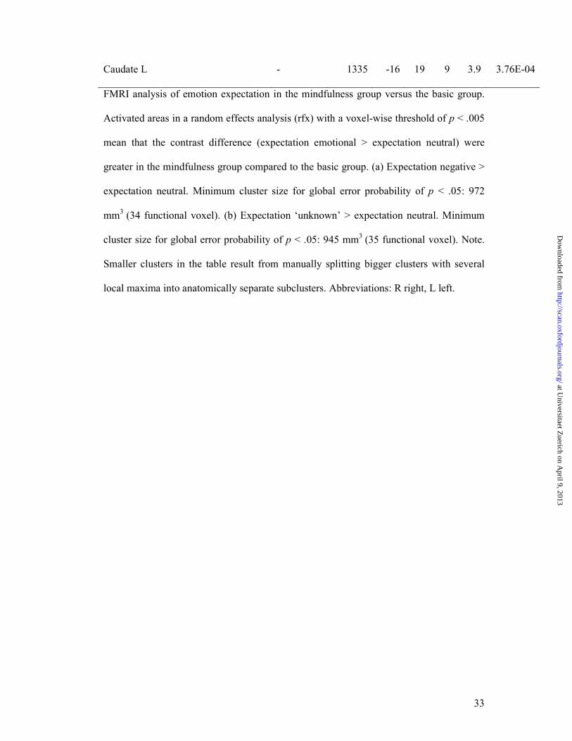
Caudate L - 1335 -16 19 9 3.9 3.76E-04
FMRI analysis of emotion expectation in the mindfulness group versus the basic group.
Activated areas in a random effects analysis (rfx) with a voxel-wise threshold of p < .005
mean that the contrast difference (expectation emotional > expectation neutral) were
greater in the mindfulness group compared to the basic group. (a) Expectation negative >
expectation neutral. Minimum cluster size for global error probability of p < .05: 972
mm3
(34 functional voxel). (b) Expectation ‘unknown’ > expectation neutral. Minimum
cluster size for global error probability of p < .05: 945 mm3
(35 functional voxel). Note.
Smaller clusters in the table result from manually splitting bigger clusters with several
local maxima into anatomically separate subclusters. Abbreviations: R right, L left.
at Universitaet Z
uerich on April 9, 2013
http://scan.oxfordjournals.org/D
ownloaded from

34
Table 3
Whole-brain group differences (mindfulness > basic) during the perception of negative
stimuli
Anatomic region Brodmann
area
Cluster
size
Peak Talairach
coordinates
t-max p-max
mm3 X Y Z
Perception negative > neutral, Clusters with Cluster-threshold: 135 mm3
Middle frontal gyrus L 46 323 -47 39 23 3.9 2.85E-04
Middle frontal gyrus L 6 165 -40 4 57 3.7 6.71E-04
Middle frontal gyrus L 6 270 -49 10 48 4.0 2.76E-04
Parahippocampal gyrus /
Hippocampus R
- 252 25 -15 -17 -3.7 6.80E-04
Insula L 692 -39 -12 10 -3.6 8.74E-04
FMRI analysis of the perception of emotional stimuli in the mindfulness group versus the
basic group. Activated areas in a random effects analysis (rfx) of the contrast negative >
neutral for a minimum cluster size of 135 mm3
mean that the contrast difference
(perception negative > expectation neutral) were greater in the mindfulness group
compared to the basic group. Minimum cluster size for global error probability (Monte
Carlo correction) of p < .05: 810 mm3 (30 functional voxels) showed no differences
between the groups, but clusters exceed a threshold of 135 mm3
(5 functional voxels).
Abbreviations: R right, L left.
at Universitaet Z
uerich on April 9, 2013
http://scan.oxfordjournals.org/D
ownloaded from

Figure 1. Illustration of experimental task and durations. Cues are enlarged for presentation reasons. Their actual height in the experiment was about 1/40 screen size.
81x126mm (300 x 300 DPI)
at Universitaet Z
uerich on April 9, 2013
http://scan.oxfordjournals.org/D
ownloaded from

Figure 3. (A) Significantly lower group mean beta weight of the mindfulness group (Mind) in the amygdala ROI (t(44) = -2.91, p < .01) compared to the basic group (Basic),in the perception of negative pictures
(ng), no group difference in the perception of neutral pictures (nt) (t(44) = - .08, p = .935). (B) Differing
time courses for the amygdala ROI between groups in the perception of negative stimuli: Similarity of time courses of the perception of negative and neutral pictures only in the mindfulness group. Error bars indicate
standard error. 181x76mm (300 x 300 DPI)
at Universitaet Z
uerich on April 9, 2013
http://scan.oxfordjournals.org/D
ownloaded from

Figure 4. A-C. Activations in the DMPFC in the group comparison “mindfulness” versus “basic” of the contrast expectation negative versus expectation neutral (eng > ent). (A) Higher DMPFC activity (yellow circle) in the mindfulness group in the expectation of negative stimuli. (B) Significantly higher mean beta
weight of the peak DMPFC voxel (x = -7, y = 7, z = -54) in the mindfulness group (Mind) (t(44) = 4.30, p < .0001) in the expectation of negative stimuli (eng) compared to the basic group (Bas), no group difference in the expectation of neutral stimuli (ent). (C) Time courses of the conditions for this region. Error bars
indicate standard error. 188x132mm (300 x 300 DPI)
at Universitaet Z
uerich on April 9, 2013
http://scan.oxfordjournals.org/D
ownloaded from

Figure 5. Correlation of individual mean beta weights of the contrast expectation negative versus expectation neutral in the left DMPFC (A) and in the right anterior insula (B) with trait mindfulness scores
(MAAS). 203x111mm (300 x 300 DPI)
at Universitaet Z
uerich on April 9, 2013
http://scan.oxfordjournals.org/D
ownloaded from

ROI of the DMPFC (blue), amygdala (orange box), insula (green). 170x57mm (300 x 300 DPI)
at Universitaet Z
uerich on April 9, 2013
http://scan.oxfordjournals.org/D
ownloaded from


