Embed Size (px)
Citation preview

Measurement of Orbital Volume after Enucleation andOrbital ImplantationOlga Lukats1*, Tamas Vızkelety2, Zsolt Markella3, Erika Maka1, Maria Kiss4, Adrienn Dobai2,
Peter Bujtar5, Attila Szucs2, Jozsef Barabas2
1 Departmentof Opthalmology, Faculty of Medicine, Semmelweis University, Budapest, Hungary, 2 Department of Oral and Maxillofacial Surgery and Dentistry, Faculty of
Dentistry, Semmelweis University, Budapest, Hungary, 3 Kalman Kando Faculty of Electrial Engineering, Obuda University, Budapest, Hungary, 4 Dunaujvaros College,
Dunaujvaros, Hungary, 5 Department of Oral and Maxillofacial Surgery, University Hospitals of Leicester, Leicester, United Kingdom
Abstract
Introduction: This article reports experience relating to the measurement of orbital volume by means of cone beamcomputed tomography (CBCT) and Cranioviewer program software in patients who have undergone enucleation andorbital implantation.
Patients and Methods: CBCT scans were made in 30 cases, 10 of which were later excluded because of various technicalproblems. The study group therefore consisted of 20 patients (8 men and 12 women). The longest follow-up time was 7years, and the shortest was 1 year. In all 20 cases, the orbital volume was measured with Cranioviewer orbital programsoftware. Slices were made in the ventrodorsal direction at 4.8 mm intervals in the frontal plane, in both bony orbits (boththat containing the orbital implant and the healthy one). Similar measurements were made in 20 patients with variousdental problems. CBCT scans were recorded for the facial region of the skull, containing the orbital region. The Cranioviewerprogram can colour the area of the slices red, and it automatically measures the area in mm.
Results: In 5 of the 20 cases, the first 4 or all 5 slices revealed that the volume of the operated orbit was significantly smallerthan that of the healthy orbit, in 12 cases only from 1 to 3 of the slices indicated such a significant difference, and in 3 casesno differences were observed between the orbits. In the control group of patients with various dental problems, there wasno significant difference between the two healthy orbits. The accuracy of the volume measurements was assessedstatistically by means of the paired samples t-test.
Summary: To date, no appropriate method is avaliable for exact measurement of the bony orbital volume, which would beof particular importance in orbital injury reconstruction. However, the use of CBCT scans and Cranioviewer orbital programsoftware appears to offer a reliable method for the measurement of changes in orbital volume.
Citation: Lukats O, Vızkelety T, Markella Z, Maka E, Kiss M, et al. (2012) Measurement of Orbital Volume after Enucleation and Orbital Implantation. PLoS ONE 7(12):e50333. doi:10.1371/journal.pone.0050333
Editor: Peter Csermely, Semmelweis University, Hungary
Received June 12, 2012; Accepted October 17, 2012; Published December 6, 2012
Copyright: � 2012 Lukats et al. This is an open-access article distributed under the terms of the Creative Commons Attribution License, which permitsunrestricted use, distribution, and reproduction in any medium, provided the original author and source are credited.
Funding: The authors’ work was part of a study which was supported by a scientific application TAMOP (Tarsadalmi Megujulas Operatıv Program) 4.2.1./B-09/1KMR 2010-001. The funders had no role in study design, data collection and analysis, decision to publish, or preparation of the manuscript.
Competing Interests: The authors have declared that no competing interests exist.
* E-mail: [email protected]
Introduction
The orbits are conical or four-sided pyramidal cavities, which
open into the midline of the face and point back into the head.
Each consist of a base, an apex and four walls. They protect the
eye from mechanical injury.
The base which opens in the face has four borders. The
folowing bones take part in their forrnation: superior margin:
frontal bone, inferior margin: maxilla and zygomatic, medial
margin: frontal, lacrimal and maxilla, lateral margin: zygomatic
and frontal. The apex lies near the medial end of superior orbital
fissure and contains the optic canal (containing the optic nerve and
ophthalmic artery) which communicates with middle cranial fossa.
The roof is formed primarily by the orbital plate frontal bone, and
also the lesser wing of sphenoid near the apex of the orbit. The
orbital surface presents medially by trochlear fossa and laterally by
lacrimal fossa.
The floor is formed by the orbital surface of maxilla, the orbital
surface of zygomatic bone and the minute orbital process of
palatine bone. Medially near the orbital margin, is located the
groove for nasolacrimal duct. Near the middle of the floor, located
infraorbital groove, which leads to the infraorbital foramen. The
floor is separated from the lateral wall by inferior orbital fissure,
which connects the orbit to pterygopalatine and infratemporal
fossa.
The medial wall is formed primally by the orbital plate of the
ethmoid, as well as contributions from the frontal process of
maxilla, the lacrimal bone and a small part of the body of the
sphenoid.
The lateral wall is formed by the frontal process of zygomatic
and more posteriorly by the orbital plate of the greater wing of
sphenoid. The bones meet at the zygomaticosphenoid suture.
PLOS ONE | www.plosone.org 1 December 2012 | Volume 7 | Issue 12 | e50333

The adult orbital margin is approximately rectangular with
horizontal dimension of 40 mm and a vertical dimension of
35 mm. The length of the medial orbital wall from the anterior
lacrimal crest is 45–50 mm, whereas the lateral wall from the rim
to the superior orbital fissure is 40 mm. The adult lateral orbital
walls are angled 90 degrees from each other or 45 degrees in the
anteroposterior direction. The divergent axis of each orbit thus
becomes half of 45 degrees or 22.5 degrees.
In the adult human the volume of the orbit is 30 ml. Because of
the complicated anatomical structure of the bony orbit, measure-
ment of its volume is difficult. The problems are mainly due to the
irregular inner border, holes and fissures. We have now made use
of Cranioviewer orbital program software [developed by Vizkelety
and Markella], together with cone beam computed tomography
(CBCT), to investigate both orbits in 20 patients with intraorbital
implants and in 20 patients with various dental problems
(otherwise healthy). To the best of our knowledge, there have
been no reports in the literature of using CBCT scans for orbital
volume measurement. Merely a few literature articles mention an
orbital volume decrease after enucleation in adults. Our primary
aim was to establish whether there is any detectable difference
between the volumes of the healthy and the operated orbits, or
between those of the healthy left and right orbits.
Patients and Methods
The study received ethical approval from Semmelweis Univer-
sity, Budapest, and complied fully with institutional ethical
protocols and the guidelines of the Helsinki Declaration. A
large-volume CBCT scanner (iCat Classic, Xoran Technologies,
Ann Arber, Michigan, USA) was used, with the following
parameters: 120 kV, pixel size 0,3 mm, slice increment 0.3, and
FOV height 6 cm. This type of CBCT can image not only the
facial skull, but also the orbital cavity. CBCT scans were made in
an attempt to detect changes within the orbital implants in vivo in
30 patients. At the time of the scan the position of the head in the
CBCT was close to the Fankfurt horizontal. The gained selection
of data may be rotated freely. In ten cases the situation of the
heads was not so good, as can be accepted, for this reason we
cannot rotate them properly for measurement. These cases were
excluded from the patients. We used only that cases whose heads
were in the desired position.
The scans utilized for orbital measurements in this study were
from 20 of these subjects (8 men, 12 women; mean age 43.85
years, range 14–76). Enucleation had been peformed because of
severe injury in 9 cases, a painful eye in 5 cases, an intraocular
malignant melanoma in 5 cases, and retinoblastoma in 1 case. FCI
synthetic hydroxyapatite had been used as the orbital implant for
volume replacement in 12 cases, and aluminium (Bioceramic) in 8
cases. In 18 cases the implantation had been primary, and in 2
cases secondary. The diameter of the implant was 20 mm in 12
cases, and 18 mm in 8 cases.
The Cranioviewer orbital program software was used in
conjuction with the CBCT scans for special volume measurement.
We set the image in a way that the most dorsal points of the right
and the left orbit entry may be connected with a straight line,
which at the same time is the level where the most ventral points of
the orbit constitute a closed circle. We set the head in vertical
position at the time of scanning, then the analogue images of the
orbit get into nearly identical levels. At the same time in the skull
we may present and measure the area of the frontal image of both
the healthy and the implanted orbit and the volume of the orbit
once we know the distance between the different levels. The
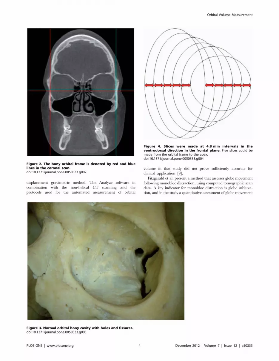
orbital frame can be denoted by red and blue lines in axial [Fig. 1]
and coronal [Fig. 2] scans. Slices were made at 4.8 mm intervals in
the ventrodorsal direction in the frontal plane. Five slices could be
made from the orbital frame to the apex [Fig: 3–4] The program
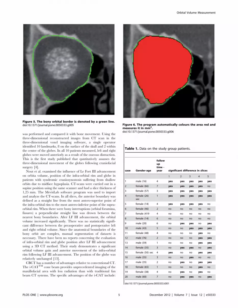
can outline the bony border and enclose the foramens and fissures
with a green line [Fig.5], fill every slice with red colour [Fig.6], and
measure the area of the slice in mm2.
The shortest follow-up time (between enucleation and CBCT
examination) was 1 year, and the longest was 7 years.
Results were derived from measurements by 3 different persons,
each of whom made each measurement 3 times. In all patients,
both orbital cavities were measured with this technique. The
paired t test was used to compare group differences. P values ,
0.05 were considered to be statistically significant.
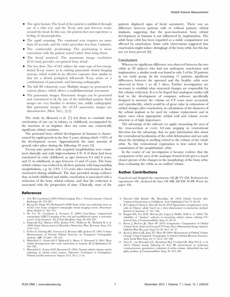
Results: [Table: 1]
In 5 patients, there was a significantly lower volume for the first
4 or all 5 of the measured slices in the orbit containing the implant
than in the patient’s own healthy orbit. [Fig. 7] [In patient No. 4,
one of the 2 secondary implantation cases, enucleation had been
performed in early childhood because of retinoblastoma. In spite
of the fact that her implantation had been carried out in
adulthood, she proved to be an example of a volume reduction
after childhood enucleation. Patient No.5 was 14, and patient
No.1 was 16 years old at the time of enucleation and primary
orbital implantation. (At these ages, the bony orbita is fully
developed and these patients therefore deserve inclusion in the
study group). In 12 patients, the measured value was significantly
lower in the operated orbit in 1,2 or 3 slices In 3 patients, there
were no significant differences between the slices in the operated
and the healthy orbits.
The control group, which consisted of 20 patients, who had
never suffered any orbital injury or undergone any orbital
opearation, exhibited a normal skeletal skull shape without any
facial asymmetry. Their CBCT scans were made because of dental
problems. In this group, no significant differences were measured
between the right and left orbital cavities [Fig. 8].
Discussion
Multislice CT and MRI allow multiplanar imaging of both the
normal and pathological anatomy. In the CT technique, the scan
plane is planned from a lateral scout to be parallel to the
infraorbital-meatal line approximating the orbital nerve plane.
Slices 3 mm thick are preferred for routine soft tissue visualization
on spiral CT because of the increased noise with thinner slices. 3D
surface-shaded models may by produced when craniofacial
surgery is planned, providing an adequent demonstration of the
bone anatomy in the context of fractures of the orbit and
craniofacial abnormalities [1].
With CT volume measurements, the task of CT scanning is
changed from creating images to determining the number of pixels
belonging in a predefined density range. Region-growing algo-
rithms have been used to determine the number of pixels of each
category for each slice. When multipled by a conversion factor, the
summation of all pixels for each category represents the estimate of
the total. Although there are variations among individuals, the
range of normal adult measurements generally lies within 2 SDs of
the mean. The bony and soft-tissue volumes are slightly greater in
men as a group than in women. There are small differences in
men as a group compared with women, but minimal differences
between the right and left orbits in the same person [4].
Ji et al. [8] examined morphologic orbit parameters of Chinese
adults. For example, the bony orbital volume, orbital foramen area
Orbital Volume Measurement
PLOS ONE | www.plosone.org 2 December 2012 | Volume 7 | Issue 12 | e50333

and orbital rim perimeter were measured on 3D models through
the use of a 3D reconstruction technique.
The differences between the two orbits and between the two
sexes were analysed. To ensure accuracy, manual segmentation
was used to define the boundary of the orbit. The borders of the
whole orbital content were defined by the four orbital walls. The
posterior boundary was the junction of the medial and lateral walls
at the optic foramen. A simulated surface was determined that
covered the orbital foramen with the orbital rim. This surface was
used to separate the whole orbital content into 2 parts, the
posterior part being defined as the bony orbit. In this way, the area
of the orbital foramen and the perimeter of the orbital rim could
be calculated automatically. The measurement method demon-
strated the high reproducibility of the results. The orbital volume
and other anatomic parameters indicated that the two orbits were
symmetric. The orbital size proved to be significantly smaller in
women than in men, but in a given individual there was no
difference between the two orbits.
Orbital blowout fractures are among the injuries most
commonly observed in the facial region. One of the most
important aspects in the reconstruction of orbital wall fractures
is restoration of the normal orbital volume. Accurate preoperative
measurement of the orbital volume is invaluable in predicting and
(if it leads to operative intervention) preventing post-injury
enophthalmos which is a common complication, optimum
reconstruction of which also demands an accurate estimation of
the orbital implant volume [11].
Thirty facial CT scans were utilized to measure 30 normal
orbits by using an image analysis program (Dextroscope,
Singapore). Calculation of the orbital volume from coronal scans
underestimates the volume as compared with axial scans, and the
criterion for the anterior limit of the measurement can affect the
volume determination. Three novel cephalometric angles that can
be obtained by 3D image analysis with stereoscopy may account
for the inaccuracies seen on coronal scans [10].
In 3 series of young, growing rabbits, varying amounts of
intraorbital tissue were removed from the right orbit by
evisceration, enucleation and excenteration. The left orbit was
maintained intact as a control for the operated right side. A
removable permanent elastic rubber-based imprint of the clean
orbit was made. The volume was calculated from the weight and
specific gravity of the orbital imprint. After the excision of orbital
tissue, the orbit continued to increase in size, but at a slower rate
than that for the unoperated orbit. In general, there was a direct
correlation between the lack of intraorbital mass and the decrease
in orbital growth [12].
Koppel et al. examined 5 dried skulls with prosthetic globes
and periorbita by non-helical scanning with an Elscint 2400 CT
scanner. The images obtained were processed with the Analyze
software package and the results were compared with the
volume of the intraorbital prosthesis determined by a volume-
Figure 1. The orbital frame is denoted by red and blue lines in the axial scan.doi:10.1371/journal.pone.0050333.g001
Orbital Volume Measurement
PLOS ONE | www.plosone.org 3 December 2012 | Volume 7 | Issue 12 | e50333
displacement gravimetric method. The Analyze software in
combination with the non-helical CT scanning and the
protocols used for the automated measurement of orbital
volume in that study did not prove sufficiently accurate for
clinical application [9].
Fitzgerald et al. present a method that assesses globe movement
following monobloc distraction, using computed tomographic scan
data. A key indicator for monobloc distraction is globe subluxa-
tion, and in the study a quantitative assessment of globe movement
Figure 2. The bony orbital frame is denoted by red and bluelines in the coronal scan.doi:10.1371/journal.pone.0050333.g002
Figure 3. Normal orbital bony cavity with holes and fissures.doi:10.1371/journal.pone.0050333.g003
Figure 4. Slices were made at 4.8 mm intervals in theventrodorsal direction in the frontal plane. Five slices could bemade from the orbital frame to the apex.doi:10.1371/journal.pone.0050333.g004
Orbital Volume Measurement
PLOS ONE | www.plosone.org 4 December 2012 | Volume 7 | Issue 12 | e50333
was performed and compared it with bone movement. Using the
three-dimensional reconstructed images from CT scan in the
three-dimensional voxel imaging software, a single operator
identified 10 landmarks, 8 on the surface of the skull and 2 within
the center of the globes. In all 10 patients measured, left and right
globes were moved anteriorly as a result of the osseous distraction.
This is the first study published that quntitatively assasses the
three-dimensional movement of the globes following craniofacial
surgery [4].
Nout et al. examined the influence of Le Fort III advancement
on orbita volume, position of the infra-orbital rim and globe in
patients with syndromic craniosynostosis suffering from shallow
orbits due to midface hypoplasia. CT-scans were carried out in a
supine position using the same scanner and had a slice thickness of
1,25 mm. The MevisLab software program was used to import
and analyze the CT-scans. In all slices, the anterior boundary was
defined as a straight line from the most antero-superior point of
the infra-orbital rim to the most antero-inferior point of the supra-
orbital rim. When there were bony interruptions (orbital foramina,
fissures) a perpendicular straight line was drown between the
nearest bony boundaries. After LF III advancement, the orbital
volume increased significantly. There was no statistically signifi-
cant difference between the preaoperative and postoperative left
and right orbital volume. Since the anatomical boundaries of the
bony orbit are complex, manual segmentation of datasets is
necessary. There have been no reports concerning the evaluation
of infra-orbital rim and globe position after LF III advancement
using a 3D CT method. Their study demonstrates a significant
orbital volume gain and anterior movement of the infra-orbital
rim following LF III advancement. The position of the globe was
relatively unchanged [13].
CBCT has a number of advantages relative to conventional CT.
The i-CATTM cone beam provides unprecedented imaging of the
maxillofacial area with less radiation than with traditional fan
beam CT systems. The specific advantages of the i-CAT include:
Figure 5. The bony orbital border is denoted by a green line.doi:10.1371/journal.pone.0050333.g005 Figure 6. The program automatically colours the area red and
measures it in mm2.doi:10.1371/journal.pone.0050333.g006
Table 1. Data on the study group patients.
case Gender-age
followuptime -year significant difference in slices
1 2 3 4 5
1 male (16) 4 yes yes yes yes yes
2 female (66) 7 yes yes yes yes no
3 female (57) 3 yes yes yes yes yes
4 female (26)sec
1 yes yes yes yes yes
5 female (14) 4 yes yes yes yes no
6 female (46) 2 no no no no no
7 female (419 4 no no no no no
8 female (14) 6 no no no no no
9 male (20) 6 no yes yes no yes
10 male (43) 5 no no yes yes yes
11 female (48) 4 no no no yes no
12 male (76) 2 no no no yes yes
13 male (59) 1 no no no yes yes
14 female (35) 3 no yes yes no yes
15 female (50) sec 4 yes no no yes yes
16 male (35) 3 no no yes no no
17 male (35) 2 no yes no yes yes
18 female (65) 1 no no no yes no
19 female (38) 4 no yes no yes no
20 male (60) 7 no yes yes no yes
doi:10.1371/journal.pone.0050333.t001
Orbital Volume Measurement
PLOS ONE | www.plosone.org 5 December 2012 | Volume 7 | Issue 12 | e50333

Figure 7. Data on patient No 1, demonstrating significant differences in the measured slices between the operated and the healthyorbit.doi:10.1371/journal.pone.0050333.g007
Figure 8. Data on the control group, demonstrating no significant difference in the measured slices between the right and leftorbits.doi:10.1371/journal.pone.0050333.g008
Orbital Volume Measurement
PLOS ONE | www.plosone.org 6 December 2012 | Volume 7 | Issue 12 | e50333
N The open format: The head of the patient is stabilized through
use of a chin rest, and the X-ray unit and detector rotate
around the head. In this way, the patient does not experience a
feeling of claustrophobia.
N The rapid scanning: The rotational scan requires no more
than 20 seconds, and the entire procedure less than 5 minutes.
N The comfortable positioning: The positioning is more
convenient with the patient seated rather than lying down.
N The detail attained: The maximum image resolution
[0.25 mm] provides exceptional bony detail.
N The low dose: The i-CAT utilizes the same type of low-energy
dental X-ray source as in existing panoramic dental imaging
systems, which results in an effective exposure dose similar to
that for a dental perinpical full-mouth X-ray series or a
combination of panoramic and bitewing radiographs.
N The full 3D volumetric scan: Multiple images we generated in
various planes, which allows a multidimensional assessment.
N The panoramic images: Panoramic images can be generated
and customized to fit the jaw of the individual patient. Such
images are very familiar to dentists, but, unlike radiographic
film panoramic images, the i-CAT panoramic images are
distortion-free (Med. Net.), [2,3].
The study by Howard et al. [7] led them to conclude that
enucleation of one eye in infancy or childhood, accompanied by
the insertion of an implant, does not cause any cosmetically
significant orbital variation.
The postnatal bony orbital development in humans is charac-
terized by rapid growth in the first 3 years, during which <50% of
the development occurs. Approximately the same amount of
growth takes place during the following 10 years [5].
Twenty-nine patients with acquired anophthalmia were exam-
ined clinically and with high-resolution CT; 8 of them had been
enucleated in early childhood, at ages between 0.4 and 8 years,
and 21 in adulthood, at ages between 15 and 53 years. The bony
orbital volume was reduced in all these patients with long-standing
anophthalmia, e.g. by 3.8% 7-13 years after enucleation in those
enucleated during adulthood. The date provided strong evidence
that, in both childhood and adults, enucleation is associated with a
reduction of the bony orbital volume, and that the reduction is
associated with the progression of time. Clinically, none of the
patients displayed signs of facial asymmetry. There was no
difference between patients with or without primary orbital
implants, suggesting that the post-enucleation bony orbital
development in humans is not influenced by implantation. The
adult bony orbit has been regarded as a stable compartment not
affected by enucleation. Some early observations suggested that
enucleation might induce shringkage of the bony orbit, but this has
not yet been proved [6].
ConclusionsWhereas no significant difference was observed between the two
orbits in 20 subjects who had not undergone enucleation and
implantation, a similar result was found in only 3 of the 20 patients
in our study group. In the remaining 17 patients, significant
differences between the operated and the healthy orbit were
observed in from 1 to all 5 slices. Further investigations are
necessary to establish what structural changes are responsible for
this volume reduction. It is to be hoped that analogous studies will
lead to the development of computer software specificially
designed to measure the volume on CT scans more accurately
and reproducibly, which would be of great value in estimations of
orbital changes after enucleation, in calculations of the diameter of
the orbital implant to be used for volume replacement and in
injury cases when appropriate orbital wall and volume recon-
struction is of high importance.
The adventage of the software we apply: measuring the area of
the cross-sections at every 4,8 mm stepping in ventrodorsal
direction has the advantage that we gain information also about
the ventrodorsal localization of the orbit deformation and not only
about the shrinking or swelling related to the volume of the whole
orbit. So this ventrodorsal registration is best suited for the
examitation of the anophthalmic orbit.
In the course of our investigation it became evident, that the
comparison of the area of the analogue frontal levels gives a much
clearer picture of the changes in the morphology of the bony orbit,
than evaluating the orbits as an unified volume.
Author Contributions
Conceived and designed the experiments: OL JB TV ZM. Performed the
experiments: OL. Analyzed the data: OL MK AD EM AS PB. Wrote the
paper: OL.
References
1. Aviv RI, Casselman J (2005) Orbital imaging: Part 1. Normal anatomy. Clinical
Radiology 60: 279–287.2. Bryant JA, Drage NA, Richmond S (2008) Study of the scan uniformity from an
i-CAT cone beam computed tomography dental imaging system. Dentomax-illofac Radiol 37: 365–374.
3. De Vos W, Casselman J, Swennen G (2009) Cone-Beam computerised
tomography [CBCT] imaging of the oral and maxillofacial region: a systematicreview of the literature. Int J Oral Maxillofac Surg 38: 609–625.
4. Fitzgerald E.J., Marucci D.D., Jeelani N.O., Witherow H., Richards R et al.(2009) Oular Advencement in Monobloc Distraction. Plast. Reconstr. Surg. 123:
1570–1577.
5. Forbes G, Gehring DG, Gorman CA, Brennan MD, Jackson IT (1985) VolumeMeasurements of Normal Orbital Structure by Computed Tomographic
Analysis. AJR 145 July: 149–145.6. Hintschich C, Zonneveld F, Baldeschi L, Bunce C, Koornnef L (2001) Bony
orbital developement after early enucleation in humans. Br J Ophthalmol 85:205–208.
7. Hintschich C, Zonneveld F, Koornneef L (2001) Normal orbital volumes and its
pathology in orbital socket surgery. Operative Techniques in Oculoplastic,Orbital and Reconstructive Surgery Vl.4. No.1: 3–14.
8. Howard GM, Kindler SR, Macmillan AS (1965) Orbital Growth After
Unilateral Enucleation in Childhood. Arch Ophthalmol Vol 73: 80–83.
9. Ji Y, Quian Z, Dong Y, Zhou H, Fan X (2010) Quantitative morphometry of the
orbit in Chinese adults based on a three-dimensional reconstruction method.
Journal of Anatomy 17: 501–506.
10. Koppel DA, Foy R.H, McCaul JA, Logan J, Hadley D.M et al. (2003) The
reliability of ‘‘Analyze’’ software in measuring orbital volume utilizing CT-
derived data. J Craniomaxillofac Surg 31: 88–91.
11. Kwon J, Barrera JE, Most SP (2010) Comparative Computation of Orbital
Volume From Axial and Coronal CT Using Three-Dimensional Image Analysis.
Ophthal Plast Reconstr Surg Vol 26: No1: 26–27.
12. Kwon J, Barrera JE, Jung TY, Most SP (2009) Mesurements of Orbital Volume
Change Using Computed Tomography in Isolated Orbital Blowout Fractures.
Arch Facial Plast Surg vol 11: No.6: 395–398.
13. Nout E., van Bezooijen J.S., Koudstaal M.J, Veenland JF, Hop W.C.J. et al.
(2012) Orbital change following Le Fort III advancement in syndromic
craniosynostosis: quantitative evaluation of orbita volume, infraorbital rim and
globe position. J Craniomaxillofac Surg. 40: 223–228.
Orbital Volume Measurement
PLOS ONE | www.plosone.org 7 December 2012 | Volume 7 | Issue 12 | e50333