Embed Size (px)
Citation preview
Article
Mathematical Modeling of Ion Quantum Tunneling RevealsNovel Properties of Voltage-Gated Channels and QuantumAspects of Their Pathophysiology inExcitability-Related Disorders
Abdallah Barjas Qaswal 1,* , Omar Ababneh 2 , Lubna Khreesha 3 , Abdallah Al-Ani 4 , Ahmad Suleihat 5
and Mutaz Abbad 5
�����������������
Citation: Qaswal, A.B.; Ababneh, O.;
Khreesha, L.; Al-Ani, A.; Suleihat, A.;
Abbad, M. Mathematical Modeling of
Ion Quantum Tunneling Reveals
Novel Properties of Voltage-Gated
Channels and Quantum Aspects of
Their Pathophysiology in
Excitability-Related Disorders.
Pathophysiology 2021, 28, 116–152.
https://doi.org/10.3390/
pathophysiology28010010
Received: 5 February 2021
Accepted: 4 March 2021
Published: 7 March 2021
Publisher’s Note: MDPI stays neutral
with regard to jurisdictional claims in
published maps and institutional affil-
iations.
Copyright: © 2021 by the authors.
Licensee MDPI, Basel, Switzerland.
This article is an open access article
distributed under the terms and
conditions of the Creative Commons
Attribution (CC BY) license (https://
creativecommons.org/licenses/by/
4.0/).
1 Department of Internship Program, Jordan University Hospital, The University of Jordan, Amman 11942, Jordan2 Department of Anesthesia and Intensive Care, School of Medicine, The University of Jordan,
Amman 11942, Jordan; [email protected] Department of Special Surgery, School of Medicine, The University of Jordan, Amman 11942, Jordan;
[email protected] School of Medicine, The University of Jordan, Amman 11942, Jordan; [email protected] Department of General Surgery, School of Medicine, The University of Jordan, Amman 11942, Jordan;
[email protected] (A.S.); [email protected] (M.A.)* Correspondence: [email protected]
Abstract: Voltage-gated channels are crucial in action potential initiation and propagation and thereare many diseases and disorders related to them. Additionally, the classical mechanics are the mainmechanics used to describe the function of the voltage-gated channels and their related abnormalities.However, the quantum mechanics should be considered to unravel new aspects in the voltage-gatedchannels and resolve the problems and challenges that classical mechanics cannot solve. In thepresent study, the aim is to mathematically show that quantum mechanics can exhibit a powerfultendency to unveil novel electrical features in voltage-gated channels and be used as a promisingtool to solve the problems and challenges in the pathophysiology of excitability-related diseases. Themodel of quantum tunneling of ions through the intracellular hydrophobic gate is used to evaluatethe influence of membrane potential and gating free energy on the tunneling probability, singlechannel conductance, and quantum membrane conductance. This evaluation is mainly based ongraphing the mathematical relationships between these variables. The obtained mathematical graphsshowed that ions can achieve significant quantum membrane conductance, which can affect theresting membrane potential and the excitability of cells. In the present work, quantum mechanicsreveals original electrical properties associated with voltage-gated channels and introduces newinsights and implications into the pathophysiology of excitability- related disorders. In addition,the present work sets a mathematical and theoretical framework that can be utilized to conductexperimental studies in order to explore the quantum aspects of voltage-gated channels and thequantum bioelectrical property of biological membranes.
Keywords: quantum tunneling; voltage-gated channel; sodium ions; potassium ions; quantumbiophysics; epilepsy; arrhythmias; pain; quantum biology
1. Introduction
Voltage-gated channels are crucial for action potential initiation and propagation [1].Thus, any disturbance in their function or structure could affect the processes and actionswithin, and of excitable cells, resulting in different diseases, such as epilepsy [2], paindisorders [3], and cardiac arrhythmias [4]. Additionally, understanding how voltage-gatedchannels operate and how they can be implicated in the pathophysiology of many diseasesis based mainly on the principles of classical mechanics and laws of thermodynamics [1].
Pathophysiology 2021, 28, 116–152. https://doi.org/10.3390/pathophysiology28010010 https://www.mdpi.com/journal/pathophysiology
Pathophysiology 2021, 28 117
Such practice ignores the role of quantum mechanics as its integration within classicalmechanics might unravel new aspects regarding the function of voltage-gated channels,which could enhance our understanding of excitable cells and their role in the patho-genesis of some diseases. Furthermore, the integration of quantum mechanics is furtherencouraged by the many puzzling challenges associated with understanding the roles ofabnormal voltage-gated channels in the pathophysiology of excitability-related disorderssuch as epilepsy [2,5], pain disorders [6], and cardiac arrhythmias [7]. Those diseasesrepresent major medical issues and hurdles in terms of how they are prevented, treated, orcontrolled [8–11].
Quantum mechanics is the field of physics that focuses on the behavior of atomic andsubatomic particles and this behavior can be studied by using the Schrödinger equation toobtain the wave function of a particle [12]. Recently, the field of quantum mechanics hasbeen extended to biology in order to understand different biological actions and eventsincluding photosynthesis, action of enzymes, olfaction, and birds’ navigation [13–15].However, many concerns have emerged along quantum biology. One of these concernsis that the hot noisy biological environment does not sustain quantum behavior. Thisconcern has been opposed by recent research, which showed that quantum properties,such as the quantum entanglement of huge number of atoms, can be maintained at hightemperatures such as those of the human body or even higher [16]. Different mechanismscould explain the persistence of quantum coherence within biological systems, includinghydrophobic pockets, as in the hydrophobic gate of the voltage-gated channels that are thefocus of this study [17–19]. Therefore, this gives researchers more motivation to pursueapplying quantum mechanics within biological systems. In recent years, researchers havebeen focusing on the quantum features of ions, such as potassium and calcium ions, andtheir role in the processes and actions of neurons [20–22]. This approach is scientificallysound as it has been observed and documented that atoms, ions, and even molecules canbehave according to the principles of quantum mechanics in a similar fashion to that ofsubatomic particles [23–26]. Moreover, the ability of scientists to explain the high selectivityof voltage-gated channels through mathematical structures acts as further evidence forthe validity of studying biological systems through a quantum perspective [22,27–29].However, a principal component of the voltage-gated channel did not receive enough at-tention. This component is the channel’s gate, which determines the channel’s conductanceand consequently, the overall membrane’s conductance and the electrical properties ofits associated tissues [1,18,19,30]. Thus, the present work is an extension of the previousworks that focused on studying the gates of voltage-gated channels through understandingthe quantum behavior of their target ions, and their quantum conductance [31,32].
This study aims to approach the function of voltage-gated channels from a quantumperspective using the quantum tunneling model [31,32]. The model is designed to portraythe novel electrical features of voltage-gated channels and shed light on the differencesthat make it distinct from the classical model of Boltzmann distribution for voltage-gatedchannels. Moreover, this quantum model is implemented to signify the contribution ofquantum behavior of ions in the pathophysiology of excitable tissue diseases. In this work,the aim is not to focus on and review the details of the challenges and puzzles in the functionof voltage-gated channels and their related diseases specifically. However, the aim is tospot hints and clues and to establish a comprehensive mathematical model to encourageresearchers to consider quantum mechanics in future works when they aim to resolve achallenge or a puzzle in the field of electrophysiology and to unveil the pathogenesis of acertain disease related to the function of voltage-gated channels. However, some of thechallenges and puzzles will be discussed in this work and the quantum model will offerreasonable explanation for them especially that they are not well explained by the classicalmodels. Additionally, the present study might be helpful to aid in developing novel agentsto treat and control epilepsy syndromes, pain disorders, and cardiac arrhythmias.
Pathophysiology 2021, 28 118
2. The Mathematical Model2.1. The Conductance of the Voltage-Gated Channels According to the Laws of Thermodynamics
The voltage-gated channels are mainly composed from two parts: (1) A selectivityfilter and (2) an intracellular gate [1]. The function of the selectivity filter is to discriminatebetween ions and to enable the channel to selectively pass specific ion [1]. On the otherhand, the intracellular gate functions as the main controller of the channel’s conductanceand that of the overall membrane [18,19,30]. The intracellular gate is a hydrophobic con-striction made by the bundles crossing of the four S6 segments of the alpha subunits of thechannel [18,19]. This gate represents an energy barrier that impends the passage of ions [30].Hence, the intracellular hydrophobic gate controls the ions’ passage and the channel’sconductance. Furthermore, the intracellular gate operates as a narrow hydrophobic pore, inwhich its ‘open’ state is characterized by an increased pore radius and a decreased energybarrier, which consequently facilities the passage of ions [33]. According to the classicalphysics of thermodynamics, the voltage gated channels have two states: (1) A closed state(C) and (2) an open state (O), which fit the Boltzmann distribution, as demonstrated in thefollowing equation [1,33]:
P =O
C + O= (1 + e
qgV1/2−qgVmKBT )
−1
, (1)
where P is the fraction of open channels from the total available channels at a certain areaof the cell membrane (the open probability), V1/2 is the membrane voltage at which halfof the channels are open, Vm is the actual membrane voltage, qg is the gating’s charge, KBis the Boltzmann’s constant (1.38× 10⁻²³ J/K), and T is the absolute body temperature(310 K). The mathematical term qgV1/2 represents the gating free energy, which is the energyassociated with the conversion from the ‘closed’ state to the ‘open’ state at Vm = 0 [34].Furthermore, the mathematical structure qgV1/2 − qgVm represents the energy requiredto switch from the ‘closed’ state to the ‘open’ state or the energy barrier that resists thepassage of ions at a certain membrane potential Vm.
According to the Boltzmann distribution, voltage-gated channels are either in the‘closed’ state that has zero conductance, or in the ‘open’ state that has a certain value ofconductance Csin gle. As a result, when there is a certain number of channels at a surfacearea of a membrane with a certain membrane voltage Vm, a fraction of this number ofchannels will be ‘open’ and be able to conduct ions. Therefore, the membrane conductanceCM due to voltage-gated channels can be calculated by the following equation:
CM = D× Csin gle × (1 + eqgV1/2−qgVm
KBT )−1
, (2)
where CM is the membrane’s conductance (S/m2), Csin gle is the single channel conductance(S), and D is the channels’ density (channels/m2).
2.2. The Conductance of the Voltage-Gated Channels According to Quantum Mechanics
According to quantum mechanics, the hydrophobic gate can be represented as apotential barrier through which ions can tunnel [31,32]. The tunneling probability (TQ)through the hydrophobic gate, as solved from Schrodinger’s equation, can be calculated byusing the following equation [12,31,32]:
TQ = e−√
8m}
X2∫X1
√(U(x)−KEdx
, (3)
where m is the mass of the ion, } is the reduced Planck’s constant (1.05× 10⁻³⁴ Js), U(x) isthe barrier’s energy with respect to the ion’s position x across the gate, KE is the kineticenergy of the ion, and x1-x2 is the forbidden region of the gate where the barrier’s energyU(x) is higher than the kinetic energy of the ion KE.
Pathophysiology 2021, 28 119
The energy required to open the gate is (qgV1/2 − qgVm), which represents the energybarrier of the gate. In other words, this energy is needed to perform the mechanical workto dilate the hydrophobic pore in order to facilitate ion conduction [33]. As a result, we candeduce that ion must have this amount of energy to overcome the energy barrier of thegate. Therefore, this amount of energy (qgV1/2 − qgVm) represents the energy barrier U(x)stated in Equation (3). However, quantum tunneling of ions does not require the dilationof the hydrophobic gate.
Here, it is claimed that this energy distributes equally along the length of the gateL. To make a mathematical connection between the barrier’s energy U(x) and the ion’sposition x through the gate, the gate can be illustrated as an electric field that opposes thepassage of ions. This electric field E can be calculated as demonstrated in the followingequation [31,32]:
E =qgV1/2 − qgVm
qionL, (4)
where qion is the charge of the ion.The barrier’s energy with respect to the ion’s position can be calculated through the
following equation:U(x) = qionEx, (5)
Then, by substituting Equation (4) in Equation (5):
U(x) =qgV1/2 − qgVm
Lx, (6)
The channel’s gate is a short hydrophobic constriction that is located at the intracellularside [18,19]. Therefore, as long as the membrane potential is negative inside with regardsto the outside, extracellular cations, such as sodium and potassium ions, will move fromoutside the cell using the membrane’s potential acquiring kinetic energy qionVm untilhitting the intracellular gate [31,32]. On the other hand, the intracellular cations will hitthe intracellular gate before going through the membrane’s potential and hence it willnot affect their kinetic energy [31,32]. However, both intracellular and extracellular ionshave a thermal energy at body temperature equals to 3
2 KBT = 0.64× 10⁻²⁰ J. Therefore, itis assumed that the kinetic energy of the ion does not change while passing through theintracellular gate, since the length of the gate is relatively short when compared with thefull thickness of the cell membrane; thus, the voltage across the gate is neglected.
The integral in Equation (3) can be solved as the following:
R =
X2∫X1
√qgV1/2 − qgVm
Lx− KEdx =
2L3(qgV1/2 − qgVm)
√(
qgV1/2 − qgVm
Lx2 − KE)
3
− 2L3(qgV1/2 − qgVm)
√(
qgV1/2 − qgVm
Lx1 − KE)
3
, (7)
x2 is at the end of the gate (x2 = L), and x1 is where U(x1) =qgV1/2−qgVm
L x1 = KE.Thus, Equation (7) becomes:
R =
X2∫X1
√qgV1/2 − qgVm
Lx− KEdx =
2L3(qgV1/2 − qgVm)
√((qgV1/2 − qgVm)− KE)3, (8)
Regarding the extracellular monovalent cations such as sodium and potassium,Equation (8) can be written as:
Ro =2L
−3(qgV1/2 − qgVm)
√(−(qgV1/2 − qgVm)− (qionVm +
32
KBT))3, (9)
Pathophysiology 2021, 28 120
Ro =2L
3(qgVm − qgV1/2)
√((qgVm − qgV1/2)− (qionVm +
32
KBT))3, (10)
In Equation (9), the minus sign is inserted to indicate that the membrane voltages (Vmand V1/2) are absolute values. This is made because the kinetic energy of the ions qionVmis a positive value and Vm should be an absolute value of the actual membrane voltage.Additionally, when tunneling probability and its related equations are encountered, themembrane’s voltage is negative on the inside with regard to the outside, and the value ofthe membrane’s voltage is an absolute value.
On the other hand, Equation (8) for the intracellular monovalent cations can bewritten as:
Ri =2L
3(qgVm − qgV1/2)
√((qgVm − qgV1/2)− (
32
KBT))3, (11)
As a result, the tunneling probability of extracellular ions TQ(o) and intracellular ionsTQ(i) can be calculated by the following equations:
TQ(o) = e−√
8m} ×
2L3(qgVm−qgV1/2)
√((qgVm−qgV1/2)−(qionVm+ 3
2 KBT))3
, (12)
TQ(i) = e−√
8m} ×
2L3(qgVm−qgV1/2)
√((qgVm−qgV1/2)−( 3
2 KBT))3
, (13)
So, from the perspective of quantum mechanics, ions can tunnel through the closedintracellular gate of the channels. Consequently, the closed channels can conduct ions viaquantum tunneling, hence the conductance is called quantum conductance. The quantumconductance of a single channel CQ can be calculated by this equation [12,31,32,35]:
CQ =q2
ionh
TQ, (14)
where h is Planck’s constant (6.6× 10⁻³⁴ Js), qion is the charge of the ion, and TQ is thetunneling probability. The unit of CQ is Siemens (S).
Moreover, at certain channels’ density D (channels/m2), the quantum membraneconductance CQM can be calculated by this equation:
CQM = D× CQ, (15)
The unit of CQM is S/m2.
3. Results
In this section, the comparison between quantum conductance and classical conduc-tance of voltage-gated channels is made by graphing the equations, which contain thevariables of conductance, gating free energy, and membrane voltage. Such presentationwill delineate how the conductance changes alongside variations in gating free energy andmembrane potential (voltage). The mathematical graphs are plotted using MATLAB. Themathematical graphing is used to provide a comprehensive evaluation of the electricalfunction of the voltage-gated channels. Moreover, by plotting the graphs and comparingthem, novel perspectives relating to the voltage-gated channels and the contribution of thequantum behavior of ions in the pathophysiology of the excitability-related disorders couldbe discussed. The mathematical plots will be based on semi-log graphing so that the com-parison between classical and quantum models can be made in a comprehensive manner.
3.1. The Conductance of the Voltage-Gated Sodium Channels According to the Boltzmann Distribution
In this study, the quantum and classical models will be applied on the Nav1.2sodium channels. These channels have gating charge qg = 9.2e = 9.2× 1.6× 10−19 =14.72× 10⁻¹⁹ C [36], and with half activation voltage V1/2 = 43 mV [37,38], the gating free
Pathophysiology 2021, 28 121
energy qgV1/2 = 6.33× 10⁻²⁰ J [39]. Furthermore, the density of sodium channels D is5× 1013 channels/m2 [1], and the single channel conductance of sodium channel Csin gle(Na)
is 15× 10−12 S [1].Considering these values in Equations (1) and (2):
PNa = (1 + eG−147.2Vm
0.43 )−1
, (16)
CM(Na) = 750× (1 + eG−147.2Vm
0.43 )−1
, (17)
where G =qgV1/210−20 .
As long as the membrane potential is negative on the inside with regard to the outside,the membrane potentials (Vm and V1/2) are substituted with their negative sign when theBoltzmann distribution is applied on the voltage-gated channels.
Thus, the open probability of sodium channels at G = −6.33 J:
PNa = (1 + e−6.33−147.2Vm
0.43 )−1
, (18)
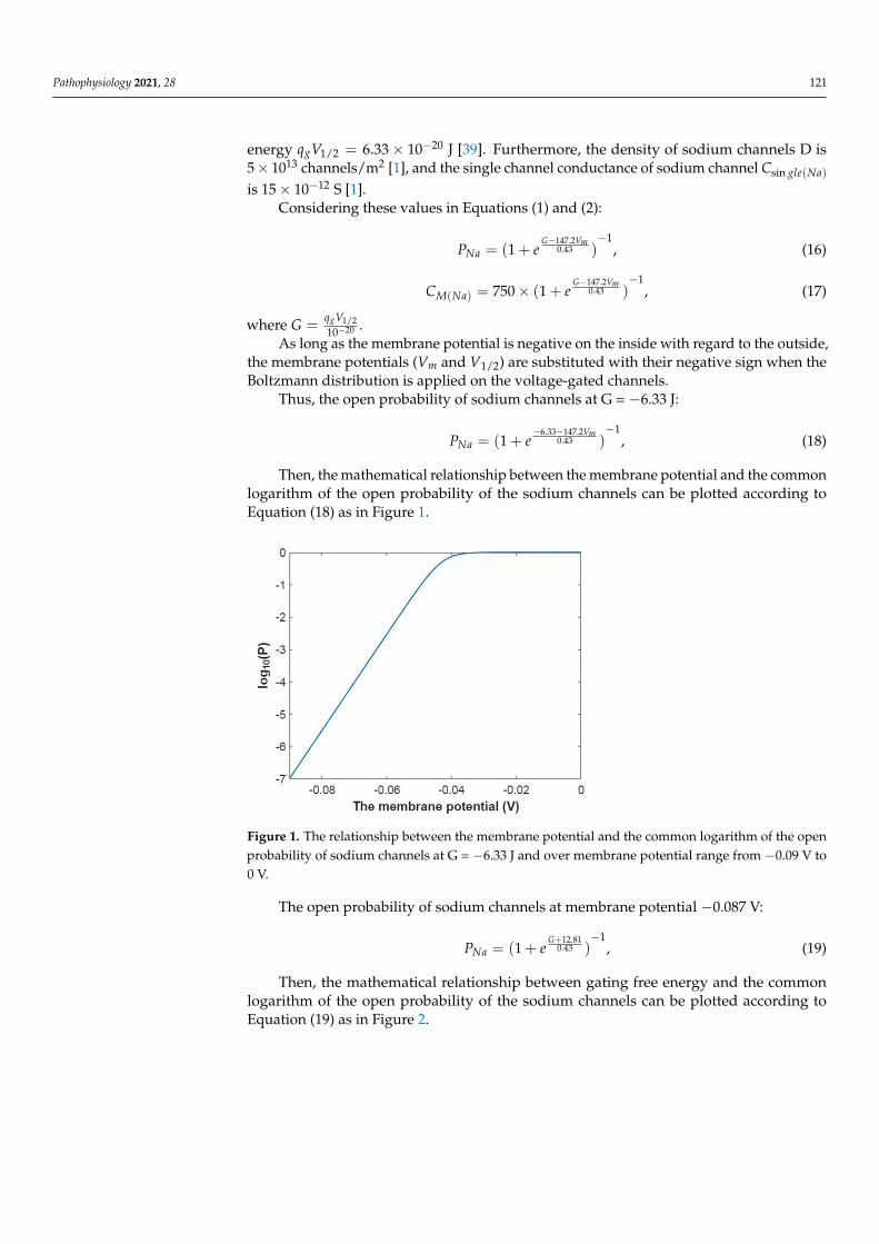
Then, the mathematical relationship between the membrane potential and the commonlogarithm of the open probability of the sodium channels can be plotted according toEquation (18) as in Figure 1.
Pathophysiology 2021, 28 122
Figure 1. The relationship between the membrane potential and the common logarithm of the open probability of sodium channels at G = −6.33 J and over membrane potential range from −0.09 V to 0 V.
The open probability of sodium channels at membrane potential −0.087 V: 12.81
10.43(1 )G
NaP e+
−= + , (19)
Then, the mathematical relationship between gating free energy and the common logarithm of the open probability of the sodium channels can be plotted according to Equation (19) as in Figure 2.
Figure 2. The relationship between gating free energy and the common logarithm of the open probability of the sodium channels at membrane potential −0.087 V and over gating free energy range from −12 J to −5 J.
The reference point of membrane potential −0.087 V will be used throughout the pa-per to carry out the calculations where appropriate. This value of membrane potential represents the resting membrane potential at physiological concentrations and resting leaky conductance values of sodium and potassium ions (See Section 3.5 for values and calculations).
The membrane conductance of sodium ions according to the Boltzmann distribution at G = −6.33 J:
Figure 1. The relationship between the membrane potential and the common logarithm of the openprobability of sodium channels at G = −6.33 J and over membrane potential range from −0.09 V to0 V.
The open probability of sodium channels at membrane potential −0.087 V:
PNa = (1 + eG+12.81
0.43 )−1
, (19)
Then, the mathematical relationship between gating free energy and the commonlogarithm of the open probability of the sodium channels can be plotted according toEquation (19) as in Figure 2.
Pathophysiology 2021, 28 122
Pathophysiology 2021, 28 122
Figure 1. The relationship between the membrane potential and the common logarithm of the open probability of sodium channels at G = −6.33 J and over membrane potential range from −0.09 V to 0 V.
The open probability of sodium channels at membrane potential −0.087 V: 12.81
10.43(1 )G
NaP e+
−= + , (19)
Then, the mathematical relationship between gating free energy and the common logarithm of the open probability of the sodium channels can be plotted according to Equation (19) as in Figure 2.
Figure 2. The relationship between gating free energy and the common logarithm of the open probability of the sodium channels at membrane potential −0.087 V and over gating free energy range from −12 J to −5 J.
The reference point of membrane potential −0.087 V will be used throughout the pa-per to carry out the calculations where appropriate. This value of membrane potential represents the resting membrane potential at physiological concentrations and resting leaky conductance values of sodium and potassium ions (See Section 3.5 for values and calculations).
The membrane conductance of sodium ions according to the Boltzmann distribution at G = −6.33 J:
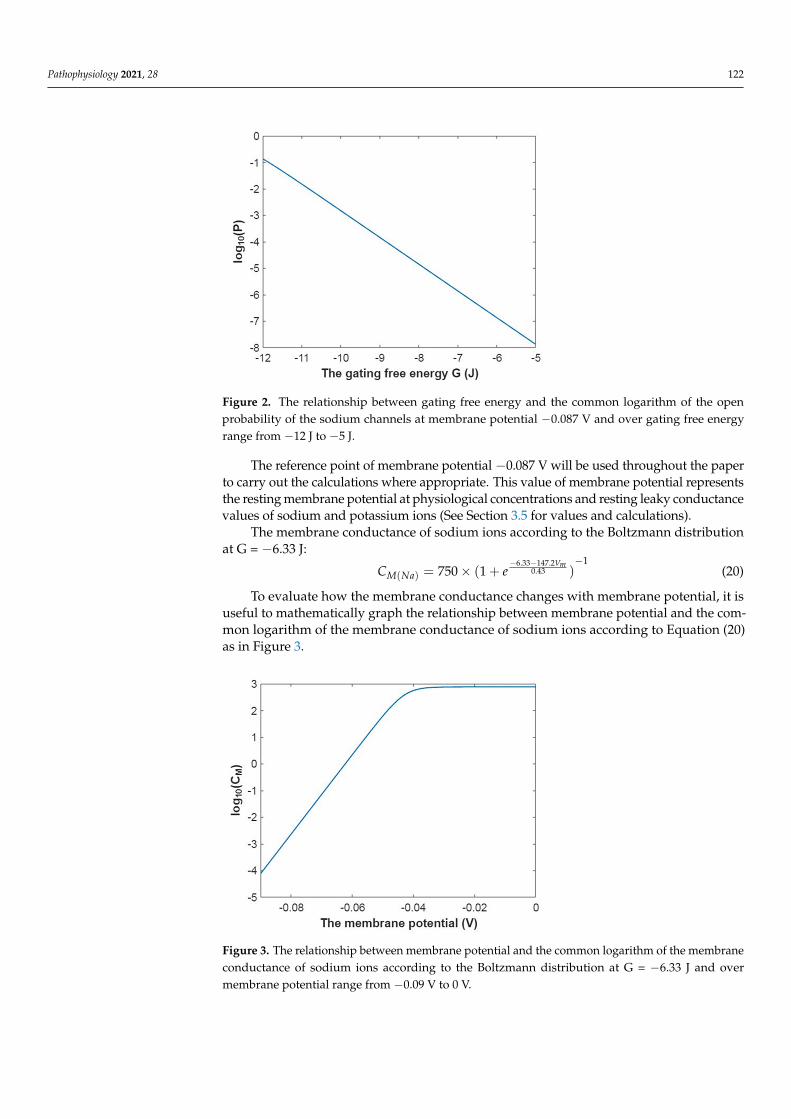
Figure 2. The relationship between gating free energy and the common logarithm of the openprobability of the sodium channels at membrane potential −0.087 V and over gating free energyrange from −12 J to −5 J.
The reference point of membrane potential −0.087 V will be used throughout the paperto carry out the calculations where appropriate. This value of membrane potential representsthe resting membrane potential at physiological concentrations and resting leaky conductancevalues of sodium and potassium ions (See Section 3.5 for values and calculations).
The membrane conductance of sodium ions according to the Boltzmann distributionat G = −6.33 J:
CM(Na) = 750× (1 + e−6.33−147.2Vm
0.43 )−1
(20)
To evaluate how the membrane conductance changes with membrane potential, it isuseful to mathematically graph the relationship between membrane potential and the com-mon logarithm of the membrane conductance of sodium ions according to Equation (20)as in Figure 3.
Pathophysiology 2021, 28 123
6.33 147.210.43
( ) 750 (1 )mV
M NaC e− −
−= × +,
(20)
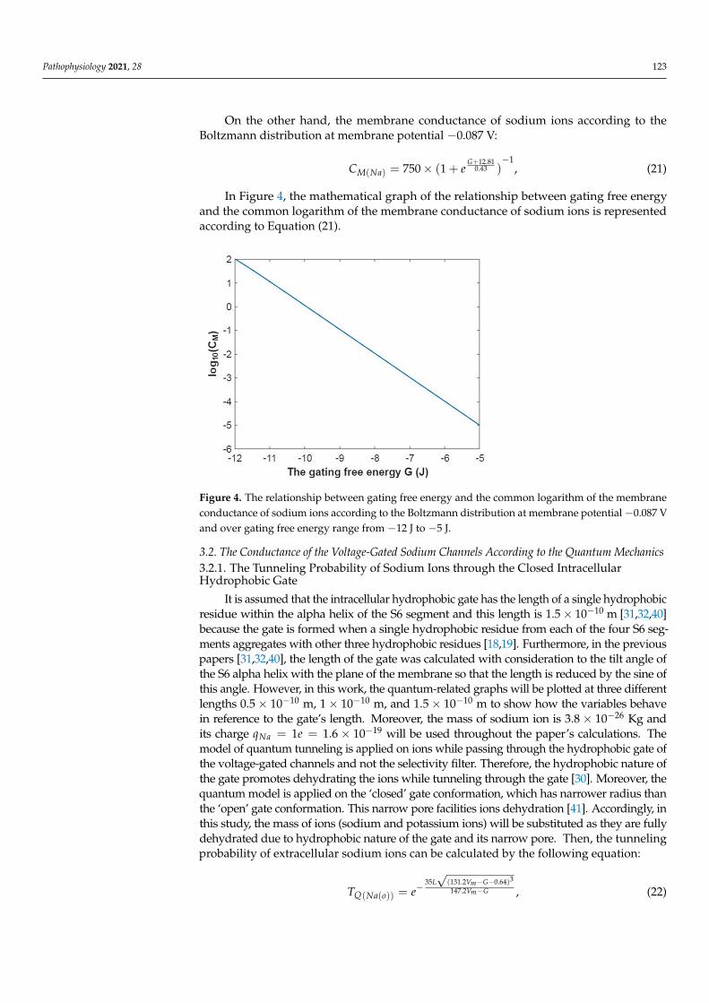
To evaluate how the membrane conductance changes with membrane potential, it is useful to mathematically graph the relationship between membrane potential and the common logarithm of the membrane conductance of sodium ions according to Equation (20) as in Figure 3.
Figure 3. The relationship between membrane potential and the common logarithm of the mem-brane conductance of sodium ions according to the Boltzmann distribution at G = −6.33 J and over membrane potential range from −0.09 V to 0 V.
On the other hand, the membrane conductance of sodium ions according to the Boltz-mann distribution at membrane potential −0.087 V:
12.8110.43
( ) 750 (1 )G
M NaC e+
−= × +,
(21)
In Figure 4, the mathematical graph of the relationship between gating free energy and the common logarithm of the membrane conductance of sodium ions is represented according to Equation (21).
Figure 3. The relationship between membrane potential and the common logarithm of the membraneconductance of sodium ions according to the Boltzmann distribution at G = −6.33 J and overmembrane potential range from −0.09 V to 0 V.
Pathophysiology 2021, 28 123
On the other hand, the membrane conductance of sodium ions according to theBoltzmann distribution at membrane potential −0.087 V:
CM(Na) = 750× (1 + eG+12.81
0.43 )−1
, (21)
In Figure 4, the mathematical graph of the relationship between gating free energyand the common logarithm of the membrane conductance of sodium ions is representedaccording to Equation (21).
Pathophysiology 2021, 28 123
6.33 147.210.43
( ) 750 (1 )mV
M NaC e− −
−= × +,
(20)
To evaluate how the membrane conductance changes with membrane potential, it is useful to mathematically graph the relationship between membrane potential and the common logarithm of the membrane conductance of sodium ions according to Equation (20) as in Figure 3.
Figure 3. The relationship between membrane potential and the common logarithm of the mem-brane conductance of sodium ions according to the Boltzmann distribution at G = −6.33 J and over membrane potential range from −0.09 V to 0 V.
On the other hand, the membrane conductance of sodium ions according to the Boltz-mann distribution at membrane potential −0.087 V:
12.8110.43
( ) 750 (1 )G
M NaC e+
−= × +,
(21)
In Figure 4, the mathematical graph of the relationship between gating free energy and the common logarithm of the membrane conductance of sodium ions is represented according to Equation (21).
Figure 4. The relationship between gating free energy and the common logarithm of the membraneconductance of sodium ions according to the Boltzmann distribution at membrane potential−0.087 Vand over gating free energy range from −12 J to −5 J.
3.2. The Conductance of the Voltage-Gated Sodium Channels According to the Quantum Mechanics3.2.1. The Tunneling Probability of Sodium Ions through the Closed IntracellularHydrophobic Gate
It is assumed that the intracellular hydrophobic gate has the length of a single hydrophobicresidue within the alpha helix of the S6 segment and this length is 1.5× 10⁻¹⁰ m [31,32,40]because the gate is formed when a single hydrophobic residue from each of the four S6 seg-ments aggregates with other three hydrophobic residues [18,19]. Furthermore, in the previouspapers [31,32,40], the length of the gate was calculated with consideration to the tilt angle ofthe S6 alpha helix with the plane of the membrane so that the length is reduced by the sine ofthis angle. However, in this work, the quantum-related graphs will be plotted at three differentlengths 0.5× 10⁻¹⁰ m, 1× 10⁻¹⁰ m, and 1.5× 10⁻¹⁰ m to show how the variables behavein reference to the gate’s length. Moreover, the mass of sodium ion is 3.8× 10⁻²⁶ Kg andits charge qNa = 1e = 1.6× 10⁻¹⁹ will be used throughout the paper’s calculations. Themodel of quantum tunneling is applied on ions while passing through the hydrophobic gate ofthe voltage-gated channels and not the selectivity filter. Therefore, the hydrophobic nature ofthe gate promotes dehydrating the ions while tunneling through the gate [30]. Moreover, thequantum model is applied on the ‘closed’ gate conformation, which has narrower radius thanthe ‘open’ gate conformation. This narrow pore facilities ions dehydration [41]. Accordingly, inthis study, the mass of ions (sodium and potassium ions) will be substituted as they are fullydehydrated due to hydrophobic nature of the gate and its narrow pore. Then, the tunnelingprobability of extracellular sodium ions can be calculated by the following equation:
TQ(Na(o)) = e−35L√
(131.2Vm−G−0.64)3
147.2Vm−G , (22)
Pathophysiology 2021, 28 124
while the tunneling probability of intracellular sodium ions can be calculated by thefollowing equation:
TQ(Na(i)) = e−35L√
(147.2Vm−G−0.64)3
147.2Vm−G , (23)
All the ranges of membrane potential and gating free energy in the following plots willbe chosen in a way by which their substitution in the aforementioned equations does notyield a negative number in the square root of the tunneling probability equation and itsrelated equations of quantum conductance, in an effort to avoid getting imaginary numbers.
The tunneling probability of extracellular sodium ions through the energy barrier ofthe hydrophobic gate at G = 6.33 J:
TQ(Na(o)) = e−35L√
(131.2Vm−6.97)3
147.2Vm−6.33 , (24)
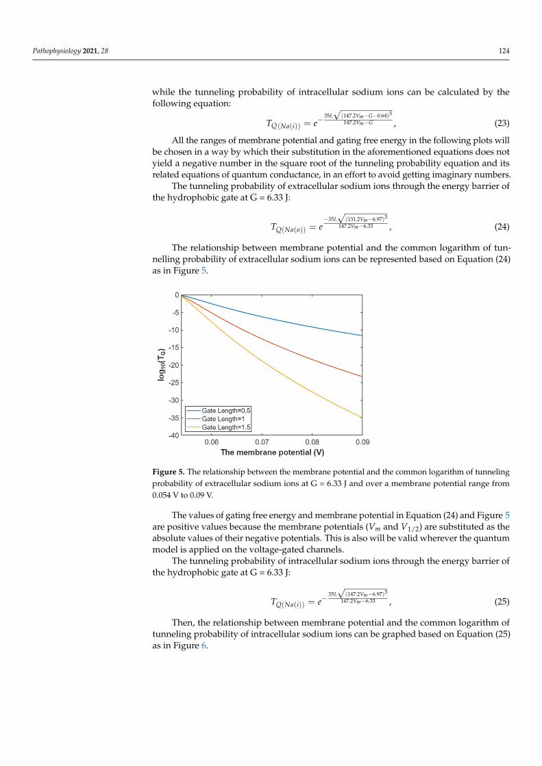
The relationship between membrane potential and the common logarithm of tun-nelling probability of extracellular sodium ions can be represented based on Equation (24)as in Figure 5.
Pathophysiology 2021, 28 125
Figure 5. The relationship between the membrane potential and the common logarithm of tunnel-ing probability of extracellular sodium ions at G = 6.33 J and over a membrane potential range from 0.054 V to 0.09 V.
The values of gating free energy and membrane potential in Equation (24) and Figure 5 are positive values because the membrane potentials (Vm and V1/2) are substituted as the absolute values of their negative potentials. This is also will be valid wherever the quan-tum model is applied on the voltage-gated channels.
The tunneling probability of intracellular sodium ions through the energy barrier of the hydrophobic gate at G = 6.33 J:
335 (147.2 6.97)147.2 6.33
( ( ))
m
m
L VV
Q Na iT e−
−−=
, (25)
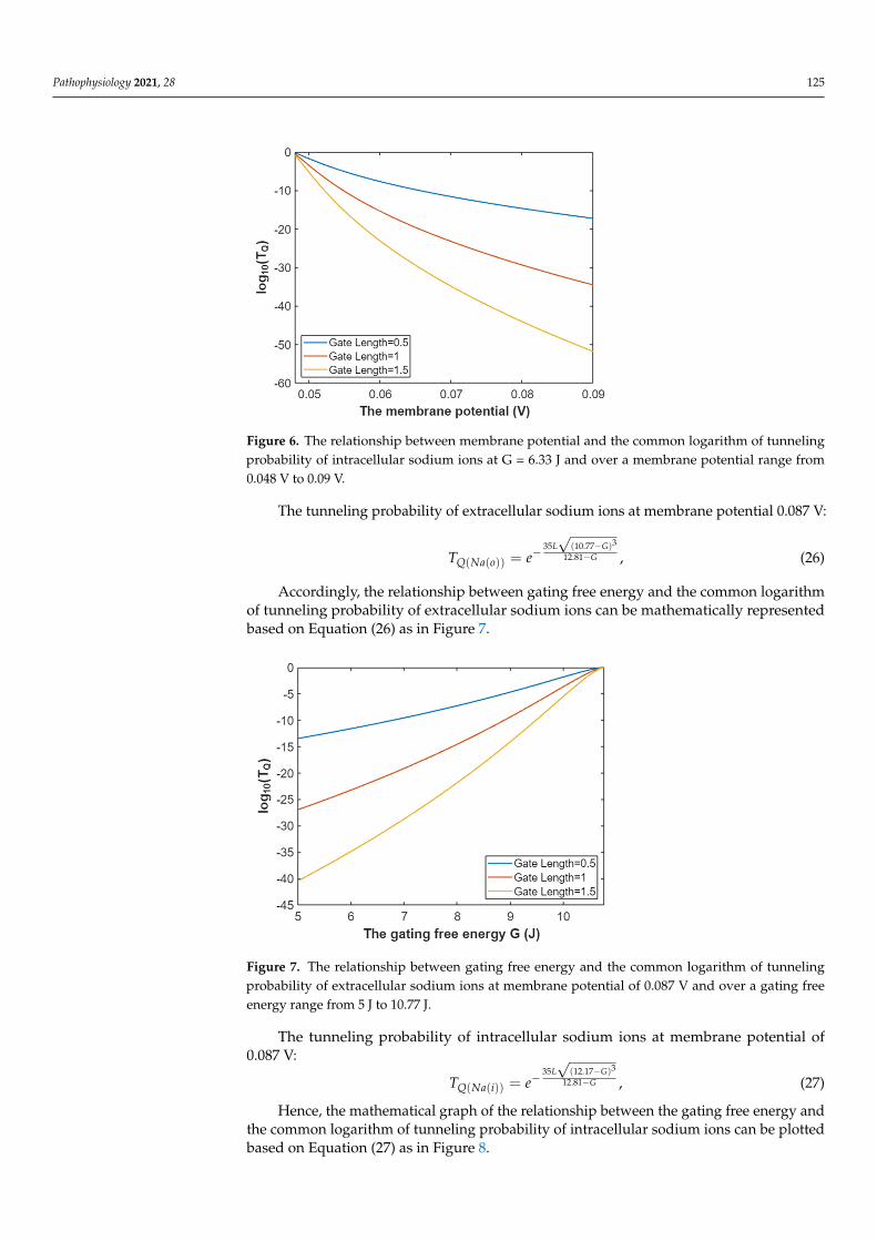
Then, the relationship between membrane potential and the common logarithm of tunneling probability of intracellular sodium ions can be graphed based on Equation (25) as in Figure 6.
Figure 6. The relationship between membrane potential and the common logarithm of tunneling probability of intracellular sodium ions at G = 6.33 J and over a membrane potential range from 0.048 V to 0.09 V.
The tunneling probability of extracellular sodium ions at membrane potential 0.087 V:
Figure 5. The relationship between the membrane potential and the common logarithm of tunnelingprobability of extracellular sodium ions at G = 6.33 J and over a membrane potential range from0.054 V to 0.09 V.
The values of gating free energy and membrane potential in Equation (24) and Figure 5are positive values because the membrane potentials (Vm and V1/2) are substituted as theabsolute values of their negative potentials. This is also will be valid wherever the quantummodel is applied on the voltage-gated channels.
The tunneling probability of intracellular sodium ions through the energy barrier ofthe hydrophobic gate at G = 6.33 J:
TQ(Na(i)) = e−35L√
(147.2Vm−6.97)3
147.2Vm−6.33 , (25)
Then, the relationship between membrane potential and the common logarithm oftunneling probability of intracellular sodium ions can be graphed based on Equation (25)as in Figure 6.
Pathophysiology 2021, 28 125
Pathophysiology 2021, 28 125
Figure 5. The relationship between the membrane potential and the common logarithm of tunnel-ing probability of extracellular sodium ions at G = 6.33 J and over a membrane potential range from 0.054 V to 0.09 V.
The values of gating free energy and membrane potential in Equation (24) and Figure 5 are positive values because the membrane potentials (Vm and V1/2) are substituted as the absolute values of their negative potentials. This is also will be valid wherever the quan-tum model is applied on the voltage-gated channels.
The tunneling probability of intracellular sodium ions through the energy barrier of the hydrophobic gate at G = 6.33 J:
335 (147.2 6.97)147.2 6.33
( ( ))
m
m
L VV
Q Na iT e−
−−=
, (25)
Then, the relationship between membrane potential and the common logarithm of tunneling probability of intracellular sodium ions can be graphed based on Equation (25) as in Figure 6.
Figure 6. The relationship between membrane potential and the common logarithm of tunneling probability of intracellular sodium ions at G = 6.33 J and over a membrane potential range from 0.048 V to 0.09 V.
The tunneling probability of extracellular sodium ions at membrane potential 0.087 V:
Figure 6. The relationship between membrane potential and the common logarithm of tunnelingprobability of intracellular sodium ions at G = 6.33 J and over a membrane potential range from0.048 V to 0.09 V.
The tunneling probability of extracellular sodium ions at membrane potential 0.087 V:
TQ(Na(o)) = e−35L√
(10.77−G)3
12.81−G , (26)
Accordingly, the relationship between gating free energy and the common logarithmof tunneling probability of extracellular sodium ions can be mathematically representedbased on Equation (26) as in Figure 7.
Pathophysiology 2021, 28 126
335 (10.77 )12.81
( ( ))
L GG
Q Na oT e−
−−= , (26)
Accordingly, the relationship between gating free energy and the common logarithm
of tunneling probability of extracellular sodium ions can be mathematically represented based on Equation (26) as in Figure 7.
Figure 7. The relationship between gating free energy and the common logarithm of tunneling probability of extracellular sodium ions at membrane potential of 0.087 V and over a gating free energy range from 5 J to 10.77 J.
The tunneling probability of intracellular sodium ions at membrane potential of 0.087 V:
335 (12.17 )12.81
( ( ))
L GG
Q Na iT e−
−−= , (27)
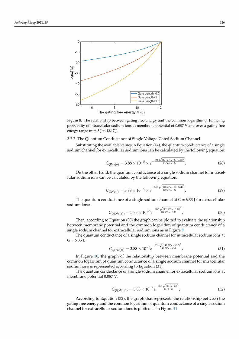
Hence, the mathematical graph of the relationship between the gating free energy and the common logarithm of tunneling probability of intracellular sodium ions can be plotted based on Equation (27) as in Figure 8.
Figure 8. The relationship between gating free energy and the common logarithm of tunneling probability of intracellular sodium ions at membrane potential of 0.087 V and over a gating free energy range from 5 J to 12.17 J.
Figure 7. The relationship between gating free energy and the common logarithm of tunnelingprobability of extracellular sodium ions at membrane potential of 0.087 V and over a gating freeenergy range from 5 J to 10.77 J.
The tunneling probability of intracellular sodium ions at membrane potential of0.087 V:
TQ(Na(i)) = e−35L√
(12.17−G)3
12.81−G , (27)
Hence, the mathematical graph of the relationship between the gating free energy andthe common logarithm of tunneling probability of intracellular sodium ions can be plottedbased on Equation (27) as in Figure 8.
Pathophysiology 2021, 28 126
Pathophysiology 2021, 28 126
335 (10.77 )12.81
( ( ))
L GG
Q Na oT e−
−−= , (26)
Accordingly, the relationship between gating free energy and the common logarithm
of tunneling probability of extracellular sodium ions can be mathematically represented based on Equation (26) as in Figure 7.
Figure 7. The relationship between gating free energy and the common logarithm of tunneling probability of extracellular sodium ions at membrane potential of 0.087 V and over a gating free energy range from 5 J to 10.77 J.
The tunneling probability of intracellular sodium ions at membrane potential of 0.087 V:
335 (12.17 )12.81
( ( ))
L GG
Q Na iT e−
−−= , (27)
Hence, the mathematical graph of the relationship between the gating free energy and the common logarithm of tunneling probability of intracellular sodium ions can be plotted based on Equation (27) as in Figure 8.
Figure 8. The relationship between gating free energy and the common logarithm of tunneling probability of intracellular sodium ions at membrane potential of 0.087 V and over a gating free energy range from 5 J to 12.17 J.
Figure 8. The relationship between gating free energy and the common logarithm of tunnelingprobability of intracellular sodium ions at membrane potential of 0.087 V and over a gating freeenergy range from 5 J to 12.17 J.
3.2.2. The Quantum Conductance of Single Voltage-Gated Sodium Channel
Substituting the available values in Equation (14), the quantum conductance of a singlesodium channel for extracellular sodium ions can be calculated by the following equation:
CQNa(o) = 3.88× 10⁻⁵ × e−35L√
(131.2Vm−G−0.64)3
147.2Vm−G , (28)
On the other hand, the quantum conductance of a single sodium channel for intracel-lular sodium ions can be calculated by the following equation:
CQNa(i) = 3.88× 10⁻⁵ × e−35L√
(147.2Vm−G−0.64)3
147.2Vm−G , (29)
The quantum conductance of a single sodium channel at G = 6.33 J for extracellularsodium ions:
CQ(Na(o)) = 3.88× 10⁻⁵e−35L√
(131.2Vm−6.97)3
147.2Vm−6.33 , (30)
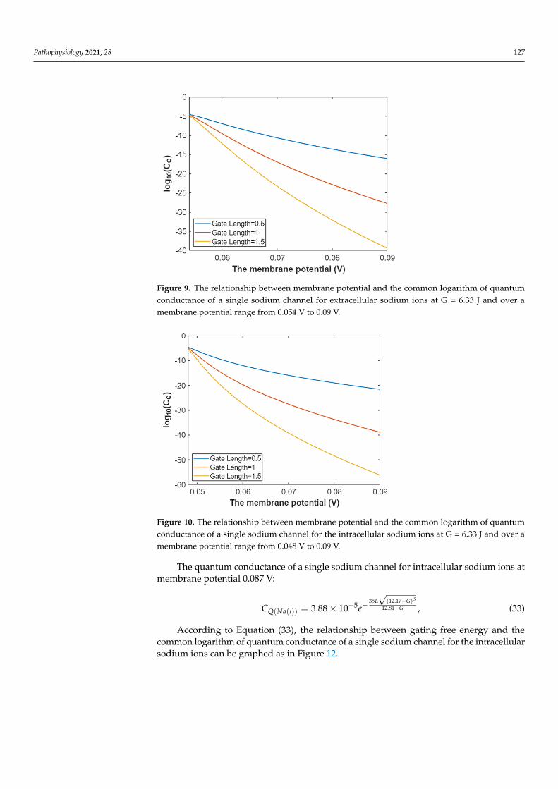
Then, according to Equation (30) the graph can be plotted to evaluate the relationshipbetween membrane potential and the common logarithm of quantum conductance of asingle sodium channel for extracellular sodium ions as in Figure 9.
The quantum conductance of a single sodium channel for intracellular sodium ions atG = 6.33 J:
CQ(Na(i)) = 3.88× 10⁻⁵e−35L√
(147.2Vm−6.97)3
147.2Vm−6.33 , (31)
In Figure 10, the graph of the relationship between membrane potential and thecommon logarithm of quantum conductance of a single sodium channel for intracellularsodium ions is represented according to Equation (31).
The quantum conductance of a single sodium channel for extracellular sodium ions atmembrane potential 0.087 V:
CQ(Na(o)) = 3.88× 10⁻⁵e−35L√
(10.77−G)3
12.81−G , (32)
According to Equation (32), the graph that represents the relationship between thegating free energy and the common logarithm of quantum conductance of a single sodiumchannel for extracellular sodium ions is plotted as in Figure 11.
Pathophysiology 2021, 28 127
Pathophysiology 2021, 28 127
3.2.2. The Quantum Conductance of Single Voltage-Gated Sodium Channel Substituting the available values in Equation (14), the quantum conductance of a sin-
gle sodium channel for extracellular sodium ions can be calculated by the following equa-tion:
335 (131.2 0.64)147.25
( ) 3.88 10m
m
L V GV G
QNa oC e− −
−−−= × × , (28)
On the other hand, the quantum conductance of a single sodium channel for intra-cellular sodium ions can be calculated by the following equation:
335 (147.2 0.64)147.25
( ) 3.88 10m
m
L V GV G
QNa iC e− −
−−−= × × , (29)
The quantum conductance of a single sodium channel at G = 6.33 J for extracellular sodium ions:
335 (131.2 6.97)147.2 6.335
( ( )) 3.88 10m
m
L VV
Q Na oC e−
−−−= × , (30)
Then, according to Equation (30) the graph can be plotted to evaluate the relationship between membrane potential and the common logarithm of quantum conductance of a single sodium channel for extracellular sodium ions as in Figure 9.
Figure 9. The relationship between membrane potential and the common logarithm of quantum conductance of a single sodium channel for extracellular sodium ions at G = 6.33 J and over a membrane potential range from 0.054 V to 0.09 V.
The quantum conductance of a single sodium channel for intracellular sodium ions at G = 6.33 J:
335 (147.2 6.97)147.2 6.335
( ( )) 3.88 10m
m
L VV
Q Na iC e−
−−−= × , (31)
In Figure 10, the graph of the relationship between membrane potential and the com-mon logarithm of quantum conductance of a single sodium channel for intracellular so-dium ions is represented according to Equation (31).
Figure 9. The relationship between membrane potential and the common logarithm of quantumconductance of a single sodium channel for extracellular sodium ions at G = 6.33 J and over amembrane potential range from 0.054 V to 0.09 V.
Pathophysiology 2021, 28 128
Figure 10. The relationship between membrane potential and the common logarithm of quantum conductance of a single sodium channel for the intracellular sodium ions at G = 6.33 J and over a membrane potential range from 0.048 V to 0.09 V.
The quantum conductance of a single sodium channel for extracellular sodium ions at membrane potential 0.087 V:
335 (10.77 )5 12.81
( ( )) 3.88 10L G
GQ Na oC e
−−− −= × , (32)
According to Equation (32), the graph that represents the relationship between the gating free energy and the common logarithm of quantum conductance of a single sodium channel for extracellular sodium ions is plotted as in Figure 11.
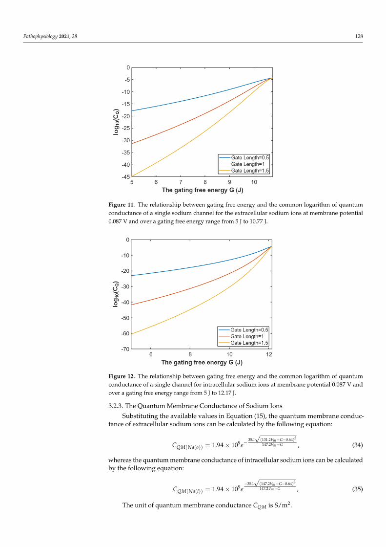
Figure 11. The relationship between gating free energy and the common logarithm of quantum conductance of a single sodium channel for the extracellular sodium ions at membrane potential 0.087 V and over a gating free energy range from 5 J to 10.77 J.
The quantum conductance of a single sodium channel for intracellular sodium ions at membrane potential 0.087 V:
Figure 10. The relationship between membrane potential and the common logarithm of quantumconductance of a single sodium channel for the intracellular sodium ions at G = 6.33 J and over amembrane potential range from 0.048 V to 0.09 V.
The quantum conductance of a single sodium channel for intracellular sodium ions atmembrane potential 0.087 V:
CQ(Na(i)) = 3.88× 10⁻⁵e−35L√
(12.17−G)3
12.81−G , (33)
According to Equation (33), the relationship between gating free energy and thecommon logarithm of quantum conductance of a single sodium channel for the intracellularsodium ions can be graphed as in Figure 12.
Pathophysiology 2021, 28 128
Pathophysiology 2021, 28 128
Figure 10. The relationship between membrane potential and the common logarithm of quantum conductance of a single sodium channel for the intracellular sodium ions at G = 6.33 J and over a membrane potential range from 0.048 V to 0.09 V.
The quantum conductance of a single sodium channel for extracellular sodium ions at membrane potential 0.087 V:
335 (10.77 )5 12.81
( ( )) 3.88 10L G
GQ Na oC e
−−− −= × , (32)
According to Equation (32), the graph that represents the relationship between the gating free energy and the common logarithm of quantum conductance of a single sodium channel for extracellular sodium ions is plotted as in Figure 11.
Figure 11. The relationship between gating free energy and the common logarithm of quantum conductance of a single sodium channel for the extracellular sodium ions at membrane potential 0.087 V and over a gating free energy range from 5 J to 10.77 J.
The quantum conductance of a single sodium channel for intracellular sodium ions at membrane potential 0.087 V:
Figure 11. The relationship between gating free energy and the common logarithm of quantumconductance of a single sodium channel for the extracellular sodium ions at membrane potential0.087 V and over a gating free energy range from 5 J to 10.77 J.
Pathophysiology 2021, 28 129
335 (12.17 )5 12.81
( ( )) 3.88 10L G
GQ Na iC e
−−− −= × , (33)
According to Equation (33), the relationship between gating free energy and the com-mon logarithm of quantum conductance of a single sodium channel for the intracellular sodium ions can be graphed as in Figure 12.
Figure 12. The relationship between gating free energy and the common logarithm of quantum conductance of a single channel for intracellular sodium ions at membrane potential 0.087 V and over a gating free energy range from 5 J to 12.17 J.
3.2.3. The Quantum Membrane Conductance of Sodium Ions Substituting the available values in Equation (15), the quantum membrane conduct-
ance of extracellular sodium ions can be calculated by the following equation:
335 (131.2 0.64)147.29
( ( )) 1.94 10m
m
L V GV G
QM Na oC e− −
−−= × , (34)
whereas the quantum membrane conductance of intracellular sodium ions can be calcu-lated by the following equation:
335 (147.2 0.64)147.29
( ( )) 1.94 10m
m
L V GV G
QM Na iC e− − −
−= × , (35)
The unit of quantum membrane conductance QMC is S/m2. The quantum membrane conductance of extracellular sodium ions at G = 6.33 J:
335 (131.2 6.97)147.2 6.339
( ( )) 1.94 10m
m
L VV
QM Na oC e− −
−= × , (36)
Then, according to Equation (36) the relationship between membrane potential and the common logarithm of quantum membrane conductance of extracellular sodium ions can be mathematically graphed as in Figure 13.
Figure 12. The relationship between gating free energy and the common logarithm of quantumconductance of a single channel for intracellular sodium ions at membrane potential 0.087 V andover a gating free energy range from 5 J to 12.17 J.
3.2.3. The Quantum Membrane Conductance of Sodium Ions
Substituting the available values in Equation (15), the quantum membrane conduc-tance of extracellular sodium ions can be calculated by the following equation:
CQM(Na(o)) = 1.94× 10⁹e−35L√
(131.2Vm−G−0.64)3
147.2Vm−G , (34)
whereas the quantum membrane conductance of intracellular sodium ions can be calculatedby the following equation:
CQM(Na(i)) = 1.94× 10⁹e−35L√
(147.2Vm−G−0.64)3
147.2Vm−G , (35)
The unit of quantum membrane conductance CQM is S/m2.
Pathophysiology 2021, 28 129
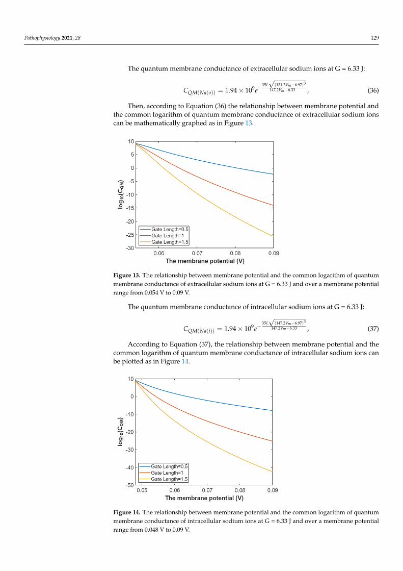
The quantum membrane conductance of extracellular sodium ions at G = 6.33 J:
CQM(Na(o)) = 1.94× 10⁹e−35L√
(131.2Vm−6.97)3
147.2Vm−6.33 , (36)
Then, according to Equation (36) the relationship between membrane potential andthe common logarithm of quantum membrane conductance of extracellular sodium ionscan be mathematically graphed as in Figure 13.
Pathophysiology 2021, 28 130
Figure 13. The relationship between membrane potential and the common logarithm of quantum membrane conductance of extracellular sodium ions at G = 6.33 J and over a membrane potential range from 0.054 V to 0.09 V.
The quantum membrane conductance of intracellular sodium ions at G = 6.33 J:
335 (147.2 6.97)147.2 6.339
( ( )) 1.94 10m
m
L VV
QM Na iC e−
−−= ×
, (37)
According to Equation (37), the relationship between membrane potential and the common logarithm of quantum membrane conductance of intracellular sodium ions can be plotted as in Figure 14.
Figure 14. The relationship between membrane potential and the common logarithm of quantum membrane conductance of intracellular sodium ions at G = 6.33 J and over a membrane potential range from 0.048 V to 0.09 V.
The quantum membrane conductance of extracellular sodium ions at membrane po-tential 0.087 V:
335 (10.77 )9 12.81
( ( )) 1.94 10L G
GQM Na oC e
−−
−= × , (38)
Figure 13. The relationship between membrane potential and the common logarithm of quantummembrane conductance of extracellular sodium ions at G = 6.33 J and over a membrane potentialrange from 0.054 V to 0.09 V.
The quantum membrane conductance of intracellular sodium ions at G = 6.33 J:
CQM(Na(i)) = 1.94× 10⁹e−35L√
(147.2Vm−6.97)3
147.2Vm−6.33 , (37)
According to Equation (37), the relationship between membrane potential and thecommon logarithm of quantum membrane conductance of intracellular sodium ions canbe plotted as in Figure 14.
Pathophysiology 2021, 28 130
Figure 13. The relationship between membrane potential and the common logarithm of quantum membrane conductance of extracellular sodium ions at G = 6.33 J and over a membrane potential range from 0.054 V to 0.09 V.
The quantum membrane conductance of intracellular sodium ions at G = 6.33 J:
335 (147.2 6.97)147.2 6.339
( ( )) 1.94 10m
m
L VV
QM Na iC e−
−−= ×
, (37)
According to Equation (37), the relationship between membrane potential and the common logarithm of quantum membrane conductance of intracellular sodium ions can be plotted as in Figure 14.
Figure 14. The relationship between membrane potential and the common logarithm of quantum membrane conductance of intracellular sodium ions at G = 6.33 J and over a membrane potential range from 0.048 V to 0.09 V.
The quantum membrane conductance of extracellular sodium ions at membrane po-tential 0.087 V:
335 (10.77 )9 12.81
( ( )) 1.94 10L G
GQM Na oC e
−−
−= × , (38)
Figure 14. The relationship between membrane potential and the common logarithm of quantummembrane conductance of intracellular sodium ions at G = 6.33 J and over a membrane potentialrange from 0.048 V to 0.09 V.
Pathophysiology 2021, 28 130
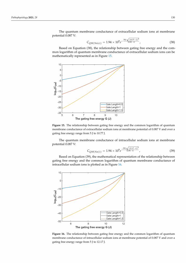
The quantum membrane conductance of extracellular sodium ions at membranepotential 0.087 V:
CQM(Na(o)) = 1.94× 10⁹e−35L√
(10.77−G)3
12.81−G , (38)
Based on Equation (38), the relationship between gating free energy and the com-mon logarithm of quantum membrane conductance of extracellular sodium ions can bemathematically represented as in Figure 15.
Pathophysiology 2021, 28 131
Based on Equation (38), the relationship between gating free energy and the common logarithm of quantum membrane conductance of extracellular sodium ions can be math-ematically represented as in Figure 15.
Figure 15. The relationship between gating free energy and the common logarithm of quantum membrane conductance of extracellular sodium ions at membrane potential of 0.087 V and over a gating free energy range from 5 J to 10.77 J.
The quantum membrane conductance of intracellular sodium ions at membrane po-tential 0.087 V:
335 (12.17 )9 12.81
( ( )) 1.94 10L G
GQM Na iC e
− −−= × , (39)
Based on Equation (39), the mathematical representation of the relationship between gating free energy and the common logarithm of quantum membrane conductance of in-tracellular sodium ions is plotted as in Figure 16.
Figure 16. The relationship between gating free energy and the common logarithm of quantum membrane conductance of intracellular sodium ions at membrane potential of 0.087 V and over a gating free energy range from 5 J to 12.17 J.
Figure 15. The relationship between gating free energy and the common logarithm of quantummembrane conductance of extracellular sodium ions at membrane potential of 0.087 V and over agating free energy range from 5 J to 10.77 J.
The quantum membrane conductance of intracellular sodium ions at membranepotential 0.087 V:
CQM(Na(i)) = 1.94× 10⁹e−35L√
(12.17−G)3
12.81−G , (39)
Based on Equation (39), the mathematical representation of the relationship betweengating free energy and the common logarithm of quantum membrane conductance ofintracellular sodium ions is plotted as in Figure 16.
Pathophysiology 2021, 28 131
Based on Equation (38), the relationship between gating free energy and the common logarithm of quantum membrane conductance of extracellular sodium ions can be math-ematically represented as in Figure 15.
Figure 15. The relationship between gating free energy and the common logarithm of quantum membrane conductance of extracellular sodium ions at membrane potential of 0.087 V and over a gating free energy range from 5 J to 10.77 J.
The quantum membrane conductance of intracellular sodium ions at membrane po-tential 0.087 V:
335 (12.17 )9 12.81
( ( )) 1.94 10L G
GQM Na iC e
− −−= × , (39)
Based on Equation (39), the mathematical representation of the relationship between gating free energy and the common logarithm of quantum membrane conductance of in-tracellular sodium ions is plotted as in Figure 16.
Figure 16. The relationship between gating free energy and the common logarithm of quantum membrane conductance of intracellular sodium ions at membrane potential of 0.087 V and over a gating free energy range from 5 J to 12.17 J.
Figure 16. The relationship between gating free energy and the common logarithm of quantummembrane conductance of intracellular sodium ions at membrane potential of 0.087 V and over agating free energy range from 5 J to 12.17 J.
Pathophysiology 2021, 28 131
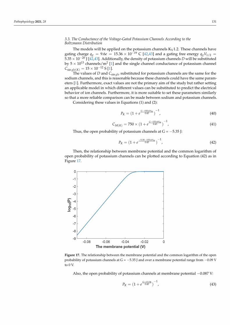
3.3. The Conductance of the Voltage-Gated Potassium Channels According to theBoltzmann Distribution
The models will be applied on the potassium channels KV1.2. These channels havegating charge qg = 9.6e = 15.36 × 10⁻¹⁹ C [42,43] and a gating free energy qgV1/2 =5.35× 10⁻²⁰ J [42,43]. Additionally, the density of potassium channels D will be substitutedby 5× 10¹³ channels/m2 [1] and the single channel conductance of potassium channelCsin gle(K) = 15× 10−12 S [1].
The values of D and Csin gle substituted for potassium channels are the same for thesodium channels, and this is reasonable because these channels could have the same param-eters [1]. Furthermore, exact values are not the primary aim of the study but rather settingan applicable model in which different values can be substituted to predict the electricalbehavior of ion channels. Furthermore, it is more suitable to set these parameters similarlyso that a more reliable comparison can be made between sodium and potassium channels.
Considering these values in Equations (1) and (2):
PK = (1 + eG−153.6Vm
0.43 )−1
, (40)
CM(K) = 750× (1 + eG−153.6Vm
0.43 )−1
, (41)
Thus, the open probability of potassium channels at G = −5.35 J:
PK = (1 + e−5.35−153.6Vm
0.43 )−1
, (42)
Then, the relationship between membrane potential and the common logarithm ofopen probability of potassium channels can be plotted according to Equation (42) as inFigure 17.
Pathophysiology 2021, 28 132
3.3. The Conductance of the Voltage-Gated Potassium Channels According to the Boltzmann Distribution
The models will be applied on the potassium channels KV1.2. These channels have gating charge 199.6 15.36 10gq e −= = × C [42,43] and a gating free energy
201/2 5.35 10gq V −= × J [42,43]. Additionally, the density of potassium channels D will be
substituted by 135 10× channels/m2 [1] and the single channel conductance of potassium channel 12
sin ( ) 15 10gle KC −= × S [1].
The values of D and sin gleC substituted for potassium channels are the same for the sodium channels, and this is reasonable because these channels could have the same pa-rameters [1]. Furthermore, exact values are not the primary aim of the study but rather setting an applicable model in which different values can be substituted to predict the electrical behavior of ion channels. Furthermore, it is more suitable to set these parameters similarly so that a more reliable comparison can be made between sodium and potassium channels.
Considering these values in Equations (1) and (2): 153.6
10.43(1 )mG V
KP e−
−= + , (40)
153.610.43
( ) 750 (1 )mG V
M KC e−
−= × + , (41)
Thus, the open probability of potassium channels at G = −5.35 J: 5.35 153.6
10.43(1 )mV
KP e− −
−= + , (42)
Then, the relationship between membrane potential and the common logarithm of open probability of potassium channels can be plotted according to Equation (42) as in Figure 17.
Figure 17. The relationship between the membrane potential and the common logarithm of the open probability of potassium channels at G = −5.35 J and over a membrane potential range from–0.09 V to 0 V.
Also, the open probability of potassium channels at membrane potential −0.087 V:
Figure 17. The relationship between the membrane potential and the common logarithm of the openprobability of potassium channels at G = −5.35 J and over a membrane potential range from −0.09 Vto 0 V.
Also, the open probability of potassium channels at membrane potential −0.087 V:
PK = (1 + eG+13.36
0.43 )−1
, (43)
Pathophysiology 2021, 28 132
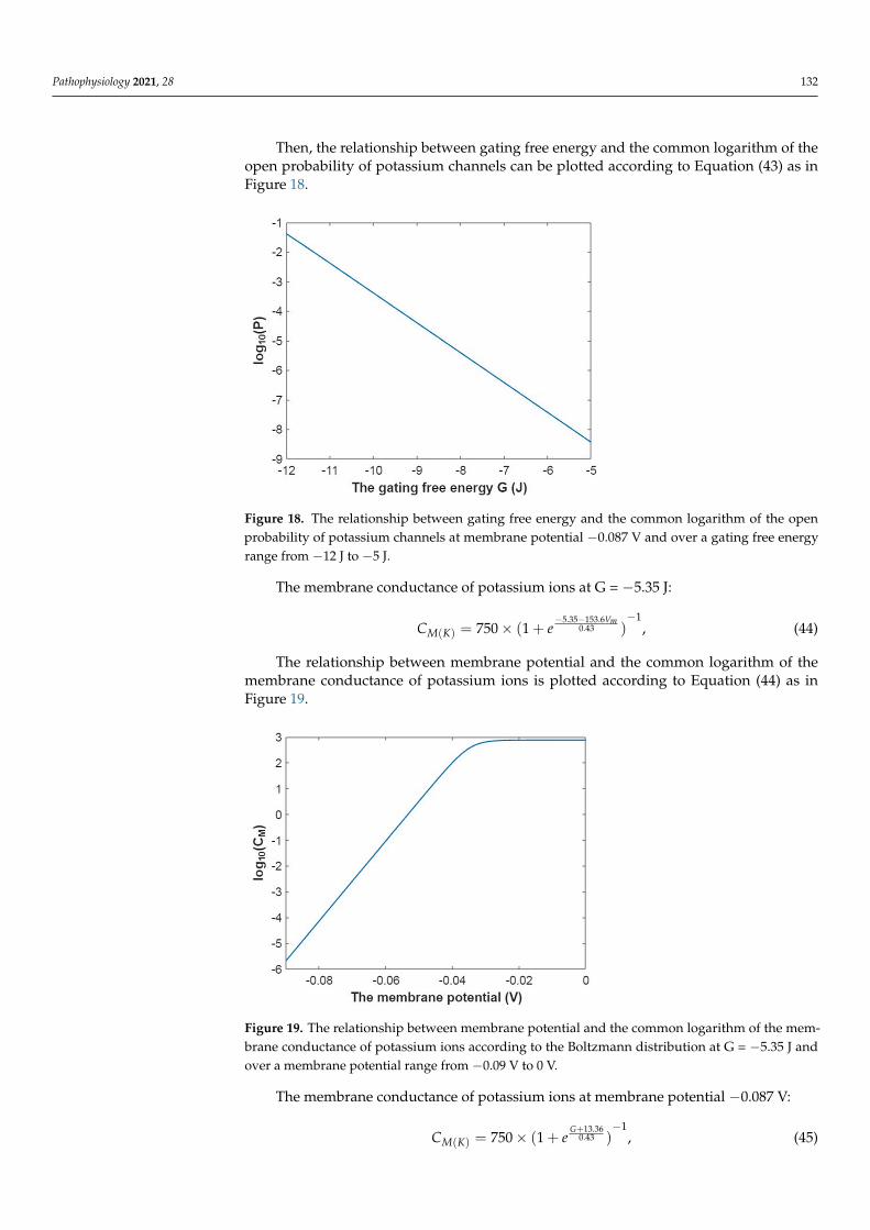
Then, the relationship between gating free energy and the common logarithm of theopen probability of potassium channels can be plotted according to Equation (43) as inFigure 18.
Pathophysiology 2021, 28 133
13.3610.43(1 )
G
KP e+
−= + , (43)
Then, the relationship between gating free energy and the common logarithm of the open probability of potassium channels can be plotted according to Equation (43) as in Figure 18.
Figure 18. The relationship between gating free energy and the common logarithm of the open probability of potassium channels at membrane potential −0.087 V and over a gating free energy range from −12 J to −5 J.
The membrane conductance of potassium ions at G = −5.35 J: 5.35 153.6
10.43( ) 750 (1 )
mV
M KC e− −
−= × + , (44)
The relationship between membrane potential and the common logarithm of the membrane conductance of potassium ions is plotted according to Equation (44) as in Fig-ure 19.
Figure 18. The relationship between gating free energy and the common logarithm of the openprobability of potassium channels at membrane potential −0.087 V and over a gating free energyrange from −12 J to −5 J.
The membrane conductance of potassium ions at G = −5.35 J:
CM(K) = 750× (1 + e−5.35−153.6Vm
0.43 )−1
, (44)
The relationship between membrane potential and the common logarithm of themembrane conductance of potassium ions is plotted according to Equation (44) as inFigure 19.
Pathophysiology 2021, 28 133
13.3610.43(1 )
G
KP e+
−= + , (43)
Then, the relationship between gating free energy and the common logarithm of the open probability of potassium channels can be plotted according to Equation (43) as in Figure 18.
Figure 18. The relationship between gating free energy and the common logarithm of the open probability of potassium channels at membrane potential −0.087 V and over a gating free energy range from −12 J to −5 J.
The membrane conductance of potassium ions at G = −5.35 J: 5.35 153.6
10.43( ) 750 (1 )
mV
M KC e− −
−= × + , (44)
The relationship between membrane potential and the common logarithm of the membrane conductance of potassium ions is plotted according to Equation (44) as in Fig-ure 19.
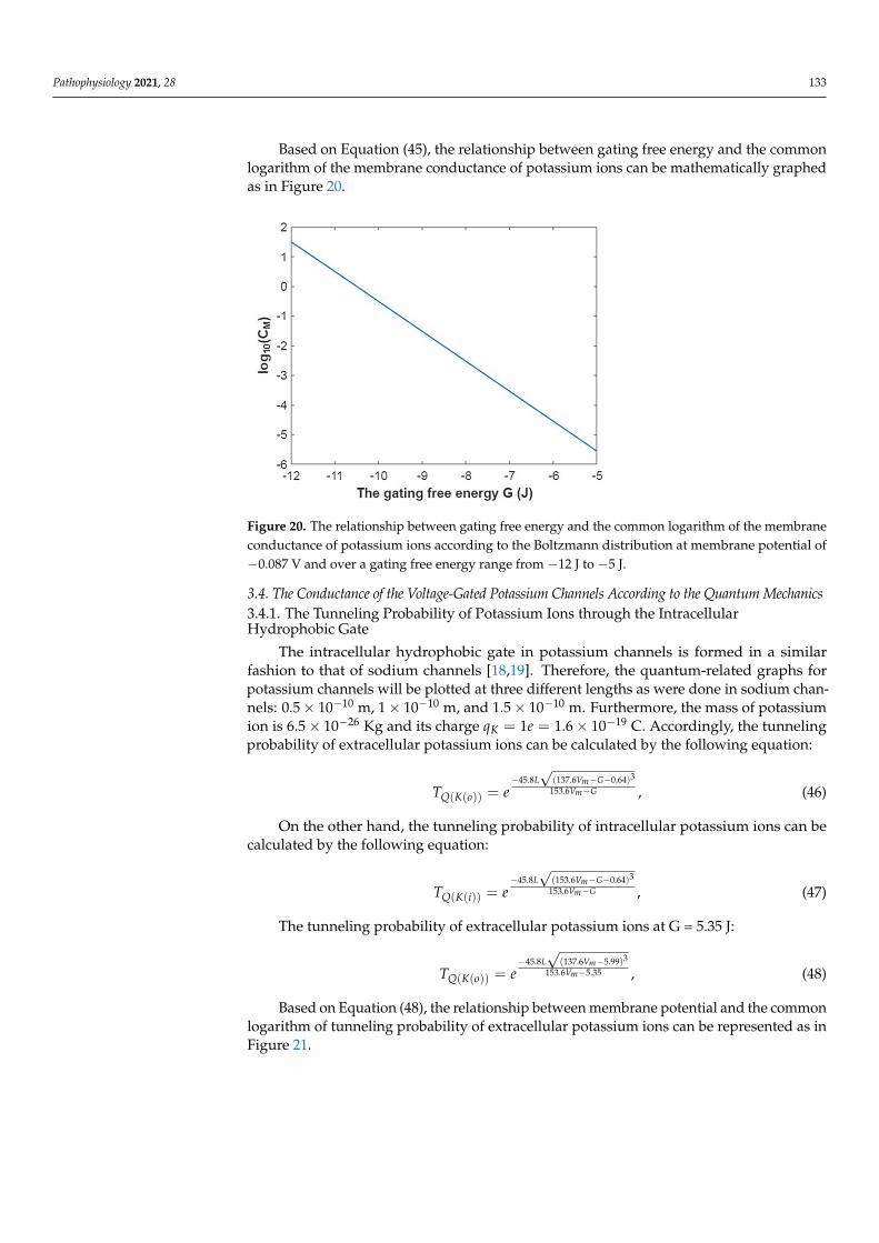
Figure 19. The relationship between membrane potential and the common logarithm of the mem-brane conductance of potassium ions according to the Boltzmann distribution at G = −5.35 J andover a membrane potential range from −0.09 V to 0 V.
The membrane conductance of potassium ions at membrane potential −0.087 V:
CM(K) = 750× (1 + eG+13.36
0.43 )−1
, (45)
Pathophysiology 2021, 28 133
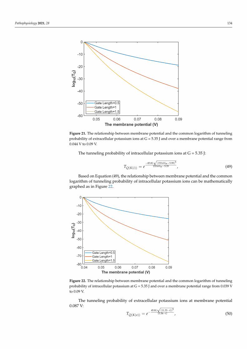
Based on Equation (45), the relationship between gating free energy and the commonlogarithm of the membrane conductance of potassium ions can be mathematically graphedas in Figure 20.
Pathophysiology 2021, 28 134
Figure 19. The relationship between membrane potential and the common logarithm of the mem-brane conductance of potassium ions according to the Boltzmann distribution at G = −5.35 J and over a membrane potential range from −0.09 V to 0 V.
The membrane conductance of potassium ions at membrane potential −0.087 V: 13.36
10.43( ) 750 (1 )
G
M KC e+
−= × + , (45)
Based on Equation (45), the relationship between gating free energy and the common logarithm of the membrane conductance of potassium ions can be mathematically graphed as in Figure 20.
Figure 20. The relationship between gating free energy and the common logarithm of the mem-brane conductance of potassium ions according to the Boltzmann distribution at membrane poten-tial of −0.087 V and over a gating free energy range from −12 J to −5 J.
3.4. The Conductance of the Voltage-Gated Potassium Channels According to the Quantum Mechanics 3.4.1. The Tunneling Probability of Potassium Ions through the Intracellular Hydropho-bic Gate
The intracellular hydrophobic gate in potassium channels is formed in a similar fash-ion to that of sodium channels [18,19]. Therefore, the quantum-related graphs for potas-sium channels will be plotted at three different lengths as were done in sodium channels:
100.5 10−× m, 101 10−× m, and 101.5 10−× m. Furthermore, the mass of potassium ion is 266.5 10−× Kg and its charge 191 1.6 10Kq e −= = × C. Accordingly, the tunneling probability of extracellular potassium ions can be calculated by the following equation:
345.8 (137.6 0.64)153.6
( ( ))
m
m
L V GV G
Q K oT e− − −
−= , (46)
On the other hand, the tunneling probability of intracellular potassium ions can be calculated by the following equation:
345.8 (153.6 0.64)153.6
( ( ))
m
m
L V GV G
Q K iT e− − −
−= , (47)
The tunneling probability of extracellular potassium ions at G = 5.35 J:
Figure 20. The relationship between gating free energy and the common logarithm of the membraneconductance of potassium ions according to the Boltzmann distribution at membrane potential of−0.087 V and over a gating free energy range from −12 J to −5 J.
3.4. The Conductance of the Voltage-Gated Potassium Channels According to the Quantum Mechanics3.4.1. The Tunneling Probability of Potassium Ions through the IntracellularHydrophobic Gate
The intracellular hydrophobic gate in potassium channels is formed in a similarfashion to that of sodium channels [18,19]. Therefore, the quantum-related graphs forpotassium channels will be plotted at three different lengths as were done in sodium chan-nels: 0.5× 10⁻¹⁰ m, 1× 10⁻¹⁰ m, and 1.5× 10⁻¹⁰ m. Furthermore, the mass of potassiumion is 6.5× 10⁻²⁶ Kg and its charge qK = 1e = 1.6× 10⁻¹⁹ C. Accordingly, the tunnelingprobability of extracellular potassium ions can be calculated by the following equation:
TQ(K(o)) = e−45.8L√
(137.6Vm−G−0.64)3
153.6Vm−G , (46)
On the other hand, the tunneling probability of intracellular potassium ions can becalculated by the following equation:
TQ(K(i)) = e−45.8L√
(153.6Vm−G−0.64)3
153.6Vm−G , (47)
The tunneling probability of extracellular potassium ions at G = 5.35 J:
TQ(K(o)) = e−45.8L√
(137.6Vm−5.99)3
153.6Vm−5.35 , (48)
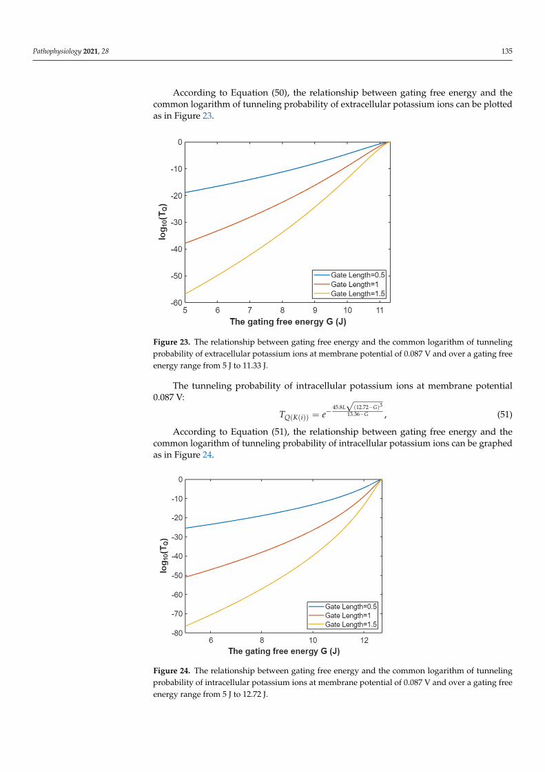
Based on Equation (48), the relationship between membrane potential and the commonlogarithm of tunneling probability of extracellular potassium ions can be represented as inFigure 21.
Pathophysiology 2021, 28 134
Pathophysiology 2021, 28 135
345.8 (137.6 5.99)153.6 5.35
( ( ))
m
m
L VV
Q K oT e− −
−= , (48)
Based on Equation (48), the relationship between membrane potential and the com-mon logarithm of tunneling probability of extracellular potassium ions can be represented as in Figure 21.
Figure 21. The relationship between membrane potential and the common logarithm of tunneling probability of extracellular potassium ions at G = 5.35 J and over a membrane potential range from 0.044 V to 0.09 V.
The tunneling probability of intracellular potassium ions at G = 5.35 J:
345.8 (153.6 5.99)153.6 5.35
( ( ))
m
m
L VV
Q K iT e− −
−= , (49)
Based on Equation (49), the relationship between membrane potential and the com-mon logarithm of tunneling probability of intracellular potassium ions can be mathemat-ically graphed as in Figure 22.
Figure 21. The relationship between membrane potential and the common logarithm of tunnelingprobability of extracellular potassium ions at G = 5.35 J and over a membrane potential range from0.044 V to 0.09 V.
The tunneling probability of intracellular potassium ions at G = 5.35 J:
TQ(K(i)) = e−45.8L√
(153.6Vm−5.99)3
153.6Vm−5.35 , (49)
Based on Equation (49), the relationship between membrane potential and the commonlogarithm of tunneling probability of intracellular potassium ions can be mathematicallygraphed as in Figure 22.
Pathophysiology 2021, 28 135
345.8 (137.6 5.99)153.6 5.35
( ( ))
m
m
L VV
Q K oT e− −
−= , (48)
Based on Equation (48), the relationship between membrane potential and the com-mon logarithm of tunneling probability of extracellular potassium ions can be represented as in Figure 21.
Figure 21. The relationship between membrane potential and the common logarithm of tunneling probability of extracellular potassium ions at G = 5.35 J and over a membrane potential range from 0.044 V to 0.09 V.
The tunneling probability of intracellular potassium ions at G = 5.35 J:
345.8 (153.6 5.99)153.6 5.35
( ( ))
m
m
L VV
Q K iT e− −
−= , (49)
Based on Equation (49), the relationship between membrane potential and the com-mon logarithm of tunneling probability of intracellular potassium ions can be mathemat-ically graphed as in Figure 22.
Figure 22. The relationship between membrane potential and the common logarithm of tunnelingprobability of intracellular potassium at G = 5.35 J and over a membrane potential range from 0.039 Vto 0.09 V.
The tunneling probability of extracellular potassium ions at membrane potential0.087 V:
TQ(K(o)) = e−45.8L√
(11.33−G)3
13.36−G , (50)
Pathophysiology 2021, 28 135
According to Equation (50), the relationship between gating free energy and thecommon logarithm of tunneling probability of extracellular potassium ions can be plottedas in Figure 23.
Pathophysiology 2021, 28 136
Figure 22. The relationship between membrane potential and the common logarithm of tunneling probability of intracellular potassium at G = 5.35 J and over a membrane potential range from 0.039 V to 0.09 V.
The tunneling probability of extracellular potassium ions at membrane potential 0.087 V:
345.8 (11.33 )13.36
( ( ))
L GG
Q K oT e−
−−= , (50)
According to Equation (50), the relationship between gating free energy and the com-mon logarithm of tunneling probability of extracellular potassium ions can be plotted as in Figure 23.
Figure 23. The relationship between gating free energy and the common logarithm of tunneling probability of extracellular potassium ions at membrane potential of 0.087 V and over a gating free energy range from 5 J to 11.33 J.
The tunneling probability of intracellular potassium ions at membrane potential 0.087 V:
345.8 (12.72 )13.36
( ( ))
L GG
Q K iT e−
−−= , (51)
According to Equation (51), the relationship between gating free energy and the com-mon logarithm of tunneling probability of intracellular potassium ions can be graphed as in Figure 24.
Figure 23. The relationship between gating free energy and the common logarithm of tunnelingprobability of extracellular potassium ions at membrane potential of 0.087 V and over a gating freeenergy range from 5 J to 11.33 J.
The tunneling probability of intracellular potassium ions at membrane potential0.087 V:
TQ(K(i)) = e−45.8L√
(12.72−G)3
13.36−G , (51)
According to Equation (51), the relationship between gating free energy and thecommon logarithm of tunneling probability of intracellular potassium ions can be graphedas in Figure 24.
Pathophysiology 2021, 28 137
Figure 24. The relationship between gating free energy and the common logarithm of tunneling probability of intracellular potassium ions at membrane potential of 0.087 V and over a gating free energy range from 5 J to 12.72 J.
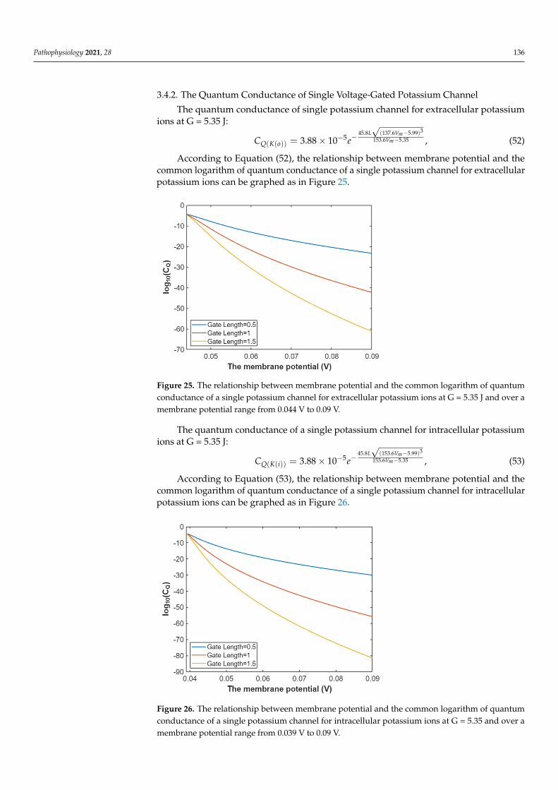
3.4.2. The Quantum Conductance of Single Voltage-Gated Potassium Channel The quantum conductance of single potassium channel for extracellular potassium
ions at G = 5.35 J:
345.8 (137.6 5.99)153.6 5.355
( ( )) 3.88 10m
m
L VV
Q K oC e−
−−−= ×
, (52)
According to Equation (52), the relationship between membrane potential and the common logarithm of quantum conductance of a single potassium channel for extracellu-lar potassium ions can be graphed as in Figure 25.
Figure 25. The relationship between membrane potential and the common logarithm of quantum conductance of a single potassium channel for extracellular potassium ions at G = 5.35 J and over a membrane potential range from 0.044 V to 0.09 V.
The quantum conductance of a single potassium channel for intracellular potassium ions at G = 5.35 J:
Figure 24. The relationship between gating free energy and the common logarithm of tunnelingprobability of intracellular potassium ions at membrane potential of 0.087 V and over a gating freeenergy range from 5 J to 12.72 J.
Pathophysiology 2021, 28 136
3.4.2. The Quantum Conductance of Single Voltage-Gated Potassium Channel
The quantum conductance of single potassium channel for extracellular potassiumions at G = 5.35 J:
CQ(K(o)) = 3.88× 10⁻⁵e−45.8L√
(137.6Vm−5.99)3
153.6Vm−5.35 , (52)
According to Equation (52), the relationship between membrane potential and thecommon logarithm of quantum conductance of a single potassium channel for extracellularpotassium ions can be graphed as in Figure 25.
Pathophysiology 2021, 28 137
Figure 24. The relationship between gating free energy and the common logarithm of tunneling probability of intracellular potassium ions at membrane potential of 0.087 V and over a gating free energy range from 5 J to 12.72 J.
3.4.2. The Quantum Conductance of Single Voltage-Gated Potassium Channel The quantum conductance of single potassium channel for extracellular potassium
ions at G = 5.35 J:
345.8 (137.6 5.99)153.6 5.355
( ( )) 3.88 10m
m
L VV
Q K oC e−
−−−= ×
, (52)
According to Equation (52), the relationship between membrane potential and the common logarithm of quantum conductance of a single potassium channel for extracellu-lar potassium ions can be graphed as in Figure 25.
Figure 25. The relationship between membrane potential and the common logarithm of quantum conductance of a single potassium channel for extracellular potassium ions at G = 5.35 J and over a membrane potential range from 0.044 V to 0.09 V.
The quantum conductance of a single potassium channel for intracellular potassium ions at G = 5.35 J:
Figure 25. The relationship between membrane potential and the common logarithm of quantumconductance of a single potassium channel for extracellular potassium ions at G = 5.35 J and over amembrane potential range from 0.044 V to 0.09 V.
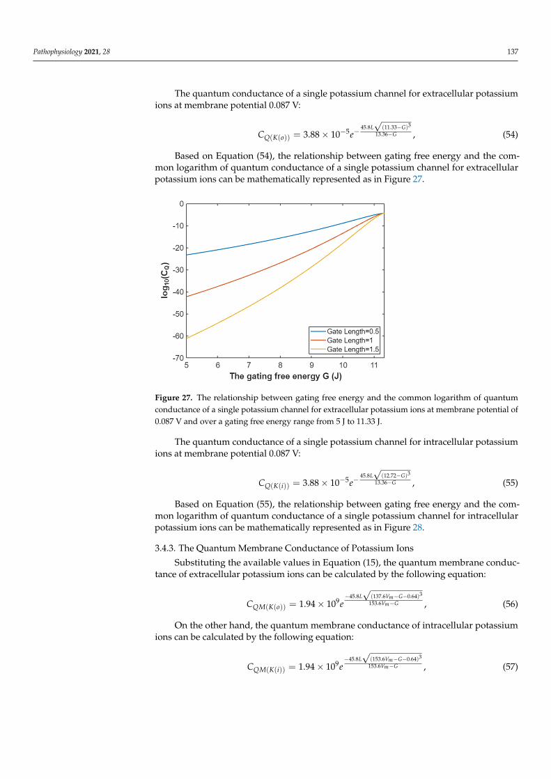
The quantum conductance of a single potassium channel for intracellular potassiumions at G = 5.35 J:
CQ(K(i)) = 3.88× 10⁻⁵e−45.8L√
(153.6Vm−5.99)3
153.6Vm−5.35 , (53)
According to Equation (53), the relationship between membrane potential and thecommon logarithm of quantum conductance of a single potassium channel for intracellularpotassium ions can be graphed as in Figure 26.
Pathophysiology 2021, 28 138
345.8 (153.6 5.99)153.6 5.355
( ( )) 3.88 10m
m
L VV
Q K iC e−
−−−= × , (53)
According to Equation (53), the relationship between membrane potential and the common logarithm of quantum conductance of a single potassium channel for intracellu-lar potassium ions can be graphed as in Figure 26.
Figure 26. The relationship between membrane potential and the common logarithm of quantum conductance of a single potassium channel for intracellular potassium ions at G = 5.35 and over a membrane potential range from 0.039 V to 0.09 V.
The quantum conductance of a single potassium channel for extracellular potassium ions at membrane potential 0.087 V:
345.8 (11.33 )5 13.36
( ( )) 3.88 10L G
GQ K oC e
−−− −= × , (54)
Based on Equation (54), the relationship between gating free energy and the common logarithm of quantum conductance of a single potassium channel for extracellular potas-sium ions can be mathematically represented as in Figure 27.
Figure 26. The relationship between membrane potential and the common logarithm of quantumconductance of a single potassium channel for intracellular potassium ions at G = 5.35 and over amembrane potential range from 0.039 V to 0.09 V.
Pathophysiology 2021, 28 137
The quantum conductance of a single potassium channel for extracellular potassiumions at membrane potential 0.087 V:
CQ(K(o)) = 3.88× 10⁻⁵e−45.8L√
(11.33−G)3
13.36−G , (54)
Based on Equation (54), the relationship between gating free energy and the com-mon logarithm of quantum conductance of a single potassium channel for extracellularpotassium ions can be mathematically represented as in Figure 27.
Pathophysiology 2021, 28 138
345.8 (153.6 5.99)153.6 5.355
( ( )) 3.88 10m
m
L VV
Q K iC e−
−−−= × , (53)
According to Equation (53), the relationship between membrane potential and the common logarithm of quantum conductance of a single potassium channel for intracellu-lar potassium ions can be graphed as in Figure 26.
Figure 26. The relationship between membrane potential and the common logarithm of quantum conductance of a single potassium channel for intracellular potassium ions at G = 5.35 and over a membrane potential range from 0.039 V to 0.09 V.
The quantum conductance of a single potassium channel for extracellular potassium ions at membrane potential 0.087 V:
345.8 (11.33 )5 13.36
( ( )) 3.88 10L G
GQ K oC e
−−− −= × , (54)
Based on Equation (54), the relationship between gating free energy and the common logarithm of quantum conductance of a single potassium channel for extracellular potas-sium ions can be mathematically represented as in Figure 27.
Figure 27. The relationship between gating free energy and the common logarithm of quantumconductance of a single potassium channel for extracellular potassium ions at membrane potential of0.087 V and over a gating free energy range from 5 J to 11.33 J.
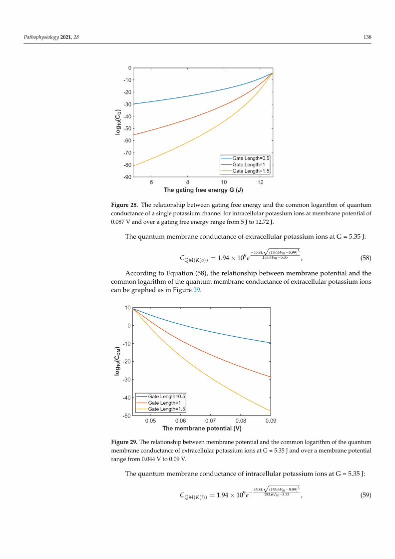
The quantum conductance of a single potassium channel for intracellular potassiumions at membrane potential 0.087 V:
CQ(K(i)) = 3.88× 10⁻⁵e−45.8L√
(12.72−G)3
13.36−G , (55)
Based on Equation (55), the relationship between gating free energy and the com-mon logarithm of quantum conductance of a single potassium channel for intracellularpotassium ions can be mathematically represented as in Figure 28.
3.4.3. The Quantum Membrane Conductance of Potassium Ions
Substituting the available values in Equation (15), the quantum membrane conduc-tance of extracellular potassium ions can be calculated by the following equation:
CQM(K(o)) = 1.94× 10⁹e−45.8L√
(137.6Vm−G−0.64)3
153.6Vm−G , (56)
On the other hand, the quantum membrane conductance of intracellular potassiumions can be calculated by the following equation:
CQM(K(i)) = 1.94× 10⁹e−45.8L√
(153.6Vm−G−0.64)3
153.6Vm−G , (57)
Pathophysiology 2021, 28 138
Pathophysiology 2021, 28 139
Figure 27. The relationship between gating free energy and the common logarithm of quantum conductance of a single potassium channel for extracellular potassium ions at membrane potential of 0.087 V and over a gating free energy range from 5 J to 11.33 J.
The quantum conductance of a single potassium channel for intracellular potassium ions at membrane potential 0.087 V:
345.8 (12.72 )5 13.36
( ( )) 3.88 10L G
GQ K iC e
−−− −= × , (55)
Based on Equation (55), the relationship between gating free energy and the common logarithm of quantum conductance of a single potassium channel for intracellular potas-sium ions can be mathematically represented as in Figure 28.
Figure 28. The relationship between gating free energy and the common logarithm of quantum conductance of a single potassium channel for intracellular potassium ions at membrane potential of 0.087 V and over a gating free energy range from 5 J to 12.72 J.
3.4.3. The Quantum Membrane Conductance of Potassium Ions Substituting the available values in Equation (15), the quantum membrane conduct-
ance of extracellular potassium ions can be calculated by the following equation:
345.8 (137.6 0.64)153.69
( ( )) 1.94 10m
m
L V GV G
QM K oC e− − −
−= × , (56)
On the other hand, the quantum membrane conductance of intracellular potassium ions can be calculated by the following equation:
345.8 (153.6 0.64)153.69
( ( )) 1.94 10m
m
L V GV G
QM K iC e− − −
−= × , (57)
The quantum membrane conductance of extracellular potassium ions at G = 5.35 J: 345.8 (137.6 5.99)
153.6 5.359( ( )) 1.94 10
m
m
L VV
QM K oC e− −
−= × , (58)
According to Equation (58), the relationship between membrane potential and the common logarithm of the quantum membrane conductance of extracellular potassium ions can be graphed as in Figure 29.
Figure 28. The relationship between gating free energy and the common logarithm of quantumconductance of a single potassium channel for intracellular potassium ions at membrane potential of0.087 V and over a gating free energy range from 5 J to 12.72 J.
The quantum membrane conductance of extracellular potassium ions at G = 5.35 J:
CQM(K(o)) = 1.94× 10⁹e−45.8L√
(137.6Vm−5.99)3
153.6Vm−5.35 , (58)
According to Equation (58), the relationship between membrane potential and thecommon logarithm of the quantum membrane conductance of extracellular potassium ionscan be graphed as in Figure 29.
Pathophysiology 2021, 28 140
Figure 29. The relationship between membrane potential and the common logarithm of the quan-tum membrane conductance of extracellular potassium ions at G = 5.35 J and over a membrane potential range from 0.044 V to 0.09 V.
The quantum membrane conductance of intracellular potassium ions at G = 5.35 J:
345.8 (153.6 5.99)153.6 5.359
( ( )) 1.94 10m
m
L VV
QM K iC e−
−−= × , (59)
According to Equation (59), the relationship between membrane potential and the common logarithm of the quantum membrane conductance of intracellular potassium ions can be graphed as in Figure 30.
Figure 30. The relationship between membrane potential and the common logarithm of the quan-tum membrane conductance of intracellular potassium ions at G = 5.35 J and over a membrane potential range from 0.039 V to 0.09 V.
The quantum membrane conductance of extracellular potassium ions at membrane potential 0.087 V:
345.8 (11.33 )9 13.36
( ( )) 1.94 10L G
GQM K oC e
−−
−= × , (60)
Figure 29. The relationship between membrane potential and the common logarithm of the quantummembrane conductance of extracellular potassium ions at G = 5.35 J and over a membrane potentialrange from 0.044 V to 0.09 V.
The quantum membrane conductance of intracellular potassium ions at G = 5.35 J:
CQM(K(i)) = 1.94× 10⁹e−45.8L√
(153.6Vm−5.99)3
153.6Vm−5.35 , (59)
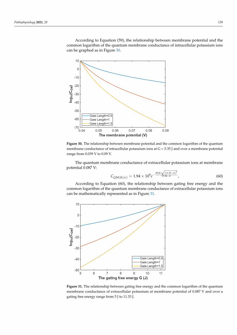
Pathophysiology 2021, 28 139
According to Equation (59), the relationship between membrane potential and thecommon logarithm of the quantum membrane conductance of intracellular potassium ionscan be graphed as in Figure 30.
Pathophysiology 2021, 28 140
Figure 29. The relationship between membrane potential and the common logarithm of the quan-tum membrane conductance of extracellular potassium ions at G = 5.35 J and over a membrane potential range from 0.044 V to 0.09 V.
The quantum membrane conductance of intracellular potassium ions at G = 5.35 J:
345.8 (153.6 5.99)153.6 5.359
( ( )) 1.94 10m
m
L VV
QM K iC e−
−−= × , (59)
According to Equation (59), the relationship between membrane potential and the common logarithm of the quantum membrane conductance of intracellular potassium ions can be graphed as in Figure 30.
Figure 30. The relationship between membrane potential and the common logarithm of the quan-tum membrane conductance of intracellular potassium ions at G = 5.35 J and over a membrane potential range from 0.039 V to 0.09 V.
The quantum membrane conductance of extracellular potassium ions at membrane potential 0.087 V:
345.8 (11.33 )9 13.36
( ( )) 1.94 10L G
GQM K oC e
−−
−= × , (60)
Figure 30. The relationship between membrane potential and the common logarithm of the quantummembrane conductance of intracellular potassium ions at G = 5.35 J and over a membrane potentialrange from 0.039 V to 0.09 V.
The quantum membrane conductance of extracellular potassium ions at membranepotential 0.087 V:
CQM(K(o)) = 1.94× 10⁹e−45.8L√
(11.33−G)3
13.36−G , (60)
According to Equation (60), the relationship between gating free energy and thecommon logarithm of the quantum membrane conductance of extracellular potassium ionscan be mathematically represented as in Figure 31.
Pathophysiology 2021, 28 141
According to Equation (60), the relationship between gating free energy and the com-mon logarithm of the quantum membrane conductance of extracellular potassium ions can be mathematically represented as in Figure 31.
Figure 31. The relationship between gating free energy and the common logarithm of the quantum membrane conductance of extracellular potassium at membrane potential of 0.087 V and over a gating free energy range from 5 J to 11.33 J.
The quantum membrane conductance of intracellular potassium ions at membrane potential 0.087 V:
345.8 (12.72 )9 13.36
( ( )) 1.94 10L G
GQM K iC e
−−
−= × , (61)
According to Equation (61), the relationship between gating free energy and the com-mon logarithm of the quantum membrane conductance of intracellular potassium ions can be mathematically represented as in Figure 32.
Figure 32. The relationship between gating free energy and the common logarithm of the quantum membrane conductance of intracellular potassium ions at membrane potential of 0.087 V and over a gating free energy range from 5 J and 12.72 J.
Figure 31. The relationship between gating free energy and the common logarithm of the quantummembrane conductance of extracellular potassium at membrane potential of 0.087 V and over agating free energy range from 5 J to 11.33 J.
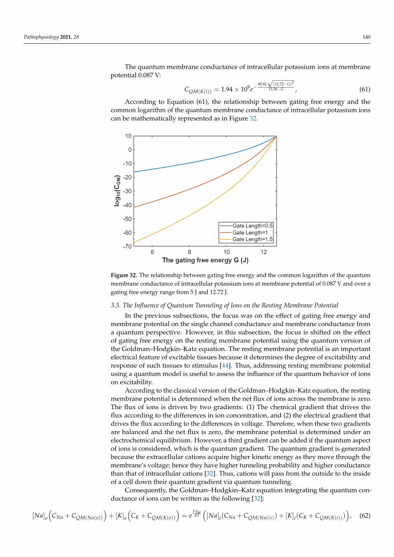
Pathophysiology 2021, 28 140
The quantum membrane conductance of intracellular potassium ions at membranepotential 0.087 V:
CQM(K(i)) = 1.94× 10⁹e−45.8L√
(12.72−G)3
13.36−G , (61)
According to Equation (61), the relationship between gating free energy and thecommon logarithm of the quantum membrane conductance of intracellular potassium ionscan be mathematically represented as in Figure 32.
Pathophysiology 2021, 28 141
According to Equation (60), the relationship between gating free energy and the com-mon logarithm of the quantum membrane conductance of extracellular potassium ions can be mathematically represented as in Figure 31.
Figure 31. The relationship between gating free energy and the common logarithm of the quantum membrane conductance of extracellular potassium at membrane potential of 0.087 V and over a gating free energy range from 5 J to 11.33 J.
The quantum membrane conductance of intracellular potassium ions at membrane potential 0.087 V:
345.8 (12.72 )9 13.36
( ( )) 1.94 10L G
GQM K iC e
−−
−= × , (61)
According to Equation (61), the relationship between gating free energy and the com-mon logarithm of the quantum membrane conductance of intracellular potassium ions can be mathematically represented as in Figure 32.
Figure 32. The relationship between gating free energy and the common logarithm of the quantum membrane conductance of intracellular potassium ions at membrane potential of 0.087 V and over a gating free energy range from 5 J and 12.72 J.
Figure 32. The relationship between gating free energy and the common logarithm of the quantummembrane conductance of intracellular potassium ions at membrane potential of 0.087 V and over agating free energy range from 5 J and 12.72 J.
3.5. The Influence of Quantum Tunneling of Ions on the Resting Membrane Potential
In the previous subsections, the focus was on the effect of gating free energy andmembrane potential on the single channel conductance and membrane conductance froma quantum perspective. However, in this subsection, the focus is shifted on the effectof gating free energy on the resting membrane potential using the quantum version ofthe Goldman–Hodgkin–Katz equation. The resting membrane potential is an importantelectrical feature of excitable tissues because it determines the degree of excitability andresponse of such tissues to stimulus [44]. Thus, addressing resting membrane potentialusing a quantum model is useful to assess the influence of the quantum behavior of ionson excitability.
According to the classical version of the Goldman–Hodgkin–Katz equation, the restingmembrane potential is determined when the net flux of ions across the membrane is zero.The flux of ions is driven by two gradients: (1) The chemical gradient that drives theflux according to the differences in ion concentration, and (2) the electrical gradient thatdrives the flux according to the differences in voltage. Therefore, when these two gradientsare balanced and the net flux is zero, the membrane potential is determined under anelectrochemical equilibrium. However, a third gradient can be added if the quantum aspectof ions is considered, which is the quantum gradient. The quantum gradient is generatedbecause the extracellular cations acquire higher kinetic energy as they move through themembrane’s voltage; hence they have higher tunneling probability and higher conductancethan that of intracellular cations [32]. Thus, cations will pass from the outside to the insideof a cell down their quantum gradient via quantum tunneling.
Consequently, the Goldman–Hodgkin–Katz equation integrating the quantum con-ductance of ions can be written as the following [32]:
[Na]o(
CNa + CQM(Na(o))
)+ [K]o
(CK + CQM(K(o))
)= e
FVmRT
([Na]i(CNa + CQM(Na(i))) + [K]i(CK + CQM(K(i)))
), (62)
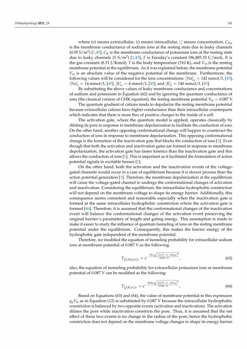
Pathophysiology 2021, 28 141
where (o) means extracellular, (i) means intracellular, [ ] means concentration, CNais the membrane conductance of sodium ions at the resting state due to leaky channels(0.05 S/m2) [1,45], CK is the membrane conductance of potassium ions at the resting statedue to leaky channels (5 S/m2) [1,45], F is Faraday’s constant (96,485.33 C/mol), R isthe gas constant (8.31 J/Kmol), T is the body temperature (310 K), and Vm is the restingmembrane potential at the equilibrium. As it was explained before, the membrane potentialVm is an absolute value of the negative potential of the membrane. Furthermore, thefollowing values will be considered for the ions concentrations: [Na]o = 142 mmol/L [45],[Na]i = 14 mmol/L [45], [K]o = 4 mmol/L [45], and [K]i = 140 mmol/L [45].
By substituting the above values of leaky membrane conductance and concentrationsof sodium and potassium in Equation (62) and by ignoring the quantum conductance ofions (the classical version of GHK equation), the resting membrane potential Vm = 0.087 V.
The quantum gradient of cations tends to depolarize the resting membrane potentialbecause extracellular cations have higher conductance than their intracellular counterpartswhich indicates that there is more flux of positive charges to the inside of a cell.
The activation gate, where the quantum model is applied, operates classically bydilating its pore in response to membrane depolarization to facilitate the conduction of ions.On the other hand, another opposing conformational change will happen to counteract theconduction of ions in response to membrane depolarization. This opposing conformationalchange is the formation of the inactivation gate that blocks the conduction of ions [1]. Eventhough that both the activation and inactivation gates are formed in response to membranedepolarization, the activation gate has faster kinetics than the inactivation gate and thisallows the conduction of ions [1]. This is important as it facilitated the formulation of actionpotential signals in excitable tissues [1].
On the other hand, both the activation and the inactivation events of the voltage-gated channels would occur in a case of equilibrium because it is slower process than theaction potential generation [1]. Therefore, the membrane depolarization at the equilibriumwill cause the voltage-gated channel to undergo the conformational changes of activationand inactivation. Considering the equilibrium, the intracellular hydrophobic constrictionwill not depend on the membrane voltage to shape its energy barrier. Additionally, thisconsequence seems consistent and reasonable especially when the inactivation gate isformed at the same intracellular hydrophobic constriction where the activation gate isformed [46]. Therefore, it is assumed that the conformational changes of the inactivationevent will balance the conformational changes of the activation event preserving theoriginal barrier’s parameters of length and gating energy. This assumption is made tomake it easier to study the influence of quantum tunneling of ions on the resting membranepotential under the equilibrium. Consequently, this makes the barrier energy of thehydrophobic gate independent of the membrane potential.
Therefore, we modified the equation of tunneling probability for extracellular sodiumions at membrane potential of 0.087 V as the following:
TQ(Na(o))r = e−35L√
(12.17−G−16Vm)3
12.81−G , (63)
also, the equation of tunneling probability for extracellular potassium ions at membranepotential of 0.087 V can be modified as the following:
TQ(K(o))r = e−45.8L√
(12.72−G−16Vm)3
13.36−G , (64)
Based on Equations (63) and (64), the value of membrane potential in this expressionqgVm as in Equation (12) is substituted by 0.087 V because the intracellular hydrophobicconstriction is balanced by two opposite events (activation and inactivation). The activationdilates the pore while inactivation constricts the pore. Thus, it is assumed that the neteffect of these two events is no change in the radius of the pore; hence the hydrophobicconstriction does not depend on the membrane voltage changes to shape its energy barrier.
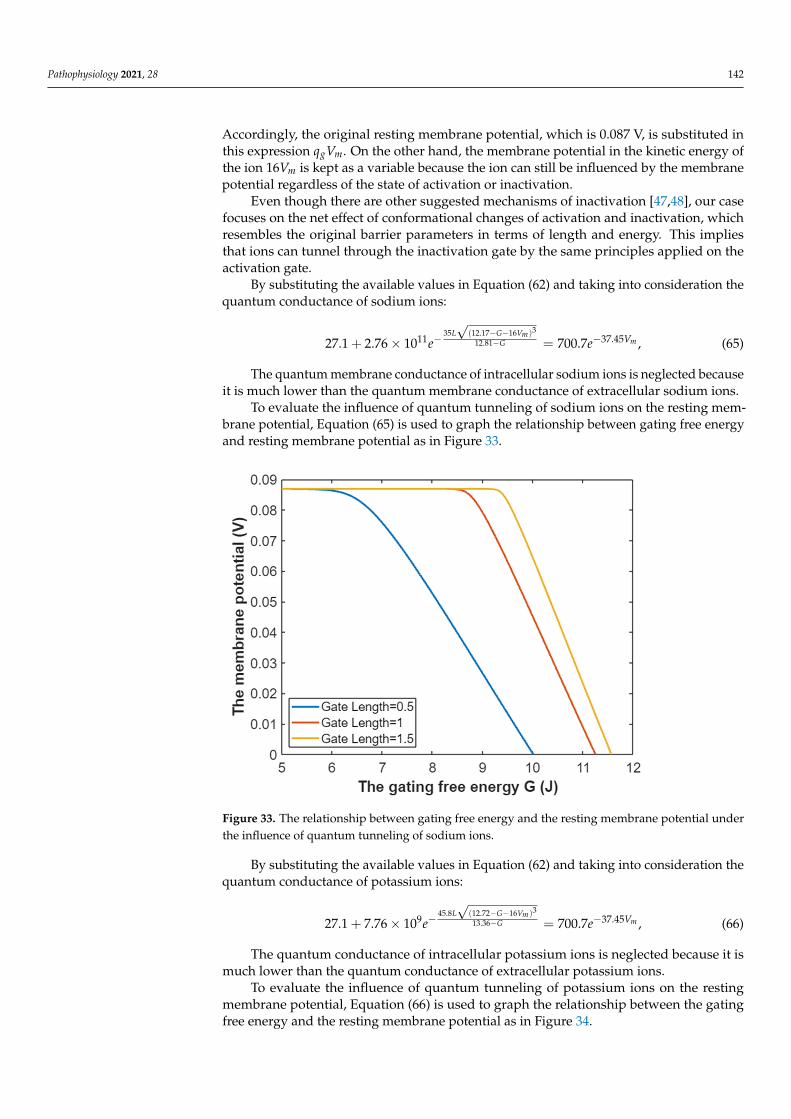
Pathophysiology 2021, 28 142
Accordingly, the original resting membrane potential, which is 0.087 V, is substituted inthis expression qgVm. On the other hand, the membrane potential in the kinetic energy ofthe ion 16Vm is kept as a variable because the ion can still be influenced by the membranepotential regardless of the state of activation or inactivation.
Even though there are other suggested mechanisms of inactivation [47,48], our casefocuses on the net effect of conformational changes of activation and inactivation, whichresembles the original barrier parameters in terms of length and energy. This impliesthat ions can tunnel through the inactivation gate by the same principles applied on theactivation gate.
By substituting the available values in Equation (62) and taking into consideration thequantum conductance of sodium ions:
27.1 + 2.76× 10¹¹e−35L√
(12.17−G−16Vm)3
12.81−G = 700.7e−37.45Vm , (65)
The quantum membrane conductance of intracellular sodium ions is neglected becauseit is much lower than the quantum membrane conductance of extracellular sodium ions.
To evaluate the influence of quantum tunneling of sodium ions on the resting mem-brane potential, Equation (65) is used to graph the relationship between gating free energyand resting membrane potential as in Figure 33.
Pathophysiology 2021, 28 144
To evaluate the influence of quantum tunneling of sodium ions on the resting mem-brane potential, Equation (65) is used to graph the relationship between gating free energy and resting membrane potential as in Figure 33.
Figure 33. The relationship between gating free energy and the resting membrane potential under the influence of quantum tunneling of sodium ions.
By substituting the available values in Equation (62) and taking into consideration the quantum conductance of potassium ions:
345.8 (12.72 16 )37.459 13.3627.1 7.76 10 700.7
m
m
L G VVGe e
− −− −−+ × = ,
(66)
The quantum conductance of intracellular potassium ions is neglected because it is much lower than the quantum conductance of extracellular potassium ions.
To evaluate the influence of quantum tunneling of potassium ions on the resting membrane potential, Equation (66) is used to graph the relationship between the gating free energy and the resting membrane potential as in Figure 34.
Figure 33. The relationship between gating free energy and the resting membrane potential underthe influence of quantum tunneling of sodium ions.
By substituting the available values in Equation (62) and taking into consideration thequantum conductance of potassium ions:
27.1 + 7.76× 10⁹e−45.8L√
(12.72−G−16Vm)3
13.36−G = 700.7e−37.45Vm , (66)
The quantum conductance of intracellular potassium ions is neglected because it ismuch lower than the quantum conductance of extracellular potassium ions.
To evaluate the influence of quantum tunneling of potassium ions on the restingmembrane potential, Equation (66) is used to graph the relationship between the gatingfree energy and the resting membrane potential as in Figure 34.
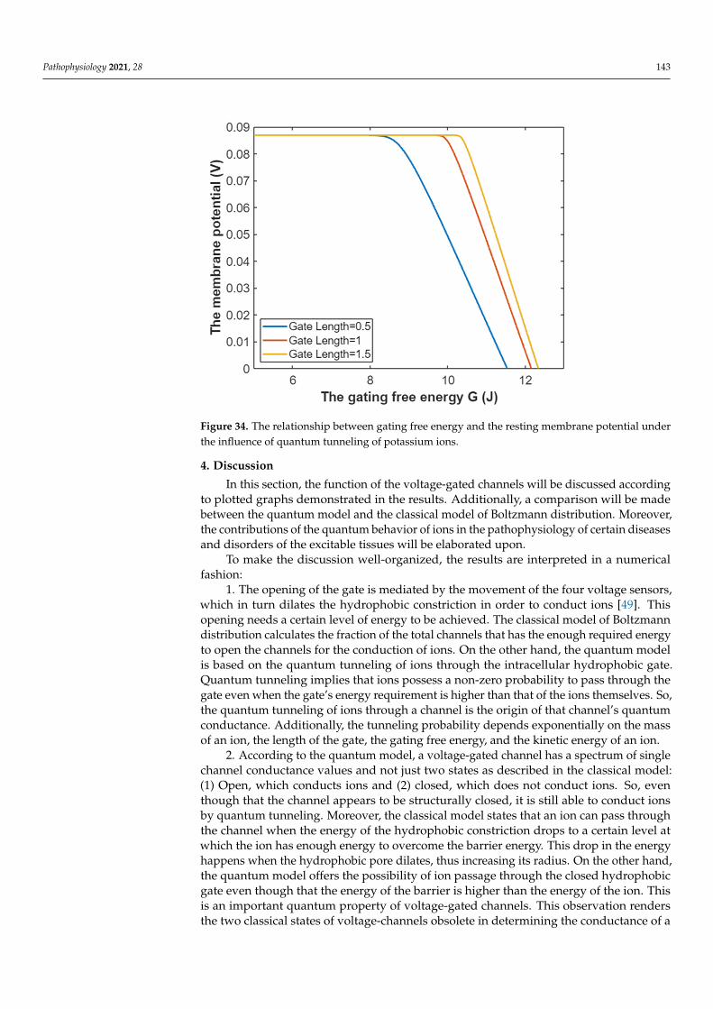
Pathophysiology 2021, 28 143Pathophysiology 2021, 28 145
Figure 34. The relationship between gating free energy and the resting membrane potential under the influence of quantum tunneling of potassium ions.
4. Discussion In this section, the function of the voltage-gated channels will be discussed according
to plotted graphs demonstrated in the results. Additionally, a comparison will be made between the quantum model and the classical model of Boltzmann distribution. Moreo-ver, the contributions of the quantum behavior of ions in the pathophysiology of certain diseases and disorders of the excitable tissues will be elaborated upon.
To make the discussion well-organized, the results are interpreted in a numerical fashion:
1. The opening of the gate is mediated by the movement of the four voltage sensors, which in turn dilates the hydrophobic constriction in order to conduct ions [49]. This open-ing needs a certain level of energy to be achieved. The classical model of Boltzmann dis-tribution calculates the fraction of the total channels that has the enough required energy to open the channels for the conduction of ions. On the other hand, the quantum model is based on the quantum tunneling of ions through the intracellular hydrophobic gate. Quantum tunneling implies that ions possess a non-zero probability to pass through the gate even when the gate’s energy requirement is higher than that of the ions themselves. So, the quantum tunneling of ions through a channel is the origin of that channel’s quan-tum conductance. Additionally, the tunneling probability depends exponentially on the mass of an ion, the length of the gate, the gating free energy, and the kinetic energy of an ion.
2. According to the quantum model, a voltage-gated channel has a spectrum of single channel conductance values and not just two states as described in the classical model: (1) Open, which conducts ions and (2) closed, which does not conduct ions. So, even though that the channel appears to be structurally closed, it is still able to conduct ions by quan-tum tunneling. Moreover, the classical model states that an ion can pass through the chan-nel when the energy of the hydrophobic constriction drops to a certain level at which the ion has enough energy to overcome the barrier energy. This drop in the energy happens when the hydrophobic pore dilates, thus increasing its radius. On the other hand, the quantum model offers the possibility of ion passage through the closed hydrophobic gate even though that the energy of the barrier is higher than the energy of the ion. This is an important quantum property of voltage-gated channels. This observation renders the two
Figure 34. The relationship between gating free energy and the resting membrane potential underthe influence of quantum tunneling of potassium ions.
4. Discussion
In this section, the function of the voltage-gated channels will be discussed accordingto plotted graphs demonstrated in the results. Additionally, a comparison will be madebetween the quantum model and the classical model of Boltzmann distribution. Moreover,the contributions of the quantum behavior of ions in the pathophysiology of certain diseasesand disorders of the excitable tissues will be elaborated upon.
To make the discussion well-organized, the results are interpreted in a numericalfashion:
1. The opening of the gate is mediated by the movement of the four voltage sensors,which in turn dilates the hydrophobic constriction in order to conduct ions [49]. Thisopening needs a certain level of energy to be achieved. The classical model of Boltzmanndistribution calculates the fraction of the total channels that has the enough required energyto open the channels for the conduction of ions. On the other hand, the quantum modelis based on the quantum tunneling of ions through the intracellular hydrophobic gate.Quantum tunneling implies that ions possess a non-zero probability to pass through thegate even when the gate’s energy requirement is higher than that of the ions themselves. So,the quantum tunneling of ions through a channel is the origin of that channel’s quantumconductance. Additionally, the tunneling probability depends exponentially on the massof an ion, the length of the gate, the gating free energy, and the kinetic energy of an ion.
2. According to the quantum model, a voltage-gated channel has a spectrum of singlechannel conductance values and not just two states as described in the classical model:(1) Open, which conducts ions and (2) closed, which does not conduct ions. So, eventhough that the channel appears to be structurally closed, it is still able to conduct ionsby quantum tunneling. Moreover, the classical model states that an ion can pass throughthe channel when the energy of the hydrophobic constriction drops to a certain level atwhich the ion has enough energy to overcome the barrier energy. This drop in the energyhappens when the hydrophobic pore dilates, thus increasing its radius. On the other hand,the quantum model offers the possibility of ion passage through the closed hydrophobicgate even though that the energy of the barrier is higher than the energy of the ion. Thisis an important quantum property of voltage-gated channels. This observation rendersthe two classical states of voltage-channels obsolete in determining the conductance of a
Pathophysiology 2021, 28 144
single channel. This provides a reasonable explanation for the permeation of ions throughthe closed channels without the need of the mechanical movement of voltage sensors todilate the pore because quantum tunneling of ions does not require the pore dilation [50,51].Moreover, the quantum model can explain that the movement of at least one voltagesensor out of four, can make a channel permeable [52]. Such phenomenon exists due tothe movement of one voltage sensor as it can partially dilate the pore, reducing the barrierenergy of the gate to enhance the quantum tunneling of ions and their conductance withoutthe need of the cooperation of the four sensors to fully dilate the pore.
3. Based on the equations and graphs in the results section, there is a probabilityof a channel opening in the classical model and there is a probability of ion tunnelingthrough the channel in the quantum model. In addition to that, there are two values ofsingle channel conductance in the classical model: (1) Zero conductance when the channelis closed and (2) Csin gle when the channel is open, but there is a range of single channelconductance values in the quantum model and not just two values. However, there ismembrane conductance in both models.
4. As aforementioned in the results, the tunneling probability, quantum conductanceof a single channel, and quantum membrane conductance were evaluated accordingto the membrane potential and gating free energy. It is obvious that the extracellularcations have higher tunneling probability, higher single channel conductance, and highermembrane conductance because they have higher kinetic energy than their intracellularcounterparts. The higher kinetic energy of the extracellular cations is attributed to themembrane potential that is negative on the inside in comparison to the outside of a cell;hence the cations that come from the outside passing through the channel until reachingthe intracellular gate will go through the membrane potential and acquire kinetic energy.Therefore, the quantum model discriminates between the extracellular and intracellularions in terms of conductance unlike the classical model, which treats them the same interms of conductance; see Figure 35.
Additionally, it is clear that sodium has higher tunneling probability and higherconductance than potassium and this discrepancy can be attributed to the fact that themass of sodium is less than the mass of potassium. Thus, the mass is an exponentiallyinfluential factor on the conductance in the quantum model.
5. The membrane conductance is the eventual electrical property that is used to assessthe influence of ions on the excitability of the membrane. By comparing the graphs ofmembrane conductance for the same ion in both models, it is evident that the quantummembrane conductance of extracellular ions can be comparable or even higher than themembrane conductance of ions due to the mechanical opening of the channels as in theclassical model. This means that ions can achieve significant quantum conductance, whichcan affect the electrical behavior of excitable cells at the resting state and during an actionpotential.
6. The membrane potential affects quantum tunneling and the conductance of ions inboth models. This is vital in the generation and propagation of action potentials because if astimulus is strong enough, it will trigger the opening of the sodium voltage-gated channelsto increase their conductance, depolarizing the membrane potential, which will furtherincrease the conductance of sodium ions to elicit a spike of depolarization. Accordingto the graphs in the results, it seems that quantum tunneling of ions contributed to thedepolarization spike of the action potential because as the membrane depolarizes, thequantum membrane conductance of sodium ions increases significantly in comparisonto the membrane conductance in the Boltzmann model. Thus, this insight gives a newperspective in the process of action potential generation involving the quantum behaviorof ions. Furthermore, this observation supports the previous works that focused on therole of quantum mechanics within action potentials [53,54].
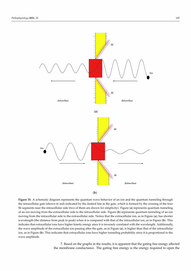
Pathophysiology 2021, 28 145
Pathophysiology 2021, 28 146
classical states of voltage-channels obsolete in determining the conductance of a single channel. This provides a reasonable explanation for the permeation of ions through the closed channels without the need of the mechanical movement of voltage sensors to dilate the pore because quantum tunneling of ions does not require the pore dilation [50,51]. Moreover, the quantum model can explain that the movement of at least one voltage sen-sor out of four, can make a channel permeable [52]. Such phenomenon exists due to the movement of one voltage sensor as it can partially dilate the pore, reducing the barrier energy of the gate to enhance the quantum tunneling of ions and their conductance with-out the need of the cooperation of the four sensors to fully dilate the pore.
3. Based on the equations and graphs in the results section, there is a probability of a channel opening in the classical model and there is a probability of ion tunneling through the channel in the quantum model. In addition to that, there are two values of single chan-nel conductance in the classical model: (1) Zero conductance when the channel is closed and (2) sin gleC when the channel is open, but there is a range of single channel conduct-ance values in the quantum model and not just two values. However, there is membrane conductance in both models.
4. As aforementioned in the results, the tunneling probability, quantum conductance of a single channel, and quantum membrane conductance were evaluated according to the membrane potential and gating free energy. It is obvious that the extracellular cations have higher tunneling probability, higher single channel conductance, and higher mem-brane conductance because they have higher kinetic energy than their intracellular coun-terparts. The higher kinetic energy of the extracellular cations is attributed to the mem-brane potential that is negative on the inside in comparison to the outside of a cell; hence the cations that come from the outside passing through the channel until reaching the intracellular gate will go through the membrane potential and acquire kinetic energy. Therefore, the quantum model discriminates between the extracellular and intracellular ions in terms of conductance unlike the classical model, which treats them the same in terms of conductance; see Figure 35.
(a)
Pathophysiology 2021, 28 147
(b)
Figure 35. A schematic diagram represents the quantum wave behavior of an ion and the quantum tunneling through the intracellular gate (shown in red) indicated by the dashed line in the gate, which is formed by the crossing of the four S6 segments near the intracellular side (two of them are shown for simplicity). Figure (a) represents quantum tunneling of an ion moving from the extracellular side to the intracellular side. Figure (b) represents quantum tunneling of an ion moving from the intracellular side to the extracellular side. Notice that the extracellular ion, as in Figure (a), has shorter wavelength (the distance from peak to peak) when it is compared with that of the intracellular ion, as in Figure (b). This indicates that extracellular ions have higher kinetic energy since it is inversely correlated with the wavelength. Addition-ally, the wave amplitude of the extracellular ion passing after the gate, as in Figure (a), is higher than that of the intracel-lular ion, as in Figure (b). This indicates that extracellular ions have higher tunneling probability since it is proportional to the wave amplitude.
Additionally, it is clear that sodium has higher tunneling probability and higher con-ductance than potassium and this discrepancy can be attributed to the fact that the mass of sodium is less than the mass of potassium. Thus, the mass is an exponentially influential factor on the conductance in the quantum model.
5. The membrane conductance is the eventual electrical property that is used to assess the influence of ions on the excitability of the membrane. By comparing the graphs of membrane conductance for the same ion in both models, it is evident that the quantum membrane conductance of extracellular ions can be comparable or even higher than the membrane conductance of ions due to the mechanical opening of the channels as in the classical model. This means that ions can achieve significant quantum conductance, which can affect the electrical behavior of excitable cells at the resting state and during an action potential.
6. The membrane potential affects quantum tunneling and the conductance of ions in both models. This is vital in the generation and propagation of action potentials because if a stimulus is strong enough, it will trigger the opening of the sodium voltage-gated channels to increase their conductance, depolarizing the membrane potential, which will further increase the conductance of sodium ions to elicit a spike of depolarization. Ac-cording to the graphs in the results, it seems that quantum tunneling of ions contributed to the depolarization spike of the action potential because as the membrane depolarizes, the quantum membrane conductance of sodium ions increases significantly in compari-son to the membrane conductance in the Boltzmann model. Thus, this insight gives a new perspective in the process of action potential generation involving the quantum behavior of ions. Furthermore, this observation supports the previous works that focused on the role of quantum mechanics within action potentials [53,54].
Figure 35. A schematic diagram represents the quantum wave behavior of an ion and the quantum tunneling throughthe intracellular gate (shown in red) indicated by the dashed line in the gate, which is formed by the crossing of the fourS6 segments near the intracellular side (two of them are shown for simplicity). Figure (a) represents quantum tunnelingof an ion moving from the extracellular side to the intracellular side. Figure (b) represents quantum tunneling of an ionmoving from the intracellular side to the extracellular side. Notice that the extracellular ion, as in Figure (a), has shorterwavelength (the distance from peak to peak) when it is compared with that of the intracellular ion, as in Figure (b). Thisindicates that extracellular ions have higher kinetic energy since it is inversely correlated with the wavelength. Additionally,the wave amplitude of the extracellular ion passing after the gate, as in Figure (a), is higher than that of the intracellularion, as in Figure (b). This indicates that extracellular ions have higher tunneling probability since it is proportional to thewave amplitude.
7. Based on the graphs in the results, it is apparent that the gating free energy affectedthe membrane conductance. The gating free energy is the energy required to open the
Pathophysiology 2021, 28 146
closed channel at membrane potential 0 V and this quantity gives information abouthow difficult it is to open a channel. Additionally, the conductance was assessed over adecreasing range of gating free energy; hence it was expected to observe increasing valuesin the conductance. The gating free energy of the channels decreases in many conditionsincluding: Channelopathies [55–57] tissue ischemia [58], and mechanical stretch of themembrane [58,59]. These conditions are present in many genetic and acquired diseasessuch as channelopathies of epilepsy [60], cardiac arrhythmias [61], and pain disorders [62].Moreover, cerebral ischemia, stroke, myocardial ischemia, and myocardial infarction areassociated with increased risk of higher excitability resulting in seizures, epilepsy, andlife-threatening arrhythmias [63–65]. Furthermore, the pathological stretch in heart failureand valvular heart disease is associated with serious arrhythmias such as ventriculartachycardia, ventricular fibrillation, and atrial fibrillation [66–69]. So, the decrease in thegating free energy can increase the risk of developing excitability-related disorders. Thiscan be explained by the two models: (1) In the classical model, the decrease in the gatingfree energy of the sodium channels will decrease the barrier energy of the gate resulting ina higher fraction of open channels and higher membrane conductance at certain membranevoltage. This means that the membrane initiates the depolarization spike of action potentialat lower thresholds in response to certain stimulus. (2) In the quantum model, the decreasein the gating free energy of the sodium channels will decrease the barrier energy of thegate resulting in higher tunneling probability and higher quantum conductance of sodiumions at certain membrane voltages. Consequently, the membrane will be more responsiveand more able to generate the depolarization spike of action potentials at a lower threshold.Furthermore, according to the quantum model, the decrease in the gating free energy resultsin a significant increase in the conductance of extracellular sodium ions that can affect themembrane potential and its excitability. Thus, the decrease in the gating free energy willdecrease the threshold required to initiate an action potential according to both models.Interestingly, there is another mechanism through which the quantum behavior of ions canincrease this excitability. This mechanism is the ability of sodium and potassium ions todepolarize the resting membrane potential in response to the decrease in the gating freeenergy as represented in Figures 33 and 34. Therefore, as the resting membrane potentialbecomes depolarized, it becomes closer to the threshold and more responsive to transmitan action potential.
8. According to the quantum model, both sodium and potassium ions can depolarizethe resting membrane potential because extracellular sodium and potassium ions havehigher tunneling probability and higher quantum conductance than intracellular sodiumand potassium ions. Consequently, there will be a quantum gradient of positive ions fromoutside to inside the cell resulting in depolarizing the resting membrane potential underthe equilibrium. However, according to the classical model, when the potassium channelsopen, potassium ions diffuse from inside to outside the cell, down their concentrationgradient, resulting in membrane hyperpolarization. Hence, the quantum model canexplain why gain-of-function mutations in potassium channels of excitatory neurons canresult in hyperexcitability instead of hypoexcitability [70]. Surprisingly, when the gain-of-function mutation in potassium channels decreases the gating free energy, this mayincrease the tunneling probability and quantum conductance of extracellular potassiumions significantly to depolarize the resting membrane potential, predisposing the membraneto hyperexcitability.
9. The quantum model treats voltage-gated channels as leaky channels that cancontribute to the resting membrane potential especially when certain mutation occurs todecrease the gating free energy. On the other hand, the classical model concerns itself withthe opening and closing of channels for initiation and propagation of action potentials.Under equilibrium, the ions can tunnel through the activation or the inactivation gateresulting in a leaky persistent quantum current that could alter the membrane potential.This serves to give a reasonable mechanism behind the persistent inward sodium currentin the mutated channels, which are implicated in higher excitability such as in epilepsy.
Pathophysiology 2021, 28 147
This current fails to be inactivated even during prolonged depolarization [71] and this isconsistent with the prediction of the quantum model, which states that ions can tunnelthrough the inactivation gate during even prolonged depolarization.
The comparisons and the implications listed above are deduced mainly from theequations and the mathematical graphs obtained in the results. Therefore, they requireexperimental evidence to be further validated. However, it is vital to propose and discussthe mathematical structure of any model before performing any experiments to test itsvalidity. Hence, the present study focuses on the mathematical aspects of the quantumbioelectrical property of the biological membrane.
To make a connection between the mathematical quantum model proposed here inthe article and future experiments to test its validity, concrete strategies and approachesare listed here to help in conducting experimental studies aimed at providing evidence onthe quantum bioelectrical property of the biological membrane. Interestingly, it has beenshown that the quantum features of ions can be used to control the membrane preparationwhich shows a high selectivity for targeted ions [72].
Before addressing the strategies and the approaches, it is important to notice that undernormal physiological parameters the quantum behavior of sodium and potassium ions hasminimal influence on the overall electrical behavior of the excitable cells [31,32]. This isclear in Figures 33 and 34 in which the gating free energy must decrease to certain level atwhich the quantum behavior has a significant effect on the membrane potential. Therefore,when electrophysiological studies are to be conducted, it is important to set an experimentalenvironment where the quantum behavior of ions can influence the membrane potential.This can be achieved by two main approaches: (1) Inducing mutations in the residues ofthe activation or inactivation gates so that the gating free energy decreases or (2) usingions with lower masses such as lithium [31,32] or hydrogen ions (protons) so that thequantum tunneling probability increases significantly without even inducing mutations inthe activation or inactivation gates.
The evidence for the quantum bioelectrical property of the biological membrane canbe provided by adopting the following experimental strategies and approaches:
1. A key difference between classical mechanics and quantum mechanics of thevoltage-gated channels is the process of ions conduction as aforementioned. According toquantum mechanics, ions can tunnel through the closed activation gate or inactivation gatewithout inducing any conformational changes in the molecular structure of the voltage-gated channel such as the movements of the voltage sensors or pore dilations. This can givea promising hint on the presence of quantum effects of ions. Thus, electrophysiologicalstudies can be conducted to measure such quantum currents while observing no mechanicalmovements or pore dilation sufficient to conduct the ions.
2. According to quantum mechanics, the closed channels can have a range of singlechannel conductance values. Hence, it is expected that different molecular closed structuresof the same mutated channel would have different values of conductance that obey theexponential equations presented here in the article. Therefore, electrophysiological studiescan be conducted to measure the conductance of the different closed structures of thesame channel before reaching the open state in which sufficient dilation has occurred topermeate ions. Then, the experimental results of the conductance values can be comparedwith the theoretical results obtained when applying the mathematical model proposedin the present work. Another similar approach is to perform different mutations on thesame channel to decrease the gating free energy. Hence, the conductance at a certainclosed structure is measured experimentally for the different mutations performed. Thenthe experimental and the theoretical values are compared to evaluate the validity of themathematical quantum model. In addition, according to the quantum model, the quantumconductance of a single channel can be significantly larger than the closed state of a channel.Thus, experimentally manipulating the parameters of the closed hydrophobic gate canresult in conductance values higher than the values of the closed state and this can provideevidence on quantum conductance.
Pathophysiology 2021, 28 148
3. One important quantum aspect in the function of the voltage-gated channel isthat extracellular cations have higher conductance than the intracellular cations and thisis applied on both sodium and potassium ions. For example, when a gain-of-functionmutation (mutation decreases the gating free energy) occurs in the potassium channel, itis expected that there will be a net inward (from the extracellular side to the intracellularside) current of potassium ions. Therefore, electrophysiological studies can be performedto measure this inward current of potassium ions through the mutated channels. This willprovide promising evidence on the presence of the quantum effects because this inwardcurrent of potassium ions cannot be explained by the conventional electrophysiologicalprinciples as potassium ions must diffuse according to their concertation gradient from theintracellular side to the extracellular side resulting in only an outward current.
4. Here, another approach is proposed to be utilized to provide the evidence onthe quantum effects of the biological membrane. The approach is to measure the restingmembrane potential at different groups of mutated channels contained in the biologicalmembrane. Then, it is expected to get experimental plots that fit the theoretical plots asin Figures 33 and 34. Additionally, observing the depolarizing effect of potassium ionswhen the gating free energy decreases due to mutations will provide favorable evidencefor the effects of quantum behavior within biological systems. This effect is not expectedaccording to the classical mechanics or the conventional electrophysiological principles.Actually, according to classical mechanics, it is not expected for the voltage-gated channelsto contribute to establishing the resting membrane potential because they mainly functionduring the action potential propagation and not at the resting state. However, according toquantum mechanics, it is expected for the mutated voltage-gated channels to contribute toestablishing the resting membrane potential as discussed before. Therefore, if experimentsprovide evidence on the relationship between the gating free energy of the voltage-gatedchannels and the resting membrane potential that are consistent with the theoretical dataproposed here, this will provide evidence that the biological membrane possesses quantumelectrical activity.
5. Furthermore, a key feature in the quantum conductance is the exponential depen-dence on the mass of the ions and the length of the gate. This will be an interesting strategyto be used to experimentally show the exponential variations in quantum conductancebetween ions according to their masses and the length of the gate.
Quantum de-coherence is one of the fastest physical processes and it represents amajor challenge for the validity of many studies that focus on applying quantum mechanicson the biological systems including our present study. We mentioned this challenge inthe introduction. Here, we provide possible mechanisms that can sustain the quantumcoherence of ions so that the quantum tunneling can occur before de-coherence takes place.These mechanisms include:
1. The short length of the hydrophobic gate (0.5− 1.5)× 10−10 m, in which the quan-tum tunneling happens, will provide enough time before de-coherence occurs be-cause it has been shown that the distance correlates inversely to the time of de-coherence [17,73,74].
2. The gate is made by the aggregation of hydrophobic amino acids. This hydropho-bic pocket will exclude water (dewetting) and may create a decoherence-free sub-space [17,75].
3. Because voltage-gated channels and ions are a part of the biological system, it is ex-pected that they could operate far from the thermal equilibrium, which indicates thations may cool down through the hydrophobic gate even at higher body temperatures.This ‘cooling down’ can sustain the quantum coherence and make the quantum effectsmore apparent [17,74–77].
4. The complex non-linearity of brain dynamics can augment the quantum fluctuationsand hence contribute to the maintenance of the quantum coherence of ions [78,79].The electromagnetic field effects possibly mediate this augmentation [80–82].
Pathophysiology 2021, 28 149
The present work has certain qualities that make it distinct from the other previousstudies [31,32]. These qualities include: (1) The present study makes a comparison betweenclassical mechanics and quantum mechanics to mathematically show the differences intheir influence on the function of voltage-gated channels. (2) The present study integratesthe effect of membrane potential on the energy barrier of the gate. Thus, this allows us toevaluate the relationship between membrane potential and quantum tunneling probability,quantum conductance of a single channel, and the quantum membrane conductance.(3) The present study uses mathematical graphs to show vividly how the aforementionedvariables change and behave according to the principles of quantum mechanics. Usingmathematical graphs allows covering a wide range of values to get a more comprehensiveunderstanding of the quantum model of the voltage-gated channels. (4) The present studyfocuses on both the activation and inactivation gate in its discussion. (5) The presentstudy sheds light on the implications of using the quantum model to provide reasonableexplanations for the pathological mechanisms in excitability-related diseases and disorders.(6) The present study sets a more comprehensive mathematical model that can be usedto measure to the quantum conductance of the channels and the biological membrane inelectrophysiological experiments.
5. Conclusions
The present study proposes the quantum model, which is based on the quantumtunneling of ions through the closed gate of these channels, to approach voltage-gatedchannels. This approach is adopted to unveil new properties in the voltage-gated channelsand to solve the challenges and problems at the molecular and clinical levels. The modelcan explain the permeation of ions through the closed channels without the movementof voltage sensors, and it can also explain the paradox of the hyperexcitability effects ofgain-of-function mutations in potassium channels. Moreover, it can explain the persistentinward sodium current in the mutated channels during prolonged depolarization. Inaddition to that, the quantum model sheds light on the contribution of the quantumtunneling of ions on the pathophysiology of epilepsy, pain, and arrhythmia. This might behelpful to develop new agents to treat and control these chronic and challenging diseases.Also, different experimental strategies are listed in this study to test the scientific validityof the proposed model. Furthermore, possible mechanisms are suggested to explain thepersistence of quantum coherence of ions to ensure that quantum tunneling of ions occursbefore de-coherence.
Author Contributions: Conceptualization, A.B.Q.; methodology, A.B.Q.; investigation, A.B.Q.; re-sources, A.B.Q.; writing—original draft preparation, A.B.Q.; supervision, O.A. and L.K.; projectadministration, O.A. and L.K.; writing—review and editing, A.A.-A. and A.S., M.A. All authors haveread and agreed to the published version of the manuscript.
Funding: This research received no external funding.
Institutional Review Board Statement: Not applicable.
Informed Consent Statement: Not applicable.
Data Availability Statement: Not applicable.
Conflicts of Interest: The author declares no conflict of interest.
References1. Bertil, H. Ion Channels of Excitable Membranes, 3rd ed.; Sinauer Associates: Sunderland, MA, USA, 2001.2. Lerche, H.; Shah, M.; Beck, H.; Noebels, J.; Johnston, D.; Vincent, A. Ion channels in genetic and acquired forms of epilepsy.
J. Physiol. 2013, 591, 753–764. [CrossRef] [PubMed]3. Dib-Hajj, S.D.; Geha, P.; Waxman, S.G. Sodium channels in pain disorders: Pathophysiology and prospects for treatment. Pain
2017, 158, S97–S107. [CrossRef]4. Dehghani-Samani, A.; Madreseh-Ghahfarokhi, S.; Dehghani-Samani, A. Mutations of voltage-gated ionic channels and risk of
severe cardiac arrhythmias. Acta Cardiol. Sin. 2019, 35, 99–110. [PubMed]
Pathophysiology 2021, 28 150
5. Tejada, J.; Costa, K.M.; Bertti, P.; Garcia-Cairasco, N. The epilepsies: Complex challenges needing complex solutions. Epilepsy Behav.2013, 26, 212–228. [CrossRef] [PubMed]
6. Ma, R.S.; Kayani, K.; Whyte-Oshodi, D.; Whyte-Oshodi, A.; Nachiappan, N.; Gnanarajah, S.; Mohammed, R. Voltage gatedsodium channels as therapeutic targets for chronic pain. J. Pain Res. 2019, 12, 2709–2722. [CrossRef]
7. Ragab, A.A.; Sitorus, G.D.; Brundel, B.B.; de Groot, N.M. The Genetic Puzzle of Familial Atrial Fibrillation. Front. Cardiovasc. Med.2020, 7, 14. [CrossRef]
8. Privitera, M. Current challenges in the management of epilepsy. Am. J. Manag. Care 2011, 17 (Suppl. 7), S195–S203.9. Domenichiello, A.F.; Ramsden, C.E. The silent epidemic of chronic pain in older adults. Prog. Neuro-Psychopharmacol. Biol. Psychiatry
2019, 93, 284–290. [CrossRef] [PubMed]10. Thomas, M.A. Pain management–the challenge. Ochsner J. 2003, 5, 15–21.11. Nattel, S.; Andrade, J.; Macle, L.; Rivard, L.; Dyrda, K.; Mondesert, B.; Khairy, P. New directions in cardiac arrhythmia
management: Present challenges and future solutions. Can. J. Cardiol. 2014, 30, S420–S430. [CrossRef] [PubMed]12. Serway, R.A.; Moses, C.J.; Moyer, C.A. Modern Physics; Cengage Learning: Boston, MA, USA, 2004.13. Lambert, N.; Chen, Y.N.; Cheng, Y.C.; Li, C.M.; Chen, G.Y.; Nori, F. Quantum biology. Nat. Phys. 2013, 9, 10–18. [CrossRef]14. Ball, P. Physics of life: The dawn of quantum biology. Nature 2011, 474, 272–274. [CrossRef]15. Kim, Y.; Bertagna, F.; D’Souza, E.M.; Heyes, D.J.; Johannissen, L.O.; Nery, E.T.; Pantelias, A.; Sanchez-Pedreño Jimenez, A.;
Slocombe, L.; Spencer, M.G.; et al. Quantum Biology: An Update and Perspective. Quantum Rep. 2021, 3, 6. [CrossRef]16. Kong, J.; Jiménez-Martínez, R.; Troullinou, C.; Lucivero, V.G.; Tóth, G.; Mitchell, M.W. Measurement-induced, spatially-extended
entanglement in a hot, strongly-interacting atomic system. Nat. Commun. 2020, 11, 1–9. [CrossRef] [PubMed]17. Arndt, M.; Juffmann, T.; Vedral, V. Quantum physics meets biology. HFSP J. 2009, 3, 386–400. [CrossRef]18. Oelstrom, K.; Goldschen-Ohm, M.P.; Holmgren, M.; Chanda, B. Evolutionarily conserved intracellular gate of voltage-dependent
sodium channels. Nat. Commun. 2014, 5, 1–9. [CrossRef] [PubMed]19. Labro, A.J.; Snyders, D.J. Being flexible: The voltage-controllable activation gate of Kv channels. Front. Pharmacol. 2012, 3, 168.
[CrossRef]20. Ingber, L.; Pappalepore, M.; Stesiak, R.R. Electroencephalographic field influence on calcium momentum waves. J. Theor. Biol.
2014, 343, 138–153. [CrossRef]21. Ingber, L. Quantum calcium-ion interactions with EEG. Sci 2019, 1, 20. [CrossRef]22. Summhammer, J.; Sulyok, G.; Bernroider, G. Quantum Mechanical Coherence of K+ Ion Wave Packets Increases Conduction in
the KcsA Ion Channel. Appl. Sci. 2020, 10, 4250. [CrossRef]23. Koch, H.J. Quantum biology: Unit membrane reduces entropy due to wave particle duality. NeuroQuantology 2017, 15. [CrossRef]24. Juffmann, T.; Milic, A.; Müllneritsch, M.; Asenbaum, P.; Tsukernik, A.; Tüxen, J.; Mayor, M.; Cheshnovsky, O.; Arndt, M. Real-time
single-molecule imaging of quantum interference. Nat. Nanotechnol. 2012, 7, 297–300. [CrossRef] [PubMed]25. Gerlich, S.; Eibenberger, S.; Tomandl, M.; Nimmrichter, S.; Hornberger, K.; Fagan, P.J.; Tüxen, J.; Mayor, M.; Arndt, M. Quantum
interference of large organic molecules. Nat. Commun. 2011, 2, 263. [CrossRef] [PubMed]26. Eibenberger, S.; Gerlich, S.; Arndt, M.; Mayor, M.; Tüxen, J. Matter–wave interference of particles selected from a molecular
library with masses exceeding 10000 amu. Phys. Chem. Chem. Phys. 2013, 15, 14696–14700. [CrossRef]27. Vaziri, A.; Plenio, M.B. Quantum coherence in ion channels: Resonances, transport and verification. New J. Phys. 2010, 12, 085001.
[CrossRef]28. Ganim, Z.; Tokmakoff, A.; Vaziri, A. Vibrational excitons in ionophores: Experimental probes for quantum coherence-assisted ion
transport and selectivity in ion channels. New J. Phys. 2011, 13, 113030. [CrossRef]29. Salari, V.; Naeij, H.; Shafiee, A. Quantum interference and selectivity through biological ion channels. Sci. Rep. 2017, 7, 41625.
[CrossRef] [PubMed]30. Aryal, P.; Sansom, M.S.; Tucker, S.J. Hydrophobic gating in ion channels. J. Mol. Biol. 2015, 427, 121–130. [CrossRef] [PubMed]31. Qaswal, A.B. Quantum tunneling of ions through the closed voltage-gated channels of the biological membrane: A mathematical
model and implications. Quantum Rep. 2019, 1, 19. [CrossRef]32. Qaswal, A.B. Quantum Electrochemical Equilibrium: Quantum Version of the Goldman–Hodgkin–Katz Equation. Quantum Rep.
2020, 2, 17. [CrossRef]33. Zhang, X.C.; Yang, H.; Liu, Z.; Sun, F. Thermodynamics of voltage-gated ion channels. Biophys. Rep. 2018, 4, 300–319. [CrossRef]
[PubMed]34. Chowdhury, S.; Chanda, B. Estimating the voltage-dependent free energy change of ion channels using the median voltage for
activation. J. Physiol. 2012, 139, 3–17. [CrossRef]35. Chen, F.; Hihath, J.; Huang, Z.; Li, X.; Tao, N.J. Measurement of single-molecule conductance. Annu. Rev. Phys. Chem. 2007,
58, 535–564. [CrossRef]36. Gonzalez, C.; Contreras, G.F.; Peyser, A.; Larsson, P.; Neely, A.; Latorre, R. Voltage sensor of ion channels and enzymes.
Biophys. Rev. 2012, 4, 1–5. [CrossRef]37. Feigenspan, A.; Dedek, K.; Weiler, R.; Thanos, S.; Schlich, K. Expression and biophysical characterization of voltage-gated sodium
channels in axons and growth cones of the regenerating optic nerve. Investig. Ophthalmol. Vis. Sci. 2010, 51, 1789–1799. [CrossRef]38. Hu, W.; Tian, C.; Li, T.; Yang, M.; Hou, H.; Shu, Y. Distinct contributions of Na v 1.6 and Na v 1.2 in action potential initiation and
backpropagation. Nat. Neurosci. 2009, 12, 996–1002. [CrossRef] [PubMed]
Pathophysiology 2021, 28 151
39. Qaswal, A.B. Magnesium Ions Depolarize the Neuronal Membrane via Quantum Tunneling through the Closed Channels.Quantum Rep. 2020, 2, 5. [CrossRef]
40. Qaswal, A.B. A Theoretical Study to Explain the Referred Pain Phenomenon and Its Characteristics via Quantum Tunneling ofPotassium Ions through the Channels of Neuronal Membrane. NeuroQuantology 2019, 17. [CrossRef]
41. Richards, L.A.; Schäfer, A.I.; Richards, B.S.; Corry, B. The importance of dehydration in determining ion transport in narrowpores. Small 2012, 8, 1701–1709. [CrossRef] [PubMed]
42. Ishida, I.G.; Rangel-Yescas, G.E.; Carrasco-Zanini, J.; Islas, L.D. Voltage-dependent gating and gating charge measurements in theKv1. 2 potassium channel. J. Physiol. 2015, 145, 345–358.
43. Islas, L.D. Functional diversity of potassium channel voltage-sensing domains. Channels 2016, 10, 202–213. [CrossRef]44. Issa, Z.F.; Miller, J.M.; Zipes, D.P. Molecular mechanisms of cardiac electrical activity. In Clinical Arrhythmology and Electrophysiology:
A Companion to Braunwald’s Heart Disease, 2nd ed.; WB Saunders: Philadelphia, PA, USA, 2012; pp. 1–9.45. Hall, J.E. Guyton and Hall Textbook of Medical Physiology e-Book; Elsevier Health Sciences: Amsterdam, The Netherlands, 2020.46. Payandeh, J.; El-Din, T.M.; Scheuer, T.; Zheng, N.; Catterall, W.A. Crystal structure of a voltage-gated sodium channel in two
potentially inactivated states. Nature 2012, 486, 135–139. [CrossRef] [PubMed]47. Cuello, L.G.; Jogini, V.; Cortes, D.M.; Perozo, E. Structural mechanism of C-type inactivation in K+ channels. Nature 2010,
466, 203–208. [CrossRef]48. Bagnéris, C.; Naylor, C.E.; McCusker, E.C.; Wallace, B.A. Structural model of the open–closed–inactivated cycle of prokaryotic
voltage-gated sodium channels. J. Physiol. 2015, 145, 5–16. [CrossRef] [PubMed]49. Catterall, W.A. Ion channel voltage sensors: Structure, function, and pathophysiology. Neuron 2010, 67, 915–928. [CrossRef]50. Soler-Llavina, G.J.; Holmgren, M.; Swartz, K.J. Defining the conductance of the closed state in a voltage-gated K+ channel. Neuron
2003, 38, 61–67. [CrossRef]51. Banh, R.; Cherny, V.V.; Morgan, D.; Musset, B.; Thomas, S.; Kulleperuma, K.; Smith, S.M.; Pomès, R.; DeCoursey, T.E. Hydrophobic
gasket mutation produces gating pore currents in closed human voltage-gated proton channels. Proc. Natl. Acad. Sci. USA 2019,116, 18951–18961. [CrossRef] [PubMed]
52. Westhoff, M.; Eldstrom, J.; Murray, C.I.; Thompson, E.; Fedida, D. IKs ion-channel pore conductance can result from individualvoltage sensor movements. Proc. Natl. Acad. Sci. USA 2019, 116, 7879–7888. [CrossRef]
53. Moradi, N.; Scholkmann, F.; Salari, V. A study of quantum mechanical probabilities in the classical Hodgkin–Huxley model.J. Integr. Neurosci. 2015, 14, 1–7. [CrossRef] [PubMed]
54. Bernroider, G.; Summhammer, J. Can quantum entanglement between ion transition states effect action potential initiation?Cognit. Comput. 2012, 4, 29–37. [CrossRef]
55. Yifrach, O.; MacKinnon, R. Energetics of pore opening in a voltage-gated K+ channel. Cell 2002, 111, 231–239. [CrossRef]56. Huang, J.; Estacion, M.R.; Zhao, P.; Dib-Hajj, F.; Schulman, B.; Abicht, A.; Kurth, I.; Brockmann, K.; Waxman, S.; Dib-Hajj, S.D. A
novel gain-of-function Nav1. 9 mutation in a child with episodic pain. Front. Neurosci. 2019, 13, 918. [CrossRef]57. Chamberlin, A.; Qiu, F.; Rebolledo, S.; Wang, Y.; Noskov, S.Y.; Larsson, H.P. Hydrophobic plug functions as a gate in voltage-gated
proton channels. Proc. Natl. Acad. Sci. USA 2014, 111, E273–E282. [CrossRef] [PubMed]58. Morris, C.E. Voltage-gated channel mechanosensitivity: Fact or friction? Front. Physiol. 2011, 2, 25. [CrossRef] [PubMed]59. Reeves, D.; Ursell, T.; Sens, P.; Kondev, J.; Phillips, R. Membrane mechanics as a probe of ion-channel gating mechanisms.
Phys. Rev. E 2008, 78, 041901. [CrossRef]60. George, A.L., Jr. Inherited channelopathies associated with epilepsy. Epilepsy Curr. 2004, 4, 65–70. [CrossRef]61. Behere, S.P.; Weindling, S.N. Inherited arrhythmias: The cardiac channelopathies. Ann. Pediatr. Cardiol. 2015, 8, 210–220.
[CrossRef] [PubMed]62. Cregg, R.; Momin, A.; Rugiero, F.; Wood, J.N.; Zhao, J. Pain channelopathies. J. Physiol. 2010, 588, 1897–1904. [CrossRef]63. Silverman, I.E.; Restrepo, L.; Mathews, G.C. Poststroke seizures. Arch. Neurol. 2002, 59, 195–201. [CrossRef]64. Ghuran, A.V.; Camm, A.J. Ischaemic heart disease presenting as arrhythmias. Br. Med. Bull. 2001, 59, 193–210. [CrossRef]65. Bhar-Amato, J.; Davies, W.; Agarwal, S. Ventricular arrhythmia after acute myocardial infarction: ‘The Perfect Storm’.
Arrhythmia Electrophysiol. Rev. 2017, 6, 134–139. [CrossRef]66. Franz, M.R.; Cima, R.; Wang, D.; Profitt, D.; Kurz, R. Electrophysiological effects of myocardial stretch and mechanical determi-
nants of stretch-activated arrhythmias. Circulation 1992, 86, 968–978. [CrossRef]67. Bigger, J.T., Jr. Why patients with congestive heart failure die: Arrhythmias and sudden cardiac death. Circulation 1987,
75, IV28–IV35.68. Horstkotte, D. Arrhythmias in the natural history of mitral stenosis. Acta Cardiol. 1992, 47, 105–113.69. Scardigli, M.; Cannazzaro, S.; Coppini, R.; Crocini, C.; Yan, P.; Loew, L.M.; Sartiani, L.; Cerbai, E.; Pavone, F.S.; Sacconi, L.; et al.
Arrhythmia susceptibility in a rat model of acute atrial dilation. Prog. Biophys. Mol. Biol. 2020, 154, 21–29. [CrossRef]70. Niday, Z.; Tzingounis, A.V. Potassium channel gain of function in epilepsy: An unresolved paradox. Neuroscientist 2018,
24, 368–380. [CrossRef]71. Stafstrom, C.E. Persistent sodium current and its role in epilepsy. Epilepsy Curr. 2007, 7, 15–22. [CrossRef]72. Zhao, Y.; Liu, Y.; Wang, C.; Ortega, E.; Wang, X.; Xie, Y.F.; Shen, J.; Gao, C.; Van der Bruggen, B. Electric field-based ionic control of
selective separation layers. J. Mater. Chem. A 2020, 8, 4244–4251. [CrossRef]
Pathophysiology 2021, 28 152
73. Salari, V.; Tuszynski, J.; Rahnama, M.; Bernroider, G. Plausibility of quantum coherent states in biological systems. J. Phys.Conf. Ser. 2011, 306, 012075. [CrossRef]
74. Hagan, S.; Hameroff, S.R.; Tuszynski, J.A. Quantum computation in brain microtubules: Decoherence and biological feasibility.Phys. Rev. E 2002, 65, 061901. [CrossRef]
75. Lidar, D.A.; Chuang, I.L.; Whaley, K.B. Decoherence-free subspaces for quantum computation. Phys. Rev. Lett. 1998, 81, 2594.[CrossRef]
76. Matsuno, K. Cell motility as an entangled quantum coherence. BioSystems 1999, 51, 15–19. [CrossRef]77. Cai, J.; Popescu, S.; Briegel, H.J. Dynamic entanglement in oscillating molecules and potential biological implications. Phys. Rev. E
2010, 82, 021921. [CrossRef]78. Jedlicka, P. Revisiting the quantum brain hypothesis: Toward quantum (neuro) biology? Front. Mol. Neurosci. 2017, 10, 366.
[CrossRef]79. Jedlicka, P. Quantum stochasticity and (the end of) neurodeterminism. Quantum Phys. Meets Philos. Mind 2014, 183–197.
[CrossRef]80. Anastassiou, C.A.; Koch, C. Ephaptic coupling to endogenous electric field activity: Why bother? Curr. Opin. Neurobiol. 2015,
31, 95–103. [CrossRef] [PubMed]81. Fröhlich, F.; McCormick, D.A. Endogenous electric fields may guide neocortical network activity. Neuron 2010, 67, 129–143.
[CrossRef]82. McFadden, J.; Al-Khalili, J. Life on the Edge: The Coming of Age of Quantum Biology; Crown Publishing Group: New York, NY, USA,
26 July 2016.


