Embed Size (px)
Citation preview
Synthesis of Methotrexate loaded Cerium fluoride nanoparticles with pH sensitive extended
release coupled with Hyaluronic acid receptor with plausible theranostic capabilities for
preclinical safety studies
MASTER THESIS
Submitted by
Nitish Manu George
In partial fulfilment for the award of the Degree of
BACHELOR & MASTER OF TECHNOLOGY (DUAL)
IN
NANOTECHNOLOGY
Under the guidance of
Dr. Mayuri Gandhi
Amity University, Noida
Uttar Pradesh, India
Indian Institute of Technology-Bombay
Mumbai, India
December – May, 2016
Acknowledgements
I'd like to extend my sincerest gratitude towards Dr. Mayuri Gandhi for accepting my request to
work under her guidance at the C.R.N.T.S. facility in I.I.T. Bombay. Her continued support, timely
interventions and sound advice throughout the course of my project work is what lead to its eventual
success. Although her nature of expecting perfection would often push me to continually improve and
hone my skills, her friendly demeanour made her a guide who was easy to approach and connect with
on a personal level. The warmth she showered upon me certainly instils in me a feeling that I wasn't
wrong in pursuing my research under her experienced guidance.
I'd also like to thank all those working in the facility for being warm and friendly. On more than
one occasion, they've extended their sincerest assistance in my project. Without some key
recommendations, I wonder whether I'd even be able to complete my project. It was a humbling
experience to see them take time out from their schedules in order to assist me with various challenges
I faced. From constant inquiries towards the status of my project to subtle words of encouragement,
the contribution of the laboratory members towards my project can't be overlooked. More often than
sometimes, I definitely would've been an annoyance to them but they never held it against me and
chose to only support me to the best of their capabilities. They are all a true inspiration in
professionalism with a hint of familial bonding in the workplace.
Furthermore, I'd like to thank all the researchers who've worked previously in the subject
enabling me to develop my own hypothesis by being inspired from their work. Among them, my own
friend Varun S., who I'd like to acknowledge as an informed researcher who'd assist me the best he
could since my project directly takes a page from his previous research. He'd been generous enough to
share all forms of relevant information in the spirit of goodwill towards research and on more than
one occasion encouraged me.
Lastly, I'd like to thank my internal guide Dr. Nidhi Chaunhan and all the faculty members of
1

Amity Institute of Nanotechnology at Amity University for their support and guidance. I'd also like to
extend my gratitude towards my parents who supported my financially and emotionally even though
there was a familial crisis at hand. I'd also like to thank my friends who helped in keeping me in high
spirits throughout the course of my project.
2
Abstract
A key challenge in drug delivery systems is the real time monitoring of delivered drug and
subsequent response. Recent advancement in nanotechnology has enabled the design and preclinical
implementation of novel drug delivery systems (DDS) with theranostic abilities. Herein, fluorescent
cerium fluoride (CeF3) nanoparticles (nps) were synthesized and their surface modified with a coat of
polyethylenimine (PEI). Thereafter, Methotrexate was conjugated upon it through glutaraldehyde cross-
linking for a pH-sensitive release. This was followed by the addition of a Hyaluronic acid (HA) receptor
via 1-Ethyl-3-(3-dimethylaminopropyl)-carbodiimide and N-hydroxysuccinimide (EDC-NHS)
chemistry to achieve a possible active drug targeting system. The obtained drug delivery nano-agent
retains and exhibits unique photo-luminescent properties attributed to the nps while exhibiting
potential theranostic capabilities.
3
Contents
Acknowledgements....................................................................................................................................................2
Abstract.......................................................................................................................................................................4
1.Introduction............................................................................................................................................................7
1.1.Cancer Research............................................................................................................................................7
1.2.Drug Delivery Systems for Cancer............................................................................................................8
1.3.Theranostics...................................................................................................................................................8
1.4.pH sensitive glutaraldehyde cross-linking.................................................................................................9
1.5.Hyaluronic Acid – Cancer receptor...........................................................................................................9
1.6.Objective.........................................................................................................................................................9
2.Materials and Methods.......................................................................................................................................11
2.1.Reagents........................................................................................................................................................11
2.2.Experiment..................................................................................................................................................11
a)Preparation of Phosphate Buffered Saline (PBS) solution...............................................................11
b)Synthesis of Cerium Fluoride nps........................................................................................................11
c)Surface Modification and Drug Loading..............................................................................................12
d)Preparation and coupling of Hyaluronic Acid....................................................................................12
e)Drug release studies.................................................................................................................................12
2.3.Characterization..........................................................................................................................................13
3.Results...................................................................................................................................................................14
Discussions and Conclusions................................................................................................................................20
References.................................................................................................................................................................21
4
List of Figures
Figure 1: Schematic representation of proposed agent preparation..............................................................13
Figure 2: TEM image..............................................................................................................................................14
Figure 3: Graph plot showing data acquired from UV-Vis Spectroscopy for prepared Samples..............15
Figure 4: XRD plot of prepared nanoparticle samples in native as well as conjugated form....................16
Figure 5: PL of samples without and with HA..................................................................................................17
Figure 6: FTIR plot of prepared samples without and with HA....................................................................18
Figure 7: Drug release studies over time-lapse...................................................................................................19
5
1. Introduction
1.1. Cancer Research
Cancer is one of the most popular diseases on the face of this earth that has existed for almost
the entirety of human history. With over 200 known variants, it is a group of diseases involving
abnormal growth of cells with the potential plausibility of invading or spreading to healthier cells or
parts of the body. Lacking in specific fingerprints and having the widest spectra of causalities, it is next
to impossible to prove the cause for a particular cancer regardless of its type. [1] Therefore, a lot of
research is under way to identify the causes of the disease as well as in developing strategies for its
prevention, diagnosis, treatment and subsequent cure.
Although a number of treatment options exist for cancers such as surgery, chemotherapy,
radiation therapy, gene therapy, immunotherapy, photo-dynamic therapy, targeted therapy, palliative care
etc.,[2-13] they still do not accommodate for the different phenotypes observed in individual cases or
provide a generic therapeutic to be followed. Also, cancer is a class of disease, it can also be argued that
it will be be highly unlikely that a single cure for cancer will ever come to exist. However, research
continues in hopes of achieving a “silver bullet” treatment technique that can be employed over a
majority of the cases.
Due to the dynamic nature of the disease, Cancer Research is one of the most funded forms of
medical research in the world with billions spent worldwide every year. Recent advancements in
molecular and cellular biology has lead to a revolution bringing about better treatments through a
variety of preclinical and clinical trials reducing the risk quotient involved with the treatment of the
disease with every passing day.[14] Perhaps one day, the silver bullet treatment that every researcher
working in the field envisions might even become a reality with all the awareness, effort and investment
being put into the field.
6
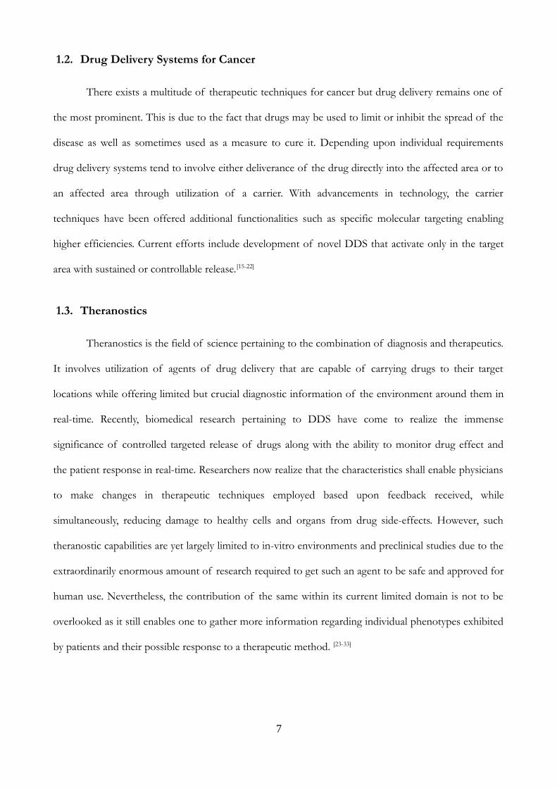
1.2. Drug Delivery Systems for Cancer
There exists a multitude of therapeutic techniques for cancer but drug delivery remains one of
the most prominent. This is due to the fact that drugs may be used to limit or inhibit the spread of the
disease as well as sometimes used as a measure to cure it. Depending upon individual requirements
drug delivery systems tend to involve either deliverance of the drug directly into the affected area or to
an affected area through utilization of a carrier. With advancements in technology, the carrier
techniques have been offered additional functionalities such as specific molecular targeting enabling
higher efficiencies. Current efforts include development of novel DDS that activate only in the target
area with sustained or controllable release.[15-22]
1.3. Theranostics
Theranostics is the field of science pertaining to the combination of diagnosis and therapeutics.
It involves utilization of agents of drug delivery that are capable of carrying drugs to their target
locations while offering limited but crucial diagnostic information of the environment around them in
real-time. Recently, biomedical research pertaining to DDS have come to realize the immense
significance of controlled targeted release of drugs along with the ability to monitor drug effect and
the patient response in real-time. Researchers now realize that the characteristics shall enable physicians
to make changes in therapeutic techniques employed based upon feedback received, while
simultaneously, reducing damage to healthy cells and organs from drug side-effects. However, such
theranostic capabilities are yet largely limited to in-vitro environments and preclinical studies due to the
extraordinarily enormous amount of research required to get such an agent to be safe and approved for
human use. Nevertheless, the contribution of the same within its current limited domain is not to be
overlooked as it still enables one to gather more information regarding individual phenotypes exhibited
by patients and their possible response to a therapeutic method. [23-33]
7
1.4. pH sensitive glutaraldehyde cross-linking
Glutaraldehyde is one of the most versatile cross-linking reagents that is prominently utilized in
biochemistry. Present in at least thirteen different forms based on solution conditions, there is
substantial literature available regarding the use of the reagent for various cross-linking applications
with various carbohydrates, lipids, nucleic acids, enzymes, and other soluble and insoluble molecules.
Owing to many discrepancies and the unique characteristics of each moiety linked with the reagent
however, cross-linking procedures are largely developed through empirical observations and its
subsequent properties are also tested in the same manner. However, many research articles elucidate
cases wherein the cross-linking acts in a pH sensitive manner with respect to linking efficiencies and
reversibility of the link formed. [33-35]
1.5. Hyaluronic Acid – Cancer receptor
Hyaluronic acid is a unique nonsulfated glycosaminoglycan which is formed in the plasma
membrane instead of the Golgi. Through literature it has been mentioned that the macromolecule
significantly contributes to cell proliferation and migration and has also been linked to the progression
of many malignant tumours. For cancer, HA acts as a receptor enabling targeting due to the over-
expression of CD44 glycoprotein in cancer cells which is recognized as a major receptor of HA. [35-44]
1.6. Objective
Through this thesis, an attempt at creating a novel DDS for Cancer, utilizing CeF3 nps is
described. The naked lanthanide nps have previously been associated with providing protection against
oxidative stress as well as being toxic. The host material was selected under the assumption that the
same characteristic shall be reproduced in preclinical safety studies even when functionalized as a drug
delivery agent. If realized, it would entail the safeguard of healthy cells from apoptosis due to oxidative
stress from introduction of nanoparticles into their environment. Furthermore, the understanding that
CeF3 can act as a scintillator makes it all the more desirable as a drug carrier for the purpose of
8
theranostic applications; at least within the scope of preclinical studies.
Utilizing PEI, the surface of the nanoparticles synthesized were modified such that
glutaraldehyde cross-linking of the amine groups in PEI with the amine group in the Cancer drug –
Methotrexate - was enabled. This entailed that the conjugation of the drug was pH-sensitive which
consequently will allow controlled release within Cancer cells alone. This would eventually reduce
chances of damaging healthier cells rendering the drug more effective than when delivered using
conventional means.
In order to impart a targeting functionality to the DDS, Hyaluronic acid was added as a
receptor. It is well established in literature as a targeting compound for Cancer cells and it was
implemented to the system via EDC-NHS coupling. With the addition of the final component, the
novel DDS that was planned was completed. With unique optical properties of CeF3 nps coupled with
the enhancements, it is believed that the DDS may prove beneficial in theranostic applications across
preclinical screens; with significant possibility of the same to be transferable to in-vivo conditions upon
optimization.
9
2. Materials and Methods
2.1. Reagents
Ammonium fluoride (NH4F), Cerium nitrate hexahydrate (Ce(NO3).6H2O), Polyethylenimine
((C2H5N)n), Methotrexate (C20H22N8O5), 25% Glutaraldehyde solution (C5H8O2), Hyaluronic acid
sodium salt (C14H22NNaO11), Sodium chloride (NaCl), Potassium chloride (KCl), Disodium phosphate
(Na2HPO4), Monopotassium phosphate (KH2PO4), 35% Hydrochloric acid, 1-Ethyl-3-(3-
dimethylaminopropyl)-carbodiimide (EDC), N-hydroxysuccinimide (NHS) were all purchased from
Sigma Aldrich. Absolute Ethanol was supplied by Changshu Hongsheng Fine Chemical Co., Ltd. De-
ionized (DI) water was procured from a pre-existing distillation unit within the department.
2.2. Experiment
a) Preparation of Phosphate Buffered Saline (PBS) solution
1 L PBS solution with pH 7.4 was prepared as per the recipe elucidated in Cold Spring Harbor
Protocols. 250 mL of which was later on adjusted to pH 5.0.[45]
b) Synthesis of Cerium fluoride nps
Cerium fluoride nanoparticles were synthesized following a slightly modified version of the
simple co-precipitation route employed by Varun et. al.[46] wherein 40 mM cerium nitrate hexahydrate
dissolved in ethanol was mixed with hot (~70º C) 120 mM ammonium fluoride hexahydrate dissolved
in DI water and stirred continuously for 10-15 minutes.
c) Surface Modification and Drug Loading
To the freshly prepared solution with cerium fluoride nps precipitation, 1% PEI in DI water
was made and added in a 1:25 ratio by solvent. It was then left on stirring for nearly 24 hours in order
to coat the nps with the polymer coating. 1 mM of the drug was then added to the solution and left to
10
stir for another hour for a uniform dispersion. This was followed by the addition of nearly 1.5 times
absolute ethanol to the amount of solvent present in the solution in order to attain desolvation. After
another hour of stirring, a few drops of 25% glutaraldehyde solution was added and the solution was
left to stir for about 12 hours. The resulting solution was made to precipitate through heating and
addition of ethanol (if required) and thereafter washed with ethanol. It was then re-dispersed in DI
water/PBS as per requirement.
d) Preparation and coupling of Hyaluronic acid
A 10 mM solution of Hyaluronic Acid Sodium Salt was prepared in 10 mM solution of HCl.
Thereafter 10 mM of EDC and NHS were added to the solution and stirred for about 5 minutes
before adding immediately to the drug-PEI-nps conjugate dispersion prepared earlier and left for
stirring for nearly 24 hours. The final product was thereafter washed with hot DI water. It was then re-
dispersed in DI water/PBS as per the requirement.
e) Drug release studies
1 mM of the drug was dispersed in 10 mL of pH 7.4 PBS solution and 300 uL of it was further
diluted in 1.2 mL of pH 7.4 PBS solution. This was then characterized using UV-Vis spectroscopy to
ascertain maximum peak of the drug. The supernatant post washings were collected and air dried. It
was then re-dispersed in 10 mL of pH 7.4 PBS and then further diluted by taking 300 ul in 1.2 mL of
pH 7.4 PBS before characterization using UV-Vis to evaluate drug loss during drug loading and
receptor attachment. Thereafter, a certain weight of the prepared agent was dispersed in 10 mL of pH
7.4 PBS and loaded into a dialysis membrane. This was suspended in 50 mL solution of pH 7.4 PBS
for nearly a day in order to remove any drug releasing at that pH. Immediately after this, the agent
loaded membrane was suspended in 150 mL of pH 5.0 PBS solution. 300 uL samples were taken out
on a time lapse and the removed PBS was replenished in order to carry out drug release studies by
creating dilutions in 1.2 mL of pH 5.0 PBS solution and then characterizing using UV-Vis
11
spectroscopy. The same weight of the drug was directly dispersed in 10 mL of pH 5.0 PBS and
characterized for drug max value.
2.3. Characterization
UV-Vis absorption spectra were acquired by using the Cary 100 UV-Vis Spectrophotometer by
Agilent Technologies. X-ray powder Diffraction (XRD) Spectroscopy was carried out on a Philips
powder diffractometer PW 3040/60 X'Pert Pro with Cu Kα radiation to determine nps characteristics.
Photo-luminescence (PL) for the emission spectra of the sample was carried out on a Horiba Jobin-
Yvon FluroLog-3 model. A Bruker Hyperion 3000 Microscope with a Vertex 80 Fourier transform
infra-red (FTIR) system was used for obtaining FTIR spectra. The data was then plotted using the open
source software GNU-PLOT.
12
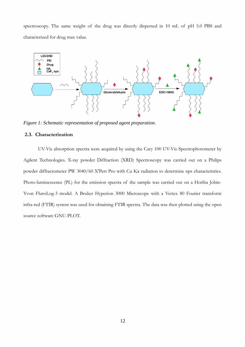
Figure 1: Schematic representation of proposed agent preparation.
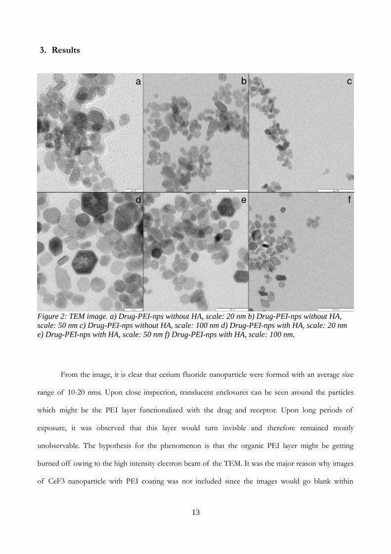
3. Results
From the image, it is clear that cerium fluoride nanoparticle were formed with an average size
range of 10-20 nms. Upon close inspection, translucent enclosures can be seen around the particles
which might be the PEI layer functionalized with the drug and receptor. Upon long periods of
exposure, it was observed that this layer would turn invisble and therefore remained mostly
unobservable. The hypothesis for the phenomenon is that the organic PEI layer might be getting
burned off owing to the high intensity electron beam of the TEM. It was the major reason why images
of CeF3 nanoparticle with PEI coating was not included since the images would go blank within
13
Figure 2: TEM image. a) Drug-PEI-nps without HA, scale: 20 nm b) Drug-PEI-nps without HA, scale: 50 nm c) Drug-PEI-nps without HA, scale: 100 nm d) Drug-PEI-nps with HA, scale: 20 nm e) Drug-PEI-nps with HA, scale: 50 nm f) Drug-PEI-nps with HA, scale: 100 nm.
seconds of exposure. With the functionalization of the organic layer however, the same phenomenon
was limited, perhaps due to the quenching of the light sensitive PEI.
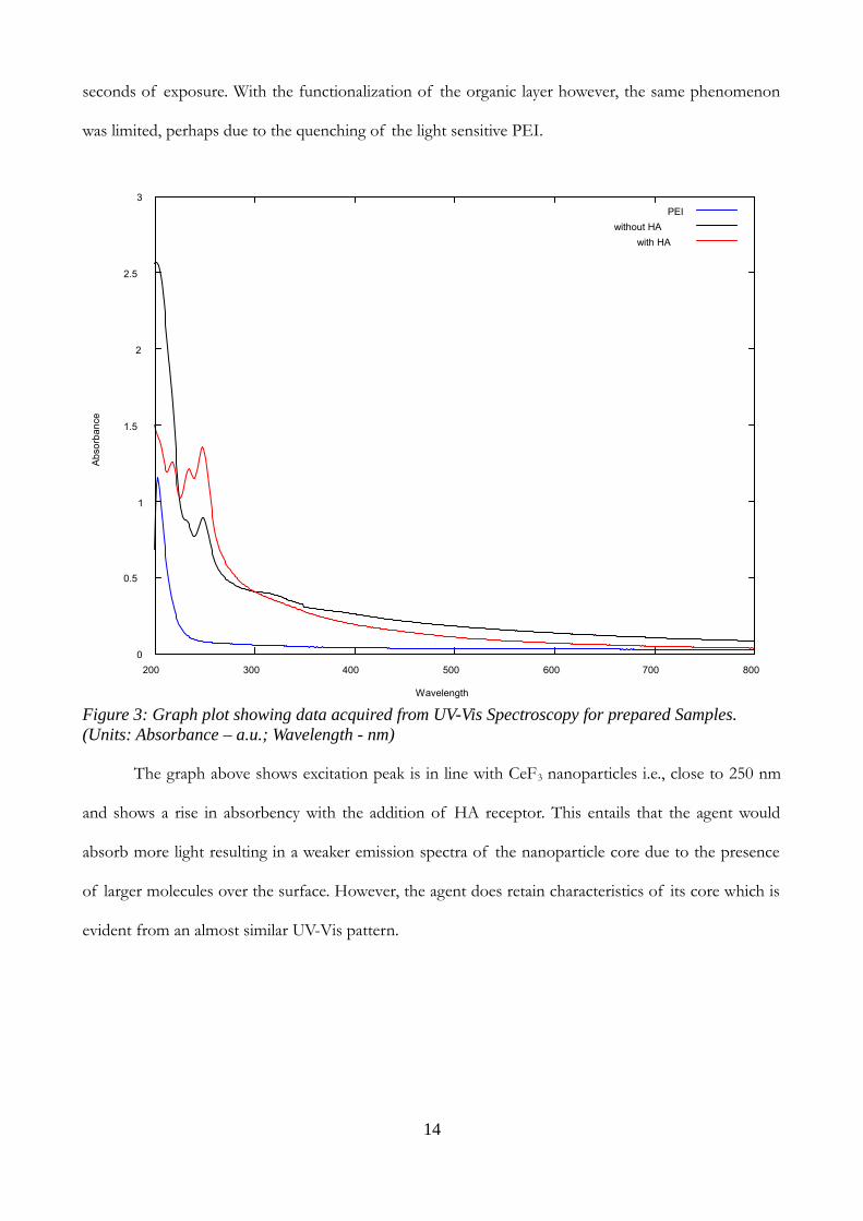
The graph above shows excitation peak is in line with CeF3 nanoparticles i.e., close to 250 nm
and shows a rise in absorbency with the addition of HA receptor. This entails that the agent would
absorb more light resulting in a weaker emission spectra of the nanoparticle core due to the presence
of larger molecules over the surface. However, the agent does retain characteristics of its core which is
evident from an almost similar UV-Vis pattern.
14
Figure 3: Graph plot showing data acquired from UV-Vis Spectroscopy for prepared Samples. (Units: Absorbance – a.u.; Wavelength - nm)
0
0.5
1
1.5
2
2.5
3
200 300 400 500 600 700 800
Ab
sorb
an
ce
Wavelength
PEI
without HA
with HA
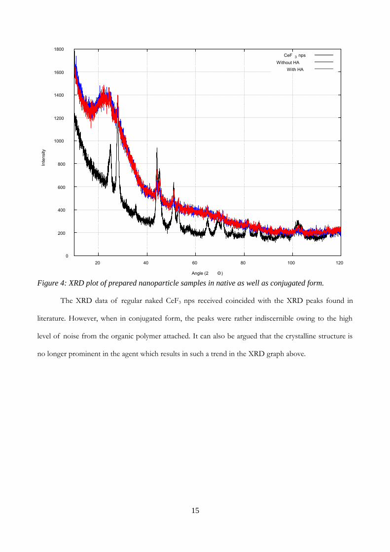
The XRD data of regular naked CeF3 nps received coincided with the XRD peaks found in
literature. However, when in conjugated form, the peaks were rather indiscernible owing to the high
level of noise from the organic polymer attached. It can also be argued that the crystalline structure is
no longer prominent in the agent which results in such a trend in the XRD graph above.
15
Figure 4: XRD plot of prepared nanoparticle samples in native as well as conjugated form.
0
200
400
600
800
1000
1200
1400
1600
1800
20 40 60 80 100 120
Inte
nsi
ty
Angle (2 Θ)
CeF 3 nps
Without HA
With HA
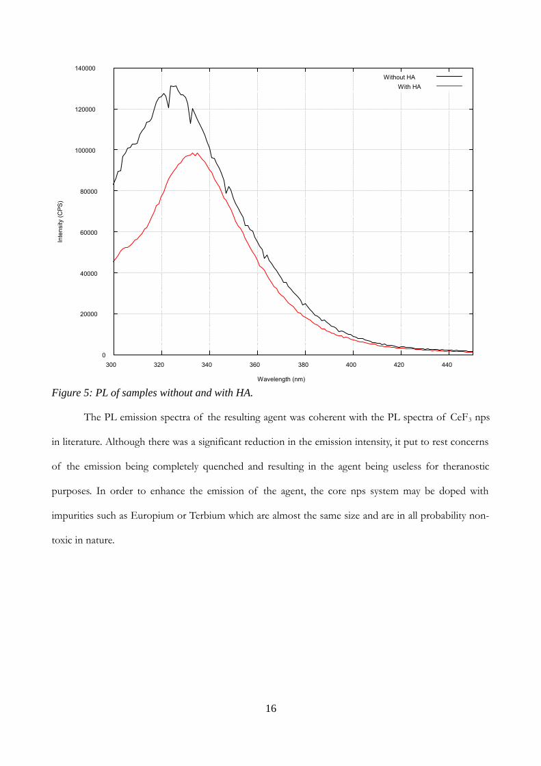
The PL emission spectra of the resulting agent was coherent with the PL spectra of CeF3 nps
in literature. Although there was a significant reduction in the emission intensity, it put to rest concerns
of the emission being completely quenched and resulting in the agent being useless for theranostic
purposes. In order to enhance the emission of the agent, the core nps system may be doped with
impurities such as Europium or Terbium which are almost the same size and are in all probability non-
toxic in nature.
16
Figure 5: PL of samples without and with HA.
0
20000
40000
60000
80000
100000
120000
140000
300 320 340 360 380 400 420 440
Inte
nsi
ty (
CP
S)
Wavelength (nm)
Without HA
With HA
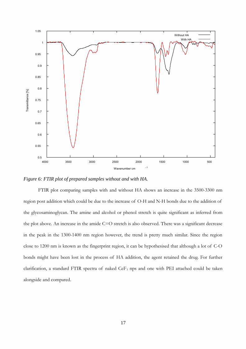
FTIR plot comparing samples with and without HA shows an increase in the 3500-3300 nm
region post addition which could be due to the increase of O-H and N-H bonds due to the addition of
the glycosaminoglycan. The amine and alcohol or phenol stretch is quite significant as inferred from
the plot above. An increase in the amide C=O stretch is also observed. There was a significant decrease
in the peak in the 1300-1400 nm region however, the trend is pretty much similar. Since the region
close to 1200 nm is known as the fingerprint region, it can be hypothesised that although a lot of C-O
bonds might have been lost in the process of HA addition, the agent retained the drug. For further
clarification, a standard FTIR spectra of naked CeF3 nps and one with PEI attached could be taken
alongside and compared.
17
Figure 6: FTIR plot of prepared samples without and with HA.
0.5
0.55
0.6
0.65
0.7
0.75
0.8
0.85
0.9
0.95
1
1.05
500 1000 1500 2000 2500 3000 3500 4000
Tra
nsm
itta
nce
[%
]
Wavenumber cm -1
Without HA
With HA
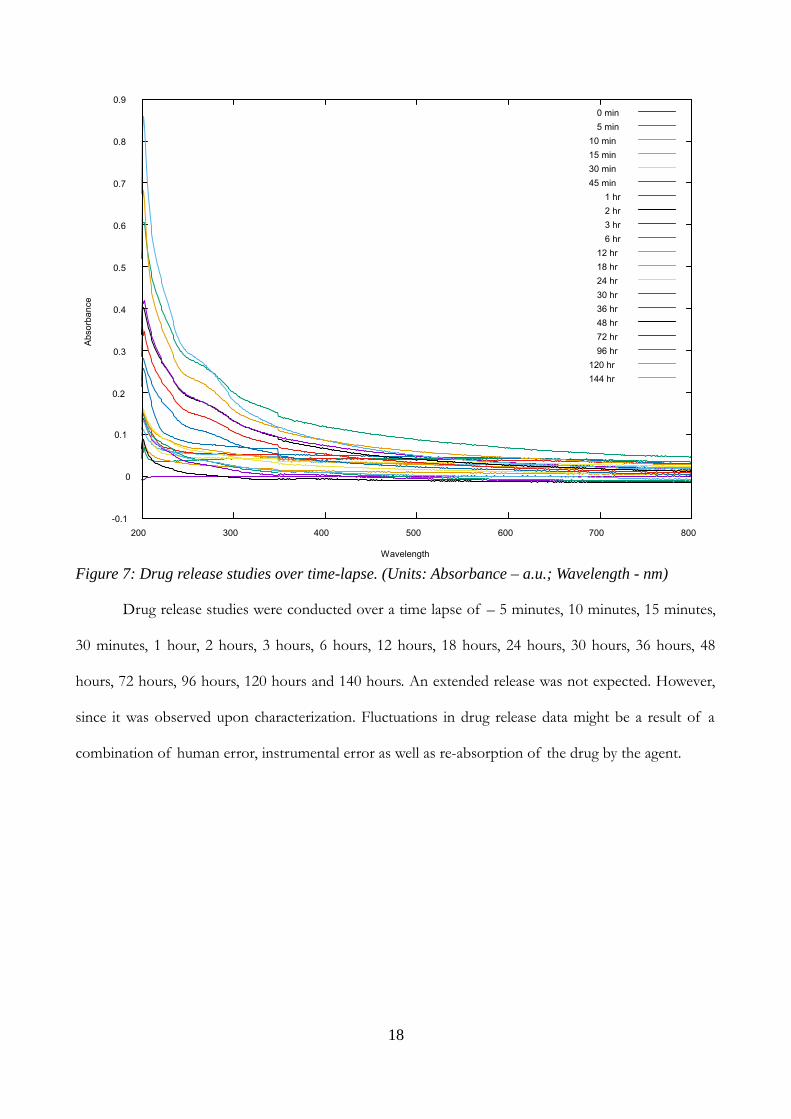
Drug release studies were conducted over a time lapse of – 5 minutes, 10 minutes, 15 minutes,
30 minutes, 1 hour, 2 hours, 3 hours, 6 hours, 12 hours, 18 hours, 24 hours, 30 hours, 36 hours, 48
hours, 72 hours, 96 hours, 120 hours and 140 hours. An extended release was not expected. However,
since it was observed upon characterization. Fluctuations in drug release data might be a result of a
combination of human error, instrumental error as well as re-absorption of the drug by the agent.
18
Figure 7: Drug release studies over time-lapse. (Units: Absorbance – a.u.; Wavelength - nm)
-0.1
0
0.1
0.2
0.3
0.4
0.5
0.6
0.7
0.8
0.9
200 300 400 500 600 700 800
Ab
sorb
an
ce
Wavelength
0 min
5 min
10 min
15 min
30 min
45 min
1 hr
2 hr
3 hr
6 hr
12 hr
18 hr
24 hr
30 hr
36 hr
48 hr
72 hr
96 hr
120 hr
144 hr
Discussions and Conclusions
Although the agent offers promising results in terms of drug release as well as
photoluminescence, and core being lanthanide would offer obstruction to X-rays offering another
method of agent monitoring within the body, it is yet to be tested for toxicity. The behaviour of the
prepared agent is but anyone's guess unless a few toxicity assays are performed to determine their
safety. Nevertheless, the system is proposed to be useful within the scope of preclinical safety studies
wherein cultured cells are utilized. In this manner, newly formulated drugs can be tested in a safe
manner without animal or human casualties. Further tests are required to conclude the targeting
capabilities owing to HA.
Due to the dynamic and versatile nature of the agent, it can be highly tuned to suit specific
needs. The luminescence can be enhanced via means of introducing impurities of other lanthanide
elements. Also, the application method can be varied. Addition of an external encapsulation could
enable drug delivery in an oral form. Transferring the agent onto a thin film can also help in facilitating
topical approaches to certain malignant tumours on the skin surface. Since nanoparticle systems have a
high penetrative capability, it can be a good way to combat skin cancer in many individuals and also in a
very cheap and easy way.
In conclusion, a novel DDS was designed utilizing nps from the lanthanide series as the core
element. The results observed seem rather promising towards evolving the current theranostic
techniques available and might prove crucial in the creation of a marketable technology. However, an
immense amount of research would be required prior to technology transfer including a number of
toxicological screens, preclinical and clinical trials along with a number of approvals. Nevertheless, if
realized, the technology developed shall revolutionize modern medicine.
19
References
[1] Mukherjee, S. (2010). The emperor of all maladies: a biography of cancer. New York, NY.
[2] Carper, S., & Meacham, S. (2013). U.S. Patent No. 8,426,387. Washington, DC: U.S. Patent and
Trademark Office.
[3] Abou-Jawde, R., Choueiri, T., Alemany, C., & Mekhail, T. (2003). An overview of targeted
treatments in cancer. Clinical therapeutics, 25(8), 2121-2137.
[4] Kanwar, R. K., A Cheung, C. H., Chang, J. Y., & Kanwar, J. R. (2010). Recent advances in anti-
survivin treatments for cancer. Current medicinal chemistry, 17(15), 1509-1515.
[5] Kumar, A., Soares, H., Wells, R., Clarke, M., Hozo, I., Bleyer, A., ... & Djulbegovic, B. (2005).
Are experimental treatments for cancer in children superior to established treatments?
Observational study of randomised controlled trials by the Children's Oncology Group. Bmj,
331(7528), 1295.
[6] Ernst, E. (1998). The prevalence of complementary/alternative medicine in cancer. Cancer,
83(4), 777-782.
[7] Van der Zouwe, N., Van Dam, F. S., Aaronson, N. K., & Hanewald, G. J. (1994). [Alternative
treatments in cancer; extent and background of utilization]. Nederlands tijdschrift voor
geneeskunde, 138(6), 300-306.
[8] Krakoff, I. H. (1996). Systemic treatment of cancer. CA: a cancer journal for clinicians, 46(3),
134-141.
[9] Robinson, J. E. (1927). Systemic Treatment Of Cancer. British medical journal, 1(3454), 541.
[10] CASSILETH, B. R., Lusk, E. J., Strouse, T. B., & Bodenheimer, B. J. (1984). Contemporary
unorthodox treatments in cancer medicine: A study of patients, treatments, and practitioners.
Annals of Internal Medicine, 101(1), 105-112.
20
[11] Downer, S. M., Cody, M. M., McCluskey, P., Wilson, P. D., Arnott, S. J., Lister, T. A., & Slevin,
M. L. (1994). Pursuit and practice of complementary therapies by cancer patients receiving
conventional treatment. Bmj, 309(6947), 86-89.
[12] Lind, M. J. (2008). Principles of cytotoxic chemotherapy. Medicine, 36(1), 19-23.
[13] Sawyers, C. (2004). Targeted cancer therapy. Nature, 432(7015), 294-297.
[14] Fujimura, Joan H. Crafting science: A sociohistory of the quest for the genetics of cancer.
Harvard University Press, 1996.
[15] Cho, K., Wang, X. U., Nie, S., & Shin, D. M. (2008). Therapeutic nanoparticles for drug
delivery in cancer. Clinical cancer research, 14(5), 1310-1316.
[16] Malam, Y., Loizidou, M., & Seifalian, A. M. (2009). Liposomes and nanoparticles: nanosized
vehicles for drug delivery in cancer. Trends in pharmacological sciences, 30(11), 592-599.
[17] Haley, B., & Frenkel, E. (2008, February). Nanoparticles for drug delivery in cancer treatment.
In Urologic Oncology: Seminars and original investigations (Vol. 26, No. 1, pp. 57-64). Elsevier.
[18] Alley, S. C., Okeley, N. M., & Senter, P. D. (2010). Antibody–drug conjugates: targeted drug
delivery for cancer. Current opinion in chemical biology, 14(4), 529-537.
[19] Poste, G. H., & Kirsh, R. (1983). Site-specific (targeted) drug delivery in cancer therapy.
Bio/Technology, 1(10), 869-878.
[20] Arap, W., Pasqualini, R., & Ruoslahti, E. (1998). Cancer treatment by targeted drug delivery to
tumor vasculature in a mouse model. Science, 279(5349), 377-380.
[21] Sharma, D., Chelvi, T. P., Kaur, J., Chakravorty, K., De, T. K., Maitra, A., & Ralhan, R. (1995).
Novel Taxol formulation: polyvinylpyrrolidone nanoparticle-encapsulated Taxol for drug
delivery in cancer therapy. Oncology research, 8(7-8), 281-286.
[22] Liu, Z., Robinson, J. T., Tabakman, S. M., Yang, K., & Dai, H. (2011). Carbon materials for
21
drug delivery & cancer therapy. Materials today, 14(7), 316-323.
[23] Josefsen, L. B., & Boyle, R. W. (2012). Unique diagnostic and therapeutic roles of porphyrins
and phthalocyanines in photodynamic therapy, imaging and theranostics. Theranostics, 2(9),
916-966.
[24] Baum, R. P., & Kulkarni, H. R. (2012). Theranostics: from molecular imaging using Ga-68
labeled tracers and PET/CT to personalized radionuclide therapy-the Bad Berka experience.
Theranostics, 2(5), 437-47.
[25] Muthu, M. S., Leong, D. T., Mei, L., & Feng, S. S. (2014). Nanotheranostics-application and
further development of nanomedicine strategies for advanced theranostics. Theranostics, 4(6),
660-677.
[26] Hayashi, K., Nakamura, M., Sakamoto, W., Yogo, T., Miki, H., Ozaki, S., ... & Ishimura, K.
(2013). Superparamagnetic nanoparticle clusters for cancer theranostics combining magnetic
resonance imaging and hyperthermia treatment. Theranostics, 3(6), 366-376.
[27] Chen, Z., Ma, L., Liu, Y., & Chen, C. (2012). Applications of functionalized fullerenes in tumor
theranostics. Theranostics, 2(3), 238-250.
[28] Zhang, Z., Wang, J., & Chen, C. (2013). Gold nanorods based platforms for light-mediated
theranostics. Theranostics, 3(3), 223-238.
[29] Xie, J., & Jon, S. (2012). Magnetic nanoparticle-based theranostics. Theranostics, 2(1), 122-124.
[30] Liu, Z., & Liang, X. J. (2012). Nano-carbons as theranostics. Theranostics, 2(3), 235-237.
[31] Luk, B. T., Fang, R. H., & Zhang, L. (2012). Lipid-and polymer-based nanostructures for
cancer theranostics. Theranostics, 2(12), 1117-1126.
[32] Lukianova-Hleb, E. Y., Oginsky, A. O., Samaniego, A. P., Shenefelt, D. L., Wagner, D. S.,
Hafner, J. H., ... & Lapotko, D. O. (2011). Tunable plasmonic nanoprobes for theranostics of
22
prostate cancer. Theranostics, 1, 3-17.
[33] Kildeeva, N. R., Perminov, P. A., Vladimirov, L. V., Novikov, V. V., & Mikhailov, S. N. (2009).
About mechanism of chitosan cross-linking with glutaraldehyde. Russian journal of bioorganic
chemistry, 35(3), 360-369.
[34] Migneault, I., Dartiguenave, C., Bertrand, M. J., & Waldron, K. C. (2004). Glutaraldehyde:
behavior in aqueous solution, reaction with proteins, and application to enzyme crosslinking.
Biotechniques, 37(5), 790-806.
[35] Jin, Y., Ma, X., Feng, S., Liang, X., Dai, Z., Tian, J., & Yue, X. (2015). Hyaluronic Acid
Modified Tantalum Oxide Nanoparticles Conjugating Doxorubicin for Targeted Cancer
Theranostics. Bioconjugate chemistry, 26(12), 2530-2541.
[36] Lokeshwar, V. B., Öbek, C., Soloway, M. S., & Block, N. L. (1997). Tumor-associated hyaluronic
acid: a new sensitive and specific urine marker for bladder cancer. Cancer research, 57(4), 773-
777.
[37] Lee, Y., Lee, H., Kim, Y. B., Kim, J., Hyeon, T., Park, H., ... & Park, T. G. (2008). Bioinspired
surface immobilization of hyaluronic acid on monodisperse magnetite nanocrystals for targeted
cancer imaging. Advanced Materials, 20(21), 4154-4157.
[38] Lokeshwar, V. B., Rubinowicz, D., Schroeder, G. L., Forgacs, E., Minna, J. D., Block, N. L., ... &
Lokeshwar, B. L. (2001). Stromal and epithelial expression of tumor markers hyaluronic acid
and HYAL1 hyaluronidase in prostate cancer. Journal of Biological Chemistry, 276(15), 11922-
11932.
[39] Choi, K. Y., Min, K. H., Na, J. H., Choi, K., Kim, K., Park, J. H., ... & Jeong, S. Y. (2009). Self-
assembled hyaluronic acid nanoparticles as a potential drug carrier for cancer therapy: synthesis,
characterization, and in vivo biodistribution. Journal of Materials Chemistry, 19(24), 4102-4107.
[40] Choi, K. Y., Yoon, H. Y., Kim, J. H., Bae, S. M., Park, R. W., Kang, Y. M., ... & Kim, K. (2011).
23
Smart nanocarrier based on PEGylated hyaluronic acid for cancer therapy. ACS nano, 5(11),
8591-8599.
[41] Coradini, D., Pellizzaro, C., Miglierini, G., Daidone, M. G., & Perbellini, A. (1999). Hyaluronic
acid as drug delivery for sodium butyrate: Improvement of the anti proliferative activity on a‐
breast cancer cell line. International journal of cancer, 81(3), 411-416.‐
[42] Gardner, M. J., Catterall, J. B., Jones, L. M. H., & Turner, G. A. (1996). Human ovarian tumour
cells can bind hyaluronic acid via membrane CD44: a possible step in peritoneal metastasis.
Clinical & experimental metastasis, 14(4), 325-334.
[43] Delpech, B., Chevallier, B., Reinhardt, N., Julien, J. P., Duval, C., Maingonnat, C., ... & Asselain,
B. (1990). Serum hyaluronan (hyaluronic acid) in breast cancer patients. International Journal of
Cancer, 46(3), 388-390.
[44] Posey, J. T., Soloway, M. S., Ekici, S., Sofer, M., Civantos, F., Duncan, R. C., & Lokeshwar, V. B.
(2003). Evaluation of the prognostic potential of hyaluronic acid and hyaluronidase (HYAL1)
for prostate cancer. Cancer research, 63(10), 2638-2644.
[45] Cold Spring Harbor Protocls, Vol. 2006, No. 1. (1 June, 2006), pdb.rec8247,
doi:10.11.1/pdb.rec8247 Key: citeulike:12802637
[46] Varun, S., Kalra, M., & Gandhi, M. (2015). White Light Emission Through Downconversion
of Terbium and Europium Doped CeF3 Nanophosphors. Journal of fluorescence, 25(5), 1501-
1505.
24


