Embed Size (px)
Citation preview
, . MALACOLOGIA, 1998, 40(1-2): 305-320
MARINE VALVATOIDEA - COMMENTS ON ANATOMY AND SYSTEMATICS WITH DESCRIPTION OF A NEW SPECIES FROM FLORIDA
(HETEROBRANCHIA: CORNIROSTRIDAE)
Rudiger Bieler1, Alexander D. Ball1•
2, and Paula M. Mikkelsen3
ABSTRACT
The "lower heterobranch" gastropod family Cornirostridae Ponder, 1990 (Valvatoidea), has been previously known from only six confirmed extant species in three genera ( Cornirostra, Noerrevangia, Tamura). Knowledge of the soft-body morphology is necessary for placement in this family. The unique "multi-tentacled, two-tailed" habitus forms a synapomorphy of this group (an appearance produced by a combination of characters of the paired anterior oral lobes, cephalic tentacles, curved foot processes, and the deeply split hindfoot). As a result of research on "lower heterobranchs" of the Florida Keys, a new species of Cornirostra, C. floridana Bieler & Mikkelsen, n. sp., is described from the Florida Keys, as the seventh known species in the family, the second known species in the genus, and the first member of Cornirostra from the Atlantic Ocean. Detailed anatomical descriptions and interpretations of the foregut and nervous system are provided from computer-assisted reconstructions of semi-thin histological sections. Shell and anatomical characters of the seven confirmed living cornirostrid species are summarized, generic and familial diagnoses are discussed, and a redescription of the family Cornirostridae is provided, based on shell and anatomical data. Distinguishing characters of other recognized families of Valvatoidea (Valvatidae, Orbitestellidae) are surveyed. The problematic assignment of fossils to this anatomically defined gastropod family is also addressed.4
Key words: Florida Keys, Gastropoda, lower Heterobranchia, Cornirostra, Atlantic Ocean, systematics, nervous system, histology.
INTRODUCTION
A focus in recent gastropod research has been on the "lower heterobranchs" (also termed Allogastropoda, Heterostropha, etc.), a grade or clade of several families that shows heterobranch anatomical organization in many organ systems, distinguishing them from the caenogastropods with which many were traditionally grouped. In addition to such relatively well-known groups as Pyramidelloidea, Architectonicoidea, and Valvatidae, this group includes families of lesser-known, small-shelled snails, such as Omalogyridae, Rissoellidae, Glacidorbidae, and the enigmatic and very recently described Tjaernoidae Waren, 1991, Hyalogyrinidae Waren & Souchet, 1992, Xylodisculidae Waren, 1992, and Cimidae Waren, 1993. The erection of new family-group taxa is partly the result of
newly discovered species, and partly because of critical reassessment of anatomical features, warranting the transfer of taxa from traditionally recognized "prosobranch" groups into the lower heterobranchs (e.g., Haszprunar, 1988; Rath, 1988; Ponder, 1991; Bieler, 1992; Waren et al., 1993).
One such example is Tamura bicaudata (Pilsbry & McGinty, 1946), described as a vitrinellid caenogastropod from Missouri Key in the Lower Florida Keys. Moore (1964) questioned its placement in Vitrinellidae, based on the uncharacteristic tentacle morphology illustrated in the original figures. In a reassessment of available anatomical data on Vitrinellidae, Bieler & Mikkelsen (1988) removed T. bicaudata from the Vitrinellidae on the basis of several head-foot characters, but also could not suggest a new taxonomic placement. While the small, thin, transparent shell
1Department of Zoology, Field Museum of Natural History, Roosevelt Road at Lake Shore Drive, Chicago, Illinois 60605, U.S.A.; [email protected] 2Current address: Zoology Department, The Natural History Museum, Cromwell Road, London, SW7 580, United Kingdom; [email protected] 3Department of Invertebrates, American Museum of Natural History, Central Park West at 79th Street, New York, New York 10024-5192, U.S.A.; [email protected] 4This paper is dedicated to the memory of our friend and colleague Dr. Donald R. Moore (1921-1997), who contributed extensively to the knowledge of Florida's micromollusk fauna.
305

306 BIELER, BALL & MIKKELSEN
of T. bicaudata is very similar in overall appearance to many vitrinellid species (particularly that of Vitrine/la helicoidea C. B. Adams, 1850), its body morphology is uniquely peculiar. The oral lobes, cephalic tentacles, and curved anterior foot processes, along with a deeply split hindfoot, make the animal appear multi-tentacled and two-tailed.
Other similar "horned-snout, cleft-tail" snails have been recently discovered. Ponder (1990b) introduced the generic name Carnirostra tor Micradiscu/a pellucida Laseron, 1954, from Australia, and placed it and Tamura in his new family Cornirostridae among the lower heterobranchs. Based on extensive anatomical comparisons, he recognized the position of this family in the Valvatoidea, a group previously thought restricted to freshwater. Ponder (1990b) suspected "Cyc/ostrema" prestani Melvill, 1906, from Ceylon, and the Mediterranean Skenea pel/ucida Monterosato, 1874, also to belong to Cornirostridae. The latter species, after more detailed anatomical study, has since been transferred to Hyalogyrinidae (Waren et al., 1993); the former remains of uncertain status. Waren et al. (1993) also transferred a Mediterranean species, Oxystele depressa Granata, 1877, to Tamura, and introduced a third cornirostrid genus, Naerrevangia, tor N. fragilis Waren & Schander (in Waren et al.), 1993, from the Faroe Islands. Most recently, Fukuda & Yamashita (1997) described the first cornirostrid species from the Western Pacific, Tamura yashima and T. himeshima. Thus to date the family is known from six confirmed extant species placed in three genera.
Ongoing research on "lower heterobranchs" of the Florida Keys has brought to light an additional extant species of Carnirostra, which is here described. Histological reconstruction and functional interpretations emphasized foregut and nervous system anatomy, particularly those structures that differed from, or were not addressed, by Ponder's studies (1990b) of cornirostrid species. Shell-morphological and anatomical characters are discussed for all known extant species, and comments are made on the reported Cretacous-Jurassic-Triassic fossil record (Schroder, 1995; Bandel, 1996).
MATERIALS AND METHODS
Specimens were collected in the Florida Keys by "rock washing" (i.e., scrubbing the
surfaces of shallow-subtidal rocks that can be lifted out of the water, including the underside normally resting on the sediment, with brush and saltwater) and by shoveling, hand-dredging, and sieving of muddy and sandy shallowwater substrata. Specimens were sorted from the resulting freshly collected material in the field laboratory under a dissecting microscope.
Carnirostra flaridana n. sp. was observed alive only once, sketched, and photographed with a single-lens reflex camera equipped with extension tubes. For this reason, observations of gross morphology remained incomplete. Anatomical descriptions are based upon histological sections of the single livecollected specimen; initially intended for DNA studies, the specimen had not been chemically fixed before alcohol preservation, so tis-
Figure and Table Abbreviations
agp accessory glandular pocket bm buccal mass bpi black pigmented layer bw body wall c cuticle cg cerebral ganglion co cornea ct connective tissue cs cuticular sheath e eye g gill h heart jt jaw tooth le lens lu lumen m muscle mm mantle margin mpp metapodial processes orl oral lobe ot oral tube PC protoconch pg pedal ganglion pig pigment pig pleural ganglion PMO pigmented mantle organ pp propodial processes pt pallial tentacle sbg subesophagealganglion sg salivary gland sh shell sn snout nerves snt snout spg supraesophageal ganglion st statocyst TC teleoconch te cephalic tentacle tn tentacle nerves v void

CORNIROSTRIDAE ANATOMY AND SYSTEMATICS 307
sue preparation was not ideal. Following rehydration from 70% ethanol, the shell was dissolved using saturated aqueous ethylene diamene tetraacetic acid (EDTA). After dehydration through an ascending graded ethanol series, the specimen was infiltrated with LR White resin (Polysciences, Inc.) and flat-embedded in fresh resin using an inverted BEEM capsule. Polymerization took 8 h at 70°C. The specimen was mounted in transverse orientation and serial-sectioned at 1 µm thickness. Sections were stained in aqueous toluidine blue, and mounted in Polymount (Polysciences, Inc.) under coverslips. The sections were drawn at 4 µm intervals using a camera lucida and the internal anatomy was reconstructed using Jandel Scientific's PC-3D software. Where greater resolution was required, specimens were reconstructed at 1 µm intervals. Individual sections were photographed using a photomicroscope with automatic camera attachment. Scanning electron micrographs (SEM) were produced from air-dried shells, coated with gold, observed and photographed using an AMRAY 1810 scanning electron microscope at FMNH. Spire angle was measured using a protractor against photographs or line drawings of shells in lateral view. Numbers of protoconch and teleoconch whorls were ascertained using the method of Taylor as summarized by Jablonski & Lutz (1980: 330, fig. 4). This method counts the initial embryonic part of the shell as part of a whorl; this explains discrepancies between our counts and those of other authors who expressly or apparently employed different methods of whorl counting.
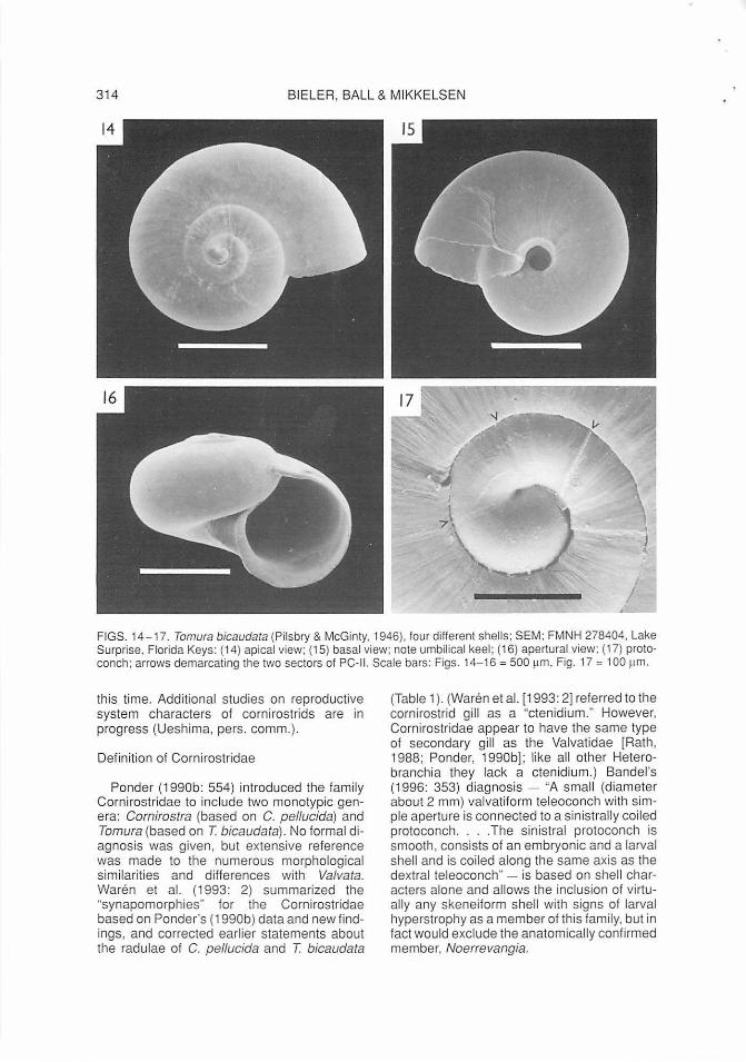
Comparative material of Tomura (Figs. 14-17) was collected in the Upper Florida Keys (Sta. FK-021, Lake Surprise, northeastern end of U. S. Route 1 causeway, Mile Marker 107.5, Monroe County, Florida, sediment/algae at approximately 1 .5 m depth, by hand dredge, salinity = 22 ppt, 9 July 1995, Bieler/Mikkelsen coll.). Voucher specimens are deposited in FMNH 278404 (including SEM material) and AMNH 289603. Specimens collected for this study were compared to the type specimen of Tamura bicaudata (ANSP 182042; Missouri Key, Lower Florida Keys, Monroe County, Florida, T. L. McGinty!, March 1945; 1.18 mm maximum shell diameter}, to Cornirosta pellucida and other material studied by Ponder (AMS), and to other "vitrinelliform" gastropods collected by Pilsbry & McGinty in the Florida Keys (ANSP). Several major collections, including AMNH, ANSP,
DMNH, and FMNH, were searched (unsuccessfully) for additional specimens of the new species.
Museum acronyms used in text are: AMNH American Museum of Natural His
tory, New York, U.S.A. AMS The Australian Museum, Sydney,
New South Wales, Australia ANSP Academy of Natural Sciences of
Philadelphia, Pennsylvania, U. S. A. DMNH Delaware Museum of Natural His
tory, Wilmington, U.S. A. FMNH Field Museum of Natural History,
Chicago, Illinois, U.S. A. HMNS Houston Museum of Natural Sci
ence, Texas, U.S.A. USNM National Museum of Natural History,
Smithsonian Institution, Washington, D.C., U.S. A.
RESULTS
Valvatoidea Gray, 18401
Cornirostridae Ponder, 1990b
Cornirostra Ponder, 1990b; type species by original designation: Microdiscula pellucida Laseron, 1954.
Cornirostra floridana Bieler & Mikkelsen, new species Figs. 1-13
Type Locality
Indian Key Fill (formerly known as "Central Supply"), Mile Marker 79, Middle Florida Keys, bay side (Gulf of Mexico), Monroe County, Florida; 24°53'25"N, 80°40'28"W.
Type Material
Holotype (FMNH 278401; shell with dried tissue remains); paratype 1 (AMNH 289256, with dried tissue remains), paratype 2 (USNM 880276, SEM specimen), and paratype 3 (FMNH 278402, SEM specimen) collected with holotype in shallow subtidal habitat by hand-dredge, 26 July 1992 (sta. RB-1582). Paratype 4 (FMNH 278405), paratype 5 (AMNH 289806), and paratype 6 (FMNH 278403, live-observed specimen, serial-sec-
1 Availability and authorship established with ICZN Direction 27 (1955); in contrast to recent references {e.g., Riedel, 1993).
308 BIELER, BALL & MIKKELSEN
tioned on microslides) from shoveled silty mud, among turtle grass ( Thalassia testudinum Banks ex Konig) and green algae [Penicillus cf. dumetosus (Lamoureux) Blainville, and Halimeda spp.], in shallow water (<1 m) at low tide, 1 October 1994 (sta. FK-001 ). All material from type locality, Bieler/Mikkelsen coll.
Dimensions:
Diameter Height Teleoconch (mm) (mm) whorls
Holotype 1.70 1.60 2 7/8-Paratype 1 1.28 0.92 2 1/10 Paratype 2 1.66 1.58 2 5/8-Paratype 3 2.10 1.80 3-
(damaged after SEM)
Paratype 4 1.42 1.04 2 3110 Paratype 5 1.20 0.90 2+
(damaged, was larger)
Etymology
floridanus, -a, -um; named for the State of Florida.
Description
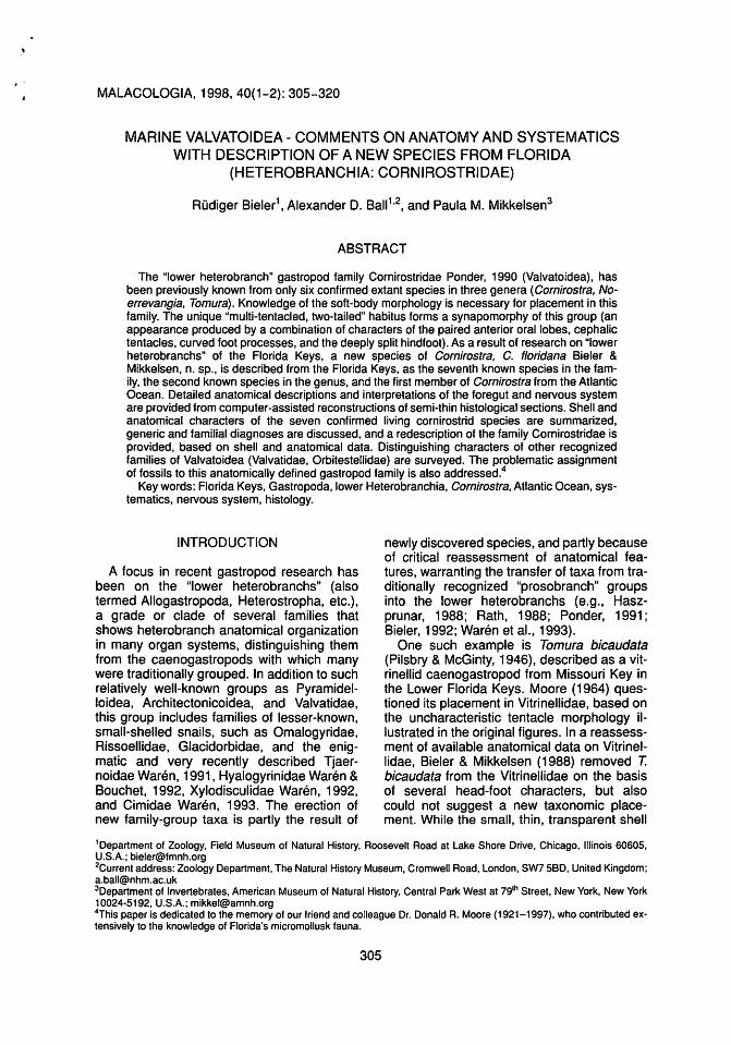
Teleoconch (Figs. 1-3): Diameter to about 2 mm at nearly 3 convex whorls; transparent, smooth with fine growth lines, high-spired (spire angle 105-110°). Base simple, smooth, umbilicate, without umbilical keel. Aperture round; peristome simple, sharp. Fresh specimens with thin transparent periostracum, imparting very fine spiral sculpture (visible under oblique microscope light).
Protoconch (Fig. 4): 180-185 µm (paratypes 3, 2, respectively), about 1.2 whorls, coiling near-planispiral but with initial hyperstrophy (tip of apex slightly sunken). Protoconch I (embryonic shell) measuring 133-141 µm (paratypes 3, 2), with reticulated sculptural pattern as shown for other cornirostrid species (Ponder, 1990b: fig. SF; Waren et al., 1993: fig. 3); protoconch II (larval shell, comprising about 1/5 of a whorl) smooth, divided into two sectors by a growth mark.
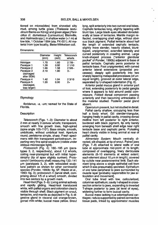
Head-foot(Figs. 5-6): Living animal actively and rapidly gliding. Head-foot translucent white, with pallial organs and coloration clearly visible through shell. Black pigment on snout, tentacles, and mantle as depicted in Fig. 5; digestive gland in visceral coil orange-brown, gonad milk-white; buccal mass yellow. Snout
long, split anteriorly into two curved oral lobes. Cephalic tentacles long, slightly tapering with blunt tips. Large black eyes situated dorsolaterally at base of tentacles. Mantle margin reflected, overlapping shell edge, without obvious black pigment. Pallial tentacle about half the length of extended cephalic tentacle, slightly more slender, heavily ciliated, blunttipped, unpigmented, extended laterally and curved posteriorly in crawling animal. Large knob-shaped process ("anterior glandular pocket" of Ponder, 1990b) adjacent to base of pallial tentacle. Cephalic penis posterior to tentacle base. Foot unpigmented, with curved anterior lateral extensions (propodial processes); deeply split posteriorly into two sharply tapering metapodial processes (of unequal length), grooved at outer lateral edge, separated by LI-shaped indentation (Fig. 6).
Anterior pedal mucus gland in anterior part of foot, extending posteriorly to pedal ganglia where it appears to fold around pedal commissure. Folded dorsal component passing anteriorly and then becoming untraceable in the material studied. Posterior pedal gland absent.
Operculum present, but not studied in detail. Pallial cavity shallow, occupying less than
half a body whorl. Gill slender-triangular, hanging freely in pallial cavity, crossing dorsal midline from left posterior to right anterior, bordered with black pigment, tip only barely emerging from beneath shell edge near right tentacle base and cephalic penis. Pulsating heart clearly visible in living animal at rear of pallial cavity.
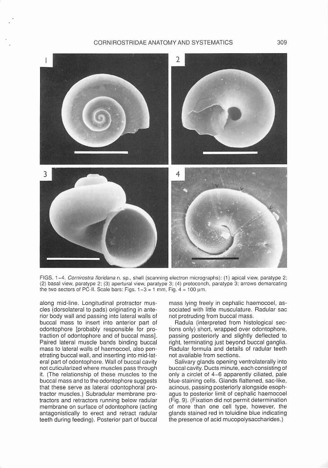
Alimentary System: Mouth ventrally directed, slit-shaped, at tip of snout. Paired jaws (Figs. 7 -8) attached to lateral walls of oral tube at approximate mid-point of its length; composed of overlapping, finely denticulate elements (8-1 O elements at widest extent; each element about 1 O µm in length), covered by cuticle near posteroventral limit. Each element lying above a single epithelial cell from which it was presumably secreted. Epithelium of oral tube peripheral to jaws surrounded by muscle layer [probably responsible for jaw articulation and movement].
Oral tube lined with low, cuticularized, columnar epithelium; cavity I-shaped in crosssection anterior to jaws, expanding to inverted T-shape posterior to jaws (at level of eyes), widening further to form buccal cavity.
Buccal mass lacking true odontophoral cartilages; radula supported by paired connective tissue pads, linked by approximator muscles
CORNIROSTRIDAE ANATOMY AND SYSTEMATICS 309
FIGS. 1- 4. Cornirostra floridana n. sp. , shell (scanning electron micrographs): (1) apical view, paratype 2; (2) basal view, paratype 2; (3) apertural view, paratype 3; (4) protoconch, paratype 3; arrows demarcating the two sectors of PC-II. Scale bars: Figs. 1-3 = 1 mm , Fig. 4 = 100 ~1m.
along mid-line. Longitudinal protractor muscles (dorsolateral to pads) originating in anterior body wall and passing into lateral walls of buccal mass to insert into anterior part of odontophore [probably responsible for protraction of odontophore and of buccal mass] . Paired lateral muscle bands binding buccal mass to lateral walls of haemocoel, also penetrating buccal wall, and inserting into mid-lateral part of odontophore. Wall of buccal cavity not cuticularized where muscles pass through it. (The relationship of these muscles to the buccal mass and to the odontophore suggests that these serve as lateral odontophoral protractor muscles.) Subradular membrane protractors and retractors running below radular membrane on surface of odontophore (acting antagonistically to erect and retract radular teeth during feeding) . Posterior part of buccal
mass lying freely in cephalic haemocoel, associated with little musculature. Radular sac not protruding from buccal mass.
Radula (interpreted from histological sections only) short, wrapped over odontophore, passing posteriorly and slightly deflected to right, terminating just beyond buccal ganglia. Radular formula and detai ls of radular teeth not available from sections.
Salivary glands opening ventrolaterally into buccal cavity. Ducts minute, each consisting of only a ci rclet of 4 - 6 apparently ciliated , pale blue-staining cells. Glands flattened, sac-like, acinous, passing posteriorly alongside esophagus to posterior limit of cephalic haemocoel (Fig. 9). (Fixation did not permit determination of more than one cell type , however, the glands stained red in toluidine blue indicating the presence of acid mucopolysaccharides.)

310 BIELER, BALL & MIKKELSEN
5 6
FIGS. 5-6. Cornirostra floridana n. sp., crawling animal: (5) dorsal view; (6) ventral view. Scale bar = 1 mm.
Remainder of alimentary canal not observed in detail, but presence of style sac and localized cuticularization of stomach lining (as described by Ponder, 1990b, for C. pellucida) confirmed.
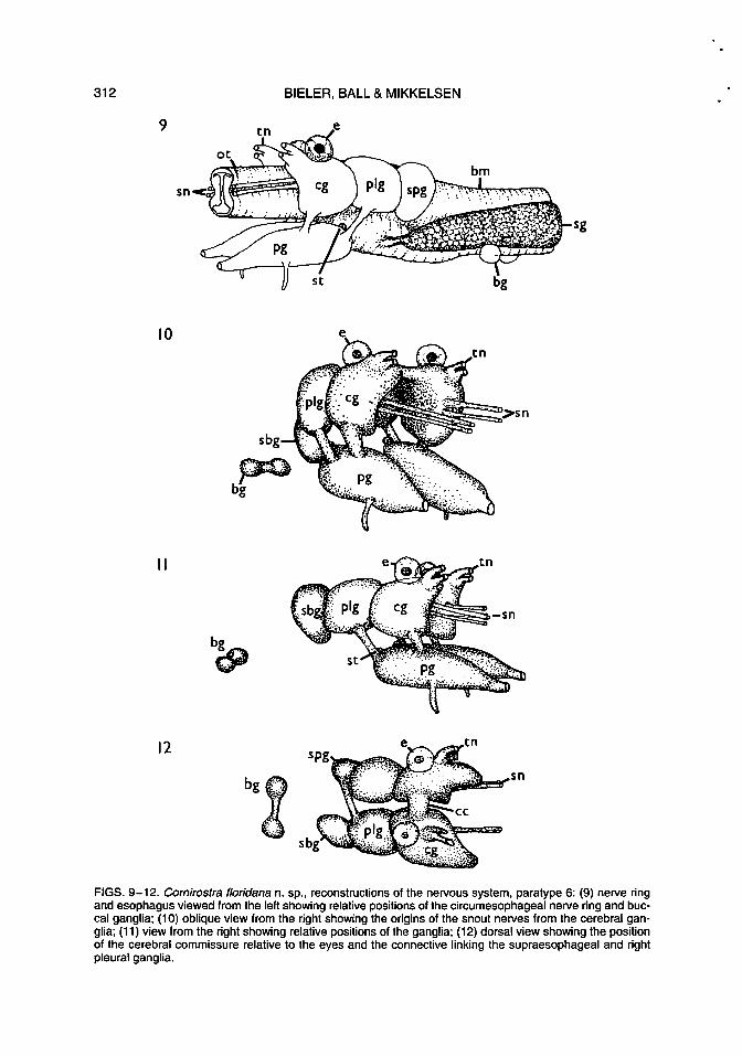
Nervous System (Figs. 9-12): Cerebral commissure lying immediately posterior to tentacle bases. Paired tentacle nerves originating on anterodorsal bulges on each cerebral ganglion. Paired snout nerves extending from each cerebral ganglion, down length of snout along esophagus; ventral nerve branches innervating parts of snout and esophagus.
Pleural ganglia lying lateral to esophagus and closely abutting cerebral ganglia.
Pedal ganglia elongated, positioned ventral to cerebral ganglia. Each cerebropedal connective attached near middle of pedal ganglion; pleuropedal connective attached near posterior limit. Statocysts small, lying dorsally just posterior to cerebropleural juncture, and each containing a single statolith.
Large single pedal nerve passing anteriorly from each pedal ganglion; another nerve passing ventrally from near anterior end of ganglion anterior to cerebropedal connective. (Only anterior pedal nerves were found.) Supraesophageal ganglion (Fig. 9) closely abutting left pleural ganglion, dorsolateral to
esophagus, connected to right pleural ganglion by short connective passing over esophagus. A further nerve passing toward pallial cavity to join osphradial ganglion which lies on floor of pallial cavity on left side. Subesophageal ganglion (Fig. 11) abutting right pleural ganglion, lateral to esophagus, at level of supraesophageal ganglion. Buccal ganglia situated posterior to nerve ring, behind buccal mass, forming dumbell-shaped mass due to short commissure. (Subesophageal connective, buccal connectives, and visceral ganglion not located.)
Eyes at tentacle bases, in close contact with dorsal surfaces of cerebral ganglia, innervated by short nerve at rear of each eye. Each a closed sphere (Fig. 13), with black granular pigment posteriorly; lens sphericalovoid, solid, interior staining evenly light blue in toluidine blue.
Comparative Remarks
This new species, compared to the six other confirmed cornirostrid species (Table 1) is most similar to the Australian Cornirostra pel/ucida (as re-described by Ponder, 1990b), especially with regard to high spire angle and black mantle pigmentation. The basic organization of the nervous system and the anterior

CORNIROSTRIDAE ANATOMY AND SYSTEMATICS 311
c
FIGS. 7-8. Cornirostra floridana n. sp., jaws (1-pm sections of paratype 6 and explanatory drawing): (7) transverse section through oral tube with paired jaw: (8) jaw elements, detail. Scale bars: Fig. 7 = 50 ~1m, Fig. 8 = 10 pm.
312 BIELER, BALL & MIKKELSEN
9
sg
10 e
sbg
11
bg
~
12
bgg·~·· .. •· .
.,., ;~
FIGS. 9-12. Comirostra floridana n. sp., reconstructions of the nervous system, paratype 6: (9) nerve ring and esophagus viewed from the left showing relative positions of the circumesophageal nerve ring and buccal ganglia; (10) oblique view from the right showing the origins of the snout nerves from the cerebral ganglia; (11) view from the right showing relative positions of the ganglia; (12) dorsal view showing the position of the cerebral commissure relative to the eyes and the connective linking the supraesophageal and right pleural ganglia.

CORNIROSTRIDAE ANATOMY AND SYSTEMATICS 313
FIG. 13. Cornirostra floridana n. sp. , eye (1-pm section of paratype 6). Scale bar = 20 pm.
pedal mucus gland are identical. Other than by geographical distribution, it differs from the latter in: smaller size, position of jaws (midpoint of oral tube versus immediately behind mouth), color of buccal mass in living animals (yellow versus white), relative position of cerebral ganglia (with commissure versus abutting). position of statocyst (near cerebropleural junctions versus on dorsal surface of pedal ganglia), number of pedal nerves (one versus two), and construction of the eye lens (solid versus hollow; also solid in Valvata spp. [Bernard, 1890; Ponder, 1990b)). Because so few cornirostrid specimens have been studied anatomically, some of these differences could be results of preservation artifact, varying degree of retraction , and histological technique, and warrant verification as additional material becomes available.
The new species differs from the single other known cornirostrid from Florida, Tamura bicaudata (Figs. 14- 17) in: spire angle (105-11 0° in C. flaridana versus 130- 140° in T. bicaudata; compare Figures 3 and 16), umbilical keel (absent versus present; Figs. 2 and 15), black mantle pigment (present versus absent), and depth of metapodial cleft (deep versus shal low).
DI SCUSSION
Carnirastra Versus Tamura and Naerrevangia
The seven confirmed cornirostrid species differ in a number of shell and anatomical
characters (Table 1 ). These species currently reside in three nominal genera (Carnirastra, two species; Tamura, four species; Naerrevangia, one species). Generic characters are difficult to summarize considering the mosaic character state distribution of the few described species, but the three taxa might in fact describe natural groups. Separating characters appear to include the shape of the marginal radular teeth: triangular in Carnirastra (although stil l unknown in C. flaridana) , rectangular in Tamura, and slender oar-like in Naerrevangia. Carnirastra is further distinguished by black mantle pigment, a relatively high spire, and a deep metapodial cleft ; Naerrevangia by a lecithotrophic-type protoconch (no PC-II) and a grooved cephalic penis. Ponder (1990b) listed the characters in which Tamura differed from Carnirastra: much smaller overall size, spiral ridge in umbilicus, non-pigmented mantle and head-foot, and radular characters. Since the transfer of Oxystele depressa to Tamura (based on gross morphology and radula; Waren et al., 1993), and the description of additional Tamura species from Japan (Fukuda & Yamashita, 1997), shell size and umbilical characters are now weakened as distinctions. T. depressa is unique in the family in having a callus-filled umbilicus, spiral sculptural ridges on the early teleoconch whorls, a small yellow pigmented mantle organ, a minute (not long) pall ial tentacle, and seven (instead of nine) radular teeth per row; its size is intermediate between those of T. bicaudata and C. pellucida.
Because of the still-incomplete dataset, no cladistic analysis of the group is attempted at
314 BIELER, BALL & MIKKELSEN
FIGS. 14-17. Tamura bicaudata (Pilsbry & McGinty, 1946), four different shells; SEM; FMNH 278404, Lake Surprise, Florida Keys: (14) apical view; (15} basal view; note umbilical keel ; (16) apertural view; (17) protoconch; arrows demarcating the two sectors of PC-II. Scale bars: Figs. 14-16 = 500 ~1m , Fig. 17 = 100 pm.
this time. Additional studies on reproductive system characters of cornirostrids are in progress (Ueshima, pers. comm.).
Definition of Cornirostridae
Ponder (1990b: 554) introduced the family Cornirostridae to include two monotypic genera: Cornirostra (based on C. pellucida) and Tamura (based on T bicaudata). No formal diagnosis was given, but extensive reference was made to the numerous morphological similarities and differences with Va/vata. Waren et al. {1993: 2) summarized the "synapomorphies" for the Cornirostridae based on Ponder's (1990b) data and new findings, and corrected earlier statements about the radulae of C. pellucida and T bicaudata
(Table 1 ). (Waren et al. [1 993: 2] referred to the cornirostrid gill as a "ctenidium. " However, Cornirostridae appear to have the same type of secondary gill as the Valvatidae [Rath, 1988; Ponder, 1990b]; like all other Heterobranchia they lack a ctenidium.) Bandel 's {1996: 353) diagnosis - "A small (diameter about 2 mm) valvatiform teleoconch with simple aperture is connected to a sinistrally coiled protoconch .... The sinistral protoconch is smooth, consists of an embryonic and a larval shell and is coiled along the same axis as the dextral teleoconch" - is based on shell characters alone and allows the inclusion of virtually any skeneiform shell with signs of larval hyperstrophy as a member of this family, but in fact would exclude the anatomically confirmed member, Noerrevangia.

TABLE 1. Comparative characters of extant cornirostrid species. Data from: 1Ponder, 1990b; 2Waren et al., 1993; 3R. Robertson, pers. comm.; 4Fukuda & Yamashita, 1997; 5this study.[*, determined from Ponder (1990b: figs. 1D-F,5, 6}; **,determined from Waren et al. (1993: figs. 1-6}; ***,determined from Waren et al. (1993: figs. 14-17), ****determined from Fukuda & Yamashita (1997: figs. 18-23)).
Carnirastra Carnirastra Tamura Tamura Tamura Tamura Naerrevangia pellucida1 flaridana5 bicaudata 1
·5 yashima4 himeshima4 depressa2 fragilis2
Shell spire 115-140°* 105-110° 130-140° > 140°**** > 140°**** 130°** 140°*** Umbilicus open, open, open, keeled open, unkeeled open, unkeeled callus-filled open, unkeeled
unkeeled unkeeled (one juvenile ()
described with 0 JJ
distinct keel) z Maximum size 2.3mm 2.1 mm 1.25 mm 1.5mm 1.9 mm 1.6mm 1.7mm Jj PC size 217 µm* 108-185 µm 165-195 µm 180-200 µm 200µm 150-175 µm 270µm 0 en PC coiling initial hyper- initial hyper- initial hyperstro- initial hyperstro- initial hyperstro- hyperstrophy orthostrophy -I
strophy strophy phy phy**** phy**** (see text) JJ PC whorls -1.25 ~1.20 -1.25 -1.50 -1.50 -1. 7** -1.1 *** 0
[Note A] )> m
TC whorls 2. 75* [Note B] 2.05-2.95 2.0-2.25 2.0-2.5 -2.25 -2.0 2.0 )>
PC-II sculpture smooth smooth smooth, initial unknown (car- smooth smooth no PC-II z part with 0-2 roded) ~ spiral ridges 0
s: TC sculpture smooth smooth smooth fine growth rough growth occassionally smooth -< lines, very lines, weak initially with )>
weak spiral spiral threads; spiral ridges z threads; with with thick, yel- CJ
en thin, transpar- lowish perios- -< ent perios- tracum en
-I tracum m
Mantle pigment black black none none none none unknown s: Glandular present present present not mentioned not mentioned not mentioned unknown ~
pocket [Note C] or figured or figured or figured 0 Cephalic penis folded back folded back folded back folded back folded back folded back folded forward en
(artifact?) Metrapodial cleft deep* deep shallow deep deep shallow** present, but
depth unknown
Pigmented pal- black mantle black mantle not found not found not found small, bright unknown lial gland strip strip yellow
(PMO) ( cantinuecf} (...) ......
(JI

316
.~ g> ra~ :s :::= ~~ <I>-.;;;
~
~)
~ ~
0>"7" c: C\I ..Qc\J
:::i Cl)
c:- !b I T""" i;;: >;;;; I
~ c;l. ~~ -S2 ::::J T""" V C: I
·- C\I E...!-
"ra ~.§ :::i -': E ID ~E :c:
~"ra
~~ ~~ :s...
"l ~~.S! :::i-@ E :::s ~ ra
15
~'° ..... ra :g c:: ·~ {g e·;:: 8.g
T"""
~ C> "";"" cC\I
..Q ,.!.
~ C> "";"" c: C\I
..Q ,.!.
"& ~
0>"7" c: C\I ..Qc\i
c: ;: 0
C>~ c: c:
..Q ::::J
~~ iij {g o·- N ·~ (j T"""
E~ C:,
~
~ Q)
"O c: Q) u;
en "O c ca ~ Q)
e ca u.
c ca Q) ..... c:
~ ~ C> Q) c: .~ ca u - Q)
~ :2
~ ~ca Q) -- ::::J ca C> c: C> c: ca c ca a. o- ca Q) --:>
b Q)
~ca ~"S ca C> c: C> c: ca c: ca a. o- ca Q) --:>
C')
ca "S C> c: ca t> ~
c: ;: 0 c:
..x: c: ::::J
I
.....
~ -~
~..c: ·g~ 0:::
ca :2 0
0:::
"O ..... ~~ca
0 Q) I
-s I(.) Cl. .C> "";"" <I> CM ~ ..Q ,.!.
ca ::::i= C> C> ca cc.::. o ca en
- ::::J Q) <(
! T"""
w _J cc ~
Q) ..c: 0 Q) ca a> c = ~ (ij~ ~ ~ :g E ·ei Cii ca ..... ca a.. a: .E :2
Q) C> c ~ c: ;: 0 c ~
BIELER, BALL & MIKKELSEN
gi I/)
I/)
c: CD E ·o CD a. C/)
C> c: ~ 0
• Cl)
- c: ~~
• cu
~~ .0 C/)
..!!i g .g ... CJ) ... o ..... :::>
i..: ~ '"-: .g ~ ';;;cf~ *~_g ~.gs .!:~-{!! C> C/) :J
.!:~ ~ c: ·- :Q cii 5~ {!!"~ O(l):JCD
oCDEfit" -0£~ !ii
_g E .5 iJi a> e. e E';;; ~-E ... ::IC/) 0 5 >. ~ (.) _.o a.o .5~_8 ~ =.:i.9~ i~ m-E cu~ 8::3 ~CUCUQ)
~-gou c:-c:.S cu c: C/) c:
..c: ::I CD CD
~~p~ ~.5a>'E .?:-'c,;~ ~ .E 5 8...s g..c: iao ,Ill 3:: :5 CD C/) C") "O C/) cu-c:CU _::::i cu.o
~~!~ g~£g
- cu ii; ... r-..-..c: 0 ..O'>-"i:: C")M-o CD CJ) LO CD iii ~g !§ 8.
·CJ) Cllrocn-C/)
- ~:;.a Q> °':'LOE C:CD··Q)
-G> "O .0 C/) .... c: 0 Q)
~if ma. ..... C/)
>. >..~·-
~~a;·;: §? g?1?~ ·-·- 0 -(!)(!)a. iii ci:aiu &
A re-description, summarizing existing shell and anatomical data (Table 1) is here attempted: small (<2.3 mm) valvatoideans with (almost) smooth skeneiform teleoconchs of 2-3 more-or-less convex whorls and simple peristome, and (weakly) sinistral protoconchs coiling around the same axis (this larval hyperstrophy might not be expressed if only protoconch I [embryonic shell] is present, e.g., Noerrevangia); snout long, with two tentaclelike oral lobes; radula with 7-9 teeth per row including 2-3 partly overlapping lateral teeth, and rachidian with highly developed lateral support; foot with propodial and metapodial extensions, appearing cleft anteriorly and posteriorly; single right pallial tentacle; bipectinate, basally attached gill; hermaphroditic reproductive system with cephalic penis .
The Cornirostrid Protoconch
A cursory survey of the literature on marine valvatoideans would imply considerable differences in the coiling of the protoconch . Cornirostra pellucida, for instance, was described as having the "apex slightly inrolled" (Ponder, 1990b: 539), members of Orbitestellidae were by a protoconch that "is not heterostrophic but the initial half whorl dips downward" (Ponder, 1990a: 513), while Tamura depressa was described (Waren et al., 1993: 3) with a "hyperstrophic" larval shell. However, all extant cornirostrids (with the exception of Noerrevangia fragilis, which lacks a larval shell [PC-11)), have a slightly hyperstrophically coiled larval shell. The hyperstrophy is most pronounced in T. depressa (Table 1 ), in which the protoconch has more whorls than in the other species.
Similarly weakly expressed larval hyperstrophy also occurs in other lower heterobranchs, such as Xenoskenea Waren & Gofas (in Waren et al.), 1993, of the Hyalogyrinidae, and in Ammonicera Vayssiere, 1893, of the Omalogyridae (Bieler & Mikkelsen, 1998). The protoconch of freshwater members of Valvatidae has been described as without such hyperstrophy (Ponder, 1991: 22). Certain valvatid species, however, do display a weak initial hyperstrophy not unlike that seen in cornirostrids. This is demonstrated, for instance, by the SEM illustrations of the protoconch of Valvata relicta Polinski, 1929, by Hadzisce et al. (1976: fig. 7), and of Valvata piscinalis (0. F. MOiier, 1774) by Riedel (1993: pl. 1). Riedel (1993: 351) explicitly referred to the "het-

CORNIROSTRIDAE ANATOMY AND SYSTEMATICS 317
erostrophic character of the apex" in the latter species [in contrast to Bandel (1996: 361 ), who described the protoconch of modern Valvata species as "planispirally coiled" with reference to the same work by Riedel, 1993]. As pointed out by Waren & Souchet (1992: 49), larval hyperstrophy is often difficult or impossible to recognize in species with lecithotrophic development (where it might be obfuscated by distortion caused by the storage of nutrients), while it can be obvious in closely related forms with multispiral protoconchs reflecting planktotrophic development.
In spite of initial larval hyperstrophy, the cornirostrid shell does not display true heterostrophy (a divergence of the coiling direction/axes between proto- and teleoconchs) as is common for many other heterobranchs, such as Pyramidellidae, Architectonicidae, and Acteocinidae (for further explanation and definition of terms, see Bieler, 1993: 16, 18).
Another apparent difference among cornirostrid species, the sculpture of the embryonic shell (PC-I), also seems based upon differences of terminology employed by the describing authors. This has been variously described as "close-set shallow pits" (Ponder, 1990b; Cornirostra pellucida), or "branching small ridges" (Waren et al., 1993; Tomura depressa), but the illustrations accompanying these descriptions show sculpture similar or identical to the reticulated pattern observed in C. floridana.
A feature not previously noted in cornirostrid protoconchs is a distinct separation into three regions as observed here in both Cornirostra floridana and Tomura bicaudata (Figs. 4, 17). In both species, the PC-II is interrupted by a distinct growth mark. In the case of T. bicaudata, two of three specimens studied by SEM showed weak spiral ridges in the first sector of the PC-II (Fig. 17), markedly differing from the smooth second sector. A fourth specimen (here illustrated in Fig. 14) displayed strong corrosion of the PC-II, clearly indicating the border between embryonic and larval shells and suggesting mineralogical differences between the two. Such a tripartite division, although less pronounced, also occurs in C. floridana (two specimens investigated by SEM; one with the first sector, the other with the second sector slightly wider; Fig. 4). The biological significance of these protoconch growth marks remains unclear. Specimens of both species also show stages of arrested growth in the first teleoconch whorl.
Cornirostridae vs. Other Valvatoidea
Ponder (1990b) compared the cornirostrid species Cornirostra pellucida and Tomura bicaudata with Valvata, particularly V. cristata (MOiier, 1774). Ponder inferred close relationship between marine Cornirostridae and freshwater Valvatidae based on many morphological and anatomical similarities (e.g., the long pallial tentacle, the type of secondary gill, the pallial renal organ and pericardium, organization of alimentary canal, nervous system, and much of the reproductive system), and placed Cornirostridae in the Valvatoidea. Valvatids differ in having, among other things, a protoconch with spiral sculpture as well as egg strings that are connected by chalazae; they lack the unique "cleft" foot and "horned" snout.
Ponder (1991: 22) considered another family of marine skeneiform gastropods, the Orbitestellidae, as "rather distantly related to the valvatids and cornirostrids." Healy (1990, 1993), in a parallel study of spermatozoa and spermatogenesis of members of the three families (plus the Hyalogyrinidae), also supported their combination as Valvatoidea. Members of the Orbitestellidae - Orbitestella wareni Ponder, 1990, and Microdiscula charoni (Tate, 1899) - were examined in detail by Ponder (1990a). Their anatomy is significantly different from both species of Cornirostra. In orbitestellids, the jaws are composed of multiple rows of cuspidate elements with a variable number of elements in each row, whereas in the Cornirostridae (Ponder, 1990b; this paper) and the Valvatidae (Cleland, 1954), the jaws are composed of a large number of teeth which interlock to form plates. The large jaws in the Orbitestellidae are associated with a muscular oral bulb, which is absent in members of Cornirostridae and Valvatidae. In addition, the oral tube is longer and the jaws lie further from the buccal cavity in cornirostrids. There are no posterior pedal glands in the two examined Cornirostra species or in Valvata. In the Orbitestellidae, there is a posterior pedal mucus gland, which passes dorsally into the cephalic haemocoel and surrounds the esophagus (Ponder, 1990a). The nervous system in Orbitestellidae species is epiathroid, and both pairs of cerebral and pleural ganglia are fused (Ponder, 1990a). The subesophageal ganglion lies ventrolateral to the buccal mass, while the supraesophageal ganglion lies dorsally and abuts the right pleural ganglion. This situation represents a clockwise rotation of the

318 BIELER, BALL & MIKKELSEN
esophageal ganglia (relative to the cerebral and pleural ganglia) compared to the two species of Cornirostra. Its significance however is unclear.
The described positions of the buccal mass relative to the nerve ring in the aforementioned descriptions should not be regarded as reliable, because the buccal mass is presumably brought forward during feeding and its position in the preserved specimen is therefore dependent on the degree of relaxation achieved before fixation.
In addition to these extant families currently grouped in the still ill-defined Valvatoidea, Bandel (1991) introduced the brackish-water Provalvatidae (Cretaceous to Upper Jurassic), which he later (1996: 353) hypothesized as possibly representing "the transition from fully marine Cornirostridae to fresh water Valvatidae." Bandel's statement (1996: 353) that the number of radular teeth per row is "regularly 7" in the Valvatoidea is erroneous. The inclusion of Cornirostridae and Orbitestellidae changed this number to range from five to nine. Recent placement of Cornirostridae and several other "lower heterobranch" groups in a superfamily Architectonicoidea (e.g., Bandel, 1996), is contradicted by available anatomical data (e.g., Healy, 1993; Huber, 1993).
Fossil Cornirostridae
Extant cornirostrids, with their non-descript, skeneiform/valvatiform/vitrinelliform shells can only be confirmed as members of this family through knowledge of their anatomical features. In their description of Noerrevangia fragilis, for instance, Waren et al. (1993: 10) stated that the shell "is featureless and it would presently be impossible to classify it from conchological characters alone." Certain protoconch characters are available, but Schroder (in a study including Jurassic and Lower Cretaceous fossils interpreted as Cornirostridae [1995: 81, our translation]) admitted that "The early ontogenetic shells of the studied Heterostropha are relatively poor in characters and are not, or only in a limited sense, useful for delimiting higher systematic units (genera, families)." Attempts to establish a deep fossil record for the family (Schroder, 1995; Bandel, 1996) by "connecting-the-dots" between Recent, Cretaceous, Jurassic, and Triassic taxa with similarly non-descript shells should thus be viewed with caution. Examples are the St. Cassian (Triassic) genus Car-
boninia Bandel, 1996, 1 the high-spired "littoriniform" shell of which has no known living counterpart in the Cornirostridae, and the Lower Cretaceous Bandellina2 laevissima Schroder, 1995, described with turbinid-like teleoconch and architectonicid-like protoconch, but placed in Cornirostridae because of its absence of sculpture (Schroder, 1995: 79).
Pacaud & Le Renard (1996: 170) also listed the British Tertiary Anomalorbis Paul, 1991, as a member of the Cornirostridae. As was already noted by Paul (1991: 40), the Anomalorbis protoconch bears some resemblance to that of modern cornirostrids, but the teleoconch differs substantially in its sinistral coiling and compressed whorls. Another taxon tentatively assigned to Cornirostridae by Pacaud & Le Renard (1996) was Bonnetella Cossmann, 1918, a new name for preoccupied Bonnetia Cossmann, 1907, non Robineau-Desvoidy, 1830 (Diptera). This taxon is based on the Paris Basin Eocene species "Bonnetia" planispira Cossmann, 1907, which has a vitrinelliform shell with a strong callus in the umbilicus. Its affinity to extant Cornirostridae is unclear. Also tentatively placed in Cornirostridae by Pacaud & Le Renard (1996) was Cyclostremiscus Pilsbry & Olsson, 1945. The anatomy of extant Cyclostremiscus species is well known (Bieler & Mikkelsen, 1988, for C. beauii [Fischer, 1857]); there is no doubt that this genus belongs to the Vitrinellidae (Caenogastropoda: Rissooidea).
ACKNOWLEDGMENTS
We thank Gary Rosenberg and Ned Gilmore (ANSP) for loan of the type specimen of Tomura bicaudata and assistance during our studies of "vitrinelliform" gastropods from the Florida Keys at ANSP; Winston F. Ponder and Ian Loch (AMS) for hospitality during RB's study of cornirostrid material in Sydney; John B. Wise (HMNS) for sharing material of T. bicaudata he collected in the Florida Keys; Robert Robertson (ANSP) for distributional data on T. bicaudata from the Bahamas; and Winston F. Ponder and Anders Waren for their reviews of the manuscript.
1"Carboninia Bandel, 1994" of Bandel (1994a: 149; 1994b: 89) and "Carboninia Bandel (in Prep.)" of ~chroder (1995: 79, 81) are nomina nuda. Bandellina Schroder, 1995. - "Bandellina
Schroder, 1993" of Bandel (1994b: 89) is a nomen nudum.
.....

,~
CORNIROSTRIDAE ANATOMY AND SYSTEMATICS 319
This project, including a postdoctoral fellowship to ADB, was supported by NSF grant DEB-9318231 to RB. Field and laboratory work in Florida was made possible through Visiting Scientist Awards from the Smithsonian Marine Station at Ft. Pierce (SMSFP); Mary E. Rice and the station staff are gratefully acknowledged for their support. Field collecting in the Florida Keys was made possible through supplementary funding from the Bertha LeBus Charitable Trust and Field Museum's Marshall Field Fund. This is SMSFP Contribution no. 456.
Note Added in Proof
A form very similar to Cornirostra floridana was recently described from Yucatan, Mexico. That species, introduced as Tomura xenoskenoides Rubio & Rolan, 1998, appears to have near-identical teleoconch morphology and dimensions, but differs in body pigmentation pattern (with only one, grayish brown, crescent-shaped mantle-band, and more-fully pigmented snout and oral tentacles) and in protoconch size (160 µm).
Rubio, F & E. Rolan. 1998. Una nueva especie de Tomura (Gastropoda, Heterobranchia, Cornirostridae) del Caribe -A new species of Tomura (Gastropoda, Heterobranchia, Cornirostridae) from the Caribbean. lberus, 16(1 ): 119-123 [June].
LITERATURE CITED
BANDEL, K., 1991, Gastropods from brackish and freshwater of the Jurassic-Cretaceous transition (a systematic evaluation). Berliner Geowissenschaftliche Abhandlungen, (A) 134: 9-55, pis. 1-6.
BANDEL, K., 1994a, Comparison of Upper Triassic and Lower Jurassic gastropods from the Peruvian Andes (Pucara group) and the Alps (Cassian Formation). Palaeontographica, Pal. A, 233: 127-160, pis. 1-5.
BANDEL, K., 1994b, Triassic Euthyneura (Gastropoda) from St. Cassian Formation (Italian Alps) with a discussion on the evolution of the Heterostropha. Freiberger Forschungsheft, C452: 79-100, incl. pis. 1 -4.
BANDEL, K., 1996, Some heterostrophic gastropods from Triassic St. Cassian Formation with a discussion on the classification of the Allogastropoda. Palaontologische Zeitschrift, 70(3/4): 325-365.
BERNARD, F., 1890, Recherches sur Valvata piscinalis. Bulletin Scientifique de la France et de la Belgique, (4)22(1 ): 253-361, pis. 12-20.
BIELER, R., 1992, Gastropod phylogeny and systematics. Annual Review of Ecology and Systematics, 23: 311-338.
BIELER, R., 1993, Architectonicidae of the lndoPacific (Mollusca, Gastropoda). Abhandlungen des Naturwissenschaftlichen Vereins in Hamburg, 30: 377 pp.
BIELER, R. & P. M. MIKKELSEN, 1988, Anatomy and reproductive biology of two western Atlantic species of Vitrinellidae, with a case of protandrous hermaphroditism in the Rissoacea. The Nautilus, 102(1 ): 1-29.
BIELER, R. & P. M. MIKKELSEN, 1998, Ammonicera in Florida-notes on the smallest living gastropod in the United States and comments on other species of Omalogyridae (Heterobranchia). The Nautilus, 111 ( 1): 1 -12.
CLELAND, D. M., 1954, A study of the habits of Valvata piscinals (MOiier) and the structure and function of the alimentary canal and reproductive system. Proceedings of the Malacological Society of London, 30(4): 167-203.
COSSMANN, M., 1907, Appendice n° 4 au catalogue illustre des coquilles fossiles de l'Eocene des environs de Paris. Anna/es de la Societe Zoologique et Malacologique de Belgique, 41 ("1906"): 186-286, pis. 5-1 o.
COSSMANN, M., 1918, Essais de paleoconchologie comparee, 11: 1-388, pis. 1-11.
FUKUDA, H. & H. YAMASHITA, 1997, Two new species of the family Cornirostridae (Gastropoda: Heterobranchia: Valvatoidea) from the Seto Inland Sea, western Japan. The Yuriyagai, Journal of the Ma/acozoo/ogica/ Association of Yamaguchi, 5(1-2): 1-16 (English with Japanese translation).
HADZISCE, S., C. M. PATIERSON, J. B. BURCH & P. T. LOVERDE, 1976, The embryonic shell surface sculpture of Gocea and Valvata. Malacological Review, 9: 1 -14.
HASZPRUNAR, G., 1988, On the origin and evolution of major gastropod groups, with special reference to the Streptoneura. Journal of Molluscan Studies, 54(4): 367-441.
HEALY, J. M., 1990, Spermatozoa and spermiogenesis of Cornirostra, Valvata and Orbitestella (Gastropoda: Heterobranchia) with a discussion of valvatoidean sperm morphology. Journal of Molluscan Studies, 56: 557-566.
HEALY, J. M., 1993, Comparative sperm ultrastructure and spermiogenesis in basal heterobranch gastropods (Valvatoidea, Architectonicoidea, Rissoelloidea, Omalogyroidea, Pyramidelloidea) (Mollusca). Zoologica Scripta, 22(3): 263-276.
HUBER, G., 1993, On the cerebral nervous system of marine Heterobranchia (Gastropoda). Journal of Molluscan Studies, 59(4): 381-420.
ICZN (International Commission on Zoological Nomenclature; F. Hemming, ed.), 1955, Direction 27. Addition to the "Official List of Family-Group Names in Zoology" of family-group names based upon the names of certain genera of non-marine Mollusca placed on the "Official List of Generic
320 BIELER, BALL & MIKKELSEN
Names in Zoology" by the ruling given in "Opinion" 355. Opinions and Declarations rendered by the International Commission on Zoological Nomenclature, 10: 481-492.
JABLONSKI, D. & R. A. LUTZ, 1980, Molluscan larval shell morphology. Ecological and paleontological applications. Pages 323-377, in: o. c. RHOADS, and R. A. LUTZ, eds., Skeletal growth of aquatic organisms. Topics in Geobiology, Vol. 1. New York and London, Plenum Press.
LASERON, C. F., 1954, Revision of the Liotiidae of New South Wales. The Australian Zoologist, 12(1 ): 1-25.
MOORE, D. R., 1964, The family Vitrinellidae in south Florida and the Gulf of Mexico. Ph.D. Dissertation, University of Miami, Miami, Florida, xi+ 235 pp. [University Microfilms, Inc., Ann Arbor, Michigan; no. 65-743].
PACAUD, J.-M. AND J. LE RENARD, 1996, Revision des mollusques paleogenes du basin de Paris IV-Liste systematique actualisee. Cossmanniana, 3(4)("1995"): 151-187 [published 11 January 1996].
PAUL, C. R. C., 1991, The morphology, palaeoecology, and taxonomic affinities of three British Tertiary species of "PlanorbiS' (Mollusca; Gastropoda). Tertiary Research, 13(1): 37-46, incl. 2 pis.
PILSBRY, H. A. & T. L. McGINTY, 1946, Vitrinellidae of Florida, Part 4. The Nautilus, 60(1 ): 12-18, pl. 2.
PONDER, W. F., 1990a, The anatomy and relationships of the Orbitestellidae (Gastropoda: Heterobranchia). Journal of Molluscan Studies, 56(4): 515-532.
PONDER, W. F., 1990b, The anatomy and relationships of a marine valvatoidean (Gastropoda: Heterobranchia). Journal of Molluscan Studies, 56(4): 533-555.
PONDER, W. F., 1991, Marine valvatoidean gastropods-implications for early heterobranch phylogeny. Journal of Molluscan Studies, 57(1 ): 21-32.
RATH, E., 1988, Organization and systematic position of the Valvatidae. Malacological Review, Supplement 4 (Prosobranch Phylogeny): 194-204.
RIEDEL, F., 1993, Early ontogenetic shell formation in some freshwater gastropods and taxonomic implications of the protoconch. Limnologica, 23(4),: 349-368.
SCHRODER, M., 1995, FrOhontogenetische Schalen jurassischer und unterkretazischer Gastropoden aus Norddeutschland und Polen. Palaeontographica, A 238(1-4): 1-95, pis. 1-15.
WAREN, A., 1991, New and little known Mollusca from Iceland and Scandinavia. Sarsia, 76: 53-124.
WAREN, A., 1992, New and little known "skeneimorph" gastropods from the Mediterranean Sea and the adjacent Atlantic Ocean. Bollettino Malacologico, 27(10-12): 149-248.
WAREN, A., 1993, New and little known Mollusca from Iceland and Scandinavia. Part 2. Sarsia, 78: 159-201.
WAREN, A. & P. SOUCHET, 1992, New records, species, genera, and a new family of gastropods from hydrothermal vents and hydrocarbon seeps. Zoologica Scripta, 22(1): 1-90.
WAREN, A., S. GOFAS, & C. SCHANDER, 1993, Systematic position of three European heterobranch gastropods. The Veliger, 36(1 ): 1-15.
Revised ms. accepted 1 June 1998


