Embed Size (px)
Citation preview

Marine Biofilm Formed by Coccoid
Cyanobacteria: Development and
Control
A Dissertation presented to the
UNIVERSITY OF PORTO
for the degree of Doctor in
Chemical and Biological Engineering
by
Sara Isabel da Silva Faria
Supervisor: Prof. Filipe J. Mergulhão
Co-supervisor: Rita Teixeira dos Santos
LEPABE – Laboratory for Process Engineering, Environment, Biotechnology and Energy
Department of Chemical Engineering
Faculty of Engineering, University of Porto
November, 2021


This PhD project was funded by “CVMAR+i – Industrial Innovation and Marine
Biotechnology Valorization” international project, financed by INTERREG V-A Spain –
Portugal (POCTEP) (0302_CVMAR_I_1_P) and by Base Funding - UIDB/00511/2020
of the Laboratory for Process Engineering, Environment, Biotechnology and Energy –
LEPABE - funded by national funds through the FCT/MCTES (PIDDAC).


“A tarefa não é tanto ver aquilo que ninguém viu, mas pensar
o que ninguém ainda pensou sobre aquilo que todo mundo vê.”
Arthur Schopenhauer

vii
Acknowledgments
In completing another journey, which led me to a PhD in Chemical and Biological
Engineering, I am grateful to those who contributed to my academic and scientific
enrichment. Therefore, the elaboration of this thesis would not be possible without the
collaboration, encouragement and support of several people.
First of all, I would like to express my sincere gratitude to my supervisor Professor
Filipe Mergulhão for the scientific supervision and support throughout my studies. I also
would like to thank his pertinent suggestions and criticisms, as well as the invaluable
trust, availability, encouragement and persistence, that contributed to improving my
skills. The knowledge I acquired from him has been precious and will definitely be crucial
to my future professional career and personal life.
I am also grateful to Dr. Rita Santos, my co-supervisor, for all suggestions,
support, and contribution throughout my studies. Definitely, she was one of the major
contributors to the realization of this thesis project and for my progress as a researcher.
Your help gave me more confidence to overcome my insecurities, improving my
scientific and academic skills. I hope our friendship is for life.
I also would like to say thank you to Dr. Luciana Gomes! Thank you for helping
me to overcome the obstacles during these three years and for your friendship. I learned
from you the importance of persevering and not giving up even when everything doesn't
seem to work.
To all those in the Department of Chemical Engineering and LEPABE, I would
like to express my sincere thanks for providing good working facilities.
I would like to thank Dr. Vítor Vasconcelos and Dr. João Morais from the
Interdisciplinary Centre of Marine and Environmental Research (CIIMAR) group for
supplying the cellular suspensions from cyanobacterial strains used on my PhD project.

viii
To Prof. Luís de Melo, Prof. Nuno Azevedo and Dr. Andreia Azevedo for sharing
their knowledge and supporting this journey.
To all my LabE007/E008 and BEL 2 colleagues, with special thanks to Maria João
Romeu, Ana Claúdia Barros, Diana Oliveira, Isabel Oliveira, Beatriz Magalhães and
Anabela Borges, who were with me during all these years, for advice and the good
atmosphere in the lab. I also would like to thank Carla Ferreira, Sílvia Faia and Paula
Pinheiro from E-101 for being always ready to help me with “stuff” problems.
I would like to thank my family, especially my parents for their patience and
unconditional support. To André Castro, I thank you for all the optimism, patience and
love. Without all of you on my side, none of my victories would make sense and nothing
of that would be possible. You are, undoubtedly, the real drivers of this adventure that is
life.
Last but not least, I would like to thank all my friends, especially Ana Azevedo,
Vanessa Dinis and Isabel Vilar, for the good times we have always had together. I hope
that our friendship will last a lifetime and that there will never cease to be stories to tell.
Thank you for helping me to increase my confidence and build a bright future!
Thank you all for the support!

ix
Scientific outputs
The results presented in this thesis are partially published in:
Papers in peer-reviewed journals
Faria, S. I., R. Teixeira-Santos, M. J. Romeu, J. Morais, V. Vasconcelos and F. J.
Mergulhão (2020). "The Relative Importance of Shear Forces and Surface
Hydrophobicity on Biofilm Formation by Coccoid Cyanobacteria." Polymers 12(3): 653.
(doi: 10.3390/polym12030653) (Chapter 3)
Faria, S. I., R. Teixeira-Santos, L. C. Gomes, E. R. Silva, J. Morais, V. Vasconcelos and
F. J. Mergulhão (2020). “Experimental assessment of the performance of two marine
coatings to curb biofilm formation of microfoulers.” Coatings 10(9):893. (doi:
10.3390/coatings10090893) (Chapter 4).
Faria, S. I., R. Teixeira-Santos, M. J. Romeu, J. Morais, E. de Jong, J. Sjollema,V.
Vasconcelos and F. J. Mergulhão (2020). “Unveiling the antifouling performance of
different marine surfaces and their effect on the development and structure of
cyanobacterial biofilms.” Microorganisms 9(5): 1102. (doi:
10.3390/microorganisms9051102) (Chapter 5).
Faria, S. I., L. C. Gomes, R. Teixeira-Santos, J. Morais, V. Vasconcelos and F. J.
Mergulhão (2021) “Developing New Marine Antifouling Surfaces: Learning from
Single-Strain Laboratory Tests.” Coatings 11(1): 90. (doi: 10.3390/coatings11010090)
(Chapter 6).
Faria, S. I., R. Teixeira-Santos, J. Morais, V. Vasconcelos and F. J. Mergulhão (2021).
“The association between the initial adhesion and cyanobacterial biofilm development.”
FEMS Microbiology Ecology 97(5): fiab052. (doi: 10.1093/femsec/fiab052) (Chapter
7).

x
Other outputs
Faria, S. I., F. J. Mergulhão (2019). “Influence of hydrodynamic conditions and surface
properties on cyanobacterial adhesion.” 3rd Doctoral Congress in Engineering in the
Symposium on Chemical and Biological Engineering, Portugal (poster presentation).
Faria, S. I. (2020). “Marine biofilm formed by coccoid cyanobacteria: development and
control.” Training School Meeting organized by European Network of multidisciplinary
research to Improve the Urinary Stents (ENIUS) (COST Action CA16217), Lublin
(Poland) (oral presentation) https://link.springer.com/article/10.1007/s00240-020-01218-
2 .
Faria, S. I., F. J. Mergulhão (2020). “The influence of shear forces and surface
hydrophobicity on coccoid cyanobacterial biofilm development.” Biofilms 9, online
international conference (poster presentation).
Silva E. R., A. V. Tulcidas, O. Ferreira, R. Bayón, A. Igartua. G. Mendoza, F. J.
Mergulhão, S. I. Faria, L. C. Gomes, S. Carvalho, J. C. M. Bordado (2021). “Assessment
of the environmental compatibility and antifouling performance of an innovative biocidal
and foul-release multifunctional marine coating”. Environmental Research 198 (doi:
10.1016/j.envres.2021.111219).
Neves, A. R., R. Ruivo, J. Sousa, L. Gomes, S. I. Faria, E. Sousa, M. Pinto, M. Santos,
F. J. Mergulhão, E. Silva, M. C. Silva (2021). “Synthesis, antibiofilm properties and
application into marine coatings of a new eco-friendly polyphenolic compound.” 4th
International Conference on Biological and Biomimetics Adhesives (ICBBA 2021),
Aveiro, Portugal, online international conference (oral presentation by Ana Neves).

xi
Nomenclature
Abbreviations
2D Two-dimensional
3D Three-dimensional
AF Antifouling
AFM Atomic force microscopy
CFD Computational fluid dynamics
CIIMAR Interdisciplinary centre of marine and environmental research
CLSM Confocal laser scanning microscopy
EPS Extracellular polymeric substances
FRC Fouling release coatings
GHG Greenhouse gases
HEMA Hydrophilic hydroxyethyl methacrylate
HPG Hyperbranched polyglycerol
IAS Invasive alien species
IMO International maritime organization
LEGE-CC Blue biotechnology and ecotoxicology culture collection
MTPs Microtiter plates
OCT Optical coherence tomography
OD Optical density
PDMS Polydimethylsiloxane
PEG Polyethylene glycol
PFA Hydrophobic perfluorodecyl acrylate
QS Quorum sensing
TBT Tributyltin
UV Ultraviolet

xii
Symbols
γLW Lifshitz van der Waals components (mJ/m2)
γ− Electron donor components (mJ/m2)
γ+ Electron acceptor character (mJ/m2)
γ𝑙𝑇𝑜𝑡 Total surface energy (mJ/m2)
γ AB Lewis acid-base components (mJ/m2)
ΔG Surface hydrophobicity (mJ/m2)
ΔGAdh Free energy of adhesion (mJ/m2)
Ra Average roughness (nm)
θ Surface angle (°)
θw Contact angles with water (°)
θF Contact angles with formamide (°)
θB Contact angles with α- bromonaphthalene (°)

xiii
Table of contents
Acknowledgments ......................................................................................................... vii
Scientific outputs ........................................................................................................... ix
Nomenclature ................................................................................................................. xi
Abbreviations ........................................................................................................................... xi
Symbols ... .............................................................................................................................. xii
Table of contents .......................................................................................................... xiii
List of figures ............................................................................................................... xix
List of tables ................................................................................................................ xxv
Abstract ..................................................................................................................... xxvii
Resumo………………………………………….. ..................................................... xxix
Chapter 1. Introduction ............................................................................................... 31
1.1. Relevance and motivation ................................................................................................. 31
1.2. Objectives and outline ...................................................................................................... 34
1.3. References ........................................................................................................................ 36
Chapter 2. Literature review ....................................................................................... 41
2.1. Biofouling development ................................................................................................... 41
2.1.1. Marine biofilms ......................................................................................................... 43
2.1.1.1. Influence of hydrodynamic conditions ................................................................. 45
2.1.1.2. Influence of surface properties ............................................................................. 46
2.2. Economic and environmental impacts of marine biofouling ........................................... 47
2.3. Antifouling strategies ....................................................................................................... 50
2.3.1. Natural biocide-based coatings.................................................................................. 51
2.3.2. Fouling-resistant coatings .......................................................................................... 51
2.3.3. Fouling-release coatings ............................................................................................ 52
2.3.4. Laboratory tests ........................................................................................................ 58
2.4. References ........................................................................................................................ 59
Chapter 3.The relative importance of shear forces and surface hydrophobicity on
biofilm formation by coccoid cyanobacteria .............................................................. 71
Abstract ................................................................................................................................. 71

xiv
3.1. Introduction ...................................................................................................................... 72
3.2. Materials and methods ..................................................................................................... 74
3.2.1. Surface preparation ................................................................................................... 74
3.2.2. Cyanobacterial strains and growth conditions ........................................................... 75
3.2.3. Biofilm formation ...................................................................................................... 75
3.2.4. Biofilm analysis......................................................................................................... 76
3.2.4.1. Cyanobacterial cell counting ................................................................................ 76
3.2.4.2. Biofilm wet weight and thickness ........................................................................ 77
3.2.4.3. Chlorophyll a quantification ................................................................................ 77
3.2.4.4. OCT ...................................................................................................................... 78
3.2.5. Data analysis.............................................................................................................. 78
3.3. Results .............................................................................................................................. 80
3.4. Discussion ........................................................................................................................ 86
3.5. References ........................................................................................................................ 88
Chapter 4.Experimental assessment of the performance of two marine coatings to
curb biofilm formation of microfoulers ...................................................................... 95
Abstract ........................................................................................................................... 95
4.1. Introduction ...................................................................................................................... 96
4.2. Materials and methods ..................................................................................................... 98
4.2.1. Surface preparation ................................................................................................... 98
4.2.2. Surface characterization ............................................................................................ 99
4.2.2.1. Atomic force microscopy (AFM) ......................................................................... 99
4.2.2.2. Hydrophobicity ................................................................................................... 100
4.2.3. Marine organisms and growth conditions ............................................................... 100
4.2.4. Biofilm formation .................................................................................................... 100
4.2.5. Biofilm analysis....................................................................................................... 101
4.2.5.1. Biofilm cell counting and wet weight ................................................................ 101
4.2.5.2. Biofilm thickness ................................................................................................ 102
4.2.5.3. CLSM ................................................................................................................. 102
4.2.6. Data analysis............................................................................................................ 103
4.3. Results ............................................................................................................................ 104
4.4. Discussion ...................................................................................................................... 111
4.5. Conclusion ...................................................................................................................... 114
4.6.References ....................................................................................................................... 114

xv
Chapter 5.Unveiling the antifouling performance of different marine surfaces and
their effect on the development and structure of cyanobacterial biofilms ............ 121
Abstract ......................................................................................................................... 121
5.1. Introduction ........................................................................................................... 122
5.2. Material and methods ............................................................................................ 124
5.2.1. Surface preparation ................................................................................................... 124
5.2.2. Surface characterization............................................................................................ 125
5.2.2.1. AFM ....................................................................................................................... 125
5.2.2.2. Thermodynamic analysis .................................................................................... 125
5.2.3. Marine organisms and growth conditions .............................................................. 127
5.2.4. Biofilm formation assays .......................................................................................... 128
5.2.4.1. Biofilm cell counting ........................................................................................... 128
5.2.4.2. Biofilm wet weight .............................................................................................. 129
5.2.4.3. Chlorophyll a content .......................................................................................... 129
5.2.4.4. Biofilm thickness and structure .......................................................................... 129
5.2.5. Statistical analysis ..................................................................................................... 130
5.3. Results ................................................................................................................... 130
5.3.1. Surface characterization of materials and cyanobacterial isolates ...................... 131
5.3.2. Quantification of biofilms developed on tested surfaces ..................................... 133
5.3.3. Structure analysis of biofilms developed on tested surfaces ............................... 136
5.4. Discussion ............................................................................................................. 140
5.5. Conclusions ........................................................................................................... 145
5.6. References ............................................................................................................. 145
Chapter 6.Developing new marine antifouling surfaces: learning from single-strain
laboratory tests ........................................................................................................... 153
Abstract ......................................................................................................................... 153
6.1. Introduction ........................................................................................................... 154
6.2. Materials and methods .......................................................................................... 156
6.2.1. Surface preparation ..................................................................................................... 156
6.2.2. Marine organisms and culture conditions ................................................................... 156
6.2.3. Single- and dual-species biofilm formation ................................................................ 157
6.2.4. Biofilm analysis .......................................................................................................... 159
6.2.4.1. Cell density and wet weight ................................................................................... 159

xvi
6.2.4.2. Thickness ................................................................................................................ 159
6.2.4.3. CLSM ..................................................................................................................... 159
6.2.5. Statistical analysis ....................................................................................................... 160
6.3. Results ................................................................................................................... 160
6.4. Discussion .............................................................................................................. 165
6.5. Conclusions ........................................................................................................... 168
6.6. References ............................................................................................................. 169
Chapter 7.The association between the initial adhesion and cyanobacterial biofilm
development ................................................................................................................ 175
Abstract ......................................................................................................................... 175
7.1. Introduction ........................................................................................................... 176
7.2. Materials and methods .......................................................................................... 178
7.2.1. Surfaces preparation .................................................................................................... 178
7.2.2. Cyanobacterial strains and growth conditions ............................................................. 179
7.2.3. Thermodynamic characterization ................................................................................ 179
7.2.4. Initial adhesion assays ................................................................................................. 179
7.2.5. Biofilm formation assays ............................................................................................ 180
7.2.5.1. Biofilm cell counting ............................................................................................ 181
7.2.5.2. Biofilm wet weight ............................................................................................... 181
7.2.5.3. Biofilm thickness .................................................................................................. 181
7.2.5.4. Chlorophyll a quantification ................................................................................ 181
7.2.5.5. OCT ...................................................................................................................... 181
7.2.6. Statistical analysis ....................................................................................................... 182
7.3. Results ................................................................................................................... 183
7.3.1. Thermodynamic analysis............................................................................................. 183
7.3.2. Initial adhesion and cyanobacterial biofilm formation ................................................ 183
7.3.3. Surface effect on cell adhesion and biofilm formation ............................................... 186
7.3.4. Hydrodynamic effect on cell adhesion and biofilm formation .................................... 187
7.3.5. Association between the initial cell adhesion and biofilm development..................... 187
7.3.6. Biofilm parameters analysis ........................................................................................ 188
7.3.7. Biofilm structure analysis ............................................................................................ 189
7.4. Discussion ............................................................................................................. 189
7.5. References ............................................................................................................. 194

xvii
Chapter 8. Conclusions and suggestions for future work ....................................... 201
8.1. Conclusions .................................................................................................................... 201
8.2. Suggestions for future work ........................................................................................... 203
8.3. References ...................................................................................................................... 205

xviii

xix
List of figures
Chapter 2
Figure 2.1. Schematic representation of (A) ship structures typically prone to biofouling
and (B) main stages of biofouling development (Antunes et al. 2019, Vinagre et al. 2020).
........................................................................................................................................ 42
Chapter 3
Figure 3.1. A representative image of water contact angle measurement. Pictures of water
droplets on glass (left) and epoxy-coated glass (right) surfaces. .................................... 80
Figure 3.2. Evaluation of the influence of hydrodynamic conditions on biofilm
development of Synechocystic salina LEGE 00041 for 42 days, on glass (A–D) and
epoxy-coated glass (E–H), respectively. The analyzed parameters refer to biofilm cells
(A and E), biofilm wet weight (B and F), biofilm thickness (C and G), and chlorophyll a
(D and H) at two different hydrodynamic conditions (● 40 rpm; ◊ 185 rpm). Symbol *
indicates significant results for p-values < 0.05, comparing the two hydrodynamic
conditions. ...................................................................................................................... 83
Figure 3.3. Evaluation of the influence of hydrodynamic conditions on biofilm
development of Cyanobium sp. LEGE 06097 for 42 days, on glass (A–D) and epoxy-
coated glass (E–H), respectively. The analyzed parameters refer to biofilm cells (A and
E), biofilm wet weight (B and F), biofilm thickness (C and G), and chlorophyll a (D and
H) at two different hydrodynamic conditions (● 40 rpm; ◊ 185 rpm). Symbol * indicates
significant results for p-values < 0.05, comparing the two hydrodynamic conditions. .. 84
Figure 3.4. Radar charts representing (A and E) the number of biofilm cells (Log
cells.cm-2), (B and F) biofilm wet weight (mg), (C and G) biofilm thickness (µm), and
(D and H) chlorophyll a content (µg.cm-2), for S. salina LEGE 00041 and Cyanobium sp.
LEGE 06097. Average values (previously represented in figures 3.2 and 3.3) are plotted
as a dashed line considering the time scale (days) indicated in each quadrant. The
following conditions are depicted in each quadrant: Q1: Gla/40 glass at 40 rpm; Q2:
Epx/40 epoxy-coated glass at 40 rpm; Q3: Epx/185 epoxy-coated glass at 185 rpm; and
Q4: Gla/185 glass at 185 rpm. The hydrodynamic effect calculated by subtracting the
values obtained at different shear forces for both glass (Q1 vs. Q4) and epoxy-coated
glass (Q2 vs. Q3) is represented by the yellow area. The surface effect determined by

xx
subtracting the values obtained for two different surfaces at lower shear (Q1 vs. Q2) and
higher shear (Q4 vs. Q3) is represented by the blue area. When these effects overlap, they
are represented by the green area. Only positive differences are represented. ............... 85
Figure 3.5. Representative images obtained by optical coherence tomography (OCT) for
S. salina LEGE 00041 biofilm (A–D) and Cyanobium sp. LEGE 06097 biofilm (E–H),
on day 42, on glass at 40 (A and E) and 185 rpm (B and F), and on epoxy-coated glass at
40 rpm (C and G) and 185 rpm (D and H). .................................................................... 86
Chapter 4
Figure 4.1. Representative images of water contact angle (θw) measurements on EpoRef
(a) and SilRef (b) coatings. ........................................................................................... 105
Figure 4.2. AFM images of EpoRef (a) and SilRef (b) surfaces with a scan range of 40
µm x 40 µm (contact mode). The color bar corresponds to the z-range of the respective
image. ........................................................................................................................... 105
Figure 4.3. Effect of two commercial coatings (■ EpoRef; ■ SilRef) on biofilm
development of Cyanobium sp. LEGE 10375 (a–c) and Pseudoalteromonas tunicata (d–
f) for 49 days. The analyzed parameters refer to the number of biofilm cells (a,d), biofilm
wet weight (b,e), and biofilm thickness (c,f). Statistical analysis was performed by paired
t-test, and significant differences between the two surfaces are indicated with *** (p <
0.01), ** (p < 0.05), and * (p < 0.1). ............................................................................ 106
Figure 4.4. Radar charts representing (a) the number of cells (108 cells/cm2), (b) wet
weight (mg), and (c) thickness (µm) for Pseudoalteromonas tunicata (Ps, right side) and
Cyanobium sp. LEGE 10375 (Cya, left side) biofilms developed on SilRef (upperside)
and EpoRef (downside) surfaces. Average values (previously represented in Figure 4.3)
are plotted as a dashed line considering the time scale (days) indicated in each quadrant.
The following conditions are depicted in each quadrant: Ps/SilR (top right quadrant), P.
tunicata on SilRef; Ps/EpoR (bottom right quadrant), P. tunicata on EpoRef; Cya/EpoR
(bottom left quadrant), Cyanobium sp. on EpoRef; Cya/SilR (bottom right quadrant),
Cyanobium sp. on SilRef. Colored areas represent the microorganism or surface effect,
which are equivalent to all positive differences observed in each parameter (biofilm cell
number, wet weight, and thickness), when subtracting the results obtained at the same
microorganism (surface effect) or with the same surface (microorganism effect). Overlap
areas are also highlighted, indicating a combined effect of the microorganism and surface.
...................................................................................................................................... 108

xxi
Figure 4.5. Representative biofilm structures of (a) Cyanobium sp. LEGE 10375 on
EpoRef surface, (b) Cyanobium sp. LEGE 10375 on SilRef surface, (c)
Pseudoalteromonas tunicata on EpoRef surface, and (d) Pseudoalteromonas tunicata on
SilRef surface after 49 days of biofilm formation. These images were obtained from
confocal z-stacks using IMARIS software and present an aerial, 3D view of the biofilms
(shadow projection on the right). The scale bar is 50 µm. ........................................... 110
Figure 4.6. Biofilm structural parameters obtained from the z-stacks acquired at the
confocal laser scanning microscopy (CLSM) after 49 days: biovolume (a, c) and surface
coverage (b,d). Cyanobium sp. LEGE 10375 (a, b) and Pseudoalteromonas tunicata (c,
d) biofilms formed on EpoRef (■) and SilRef (■) surfaces. Standard deviations for three
independent experiments are presented. Statistical analysis was performed by using
paired t-test, and significant differences between two surfaces are indicated with *** (p
< 0.01) and ** (p < 0.05). ............................................................................................. 111
Chapter 5
Figure 5.1. Two-dimensional AFM images of perspex (a), silicone hydrogel coating (b),
polystyrene (c), glass (d), and epoxy-coated glass (e) surfaces with a scan range of 75 x
75 μm (contact mode). The color bar corresponds to the z-range (surface height range) of
the respective image. .................................................................................................... 132
Figure 5.2. Biofilm development of S. salina LEGE 00041 (1), Cyanobium sp. LEGE
06098 (2), and Cyanobium sp. LEGE 10375 (3) on perspex ■, silicone hydrogel coating
■, polystyrene ■, glass ■, and epoxy-coated glass ■ surfaces after 49 days. The analyzed
parameters refer to the number of biofilm cells (a), biofilm wet weight (b), chlorophyll a
content (c), and biofilm thickness (d). Error bars indicate the standard error of the mean.
For each cyanobacterial isolate, different lowercase letters indicate significant differences
between surfaces with a confidence level greater than 95% (p < 0.05). ...................... 135
Figure 5.3. Representative 3D OCT images obtained for S. salina LEGE 00041,
Cyanobium sp. LEGE 06098, and Cyanobium sp. LEGE 10375 biofilms formed on
perspex, silicone hydrogel, polystyrene, glass, and epoxy-coated glass surfaces after 49
days. The color scale shows the range of biofilm thickness. ........................................ 138
Figure 5.4. Mean percentage (a) and size (b) of empty spaces obtained for S. salina LEGE
00041, Cyanobium sp. LEGE 06098, and Cyanobium sp. LEGE 10375 biofilms
developed on perspex ■, silicone hydrogel coating ■, polystyrene ■, glass ■, and epoxy-
coated glass ■ surfaces after 49 days. For each cyanobacterial isolate, different lowercase

xxii
letters indicate significant differences between surfaces with a confidence level greater
than 95% (p < 0.05). ..................................................................................................... 139
Figure 5.5. Representative 2D cross-sectional OCT images obtained for S. salina LEGE
00041, Cyanobium sp. LEGE 06098, and Cyanobium sp. LEGE 10375 biofilms formed
on perspex, silicone hydrogel coating, polystyrene, glass, and epoxy-coated glass surfaces
after 49 days. The empty spaces are indicated in orange (scale bars = 100 µm). ........ 140
Chapter 6
Figure 6.1. Number of Cyanobium sp. cells growing in Våatanen nine salt solution
(VNSS) and Z8 medium attached on polymer epoxy resin after 49 days of incubation.
...................................................................................................................................... 158
Figure 6.2. (A - C) Single- and dual-species biofilm formation on gel-coated glass
surfaces during 49 days: ● - Cyanobium sp. LEGE 10375, ■ - Pseudoalteromonas
tunicata, and ▲ - Pseudoalteromonas tunicata - Cyanobium sp. LEGE 10375. The
biofilm parameters are (A) number of cells, (B) wet weight, and (C) thickness. Letters
were assigned in alphabetic order from the highest to the lowest value (from a to c) for
each time point. These assignments were made as long as statistically significant
differences existed between the biofilms with a confidence level greater than 95% (p <
0.05). The color of the letters allows the association with the type of biofilm formed
(green - Cyanobium sp. LEGE 10375, black - Pseudoalteromonas tunicata, and brown -
Pseudoalteromonas tunicata - Cyanobium sp. LEGE 10375). The means ± SDs for three
independent experiments are illustrated. (D - F) Association between the (D) number of
biofilm cells, (E) wet weight, and (F) thickness, and single- and dual-species biofilms.
Dual-species biofilms were used as the reference condition. Linear regression models
were adjusted for incubation days. Results were represented as beta estimates (β) and the
corresponding 95% confidence interval (95% CI). ...................................................... 163
Figure 6.3. (A) Biovolume of single- and dual-species biofilms established on gel-coated
glass surfaces at days 21, 35 and 49: ■ - Cyanobium sp. LEGE 10375, ■ -
Pseudoalteromonas tunicata, and ■ - Pseudoalteromonas tunicata - Cyanobium sp. LEGE
10375. Letters were assigned in alphabetic order from the highest to the lowest value
(from a to c) for each time point. These assignments were made as long as statistically
significant differences exist between the biofilms with a confidence level greater than
95% (p < 0.05). The means ± SDs for three independent experiments are illustrated. (B)
Association between the biovolume and single- and dual-species biofilms. Dual-species

xxiii
biofilm was used as the reference condition. Linear regression models were adjusted for
incubation days. Results were represented as beta estimates (β) and the corresponding
95% confidence interval (95% CI). .............................................................................. 164
Figure 6.4. 3D-projections of single- (Cyanobium sp. and P. tunicata) and dual-species
biofilms formed on gel-coated glass surfaces after 21, 35 and 49 days. The representative
images were obtained from confocal 𝑧-stacks using IMARIS software and present an
aerial view of the biofilms (shadow projection on the right). The white scale bar
corresponds to 50 µm. .................................................................................................. 165
Chapter 7
Figure 7.1. Association between initial cell adhesion and biofilm formation of three
cyanobacteria strains (Synechocystis salina LEGE 00041, S. salina LEGE 06155 and
Cyanobium sp. LEGE 06097) on glass and polymer epoxy resin surfaces under different
hydrodynamic conditions. Mean of the cumulative number of cyanobacterial cells
attached on glass and polymer epoxy resin surfaces at 40 and 185 rpm for cell adhesion
(1A) and biofilm formation (1B) assays. Bar charts represent the mean of LEGE 00041,
LEGE 06155 and LEGE 06097 attached cells on glass and polymer epoxy resin surfaces
obtained for cell adhesion (2A) and biofilm formation (2B) assays, and the mean of LEGE
00041, LEGE 06155 and LEGE 06097 attached cells under 40 and 185 rpm registered for
cell adhesion (3A) and biofilm formation (3B) assays. Linear regression models (LRMs)
between the number of cells and the independent variables performed for initial adhesion
(4A) and biofilm formation (4B). Glass and 40 rpm were used as the reference conditions.
LRMs were adjusted for strain and incubation periods. Results are presented as beta
estimates (β) and the corresponding 95% confidence interval (CI 95%). .................... 185
Figure 7.2. Associations between biofilm wet weight (A), thickness (B) and chlorophyll
a content (C) and the independent variables. Glass and 40 rpm were used as the reference
conditions. LRMs were adjusted for strain and incubation periods. Results are presented
as beta estimates (β) and the corresponding 95% confidence interval (CI 95%). ........ 189
Figure 7.3. Representative images of biofilm structures captured on day 42 using OCT
for S. salina LEGE 00041 (1), Cyanobium sp. 06097 (2) and S. salina 06155 (3) biofilms
formed on glass at 40 and 185 rpm (A and B, respectively) and on polymer epoxy resin
at 40 and 185 rpm (C and D, respectively). ................................................................. 190

xxiv

xxv
List of tables
Chapter 2
Table 2.1. Estimated economic losses caused by biofouling for marine industries
(Adapted of Carvalho, 2018). ......................................................................................... 49
Table 2.2. Main future directions of marine antifouling coatings that aim to inhibit
biofilm formation. ........................................................................................................... 55
Chapter 3
Table 3.1. p-values obtained for the differences between the hydrodynamic conditions
(40 vs 185 rpm) on biofilm formation (p-values < 0.05 are shown in bold). ................ 79
Table 3.2 p-values obtained for the differences between surface hydrophobicity (glass vs
epoxy-coated glass) on biofilm formation (p-values are shown in bold)....................... 79
Chapter 4
Table 4.1. Contact angles with water (θw), formamide (θF) and α-bromonaphthalene (θB),
hydrophobicity (∆G) and roughness (Ra) determined for the SilRef and EpoRef surfaces.
Values are presented as means ± standard deviations. ................................................. 105
Chapter 5
Table 5.1. The contact angles with water (θw), formamide (θF), and α-bromonaphthalene
(θB), hydrophobicity (according to Equation (4)) (∆G), and roughness (Ra) determined for
the tested surfaces. Values are presented as the mean ± standard deviation. ............... 132
Table 5.2. The contact angles with water (θw), formamide (θF), and ɑ-bromonaphthalene
(θB) and the hydrophobicity (∆G) for cyanobacterial strains, calculated according to
Equation (4). ................................................................................................................. 132
Table 5.3. Free energy of the interaction between cyanobacterial strains and tested
surfaces (according to Equation (5)). ........................................................................... 133

xxvi
Chapter 7
Table 7.1. Contact angle measurements, surface tension parameters and free energy of
interaction of cyanobacterial strains and tested surfaces. ............................................. 184
Table 7.2. Free energy of the interaction between cyanobacterial strains and tested
surfaces. ........................................................................................................................ 184
Table 7.3. Association between the number of biofilm cells on day 42 and the number of
adhered cells at 7.5 hours, surface, and hydrodynamic conditions. LRM was adjusted for
strain. Glass and 40 rpm were used as the reference conditions. Results were represented
as beta estimates (β) and the corresponding 95% confidence interval (CI 95%). Significant
results were considered for p-values < 0.05. ................................................................ 188

xxvii
Abstract
Marine biofouling is an undeniable problem for the marine sector since it is
responsible for several economic losses and ecological problems all around the world.
The main goal of this thesis was to understand the initial process of biofouling
development, from adhesion to the formation of marine biofilms by microfouler
organisms, and evaluate the antifouling (AF) performance of marine surfaces on the
reduction and/or control of marine microfouling.
The hydrodynamic conditions and surface properties have been described as
relevant modulating factors of the biofilm formation process. Thus, the relative
importance of these parameters was first assessed in this study, during the cyanobacterial
biofilm formation process. On this first task, the hydrodynamic conditions demonstrated
to have a higher impact on coccoid cyanobacteria biofilm development than surface
hydrophobicity, although a combined effect of these two parameters was also verified.
The surface properties, and their interaction with microfoulers in the biofilm
formation process, were evaluated in more detail in order to clarify and encourage the
research community to improve the effectiveness of existing AF surfaces and/or develop
new environmentally friendly AF materials for marine applications. The AF performance
of several surfaces was tested. A commercial silicone-based paint and an epoxy resin
showed to reduce the amount of biofilm formed and affect the biofilm structure
(thickness, density, porosity, and homogeneity).
The most realistic studies available to assess the performance of AF surfaces in
marine contexts are the immersion of marine surfaces in seawater. Nevertheless, this
strategy can be dangerous for non-target marine organisms since the tested components
can be toxic, and results are highly dependent on sea conditions. Therefore, there has been
a concern to create in vitro safe tests that mimic the real marine environment as much as
possible. Throughout this Ph.D. project, they demonstrated to be a useful tool for initial
screening and comparing the effectiveness of AF surfaces, with the advantage of
mimicking a spectrum of marine conditions.
In this work, the impact of microorganisms diversity was evaluated to understand
whether using a mixed population for the in vitro tests yields substantially different results
than using single strains. It was shown that the use of a single-strain strategy represents a

xxviii
good compromise between the high complexity of in vivo marine ecosystems and the
convenience of in vitro testing.
Additionally, the potential of short-time adhesion assays to estimate how biofilm
development occurs was evaluated, and the results revealed a significant correlation
between the number of adhered and biofilm cells. These findings demonstrated the high
potential of initial adhesion assays to estimate marine biofilm development and as a
screening tool for novel AF marine surfaces.
In conclusion, this study contributed to understanding the biofilm formation
process of microfouler organisms and defining the experimental conditions to be used in
marine in vitro tests to assess the AF potential of surfaces. The results are likely to
contribute to the development of more efficient approaches to control bio-encrustation in
the marine context.
Keywords: Biofouling, marine biofilm, microfouler organisms, cyanobacterial,
hydrodynamic conditions, surface properties, antifouling strategies, antifouling surfaces,
in vitro assay.

xxix
Resumo
A bioincrustação marinha é um problema inegável para o setor marinho uma vez
que é responsável por diversas perdas económicas e problemas ecológicos em todo o
mundo. O principal objetivo desta tese foi compreender o processo inicial de formação
da bioincrustação, da adesão até à formação de biofilmes marinhos, e avaliar o potencial
de superfícies marinhas em controlar e/ou reduzir a bioincrustação por microorganismos.
As condições hidrodinâmicas e as propriedades de superfícies têm sido descritas
como fatores determinantes no processo de formação de biofilme. Assim, na primeira
fase deste estudo, a importância relativa desses parâmetros foi investigada durante o
processo de formação de biofilme de cianobactérias. As condições hidrodinâmicas
testadas tiveram um maior impacto no desenvolvimento de biofilmes de cianobactérias
cocóides do que a hidrofobicidade da superfície, embora um efeito combinado desses dois
parâmetros também tenha sido verificado.
As propriedades da superfície e a sua interação com microrganismos, durante o
processo de formação de biofilme, foram testadas com detalhe neste estudo, de forma a
esclarecer e a encorajar a comunidade científica a melhorar a eficácia das superfícies anti-
incrustrantes existentes e/ou desenvolver novos materiais anti-incrustantes, amigos do
ambiente, para aplicações marinhas. O desempenho anti-incrustante de várias superfícies
foi testado, e uma tinta à base de silicone comercial e uma resina epóxi mostraram ser
capazes de reduzir a quantidade de biofilme formado, tendo causado impacto na estrutura
do biofilme (espessura, densidade, porosidade e homogeneidade).
Os estudos atualmente disponíveis para avaliar o desempenho de superfícies anti-
incrustantes em contexto marinho incluem a imersão de superfícies marinhas na água do
mar. No entanto, esta estratégia pode ser perigosa para os organismos marinhos uma vez
que os componentes testados podem ser tóxicos, para além de que os resultados podem
ser fortemente influenciados pelas condições marítimas. Logo, tem havido uma
preocupação crescente em implementar testes in vitro seguros e que mimetizem o
ambiente marinho real. Ao longo deste projeto de Doutoramento, os testes in vitro
demonstraram ser uma ferramenta útil para estudos iniciais de avaliação da eficácia das
superfícies anti-incrustantes, com a vantagem de permitir avaliar facilmente um amplo
espectro de condições marinhas.

xxx
Neste trabalho, o impacto da diversidade biológica foi avaliado a fim de entender
se o uso de uma população mista para os testes in vitro produz resultados
substancialmente diferentes comparativamente ao uso de uma única espécie. Este estudo
demonstrou que o uso de monocultura representa um bom compromisso entre a elevada
complexidade dos ecossistemas marinhos e a conveniência dos testes in vitro.
Além disso, o potencial dos ensaios de adesão de curto prazo, relativamente ao
desenvolvimento do biofilmes, foi avaliado e os resultados revelaram existir correlação
entre o número de células aderidas e o número de células do biofilme. Esses resultados
demonstraram o alto potencial dos ensaios de adesão inicial para estimar o
desenvolvimento do biofilme marinho, podendo ser utilizados para testes preliminares de
novas superfícies marinhas com propriedades anti-incrustantes.
Em conclusão, este estudo permitiu compreender o processo de formação dos
biofilmes marinhos e ajudou na definição das condições experimentais ideais para testes
in vitro de avaliação do potencial anti-incrustamento de superfícies. Os resultados obtidos
poderão ter impacto no desenvolvimento de abordagens mais eficientes para controlar o
problema da bio-incrustação em contexto marinho.
Palavras-chave: Bio-incrustação, biofilme marinho, microorganismos marinhos,
cianobactérias, condições hidrodinâmicas, propriedades de superfície, estratégias anti-
incrustamento, superfícies anti-incrustantes, ensaios in vitro.

31
1. 1. Introduction
1.1. Relevance and motivation
In marine environments, submerged surfaces and marine structures are quickly
colonized by marine micro and macroorganisms, in a process known as biofouling.
Biofouling is a natural process responsible for several economic and environmental problems
that affect different maritime sectors around the world, being considered one of the most
important issues facing marine technology (Silva et al. 2019, Tian et al. 2020). The
attachment of fouling organisms to marine vessels promotes surface corrosion and increases
drag resistance. Consequently, it leads to greater maintenance costs and increases fuel
consumption (Almeida et al. 2007, Tian et al. 2020). Higher fuel consumption increases the
emission of greenhouse gases (GHG) and other harmful carcinogenic effluents that
contribute to environmental pollution, global warming and climate change, as well as health
problems (Mathew et al. 2021).
In addition to the problems associated with frictional drag, marine biofouling can
have other environmental implications such as the bioinvasion of exotic species from
different geographic areas when fouling organism travel in marine vessels, having a negative
impact on global biodiversity (King et al. 2006, Lacoursière-Roussel et al. 2012, Tian et al.
2020). This phenomenon is also aggravated at both community (species richness) and
Chapter 1. Introdution
32
population (genetic diversity) levels by the intense shipping activity in ports (Lacoursière-
Roussel et al. 2012, Neves et al. 2020).
Marine biofouling also affects the underwater structures used for on-site weekly,
monthly or continuous monitoring of parameters such as dissolved oxygen, turbidity,
conductivity, pH, or fluorescence (Delauney et al. 2010). Bio-incrustation causes incorrect
measurements on optical and electrochemical marine devices such as sensors and housings
in less than a week (Delauney et al. 2010, Romeu et al. 2019). Additionally, biofouling can
damage a wide diversity of underwater marine facilities, including net cages for aquaculture,
bridges, pontoons and oil production platforms (Tian et al. 2020).
Overall, the consequences of marine biofouling involve an increase of direct costs
either for maintenance and cleaning procedures as well as indirect costs resulting from
productivity loss of maritime industries (Bannister et al. 2019, Mathew et al. 2021).
Biofouling is a dynamic process that involves several marine organisms (micro and
macroorganisms) and their interactions in sequential steps (Vladkova et al. 2014). Firstly, a
conditional film is formed on the marine surface, which promote bacteria and algae
(microfoulers) surface adhesion and biofilm formation. This microfouling layer is the basis
for later settlement of macrofouling organisms (e.g., bryozoans, mollusks, polychaeta,
tunicates, coelenterates or fungi) (Jamal et al. 2018). The biofilm is characterized as a
complex and organized consortium of microorganisms, attached to the surface, and
embedded on a slimy matrix of extracellular polymeric substances (EPS) that confer
protection against adverse conditions (Bannister et al. 2019). The biofilm formation is
influenced by innumerous factors, including surface properties, hydrodynamic conditions,
temperature, pH and nutrient availability (Allen et al. 2018, Rao 2010). Amongst all the
parameters, the physicochemical factors related to the surface and flow velocity play a crucial
role in marine biofilms development (Faria et al. 2020, Romeu et al. 2019). Therefore, it is
important to understand the impact of these factors on adhesion and biofilm formation
processes in marine environments in order to design new strategies to control biofouling and,
consequently, mitigate its implications.
Another promising approach to prevent and control biofouling is the use of efficient
AF compounds that inhibit or delay the adhesion and biofilm formation on submerged

Chapter 1. Introdution
33
surfaces since these steps correspond to the early stage of biofouling development and can
prevent the attachment of macroorganisms (Eduok et al. 2017, Silva et al. 2019). Up to date,
a wide range of control and prevention strategies have been exploited, such as the use of
chemical agents and other compounds, which are effective in fouling mitigation. However,
the use of some of these compounds has limitations since often they do not comply with the
evolving regulatory framework. Most of them affect marine ecosystems and cause significant
health issues to humans through air, land and water pollution (Amara et al. 2018, Breinlinger
et al. 2021, Mathew et al. 2021).
Therefore, the necessity to develop AF strategies continues to require efforts from the
scientific community to overcome these problems and create an acceptable, sustainable,
universal and environmentally friendly AF strategies (Miller et al. 2020, Silva et al. 2019).
Recently, different coatings have been developed to overcome some of the disadvantages of
common chemical agents typically used in the past (Basu et al. 2020, Silva et al. 2019).
However, their efficiency and effectiveness are often not verified using laboratory testing
conditions that simulate the real marine environment. Thus, results indicating poor surface
performance are only attained after prolonged and expensive field tests.
In the real marine environment, marine structures are exposed to different species of
micro and macroorganisms, and marine biofilms are mainly composed of diverse species of
bacteria and diatoms (de Carvalho 2018). Therefore, for AF studies, it is important to verify
if the behavior of single species marine biofilms is similar to mixed culture biofilms
organisms in order to evaluate if singles species can be used as a first screening approach in
controlled conditions.
A recommended first step for testing new AF surface consists of exposing them to
microfouling organisms under laboratory conditions that mimic the marine environments and
analyse for several weeks since the direct evaluation of coatings in the ocean can be very
expensive and the agents tested can be toxic to marine organisms (Faria et al. 2020, Romeu
et al. 2019, Romeu et al. 2020, Zecher et al. 2018). Recent work has shown that the results
obtained with these tests can be comparable to those obtained with field tests performed in
the Atlantic Ocean (Silva et al. 2021). Although laboratory assays provide the first indications

Chapter 1. Introdution
34
about AF performance and allow a better understanding of the dynamics of biofilm formation
they are laborious and time-consuming, requiring on average 6 weeks to provide
representative results of a real marine scenario (Silva et al. 2021). In this sense, it may be
wiser to develop alternative short-term assays that can serve as a primary screening procedure
of the AF potential of developed coatings.
1.2. Objectives and outline
The main objective of this thesis was to understand the dynamic of adhesion and
biofilm formation of coccoid cyanobacterial under distinct hydrodynamic conditions and
substratum surfaces, simulating what happens in the real marine environment, in order to
develop strategies to prevent and control marine microfouling, and consequently biofouling
development and associated problems. It was also an important objective to study strategies
that contribute to improving the conditions of in vitro tests in order to obtain more reliable
results in a more efficient manner.
In order to answer these main goals, the following specific objectives were defined:
1. Assessment of the relative importance of shear forces and surface hydrophobicity
on biofilm formation by coccoid cyanobacterial;
2. Evaluate the AF potential of different marine surfaces to prevent or reduce biofilm
formation by marine microorganisms, under controlled hydrodynamic conditions.
3. Improving the assay conditions used to evaluate AF potential of marine surfaces.
a. Understanding if biofilm forming by single species of marine
microorganisms can be indicative of the AF performance of a surface;
b. Evaluate the potential of short-time adhesion assays to assess the AF
behavior of surfaces.

Chapter 1. Introdution
35
This thesis is outlined as follows:
Chapter 2 consists of a brief literature review describing the state of the art, which was the
basis for the development of this thesis.
In Chapter 3, a detailed study was made to evaluate the relative importance of shear forces
and surface hydrophobicity on cyanobacterial biofilm development in marine settings. In that
study, we followed biofilm development in defined hydrodynamic conditions (including
those that can be found in harbors), using two cyanobacterial coccoid strains with different
biofilm-forming capacities and two model surfaces with different hydrophobicity (glass and
an epoxy polymeric coating).
After this initial evaluation, which concluded that hydrodynamics had a stronger impact on
biofilm development than surface hydrophobicity, the following studies analysed the
performance of marine AF surfaces under the hydrodynamic conditions prevailing around a
ship hull in a harbor. Understanding how AF surfaces interact with microfoulers can clarify
and help to improve their efficacy, leading to the development of new AF coatings to control
and/or reduce biofouling.
In Chapter 4, the performance of AF marine surfaces (silicone-based paint and an epoxy
resin) against biofilm formation by two common microfouling organisms,
Pseudoalteromonas tunicata and Cyanobium sp. LEGE 10375 was evaluated, under defined
hydrodynamic conditions.
In Chapter 5, the AF performance of five surface materials (glass, perspex, polystyrene,
epoxy-coated glass, and a silicone hydrogel coating) against biofilm formation by
cyanobacteria, under defined hydrodynamic conditions, were evaluated. The surface effects
on biofilm architecture were analysed.
The microorganisms selected in the previous studies, coccoid cyanobacteria and marine
bacteria, were used as model microorganisms since they are one of the first colonizers of

Chapter 1. Introdution
36
marine surfaces. However, the marine environment presents a much higher biological
complexity. The time spent in screening the AF potential of surfaces and coatings is also an
important aspect to bear in mind during development. The optimization of in vitro in terms
of biological complexity and assay duration may enable faster surface development.
In Chapter 6, we assessed if there are benefits in varying the degree of complexity of marine
cultures when the goal is to get the first indications about the AF materials’ performance. A
polymeric coating commonly used to coat the hulls of small recreational vessels and with
known antibiofilm activity was tested against mono-species and dual-species cultures, in
order to identify which culture conditions generate the worst-case scenario, being the most
appropriate for in vitro experiments, particularly during the initial screening of the
performance of novel AF marine surfaces.
In Chapter 7, the association between the initial adhesion and biofilm formation was
investigated to evaluate the potential of short-time adhesion assays to estimate the biofilm
development and, consequently, the AF efficacy of a given surface. For this purpose, the
initial adhesion and biofilm formation of three coccoid cyanobacteria isolated from different
geographic areas were evaluated using different surfaces and hydrodynamic conditions.
Finally, Chapter 8 contains the main conclusions of this thesis and some suggestions for
future work.
1.3. References
Allen, A., O. Habimana and E. Casey (2018). "The effects of extrinsic factors on the
structural and mechanical properties of Pseudomonas fluorescens biofilms: A combined
study of nutrient concentrations and shear conditions." Colloids and Surfaces B:
Biointerfaces 165: 127-134.
Almeida, E., T. C. Diamantino and O. de Sousa (2007). "Marine paints: the particular
case of antifouling paints." Progress in Organic Coatings 59(1): 2-20.

Chapter 1. Introdution
37
Amara, I., W. Miled, R. B. Slama and N. Ladhari (2018). "Antifouling processes and
toxicity effects of antifouling paints on marine environment. A review." Environmental
toxicology and pharmacology 57: 115-130.
Bannister, J., M. Sievers, F. Bush and N. Bloecher (2019). "Biofouling in marine
aquaculture: a review of recent research and developments." Biofouling 35(6): 631-648.
Basu, S., B. M. Hanh, J. I. Chua, D. Daniel, M. H. Ismail, M. Marchioro, S. Amini,
S. A. Rice and A. Miserez (2020). "Green biolubricant infused slippery surfaces to combat
marine biofouling." Journal of colloid and interface science 568: 185-197.
Breinlinger, S., T. J. Phillips, B. N. Haram, J. Mareš, J. A. M. Yerena, P. Hrouzek, R.
Sobotka, W. M. Henderson, P. Schmieder and S. M. Williams (2021). "Hunting the eagle
killer: A cyanobacterial neurotoxin causes vacuolar myelinopathy." Science 371(6536).
de Carvalho, C. C. (2018). "Marine biofilms: a successful microbial strategy with
economic implications." Frontiers in Marine Science 5: 126.
Delauney, L., C. Compere and M. Lehaitre (2010). "Biofouling protection for marine
environmental sensors." Ocean Science 6(2): 503-511.
Eduok, U., O. Faye and J. Szpunar (2017). "Recent developments and applications of
protective silicone coatings: A review of PDMS functional materials." Progress in Organic
Coatings 111: 124-163.
Faria, S. I., R. Teixeira-Santos, M. J. Romeu, J. Morais, V. Vasconcelos and F. J.
Mergulhão (2020). "The Relative Importance of Shear Forces and Surface Hydrophobicity
on Biofilm Formation by Coccoid Cyanobacteria." Polymers 12(3): 653.
Jamal, M., W. Ahmad, S. Andleeb, F. Jalil, M. Imran, M. A. Nawaz, T. Hussain, M.
Ali, M. Rafiq and M. A. Kamil (2018). "Bacterial biofilm and associated infections." Journal
of the Chinese Medical Association 81(1): 7-11.
King, R. K., G. J. Flick, S. A. Smith, M. D. Pierson, G. D. Boardman and C. W. Coale
(2006). "Comparison of bacterial presence in biofilms on different materials commonly
found in recirculating aquaculture systems." Journal of Applied Aquaculture 18(1): 79-88.
Lacoursière-Roussel, A., B. M. Forrest, F. Guichard, R. F. Piola and C. W.
McKindsey (2012). "Modeling biofouling from boat and source characteristics: a
comparative study between Canada and New Zealand." Biological invasions 14(11): 2301-
2314.

Chapter 1. Introdution
38
Mathew, N. T., J. Kronholm, K. Bertilsson, M. Despeisse and B. Johansson (2021).
Environmental and Economic Impacts of Biofouling on Marine and Coastal Heat
Exchangers. EcoDesign and Sustainability II. Springer: 385-398.
Miller, R. J., A. S. Adeleye, H. M. Page, L. Kui, H. S. Lenihan and A. A. Keller
(2020). "Nano and traditional copper and zinc antifouling coatings: metal release and impact
on marine sessile invertebrate communities." Journal of Nanoparticle Research 22: 1-15.
Neves, A. R., J. R. Almeida, F. Carvalhal, A. Câmara, S. Pereira, J. Antunes, V.
Vasconcelos, M. Pinto, E. R. Silva and E. Sousa (2020). "Overcoming environmental
problems of biocides: Synthetic bile acid derivatives as a sustainable alternative."
Ecotoxicology and environmental safety 187: 109812.
Rao, T. (2010). "Comparative effect of temperature on biofilm formation in natural
and modified marine environment." Aquatic Ecology 44(2): 463-478.
Romeu, M. J., P. Alves, J. Morais, J. M. Miranda, E. D. de Jong, J. Sjollema, V.
Ramos, V. Vasconcelos and F. J. Mergulhão (2019). "Biofilm formation behaviour of marine
filamentous cyanobacterial strains in controlled hydrodynamic conditions." Environmental
microbiology 21(11): 4411-4424.
Romeu, M. J., D. Domınguez-Pérez, D. Almeida, J. Morais, A. Campos, V.
Vasconcelos and F. J. Mergulhão (2020). "Characterization of planktonic and biofilm cells
from two filamentous cyanobacteria using a shotgun proteomic approach." Biofouling 36(6):
631-645.
Silva, E., O. Ferreira, P. Ramalho, N. Azevedo, R. Bayón, A. Igartua, J. Bordado and
M. Calhorda (2019). "Eco-friendly non-biocide-release coatings for marine biofouling
prevention." Science of the Total Environment 650: 2499-2511.
Silva, E. R., A. V. Tulcidas, O. Ferreira, R. Bayón, A. Igartua, G. Mendoza, F. J.
Mergulhão, S. I. Faria, L. C. Gomes and S. Carvalho (2021). "Assessment of the
environmental compatibility and antifouling performance of an innovative biocidal and foul-
release multifunctional marine coating." Environmental Research 198: 111219.
Tian, L., Y. Yin, H. Jin, W. Bing, E. Jin, J. Zhao and L. Ren (2020). "Novel marine
antifouling coatings inspired by corals." Materials Today Chemistry 17: 100294.

Chapter 1. Introdution
39
Vladkova, T., D. Akuzov, A. Klöppel and F. Brümmer (2014). "Current Approaches
to Reduction of Marine Biofilm Formation." Journal of Chemical Technology & Metallurgy
49(4).
Zecher, K., V. P. Aitha, K. Heuer, H. Ahlers, K. Roland, M. Fiedel and B. Philipp
(2018). "A multi-step approach for testing non-toxic amphiphilic antifouling coatings against
marine microfouling at different levels of biological complexity." Journal of microbiological
methods 146: 104-114.

Chapter 1. Introdution
40

41
2. 2. Literature review
2.1. Biofouling development
Marine biofouling is an undesirable natural process in which submerged marine
structures, including ship hulls, oil production platforms, oceanographic instrumentation,
heat exchangers, and aquaculture systems, are attached by different molecules and colonized
by marine organisms (Carteau et al. 2014, Caruso 2020, Selim et al. 2017). This process
occurs spontaneously in marine environments and involves two major groups of organisms
– micro- and macrofouling organisms – divided according to their size (Abioye et al. 2019),
which participate in different stages of biofouling development (Selim et al. 2017, Telegdi et
al. 2016). While microfouling organisms such as bacteria, cyanobacteria, and diatoms, are
the first colonizers of underwater surfaces and lead to biofilm formation (Arrhenius et al.
2014), macrofouling organisms, including bryozoans, molluscs, polychaeta, coelenterates,
and fungi, settle later and form mature fouling communities (Menesses et al. 2017, Telegdi
et al. 2016). The diversity and prevalence of fouling organisms are dependent on geographic
location and seasonal variations (Salta et al. 2013).
Marine biofouling involves several consecutive steps and is modulated by different
factors related to both environmental conditions and the physical-chemical nature of the
substrates (Caruso 2020). In particular, in ship hulls, there is a wide diversity of materials
and structures that are prone to biofouling development (Salta et al. 2013) (Figure 2.1. A).
The surface colonization process is described as a succession of three main stages, as
shown in Figure 1B: conditioning film formation, surface colonization and biofilm formation

Chapter 2. Literature review
42
by microfouling organisms (microfouling), and settlement of macrofouling organisms
(macrofouling) (Caruso 2020, Salta et al. 2013).
Submerged surfaces rapidly adsorb organic molecules that are dissolved in the
surrounding aquatic environment forming conditioning films (Vinagre et al. 2020). These
are mostly composed of glycoproteins, proteoglycans, and polysaccharides, which facilitate
the initial attachment of microfouling organisms (Caruso 2020, Flemming and Wingender
2010, Selim et al. 2017) (Figure 2.1. B). At the initial stage of surface colonization, the
pioneer species produce biomolecules that favour microbial adhesion to the substrate (Caruso
2020). Cell attachment to marine surfaces is a reversible process that occurs through
microbial appendages and weak physical forces, such as Van der Waals forces and hydrogen
Figure 2.1. Schematic representation of (A) ship structures typically prone to biofouling and (B) main
stages of biofouling development (adapted from Antunes et al. 2019, Vinagre et al. 2020).
Chapter 2. Literature review
43
bonds (Telegdi et al. 2016). Subsequently, when cell attachment becomes irreversible, the
formation of microcolonies by pioneer species builds a suitable substrate for colonization by
other microfouling organisms leading to biofilm formation (Figure 2.1. B). When the biofilm
reaches its maturation stage presenting three-dimensional structures, the nutritional
competition among microfouling organisms leads to cell death and/or detachment of biofilm
cells which are transported by flow and colonize other surface locations (Caruso 2020),
allowing biofilm proliferation (Arunasri and Mohan 2019, Jamal et al. 2018, Kumar et al.
2017) (Figure 2.1. B).
Biofilm formation is the first step in biofouling development, constituting the
substrate for the colonization and settlement of macrofouling organisms (Caruso 2020). The
microbial communities that initially attach to marine surfaces are important drivers of the
biofouling process, influencing its later stages (Salta et al. 2013). Within days to weeks,
macrofouling organisms settle and form macrofouling communities (Figure 2.1. B), creating
microhabitats that favor further settlements. Biofouling communities reach their maturity
within a few years, increasing their species diversity and richness (Vinagre et al. 2020).
The influence of biofilms on the settlement of macrofouling organisms is modulated
by the spatial and temporal heterogeneity of marine environments, which suffer changes in
terms of hydrodynamics, topography, nutrients and organic matter availability, and
biological dispersal and aggregation at the level of microhabitats (Caruso 2020).
2.1.1. Marine biofilms
Since biofilm formation is the initial colonization stage of marine biofouling, building
the basis and driving later settlement by macrofouling organisms, it deserves particular
attention.
Marine biofilms consist of organized communities of mixed microfouling organisms,
surrounded by a self-produced matrix of EPS, whose composition depends on the microbial
interactions with environmental factors (Antunes et al. 2019, Salta et al. 2013). EPS has a
significant role in microbial adhesion, acting as a glue, as well as in cohesion and protection
of biofilms against environmental alterations (Bruno and Valle 2017, Telegdi et al. 2016). In

Chapter 2. Literature review
44
addition to EPS and microorganisms, biofilms are also composed of water (75-90%) and
organic and inorganic compounds (Jamal et al. 2018, Telegdi et al. 2016).
Biofilm formation results from both cell-surface and cell-cell interactions. In turn,
cell-to-cell communication is mediated by quorum sensing (QS) mechanisms involving
signaling molecules called autoinducers, which command diverse cellular processes,
including cell division and growth (Jamal et al. 2018, Selim et al. 2017).
Biofilm structure is extensively characterized by its heterogeneity and by the presence
of pores, or empty spaces, which allow nutrient flow and confer to cells a higher resistance
to physical and chemical stresses, including pH changes, ultraviolet (UV) radiation, osmotic
shock, desiccation, and antimicrobial effect of biocides (Selim et al. 2017). In general,
biofilms are dynamic systems that include different structural and functional complexity
levels, which are constantly changing in response to marine environmental variations
(Antunes et al. 2019, Arunasri and Mohan 2019, Caruso 2020, Jamal et al. 2018).
Although marine biofilms are composed of a vast diversity of marine species,
bacteria, diatoms, and cyanobacteria are typically initial colonizers and boosters of biofilm
development (Antunes et al. 2019, Arrhenius et al. 2014). In particular, cyanobacteria are
found to be the most important colonizers of marine surfaces being able to determine the
structure and function of mature biofilms (de Carvalho 2018). Cyanobacteria are prokaryotic
photoautotrophic bacteria with a large variety of species and diverse morphology. These
microorganisms exist in filamentous and coccoid forms and can be found in practically all
habitats in the world (e.g., rocks, soils, forests, lakes, reservoirs, ice, desert, etc.) due to their
high adaptability to a wide range of environmental conditions (Brito et al. 2012, Popović et
al. 2016). The successful adaptation of cyanobacteria is largely due to their morphology,
functional versatility, and capability to produce large amounts of EPS, which allow them to
maintain the biofilm structure and contribute to a higher attachment of microbial cells on
marine surfaces (Brito et al. 2012). The initial attachment of these organisms modifies surface
properties and influences the attachment of subsequent colonizers (Antunes et al. 2019).
Several factors related to environmental conditions and substrate surface nature have
been indicated as modulators of microbial adhesion and biofilm development (Caruso 2020,
Doiron et al. 2018). The biotic factors are related to the biology of the different microfouling
organisms and their specific interactions, which will dictate how biofilm development occurs

Chapter 2. Literature review
45
on submerged marine surfaces (Vinagre et al. 2020). In turn, the abiotic factors include the
physical-chemical features of aquatic environments, such as temperature, salinity, pH,
nutrient concentration, and light availability (Caruso 2020). Besides, hydrodynamic
conditions and surface properties have been described as one of the most relevant factors
affecting adhesion and marine biofilm formation on the initial stage and its structure in
maturation phases (Flemming and Wingender 2010, Romeu et al. 2019, Telegdi et al. 2016).
2.1.1.1. Influence of hydrodynamic conditions
In marine environments, biofilms are exposed to a wide range of hydrodynamic
conditions, which depend on their geographical location associated with the movement of
the waves and/or tidal currents (Shields et al. 2011). The hydrodynamic forces influence not
only the diffusional flow of nutrients and oxygen supply, but also modulate microbial cell
adhesion and biofilm development on a given surface (Busscher and van der Mei 2006, Catão
et al. 2019, Chang et al. 2020, Risse-Buhl et al. 2017, Simoes et al. 2007). Moreover,
hydrodynamic also affect the composition and architecture of marine biofilms (Catão et al.
2019, Chang et al. 2020, Risse-Buhl et al. 2017). In fact, shear forces have been associated
with biofilm mass transfer, growth rate, EPS production, energy metabolism, cell
concentration, detachment, and the occurrence of genetic and/or molecular modifications
during biofilm development (Catão et al. 2019, Krsmanovic et al. 2020, Moreira et al. 2015,
Paul et al. 2012, Romeu et al. 2020, Romeu et al. 2021).
Several studies have shown that biofilms developed under higher shear forces are
thinner and display a lower concentration of microfouling organisms. However, biofilm cells
are strongly adhered and there are few pores or empty spaces in the biofilm structure (Chang
et al. 2020, Horn et al. 2003, Howell 2009, Rickard et al. 2004, Tsai 2005). Higher flow rates,
are usually associated with high shear forces and higher diffusional flux of nutrients, that
increase erosion and detachment of biofilm portions, thus decreasing the amount of biomass
attached to surfaces (Catão et al. 2019, Chang et al. 2020, Risse-Buhl et al. 2017). On the
other hand, a higher transport of nutrients within the biofilm channels facilitates the growth
of microorganisms that do not have direct access to the nutrients, contributing to a higher
microbial development and EPS production (Eberl et al. 2000).

Chapter 2. Literature review
46
In opposition, biofilms developed at lower shear forces present higher species
diversity, diffusivity, and cell density (Chang et al. 2020, Horn et al. 2003, Howell 2009,
Rickard et al. 2004, Tsai 2005).
In general, hydrodynamic studies show that fluid flow velocity modulates biofilm
formation at different levels during its development (Howell 2009, Krsmanovic et al. 2020,
Rickard et al. 2004, Tsai 2005).
2.1.1.2. Influence of surface properties
The biofilm formation process begins with the attachment of planktonic microbial
cells to submerged substrate surfaces (Caruso 2020, Flemming and Wingender 2010, Jamal
et al. 2018, Selim et al. 2017). Microorganisms easily adhere to marine surfaces since they
confer protection against adverse environmental conditions and facilitate access to nutrients
and light (Doiron et al. 2018). However, surface properties, including roughness, wettability,
hydrophobicity, surface polarization, and tension may affect the extension of microbial
adhesion and, consequently, biofilm formation (Caruso 2020, Kuliasha et al. 2019, Palmer et
al. 2007, Telegdi et al. 2016, Wilhelm et al. 2014).
Surface properties exert a greater impact, particularly during the early stages of
biofilm development, where cell-surface interactions are very important (Zang et al. 2015,
Wang et al. 2021). The physical-chemical properties of marine surfaces can, affect the
chemistry of the water-substratum interface, influencing the formation and composition of
conditioning films. The formation of these films can change surface properties, affecting the
diversity and density of microorganisms present on the substratum and thus facilitating
biofilm formation (Hwang et al. 2012, Lorite et al. 2011, Talluri et al. 2020, Vinagre et al.
2020, Wang et al. 2021). However, the microorganisms' response to surface materials
depends on other factors such as species, temperature, and depth (Vinagre et al. 2020).
Subsequently, during biofilm development, the impact of surface properties is attenuated
since the surface is progressively covered by microorganisms and other biofilm components
(Wang et al. 2021).
Several studies have demonstrated that surface topography and roughness influences
the physical and environmental conditions presented to microorganisms (Dantas et al. 2016,

Chapter 2. Literature review
47
De-la-Pinta et al. 2019, Vinagre et al. 2020). It is known that more heterogeneous substrata
display a larger surface area, allowing to accommodate a greater density and diversity of
microorganisms with low competition between species, and protect them from adverse
environmental conditions (Caruso 2020, Vinagre et al. 2020). In turn, surfaces displaying
lower roughness are directly related to low microbial adhesion (Dantas et al. 2016). Also,
surface hydrophobicity influences the initial microbial attachment (Cerca et al. 2005).
Several authors have demonstrated that the adhesion to hydrophobic and non-polar substrata
(e.g., teflon, silicone and plastics) occurs to a greater extent than that to hydrophilic surface
materials like metals and glass (Caruso 2020, Cerca et al. 2005, Donlan 2002, Jamal et al.
2018, Vinagre et al. 2020). However, since microbial adhesion depends on several biotic and
abiotic factors, it is not possible to establish a direct association between surface properties
and the adhesion strength of microorganisms.
2.2. Economic and environmental impacts of marine biofouling
Biofouling is one of the major challenges faced by the marine industry, having severe
economic and environmental implications worldwide (Kuliasha et al. 2019, Silva et al. 2019,
Tian et al. 2020). This natural phenomenon is responsible for a considerable number of
undesirable effects, including increased marine shipping and maintenance costs, increased
fuel consumption and carbon dioxide (CO2) emission, and the introduction of exotic species
into local ecosystems (Kuliasha et al. 2019).
A wide diversity of marine structures, such as ship hulls, oil production platforms,
aquaculture systems, and industrial water inlet systems (Caruso 2020, Silva et al. 2019, Tian
et al. 2020), can be easily colonized and damaged by fouling organisms, leading to substantial
economic losses for marine industries (Table 2.1.) (Tian et al. 2020). Similarly, biofouling
can cause incorrect measurements in underwater equipment such as devices or sensors
(Delgado et al. 2021, Romeu et al. 2019, Tian et al. 2020). Moreover, fouling organisms
attached to marine vessel hulls accelerate surface corrosion, resulting in higher dry-docking
periods and maintenance costs (Kuliasha et al. 2019, Tian et al. 2020, Zhu et al. 2020). In
addition, biofouling development increases drag resistance and, consequently, decreases

Chapter 2. Literature review
48
vessel speed (Basu et al. 2020, Neves et al. 2020, Silva et al. 2019). As such, higher fuel
consumption is required to attain reasonable cruise speeds, increasing GHG emissions and
atmospheric pollution (Neves et al. 2020, Silva et al. 2019). According to the International
Maritime Organization (IMO), without preventive and/or corrective actions, gas emissions
from world shipping fleets could increase from 150% to 250% by 2050, compared to the
emissions in 2007 (IMO 2009) and, in an extreme scenario, the CO2 emissions can even
double by 2030, posing serious environmental problems (IMO, 2009, Silva et al. 2019).
Marine biofouling is also associated with the introduction of non-indigenous species
traveling in marine vessels (ships, yachts, or sailing boats) between different geographic
areas into local ecosystems, which play a negative impact on global diversity (Lacoursière-
Roussel et al. 2016, Mineur et al. 2007, Neves et al. 2020). This process is aggravated at both
community (species richness) and population (genetic diversity) levels by the intense
shipping activity in ports (Lacoursière-Roussel et al. 2016, Neves et al. 2020). The negative
impact of bio-invasion promoted by biofouling has motivated marine conservation entities to
investigate strategies to reduce the introduction and spread of fouling species in local
ecosystems. In 2015, the European regulation on invasive alien species (IAS), regulation
1143/2014), entered into force, setting out rules to prevent, minimize and mitigate the adverse
impacts caused by invasive species through the study of their introduction routes and spread
(Vinagre et al. 2020). Likewise, in other countries, strict regulations and control measures
have already been implemented to maintain the vessel hulls clean (Georgiades and Kluza
2017, Hellio and Yebra 2009, McClay et al. 2015, Zecher et al. 2018). However, regular
cleaning procedures are also associated with significant costs for the marine sector (Zecher
et al. 2018).
Overall, the economic and environmental consequences of marine biofouling have
prompted research on AF strategies that inhibit microbial colonization and consequent
biofilm formation.

Chapter 2. Literature review
49
Table 2.1. Estimated economic losses caused by biofouling for marine industries (Adapted from de Carvalho,
2018).
INDUSTRY PROBLEM ESTIMATED COST REFERENCES
Aquaculture
Biofouling on marine facilities
(aquaculture nets, offshore oil
platforms and ship hulls)
1.5 - 3 billion U.S dollars
per year
(Fitridge et al. 2012,
Schultz et al. 2011)
Biofouling on fish cages and
shellfish harvesting sites
5-10% of the industry
value in Europe
(approximately 260
million euros per year)
(Lane and Willemsen
2004)
Replacement of cages and
application of AF compounds
Approximately 120.000
euros per year
(Willemsen 2005)
Oil and gas
industry Microbial corrosion.
20-30% of corrosion-
related costs (Skovhus et al. 2017)
Maritime
transport
Effects in hydrodynamic
performance (increased drag
resistance)
35-50% increased fuel
consumption
(Flemming and
Wingender 2010, Schultz
et al. 2011)
Biofouling of ballast water
tanks with non-indigenous
species
1.6-4% of annual
operating costs for a ship (Fernandes et al. 2016)
Coating and Cleaning
procedures 56 million euros per year (Schultz et al. 2011)
Activities related to invasive
species (cleaning procedures
and regulation
implementation)
513.9 million dollars in
1999 and 631.5 million
dollars in 2000
(Warziniack et al. 2021)
Water desalination Biofouling of reverse osmosis
membranes
5-20% of operational
costs for cleaning
(Flemming 1997,
Maddah and Chogle
2017)
Miscellaneous-
Heat exchanger
Decreased heat transfer and
induced corrosion
Approximately 7.5% of
maintenance costs of a
process plant
(Ibrahim 2012)

Chapter 2. Literature review
50
2.3. Antifouling strategies
In the last decades, numerous efforts have been made to develop efficient marine AF
strategies to inhibit biofilm formation by microfouling organisms since it precedes settlement
by macrofouling organisms, thus decreasing biofouling development and its undesirable
consequences (Delgado et al. 2021, Jayaprakashvel et al. 2020, Silva et al. 2019).
One of the most successful coatings throughout history was the self-polishing
copolymer coatings containing tributyltin (TBT), which have not only highly effective AF
properties but also are self-polishing and smoothing (Howell and Behrends 2010). Although
TBT was developed about 150 years ago, it only was applied as an AF agent in the early
1970s, in combination with several copper compounds (Batista-Andrade et al. 2018, Han et
al. 2021, Howell and Behrends 2010, Ulaeto et al. 2017). However, rapidly TBT was found
to be toxic for non-target organisms and able to persist for long periods in the aquatic
environment (Batista-Andrade et al. 2018, Han et al. 2021, Hellio and Yebra 2009, Howell
and Behrends 2010, Ulaeto et al. 2017), and in 2008, IMO banned its use (Howell and
Behrends 2010). Beyond its negative environmental impact, several authors have found that
TBT causes adverse effects to the human body due to its bioaccumulation on the marine
environment and its potential biomagnification in the food webs (Antizar-Ladislao 2008). In
addition to TBT, other toxic agents for marine organisms and human health have also been
banned (Delgado et al. 2021).
In this context, the desirable characteristics of a marine AF coating are to be effective
against a wide range of fouling organisms at the site of release (e.g., ship hulls), but non-
toxic to non-target organisms, as well as present high degradation in the marine environment
and a low bioaccumulation rate (Howell and Behrends 2010). Therefore, the development of
non-toxic, environmentally-friendly, inexpensive, and efficient antifouling coatings is urgent
(Selim et al. 2017, Tian et al. 2020).
Given the growing concern on the harmful side effects of non-natural biocidal coating
on the environment, three main strategies have been applied in designing environmental-
friendly AF coatings (Table 2.2.): (1) matrix-degrading and killing of fouling organisms; (2)
fouling resistance (preventing the attachment of fouling organisms); (3) fouling release
(reducing the strength of adhesion of fouling organisms).

Chapter 2. Literature review
51
2.3.1. Natural biocide-based coatings
Natural products and metabolites from a large variety of microorganisms and
biological resources have been investigated as promising AF agents able to control marine
biofouling development (Cui et al. 2014, Jayaprakashvel et al. 2020, Vilas-Boas et al. 2020).
Coatings based on the biocidal activity of natural compounds may be grouped into three
categories according to their release mechanisms: i) contact leaching coatings; ii) controlled
depletion polymer coatings; and iii) self-polishing copolymer coatings. Among them,
enzymatic AF coatings have been extensively explored over the past 20 years, and their mode
of action consists of the application of biocidal or adhesive enzymes and enzymatic
generation of biocides from substrates or coating compounds (Pradhan et al. 2019).
Compared to organometallic AF agents, enzymes have more advantages, including high
efficiency and biodegradability (Wang et al. 2019). Several authors have already
demonstrated that enzymes produced by marine organisms have evident inhibitory effects on
biofilm formation by diatoms (Table 2.2.) (Essock-Burns et al. 2016, Wang et al. 2019).
However, it is difficult to obtain sufficient amounts of these AF agents from the ocean and
their chemical synthesis is expensive, which may impair their massive application on marine
structures (Gu et al. 2020).
2.3.2. Fouling-resistant coatings
Fouling-resistant coatings are essentially composed of amphiphilic polymers, which
present low polymer-water interfacial energies conferring higher resistance to protein
adsorption and settlement of fouling organisms (Hu et al. 2020, Selim et al. 2017). Several
hydrophilic polymer coatings, including polyethylene glycol (PEG), hydrogel, zwitterionic,
and hyperbranched polymers (Table 2.2.), have been developed in the last years as marine
AF coatings (Hu et al. 2020, Selim et al. 2017).
Polyethylene materials have been explored due to their ability to increase surface
hydrophilicity and decrease the attraction forces with fouling organisms by the formation of
hydrogen bonds with water (Misdan et al. 2016, Pradhan et al. 2019, Selim et al. 2017). PEG

Chapter 2. Literature review
52
is a non-toxic, highly hydrophilic, and neutrally charged compound (Selim et al. 2017).
Currently, a typical strategy to prevent protein adhesion and fouling organisms attachment is
the employment of PEG nanocomposites (Table 2.2.). Sham et al. developed a PEG-
polydimethylsiloxane (PDMS) composite that reduced the viability of adhered bacteria by 4
Log (Sham et al. 2017).
Likewise, hydrogels composed of hydrophilic polymer networks and with high water
content (80%) are effective in inhibiting protein adhesion (Selim et al. 2017). In addition,
hydrogels are non-toxic, highly elastic, and inert against bio-macromolecule adhesion. In
2008, Ekblad et al. demonstrated that PEG-hydrogel composites were able to reduce the cell
density of green algae and diatoms by 95% compared to glass surfaces (Ekblad et al. 2008).
In the last decade, zwitterionic polymers have been widely explored as a new
generation of fouling-resistant materials because of their excellent hydration capacity
combined with strong hydrophilicity (Pradhan et al. 2019, Selim et al. 2017). However,
although there are few studies about the performance of zwitterionic polymers in inhibiting
biofilm formation, Hibbs et al. demonstrated that this type of coating provides minimal
resistance against bacterial biofilm formation but facilitates the removal of attached
microbial biomass by exposure to a water-jet apparatus that generated hydrodynamic
shearing forces (Hibbs et al. 2015).
Lastly, highly branched coatings with hydrophilic terminals have also been explored
to confer biofouling resistance to surfaces. In fact, hyperbranched composites provide
surfaces with compositional and topographical complexities that hamper interactions with
adhesive biomolecules secreted by marine organisms (Selim et al. 2017). In 2016, Pranantyo
et al. demonstrated that the hyperbranched polyglycerol (HPG) coatings significantly
reduced the adhesion and biofilm formation of marine bacteria and that bacterial adhesion
was dependent on coating density and surface hydrophilicity (Pranantyo et al. 2016).
2.3.3. Fouling-release coatings
Fouling release coatings (FRC) have been presented as successful alternatives that
inhibit the attachment of marine organisms by conferring to surfaces low drag resistance and
high smooth topology, which hinders the interaction between organisms and surface (Selim

Chapter 2. Literature review
53
et al. 2017). Thus, the attached organisms can be rapidly removed by the water shear force
coming from mechanical cleaning or for the shear forces of the ship’s navigation (Hu et al.
2020). Several authors have demonstrated that these coatings are able to inhibit the biofilm
development by different microorganisms typically found in the marine environment
(Dobretsov and Thomason 2011, Martinelli et al. 2011, Selim et al. 2016, Selim et al. 2019)
The currently used FRC are fluoropolymers and silicone-based coatings, such as
superhydrophobic coatings, photo-induced coatings and amphiphilic coatings (Hu et al.
2020, Selim et al. 2017).
Fluoropolymers form non-porous and smooth surfaces with low surface tension and
good antiadhesive properties against fouling organisms. Dobretsov and Thomason developed
a commercial fluoropolymer coating, Intersleek 900, which was able to significantly reduce
the number of adhered cells of marine bacteria (Dobretsov and Thomason 2011).
In turn, silicone-based coatings are characterized by their superior anti-fouling
properties, surface inertness, hydrophobicity, and high heat resistance. The mode of action
of these coatings consists of decreasing the interfacial tension of the surface and
enhancing its hydrophobicity and self-cleaning performance (Selim et al. 2017).
Super hydrophobic FRC gained worldwide attention due to their easy cleaning, non-
stick, and eco-friendly characteristics, and high potential to repel water (superhydrophobic
or lotus effect, e.g., biomimetic surfaces). Selim et al. developed a superhydrophobic FR β-
MnO2 nanocomposite which was capable to significantly reduce the number of viable
adhered cells compared to control (PDMS). The high performance of the β-MnO2
nanocomposite was achieved due to suitable roughness, superhydrophobicity and low free
energy of surface (Selim et al. 2019).
Photo-induced coatings change their wettability by UV light exposure, decreasing
fouling settlement and maintenance costs by reducing the surface free energy for self-
cleaning operations (Selim et al. 2017). Selim et al. demonstrated that TiO2 nanocomposites
induced by UV light radiation displayed biofilms with low surface coverage compared to
control (PDMS blank) (Selim et al. 2016).
Lastly, amphiphilic coatings combine hydrophilic and hydrophobic materials
providing surfaces with high antifouling properties (Selim et al. 2017). In 2017, Bucs et al.
synthetized a hydrophilic hydroxyethyl methacrylate (HEMA) and hydrophobic

Chapter 2. Literature review
54
perfluorodecyl acrylate (PFA) coating that decreased 6-fold the biomass of marine biofilms
and affected their composition (lower polysaccharide and higher protein content than the
control (Bucs et al. 2017).
Although there are a wide diversity of available AF strategies, the efficacy of most of
them is not well characterized concerning biofilm prevention and control, and further
research is needed in this field.

Chapter 2. Literature review
55
Table 2.2. Main future directions of marine antifouling coatings that aim to inhibit biofilm formation.
Strategy Antifouling
compounds/Coatings Mechanism of action Composites Major Conclusions Ref.
Matrix-degrading
and killing of
fouling organisms
Enzyme-based coatings
Release of active molecules
embedded in a polymer
matrix.
Proteases
Proteases have a significant effect
on the decomposition of barnacle
cement and diatom secretion.
(Wang et al. 2019)
Amylase,
trypsin and
lysozyme
Enzymes effectively remove the
biofilm of diatoms (60 – 90%).
(Essock-Burns et al.
2016)
Fouling resistant
Amphiphilic polymer
PEG
Increase surface
hydrophilicity and
minimize
attraction forces with
fouling organisms.
PEG-PDMS
composites
Marine bacteria adhered to PEG-
PDMS films reduced their viability
by 4 Log.
(Sham et al. 2017)
Hydrogel
Decrease protein adhesion
avoiding fouling
attachment.
PEG-based
hydrogel
The density of green algae and
diatoms cells on the hydrogel
surface was reduced by 95%
compared to glass.
(Ekblad et al. 2008)
Zwitterionic
These polymers comprise
positive and negative
charges, establishing potent
and stable ionic bonds with
water molecules and
increasing surface
hydrophilicity.
Polysulfone and
polyacrylate-
based
zwitterionic
coatings
The zwitterionic polymer coatings
provided minimal resistance against
bacterial biofilm retention and
microalgal cell attachment but
facilitated the removal of attached
microbial biomass.
(Hibbs et al. 2015)

Chapter 2. Literature review
56
Strategy Antifouling
Compounds/Coatings Mechanism of action Composites Major Conclusions Ref.
Fouling resistant Hyperbranched
polymers
Form extremely
hydrophilic surfaces with
high compositional and
topographical complexities
that hamper any
interactions with adhesive
biomolecules secreted by
marine organisms.
HPG
HPG modified surfaces
significantly reduced
the adhesion of Gram-
negative and -positive
marine bacteria and
inhibited biofilm
formation (10-fold).
(Pranantyo et al. 2016)
Fouling release
coatings
Fluoropolymers
Produce non-porous and
smooth surfaces with low
surface tension and good
anti-adhesion performance
toward fouling organisms;
these coatings also create a
weak interface with
adhesive molecules and
thus fouling attachment can
be disrupted by shear stress.
Fluoropolymer coating
(Intersleek 900)
Biofilms developed on
Intersleek 900-coated
surfaces were
significantly thinner and
showed lower bacterial
density compared to
control (tie coat).
(Dobretsov and
Thomason 2011)
Silicone-based coatings
Superhydrophobic FR
paints
Decrease surface friction
resistance. β-MnO2 nanocomposites
The number of adhered
viable cells was lower for
β-MnO2 nanocomposite
than for PDMS (control).
(Selim et al. 2019)
Photo-induced
superhydrophilic
coatings
Increase surface
hydrophilicity by UV light
irradiation
decreasing fouling
settlement.
Nanocomposites of
TiO2 photo-induced
by UV light
radiation
Photo-induced nanocomposites
reduced biofilm surface coverage.
(Selim et al. 2016)

Chapter 2. Literature review
57
Strategy Antifouling
compounds/Coatings Mechanism of action Composites Major Conclusions Ref.
Silicone-based coatings
Fouling release
coatings Amphiphilic coatings
Combines hydrophilic and
hydrophilic materials
improving surface fouling-
prevention characteristics.
Amphiphilic
copolymers
(HEMA and PFA)
Inhibition of marine biofilm
development composition (6
times lower active biomass than
control) and affected biofilm
composition (23% lower
polysaccharide and 132% higher
protein content than control).
(Bucs et al. 2017)

58
2.3.4. Laboratory tests
The most reliable method for testing the AF performance of marine surfaces is the
immersion of samples in the ocean. However, sea trials require long periods to collect data,
are dependent on sample location and the time of year, and may be expensive and
ecologically toxic for non-target marine organisms (Briand 2009, Salta et al. 2010, Zecher et
al. 2018). In this context, alternative methods have been developed to evaluate the
performance of antifouling surfaces under laboratory conditions that mimic marine
environments. Given the vast diversity of microfouling organisms and biological factors
underlying their attachment in marine scenarios (Stafslien et al. 2011), several parameters
should be considered when designing laboratory assays, including microorganisms species,
temperature, and hydrodynamic conditions (Briand 2009).
The selection of fouling organisms plays a significant role in laboratory assays since
the microfouling process begins with the attachment of early colonizers, which rapidly form
microfouling communities composed of a wide diversity of organisms. Between the early
colonizer organisms, cyanobacteria species are one of the most dominant bacterial phyla
colonizing different surfaces at diverse locations, particularly in the early stages of
colonization (de Carvalho 2018; Angelova et al. 2019). Several authors have shown that
mixed biofilms exhibit enhanced resistance compared to single-species biofilms (Gomes et
al. 2018, Lara and Lopez-Ribot 2020, Lobo et al. 2019). However, marine surfaces are
typically tested against single-species biofilms (Briand 2009).
Hydrodynamics are one of the most important factors affecting the microorganism’s
attachment and biofilm development (Paul et al. 2012, Moreira et al. 2015, Catão et al. 2019).
In marine environments, substrata surfaces are in contact with a wide range of hydrodynamic
conditions according to their location, which is associated with wave’s forces and movement
(breaking waves) and/or tidal currents that affect the normal flow conditions (Shields et al.
2011). Thus, the hydrodynamic condition selection should take into account the real
application of the AF surface under study in order to obtain representative results.

Chapter 2. Literature review
59
Although laboratory assays allow a better understanding of the dynamics of biofilm
formation under conditions representative of real scenarios and some of them show a
significant correlation with field assays, they are laborious and time-consuming, requiring on
average 6 weeks to provide results (Romeu et al. 2019, Silva et al. 2021).
In order to overcome these limitations, short-term assays (2-48 h) have been
developed as screening tools for AF marine surfaces (Stafslien et al. 2007, Salta et al. 2010,
Stafslien et al. 2011). Most of these assays are based on the enumeration of attached cells by
direct counting under a microscope after their staining (Briand 2009, Salta et al. 2010) or on
the spectrophotometric or fluorometric quantification of chlorophyll (Briand 2009). Cell
staining usually requires several methodological steps (e.g. cell fixation, staining and
washing) to improve analysis specificity and sensitivity (Briand 2009, Salta et al. 2010). In
turn, chlorophyll quantification may only be applied to chlorophyll-producing organisms
(Stafslien et al. 2007, Briand 2009). Although these assays are very useful as screening tools,
developing more accurate and simpler to perform alternative short-term assays is necessary.
Overall, the economic and environmental impact of marine biofouling and the limited
effectiveness of available antifouling strategies continue to motivate research in this field in
the development of preventive and control strategies at the level of marine biofilm formation.
2.4. References
Abioye, O. P., C. Loto and O. Fayomi (2019). "Evaluation of anti-biofouling
progresses in marine application." Journal of Bio-and Tribo-Corrosion 5(1): 22.
Angelova, A. G., G. A. Ellis, H. W. Wijesekera and G. J. Vora (2019). "Microbial
Composition and Variability of Natural Marine Planktonic and Biofouling Communities
From the Bay of Bengal." Frontiers in Microbiology 10(2738).
Antizar-Ladislao, B. (2008). "Environmental levels, toxicity and human exposure to
tributyltin (TBT)-contaminated marine environment. A review." Environment international
34(2): 292-308.

Chapter 2. Literature review
60
Antunes, J., P. Leão and V. Vasconcelos (2019). "Marine biofilms: diversity of
communities and of chemical cues." Environmental microbiology reports 11(3): 287-305.
Arrhenius, Å., T. Backhaus, A. Hilvarsson, I. Wendt, A. Zgrundo and H. Blanck
(2014). "A novel bioassay for evaluating the efficacy of biocides to inhibit settling and early
establishment of marine biofilms." Marine pollution bulletin 87(1-2): 292-299.
Arunasri, K. and S. V. Mohan (2019). Biofilms: Microbial life on the electrode
surface. Microbial Electrochemical Technology. S. V. Mohan, S. Varjani and A. Pandey.
Hyderabad, India, Elsevier: 295-313.
Basu, S., B. M. Hanh, J. I. Chua, D. Daniel, M. H. Ismail, M. Marchioro, S. Amini,
S. A. Rice and A. Miserez (2020). "Green biolubricant infused slippery surfaces to combat
marine biofouling." Journal of colloid and interface science 568: 185-197.
Batista-Andrade, J. A., S. S. Caldas, R. M. Batista, I. B. Castro, G. Fillmann and E.
G. Primel (2018). "From TBT to booster biocides: levels and impacts of antifouling along
coastal areas of Panama." Environmental pollution 234: 243-252.
Briand, J.-F. (2009). "Marine antifouling laboratory bioassays: an overview of their
diversity." Biofouling 25(4): 297-311.
Brito, A., V. Ramos, R. Seabra, A. Santos, C. L. Santos, M. Lopo, S. Ferreira, A.
Martins, R. Mota and B. Frazao (2012). "Culture-dependent characterization of
cyanobacterial diversity in the intertidal zones of the Portuguese coast: a polyphasic study."
Systematic and Applied Microbiology 35(2): 110-119.
Bruno, L. and V. Valle (2017). "Effect of white and monochromatic lights on
cyanobacteria and biofilms from Roman Catacombs." International Biodeterioration &
Biodegradation 123: 286-295.
Bucs, S. S., R. V. Linares, N. Farhat, A. Matin, Z. Khan, M. C. van Loosdrecht, R.
Yang, M. Wang, K. K. Gleason and J. C. Kruithof (2017). "Coating of reverse osmosis
membranes with amphiphilic copolymers for biofouling control." Desalination and water
treatment 68: 1-11.
Busscher, H. J. and H. C. van der Mei (2006). "Microbial adhesion in flow
displacement systems." Clinical microbiology reviews 19(1): 127-141.
Carteau, D., K. Vallée-Réhel, I. Linossier, F. Quiniou, R. Davy, C. Compère, M.
Delbury and F. Faÿ (2014). "Development of environmentally friendly antifouling paints

Chapter 2. Literature review
61
using biodegradable polymer and lower toxic substances." Progress in Organic Coatings
77(2): 485-493.
Caruso, G. (2020). "Microbial colonization in marine environments: overview of
current knowledge and emerging research topics." Journal of Marine Science and
Engineering 8(2): 78.
Catão, E. C., T. Pollet, B. Misson, C. Garnier, J.-F. Ghiglione, R. Barry-Martinet, M.
Maintenay, C. Bressy and J.-F. Briand (2019). "Shear stress as a major driver of marine
biofilm communities in the NW Mediterranean Sea." Frontiers in microbiology 10: 1768.
Cerca, N., G. B. Pier, M. Vilanova, R. Oliveira and J. Azeredo (2005). "Quantitative
analysis of adhesion and biofilm formation on hydrophilic and hydrophobic surfaces of
clinical isolates of Staphylococcus epidermidis." Research in microbiology 156(4): 506-514.
Chang, J., X. He, X. Bai and C. Yuan (2020). "The impact of hydrodynamic shear
force on adhesion morphology and biofilm conformation of Bacillus sp." Ocean Engineering
197: 106860.
Cui, Y. T., S. L. Teo, W. Leong and C. L. Chai (2014). "Searching for
“environmentally-benign” antifouling biocides." International journal of molecular sciences
15(6): 9255-9284.
Dantas, L. C. d. M., J. P. d. Silva-Neto, T. S. Dantas, L. Z. Naves, F. D. das Neves
and A. S. da Mota (2016). "Bacterial adhesion and surface roughness for different clinical
techniques for acrylic polymethyl methacrylate." International journal of dentistry 2016.
de Carvalho, C. C. (2018). "Marine biofilms: a successful microbial strategy with
economic implications." Frontiers in Marine Science 5: 126.
De-la-Pinta, I., M. Cobos, J. Ibarretxe, E. Montoya, E. Eraso, T. Guraya and G.
Quindós (2019). "Effect of biomaterials hydrophobicity and roughness on biofilm
development." Journal of Materials Science: Materials in Medicine 30(7): 1-11.
Delgado, A., C. Briciu-Burghina and F. Regan (2021). "Antifouling Strategies for
Sensors Used in Water Monitoring: Review and Future Perspectives." Sensors 21(2): 389.
Dobretsov, S. and J. C. Thomason (2011). "The development of marine biofilms on
two commercial non-biocidal coatings: a comparison between silicone and fluoropolymer
technologies." Biofouling 27(8): 869-880.

Chapter 2. Literature review
62
Doiron, K., L. Beaulieu, R. St-Louis and K. Lemarchand (2018). "Reduction of
bacterial biofilm formation using marine natural antimicrobial peptides." Colloids and
surfaces B: Biointerfaces 167: 524-530.
Donlan, R. M. (2002). "Biofilms: microbial life on surfaces." Emerging infectious
diseases 8(9): 881.
Eberl, H., C. Picioreanu, J. Heijnen and M. Van Loosdrecht (2000). "A three-
dimensional numerical study on the correlation of spatial structure, hydrodynamic
conditions, and mass transfer and conversion in biofilms." Chemical Engineering Science
55(24): 6209-6222.
Ekblad, T., G. Bergstrom, T. Ederth, S. L. Conlan, R. Mutton, A. S. Clare, S. Wang,
Y. Liu, Q. Zhao and F. D’Souza (2008). "Poly (ethylene glycol)-containing hydrogel surfaces
for antifouling applications in marine and freshwater environments." Biomacromolecules
9(10): 2775-2783.
Essock-Burns, T., A. Wepprich, A. Thompson and D. Rittschof (2016). "Enzymes
manage biofilms on crab surfaces aiding in feeding and antifouling." Journal of Experimental
Marine Biology and Ecology 479: 106-113.
Fernandes, J. A., L. Santos, T. Vance, T. Fileman, D. Smith, J. D. Bishop, F. Viard,
A. M. Queirós, G. Merino and E. Buisman (2016). "Costs and benefits to European shipping
of ballast-water and hull-fouling treatment: Impacts of native and non-indigenous species."
Marine Policy 64: 148-155.
Fitridge, I., T. Dempster, J. Guenther and R. de Nys (2012). "The impact and control
of biofouling in marine aquaculture: A review." Biofouling 28(7): 649-669.
Flemming, H.-C. (1997). "Reverse osmosis membrane biofouling." Experimental
thermal and fluid science 14(4): 382-391.
Flemming, H.-C. and J. Wingender (2010). "The biofilm matrix." Nature reviews
microbiology 8(9): 623-633.
Georgiades, E. and D. Kluza (2017). "Evidence-based decision making to underpin
the thresholds in New Zealand's craft risk management standard: biofouling on vessels
arriving to New Zealand." Marine Technology Society Journal 51(2): 76-88.
Gomes, L., J. Deschamps, R. Briandet and F. J. Mergulhão (2018). "Impact of
modified diamond-like carbon coatings on the spatial organization and disinfection of mixed-

Chapter 2. Literature review
63
biofilms composed of Escherichia coli and Pantoea agglomerans industrial isolates."
International journal of food microbiology 277: 74-82.
Gu, Y., L. Yu, J. Mou, D. Wu, M. Xu, P. Zhou and Y. Ren (2020). "Research
strategies to develop environmentally friendly marine antifouling coatings." Marine Drugs
18(7): 371.
Han, X., J. Wu, X. Zhang, J. Shi, J. Wei, Y. Yang, B. Wu and Y. Feng (2021). "Special
issue on advanced corrosion-resistance materials and emerging applications. The progress on
antifouling organic coating: from biocide to biomimetic surface." Journal of Materials
Science & Technology 61: 46-62.
Hellio, C. and D. Yebra (2009). Advances in marine antifouling coatings and
technologies. Cambridge, U.K., Woodshead Publishing. PART I: 17-31
Hibbs, M. R., B. A. Hernandez-Sanchez, J. Daniels and S. J. Stafslien (2015).
"Polysulfone and polyacrylate-based zwitterionic coatings for the prevention and easy
removal of marine biofouling." Biofouling 31(7): 613-624.
Horn, H., H. Reiff and E. Morgenroth (2003). "Simulation of growth and detachment
in biofilm systems under defined hydrodynamic conditions." Biotechnology and
Bioengineering 81(5): 607-617.
Howell, D. (2009). 17- Testing the impact of biofilms on the performance of marine
antifouling coatings. Advances in marine antifouling coatings and technologies. C. Hellio
and D. Yebra. Cambridge, UK, Woodhead Publishing Series in Metals and Surface
Engineering: 422-442.
Howell, D. and B. Behrends (2010). Consequences of antifouling coatings–the
chemist’s perspective. Biofouling. S. Dürr and J. C. Thomason. Chichester, U.K. , Wiley-
Blackwell: 226-242.
Hu, P., Q. Xie, C. Ma and G. Zhang (2020). "Silicone-based fouling-release coatings
for marine antifouling." Langmuir 36(9): 2170-2183.
Hwang, G., S. Kang, M. G. El-Din and Y. Liu (2012). "Impact of conditioning films
on the initial adhesion of Burkholderia cepacia." Colloids and Surfaces B: Biointerfaces 91:
181-188.

Chapter 2. Literature review
64
IMO-International Maritime Organization (2009). Prevention of Air Pollution From
Ships, Second IMO Study on Greenhouse Gas (GHG) Emissions From Ships. MEPC
59/INF.10.
Ibrahim, H. A.-H. (2012). Fouling in heat exchangers. MATLAB-A Fundamental
Tool for Scientific Computing and Engineering Applications-Volume 3. V. N. Katsikis.
Rijeka, Croatia, InTech: 57-96.
Jamal, M., W. Ahmad, S. Andleeb, F. Jalil, M. Imran, M. A. Nawaz, T. Hussain, M.
Ali, M. Rafiq and M. A. Kamil (2018). "Bacterial biofilm and associated infections." Journal
of the Chinese Medical Association 81(1): 7-11.
Jayaprakashvel, M., M. Sami and R. Subramani (2020). Antibiofilm, Antifouling, and
Anticorrosive Biomaterials and Nanomaterials for Marine Applications. Nanostructures for
Antimicrobial and Antibiofilm Applications. R. Prasad, B. Siddhardha and M. Dyavaiah.
Gewerbestrasse, Switzerland, Springer: 233-272.
Krsmanovic, M., D. Biswas, H. Ali, A. Kumar, R. Ghosh and A. K. Dickerson (2020).
"Hydrodynamics and surface properties influence biofilm proliferation." Advances in
Colloid and Interface Science: 102336.
Kuliasha, C. A., R. L. Fedderwitz, J. A. Finlay, S. C. Franco, A. S. Clare and A. B.
Brennan (2019). "Engineered Chemical Nanotopographies: Reversible Addition–
Fragmentation Chain-Transfer Mediated Grafting of Anisotropic Poly (acrylamide) Patterns
on Poly (dimethylsiloxane) To Modulate Marine Biofouling." Langmuir 36(1): 379-387.
Kumar, A., A. Alam, M. Rani, N. Z. Ehtesham and S. E. Hasnain (2017). "Biofilms:
Survival and defense strategy for pathogens." International Journal of Medical Microbiology
307(8): 481-489.
Lacoursière-Roussel, A., D. G. Bock, M. E. Cristescu, F. Guichard and C. W.
McKindsey (2016). "Effect of shipping traffic on biofouling invasion success at population
and community levels." Biological Invasions 18(12): 3681-3695.
Lane, A. and P. Willemsen (2004). "Collaborative effort looks into biofouling." Fish
Farming Int 44: 34-35.
Lara, H. H. and J. L. Lopez-Ribot (2020). "Inhibition of mixed biofilms of Candida
albicans and methicillin-resistant Staphylococcus aureus by positively charged silver
nanoparticles and functionalized silicone elastomers." Pathogens 9(10): 784.

Chapter 2. Literature review
65
Lobo, C. I. V., T. B. Rinaldi, C. M. S. Christiano, L. De Sales Leite, P. A. Barbugli
and M. I. Klein (2019). "Dual-species biofilms of Streptococcus mutans and Candida
albicans exhibit more biomass and are mutually beneficial compared with single-species
biofilms." Journal of oral microbiology 11(1): 1581520.
Lorite, G. S., C. M. Rodrigues, A. A. De Souza, C. Kranz, B. Mizaikoff and M. A.
Cotta (2011). "The role of conditioning film formation and surface chemical changes on
Xylella fastidiosa adhesion and biofilm evolution." Journal of colloid and interface science
359(1): 289-295.
Maddah, H. and A. Chogle (2017). "Biofouling in reverse osmosis: phenomena,
monitoring, controlling and remediation." Applied Water Science 7(6): 2637-2651.
Martinelli, E., M. Suffredini, G. Galli, A. Glisenti, M. E. Pettitt, M. E. Callow, J. A.
Callow, D. Williams and G. Lyall (2011). "Amphiphilic block copolymer/poly
(dimethylsiloxane)(PDMS) blends and nanocomposites for improved fouling-release."
Biofouling 27(5): 529-541.
McClay, T., C. Zabin, I. Davidson, R. Young and D. Elam (2015). Vessel biofouling
prevention and management options report, Coast Guard New London CT Researh and
Development Center.
Menesses, M., J. Belden, N. Dickenson and J. Bird (2017). "Measuring a critical stress
for continuous prevention of marine biofouling accumulation with aeration." Biofouling
33(9): 703-711.
Mineur, F., M. P. Johnson, C. A. Maggs and H. Stegenga (2007). "Hull fouling on
commercial ships as a vector of macroalgal introduction." Marine biology 151(4): 1299-
1307.
Misdan, N., A. F. Ismail and N. Hilal (2016). "Recent advances in the development
of (bio) fouling resistant thin film composite membranes for desalination." Desalination 380:
105-111.
Moreira, J., L. Gomes, M. Simões, L. Melo and F. Mergulhão (2015). "The impact of
material properties, nutrient load and shear stress on biofouling in food industries." Food and
Bioproducts Processing 95: 228-236.
Neves, A. R., J. R. Almeida, F. Carvalhal, A. Câmara, S. Pereira, J. Antunes, V.
Vasconcelos, M. Pinto, E. R. Silva and E. Sousa (2020). "Overcoming environmental

Chapter 2. Literature review
66
problems of biocides: Synthetic bile acid derivatives as a sustainable alternative."
Ecotoxicology and environmental safety 187: 109812.
Palmer, J., S. Flint and J. Brooks (2007). "Bacterial cell attachment, the beginning of
a biofilm." Journal of industrial microbiology & biotechnology 34(9): 577-588.
Paul, E., J. C. Ochoa, Y. Pechaud, Y. Liu and A. Liné (2012). "Effect of shear stress
and growth conditions on detachment and physical properties of biofilms." Water research
46(17): 5499-5508.
Popović, S., G. S. SIMIĆ, A. Korać, I. Golić and J. Komárek (2016). "Nephrococcus
serbicus, a new coccoid cyanobacterial species from Božana Cave, Serbia." Phytotaxa
289(2): 135-146.
Pradhan, S., S. Kumar, S. Mohanty and S. K. Nayak (2019). "Environmentally benign
fouling-resistant marine coatings: a review." Polymer-Plastics Technology and Materials
58(5): 498-518.
Pranantyo, D., L. Q. Xu, K. G. Neoh, E.-T. Kang and S. L.-M. Teo (2016).
"Antifouling coatings via tethering of hyperbranched polyglycerols on biomimetic anchors."
Industrial & Engineering Chemistry Research 55(7): 1890-1901.
Rickard, A. H., A. J. McBain, A. T. Stead and P. Gilbert (2004). "Shear rate moderates
community diversity in freshwater biofilms." Applied and Environmental Microbiology
70(12): 7426-7435.
Risse-Buhl, U., C. Anlanger, K. Kalla, T. R. Neu, C. Noss, A. Lorke and M. Weitere
(2017). "The role of hydrodynamics in shaping the composition and architecture of epilithic
biofilms in fluvial ecosystems." Water research 127: 211-222.
Romeu, M. J., P. Alves, J. Morais, J. M. Miranda, E. D. de Jong, J. Sjollema, V.
Ramos, V. Vasconcelos and F. J. Mergulhão (2019). "Biofilm formation behaviour of marine
filamentous cyanobacterial strains in controlled hydrodynamic conditions." Environmental
Microbiology 21(11): 4411-4424.
Romeu, M. J., D. Domınguez-Pérez, D. Almeida, J. Morais, A. Campos, V.
Vasconcelos and F. J. Mergulhão (2020). "Characterization of planktonic and biofilm cells
from two filamentous cyanobacteria using a shotgun proteomic approach." Biofouling 36:
631–645.

Chapter 2. Literature review
67
Romeu, M. J., D. Domınguez-Pérez, D. Almeida, J. Morais, M. Araújo, H. Osório,
A. Campos, V. Vasconcelos and F. J. Mergulhão (2021). " Quantitative proteomic analysis
of marine biofilms formed by filamentous cyanobacterium." Environmental Reseach (in
press).
Salta, M., J. A. Wharton, Y. Blache, K. R. Stokes and J. F. Briand (2013). "Marine
biofilms on artificial surfaces: structure and dynamics." Environmental microbiology 15(11):
2879-2893.
Salta, M., J. A. Wharton, P. Stoodley, S. P. Dennington, L. R. Goodes, S. Werwinski,
U. Mart, R. J. Wood and K. R. Stokes (2010). "Designing biomimetic antifouling surfaces."
Philosophical Transactions of the Royal Society A: Mathematical, Physical and Engineering
Sciences 368(1929): 4729-4754.
Schultz, M., J. Bendick, E. Holm and W. Hertel (2011). "Economic impact of
biofouling on a naval surface ship." Biofouling 27(1): 87-98.
Selim, M. S., S. A. El-Safty, M. A. El-Sockary, A. I. Hashem, O. M. A. Elenien, A.
M. EL-Saeed and N. A. Fatthallah (2016). "Smart photo-induced silicone/TiO2
nanocomposites with dominant exposed surfaces for self-cleaning foul-release coatings of
ship hulls." Materials & Design 101: 218-225.
Selim, M. S., M. Shenashen, S. A. El-Safty, S. Higazy, M. M. Selim, H. Isago and A.
Elmarakbi (2017). "Recent progress in marine foul-release polymeric nanocomposite
coatings." Progress in Materials Science 87: 1-32.
Selim, M. S., H. Yang, S. A. El-Safty, N. A. Fatthallah, M. A. Shenashen, F. Q. Wang
and Y. Huang (2019). "Superhydrophobic coating of silicone/β–MnO2 nanorod composite
for marine antifouling." Colloids and Surfaces A: Physicochemical and Engineering Aspects
570: 518-530.
Sham, R. A. M., A. A. Zainuddin, S. N. S. Samion, A. Ahmad, F. H. Anuar and W.
S. Aqma (2017). "Characterisation of Antibacterial and Antibiofilm Activities of
Poly(Ethylene Glycol)-PolyDimethylsiloxane (PEG-PDMS) Polyurethane Copolymers
Towards the Formation of Marine biofilm in Staphylococcus sp." Undergraduate Research
Journal for Biomolecular Sciences and Biotechnology advances 1: 92-99.
Shields, M. A., D. K. Woolf, E. P. Grist, S. A. Kerr, A. C. Jackson, R. E. Harris, M.
C. Bell, R. Beharie, A. Want and E. Osalusi (2011). "Marine renewable energy: The

Chapter 2. Literature review
68
ecological implications of altering the hydrodynamics of the marine environment." Ocean &
coastal management 54(1): 2-9.
Silva, E., O. Ferreira, P. Ramalho, N. Azevedo, R. Bayón, A. Igartua, J. Bordado and
M. Calhorda (2019). "Eco-friendly non-biocide-release coatings for marine biofouling
prevention." Science of the Total Environment 650: 2499-2511.
Silva, E. R., A. V. Tulcidas, O. Ferreira, R. Bayón, A. Igartua, G. Mendoza, F. J.
Mergulhão, S. I. Faria, L. C. Gomes and S. Carvalho (2021). "Assessment of the
environmental compatibility and antifouling performance of an innovative biocidal and foul-
release multifunctional marine coating." Environmental Research 198: 111219.
Simoes, M., M. O. Pereira, S. Sillankorva, J. Azeredo and M. J. Vieira (2007). "The
effect of hydrodynamic conditions on the phenotype of Pseudomonas fluorescens biofilms."
Biofouling 23(4): 249-258.
Skovhus, T. L., D. Enning and J. S. Lee (2017). Corrosion in Oil and Gas Production.
Microbiologically Influenced Corrosion in the Upstream Oil and Gas Industry. London, New
York, CRC Press: 3-33.
Stafslien, S. J., J. Bahr, J. Daniels, D. A. Christianson and B. J. Chisholm (2011).
"High-throughput screening of fouling-release properties: an overview." Journal of adhesion
science and technology 25(17): 2239-2253.
Stafslien, S. J., J. A. Bahr, J. W. Daniels, L. V. Wal, J. Nevins, J. Smith, K. Schiele
and B. Chisholm (2007). "Combinatorial materials research applied to the development of
new surface coatings VI: An automated spinning water jet apparatus for the high-throughput
characterization of fouling-release marine coatings." Review of Scientific Instruments 78(7):
072204.
Talluri, S. N., R. M. Winter and D. R. Salem (2020). "Conditioning film formation
and its influence on the initial adhesion and biofilm formation by a cyanobacterium on
photobioreactor materials." Biofouling 36(2): 183-199.
Telegdi, J., L. Trif and L. Románszki (2016). Smart anti-biofouling composite
coatings for naval applications. Smart Composite Coatings and Membranes. M. F.
Montemos, Woodhead Publishing 123-155.
Tian, L., Y. Yin, H. Jin, W. Bing, E. Jin, J. Zhao and L. Ren (2020). "Novel marine
antifouling coatings inspired by corals." Materials Today Chemistry 17: 100294.

Chapter 2. Literature review
69
Tsai, Y.-P. (2005). "Impact of flow velocity on the dynamic behaviour of biofilm
bacteria." Biofouling 21(5-6): 267-277.
Ulaeto, S. B., R. Rajan, J. K. Pancrecious, T. Rajan and B. Pai (2017). "Developments
in smart anticorrosive coatings with multifunctional characteristics." Progress in Organic
Coatings 111: 294-314.
Vilas-Boas, C., F. Carvalhal, B. Pereira, S. Carvalho, E. Sousa, M. M. Pinto, M. J.
Calhorda, V. Vasconcelos, J. R. Almeida and E. R. Silva (2020). "One Step Forward towards
the Development of Eco-Friendly Antifouling Coatings: Immobilization of a Sulfated
Marine-Inspired Compound." Marine drugs 18(10): 489.
Vinagre, P. A., T. Simas, E. Cruz, E. Pinori and J. Svenson (2020). "Marine
biofouling: A European database for the marine renewable energy sector." Journal of marine
science and engineering 8(7): 495.
Wang, L., L. Yu and C. Lin (2019). "Extraction of protease produced by sea mud
bacteria and evaluation of antifouling performance." Journal of Ocean University of China
18(5): 1139-1146.
Wang, Y., L. P. Samaranayake and G. A. Dykes (2021). "Plant components affect
bacterial biofilms development by altering their cell surface physicochemical properties: a
predictability study using Actinomyces naeslundii." FEMS Microbiology Ecology 97(1):
fiaa217.
Warziniack, T., R. G. Haight, D. Yemshanov, J. L. Apriesnig, T. P. Holmes, A. M.
Countryman, J. D. Rothlisberger and C. Haberland (2021). Economics of invasive species.
Invasive Species in Forests and Rangelands of the United States, Springer: 305-320.
Wilhelm, L., K. Besemer, C. Fasching, T. Urich, G. A. Singer, C. Quince and T. J.
Battin (2014). "Rare but active taxa contribute to community dynamics of benthic biofilms
in glacier‐fed streams." Environmental microbiology 16(8): 2514-2524.
Willemsen, P. (2005). "Biofouling in European aquaculture: is there an easy
solution." European Aquaculture Society Special Publ 35: 82-87.
Zecher, K., V. P. Aitha, K. Heuer, H. Ahlers, K. Roland, M. Fiedel and B. Philipp
(2018). "A multi-step approach for testing non-toxic amphiphilic antifouling coatings against
marine microfouling at different levels of biological complexity." Journal of microbiological
methods 146: 104-114.

Chapter 2. Literature review
70
Zhu, H.-W., J.-N. Zhang, P. Su, T. Liu, C. He, D. Feng and H. Wang (2020). "Strong
adhesion of poly (vinyl alcohol)–glycerol hydrogels onto metal substrates for marine
antifouling applications." Soft matter 16(3): 709-717.

71
3.
3. The relative importance of shear forces and surface
hydrophobicity on biofilm formation by coccoid
cyanobacteriaa
Abstract
Understanding the conditions affecting cyanobacterial biofilm development is crucial
to develop new antibiofouling strategies and decrease the economic and environmental
impact of biofilms in marine settings. In this study, we investigated the relative importance
of shear forces and surface hydrophobicity on biofilm development by two coccoid
cyanobacteria with different biofilm formation capacities. The strong biofilm-forming
Synechocystis salina was used along with the weaker biofilm-forming Cyanobium sp.
Biofilms were developed in defined hydrodynamic conditions using glass (a model
hydrophilic surface) and a polymeric epoxy coating (a hydrophobic surface) as substrates.
Biofilms developed in both surfaces at lower shear conditions contained a higher number of
a The content of this chapter was adapted from the following publication(s):
Faria, S. I., R. Teixeira-Santos, M. J. Romeu, J. Morais, V. Vasconcelos and F. J. Mergulhão (2020). "The
relative importance of shear forces and surface hydrophobicity on biofilm formation by coccoid cyanobacteria."
Polymers 12(3): 653.

Chapter 3. The relative importance of shear forces and surface hydrophobicity on biofilm formation by coccoid cyanobacteria
72
cells and presented higher values for wet weight, thickness, and chlorophyll a content. The
impact of hydrodynamics on biofilm development was generally stronger than the impact of
surface hydrophobicity, but a combined effect of these two parameters strongly affected
biofilm formation for the weaker biofilm producing organism. The antibiofilm performance
of the polymeric coating was confirmed at the hydrodynamic conditions prevailing in ports.
Shear forces were shown to have a profound impact on biofilm development in marine
settings regardless of the fouling capacity of the existing flora and the hydrophobicity of the
surface.
3.1. Introduction
Marine biofouling is an area of intense research particularly due to the considerable
economic impacts on marine transport. Biofouling on ship hulls increases frictional drag and
may result in a fuel consumption increase ranging from 6% to 45%, depending on the size of
the vessel (Banerjee et al. 2011, Nurioglu and Esteves 2015, Schultz et al. 2011, Silva et al.
2019). This is associated with increased emissions of GHG and environmental pollution
(Silva et al. 2019). In addition to the problems associated with frictional drag, marine
biofouling poses other environmental problems such as the introduction of nonindigenous
species in different habitats including the transport of pathogenic species (King et al. 2006,
Lacoursière-Roussel et al. 2012).
Biofouling by macrofouling organisms such as bryozoans, mollusks, polychaeta,
tunicates, coelenterates, or fungi occurs after biofilm formation by microfouling organisms
such as cyanobacteria and diatoms which are early colonizers (Camps et al. 2011, de
Carvalho 2018, Essock-Burns et al. 2016, Telegdi et al. 2016). Thus, it has been suggested
that reducing biofilm formation may be a good strategy to delay macrofouling (Cao et al.
2011, Chambers et al. 2006, Palmer et al. 2007).
Port authorities in different countries are moving towards a “clean hull” policy
where vessels must provide evidence of biofouling management before they arrive
(Georgiades and Kluza 2017, McClay et al. 2015, Takata et al. 2006). The enforcement of
these policies is likely to be more intense in large-sized vessels whereas small recreational

Chapter 3. The relative importance of shear forces and surface hydrophobicity on biofilm formation by coccoid cyanobacteria
73
vessels may be subjected to less stringent control and may have a significant impact on the
introduction of nonindigenous species. It has been recognized that around 87% of
nonindigenous marine species in New Zealand are associated with biofouling on
international vessels (Industries 2018, Matua 2018) and small recreational vessels may play
an important role in this process as compliance with regulations is harder to guarantee.
Several parameters have been indicated as modulators of biofilm development,
including surface hydrophobicity and hydrodynamic conditions (Donlan 2002, Telegdi et al.
2016). Recently, Romeu et al. demonstrated that lower shear forces promoted biofilm
formation using different filamentous cyanobacterial strains, while the surface properties had
a less pronounced effect (Romeu et al. 2019).
In this chapter, we have tested a polymeric epoxy resin commonly used to coat the
hulls of small recreational vessels (such as powerboats, yachts, and sailing boats) (King et al.
2006, Taylor 1996). Particularly used in fiberglass hulls, epoxy resins are selected due to
their mechanical strength and chemical resistance (Patel et al. 2018). Epoxy composites offer
improved resistance to fatigue, hull durability, and enable the production of cosmetically
attractive surfaces even after exposure to saltwater and UV light (Hoge and Leach 2016). In
addition to the problems in ship hulls, marine biofouling also affects other surfaces. Glass
surfaces can be found in underwater windows of boats, flotation spheres, moored buoys,
underwater cameras, measuring devices, or sensors (Blain et al. 2004, Taylor 1996).
Particularly in these latter cases, when the optical properties of glass windows are
compromised, these devices produce incorrect readings and require frequent cleaning and
maintenance during their operational lifetime (Delauney et al. 2010).
The main goal of this study was to evaluate the relative importance of shear forces
and surface hydrophobicity on cyanobacterial biofilm development in marine settings. For
that purpose, we have followed biofilm development in defined hydrodynamic conditions
(including those that can be found in harbors), using two cyanobacterial coccoid strains with
different biofilm-forming capacities and two model surfaces with different hydrophobicity
(glass and an epoxy polymeric coating). These surfaces can be found in ship hulls and also
in the windows of underwater sensors and measuring devices.

Chapter 3. The relative importance of shear forces and surface hydrophobicity on biofilm formation by coccoid cyanobacteria
74
3.2. Materials and methods
3.2.1. Surface preparation
In order to assess the cyanobacterial biofilm development, two different surfaces,
glass and a polymeric epoxy resin, were used. Glass coupons (1 x 1 cm; Vidraria Lousada,
Lda, Lousada, Portugal) were immersed in a 2% (v/v) TEGO 2000® (JohnsonDiversey,
Northampton, United Kingdom) solution, an amphoteric disinfectant used for cleaning and
disinfecting surfaces (Gomes et al. 2018), for 20 min (Meireles et al. 2017) under agitation
(150 rpm). Then, the coupons were washed in sterile distilled water to remove any remaining
disinfectant residues, air-dried, and sterilized by autoclaving (121 ᴼC, 15 min) (Azevedo et
al. 2006). For the preparation of epoxy-coated glass surfaces, after the washing procedures,
glass coupons were gently coated with 150 µL of epoxy resin and dried in two steps: i) 12 h
at room temperature (approximately 25 ᴼC), and ii) 3 h at 60 ᴼC, according to the instructions
from the manufacturer. The polymeric epoxy resin (produced by HB Química company,
Matosinhos, Porto, Portugal) is a commercial resin constituted by HB Eposurf 2 resin and
HB Eposurf hardener, in a ratio of 10:3. The HB Eposurf 2 resin is composed by bisphenol-
A- (epichlorohydrin) and epoxy resins (average molecular weight below 700 KDa), Poly
(Bisphenol A-co-epichlorohydrin), 4-4`- Isopropylidenediphenol, oligomeric reaction
products with 1-cloro-2,3-epoxypropene, phenol, polymer with formaldehyde,
oxiranylmethyl ether Poly[(phenyl glycidyl ether)-co-formaldehyde] and 1,6-
Hexamethylenediol diglycidyl ether. The HB Eposurf hardener is composed by 3-
aminomethyl-3,5,5-trimethylcyclohexylamine, 5-Amino-1,3,3-
trimethylcyclohexanemethylamine, mixture of cis and trans, 5-Amino-1,3,3-
trimethylcyclohexanemethylamine, mixture of cis and trans, 3-aminomethyl-3,5,5-
trimethylcyclohexylamine.
Coated coupons were immersed in 70% (v/v) ethanol (VWR International S.A.A.,
Fontenay-sous-Bois, France) for 20 min to sterilize them, according to the indications from
the manufacturer. After drying, the initial weight of each coupon was registered.
The water contact angle of both surfaces was determined in three independent
measurements performed at 25 ± 2 O C, by the sessile drop method using a contact angle meter

Chapter 3. The relative importance of shear forces and surface hydrophobicity on biofilm formation by coccoid cyanobacteria
75
(Dataphysics OCA 15 Plus, Filderstadt, Germany), as described in Gomes et al. 2015 (Gomes
et al. 2015). In each experiment, at least 25 determinations for each material were performed.
3.2.2. Cyanobacterial strains and growth conditions
Cyanobacterial strains were obtained from the Blue Biotechnology and
Ecotoxicology Culture Collection (LEGE-CCB) deposited at the Interdisciplinary Centre of
Marine and Environmental Research (CIIMAR), Porto, Portugal (Ramos et al. 2018).
Synechocystis salina LEGE 00041 (order Synechococcales) was originally obtained from a
seawater sample, collected on June 2000, at Espinho beach (41.00847 N 8.646958 W) located
in the north coast of Portugal (Ramos et al. 2018). Cyanobium sp. LEGE 06097 (order
Synechococcales) was isolated from the intertidal zone, on green macroalga, collected on
July 2006, at Martinhal beach (37.01869 N 8.926714 W) located in Vila do Bispo, Portugal
(Ramos et al. 2018). Cyanobacterial cells were grown in 750 mL Z8 medium (Kotai 1972)
supplemented with 25 g/L of synthetic sea salts (Tropic Marin) and vitamin B12 (Sigma
Aldrich, Merck, Saint Louis, MO, USA). Cultures were grown under 14 h light (10–30 mol
photons m−2 s−1, λ = 380–700 nm)/10 h dark cycles at 25 ᴼC.
3.2.3. Biofilm formation
Biofilm assays were performed on 12-well plates (VWR International, Carnaxide,
Portugal) under previously optimized conditions (Romeu et al. 2019). Briefly, transparent
double-sided adhesive tape was used to fix the coupons to the wells. The plates were
subjected to UV sterilization for 30 min and, then, the sterile coupons were fixed. Each well
was incubated with 3 mL of cyanobacterial suspension at a concentration of 1 × 10⁸ cell/mL.
Microtiter plates (MTPs) were incubated at 25 ᴼC in an orbital shaker with a 25 mm orbital
diameter (Agitorb 200ICP, Norconcessus, Ermesinde, Portugal) at 40 and 185 rpm and under
alternate light cycles of 14 h light (10–30 mol photons m−2 s−1)/10 h dark. The selection of
the hydrodynamic conditions was based on a previous study describing that a shaking
frequency of 185 rpm in this incubator corresponds to an average shear rate of 40 s−1 and a

Chapter 3. The relative importance of shear forces and surface hydrophobicity on biofilm formation by coccoid cyanobacteria
76
maximum of 120 s−1, while 40 rpm corresponds to an average shear rate of 4 s−1 and a
maximum of 11 s−1 as determined by computational fluid dynamics (CFD) (Romeu et al.
2019). As the shear rate of 50 s−1 was estimated for a ship in a harbor (Bakker et al. 2003),
and lower shear rates promote marine biofouling (Flemming et al. 2009, Minchin and
Gollasch 2003), both hydrodynamic conditions were evaluated.
Biofilm formation was followed for 6 weeks (42 days), every seven days. During the
incubation period, the culture medium was replaced twice a week. Biofilm formation
experiments were performed with two technical replicates and in two independent assays
(biological replicates).
3.2.4. Biofilm analysis
At each sampling point, two coupons of each experimental condition were analyzed
concerning i) the number of biofilm cells, ii) biofilm wet weight, iii) biofilm thickness, and
iv) chlorophyll a content. The biofilm structure was analyzed at day 42 by optical coherence
tomography (OCT). Before sampling the culture medium was carefully removed and, then,
the coupons were gently rinsed with a sterile sodium chloride (NaCl) solution (8.5 g/L, VWR
International, Carnaxide, Portugal) in order to remove loosely attached cyanobacteria.
3.2.4.1. Cyanobacterial cell counting
Cyanobacterial cells were detached from the coupons by dipping each coupon in 2
mL of 8.5 g/L sodium chloride solution and vortexing for 3 min at maximum power. Then,
10 µL of cellular suspension was placed on each side of a Neubauer chamber and observed
under the microscope (Nikon Eclipse LV100 microscope, Nikon Corporation, Tokyo, Japan).
After vortexing, the coupons were observed by microscopy in order to confirm complete cell
detachment.

Chapter 3. The relative importance of shear forces and surface hydrophobicity on biofilm formation by coccoid cyanobacteria
77
3.2.4.2. Biofilm wet weight and thickness
To determine the biofilm wet weight, coupons were detached from the wells with a
sterile tweezer and weighted. Biofilm wet weight was obtained by the difference between
initial coupon weight, determined prior to inoculation, and the weight after sampling.
Biofilm thickness was assessed using a Nikon Eclipse LV100 microscope coupled to
a joystick (Prior Scientific Ltd, Cambridge, UK), connected to a camera (Nikon digital sight
DS-RI 1, Tokyo, Japan), and analyzed using the NIS-Elements AR (Advanced Research)
4.13.05 software package. This tool features fully automated acquisition and device control
through multi-dimensional image acquisition and analysis. For each coupon, a minimum of
five representative independent fields were analyzed to obtain accurate and reproducible
results.
3.2.4.3. Chlorophyll a quantification
Chlorophyll a quantification is a common method to estimate the biomass on marine
environments because this pigment is unique and predominant in all groups of cyanobacteria
(Boyer et al. 2009). Detached cells were harvested by centrifugation (3202 x g, for 5 min at
room temperature) and the supernatant discarded. Since chlorophyll pigments are light-
sensitive, the following chlorophyll extraction procedures were performed in the dark, as
previously reported (Romeu et al. 2019). Briefly, 2 mL of 99.8% methanol (VWR
International, Carnaxide, Portugal) was added to the pellet for chlorophyll extraction. Then,
cell suspensions were incubated at 4 ᴼ C, during a period of 24 h for a maximal chlorophyll
a extraction. The absorbance at 750 nm (turbidity), 665 nm (chlorophyll a) and 652 nm
(chlorophyll b) were measured on a V-1200 spectrophotometer (VWR International China
Co., Ltd, Shanghai, China). The chlorophyll a concentration (µg/cm2) was calculated using
the following Equation (1) (Porra et al. 1989).
Chl a (µg/mL) = 16.29 × A665 − 8.54 × A652 (1)

Chapter 3. The relative importance of shear forces and surface hydrophobicity on biofilm formation by coccoid cyanobacteria
78
3.2.4.4. OCT
On day 42, the biofilms were imaged by OCT using a Thorlabs Ganymede instrument
(Thorlabs GmbH, Dachau, Germany) with a central wavelength of 930 nm. After the gentle
rinsing, the wells were filled with 3 mL of a sterile NaCl solution (8.5 g/L) and imaged. The
captured volume was 3.66 x 1.52 x 2.98 mm3 (509 x 313 x 1024 pixels). The refractive index
was set to 1.40, since this value produced optimal results in a previous study (Romeu et al.
2019). For each coupon, two-dimensional (2D) imaging was performed with a minimum of
five fields of view to ensure the accuracy and reproducibility of the results obtained.
3.2.5. Data analysis
Descriptive statistics were used to compute mean and standard deviation for sample
parameters (the number of biofilm cells, biofilm wet weight, biofilm thickness, and
chlorophyll a content). Results were presented as the percentage increase between shear
forces (obtained at 40 and 185 rpm).
Data analysis was performed using the GraphPad Prism® for Windows, version 6.01
(GraphPad Software, Inc., San Diego, CA, USA). Since the distribution of some variables
was not normal, both parametric and nonparametric tests were used. Student’s t-test was used
to compare biofilm formation under lower and higher shear forces, either for glass or epoxy-
coated glass surfaces. For the determination of the impact of the hydrodynamic conditions
and surface hydrophobicity on biofilm formation, the Mann–Whitney test was used (Table
3.1 and Table 3.2). Significant results were considered for p-values < 0.05
The impact of the hydrodynamic condition and surface hydrophobicity on biofilm
development was estimated for each analyzed parameter (the number of biofilm cells, wet
weight, thickness, and chlorophyll a content) and represented in radar charts. Radar charts
were divided into four quadrants, where each one depicts the average values obtained in each
sampling point (days) under the following experimental conditions: Q1) glass at 40 rpm
(Gla/40), Q2) epoxy-coated glass at 40 rpm (Epx/40), Q3) epoxy-coated glass at 185 rpm
(Epx/185), and Q4) glass at 185 rpm (Gla/185). The impact of the hydrodynamic conditions
was calculated by subtracting the values obtained at different shear forces for both glass (Q1

Chapter 3. The relative importance of shear forces and surface hydrophobicity on biofilm formation by coccoid cyanobacteria
79
vs. Q4) and epoxy-coated glass (Q2 vs. Q3); whereas the impact of the surface
hydrophobicity was determined by subtracting the values obtained for two different surfaces
at lower shear (Q1 vs. Q2) and higher shear (Q4 vs. Q3). All positive differences were
considered as increments resulting from hydrodynamic condition or surface hydrophobicity
and represented by a colored area (hydrodynamic effect—yellow area; surface effect—blue
area). The combined effect (green area) has been plotted whenever the surface effect
overlapped the hydrodynamic effect.
Table 3.1. p-values obtained for the differences between the hydrodynamic conditions (40 vs 185 rpm) on
biofilm formation (p-values < 0.05 are shown in bold).
Table 3.2 p-values obtained for the differences between surface hydrophobicity (glass vs epoxy-coated glass)
on biofilm formation (p-values are shown in bold).
S. salina 00041 Cyanobium sp. 06097
Lower shear Higher shear Lower shear Higher shear
Biofilm cells 0.161 0.589 0.008 0.137
Biofilm wet
weight 0.632 0.018 0.003 0.007
Biofilm thickness 0.053 < 0.001 0.001 0.202
Chlorophyll a
content 0.726 0.208 0.079 0.160
S. salina 00041 Cyanobium sp. 06097
Glass Epoxy-coated
glass Glass
Epoxy-coated
glass
Biofilm cells 0.091 0.275 0.282 0.048
Biofilm wet
weight 0.083 0.685 0.698 0.933
Biofilm thickness 0.018 < 0.001 0.001 0.035
Chlorophyll a
content < 0.001 < 0.001 0.303 0.751

Chapter 3. The relative importance of shear forces and surface hydrophobicity on biofilm formation by coccoid cyanobacteria
80
3.3. Results
In this study, we investigated the impact of shear forces and surface hydrophobicity
on biofilm development by two coccoid cyanobacteria. The different shear forces were
obtained by using distinct shaking frequencies (40 and 185 rpm) in an orbital incubator
generating average shear rates of 4 and 40 s−1, respectively, as determined by CFD (Romeu
et al. 2019). Surface hydrophobicity was evaluated by determining the water contact angle.
A value of 39.5ᴼ ± 3.5 was obtained for glass whereas for the epoxy-coated surface it was
90.2ᴼ ± 4.3 (Figure 3.1 — A representative image of water contact angle measurement). While
glass is clearly hydrophilic, the epoxy-coated surface is slightly hydrophobic (Bayoudh et al.
2006, Ma et al. 2007).
For both S. salina (high biofilm former) and Cyanobium sp. (low biofilm former), the
number of biofilm cells was higher at lower shear for all the time points tested. Biofilms
developed on glass at lower shear displayed on average a higher number of cells of S. salina
(35%, p < 0.05 for 44.4% of the time points, Figure 3.2 A) and Cyanobium sp. (32%, p <
0.05 for 55.6% of the time points, Figure 3.3 A).
In turn, biofilms formed on the epoxy-coated glass surface at lower shear also had on
average a higher number of cells of S. salina (31%, p < 0.05 for 44.4% for the time points,
Figure 3.2 E) and Cyanobium sp. (14%, p < 0.05 for 55.6% for the time points, Figure 3.3
2E).
Figure 3.1. A representative image of water contact angle measurement. Pictures of
water droplets on glass (left) and epoxy-coated glass (right) surfaces.

Chapter 3. The relative importance of shear forces and surface hydrophobicity on biofilm formation by coccoid cyanobacteria
81
Biofilms formed on glass at lower shear had, on average, a higher mass for S. salina
(17%, p < 0.05 for 55.6% of the time points, Figure 3.2 B) and Cyanobium sp. (12%, p <
0.05 for 33.3% of the time points, Figure 3.3 B). On epoxy-coated glass, increased wet weight
values were also obtained at low shear for S. salina (26%, p < 0.05 for 77.8% of the time
points, Figure 3.2 F) and Cyanobium sp. (10%, p < 0.05 for 22.2% of the time points, Figure
3.3 F).
Likewise, biofilms developed on glass at lower shear displayed, on average, a higher
thickness for S. salina (28%, p < 0.05 for 44.4% of the time points, Figure 3.2 C) and
Cyanobium sp. (41%, p < 0.05 for all the time points tested, Figure 3.3 C). On epoxy-coated
glass, biofilm thickness was also, on average, higher at lower shear for S. salina (52%, p <
0.05 for 77.8% of the time points, Figure 3.2 G) and Cyanobium sp. (34%, p < 0.05 for all
the time points tested, Figure 3.3 G).
In addition, biofilms formed on glass at lower shear had, on average, a higher content
of chlorophyll a for S. salina (80%, p < 0.05 for 44.4% of the time points, Figure 3.2 D) and
Cyanobium sp. (73%, p < 0.05 for 44.4% of the time points, Figure 3.3 D). Chlorophyll a
content produced on epoxy coated glass at lower shear was also, on average, higher for S.
salina (95%, p < 0.05 for 66.7% of time points, Figure 3.2 H) and Cyanobium sp. (35%, p <
0.05 for 44.4% for time points, Figure 3.3 H).
For both S. salina and Cyanobium sp., biofilms formed on glass displayed a slightly
higher number of cells compared to those formed on epoxy-coated glass surfaces. Likewise,
increased wet weight, thickness, and chlorophyll a content values were observed for biofilms
developed on glass (Figure 3.4).
For S. salina and for both surfaces, the hydrodynamic conditions had a high impact
on the increase of the number of biofilm cells, biofilm wet weight and thickness, and
chlorophyll a content (Figure 3.4 A–D), as represented by the yellow area. This increase was
observed in all the stages of biofilm formation (from day 1 to 42).
The increase in biofilm wet weight, thickness, and chlorophyll a content also resulted
from a combined effect between hydrodynamics and surface hydrophobicity (represented by
the green area) (Figure 3.4 B–D). However, the pure effect of hydrodynamics was stronger
than the combined effect between surface and hydrodynamics (yellow versus green area).

Chapter 3. The relative importance of shear forces and surface hydrophobicity on biofilm formation by coccoid cyanobacteria
82
Conversely, surface hydrophobicity only had an influence on the wet weight and thickness
of biofilms developed at higher shear (Figure 3.4 B-C, blue area).
For Cyanobium sp. a combined effect resulting from hydrodynamics and surface
hydrophobicity was responsible for a higher number of biofilm cells, biofilm wet weight and
thickness, and chlorophyll a content (Figure 3.4 E–H, green area). This increment was
observed for all sampling points. However, Figures 3.4 E-H show that hydrodynamics had a
smaller effect on the increment of these parameters for both glass and epoxy-coated glass
surfaces (Figure 3.4 E–H, yellow area), while surface hydrophobicity only induced an
increase on these parameters at lower shear (Figure 3.4 E–H, blue area).
Biofilm structures were evaluated on day 42 using OCT for S. salina and Cyanobium
sp. (Figure 3.5). For both strains, biofilms developed on glass at 40 rpm were more prominent
(Figure 3.5 A and 3.5 E). Moreover, the presence of three-dimensional structures was more
noticeable for biofilms formed at lower shear stress for both glass and epoxy-coated glass
surfaces (Figure 3.5 A, C, E, G).

Chapter 3. The relative importance of shear forces and surface hydrophobicity on biofilm formation by coccoid cyanobacteria
83
Figure 3.2. Evaluation of the influence of hydrodynamic conditions on biofilm development of Synechocystic
salina LEGE 00041 for 42 days, on glass (A–D) and epoxy-coated glass (E–H), respectively. The analyzed
parameters refer to biofilm cells (A and E), biofilm wet weight (B and F), biofilm thickness (C and G), and
chlorophyll a (D and H) at two different hydrodynamic conditions (● 40 rpm; ◊ 185 rpm). Symbol * indicates
significant results for p-values < 0.05, comparing the two hydrodynamic conditions.

Chapter 3. The relative importance of shear forces and surface hydrophobicity on biofilm formation by coccoid cyanobacteria
84
Figure 3.3. Evaluation of the influence of hydrodynamic conditions on biofilm development of Cyanobium sp.
LEGE 06097 for 42 days, on glass (A–D) and epoxy-coated glass (E–H), respectively. The analyzed parameters
refer to biofilm cells (A and E), biofilm wet weight (B and F), biofilm thickness (C and G), and chlorophyll a (D
and H) at two different hydrodynamic conditions (● 40 rpm; ◊ 185 rpm). Symbol * indicates significant results
for p-values < 0.05, comparing the two hydrodynamic conditions.

Chapter 3. The relative importance of shear forces and surface hydrophobicity on biofilm formation by coccoid cyanobacteria
85
Figure 3.4. Radar charts representing (A and E) the number of biofilm cells (Log cells.cm-2), (B and F) biofilm wet
weight (mg), (C and G) biofilm thickness (µm), and (D and H) chlorophyll a content (µg.cm-2), for S. salina LEGE
00041 and Cyanobium sp. LEGE 06097. Average values (previously represented in figures 3.2 and 3.3) are plotted
as a dashed line considering the time scale (days) indicated in each quadrant. The following conditions are depicted
in each quadrant: Q1: Gla/40 glass at 40 rpm; Q2: Epx/40 epoxy-coated glass at 40 rpm; Q3: Epx/185 epoxy-coated
glass at 185 rpm; and Q4: Gla/185 glass at 185 rpm. The hydrodynamic effect calculated by subtracting the values
obtained at different shear forces for both glass (Q1 vs. Q4) and epoxy-coated glass (Q2 vs. Q3) is represented by the
yellow area. The surface effect determined by subtracting the values obtained for two different surfaces at lower shear
(Q1 vs. Q2) and higher shear (Q4 vs. Q3) is represented by the blue area. When these effects overlap, they are
represented by the green area. Only positive differences are represented.

Chapter 3. The relative importance of shear forces and surface hydrophobicity on biofilm formation by coccoid cyanobacteria
86
Figure 3.5. Representative images obtained by OCT for S. salina LEGE 00041 biofilm (A–D) and Cyanobium
sp. LEGE 06097 biofilm (E–H), on day 42, on glass at 40 (A and E) and 185 rpm (B and F), and on epoxy-
coated glass at 40 rpm (C and G) and 185 rpm (D and H).
3.4. Discussion
Our study clearly demonstrated that shear forces and surface properties have a
significant impact on biofilm formation by coccoid cyanobacteria, as confirmed by the
number of biofilm cells, biofilm wet weight and thickness, and chlorophyll a content.
Cyanobacterial biofilms developed at lower shear (obtained at 40 rpm) presented a
higher number of biofilm cells when compared to those developed at higher shear (185 rpm),
both on glass and epoxy-coated glass surfaces. Similar results were obtained for wet weight
and thickness. This later parameter has a strong impact on the performance of underwater
devices (Delauney et al. 2010), and therefore, its assessment during biofilm formation is
important not only for the development and maintenance of marine devices, but also to better
understand the marine biofilm behaviour.
Likewise, several studies have proposed the determination of chlorophyll a content
as a good indicator of cyanobacterial biofilm growth (Bartram and Chorus 1999, Rees and
Bartram 2002). Our results also demonstrated that cyanobacterial biofilms growing at lower
shear produced higher amounts of chlorophyll a, which is consistent with the higher number

Chapter 3. The relative importance of shear forces and surface hydrophobicity on biofilm formation by coccoid cyanobacteria
87
of biofilm cells obtained in this condition. These results are corroborated by a previous study
demonstrating that biofilm development by filamentous cyanobacteria is also promoted at
low shear forces (Romeu et al. 2019).
Concerning the surface hydrophobicity, results demonstrated that biofilms formed on
glass displayed a higher number of cells than on epoxy-coated glass. Similarly, the biofilm
wet weight and thickness, and chlorophyll a content were higher on glass than on epoxy-
coated glass. This result suggests that, in this assay, the hydrophilic surface promoted biofilm
formation. Other studies referred that adhesion and consequent biofilm formation may occur
to a greater extent on hydrophobic surfaces rather than on hydrophilic surfaces (Cerca et al.
2005). However, according to Mazumder et al., biofilm formation may induce alterations in
the hydrophobicity of the substratum surfaces, indicating that bacterial cells already attached
can modify the surface properties (Mazumder et al. 2010).
The tendencies verified for the analyzed parameters were validated for both S. salina
and Cyanobium sp. independently of their capacity to form a biofilm. However, our data
analysis demonstrated that, for S. salina, the increase in biofilm parameters is mainly due to
shear forces (yellow shadowed area is greater than blue shadowed area, Figure 3.4 A–D),
whereas for Cyanobium sp., a combined effect resulting from the shear force and surface
hydrophobicity is responsible for the biofilm development behaviour (green shadowed area
prevails over yellow and blue shadowed areas, Figure 3.4 E–H). Therefore, it was shown that
shear forces exert a crucial impact on the development of cyanobacterial biofilms, which is
important not only in the early stage of biofilm formation but also during maturation. It is
known that lower shear forces promote uniform biofilm formation during all stages of its
development, while higher shear forces not only promote uneven biofilm formation but may
also induce biofilm detachment and deformation (Thomen et al. 2017). Moreover, it has been
reported that higher shear forces cause several functional and morphological changes in
biofilms, including quorum-sensing impairment (Kim et al. 2016, Kirisits et al. 2007) and
metabolic switching (Liu and Tay 2001), which may hinder their development.
For strains with low biofilm formation capability, although hydrodynamics also play
an important role in biofilm development, surface hydrophobicity becomes more important
than in high biofilm producers. In this case, a combined effect between hydrodynamics and
hydrophobicity becomes relevant. Surface properties such as hydrophobicity may promote

Chapter 3. The relative importance of shear forces and surface hydrophobicity on biofilm formation by coccoid cyanobacteria
88
cell/surface interactions that, even in a low magnitude, may facilitate bacterial retention and
contribute to biofilm development when combined with lower shear (Thomen et al. 2017).
Furthermore, at a higher shear, these cell/surface interactions seem to become crucial for
biofilm formation.
OCT analysis highlighted the impact of shear forces on cyanobacterial biofilm
development, demonstrating that higher biofilm amounts were obtained at lower shear. It was
also possible to observe the presence of three-dimensional structures of streamers in biofilms
formed under these conditions on both surfaces. According to Drescher et al., the presence
of these structures may contribute to biofilm growth by facilitating the capture of new cells
and other components to the biofilm (Drescher et al. 2013).
In ship hulls, epoxy composites are used due to their strong adhesion to the
construction material, high strength, and great chemical resistance (Patel et al. 2018). Our
results show that the polymeric epoxy resin also has a very good antifouling performance for
the specific application that it was designed for. It is known that fouling in ship hulls mainly
occurs when the ship is docked due to the lower shear stress when compared to sailing
conditions. In this study, we have mimicked the shear forces acting on a ship hull while
staying in a port and the results obtained with the polymeric coating suggest that it can
decrease biofilm formation. Therefore, it may have the potential to delay hull fouling thus
reducing problems associated with frictional drag, fuel consumption, and the introduction of
nonindigenous species in different habitats.
3.5. References
Azevedo, N., A. Pacheco, C. Keevil and M. Vieira (2006). "Adhesion of water
stressed Helicobacter pylori to abiotic surfaces." Journal of Applied Microbiology 101(3):
718-724.
Bakker, D., A. Van der Plaats, G. Verkerke, H. Busscher and H. Van Der Mei (2003).
"Comparison of velocity profiles for different flow chamber designs used in studies of
microbial adhesion to surfaces." Appl Environ Microbiol 69(10): 6280-6287.

Chapter 3. The relative importance of shear forces and surface hydrophobicity on biofilm formation by coccoid cyanobacteria
89
Banerjee, I., R. C. Pangule and R. S. Kane (2011). "Antifouling coatings: recent
developments in the design of surfaces that prevent fouling by proteins, bacteria, and marine
organisms." Advanced materials 23(6): 690-718.
Bartram, J. and I. Chorus (1999). Toxic cyanobacteria in water: a guide to their public
health consequences, monitoring and management. Boca Raton, FL, USA, CRC Press.
Bayoudh, S., A. Othmane, F. Bettaieb, A. Bakhrouf, H. B. Ouada and L. Ponsonnet
(2006). "Quantification of the adhesion free energy between bacteria and hydrophobic and
hydrophilic substrata." Materials Science and Engineering: C 26(2-3): 300-305.
Blain, S., J. Guillou, P. Treguer, P. Woerther, L. Delauney, E. Follenfant, O. Gontier,
M. Hamon, B. Leilde and A. Masson (2004). "High frequency monitoring of the coastal
marine environment using the MAREL buoy." Journal of Environmental Monitoring 6(6):
569-575.
Boyer, J. N., C. R. Kelble, P. B. Ortner and D. T. Rudnick (2009). "Phytoplankton
bloom status: Chlorophyll a biomass as an indicator of water quality condition in the southern
estuaries of Florida, USA." Ecological indicators 9(6): S56-S67.
Camps, M., J.-F. Briand, L. Guentas-Dombrowsky, G. Culioli, A. Bazire and Y.
Blache (2011). "Antifouling activity of commercial biocides vs. natural and natural-derived
products assessed by marine bacteria adhesion bioassay." Marine pollution bulletin 62(5):
1032-1040.
Cao, S., J. Wang, H. Chen and D. Chen (2011). "Progress of marine biofouling and
antifouling technologies." Chinese Science Bulletin 56(7): 598-612.
Cerca, N., G. B. Pier, M. Vilanova, R. Oliveira and J. Azeredo (2005). "Quantitative
analysis of adhesion and biofilm formation on hydrophilic and hydrophobic surfaces of
clinical isolates of Staphylococcus epidermidis." Research in microbiology 156(4): 506-514.
Chambers, L. D., K. R. Stokes, F. C. Walsh and R. J. Wood (2006). "Modern
approaches to marine antifouling coatings." Surface and Coatings Technology 201(6): 3642-
3652.
de Carvalho, C. C. (2018). "Marine biofilms: a successful microbial strategy with
economic implications." Frontiers in Marine Science 5: 126.
Delauney, L., C. Compere and M. Lehaitre (2010). "Biofouling protection for marine
environmental sensors." Ocean Science 6(2): 503-511.

Chapter 3. The relative importance of shear forces and surface hydrophobicity on biofilm formation by coccoid cyanobacteria
90
Donlan, R. M. (2002). "Biofilms: microbial life on surfaces." Emerging infectious
diseases 8(9): 881.
Drescher, K., Y. Shen, B. L. Bassler and H. A. Stone (2013). "Biofilm streamers cause
catastrophic disruption of flow with consequences for environmental and medical systems."
Proceedings of the National Academy of Sciences 110(11): 4345-4350.
Essock-Burns, T., A. Wepprich, A. Thompson and D. Rittschof (2016). "Enzymes
manage biofilms on crab surfaces aiding in feeding and antifouling." Journal of Experimental
Marine Biology and Ecology 479: 106-113.
Flemming, H.-C., P. S. Murthy, R. Venkatesan and K. Cooksey (2009). Marine and
industrial biofouling. Berlin/Heiderberg, Germany, Springer.
Georgiades, E. and D. Kluza (2017). "Evidence-based decision making to underpin
the thresholds in New Zealand's craft risk management standard: biofouling on vessels
arriving to New Zealand." Marine Technology Society Journal 51(2): 76-88.
Gomes, L., J. Deschamps, R. Briandet and F. J. Mergulhão (2018). "Impact of
modified diamond-like carbon coatings on the spatial organization and disinfection of mixed-
biofilms composed of Escherichia coli and Pantoea agglomerans industrial isolates."
International journal of food microbiology 277: 74-82.
Gomes, L., L. Silva, M. Simões, L. Melo and F. Mergulhão (2015). "Escherichia coli
adhesion, biofilm development and antibiotic susceptibility on biomedical materials."
Journal of Biomedical Materials Research Part A 103(4): 1414-1423.
Hoge, J. and C. Leach (2016). "Epoxy resin infused boat hulls." Reinforced Plastics
60(4): 221-223.
Kim, M. K., F. Ingremeau, A. Zhao, B. L. Bassler and H. A. Stone (2016). "Local and
global consequences of flow on bacterial quorum sensing." Nature microbiology 1(1): 1-5.
King, R. K., G. J. Flick, S. A. Smith, M. D. Pierson, G. D. Boardman and C. W. Coale
(2006). "Comparison of bacterial presence in biofilms on different materials commonly
found in recirculating aquaculture systems." Journal of applied aquaculture 18(1): 79-88.
Kirisits, M. J., J. J. Margolis, B. L. Purevdorj-Gage, B. Vaughan, D. L. Chopp, P.
Stoodley and M. R. Parsek (2007). "Influence of the hydrodynamic environment on quorum
sensing in Pseudomonas aeruginosa biofilms." Journal of bacteriology 189(22): 8357-8360.

Chapter 3. The relative importance of shear forces and surface hydrophobicity on biofilm formation by coccoid cyanobacteria
91
Kotai, J. (1972). Instructions for Preparation of Modified Nutrient Solution Z8 for
AlgaeNorwegian. Institute for Water Research, Blindern. Blindern, Oslo, Norway.
Lacoursière-Roussel, A., B. M. Forrest, F. Guichard, R. F. Piola and C. W.
McKindsey (2012). "Modeling biofouling from boat and source characteristics: a
comparative study between Canada and New Zealand." Biological invasions 14(11): 2301-
2314.
Liu, Y. and J. H. Tay (2001). "Metabolic response of biofilm to shear stress in fixed‐
film culture." Journal of applied microbiology 90(3): 337-342.
Ma, Y., X. Cao, X. Feng, Y. Ma and H. Zou (2007). "Fabrication of super-
hydrophobic film from PMMA with intrinsic water contact angle below 90." Polymer 48(26):
7455-7460.
Mazumder, S., J. O. Falkinham III, A. M. Dietrich and I. K. Puri (2010). "Role of
hydrophobicity in bacterial adherence to carbon nanostructures and biofilm formation."
Biofouling 26(3): 333-339.
McClay, T., C. Zabin, I. Davidson, R. Young and D. Elam (2015). Vessel biofouling
prevention and management options report. New London, CT, USA, U. S. Coast Guard R&D
Center.
Meireles, A., R. Fulgêncio, I. Machado, F. Mergulhão, L. Melo and M. Simões
(2017). "Characterization of the heterotrophic bacteria from a minimally processed
vegetables plant." LWT-Food Science and Technology 85: 293-300.
Minchin, D. and S. Gollasch (2003). "Fouling and ships' hulls: how changing
circumstances and spawning events may result in the spread of exotic species." Biofouling
19(S1): 111-122.
Ministry for Primary Industries. (2018). Craft Risk Management Standard:
Biofouling on Vessels Arriving to New Zealand. Wellington, New Zealand, New Zeland
Government.
Ministry for Primary Industries.. (2018). The Craft Risk Management Standard for
Biofouling: Frquently Asked Questions. Wellington, New Zealand, New Zealand
Government.

Chapter 3. The relative importance of shear forces and surface hydrophobicity on biofilm formation by coccoid cyanobacteria
92
Nurioglu, A. G. and A. C. C. Esteves (2015). "Non-toxic, non-biocide-release
antifouling coatings based on molecular structure design for marine applications." Journal of
Materials Chemistry B 3(32): 6547-6570.
Palmer, J., S. Flint and J. Brooks (2007). "Bacterial cell attachment, the beginning of
a biofilm." Journal of industrial microbiology & biotechnology 34(9): 577-588.
Patel, A., O. Kravchenko and I. Manas-Zloczower (2018). "Effect of curing rate on
the microstructure and macroscopic properties of epoxy fiberglass composites." Polymers
10(2): 125.
Porra, R. J., W. A. Thompson and P. E. Kriedemann (1989). "Determination of
accurate extinction coefficients and simultaneous equations for assaying chlorophylls a and
b extracted with four different solvents: verification of the concentration of chlorophyll
standards by atomic absorption spectroscopy." Biochimica et Biophysica Acta (BBA) -
Bioenergetics 975(3): 384-394.
Ramos, V., J. Morais, R. Castelo-Branco, Â. Pinheiro, J. Martins, A. Regueiras, A.
L. Pereira, V. R. Lopes, B. Frazão and D. Gomes (2018). "Cyanobacterial diversity held in
microbial biological resource centers as a biotechnological asset: the case study of the newly
established LEGE culture collection." Journal of applied phycology 30(3): 1437-1451.
Rees, G. and J. Bartram (2002). Monitoring bathing waters: a practical guide to the
design and implementation of assessments and monitoring programmes. Boca Raton, FL,
USA, CRC Press.
Romeu, M. J., P. Alves, J. Morais, J. M. Miranda, E. D. de Jong, J. Sjollema, V.
Ramos, V. Vasconcelos and F. J. Mergulhão (2019). "Biofilm formation behaviour of marine
filamentous cyanobacterial strains in controlled hydrodynamic conditions." Environmental
Microbiology 21(11): 4411-4424.
Schultz, M., J. Bendick, E. Holm and W. Hertel (2011). "Economic impact of
biofouling on a naval surface ship." Biofouling 27(1): 87-98.
Silva, E., O. Ferreira, P. Ramalho, N. Azevedo, R. Bayón, A. Igartua, J. Bordado and
M. Calhorda (2019). "Eco-friendly non-biocide-release coatings for marine biofouling
prevention." Science of the Total Environment 650: 2499-2511.

Chapter 3. The relative importance of shear forces and surface hydrophobicity on biofilm formation by coccoid cyanobacteria
93
Takata, L. T., M. B. Falkner and S. Gilmore (2006). Analysis, Evaluation, and
Recommendations to Reduce Nonindigenous Species Release from the Non-Ballast Water
Vector. Sacramento, CA, USA, California State Lands Commission.
Taylor, D. A. (1996). Introduction to marine engineering. Amsterdam, The
Netherlands, Elsevier.
Telegdi, J., L. Trif and L. Románszki (2016). Smart anti-biofouling composite
coatings for naval applications. Smart Composite Coatings and Membranes. M. F.
Montemos, Woodhead Publishing: 123-155.
Thomen, P., J. Robert, A. Monmeyran, A.-F. Bitbol, C. Douarche and N. Henry
(2017). "Bacterial biofilm under flow: first a physical struggle to stay, then a matter of
breathing." PloS one 12(4).

Chapter 3. The relative importance of shear forces and surface hydrophobicity on biofilm formation by coccoid cyanobacteria
94

95
4.
4. Experimental assessment of the performance of two
marine coatings to curb biofilm formation of
microfoulersb
Abstract
Biofilms formed on submerged marine surfaces play a critical role in the fouling
process, causing increased fuel consumption, corrosion, and high maintenance costs. Thus,
marine biofouling is a major issue and motivates the development of AF coatings. In this
study, the performance of two commercial marine coatings, a foul-release silicone-based
paint (SilRef) and an epoxy resin (EpoRef), was evaluated regarding their abilities to prevent
biofilm formation by Cyanobium sp. and Pseudoalteromonas tunicata (common
microfoulers). Biofilms were developed under defined hydrodynamic conditions to simulate
marine settings, and the number of biofilm cells, wet weight, and thickness were monitored
b The content of this chapter was adapted from the following publication(s):
Faria, S. I., R. Teixeira-Santos, L. C. Gomes, E. R. Silva, J. Morais, V. Vasconcelos and F. J. Mergulhão (2020).
"Experimental assessment of the performance of two marine coatings to curb biofilm formation of
microfoulers." Coatings 10(9): 893.

Chapter 4. Experimental assessment of the performance of two marine coatings to curb biofilm formation of microfoulers
96
for 7 weeks. The biofilm structure was analyzed by confocal laser scanning microscopy
(CLSM) at the end-point. Results demonstrated that EpoRef surfaces were effective in
inhibiting biofilm formation at initial stages (until day 28), while SilRef surfaces showed
high efficacy in decreasing biofilm formation during maturation (from day 35 onwards). Wet
weight and thickness analysis, as well as CLSM data, indicate that SilRef surfaces were less
prone to biofilm formation than EpoRef surfaces. Furthermore, the efficacy of SilRef
surfaces may be dependent on the fouling microorganism, while the performance of EpoRef
was strongly influenced by a combined effect of surface and microorganism.
4.1. Introduction
In marine environments, aquatic micro and macroorganisms - such as bacteria, algae,
and invertebrates - spontaneously colonize submerged surfaces in a process known as
biofouling (Silva et al. 2019, Tian et al. 2020). This natural phenomenon has severe economic
and environmental implications all over the world (Kuliasha et al. 2019, Silva et al. 2019,
Tian et al. 2020). Fouling organisms attached to marine vessel hulls promote surface
corrosion and increase frictional drag, leading to higher dry-docking periods and fuel
consumption, respectively (Tian et al. 2020). Additionally, biofouling can damage several
marine facilities, including oil production platforms, aquaculture systems, and industrial
marine water inlet systems, causing substantial economic losses (Tian et al. 2020).
Furthermore, marine biofouling promotes the bio-invasion of non-indigenous species
traveling with marine vessels (ships, yachts, or sailing boats) between different ecosystems,
playing a negative impact on global biodiversity (Lacoursière-Roussel et al. 2012, Mineur et
al. 2007, Neves et al. 2020). Therefore, inhibiting biofouling is one of the most important
challenges faced by marine industries and environmental agencies.
The biofouling process begins with the colonization of microfoulers (e.g.,
cyanobacteria, bacteria, algae, and diatoms) and biofilm formation (Gu 2018, Selim et al.
2017). Biofilms formed on marine surfaces protect microorganisms from the action of
adverse environmental conditions and allow the occurrence of macrofouling by bryozoans,
mollusks, polychaeta, tunicates, coelenterates, or fungi (marine macrofoulers) (Tu et al.

Chapter 4. Experimental assessment of the performance of two marine coatings to curb biofilm formation of microfoulers
97
2020). In recent decades, several authors have sought to develop efficient and
environmentally friendly marine coatings, aiming to inhibit biofilm formation of
microfoulers as a preventive strategy to reduce or delay marine biofouling and its undesirable
consequences.
In this chapter, the performance of two commercial marine coatings - a silicone-based
paint (SilRef) and an epoxy resin (EpoRef) - was evaluated to prevent biofilm formation by
marine microfoulers. The silicone-based paint is a third-generation hydrogel-based FRC,
frequently used to coat the ship’s hulls, marine water inlet piping, and grids in power stations
(Hempel 2016). It combines hydrogel polymers with silicone fouling-release technology to
originate PDMS matrices with self-stratifying hydrogel-promoting polymers, conferring,
upon water contact, a more hydrophilic character to the hydrophobic polymer matrix and
additives. It is thus able to prevent or weaken hydrophobic or hydrophilic interactions,
minimizing the adsorption of proteins, bacteria, and complementing the ability of the original
FRC approach (Banerjee et al. 2011). In recent years, FRCs - including PDMS-based
matrices - have attracted considerable attention because they are biocide-free and hence
environmentally friendly, efficient, and have a relatively long life-cycle (Hu et al. 2020,
Selim et al. 2017, Tian et al. 2020). Silicone polymers have been widely employed in FRC
development due to their ability to provide coating films of low surface tension and porosity,
excellent elasticity, heat resistance, and durability against UV irradiation (Selim et al. 2017);
as well as proven efficacy against common marine algae, diatoms, tube worms, hydrozoans,
barnacles, and mussels (Hu et al. 2020, Krishnan et al. 2008). Additionally, among the most
used coating materials, hydrogel polymers combined with diverse other polymers have
demonstrated high AF performance against marine organisms, becoming a focus of
increasing interest (Murosaki et al. 2012, Zhu et al. 2020). In turn, the polymeric epoxy resin
is commonly used to coat the hulls of small recreational vessels (Chambers et al. 2006,
Palmer et al. 2007) due to its unique physical, chemical, and mechanical properties, safety
issues, and low cost (Mostafaei and Nasirpouri 2013). Epoxy composites also demonstrated
high durability and resistance to fatigue and UV irradiation (Hoge and Leach 2016).
Although both commercial coatings are typically used in vessel hulls, the
microfoulers’ response to these surfaces is not adequately characterized. The present chapter
aims to evaluate the long-time performance of a silicone-based paint and an epoxy resin

Chapter 4. Experimental assessment of the performance of two marine coatings to curb biofilm formation of microfoulers
98
against biofilm formation by two common microfouling organisms, Pseudoalteromonas
tunicata and Cyanobium sp. LEGE 10375, under defined hydrodynamic conditions in order
to predict the antibiofouling response in real marine settings where these surfaces can be
used. The fouling behavior of P. tunicata has been widely described (Abouelkheir et al. 2020,
Rao et al. 2005) and this organism has become one of the preferred test organisms for surface
fouling evaluation. Cyanobium sp. is distributed in a wide range of habitats like brackish,
marine, and freshwater environments around the planet (Komárek and Anagnostidis 1998).
Given the fouling potential demonstrated in the current study, it is surprising that only one
publication assessed the biofilm formation behavior of a strain from this genus (Cyanobium
sp. LEGE 06097) in controlled hydrodynamics (Faria et al. 2020). To the best of our
knowledge, the present chapter is the first to describe the performance of AF marine coatings
against Cyanobium sp. LEGE 10375. Understanding how AF surfaces interact with
microfoulers can clarify and help to improve their efficacy, leading to the development of
new AF coatings to address this challenge.
4.2. Materials and methods
4.2.1. Surface preparation
Two commercial marine coatings, a silicone-based paint (SilRef) and an epoxy resin
(EpoRef) were used in this study. Glass coupons (1 cm x 1 cm; Vidraria Lousada, Lda,
Lousada, Portugal) were cleaned and disinfected (Faria et al. 2020) before being used as a
substrate for coating. The silicone-based paint was provided by Hempel A/S (HEMPASIL
X3+ 87500, Copenhagen, Denmark) and it is a two-component system obtained from
blending a base resin (Hempasil Base 87509) and curing agent (Hempasil Crosslinker 98951)
in a ratio of 17.8/2.2 (v/v). SilRef-coated glass surfaces were prepared using conventional
brush painting, following the recommendations of the manufacturer: a prior glass coupons
coating with a universal tie-coat (base resin XA17-17310 and curing agent XA18-RD003)
and a curing step at room temperature (about 23 °C) for 8 h, followed by a second coating
with the HEMPASIL system, and curing step at room temperature (about 23 °C) for 24 h.

Chapter 4. Experimental assessment of the performance of two marine coatings to curb biofilm formation of microfoulers
99
Finally, the cured SilRef-coated glass coupons were sterilized with UV radiation for 30 min
before use.
The epoxy resin was produced by HB Química (Matosinhos, Porto, Portugal) and
consisted of a mixture of HB Eposurf 2 resin and HB Eposurf hardener in a ratio of 10:3 (v/v)
(Faria et al. 2020). For the production of EpoRef-coated glass surfaces, 70 µL of epoxy resin
was deposited on top of the glass coupons using spin coating (Spin150 PolosTM, Paralab,
Porto, Portugal) at 6000 rpm, with increments of 1000 rpm, for 40 s. Then, the surfaces were
dried in two steps: (i) 12 h at room temperature (approximately 25 ᴼC), and (ii) 3 h at 60 ᴼC,
according to the instructions from the manufacturer (Faria et al. 2020), and sterilized by using
immersion in 70% (v/v) ethanol (VWR International S.A.S., Fontenay-sous-Bois, France)
for 20 min. Before the biofilm formation experiments, the initial weight of each coupon was
determined.
4.2.2. Surface characterization
4.2.2.1. Atomic force microscopy (AFM)
AFM studies were carried out using a PicoPlus scanning probe microscope interfaced
with a Picoscan 2500 controller (both from Keysight Technologies, Santa Rosa, CA, USA).
Each sample was imaged with a 100 x 100 µm2 piezo-scanner. The surface roughness was
determined in 40 x 40 µm2 scanned areas in three randomly chosen locations per sample
(total of three replicates) at room temperature. The SilRef and EpoRef surfaces were analyzed
through contact mode, with a v-shape silicon tip, with a spring constant of 0.085 N/m and
0.284 N/m (AppNano, Mountain View, CA, USA), respectively. The scan speed was set at
1.0 L/s. The WSxM5.0 software (Nanotec Electronica, Feldkirchen, Germany) was used to
perform the roughness surface measurements and to obtain the 2D images (Horcas et al.
2007). The roughness height parameter calculated was the average roughness (Ra).

Chapter 4. Experimental assessment of the performance of two marine coatings to curb biofilm formation of microfoulers
100
4.2.2.2. Hydrophobicity
Surface hydrophobicity was evaluated according to the approach developed by Oss
et al. (van Oss 1994). Contact angles were determined at 25 ± 2 O C by the sessile drop method
through a contact angle meter (Dataphysics OCA 15 Plus, Filderstadt, Germany), as fully
described by Gomes et al. (Gomes et al. 2015) in three independent assays. For each assay,
at least 25 measurements were performed for each material.
4.2.3. Marine organisms and growth conditions
Pseudoalteromonas tunicata DSM 14096 (DSMZ, Braunschweig, Germany) and
Cyanobium sp. LEGE 10375 were used in this study because they have been described as
early colonizers (microfoulers) of marine surfaces (Lee et al. 2008, Sekar et al. 2004). Marine
P. tunicata was stored at -80 ᴼC in 20% (v/v) glycerol; before experiments, bacteria were
spread on the complex marine medium Våatanen Nine Salt Solution (VNSS) prepared
according to Holmström et al. (Holmstrõm et al. 1998) and supplemented with 15 g/L agar
(VWR International S.A.S., Fontenay-sous-Bois, France), and incubated at 25 ᴼC for 24 h.
Then, an overnight culture (16–18 h) of P. tunicata was prepared by transferring colonies
from the VNSS agar plate to 150 mL of VNSS broth and incubating at 25 ᴼC, 160 rpm.
Cyanobium sp. strain was isolated from the intertidal zone, on a marine sponge, collected in
October 2010, at São Bartolomeu do Mar beach (41.57378 N 8.798556 W) located in
Esposende, Portugal. This cyanobacterial strain belongs to the LEGE-CC deposited at the
CIIMAR, Porto, Portugal (Ramos et al. 2018). Cyanobacteria were grown in 750 mL Z8
medium supplemented (Kotai 1972) with 25 g/L of synthetic sea salts (Tropic Marin) and
vitamin B12 (Sigma Aldrich, Merck, Saint Louis, MO, USA) under 14 h light (10–30 mol
photons/m2/s, λ = 380–700 nm)/10 h dark cycles at 25 ᴼC.
4.2.4. Biofilm formation
Biofilm experiments were performed using 12-well MTPs (VWR International,
Carnaxide, Portugal) under controlled hydrodynamic conditions. Sterilized SilRef and

Chapter 4. Experimental assessment of the performance of two marine coatings to curb biofilm formation of microfoulers
101
EpoRef coupons were fixed to the microplate wells with double-sided adhesive tape (Faria
et al. 2020). A P. tunicata suspension at an optical density (OD) 610 nm) of 0.1 (which
corresponds to 108 cells/mL) was prepared from an overnight culture, while the
cyanobacterial culture was centrifuged to remove any traces of the supplemented Z8 medium
and the pellet was resuspended in fresh VNSS medium to a final concentration of 108
cell/mL. Subsequently, 3 mL of each cell suspension was added to the wells, and the plates
were incubated at 25 ᴼC in an orbital shaker with 25 mm diameter (Agitorb 200ICP,
Norconcessus, Ermesinde, Portugal) at 185 rpm under alternate light cycles of 14 h light (10–
30 mol photons/m2/s)/10 h dark. According to previous computational fluid dynamic studies
performed by the group (Faria et al. 2020, Romeu et al. 2019), the selected shaking frequency
of 185 rpm produces an average shear rate of 40 s-1 and a maximum of 120 s-1 at the plate
bottom, including for instance the shear rate estimated for a ship in a harbor (50 s-1) (Bakker
et al. 2003). Biofilm formation was monitored for seven weeks (49 days) and sampled every
seven days. During the incubation period, the culture medium was replaced twice a week.
Three independent biofilm formation experiments (biological replicates) were performed
with two technical replicates each (two coupons of SilRef or EpoRef).
4.2.5. Biofilm analysis
Every seven days, the culture medium was carefully removed, and the coupons were
gently washed with 3 mL of 0.85% (v/v) sterile saline solution to remove loosely attached
microorganisms. Two coupons from each experimental condition were analyzed regarding
(i) the number of biofilm cells, (ii) biofilm wet weight, and (iii) biofilm thickness.
Additionally, the biofilm structure was analyzed at day 49 using CLSM.
4.2.5.1. Biofilm cell counting and wet weight
For cell counting, coupons were taken out of the wells, dipped in 2 mL of 0.85% (v/v)
sterile saline solution and vortexed for 3 min at maximum power to release biofilm cells.
Then, 10 µL of each cell suspension was placed on each side of a Neubauer chamber and

Chapter 4. Experimental assessment of the performance of two marine coatings to curb biofilm formation of microfoulers
102
observed in a bright field microscope (Nikon Eclipse LV100 microscope, Nikon Corporation,
Tokyo, Japan).
To assess the wet weight of biofilms, coupons were removed from the wells with a
tweezer and weighted on an analytical balance. The biofilm wet weight was determined by
the difference between the initial weight of coupon (before inoculation) and the weight
measured on the sampling day.
4.2.5.2. Biofilm thickness
Biofilm thickness was assessed through OCT (Thorlabs Ganymede Spectral Domain
Optical Coherence Tomography system, Thorlabs GmbH, Dachau, Germany) with a central
wavelength of 930 nm. Before biofilms were imaged, the culture medium was carefully
removed from the microplate wells, the coupons were gently washed, and the wells were
filled with 3 mL of 8.5 g/L NaCl sterile solution. Since biofilms are essentially composed of
water (Telegdi et al. 2016), the established refractive index was 1.40, close to the refractive
index of water (1.33). At each sampling day, a minimum of five different fields of view (2D
images) per material and microorganism were analyzed and captured. Image analysis was
performed using a routine developed in the Image Processing Toolbox from MATLAB 8.0
and Statistics Toolbox 8.1 (The MathWorks, Inc., Natick, MA, USA), as described by Romeu
et al. (Romeu et al. 2020).
4.2.5.3. CLSM
At day 49, coupons containing the biofilms were removed from the microplates,
washed with saline solution, and stained with 6 µM Syto9 (Thermo Fisher Scientific,
Waltham, MA, USA) - a green cell-permeant nucleic acid marker - for 10 min at room
temperature. Each stained sample was mounted on a microscopic slide and image acquisition
was performed using a Leica TCS SP5 II Confocal Laser Scanning Microscope (Leica
Microsystems, Wetzlar, Germany). All biofilms were scanned at 400 Hz using a 40x water
objective lens (LEICA HCX PL APO CS 40.0x/1.10WATER UV) with a 488-nm argon laser
set at 25% intensity. The emitted fluorescence was recorded within the range of 500–600 nm.

Chapter 4. Experimental assessment of the performance of two marine coatings to curb biofilm formation of microfoulers
103
A minimum of five stacks of horizontal-plane images (512 x 512 pixels, corresponding to
387.5 µm x 387.5 µm) with a z-step of 1 µm were acquired per sample.
Three-dimensional (3D) projections of the biofilms were constructed from the CLSM
acquisitions using the “Easy 3D” function of the IMARIS 9.1 software (Bitplane, Zurich,
Switzerland). Quantitative structural parameters (biovolume and surface coverage) were
extracted from confocal image series with the plug-in COMSTAT2 (Heydorn et al. 2000) run
in ImageJ 1.48v software (Schneider et al. 2012). While the biovolume provides an estimate
of the biomass in the biofilm (µm3/µm2), the surface coverage corresponds to the percentage
of surface area covered on the biofilm base.
4.2.6. Data analysis
Descriptive statistics were used to calculate the mean and standard deviation for the
different biofilm parameters (number of biofilm cells, wet weight, thickness, biovolume, and
surface coverage).
Data analysis was performed using the GraphPad Prism® for Windows, version 8
(GraphPadSoftware, Inc., San Diego, CA, USA). Since the variable distribution was normal,
Student’s t-test was used to compare biofilm formation between the two marine surfaces
tested (SilRef and EpoRef) for both P. tunicata and Cyanobium sp. LEGE 10375 strains. The
same statistical test was used to compare the biovolume and surface coverage of P. tunicata
and Cyanobium sp. LEGE 10375 biofilms between the two tested surfaces at the end of the
experiment (day 49). Significant results were considered for p-values < 0.1.
In order to ascertain the performance of SilRef and EpoRef marine coatings to curb
biofilm formation of main microfoulers, the surface and microorganism influence were
estimated for each parameter monitored over time (number of biofilm cells, wet weight, and
thickness) and represented in radar charts. Radar charts were split into four quadrants and
each one represents the average values obtained in each sampling point (days) for the
following assays: P. tunicata biofilm formation on SilRef (Ps/SilR, top right quadrant); P.
tunicata biofilm formation on EpoRef (Ps/EpoR, bottom right quadrant); Cyanobium sp.
biofilm formation on EpoRef (Cya/EpoR, bottom left quadrant); and Cyanobium sp. biofilm
formation on SilRef (Cya/SilR, top left quadrant). The influence of the microorganism on

Chapter 4. Experimental assessment of the performance of two marine coatings to curb biofilm formation of microfoulers
104
biofilm development was calculated by subtracting the values obtained for the two
microfoulers for both SilRef (Ps/SilR vs. Cya/SilR) and EpoRef (Ps/EpoR vs. Cya/EpoR)
surfaces, whereas the influence of the surface on biofilm development was determined by
subtracting the values obtained for two different surfaces for P. tunicata (Ps/SilR vs.
Ps/EpoR) and Cyanobium sp. (Cya/SilR vs. Cya/EpoR) microfoulers. All positive differences
were considered as increments resulting from the microorganism or surface effect and
represented by a colored area (microorganism effect - orange area; surface effect - red area).
The combined effect (blue area) has been plotted whenever the surface effect overlapped the
microorganism effect.
4.3. Results
In this study, the antibiofilm efficacy of two commercial marine coatings, a silicone-
based paint (SilRef) and an epoxy resin (EpoRef), was determined through the analysis of
Cyanobium sp. and P. tunicata biofilms developed on SilRef- and EpoRef-coated glass
substrates for 49 days under hydrodynamic conditions that mimic the marine environment.
The SilRef and EpoRef surfaces were first analyzed concerning their hydrophobicity
and roughness (Table 4.1.). Both water contact angles (θw) and degree of hydrophobicity
(∆G) indicated that the SilRef is more hydrophobic than the EpoRef surface (θw EpoRef =
69.4ᴼ, vs. θw SilRef = 108.4ᴼ Figure 4.1.; ∆G EpoRef = -26.7 mJ/m2 vs. ∆G SilRef = -55.8 mJ/m2).
The surface topography of SilRef and EpoRef surfaces was examined by AFM in
contact mode (Figure 4.2.). The topography images revealed that both coatings are
homogeneous and very smooth. In fact, the roughness of both surfaces is at a nanoscale,
suggesting that there are no features in the surfaces to act as niches for the microfoulers.
Nevertheless, the SilRef surface displayed higher roughness with an average value (Ra) of
49.7 nm, in opposition to Ra = 12.9 nm for the EpoRef surface (Table 4.1.).

Chapter 4. Experimental assessment of the performance of two marine coatings to curb biofilm formation of microfoulers
105
Table 4.1. Contact angles with water (θw), formamide (θF) and α-bromonaphthalene (θB), hydrophobicity (∆G)
and roughness (Ra) determined for the SilRef and EpoRef surfaces. Values are presented as means ± standard
deviations.
Figure 4.2. AFM images of EpoRef (a) and SilRef (b) surfaces with a scan range of 40 µm x 40 µm (contact
mode). The color bar corresponds to the z-range of the respective image.
Results of biofilm analysis - including the number of biofilm cells, wet weight, and
thickness - are presented in Figure 4.3. In general, regardless of the surface material, the
cyanobacterial strain has higher biofilm-forming capacity than P. tunicata. This is
Surface
Contact angles (°) Hydrophobicity
(mJ/m2)
∆G
Roughness
(nm)
Ra θw θF θB
EpoRef 69.4 ± 3.0 56.8 ± 3.0 23.3 ± 2.2 -26.7 12.9 ± 2.9
SilRef 108.4 ± 3.5 104.0 ± 1.9 70.0 ± 2.0 -55.8 49.7 ± 8.3
Figure 4.1. Representative images of water contact angle (θw) measurements on EpoRef (a)
and SilRef (b) coatings.

Chapter 4. Experimental assessment of the performance of two marine coatings to curb biofilm formation of microfoulers
106
particularly evident in the last two weeks of the experiment (between days 35 and 49), in
which the wet weight and mean thickness values obtained for the cyanobacterial biofilms
(Figure 4.3.b,c) were about 26% higher than those of bacterial biofilms (Figure 4.3.e,f). On
the other hand, it is possible to globally observe that, while the biofilm parameters for
cyanobacteria (in particular, the wet weight and thickness) followed an increasing tendency
over time, there was a certain stagnation of the biofilm values for the marine bacteria,
regardless of the surface (Figure 4.3.).
Looking at the cell number of Cyanobium sp. biofilms (Figure 4.3.a), there was a
growing trend for EpoRef surfaces from day 7 (with 8.39 x 10⁷ cells/cm2) to the end of the
experiment (with 1.25 x 10⁹ cells/cm2). The beneficial effect of the SilRef surfaces was
particularly evident towards the end of the experimental period, when a reduction in the cell
number of about 26% was registered compared to EpoRef surfaces for day 49 (p < 0.05,
Figure 4.3.a). For P. tunicata biofilms (Figure 4.3.d), the number of cells increased until day
21 and then kept more or less constant for both surfaces (on average, 7.66 x 10⁸ cells/cm2).
Again, at day 49, the SilRef-coated surfaces presented 17% less cells than the EpoRef
surfaces (p < 0.1, Figure 4.3.b).
Figure 4.3. Effect of two commercial coatings (■ EpoRef; ■ SilRef) on biofilm development of Cyanobium
sp. LEGE 10375 (a–c) and Pseudoalteromonas tunicata (d–f) for 49 days. The analyzed parameters refer
to the number of biofilm cells (a,d), biofilm wet weight (b,e), and biofilm thickness (c,f). Statistical analysis
was performed by paired t-test, and significant differences between the two surfaces are indicated with ***
(p < 0.01), ** (p < 0.05), and * (p < 0.1).

Chapter 4. Experimental assessment of the performance of two marine coatings to curb biofilm formation of microfoulers
107
The wet weight of Cyanobium sp. biofilms developed either on SilRef or EpoRef
surfaces (Figure 4.3.b) increased until day 35. In all sampling points, statistically significant
differences were found between the wet weight of biofilms developed on EpoRef and SilRef
surfaces. Indeed, on SilRef surfaces, Cyanobium sp. biofilms weighted, on average, 33% less
than the biofilms formed on EpoRef surfaces.
The wet weight of P. tunicata biofilms (Figure 4.3.e) increased on both surfaces until
day 35 and then remained constant, with exception of biofilms formed on SilRef surfaces
whose wet weight decreased on day 49. An average wet weight reduction of 35% was
obtained for biofilms developed on SilRef compared to those formed on EpoRef surfaces
(Figure 4.3.e).
The mean thickness of Cyanobium sp. biofilms generally increased until day 49
(Figure 4.3.c) and biofilms formed on SilRef surfaces had, on average, 27% lower thickness
than those developed on EpoRef surfaces throughout the experiment. This difference was
particularly visible on day 49, when the biofilm formed on SilRef had almost half the mean
thickness of the biofilm grown on EpoRef surfaces (p < 0.01, Figure 4.3.c). The thickness of
P. tunicata biofilms also increased over time (Figure 4.3.f), stabilizing on day 42 for biofilms
developed on SilRef surfaces. On average, P. tunicata biofilms developed on SilRef surfaces
were approximately 15% thinner than those developed on EpoRef surfaces.
The relative importance of surface properties and microorganism type on biofilm
formation was estimated for each analyzed parameter (Figure 4.3.) and graphically
represented in Figure 4.4 where only increases in the assayed parameters are depicted. For
Cyanobium sp. biofilms developed on SilRef surfaces (top left quadrant), the microfouler
organism had a strong influence on the increase of biofilm cells, wet weight, and thickness
(Figure 4.4.a–c), as represented by the orange area. Additionally, a subtle combined effect
was observed between the surface and microorganism (blue area) that contributed to a higher
number of cells at an early stage of Cyanobium sp. biofilm formation (between day 7 and 28)
(Figure 4.4.a). For Cyanobium sp. biofilms formed on EpoRef surfaces (bottom left
quadrant), a combined effect between the surface and microorganism (blue area) was
responsible for a higher number of biofilm cells, wet weight, and thickness (Figure 4.4.a–c).
This effect was more noticeable from day 35 of biofilm formation. Although the

Chapter 4. Experimental assessment of the performance of two marine coatings to curb biofilm formation of microfoulers
108
microorganism type affected biofilm formation, the combined effect was stronger (blue area
> orange area) (Figure 4.4.a–c). Moreover, the surface effect (red area) contributed to an
increase of Cyanobium sp. biofilm wet weight at an early stage of its development (Figure
4.4.b).
For P. tunicata biofilms formed on SilRef surfaces (top right quadrant), an increase
in the assayed parameters is not seen due to the effect of the microorganism type (orange
area) as only a slight increase in wet weight can be seen in Figure 4.4.b. In turn, for P. tunicata
biofilms formed on EpoRef surfaces (bottom right quadrant), a strong influence of the surface
(red area) was visible, contributing to a higher number of biofilm cells, wet weight, and
thickness (Figure 4.4.a–c). Additionally, the combined effect between the surface and
microorganism had a small influence on the biofilm wet weight at an early development stage
(days 7 and 14) (Figure 4.4.a).
Figure 4.4. Radar charts representing (a) the number of cells (108 cells/cm2), (b) wet weight (mg), and (c)
thickness (µm) for Pseudoalteromonas tunicata (Ps, right side) and Cyanobium sp. LEGE 10375 (Cya, left
side) biofilms developed on SilRef (upperside) and EpoRef (downside) surfaces. Average values
(previously represented in Figure 4.3) are plotted as a dashed line considering the time scale (days) indicated
in each quadrant. The following conditions are depicted in each quadrant: Ps/SilR (top right quadrant), P.
tunicata on SilRef; Ps/EpoR (bottom right quadrant), P. tunicata on EpoRef; Cya/EpoR (bottom left
quadrant), Cyanobium sp. on EpoRef; Cya/SilR (bottom right quadrant), Cyanobium sp. on SilRef. Colored
areas represent the microorganism or surface effect, which are equivalent to all positive differences
observed in each parameter (biofilm cell number, wet weight, and thickness), when subtracting the results
obtained at the same microorganism (surface effect) or with the same surface (microorganism effect).
Overlap areas are also highlighted, indicating a combined effect of the microorganism and surface.

Chapter 4. Experimental assessment of the performance of two marine coatings to curb biofilm formation of microfoulers
109
The structural differences between the Cyanobium sp. and P. tunicata single-species
biofilms formed on the two tested surfaces at the end of the experiment (49 days) were
evaluated using CLSM. Examples of 3D biofilm reconstructions are presented in Figure 4.5.
It is possible to observe that denser and thicker biofilms grew on the epoxy resin, regardless
of the microorganism (Figure 4.5.a,c), which confirms the results of the biofilm cell number
(Figure 4.3.a,b) and thickness (Figure 4.3.c,d) obtained by bright field microscopy and OCT,
respectively. On the contrary, biofilms formed on the silicone-based coating were thinner
and did not cover the entire surface area (Figure 4.5.b,d) due to the formation of large cell
aggregates, which are particularly visible in the P. tunicata biofilm (Figure 4.5.d). In fact,
while in the EpoRef surfaces, both Cyanobium sp. and P. tunicata biofilms displayed a
surface coverage above 70%, it was only around 45% in the SilRef surfaces (p < 0.01, Figure
4.6.b,d). With regard to biovolumes (Figure 4.6.a,c), they were also significantly higher for
the EpoRef surfaces when compared to the SilRef surfaces. In the case of cyanobacteria, the
biovolume determined for the EpoRef surfaces was about 3 times higher than that found for
the SilRef surfaces after 49 days of biofilm development. This is correlated to the higher cell
density and weight of cyanobacterial biofilms on the EpoRef coating at the experimental end-
point (Figure 4.3.a,b).

Chapter 4. Experimental assessment of the performance of two marine coatings to curb biofilm formation of microfoulers
110
Figure 4.5. Representative biofilm structures of (a) Cyanobium sp. LEGE 10375 on EpoRef surface, (b)
Cyanobium sp. LEGE 10375 on SilRef surface, (c) Pseudoalteromonas tunicata on EpoRef surface, and (d)
Pseudoalteromonas tunicata on SilRef surface after 49 days of biofilm formation. These images were obtained
from confocal z-stacks using IMARIS software and present an aerial, 3D view of the biofilms (shadow projection
on the right). The scale bar is 50 µm.

Chapter 4. Experimental assessment of the performance of two marine coatings to curb biofilm formation of microfoulers
111
4.4. Discussion
In this chapter, the long-term performance of two commercial marine coatings, a
silicone-based paint and an epoxy resin, to prevent biofilm formation by common
microfoulers was demonstrated through the kinetic evaluation of different biofilm
parameters, including the number of biofilm cells, wet weight, and thickness. For this
purpose, Cyanobium sp. and P. tunicata biofilms were developed on both surfaces for seven
weeks under controlled hydrodynamic conditions that mimic those found in some marine
Figure 4.6. Biofilm structural parameters obtained from the z-
stacks acquired at the confocal laser scanning microscopy
(CLSM) after 49 days: biovolume (a, c) and surface coverage
(b,d). Cyanobium sp. LEGE 10375 (a, b) and
Pseudoalteromonas tunicata (c, d) biofilms formed on
EpoRef (■) and SilRef (■) surfaces. Standard deviations for
three independent experiments are presented. Statistical
analysis was performed by using paired t-test, and significant
differences between two surfaces are indicated with *** (p <
0.01) and ** (p < 0.05).

Chapter 4. Experimental assessment of the performance of two marine coatings to curb biofilm formation of microfoulers
112
settings. As the hydrodynamic forces bring a strong impact on marine biofilm development
(Ali et al. 2020, Fang et al. 2020, Faria et al. 2020, Romeu et al. 2019), the experiments were
performed under specific shear forces that include the shear rate estimated for a ship in a
harbor (50/s) (Bakker et al. 2003) in order to increase the predictive value of the results.
At an initial development stage (until day 28), Cyanobium sp. biofilms formed on
SilRef-coated surfaces presented, on average, a higher number of cells compared to biofilms
formed on EpoRef surfaces. However, from day 35 onwards, the number of biofilm cells
stabilized on SilRef surfaces, while on EpoRef surfaces this number increased up to day 49.
A similar biofouling response was detected for P. tunicata biofilms. Until day 28, the number
of cells of biofilms formed on SilRef surfaces was slightly higher than those obtained for
biofilms formed on EpoRef. However, on SilRef surfaces, the biofilm cell number was
maintained, while on EpoRef surfaces, it increased up to day 49. Based on this parameter,
the EpoRef surfaces effectively inhibited biofilm formation at initial stages (until day 28),
while SilRef surfaces showed higher AF activity during biofilm maturation (from day 35
onwards). Indeed, surface characterization analysis demonstrated that the EpoRef surface is
less hydrophobic and rough, which may explain its higher performance at initial stages of
biofilm formation. In opposition, the higher hydrophobicity and roughness of SilRef may
have hindered their initial AF activity. However, despite several studies reporting a
correlation between the surface hydrophobicity and roughness and cell attachment (De-la-
Pinta et al. 2019, Ozkan and Berberoglu 2013), there is evidence that the biofilm formation
induces changes in the surface properties (Mazumder et al. 2010, Moreira et al. 2017). These
findings corroborate the higher AF activity of SilRef surfaces during the biofilm maturation
stages.
SilRef is a commercial coating, and accordingly with its technical properties, is based
on a dual-mode of action, a ‘non-stick’ ability and a foul-release effect, allied to its relative
higher elasticity (Young’s modulus). These properties may contribute to decreasing fouling
settlements and cell cohesion interactions (Bakker et al. 2003, Hempel 2016, Selim et al.
2017), as confirmed by using confocal microscopy for both studied marine strains. Besides,
this dual-action has improved the FRC original properties by the hydrogel contribution in its
singular hydrogel-PDMS polymeric matrix, which acquires a more hydrophilic character
upon contact with water. Thus, it can prevent either weaker hydrophobic or hydrophilic

Chapter 4. Experimental assessment of the performance of two marine coatings to curb biofilm formation of microfoulers
113
interactions, which can delay the adsorption of proteins, bacteria, and subsequent fouling
(Banerjee et al. 2011, Hu et al. 2020, Murosaki et al. 2012).
Recently, Faria et al. demonstrated the impact of the surface properties on marine
biofilm formation under controlled hydrodynamic conditions (Faria et al. 2020). Indeed,
surface properties may promote cell–surface interactions that, even in a low magnitude, may
facilitate microfouler retention and contribute to biofilm development (Thomen et al. 2017).
Although there was a shift between the number of cells of biofilms formed on SilRef
and EpoRef surfaces that may be dependent on the biofilm age, wet weight and thickness
analysis revealed that, in all development stages, Cyanobium sp. and P. tunicata biofilms
formed on SilRef surfaces showed lower weight and were thinner than those developed on
EpoRef surfaces. These results should be interpreted considering that a biofilm is an
organized community of aggregated cells, living at an interface between a surface and a liquid
phase, and embedded in a self-produced organic polymeric matrix (Tu et al. 2020) whose
behavior is driven by cell–cell, cell–extracellular polymer, and cell–surface interactions (Di
Martino 2018). As a consequence of these dynamic interactions, biofilm size, matrix
composition, architecture, and cellular physiology can change during biofilm development
(Serra et al. 2013, Vidakovic et al. 2018). Thus, the discrepancy initially observed between
the number of biofilm cells and biofilm weight and thickness may be an indirect effect of the
cell–surface interactions. Nevertheless, both biofilm wet weight and thickness analysis also
suggested that SilRef-coated surfaces had a better performance in preventing Cyanobium sp.
and P. tunicata biofilm formation than EpoRef surfaces. Moreover, CLSM analysis revealed
that the biovolume and surface coverage of 49-old-biofilms grown on SilRef surfaces were
lower than on EpoRef surfaces. These results can be explained by the foul release properties
of this coating that prevent fouling settlements and provide extremely smooth and self-
cleaning surfaces (Banerjee et al. 2011, Gittens et al. 2013, Hempel 2016, Murosaki et al.
2012, Selim et al. 2017).
The properties of the fouling surfaces, together with the features of the fouling
microorganisms, are the main factors that govern the fouling impact and strength of the
biofouling interface (Kuliasha et al. 2019). The results demonstrated that the efficacy of
SilRef-coated surfaces may be compromised by the microorganism. Indeed, for Cyanobium
sp. biofilms, the increase observed in all biofilm parameters was mainly due to the

Chapter 4. Experimental assessment of the performance of two marine coatings to curb biofilm formation of microfoulers
114
microorganism features. Accordingly, SilRef surfaces displayed higher activity against P.
tunicata than Cyanobium sp. biofilms. For Cyanobium sp. biofilms formed on EpoRef
surfaces, a combined effect between the surface and microorganism features played a
considerable role in biofilm development at the maturation stage. The microfoulers features
also determined, to a lower extent, the biofilm formation. In addition, the properties of
EpoRef surfaces contributed to a higher biofilm weight at the initial biofilm development
stage. Lastly, the surface properties exerted a significant effect on P. tunicata biofilm
development, contributing to a higher number of cells, wet weight, and thickness, especially
in the biofilm maturation stage.
4.5. Conclusion
This chapter suggests that the dynamic behavior of biofilms developed on the
different marine coatings is affected by the properties of the coatings, the microfoulers’
features, and the interactions that are established between them in the different stages of
biofilm development. Furthermore, it was shown that the SilRef marine coating is an
effective, non-toxic commercial alternative to toxic biocide AF paints to control biofilm
formation of common microfoulers.
4.6. References
Abouelkheir, S. S., E. A. A. H. A. Ghozlan and S. A. Sabry (2020). "Characterization
of Biofilm Forming Marine Pseudoalteromonas spp." Journal of Marine Science 2(01).
Ali, A., M. I. Jamil, J. Jiang, M. Shoaib, B. U. Amin, S. Luo, X. Zhan, F. Chen and
Q. Zhang (2020). "An overview of controlled-biocide-release coating based on polymer resin
for marine antifouling applications." Journal of Polymer Research 27(4): 1-17.
Bakker, D., A. Van der Plaats, G. Verkerke, H. Busscher and H. Van Der Mei (2003).
"Comparison of velocity profiles for different flow chamber designs used in studies of
microbial adhesion to surfaces." Appl Environ Microbiol 69(10): 6280-6287.

Chapter 4. Experimental assessment of the performance of two marine coatings to curb biofilm formation of microfoulers
115
Banerjee, I., R. C. Pangule and R. S. Kane (2011). "Antifouling coatings: recent
developments in the design of surfaces that prevent fouling by proteins, bacteria, and marine
organisms." Advanced materials 23(6): 690-718.
Chambers, L. D., K. R. Stokes, F. C. Walsh and R. J. Wood (2006). "Modern
approaches to marine antifouling coatings." Surface and Coatings Technology 201(6): 3642-
3652.
De-la-Pinta, I., M. Cobos, J. Ibarretxe, E. Montoya, E. Eraso, T. Guraya and G.
Quindós (2019). "Effect of biomaterials hydrophobicity and roughness on biofilm
development." Journal of Materials Science: Materials in Medicine 30(7): 77.
Di Martino, P. (2018). "Extracellular polymeric substances, a key element in
understanding biofilm phenotype." AIMS microbiology 4(2): 274.
Fang, H., L. Huang, H. Zhao, W. Cheng, Y. Chen, M. Fazeli and Q. Shang (2020).
Biofilm Growth and the Impacts on Hydrodynamics. Mechanics of Bio-Sediment Transport.
Berlin/Heidelberg, Germany, Springer: 153-208.
Faria, S. I., R. Teixeira-Santos, M. J. Romeu, J. Morais, V. Vasconcelos and F. J.
Mergulhão (2020). "The Relative Importance of Shear Forces and Surface Hydrophobicity
on Biofilm Formation by Coccoid Cyanobacteria." Polymers 12(3): 653.
Gittens, J. E., T. J. Smith, R. Suleiman and R. Akid (2013). "Current and emerging
environmentally-friendly systems for fouling control in the marine environment."
Biotechnology advances 31(8): 1738-1753.
Gomes, L., L. Silva, M. Simões, L. Melo and F. Mergulhão (2015). "Escherichia coli
adhesion, biofilm development and antibiotic susceptibility on biomedical materials."
Journal of Biomedical Materials Research Part A 103(4): 1414-1423.
Gu, J.-D. (2018). Microbial Biofilms, Fouling, Corrosion, and Biodeterioration of
Materials. Handbook of Environmental Degradation of Materials. Oxford, UK, Elsevier:
273-298.
Hempel (2016). Product Data HEMPASIL X3+ 87500. Hempel.
Heydorn, A., A. T. Nielsen, M. Hentzer, C. Sternberg, M. Givskov, B. K. Ersbøll and
S. Molin (2000). "Quantification of biofilm structures by the novel computer program
COMSTAT." Microbiology 146(10): 2395-2407.

Chapter 4. Experimental assessment of the performance of two marine coatings to curb biofilm formation of microfoulers
116
Hoge, J. and C. Leach (2016). "Epoxy resin infused boat hulls." Reinforced Plastics
60(4): 221-223.
Holmstrõm, C., S. James, B. A. Neilan, D. C. White and S. Kjelleberg (1998).
"Pseudoalteromonas tunicata sp. nov., a bacterium that produces antifouling agents."
International Journal of Systematic and Evolutionary Microbiology 48(4): 1205-1212.
Horcas, I., R. Fernández, J. Gomez-Rodriguez, J. Colchero, J. Gómez-Herrero and A.
Baro (2007). "WSXM: a software for scanning probe microscopy and a tool for
nanotechnology." Review of scientific instruments 78(1): 013705.
Hu, P., Q. Xie, C. Ma and G. Zhang (2020). "Silicone-based fouling-release coatings
for marine antifouling." Langmuir 36(9): 2170-2183.
Komárek, J. and K. Anagnostidis (1998). Cyanoprokaryota, part 1: Chroococcales,
Gustav Fischer Verlag Stuttgart, Germany.
Kotai, J. (1972). Instructions for Preparation of Modified Nutrient Solution Z8 for
AlgaeNorwegian. Institute for Water Research. Oslo, Norway.
Krishnan, S., C. J. Weinman and C. K. Ober (2008). "Advances in polymers for anti-
biofouling surfaces." Journal of Materials Chemistry 18(29): 3405-3413.
Kuliasha, C. A., R. L. Fedderwitz, J. A. Finlay, S. C. Franco, A. S. Clare and A. B.
Brennan (2019). "Engineered Chemical Nanotopographies: Reversible Addition–
Fragmentation Chain-Transfer Mediated Grafting of Anisotropic Poly (acrylamide) Patterns
on Poly (dimethylsiloxane) To Modulate Marine Biofouling." Langmuir 36(1): 379-387.
Lacoursière-Roussel, A., B. M. Forrest, F. Guichard, R. F. Piola and C. W.
McKindsey (2012). "Modeling biofouling from boat and source characteristics: a
comparative study between Canada and New Zealand." Biological invasions 14(11): 2301-
2314.
Lee, J.-W., J.-H. Nam, Y.-H. Kim, K.-H. Lee and D.-H. Lee (2008). "Bacterial
communities in the initial stage of marine biofilm formation on artificial surfaces." The
journal of microbiology 46(2): 174-182.
Mazumder, S., J. O. Falkinham III, A. M. Dietrich and I. K. Puri (2010). "Role of
hydrophobicity in bacterial adherence to carbon nanostructures and biofilm formation."
Biofouling 26(3): 333-339.

Chapter 4. Experimental assessment of the performance of two marine coatings to curb biofilm formation of microfoulers
117
Mineur, F., M. P. Johnson, C. A. Maggs and H. Stegenga (2007). "Hull fouling on
commercial ships as a vector of macroalgal introduction." Marine biology 151(4): 1299-
1307.
Moreira, J., L. Gomes, K. Whitehead, S. Lynch, L. Tetlow and F. Mergulhão (2017).
"Effect of surface conditioning with cellular extracts on Escherichia coli adhesion and initial
biofilm formation." Food and Bioproducts Processing 104: 1-12.
Mostafaei, A. and F. Nasirpouri (2013). "Preparation and characterization of a novel
conducting nanocomposite blended with epoxy coating for antifouling and antibacterial
applications." Journal of Coatings Technology and Research 10(5): 679-694.
Murosaki, T., N. Ahmed and J. P. Gong (2012). "Antifouling properties of
hydrogels." Science and Technology of Advanced Materials.
Neves, A. R., J. R. Almeida, F. Carvalhal, A. Câmara, S. Pereira, J. Antunes, V.
Vasconcelos, M. Pinto, E. R. Silva and E. Sousa (2020). "Overcoming environmental
problems of biocides: Synthetic bile acid derivatives as a sustainable alternative."
Ecotoxicology and environmental safety 187: 109812.
Ozkan, A. and H. Berberoglu (2013). "Cell to substratum and cell to cell interactions
of microalgae." Colloids and surfaces B: Biointerfaces 112: 302-309.
Palmer, J., S. Flint and J. Brooks (2007). "Bacterial cell attachment, the beginning of
a biofilm." Journal of industrial microbiology & biotechnology 34(9): 577-588.
Ramos, V., J. Morais, R. Castelo-Branco, Â. Pinheiro, J. Martins, A. Regueiras, A.
L. Pereira, V. R. Lopes, B. Frazão, D. Gomes, C. Moreira, M. S. Costa, S. Brûle, S. Faustino,
R. Martins, M. Saker, J. Osswald, P. N. Leão and V. M. Vasconcelos (2018). "Cyanobacterial
diversity held in microbial biological resource centers as a biotechnological asset: the case
study of the newly established LEGE culture collection." Journal of Applied Phycology
30(3): 1437-1451.
Rao, D., J. S. Webb and S. Kjelleberg (2005). "Competitive interactions in mixed-
species biofilms containing the marine bacterium Pseudoalteromonas tunicata." Applied and
environmental microbiology 71(4): 1729-1736.
Romeu, M. J., P. Alves, J. Morais, J. M. Miranda, E. D. de Jong, J. Sjollema, V.
Ramos, V. Vasconcelos and F. J. Mergulhão (2019). "Biofilm formation behaviour of marine

Chapter 4. Experimental assessment of the performance of two marine coatings to curb biofilm formation of microfoulers
118
filamentous cyanobacterial strains in controlled hydrodynamic conditions." Environmental
Microbiology 21(11): 4411-4424.
Romeu, M. J., D. Domınguez-Pérez, D. Almeida, J. Morais, A. Campos, V.
Vasconcelos and F. J. Mergulhão (2020). "Characterization of planktonic and biofilm cells
from two filamentous cyanobacteria using a shotgun proteomic approach." Biofouling.
Schneider, C. A., W. S. Rasband and K. W. Eliceiri (2012). "NIH Image to ImageJ:
25 years of image analysis." Nature methods 9(7): 671-675.
Sekar, R., V. Venugopalan, K. Satpathy, K. Nair and V. Rao (2004). Laboratory
studies on adhesion of microalgae to hard substrates. Asian Pacific Phycology in the 21st
Century: Prospects and Challenges. Amsterdam, The Netherlands, Springer: 109-116.
Selim, M. S., M. Shenashen, S. A. El-Safty, S. Higazy, M. M. Selim, H. Isago and A.
Elmarakbi (2017). "Recent progress in marine foul-release polymeric nanocomposite
coatings." Progress in Materials Science 87: 1-32.
Serra, D. O., A. M. Richter, G. Klauck, F. Mika and R. Hengge (2013).
"Microanatomy at cellular resolution and spatial order of physiological differentiation in a
bacterial biofilm." MBio 4(2).
Silva, E., O. Ferreira, P. Ramalho, N. Azevedo, R. Bayón, A. Igartua, J. Bordado and
M. Calhorda (2019). "Eco-friendly non-biocide-release coatings for marine biofouling
prevention." Science of the Total Environment 650: 2499-2511.
Telegdi, J., L. Trif and L. Románszki (2016). Smart anti-biofouling composite
coatings for naval applications. Smart Composite Coatings and Membranes. Amsterdam, The
Netherlands, Elsevier: 123-155.
Thomen, P., J. Robert, A. Monmeyran, A.-F. Bitbol, C. Douarche and N. Henry
(2017). "Bacterial biofilm under flow: first a physical struggle to stay, then a matter of
breathing." PloS one 12(4): e0175197.
Tian, L., Y. Yin, H. Jin, W. Bing, E. Jin, J. Zhao and L. Ren (2020). "Novel marine
antifouling coatings inspired by corals." Materials Today Chemistry 17: 100294.
Tu, C., T. Chen, Q. Zhou, Y. Liu, J. Wei, J. J. Waniek and Y. Luo (2020). "Biofilm
formation and its influences on the properties of microplastics as affected by exposure time
and depth in the seawater." Science of The Total Environment: 139237.

Chapter 4. Experimental assessment of the performance of two marine coatings to curb biofilm formation of microfoulers
119
van Oss, C. (1994). Interfacial forces in aqueous media. Marcel Dekker. New York,
NY, USA.
Vidakovic, L., P. K. Singh, R. Hartmann, C. D. Nadell and K. Drescher (2018).
"Dynamic biofilm architecture confers individual and collective mechanisms of viral
protection." Nature microbiology 3(1): 26-31.
Zhu, H.-W., J.-N. Zhang, P. Su, T. Liu, C. He, D. Feng and H. Wang (2020). "Strong
adhesion of poly (vinyl alcohol)–glycerol hydrogels onto metal substrates for marine
antifouling applications." Soft Matter(16): 709-717.

Chapter 4. Experimental assessment of the performance of two marine coatings to curb biofilm formation of microfoulers
120

121
5. 5. Unveiling the antifouling performance of different
marine surfaces and their effect on the development
and structure of cyanobacterial biofilmsc
Abstract
Since biofilm formation by microfoulers significantly contributes to the fouling
process, it is important to evaluate the performance of marine surfaces to prevent biofilm
formation, as well as understand their interactions with microfoulers and how these affect
biofilm development and structure. In this study, the long-term performance of five surface
materials - glass, perspex, polystyrene, epoxy-coated glass, and a silicone hydrogel coating -
in inhibiting biofilm formation by cyanobacteria was evaluated. For this purpose,
cyanobacterial biofilms were developed under controlled hydrodynamic conditions typically
found in marine environments, and the biofilm cell number, wet weight, chlorophyll a
content, and biofilm thickness and structure were assessed after 49 days. In order to obtain
more insight into the effect of surface properties on biofilm formation, they were
characterized concerning their hydrophobicity and roughness. Results demonstrated that
c The content of this chapter was adapted from the following publication(s):
Faria, S. I., R. Teixeira-Santos, M. J. Romeu, J. Morais, E. d. Jong, J. Sjollema, V. Vasconcelos and F. J.
Mergulhão (2021). "Unveiling the Antifouling Performance of Different Marine Surfaces and Their Effect on
the Development and Structure of Cyanobacterial Biofilms." Microorganisms 9(5): 1102.

Chapter 5. Unveiling the antifouling performance of different marine surfaces and their effect on the development and structure
of cyanobacterial biofilms
122
silicone hydrogel surfaces were effective in inhibiting cyanobacterial biofilm formation. In
fact, biofilms formed on these surfaces showed a lower number of biofilm cells, chlorophyll
a content, biofilm thickness, and percentage and size of biofilm empty spaces compared to
remaining surfaces. Additionally, our results demonstrated that the surface properties,
together with the features of the fouling microorganisms, have a considerable impact on
marine biofouling potential.
5.1. Introduction
Marine biofouling is the attachment of undesirable molecules and micro- and
macroorganisms to submerged surfaces, posing serious economic and environmental
implications. Biofouling on ship hulls causes an increase in frictional drag, which leads to
higher fuel consumption, maintenance costs, and downtimes (Rajeev et al. 2020, Tian et al.
2020, Zecher et al. 2018). Moreover, submerged marine facilities and equipment can be
damaged by biofouling, representing additional costs for marine industries (Tian et al. 2020).
Furthermore, this natural process allows the bio-invasion of exotic species whenever fouling
organisms travel in vessel hulls (ships, yachts, or sailing boats) across different geographic
areas, compromising the conservation of marine ecosystems (Lacoursière-Roussel et al.
2016, Neves et al. 2020). For these reasons, there is a clear need to develop more efficient
AF coatings and understand their interactions with microfouling organisms during biofilm
formation, since this is the initial colonization stage.
Biofouling occurs spontaneously via the adhesion of microfouling organisms (e.g.,
cyanobacteria and diatoms) to underwater surfaces with consequent biofilm formation, which
builds the basis for the later settlement of macrofouling organisms (e.g., bryozoans, mollusks,
polychaeta, tunicates, coelenterates, or fungi) (Arrhenius et al. 2014). This is a dynamic
process that involves several consecutive steps and is modulated by different factors, such as
the surface properties, hydrodynamic conditions, and microbial composition (Faria et al.
2020). At the initial stages of biofouling, the physicochemical properties of marine surfaces,
including surface free energy, roughness, and hydrophobicity may have a significant impact

Chapter 5. Unveiling the antifouling performance of different marine surfaces and their effect on the development and structure
of cyanobacterial biofilms
123
on the rate and extent of microorganisms adhesion and biofilm formation (Donlan 2002,
Jindal et al. 2016). Moreover, microorganisms live in microenvironments subject to external
factors that condition local nutrient transport and chemical gradients, creating specialized
niches and shaping the biofilm structure (Aufrecht et al. 2019). The spatial organization of
microorganisms can influence emergent phenomena like QS, intracellular communication,
and biofilm formation, conferring them greater resistance to mechanical and chemical
stresses (e.g., fluid shear, detergents, and AF compounds) (Hou et al. 2019).
Although it is known that microbial biofilms are complex systems that shape, and are
shaped by, their local microenvironments (Aufrecht et al. 2019), there are few studies about
how marine surfaces influence microfouler attachment and biofilm formation (Kanematsu
and Barry 2020).
In this study, the long-term performance of five surface materials - glass, perspex,
polystyrene, epoxy-coated glass, and a silicone hydrogel coating - in inhibiting or delaying
biofilm formation by microfoulers was evaluated. Glass, perspex, and polystyrene materials
are commonly found on different marine facilities and equipment, including underwater
windows of boats, aquaculture systems, flotation spheres, moored buoys, underwater
cameras, measuring devices or sensors, pontoons, and floating docks (Romeu et al. 2019,
Turner 2020). In turn, polymer epoxy resin and silicone hydrogel are two commercial marine
coatings; the first is used to coat the hulls of small recreation vessels (e.g., powerboats,
yachts, and sailing boats) (Blain et al. 2004, Taylor 1996), while the second is frequently
used to coat ship hulls, marine water inlet piping, and grids in power stations (Faria et al.
2020).
Although the presented materials are typically found in marine environments, the
microfouler response to these surfaces is not adequately characterized, and their effect on the
development and structure of marine biofilms is unexplored. Hence, the present study aimed
to evaluate the long-time performance of these surface materials against biofilm formation
by one of the most common microfouling organisms (Angelova et al. 2019, de Carvalho
2018), cyanobacteria, under defined hydrodynamic conditions, to estimate their AF
performance and to assess their effects on biofilm architecture in conditions mimicking
marine settings.

Chapter 5. Unveiling the antifouling performance of different marine surfaces and their effect on the development and structure
of cyanobacterial biofilms
124
5.2. Material and methods
5.2.1. Surface preparation
Cyanobacterial biofilm formation was studied using five different marine surfaces,
glass, perspex, polystyrene, epoxy-coated glass, and a silicone hydrogel coating.
Glass, perspex, and polystyrene surfaces were cut into squares (1 x 1 cm) designated
by coupons. The epoxy resin and silicone hydrogel coatings were prepared using glass
coupons as a substrate, as described below.
Glass (Vidraria Lousada Lda, Lousada, Portugal), perspex (Neves & Neves Lda,
Porto, Portugal), and polystyrene (VWR, International, Carnaxide, Portugal) coupons were
cleaned and disinfected by immersion in a 2% (v/v) TEGO 2000® solution (an amphoteric
disinfectant; JohnsonDiversey, Northampton, United Kingdom), for 20 min under agitation
(150 rpm) (Gomes et al. 2018, Meireles et al. 2017). Subsequently, coupons were washed
with sterile distilled water to remove possible remains of the disinfectant solution, air-dried,
and sterilized by autoclaving at 121° for 15 min (glass) or UV radiation for 30 min (perspex
and polystyrene).
Epoxy resin- and silicone hydrogel-coated surfaces were prepared using glass as a
substrate following the protocol described by Faria et al. (Faria et al. 2020). Briefly, 70 µL
of epoxy resin (HB Química, Porto, Portugal) was deposited on the top of glass coupons by
spin coating (Spin150 PolosTM, Paralab, Portugal) at 6000 rpm, with increments of 1000
rpm, for 40 s. Afterward, surfaces were dried in two sequential steps (12 h at room
temperature and 3 h at 60 °C) and sterilized by immersion in 70% (v/v) ethanol (VWR
International S.A.S., Fontenay-sous-Bois, France) for 20 min (Faria et al. 2020). The silicone
hydrogel surfaces (HEMPASIL X3+ 87500, Copenhagen, Denmark) were prepared using
conventional brush painting following the recommendations of the manufacturer and
sterilized by UV radiation for 30 min (Faria et al. 2020).
Before the biofilm formation experiments, the initial weight of each coupon was
determined.

Chapter 5. Unveiling the antifouling performance of different marine surfaces and their effect on the development and structure
of cyanobacterial biofilms
125
5.2.2. Surface characterization
5.2.2.1. AFM
AFM studies were performed using a Bruker Catalyst microscope in contact mode
with a DNP-D cantilever with a spring constant of 0.06 N/m (Bruker Billerica,
Massachusetts, USA). The surface roughness was determined from three random areas (75 x
75 µm) on three samples at room temperature. The scan speed was set to 1 Hz. Surface
roughness calculations and 2D images were made using the Nanoscope Analysis Software
from Bruker. The roughness height parameter determined was the average roughness (Ra).
5.2.2.2. Thermodynamic analysis
The hydrophobicity of the surfaces and cyanobacteria cells was determined by
contact angle measurement (Ma et al. 2007) and, subsequently, estimated using the van Oss
approach (Van Oss 1994).
Cyanobacterial substrata were prepared by filtering cell suspensions containing 1 x
109 cells/mL using cellulose membranes following the protocol developed by Busscher et al.
(Busscher et al. 1984). The contact angles of materials and cyanobacteria cells were
determined automatically at 25 ± 2 °C by the sessile drop method in a contact angle meter
(Dataphysics OCA 15 Plus, Filderstadt, Germany) using water, formamide, and α-
bromonaphthalene as reference liquids, in three independent assays. For each experiment, at
least 25 measurements were performed.
Water contact angles (θw) indicate the surface hydrophobicity (θw < 90° indicates that
a surface is hydrophilic, while θw > 90° indicates that it is hydrophobic) (Ma et al. 2007).
In turn, based on the van Oss approach (Van Oss 1994), the total surface free energy
(γTOT) of a pure substance results from the sum of the apolar Lifshitz–van der Waals
component of the surface free energy (γLW) and the polar Lewis acid–base component (γAB).

Chapter 5. Unveiling the antifouling performance of different marine surfaces and their effect on the development and structure
of cyanobacterial biofilms
126
𝛾𝑇𝑂𝑇 = 𝛾𝐿𝑊 + 𝛾𝐴𝐵 (1)
The polar AB component comprises the electron acceptor, γ+, and electron donor, γ−,
parameters and is given by
𝛾𝐴𝐵 = 2√𝛾+𝛾− (2)
The surface free energy components of a solid surface (s) are obtained by measuring
the contact angles (θ) with three different liquids (l) with known surface tension components
(Janczuk et al. 1993), followed by the simultaneous resolution of three equations of the
following type:
(1 + cos 𝜃)𝛾𝑙 = 2 (√𝛾s𝐿𝑊𝛾𝑙
𝐿𝑊 + √𝛾𝑠+𝛾𝑙
− + √𝛾s+𝛾𝑙
−) (3)
The degree of hydrophobicity of a given surface is expressed as the free energy of
interaction (ΔG, mJ.m−2) between two entities of that surface immersed in polar liquid (such
as water (w) as a model solvent). Therefore, ΔG is calculated using the following equation:
𝛥𝐺 = −2 (√𝛾𝑠𝐿𝑊 − √𝛾𝑤
𝐿𝑊)2
− 4 (√𝛾𝑠+𝛾𝑤
− + √𝛾𝑠−𝛾𝑤
+ − √𝛾𝑠+𝛾𝑠
− − √𝛾𝑤+𝛾𝑤
−) (4)
According to this approach, if the interaction between the two entities is stronger than
the interaction of each one with water (ΔG < 0 mJ/m2), the material is considered hydrophobic
(free energy of interaction is attractive); contrarily, if ΔG > 0 mJ/m2, the material is
hydrophilic (free energy of interaction is repulsive) (Bayoudh et al. 2006).
When studying the interaction (free energy of adhesion) between the surface (s) and
cyanobacteria cells (b), the total interaction energy, ΔGAdh, can be expressed as:

Chapter 5. Unveiling the antifouling performance of different marine surfaces and their effect on the development and structure
of cyanobacterial biofilms
127
𝛥𝐺 𝐴𝑑ℎ = 𝛾𝑠𝑏𝐿𝑤 − 𝛾𝑠𝑤
𝐿𝑤 − 𝛾𝑏𝑤𝐿𝑤 + 2 [√𝛾𝑤
+(√𝛾𝑠− + √𝛾𝑏
− − √𝛾𝑤−) + √𝛾𝑤
− (√𝛾𝑠+ + √𝛾𝑏
+ − √𝛾𝑤+) −
√𝛾𝑠+𝛾𝑏
− − √𝛾𝑠−𝛾𝑏
+] (5)
Thermodynamically, if ΔGAdh < 0 mJ/m2, the adhesion of cyanobacteria to the
material is favored; on the other hand, the adhesion is thermodynamically not favorable when
ΔGAdh > 0 mJ/m2.
5.2.3. Marine organisms and growth conditions
Three coccoid cyanobacteria isolates, Synechocystis salina LEGE 00041, Cyanobium
sp. LEGE 06098, and Cyanobium sp. LEGE 10375, from the LEGE-CC, deposited at the
CIIMAR, Matosinhos, Portugal, were used in this study.
S. salina LEGE 00041 was isolated from a tide pool, on the intertidal zone, in June
2000, at Espinho beach (41.00847 N 8.646958 W) located on the north coast of Portugal.
Cyanobium sp. LEGE 06098 was originally obtained from an intertidal zone, in a green
macroalga, collected in July 2006, at Martinhal beach (37.01869 N 8.926714 W) located in
Vila do Bispo, Portugal. Cyanobium sp. LEGE 10375 was isolated from the intertidal zone,
in a marine sponge, collected in October 2010, at São Bartolomeu do Mar beach (41.57378
N 8.798556 W) located in Esposende, Portugal (Ramos et al. 2018). The organisms used in
this study comprise cyanobacterial isolates from different geographical locations and
taxonomic genera with the aim of assessing the influence of different genotypic and
phenotypic profiles on surface material performance.
Cyanobacterial isolates were grown in Z8 medium (Kotai 1972) supplemented with
25 g·L−1 of synthetic sea salts (Tropic Marin) and vitamin B12 (Sigma Aldrich, Merck, Saint
Louis, MO, USA), under 14 h light (10–30 mol photons m−2 s−1, λ = 380–700 nm)/10 h dark
cycles at 25 °C (Faria et al. 2020).

Chapter 5. Unveiling the antifouling performance of different marine surfaces and their effect on the development and structure
of cyanobacterial biofilms
128
5.2.4. Biofilm formation assays
Cyanobacterial biofilm formation assays were performed using 12-well plates (VWR
International, Carnaxide, Portugal) under controlled hydrodynamic conditions. Sterilized
coupons of glass, perspex, polystyrene, epoxy-coated glass, and silicone hydrogel coating
were fixed to the microplate wells using transparent double-sided adhesive tape. Then, 3 mL
of cyanobacterial suspension at a final concentration of 1 × 10⁸ cells/mL was added to each
well, and plates were incubated at 25 °C in an orbital shaker with a 25 mm diameter (Agitorb
200ICP, Norconcessus, Ermesinde, Portugal) at 185 rpm, under alternate light cycles of 14 h
light (10–30 mol photons m−2 s−1)/10 h dark. According to previous computational fluid
dynamic studies using this type of incubator (Romeu et al. 2019), a shaking frequency of 185
rpm corresponds to an average shear rate of 40 s−1 and a maximum of 120 s−1, which
encompasses the shear rate estimated for a ship in a harbor (50 s−1) (Bakker et al. 2003).
Biofilm formation experiments were monitored for 7 weeks (49 days) since this
period corresponds on average to half of the minimal economically viable interval accepted
for the maintenance of underwater systems (Blain et al. 2004) and hull cleaning (Akinfijevs
et al. 2007, Schultz et al. 2011). During this period, the culture medium was replaced twice
a week. On day 49, two coupons of each material were removed and gently rinsed in a sterile
NaCl solution (8.5 mg/mL) to remove loosely attached cyanobacteria. Subsequently,
coupons were analyzed concerning the number of biofilm cells, biofilm wet weight,
chlorophyll a content, and biofilm thickness and structure.
Biofilm experiments were performed in duplicate and in three independent assays.
5.2.4.1. Biofilm cell counting
The biofilm cell counting was performed as described in chapter 3, section 3.2.4.1

Chapter 5. Unveiling the antifouling performance of different marine surfaces and their effect on the development and structure
of cyanobacterial biofilms
129
5.2.4.2. Biofilm wet weight
To evaluate the biofilm wet weight, coupons were removed from the microplate wells
using a sterile tweezer and weighted. The biofilm wet weight was determined by the
difference between the initial weight of coupons (before inoculation) and the weight
measured on day 49.
5.2.4.3. Chlorophyll a content
The chlorophyll a content was performed as described in chapter 3, section 3.2.4.3.
5.2.4.4. Biofilm thickness and structure
The evaluation of biofilm thickness and structure was performed on day 49 through
OCT using a Thorlabs Ganymede Spectral Domain Optical Coherence Tomography system
with a central wavelength of 930 nm (Thorlabs GmbH, Dachau, Germany). Before biofilm
analysis, the culture medium was carefully removed from the microplate wells, coupons were
washed once, and wells filled with 3 mL of 8.5 g/L NaCl sterile solution. Images from
cyanobacterial biofilms developed on studied surfaces were captured and analyzed as
previously described by Romeu et al. (Romeu et al. 2019). For each coupon, 2D and 3D
imaging were performed with a minimum of three fields of view, to ensure the accuracy and
reliability of the obtained results. For image analysis, the bottom of the biofilm was
determined as the best-fitting parabole and hyperboloid, in 2D and 3D images, respectively,
that connected the white pixels resulting from light reflection on the substratum surface. A
gray-value threshold that separates the biofilm from the background was calculated on the
basis of the gray-value histogram of the entire image (Otsu 1979). The upper contour line of
the biofilm was defined as those pixels in the image that have a gray value just higher than
the gray-value threshold and are connected to the biofilm bottom. Objects not connected to
the bottom were rejected from the biofilm structure, and the mean biofilm thickness was

Chapter 5. Unveiling the antifouling performance of different marine surfaces and their effect on the development and structure
of cyanobacterial biofilms
130
calculated as a function of the number of pixels between the bottom of the biofilm and the
upper contour line for each vertical line in the image.
5.2.5. Statistical analysis
Descriptive statistics were used to calculate the mean and standard deviation for the
contact angles, surface roughness, number of biofilm cells, biofilm wet weight, chlorophyll
a content, biofilm thickness, and percentage and size of biofilm empty spaces.
Differences in the number of biofilm cells, biofilm wet weight, chlorophyll a content,
biofilm thickness, and biofilm empty spaces obtained for tested surfaces (glass, perspex,
polystyrene, epoxy-coated glass, and silicone hydrogel coating) were evaluated using
Kruskal–Wallis and Mann–Whitney tests since the variables were not normally distributed.
Statistically significant differences were considered for p-values <0.05. Letters were
assigned in alphabetic order from the highest to the lowest value (from a to e) for each
surface. These assignments were made as long as statistically significant differences existed
between the biofilms.
Data analysis was performed using the IBM SPSS Statistics version 24.0 for
Windows (IBM SPSS, Inc., Chicago, IL, USA).
5.3. Results
In this study, the AF performance of five different marine surface materials, glass,
perspex, polystyrene, epoxy-coated glass, and a silicone hydrogel coating, was evaluated
through the analysis of cyanobacterial biofilms formation on those substrates for 49 days
under controlled hydrodynamic conditions.

Chapter 5. Unveiling the antifouling performance of different marine surfaces and their effect on the development and structure
of cyanobacterial biofilms
131
5.3.1. Surface characterization of materials and cyanobacterial
isolates
Since it is known that surface properties influence cell adhesion and subsequent
biofilm formation (Spengler et al. 2019, Zheng et al. 2021), surface materials were first
analyzed regarding their hydrophobicity, topography, and roughness. Table 5.1. presents the
contact angles, hydrophobicity, and roughness values for the tested materials.
The hydrophobicity was evaluated by contact angle measurement and based on the
van Oss approach (Van Oss 1994). Considering that water contact angles (θw) values <90°
and free energy of interaction (ΔG) values >0 mJ·m−2 indicate that a surface is hydrophilic,
both measures showed that glass is the most hydrophilic material (θw = 27.8° ± 4.0°; ∆G =
32.5 mJ/m2) followed by epoxy-coated glass (θw = 69.4° ± 3.0°; ∆G = −6.7 mJ/m2), perspex
(θw = 72.6° ± 3.2°; ∆G = −42.7 mJ/m2), and polystyrene (θw = 77.9° ± 3.6°; ∆G = −43.8
mJ/m2). Moreover, the hydrophobic behavior of the silicone hydrogel coating was also
demonstrated by the water contact angle (θw = 108.4° ± 3.5°; θw > 90°), as well as the degree
of hydrophobicity (ΔG = −55.8 mJ/m2; ∆G < 0 mJ/m2).
The surface topography and roughness of the five materials were evaluated by AFM
in contact mode as these parameters are directly related to cell adhesion (Crawford et al.
2012, Dantas et al. 2016). The topography images revealed that perspex and glass are the
most homogeneous and smooth materials (Figure 5.1.a,d). In fact, these surfaces displayed,
on average, a lower roughness value (Ra = 6.2 nm) compared to the other surfaces. The
polystyrene and epoxy-coated glass showed Ra values of 10.1 and 13.4 nm, respectively
(Table 5.1.). In opposition, the silicone hydrogel coating registered the highest Ra value (49.7
nm).
Because cell adhesion and biofilm formation are also influenced by the
physicochemical properties of the microorganisms (Wang et al. 2011, Zhang et al. 2015), the
water contact angles and degree of hydrophobicity of cells were also assessed (Table 5.2.).

Chapter 5. Unveiling the antifouling performance of different marine surfaces and their effect on the development and structure
of cyanobacterial biofilms
132
Table 5.1. The contact angles with water (θw), formamide (θF), and α-bromonaphthalene (θB), hydrophobicity
(according to Equation (4)) (∆G), and roughness (Ra) determined for the tested surfaces. Values are presented
as the mean ± standard deviation.
Surface Contact Angle (°)
∆G (mJ/m2) Ra (nm) θw θF θB
Perspex 72.6 ± 3.2 52.2 ± 3.2 22.4 ± 1.7 −42.7 6.2 ± 1.7
Silicone hydrogel 108.4 ± 3.5 104.0 ± 1.9 70.0 ± 2.0 −55.8 49.7 ± 8.3
Polystyrene 77.9 ± 3.6 62.1 ± 2.3 28.4 ± 2.6 −43.8 10.1 ± 2.2
Glass 27.8 ± 4.0 36.5 ± 3.9 44.3 ± 4.0 32.5 6.2 ± 0.9
Epoxy-coated glass 69.4 ± 3.0 56.8 ± 3.0 23.3 ± 2.2 −26.7 13.4 ± 4.1
Cyanobium sp. LEGE 10375 showed the highest ΔG value and, thus, this strain is
relatively more hydrophilic (∆G > 0 mJ/m2) than the other cyanobacteria (ΔG10375 = 63.2
mJ.m−2 > ΔG06098 = 53.1 mJ/m2 > ΔG00041 = 42.6 mJ/m2).
As the free energy of the interaction between cyanobacterial isolates and tested
surfaces can estimate the extent of cell adhesion (Zhang et al. 2015), it was calculated and
Figure 5.1. Two-dimensional AFM images of perspex (a), silicone hydrogel coating (b), polystyrene (c),
glass (d), and epoxy-coated glass (e) surfaces with a scan range of 75 x 75 μm (contact mode). The color bar
corresponds to the z-range (surface height range) of the respective image.

Chapter 5. Unveiling the antifouling performance of different marine surfaces and their effect on the development and structure
of cyanobacterial biofilms
133
the results are presented in Table 5.3. The values of free energy of adhesion (ΔGAdh) obtained
for the different cyanobacteria strains were similar for the same material and indicated that
cell adhesion on the silicone hydrogel coating and perspex (lower ΔGAdh values) is
thermodynamically more favorable compared to other materials, particularly to glass.
Table 5.2. The contact angles with water (θw), formamide (θF), and ɑ-bromonaphthalene (θB) and the
hydrophobicity (∆G) for cyanobacterial strains, calculated according to Equation (4).
Microorganism Contact Angle (°)
∆G (mJ/m2) θw θF θB
S. salina LEGE 00041 32.3 ± 4.5 43.2 ± 5.1 45.5 ± 5.5 42.6
Cyanobium sp. LEGE 06098 23.4 ± 3.1 39.8 ± 5.3 36.0 ± 5.4 53.1
Cyanobium sp. LEGE 10375 41.7 ± 3.9 63.9 ± 3.9 33.3 ± 4.0 63.2
Table 5.3. Free energy of the interaction between cyanobacterial strains and tested surfaces (according to
Equation (5)).
Microorganism ΔGAdh (mJ/m2)
Perspex Silicone Hydrogel Polystyrene Glass Epoxy-Coated Glass
S. salina LEGE 00041 2.4 0.5 4.5 38.5 12.3
Cyanobium sp. LEGE 06098 4.8 4.3 7.4 42.2 14.4
Cyanobium sp. LEGE 10375 6.8 6.4 9.7 46.6 18.3
ΔGAdh - free energy of adhesion.
5.3.2. Quantification of biofilms developed on tested surfaces
Cyanobacterial biofilm formation on the tested surfaces was assessed on day 49
through an analysis of the number of biofilm cells, biofilm wet weight, chlorophyll a content,
and biofilm thickness (Figure 5.2.) in order to evaluate the performance of tested surface
materials. Regardless of surface material, S. salina LEGE 00041 had a lower biofilm-forming
capacity than the other cyanobacteria, as demonstrated by the low number of adhered cells.
Concerning the number of biofilm cells (Figure 5.2.a), the glass and epoxy-coated
glass surfaces showed, on average, a higher number of attached cells for S. salina LEGE
00041 (3.83 × 10⁸ ± 1.27 × 10⁷ and 3.55 × 10⁸ ± 3.24 × 10⁷ cells/cm2, respectively; Figure

Chapter 5. Unveiling the antifouling performance of different marine surfaces and their effect on the development and structure
of cyanobacterial biofilms
134
5.2.(1a)) and Cyanobium sp. LEGE 06098 (2.82 × 10⁹ ± 4.72 × 10⁸ and 3.12 × 10⁹ ± 3.49 ×
108 cells/cm2, respectively; Figure 5.2.(2a)). Cyanobium sp. LEGE 10375 displayed, on
average, a higher number of biofilm cells on epoxy-coated glass surfaces (1.76 × 10⁹ ± 2.08
× 108 cells/cm2; Figure 5.2.(3a)). Conversely, the silicone hydrogel-coated surfaces
registered, on average, a lower number of biofilm cells for Cyanobium sp. LEGE 06098 (5.24
× 108 ± 4.65 × 10⁸ cells/cm2; Figure 5.2.(2a)) and Cyanobium sp. LEGE 10375 (5.49 × 10⁸
± 1.34 × 10⁸ cells/cm2; Figure 5.2.(3a)). For S. salina LEGE 00041, the lowest number of
biofilm cells was registered for the perspex (3.61 × 10⁷ ± 3.13 × 10⁶ cells/cm2) and silicone
hydrogel (4.11 × 10⁷ ± 3.81 × 10⁶ cells/cm2) surfaces (Figure 5.2.(1a)). These results
suggested that silicone hydrogel is among the surfaces with fewer adhered cells.
Considering the biofilm wet weight (Figure 5.2.b), S. salina LEGE 00041 showed no
significant differences across the tested surfaces, with biofilms weighing about 30 mg on
average (Figure 5.2.(1b)). For Cyanobium sp. LEGE 06098, biofilms formed on epoxy-glass
surfaces showed, on average, lower wet weight (28.6 ± 5.5 mg; Figure 5.2.(2b)). In turn,
Cyanobium sp. LEGE 10375 biofilms displayed, on average, lower wet weight when formed
on glass (52.9 ± 4.3 mg; Figure 5.2.(3b)).
Regarding the chlorophyll a production (Figure 5.2.c), S. salina LEGE 00041
biofilms showed, on average, lower chlorophyll a content when formed on the silicone
hydrogel (0.07 ± 0.04 µg/cm2) than the rest of surfaces (Figure 5.2.(1c)). Likewise,
Cyanobium sp. LEGE 06098 and Cyanobium sp. LEGE 10375 biofilms (Figure 5.2.(2c) and
5.2(3c)) produced, on average, a lower chlorophyll a amount on silicone hydrogel surfaces
(0.48 ± 0.10 and 0.56 ± 0.25 µg/cm2, respectively). These results are consistent with the
number of biofilm cells.
Lastly, the thickness of S. salina LEGE 00041 biofilms (Figure 5.2.(1d)) was equal
for the tested surfaces. Cyanobium sp. LEGE 06098 biofilms showed lower thickness when
formed on silicone hydrogel surfaces (41.3 ± 7.9 µm; Figure 5.2.(2d)), while Cyanobium sp.
LEGE 10375 biofilms were thinner on glass (74.1 ± 10.6 µm) and silicone hydrogel surfaces
(84.8 ± 7.7 µm) (Figure 5.2.(3d)).

Chapter 5. Unveiling the antifouling performance of different marine surfaces and their effect on the development and structure
of cyanobacterial biofilms
135
Figure 5.2. Biofilm development of S. salina LEGE 00041 (1), Cyanobium sp. LEGE 06098 (2), and
Cyanobium sp. LEGE 10375 (3) on perspex ■, silicone hydrogel coating ■, polystyrene ■, glass ■, and epoxy-
coated glass ■ surfaces after 49 days. The analyzed parameters refer to the number of biofilm cells (a), biofilm
wet weight (b), chlorophyll a content (c), and biofilm thickness (d). Error bars indicate the standard error of the
mean. For each cyanobacterial isolate, different lowercase letters indicate significant differences between
surfaces with a confidence level greater than 95% (p < 0.05).
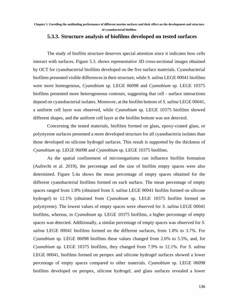
Chapter 5. Unveiling the antifouling performance of different marine surfaces and their effect on the development and structure
of cyanobacterial biofilms
136
5.3.3. Structure analysis of biofilms developed on tested surfaces
The study of biofilm structure deserves special attention since it indicates how cells
interact with surfaces. Figure 5.3. shows representative 3D cross-sectional images obtained
by OCT for cyanobacterial biofilms developed on the five surface materials. Cyanobacterial
biofilms presented visible differences in their structure, while S. salina LEGE 00041 biofilms
were more homogenous, Cyanobium sp. LEGE 06098 and Cyanobium sp. LEGE 10375
biofilms presented more heterogeneous contours, suggesting that cell - surface interactions
depend on cyanobacterial isolates. Moreover, at the biofilm bottom of S. salina LEGE 00041,
a uniform cell layer was observed, while Cyanobium sp. LEGE 10375 biofilms showed
different shapes, and the uniform cell layer at the biofilm bottom was not detected.
Concerning the tested materials, biofilms formed on glass, epoxy-coated glass, or
polystyrene surfaces presented a more developed structure for all cyanobacteria isolates than
those developed on silicone hydrogel surfaces. This result is supported by the thickness of
Cyanobium sp. LEGE 06098 and Cyanobium sp. LEGE 10375 biofilms.
As the spatial confinement of microorganisms can influence biofilm formation
(Aufrecht et al. 2019), the percentage and the size of biofilm empty spaces were also
determined. Figure 5.4a shows the mean percentage of empty spaces obtained for the
different cyanobacterial biofilms formed on each surface. The mean percentage of empty
spaces ranged from 1.8% (obtained from S. salina LEGE 00041 biofilm formed on silicone
hydrogel) to 12.1% (obtained from Cyanobium sp. LEGE 10375 biofilm formed on
polystyrene). The lowest values of empty spaces were observed for S. salina LEGE 00041
biofilms, whereas, in Cyanobium sp. LEGE 10375 biofilms, a higher percentage of empty
spaces was detected. Additionally, a similar percentage of empty spaces was observed for S.
salina LEGE 00041 biofilms formed on the different surfaces, from 1.8% to 3.7%. For
Cyanobium sp. LEGE 06098 biofilms these values changed from 2.6% to 5.5%, and, for
Cyanobium sp. LEGE 10375 biofilms, they changed from 7.9% to 12.1%. For S. salina
LEGE 00041, biofilms formed on perspex and silicone hydrogel surfaces showed a lower
percentage of empty spaces compared to other materials. Cyanobium sp. LEGE 06098
biofilms developed on perspex, silicone hydrogel, and glass surfaces revealed a lower

Chapter 5. Unveiling the antifouling performance of different marine surfaces and their effect on the development and structure
of cyanobacterial biofilms
137
percentage of empty spaces than on polystyrene and epoxy-coated glass surfaces, while
Cyanobium sp. LEGE 10375 biofilms formed on the silicone hydrogel presented a lower
percentage of empty spaces compared to perspex and polystyrene surfaces.
Figure 5.4.b shows the mean size of empty spaces obtained for the different
cyanobacterial biofilms. In addition, a graphical representation of the biofilm empty spaces
for each surface and cyanobacteria strain is presented in Figure 5.5. The mean size of empty
spaces ranged from 24 µm2 (obtained from S. salina LEGE 00041 biofilm formed on
polystyrene) to 119 µm2 (obtained from Cyanobium sp. LEGE 10375 biofilm also formed on
polystyrene). Regardless of the surface, the lower values of mean size of empty spaces were
observed for S. salina LEGE 00041, and the higher values were observed for Cyanobium sp.
LEGE 10375. In addition, S. salina LEGE 00041 biofilms displayed similar values of mean
size of empty spaces (around 30 µm2) for glass, epoxy-coated glass, and silicone hydrogel
surfaces (Figure 5.4.b). In turn, Cyanobium sp. LEGE 06098 and Cyanobium sp. 10375
biofilms developed on silicone hydrogel surfaces showed, on average, a smaller size of empty
spaces compared to glass, epoxy-coated glass, and polystyrene surfaces.
These results suggested that biofilm structure is not only dependent on the surface but
also on the cyanobacterial isolate, as previously shown by Zheng et al. (Zheng et al. 2021).

Chapter 5. Unveiling the antifouling performance of different marine surfaces and their effect on the development and structure
of cyanobacterial biofilms
138
Figure 5.3. Representative 3D OCT images obtained for S. salina LEGE 00041, Cyanobium sp. LEGE 06098,
and Cyanobium sp. LEGE 10375 biofilms formed on perspex, silicone hydrogel, polystyrene, glass, and
epoxy-coated glass surfaces after 49 days. The color scale shows the range of biofilm thickness.

Chapter 5. Unveiling the antifouling performance of different marine surfaces and their effect on the development and structure
of cyanobacterial biofilms
139
Figure 5.4. Mean percentage (a) and size (b) of empty spaces obtained for S. salina LEGE 00041,
Cyanobium sp. LEGE 06098, and Cyanobium sp. LEGE 10375 biofilms developed on perspex
■, silicone hydrogel coating ■, polystyrene ■, glass ■, and epoxy-coated glass ■ surfaces after
49 days. For each cyanobacterial isolate, different lowercase letters indicate significant
differences between surfaces with a confidence level greater than 95% (p < 0.05).

Chapter 5. Unveiling the antifouling performance of different marine surfaces and their effect on the development and structure
of cyanobacterial biofilms
140
5.4. Discussion
In this study, the long-term performance of five different material surface
characteristics in inhibiting biofilm formation by coccoid cyanobacteria was evaluated under
hydrodynamic conditions found in marine environments, through an analysis of biofilm cell
number, biofilm wet weight, chlorophyll a content, and biofilm thickness and architecture.
Since marine biofilm formation is influenced by several factors, including the surface
properties and microfouler type (Faria et al. 2020, Telegdi et al. 2016), an extensive
characterization of the surface materials and microorganisms was also performed in order to
obtain more insight into the tested surfaces and their interactions with cyanobacterial isolates.
It is known that surface properties, including hydrophobicity and roughness,
influence cell adhesion and subsequent biofilm formation (Spengler et al. 2019, Zhang et al.
2015). In this study, the results from thermodynamic analysis classified glass as the most
Figure 5.5. Representative 2D cross-sectional OCT images obtained for S. salina LEGE 00041, Cyanobium sp.
LEGE 06098, and Cyanobium sp. LEGE 10375 biofilms formed on perspex, silicone hydrogel coating,
polystyrene, glass, and epoxy-coated glass surfaces after 49 days. The empty spaces are indicated in orange
(scale bars = 100 µm).

Chapter 5. Unveiling the antifouling performance of different marine surfaces and their effect on the development and structure
of cyanobacterial biofilms
141
hydrophilic material, followed by epoxy-coated glass, perspex, and polystyrene surfaces
(Table 5.1.). Conversely, the silicone hydrogel surface was characterized as hydrophobic.
These results are in accordance with previous studies (Faria et al. 2020, Romeu et al. 2019,
Thukkaram et al. 2014). In addition, the calculation of the free energy of adhesion indicates
that cyanobacterial cell adhesion to glass and epoxy-coated glass is thermodynamically less
favorable than to the silicone hydrogel coating, perspex, and polystyrene surfaces (Table
5.3.). Concerning AFM analysis, results revealed that glass and perspex are the smoothest
tested materials, displaying a lower Ra value, followed by polystyrene and epoxy-coated glass
surfaces (Table 5.1.). In opposition, the silicone hydrogel surface showed higher roughness,
as found in a previous study (Faria et al. 2020). According to Dantas et al. (Dantas et al.
2016), a reduction in surface roughness is directly related to a decrease in bacterial adhesion.
Thus, both hydrophobicity and roughness results suggest that glass and epoxy-coated glass
surfaces may be more efficient materials in controlling cyanobacteria biofilm formation than
silicone hydrogel surfaces. Nevertheless, in general, the analysis of biofilm parameters
indicated that cyanobacterial biofilms formed on glass and epoxy-coated glass surfaces were
more developed than those formed on the silicone hydrogel coated surfaces (Figure 5.2.(1–
2a), (c), and (2d)). In fact, this adds to the debate on whether surfaces displaying higher
degrees of hydrophobicity and roughness favor bacterial adhesion (Cerca et al. 2005, De-la-
Pinta et al. 2019, Ozkan and Berberoglu 2013). It has been shown, in particular for
cyanobacterial adhesion, that it is not always possible to correlate surface hydrophobicity and
roughness with cell attachment (Faria et al. 2020, Irving and Allen 2011, Mazumder et al.
2010, Talluri et al. 2020). Furthermore, there is evidence that biofilm formation induces
changes in the substratum surfaces since already attached cells modify surface properties
(De-la-Pinta et al. 2019, Mazumder et al. 2010, Moreira et al. 2017, Ozkan and Berberoglu
2013).
The discrepancy observed between the material characterization and biofilm analysis
may be explained by the formation of conditioning films resulting from the adsorption of
molecules on the substrates that change the adhesion conditions for microorganisms (Hwang
et al. 2012, Lorite et al. 2011). The nature of formed films depends on the material type,
surrounding environment, and microorganisms (Lorite et al. 2011, Talluri et al. 2020). It is
known that these conditioning films play an important role in cyanobacterial adhesion and

Chapter 5. Unveiling the antifouling performance of different marine surfaces and their effect on the development and structure
of cyanobacterial biofilms
142
subsequent biofilm formation (Talluri et al. 2020). Therefore, despite surface properties
being of extreme importance, particularly during the adhesion phase (Zhang et al. 2015), our
results suggest that biofilm formation may also be modulated by other factors.
According to Zhang et al. (Zhang et al. 2015), biofilm formation is also influenced
by the physicochemical properties of the microorganisms. Indeed, the thermodynamic
analysis of cyanobacterial cells indicated that S. salina LEGE 00041 is relatively more
hydrophilic than Cyanobium sp. LEGE 06098 and Cyanobium sp. LEGE 10375 (Table 5.2).
Moreover, the free energy of adhesion revealed that there is a tendency for S. salina LEGE
00041 to adhere more to all tested surfaces (Table 5.3.). These results are corroborated by
the biofilm parameter analysis, i.e., number of cells, chlorophyll a content, and biofilm
thickness, which demonstrated that S. salina LEGE 00041 had a lower biofilm-forming
capacity on these surfaces than the other cyanobacteria (Figure 5.2.).
Concerning the performance of surface materials, the biofilm analysis indicated that
cyanobacterial biofilms formed on glass and epoxy glass surfaces showed, on average, a
higher number of S. salina LEGE 00041 and Cyanobium sp. LEGE 06098 cells compared to
perspex, silicone hydrogel, and polystyrene surfaces. Cyanobium sp. LEGE 10375 biofilms
formed on epoxy-coated glass exhibited a higher number of cells than the other surfaces,
whereas the performance of perspex and polystyrene was different between cyanobacterial
isolates. Furthermore, silicone hydrogel surfaces were among the surfaces that exhibited a
low number of adhered cells (Figure 5.2.a). Although these results were dependent on
cyanobacterial isolates, they are supported by the literature. Glass, perspex, and polystyrene
are not considered AF surfaces and are frequently used as positive fouling controls in several
studies (Jain et al. 2007, Lou et al. 2021, Sekar et al. 2004). Likewise, epoxy resin coatings
have not completely emerged in marine applications, especially where high fouling resistance
is needed (Qu and Zhang 2012). Lastly, silicone hydrogel surfaces are among the most
successful AF coatings to prevent marine biofouling (Nurioglu et al. 2015). The commercial
silicone hydrogel coating exerts a dual-mode of action, a ‘nonstick’ ability and a fouling-
release effect, associated with relative higher elasticity. These features may decrease fouling
settlements and cell cohesion interactions (Faria et al. 2020, Selim et al. 2017). Additionally,
this is a third-generation hydrogel-based FRC, which acquires a more hydrophilic behavior
upon contact with water (Tulcidas et al. 2015, Xie et al. 2011). Thus, it can prevent either

Chapter 5. Unveiling the antifouling performance of different marine surfaces and their effect on the development and structure
of cyanobacterial biofilms
143
hydrophobic or hydrophilic interactions, delaying the adsorption of proteins, bacteria, and
subsequent fouling (Banerjee et al. 2011, Hu et al. 2020, Murosaki et al. 2012, Sjollema et
al. 2017).
Our results indicated that S. salina LEGE 00041 biofilms presented, on average, a
similar wet weight for all tested surfaces, while Cyanobium sp. LEGE 06098 and Cyanobium
sp. LEGE 10375 biofilms, formed on epoxy-coated glass and glass surfaces, respectively,
exhibited a lower weight (Figure 5.2.b). Indeed, once adhered, cells express different QS-
related signaling molecules that stimulate or block EPS formation (Nahar et al. 2018), which
may justify the observed differences in wet weight concerning other biofilm parameters.
Regarding the chlorophyll a content, results were consistent with the number of
biofilm cells (Figure 5.2.a,c), which would be expected since several authors have proposed
pigment quantification as a good indicator of cyanobacterial biofilm growth (Bartram and
Chorus 1999, Rees and Bartram 2002). On the other hand, S. salina LEGE 00041 biofilms
presented, on average, similar thickness values for all tested surfaces, while Cyanobium sp.
LEGE 06098 biofilms formed on silicone hydrogel surfaces were thinner (supporting the
biofilm cell number and chlorophyll a content) (Figure 5.2.d). Cyanobium sp. LEGE 10375
biofilms developed on glass and silicone hydrogel surfaces showed a lower thickness than
on the remaining surfaces (Figure 5.2.d). Results demonstrated that there was no direct
association between the number of biofilm cells and biofilm thickness. In fact, biofilm
thickness, analogous to biofilm wet weight, is linked to several features of biofilm
architecture, such as density, shape, and porosity, and cannot easily be isolated from
environmental factors (e.g., flow, nutrient conditions, development age of the biofilm,
carbon–nitrogen ratios, and temperature) (Suarez et al. 2019).
Considering that heterogeneous structures may influence the biofilm resistance to
mechanical and chemical challenges, such as fluid shear, detergents, and AF compounds
(Hou et al. 2019), the study of biofilm architecture deserves special attention.
Microorganisms often live in heterogeneous microenvironments with conditions that
modulate local nutrient transport and chemical gradients, creating specialized niches for
them. The spatial confinement of microorganisms can influence emergent phenomena,
including QS, intracellular communication, and biofilm. In fact, these microenvironment
factors shape the structure of microbial communities and contribute to their phenotype

Chapter 5. Unveiling the antifouling performance of different marine surfaces and their effect on the development and structure
of cyanobacterial biofilms
144
diversity and synergism (Aufrecht et al. 2019, Boedicker et al. 2009, Kim et al. 2008). Our
OCT analysis demonstrated that cyanobacterial biofilms presented visible differences in their
structure; while S. salina LEGE 00041 biofilms were more homogenous, Cyanobium sp.
LEGE 06098 and Cyanobium sp. LEGE 10375 biofilms presented more heterogeneous
contours (Figure 5.3.). Indeed, biofilms of Cyanobium sp. LEGE 10375 showed a higher
biofilm wet weight and thickness when compared with S. salina LEGE 00041 (Figure
5.2.b,d). Moreover, biofilms formed on the glass, epoxy-coated glass and polystyrene
surfaces presented more developed structures, contrary to silicone hydrogel surfaces (Figure
5.3.). These results are corroborated by the biofilm cell density.
In addition, the analysis of biofilm empty spaces demonstrated that S. salina LEGE
00041 biofilms showed lower percentage and mean size values of empty spaces compared to
Cyanobium sp. LEGE 10375 (Figure 5.4.). S. salina LEGE 00041 and Cyanobium sp. LEGE
06098 biofilms formed on silicone hydrogel surfaces showed, on average, a lower percentage
of empty spaces compared to polystyrene and epoxy-glass surfaces (Figure 5.4.a).
Furthermore, Cyanobium sp. LEGE 10375 biofilms formed on silicone hydrogel surfaces
showed, on average, a lower percentage of empty spaces compared to polystyrene (Figure
5.4.a). While there were no significant differences in the size of empty spaces of S. salina
LEGE 00041 biofilms formed on the glass, epoxy-coated glass, and silicone hydrogel
surfaces, Cyanobium sp. LEGE 06098 and Cyanobium sp. LEGE 10375 biofilms developed
on silicone hydrogel surfaces showed, on average, a lower size of empty spaces compared to
polystyrene, glass, and epoxy-coated glass surfaces (Figure 5.4.b). Overall, results from OCT
analysis suggest that the biofilms formed on silicone hydrogel surfaces show, in general, a
less developed and heterogeneous structure compared to polystyrene, glass, and epoxy-
coated glass surfaces, while also presenting a low percentage and size of empty spaces.
However, these results should be interpreted with caution as they vary among cyanobacterial
isolates. According to Aufrecht et al. (Aufrecht et al. 2019), the spatial distribution of
microorganisms in their heterogenous network is determined by the EPS production ability
and biofilm expansion over the biofilm formation process. In fact, biofilms developed on
silicone hydrogel surfaces exhibited lower biofilm thickness, which may be related to their
lower cellular growth and expansion.

Chapter 5. Unveiling the antifouling performance of different marine surfaces and their effect on the development and structure
of cyanobacterial biofilms
145
5.5. Conclusions
Our results demonstrated high AF performance of the silicone hydrogel coating in
inhibiting or delaying cyanobacterial biofilm formation. Additionally, the comprehensive
analysis carried out in this study revealed that the surface material properties, together with
the features of the fouling microorganisms, play a considerable role in marine biofouling.
5.6. References
Akinfijevs, T., A. Janvaevskis and E. Lavendelis (2007). A Brief Survey of Ship Hull
Cleaning Devices. RTU. Riga, Latvia. 24: 133-146.
Angelova, A. G., G. A. Ellis, H. W. Wijesekera and G. J. Vora (2019). "Microbial
Composition and Variability of Natural Marine Planktonic and Biofouling Communities
From the Bay of Bengal." Frontiers in Microbiology 10(2738).
Arrhenius, A., T. Backhaus, A. Hilvarsson, I. Wendt, A. Zgrundo and H. Blanck
(2014). "A novel bioassay for evaluating the efficacy of biocides to inhibit settling and early
establishment of marine biofilms." Mar Pollut Bull 87(1-2): 292-299.
Aufrecht, J. A., J. D. Fowlkes, A. N. Bible, J. Morrell-Falvey, M. J. Doktycz and S.
T. Retterer (2019). "Pore-scale hydrodynamics influence the spatial evolution of bacterial
biofilms in a microfluidic porous network." PLoS One 14(6): e0218316.
Bakker, D., A. Van der Plaats, G. Verkerke, H. Busscher and H. Van der Mei (2003).
"Comparison of velocity profiles for different flow chamber designs used in studies of
microbial adhesion to surfaces." Applied and environmental microbiology 69(10): 6280-
6287.
Banerjee, I., R. C. Pangule and R. S. Kane (2011). "Antifouling coatings: recent
developments in the design of surfaces that prevent fouling by proteins, bacteria, and marine
organisms." Advanced materials 23(6): 690-718.

Chapter 5. Unveiling the antifouling performance of different marine surfaces and their effect on the development and structure
of cyanobacterial biofilms
146
Bartram, J. and I. Chorus (1999). Toxic cyanobacteria in water: a guide to their public
health consequences, monitoring and management. Boca Raton, FL, USA, CRC Press.
Bayoudh, S., A. Othmane, F. Bettaieb, A. Bakhrouf, H. B. Ouada and L. Ponsonnet
(2006). "Quantification of the adhesion free energy between bacteria and hydrophobic and
hydrophilic substrata." Materials Science and Engineering: C 26(2-3): 300-305.
Blain, S., J. Guillou, P. Tréguer, P. Woerther, L. Delauney, E. Follenfant, O. Gontier,
M. Hamon, B. Leildé, A. Masson, C. Tartu and R. Vuillemin (2004). "High frequency
monitoring of the coastal marine environment using the MAREL buoy." Journal of
Environmental Monitoring 6(6): 569-575.
Boedicker, J. Q., M. E. Vincent and R. F. Ismagilov (2009). "Microfluidic
confinement of single cells of bacteria in small volumes initiates high-density behavior of
quorum sensing and growth and reveals its variability." Angew Chem Int Ed Engl 48(32):
5908-5911.
Busscher, H. J., A. H. Weerkamp, H. C. van der Mei, A. W. van Pelt, H. P. de Jong
and J. Arends (1984). "Measurement of the surface free energy of bacterial cell surfaces and
its relevance for adhesion." Applied and environmental microbiology 48(5): 980-983.
Cerca, N., G. B. Pier, M. Vilanova, R. Oliveira and J. Azeredo (2005). "Quantitative
analysis of adhesion and biofilm formation on hydrophilic and hydrophobic surfaces of
clinical isolates of Staphylococcus epidermidis." Res Microbiol 156(4): 506-514.
Crawford, R. J., H. K. Webb, V. K. Truong, J. Hasan and E. P. Ivanova (2012).
"Surface topographical factors influencing bacterial attachment." Advances in Colloid and
Interface Science 179-182: 142-149.
Dantas, L. C. d. M., J. P. da Silva-Neto, T. S. Dantas, L. Z. Naves, F. D. das Neves
and A. S. da Mota (2016). "Bacterial Adhesion and Surface Roughness for Different Clinical
Techniques for Acrylic Polymethyl Methacrylate." International journal of dentistry:
8685796-8685796.
De-la-Pinta, I., M. Cobos, J. Ibarretxe, E. Montoya, E. Eraso, T. Guraya and G.
Quindós (2019). "Effect of biomaterials hydrophobicity and roughness on biofilm
development." Journal of Materials Science: Materials in Medicine 30(7): 77.
de Carvalho, C. C. C. R. (2018). "Marine Biofilms: A Successful Microbial Strategy
With Economic Implications." Frontiers in Marine Science 5(126).

Chapter 5. Unveiling the antifouling performance of different marine surfaces and their effect on the development and structure
of cyanobacterial biofilms
147
Donlan, R. M. (2002). "Biofilms: microbial life on surfaces." Emerg Infect Dis 8(9):
881-890.
Faria, S. I., R. Teixeira-Santos, L. C. Gomes, E. R. Silva, J. Morais, V. Vasconcelos
and F. J. M. Mergulhão (2020). "Experimental Assessment of the Performance of Two
Marine Coatings to Curb Biofilm Formation of Microfoulers." Coatings 10(9).
Faria, S. I., R. Teixeira-Santos, M. J. Romeu, J. Morais, V. Vasconcelos and F. J.
Mergulhão (2020). "The Relative Importance of Shear Forces and Surface Hydrophobicity
on Biofilm Formation by Coccoid Cyanobacteria." Polymers 12(3).
Gomes, L. C., J. Deschamps, R. Briandet and F. J. Mergulhao (2018). "Impact of
modified diamond-like carbon coatings on the spatial organization and disinfection of mixed-
biofilms composed of Escherichia coli and Pantoea agglomerans industrial isolates." Int J
Food Microbiol 277: 74-82.
Hou, J., C. Wang, R. T. Rozenbaum, N. Gusnaniar, E. D. de Jong, W. Woudstra, G.
I. Geertsema-Doornbusch, J. Atema-Smit, J. Sjollema, Y. Ren, H. J. Busscher and H. C.
van der Mei (2019). "Bacterial Density and Biofilm Structure Determined by Optical
Coherence Tomography." Scientific Reports 9(1): 9794.
Hu, P., Q. Xie, C. Ma and G. Zhang (2020). "Silicone-based fouling-release coatings
for marine antifouling." Langmuir 36(9): 2170-2183.
Hwang, G., S. Kang, M. G. El-Din and Y. Liu (2012). "Impact of conditioning films
on the initial adhesion of Burkholderia cepacia." Colloids Surf B Biointerfaces 91: 181-188.
Irving, T. E. and D. G. Allen (2011). "Species and material considerations in the
formation and development of microalgal biofilms." Appl Microbiol Biotechnol 92(2): 283-
294.
Jain, A., K. K. Nishad and N. B. Bhosle (2007). "Effects of DNP on the cell surface
properties of marine bacteria and its implication for adhesion to surfaces." Biofouling 23(3):
171-177.
Janczuk, B., E. Chibowski, J. M. Bruque, M. L. Kerkeb and F. G. Caballero (1993).
"On the Consistency of Surface Free Energy Components as Calculated from Contact Angles
of Different Liquids: An Application to the Cholesterol Surface." Journal of Colloid and
Interface Science 159(2): 421-428.

Chapter 5. Unveiling the antifouling performance of different marine surfaces and their effect on the development and structure
of cyanobacterial biofilms
148
Jindal, S., S. Anand, K. Huang, J. Goddard, L. Metzger and J. Amamcharla (2016).
"Evaluation of modified stainless steel surfaces targeted to reduce biofilm formation by
common milk sporeformers." J Dairy Sci 99(12): 9502-9513.
Kanematsu, H. and D. M. Barry (2020). "Biofilm on Materials’ Surfaces in Marine
Environments." Monitoring Artificial Materials and Microbes in Marine Ecosystems:
Interactions and Assessment Methods 2: 177-187.
Kim, H. J., J. Q. Boedicker, J. W. Choi and R. F. Ismagilov (2008). "Defined spatial
structure stabilizes a synthetic multispecies bacterial community." Proceedings of the
National Academy of Sciences 105(47): 18188.
Kotai, J. (1972). Instructions for Preparation of Modified Nutrient Solution Z8 for
AlgaeNorwegian. Institute for Water Research. Blindern, Oslo, Norway.
Lacoursière-Roussel, A., D. G. Bock, M. E. Cristescu, F. Guichard and C. W.
McKindsey (2016). "Effect of shipping traffic on biofouling invasion success at population
and community levels." Biological Invasions 18(12): 3681-3695.
Lorite, G. S., C. M. Rodrigues, A. A. de Souza, C. Kranz, B. Mizaikoff and M. A.
Cotta (2011). "The role of conditioning film formation and surface chemical changes on
Xylella fastidiosa adhesion and biofilm evolution." Journal of Colloid and Interface Science
359(1): 289-295.
Lou, T., X. Bai, X. He and C. Yuan (2021). "Antifouling performance analysis of
peptide-modified glass microstructural surfaces." Applied Surface Science 541: 148384.
Ma, Y., X. Cao, X. Feng, Y. Ma and H. Zou (2007). "Fabrication of super-
hydrophobic film from PMMA with intrinsic water contact angle below 90°." Polymer
48(26): 7455-7460.
Mazumder, S., J. O. Falkinham, A. M. Dietrich and I. K. Puri (2010). "Role of
hydrophobicity in bacterial adherence to carbon nanostructures and biofilm formation."
Biofouling 26(3): 333-339.
Meireles, A., R. Fulgêncio, I. Machado, F. Mergulhão, L. Melo and M. Simões
(2017). "Characterization of the heterotrophic bacteria from a minimally processed
vegetables plant." LWT - Food Science and Technology 85: 293-300.

Chapter 5. Unveiling the antifouling performance of different marine surfaces and their effect on the development and structure
of cyanobacterial biofilms
149
Moreira, J., L. Gomes, K. Whitehead, S. Lynch, L. Tetlow and F. Mergulhão (2017).
"Effect of surface conditioning with cellular extracts on Escherichia coli adhesion and initial
biofilm formation." Food and Bioproducts Processing 104: 1-12.
Murosaki, T., N. Ahmed and J. P. Gong (2012). "Antifouling properties of
hydrogels." Science and Technology of Advanced Materials 12.
Nahar, S., M. F. R. Mizan, A. J.-w. Ha and S.-D. Ha (2018). "Advances and Future
Prospects of Enzyme-Based Biofilm Prevention Approaches in the Food Industry."
Comprehensive Reviews in Food Science and Food Safety 17(6): 1484-1502.
Neves, A. R., J. R. Almeida, F. Carvalhal, A. Câmara, S. Pereira, J. Antunes, V.
Vasconcelos, M. Pinto, E. R. Silva, E. Sousa and M. Correia-da-Silva (2020). "Overcoming
environmental problems of biocides: Synthetic bile acid derivatives as a sustainable
alternative." Ecotoxicology and Environmental Safety 187: 109812.
Nurioglu, A. G., A. C. C. Esteves and G. de With (2015). "Non-toxic, non-biocide-
release antifouling coatings based on molecular structure design for marine applications."
Journal of Materials Chemistry B 3(32): 6547-6570.
Otsu, N. (1979). "A Threshold Selection Method from Gray-Level Histograms."
IEEE Transactions on Systems, Man, and Cybernetics 9(1): 62-66.
Ozkan, A. and H. Berberoglu (2013). "Cell to substratum and cell to cell interactions
of microalgae." Colloids and Surfaces B: Biointerfaces 112: 302-309.
Qu, Y.-Y. and S.-F. Zhang (2012). "Preparation and characterization of novel
waterborne antifouling coating." Journal of Coatings Technology and Research 9(6): 667-
674.
Rajeev, M., T. J. Sushmitha, K. G. Prasath, S. R. Toleti and S. K. Pandian (2020).
"Systematic assessment of chlorine tolerance mechanism in a potent biofilm-forming marine
bacterium Halomonas boliviensis." International Biodeterioration & Biodegradation 151:
104967.
Ramos, V., J. Morais, R. Castelo-Branco, Â. Pinheiro, J. Martins, A. Regueiras, A.
L. Pereira, V. R. Lopes, B. Frazão, D. Gomes, C. Moreira, M. S. Costa, S. Brûle, S. Faustino,
R. Martins, M. Saker, J. Osswald, P. N. Leão and V. M. Vasconcelos (2018). "Cyanobacterial
diversity held in microbial biological resource centers as a biotechnological asset: the case

Chapter 5. Unveiling the antifouling performance of different marine surfaces and their effect on the development and structure
of cyanobacterial biofilms
150
study of the newly established LEGE culture collection." Journal of Applied Phycology
30(3): 1437-1451.
Rees, G. and J. Bartram (2002). Monitoring bathing waters: a practical guide to the
design and implementation of assessments and monitoring programmes. Boca Raton, FL,
USA, CRC Press.
Romeu, M. J., P. Alves, J. Morais, J. M. Miranda, E. D. de Jong, J. Sjollema, V.
Ramos, V. Vasconcelos and F. J. M. Mergulhão (2019). "Biofilm formation behaviour of
marine filamentous cyanobacterial strains in controlled hydrodynamic conditions." Environ
Microbiol 21(11): 4411-4424.
Schultz, M. P., J. A. Bendick, E. R. Holm and W. M. Hertel (2011). "Economic
impact of biofouling on a naval surface ship." Biofouling 27(1): 87-98.
Sekar, R., V. P. Venugopalan, K. K. Satpathy, K. V. K. Nair and V. N. R. Rao (2004).
"Laboratory studies on adhesion of microalgae to hard substrates." Hydrobiologia 512(1):
109-116.
Selim, M. S., M. A. Shenashen, S. A. El-Safty, S. A. Higazy, M. M. Selim, H. Isago
and A. Elmarakbi (2017). "Recent progress in marine foul-release polymeric nanocomposite
coatings." Progress in Materials Science 87: 1-32.
Sjollema, J., H. Keul, H. van der Mei, R. Dijkstra, M. Rustema-Abbing, J. de Vries,
T. Loontjens, T. Dirks and H. Busscher (2017). "A Trifunctional, Modular Biomaterial
Coating: Nonadhesive to Bacteria, Chlorhexidine-Releasing and Tissue-Integrating."
Macromolecular Bioscience 17(4): 1600336.
Spengler, C., F. Nolle, J. Mischo, T. Faidt, S. Grandthyll, N. Thewes, M. Koch, F.
Müller, M. Bischoff, M. A. Klatt and K. Jacobs (2019). "Strength of bacterial adhesion on
nanostructured surfaces quantified by substrate morphometry." Nanoscale 11(42): 19713-
19722.
Suarez, C., M. Piculell, O. Modin, S. Langenheder, F. Persson and M. Hermansson
(2019). "Thickness determines microbial community structure and function in nitrifying
biofilms via deterministic assembly." Scientific Reports 9(1): 5110.
Talluri, S. N. L., R. M. Winter and D. R. Salem (2020). "Conditioning film formation
and its influence on the initial adhesion and biofilm formation by a cyanobacterium on
photobioreactor materials." Biofouling 36(2): 183-199.

Chapter 5. Unveiling the antifouling performance of different marine surfaces and their effect on the development and structure
of cyanobacterial biofilms
151
Taylor, D. A. (1996). Introduction to Marine Engineering. Oxford, UK, Butterworth-
Heinemann.
Telegdi, J., L. Trif and L. Románszki (2016). 5 - Smart anti-biofouling composite
coatings for naval applications. Smart Composite Coatings and Membranes. M. F.
Montemor. Sawston, Cambridge, Woodhead Publishing: 123-155.
Thukkaram, M., S. Sitaram, S. k. Kannaiyan and G. Subbiahdoss (2014).
"Antibacterial Efficacy of Iron-Oxide Nanoparticles against Biofilms on Different
Biomaterial Surfaces." International Journal of Biomaterials 2014: 716080.
Tian, L., Y. Yin, H. Jin, W. Bing, E. Jin, J. Zhao and L. Ren (2020). "Novel marine
antifouling coatings inspired by corals." Materials Today Chemistry 17: 100294.
Tulcidas, A. V., R. Bayón, A. Igartua, J. C. M. Bordado and E. R. Silva (2015).
"Friction reduction on recent non-releasing biocidal coatings by a newly designed friction
test rig." Tribology International 91: 140-150.
Turner, A. (2020). "Foamed Polystyrene in the Marine Environment: Sources,
Additives, Transport, Behavior, and Impacts." Environmental Science & Technology 54(17):
10411-10420.
Van Oss, C. J. (1994). Interfacial forces in aqueous media. Marcel Dekker Inc. New
York, USA.
Wang, H., M. Sodagari, Y. Chen, X. He, B.-m. Z. Newby and L.-K. Ju (2011). "Initial
bacterial attachment in slow flowing systems: Effects of cell and substrate surface
properties." Colloids and Surfaces B: Biointerfaces 87(2): 415-422.
Xie, L., F. Hong, C. He, C. Ma, J. Liu, G. Zhang and C. Wu (2011). "Coatings with
a self-generating hydrogel surface for antifouling." Polymer 52(17): 3738-3744.
Zecher, K., V. P. Aitha, K. Heuer, H. Ahlers, K. Roland, M. Fiedel and B. Philipp
(2018). "A multi-step approach for testing non-toxic amphiphilic antifouling coatings against
marine microfouling at different levels of biological complexity." J Microbiol Methods 146:
104-114.
Zhang, X., Q. Zhang, T. Yan, Z. Jiang, X. Zhang and Y. Y. Zuo (2015).
"Quantitatively predicting bacterial adhesion using surface free energy determined with a
spectrophotometric method." Environmental science & technology 49(10): 6164-6171.

Chapter 5. Unveiling the antifouling performance of different marine surfaces and their effect on the development and structure
of cyanobacterial biofilms
152
Zheng, S., M. Bawazir, A. Dhall, H.-E. Kim, L. He, J. Heo and G. Hwang (2021).
"Implication of Surface Properties, Bacterial Motility, and Hydrodynamic Conditions on
Bacterial Surface Sensing and Their Initial Adhesion." Frontiers in Bioengineering and
Biotechnology 9(82).

153
6. 6. Developing new marine antifouling surfaces: learning
from single-strain laboratory testsd
Abstract
The development of AF technology for marine environments is an area of intense
research given the severe economic and ecological effects of marine biofouling. Preliminary
data from in vitro assays is frequently used to screen the performance of AF coatings. It is
intuitive that the microbial composition plays a major role in surface colonization. The
rationale behind this study is to investigate whether using a mixed population for the in vitro
tests yields substantially different results than using single-strains during initial screening. A
polymeric coating was tested against single- and dual-species cultures of two common
microfouler organisms for 49 days. A bacterium (Pseudoaltermonas tunicata) and a
cyanobacterium (Cyanobium sp. LEGE 10375) were used in this chapter. Linear regression
analysis revealed that Cyanobium sp. biofilms were significantly associated with a higher
number of cells, wet weight, thickness, and biovolume compared to dual-species biofilms. P.
tunicata alone had a biofilm growth kinetics similar to dual-species biofilms, although the P.
tunicata - Cyanobium sp. mixture developed less dense and thinner biofilms compared to
d The content of this chapter was adapted from the following publication(s):
Faria, S. I., L. C. Gomes, R. Teixeira-Santos, J. Morais, V. Vasconcelos and F. J. Mergulhão (2021).
"Developing New Marine Antifouling Surfaces: Learning from Single-Strain Laboratory Tests." Coatings
11(1): 90.

Chapter 6. Developing new marine antifouling surfaces: learning from single-strain laboratory tests
154
both single-species biofilms. Cyanobium sp. LEGE 10375 biofilms provided the worst-case
scenario, i.e., the conditions that caused higher biofilm amounts on the surface material under
test. Therefore, it is likely that assessing the AF performance of new coatings using the most
stringent conditions may yield more robust results than using a mixed population, as
competition between microfouler organisms may reduce the biofilm formation capacity of
the consortium.
6.1. Introduction
Marine biofouling is a colonization process that starts when a surface material is
immersed in seawater and leads to the development of complex biological communities
called biofilms (Caruso 2020). This undesirable attachment of molecules and fouling
organisms to submerged surfaces causes economic losses to human activities in the sea,
including maritime transport, water desalination, aquaculture, and oil and gas industries (de
Carvalho 2018). Indeed, the main problems of biofouling on marine vessels are related to the
corrosion and increased frictional effects created by the presence of organisms on the vessel
surface (Tian et al. 2020). This can reduce the maneuverability of ships due to their increased
weight and reduced speed, resulting in increased fuel consumption (Brooks and Waldock
2009, Tian et al. 2020). High levels of biofouling activity can also lead to increased frequency
of dry-docking operations and an overall reduction of the integrity of the ship hulls, factors
that have significant financial impacts to the vessel owners (Brooks and Waldock 2009).
Additionally, biofouling communities have the potential to transport invasive non-native
species across geographical niches, which can have disastrous effects on native populations
and communities (Lacoursière-Roussel et al. 2016). Therefore, the need to protect submerged
surfaces from biofouling organisms is of economic and environmental importance.
The scientific community has been focusing its efforts on overcoming these problems
by developing AF coatings (Miller et al. 2020). The most well-consolidated and
commercially established AF coatings are biocide-containing paints (Silva et al. 2019),
typically employing copper or zinc as the active ingredient (Amara et al. 2018, Miller et al.
2020). However, alternative protective AF coatings are being developed taking into account

Chapter 6. Developing new marine antifouling surfaces: learning from single-strain laboratory tests
155
new rigid international regulations and consumer environmental concerns. At present, there
are two major environmental-friendly AF strategies, which are foul release coatings (Faria et
al. 2020a, Silva et al. 2019) and bioinspired coatings that prevent the settlement of organisms
through their surface properties (e.g., micro- or nano-structured “self-cleaning” surfaces) (Li
and Guo 2019, Salta et al. 2010).
For testing the performance of novel AF coatings, these should be first screened under
laboratory conditions that mimic the marine environment since the direct evaluation of
coatings in the ocean would be very expensive due to the large quantities of test products and
prolonged immersion times that are required. Additionally, field tests have their own
drawbacks, like the ecotoxicity of some biocide-release coatings (Zecher et al. 2018).
Several factors may influence the efficacy of an AF coating, including the water
temperature and salinity, available nutrients, hydrodynamics, and organisms that can vary
significantly depending on the sea site (Caruso 2020, Romeu et al. 2019). It is described that
marine biofilms are mainly composed of different species of bacteria and diatoms (de
Carvalho 2018). The most studied biofilm communities are those established in the euphotic
zones of aquatic habitats, comprising photoautotrophic microorganisms, such as diatoms,
green algae, and cyanobacteria, which produce the organic carbon that fuels the life of
heterotrophic microorganisms, such as bacteria (Buhmann et al. 2012). Although diatom-
bacteria co-cultures under photoautotrophic conditions would be more realistic than single
monocultures, there are only a few test systems at a laboratory scale using such mixed
cultures (Buhmann et al. 2012, Zecher et al. 2018). The study of multispecies biofilms may
be limited by the complexity of each community and the lack of knowledge regarding the
identity and abundance of each biofilm resident, which makes it difficult to select the
organisms for the in vitro assays, as well as by some technical limitations associated with
different biofilm setups (Magana et al. 2018). Furthermore, although it is described that the
physiology and function of these complex communities differ from those of the individual
species when examined as monocultures (Burmølle et al. 2014, Røder et al. 2016), it has
recently been proven that increasing culture diversity beyond a threshold has little effect on
interspecies interactions and biomass production (Azevedo et al. 2020, Yu et al. 2019). The
rationale behind the present chapter is whether there are benefits in varying the degree of
complexity of marine cultures when the goal is to get the first indications about the AF

Chapter 6. Developing new marine antifouling surfaces: learning from single-strain laboratory tests
156
materials´ performance. A polymeric coating commonly used to coat the hulls of small
recreational vessels and with known antibiofilm activity (Faria et al. 2020b) was tested
against single mono-species (Pseudoalteromonas tunicata or Cyanobium sp. LEGE 10375)
and dual-species cultures (P. tunicata - Cyanobium sp. LEGE 10375) in order to identify
which culture conditions generate the worst-case scenario, and thus the most appropriate in
vitro experiments for the initial screening of the performance of novel AF marine surfaces.
6.2. Materials and methods
6.2.1. Surface preparation
A glass surface coated with a polymer epoxy resin was tested for single- and dual-
species biofilm formation. Epoxy resins are typically used to coat the hulls of small recreation
vessels (e.g. powerboats, yachts, sailing boats) (Blain et al. 2004, Taylor 1996) since they
contribute to the production of stiffer, stronger, lighter hulls with improved structural stability
and hydrodynamic performance (Hoge and Leach 2016). Furthermore, they can have AF
activity (Faria et al. 2020b). Glass coupons (1 x 1 cm2; Vidraria Lousada, Lda, Lousada,
Portugal) were firstly washed and sterilized as fully described in Faria el al. 2020 (Faria et
al. 2020b), and then coated with 70 µL of a polymer epoxy resin (HB Química company,
Matosinhos, Portugal) by spin coating (Spin150 PolosTM , Paralab, Porto, Portugal) at 6000
rpm for 40 s, with ascends of 1000 rpm. Coated surfaces were dried in two different steps
(12 h at room temperature and 3 h at 60 °C) as previously described (Faria et al. 2020b).
Surfaces were sterilized by immersion in 70% (v/v) ethanol (VWR International S.A.A.,
Fontenay-sous-Bois, France) for 20 min and air-dried inside a flow chamber to maintain
sterility. The initial weight of each coupon was determined before experiments.
6.2.2. Marine organisms and culture conditions
One marine bacteria - Pseudoalteromonas tunicata DSM 14096 - and one
cyanobacteria - Cyanobium sp. LEGE 10375 - were the microorganisms chosen for this study

Chapter 6. Developing new marine antifouling surfaces: learning from single-strain laboratory tests
157
since they are recognized as early colonizers in the fouling process (de Carvalho 2018).
Additionally, they are typically present in the euphotic zone of a marine ecosystem, where
the best characterized multispecies biofilms are developed (Buhmann et al. 2012).
P. tunicata (DSMZ, Braunschweig, Germany) was stored at -80 °C in 20% (v/v)
glycerol (Fisher Scientific, Geel, Belgium). Before the experiments, bacteria were
subcultured twice on the complex marine medium Våatanen Nine Salt Solution (VNSS)
(Holmström et al. 1998) supplemented with 15 g/L agar (VWR International S.A.A.,
Fontenay-sous-Bois, France) for 24 h at 25 °C. An overnight culture was then prepared by
transferring colonies from a VNSS agar plate to 150 mL of VNSS medium and incubating at
25 °C with agitation.
Cyanobium sp. LEGE 10375 was isolated from an intertidal zone, on a marine sponge,
at São Bartolomeu do Mar beach (Esposende, Braga, Portugal) (41.57378 N 8.798556 W).
This cyanobacteria was obtained from LEGE-CC, located at the CIIMAR, Matosinhos,
Portugal (Ramos et al. 2018). Cyanobacteria were growth in 750 mL of Z8 medium
supplemented with 25 g/L of synthetic sea salts (Tropic Marin, Montague, MA, USA) and
vitamin B12 (Sigma Aldrich, Merck, Saint Louis, MO, USA), at 25 °C under 14 h light (10
- 30 mol photons/m2 s, λ = 380 - 700 nm)/10 h dark cycles (Faria et al. 2020b).
6.2.3. Single- and dual-species biofilm formation
The ability of P. tunicata and Cyanobium sp. alone and in co-culture to colonize the
coated surface was monitored for 7 weeks (49 days) using 12-well microplates (VWR
International, Carnaxide, Portugal) under controlled hydrodynamic conditions. Biofilm
development was followed for 49 days because this period corresponds to approximately half
of the minimal economically viable interval accepted for the maintenance of underwater
systems (Blain et al. 2004) and hull cleaning (Schultz et al. 2011). Biofilms were grown in
an orbital shaker with a 25 mm diameter (Agitorb 200ICP, Norconcessus, Ermesinde,
Portugal) at 185 rpm, which corresponds to an average shear rate of 40 s-1 and a maximum
of 120 s-1 (Faria et al. 2020a, Romeu et al. 2019), comprising the shear rate value of 50 s-1
estimated for a ship in a harbour (Bakker et al. 2003).

Chapter 6. Developing new marine antifouling surfaces: learning from single-strain laboratory tests
158
P. tunicata DSM 14096 and Cyanobium sp. LEGE 10375 suspensions at a final
concentration of 1 x 10⁸ CFU/mL were prepared in VNSS medium from the corresponding
overnight cultures. For Cyanobium sp., a 1 x 10⁸ CFU/mL cell suspension was also prepared
in Z8 medium as a growth control (Figure 6.1.) since this is the recommended medium for
the cultivation of these species of cyanobacteria (Faria et al. 2020b). For dual-species
biofilms of P. tunicata and Cyanobium sp., both cell suspensions were mixed at a 1:1 ratio
(Gomes et al. 2018) in order to obtain a final concentration of 1 x 10⁸ CFU/mL. The coated
glass coupons were first fixed to the plate wells using double-sided adhesive tape UV-
sterilized for 30 min (Faria et al. 2020b). Then, 3 mL of each single- and dual-species culture
were added to the wells, and the microplates were incubated with the alternate light cycles
of 14 h light/10 h dark cycles, at 25 °C for 49 days. Additionally, 3 mL of VNSS medium
were added to the wells containing coated glass coupons in order to control the surfaces´
sterility throughout the 49 days of the experiment (negative control). Two coupons for each
experimental condition were removed every 7 days for biofilm analysis. During the
incubation period, the culture medium was carefully replaced twice a week. Three
independent biofilm formation assays, with two technical replicates each, were performed.
0,00E+00
5,00E+08
1,00E+09
1,50E+09
2,00E+09
VNSS medium Z8 medium
Bio
film
cell
s (c
ell
s/cm
2)
Figure 6.1. Number of Cyanobium sp. cells growing in
Våatanen nine salt solution (VNSS) and Z8 medium attached
on polymer epoxy resin after 49 days of incubation.

Chapter 6. Developing new marine antifouling surfaces: learning from single-strain laboratory tests
159
6.2.4. Biofilm analysis
The removed coupons were gently washed with 3 mL of 0.85 % (v/v) sterile saline
solution to remove non-attached microorganisms (Faria et al. 2020a) and analyzed regarding
the number of biofilm cells, biofilm wet weight, and biofilm thickness. The biofilm
architecture was also evaluated through CLSM after 21, 35 and 49 days.
6.2.4.1. Cell density and wet weight
The cell counting and biofilm wet weight determination was performed as was
described in chapter 4, section 4.2.5.1.
6.2.4.2. Thickness
The biofilm thickness analysis was performed as was described in chapter 4, section
4.2.5.2.
6.2.4.3. CLSM
Single- and dual-species biofilms of P. tunicata and Cyanobium sp. were imaged
using a Leica TCS SP5 II Confocal Laser Scanning Microscope (Leica Microsystems,
Wetzlar, Germany) after 21, 35 and 49 days of biofilm formation. Biofilm samples were
counter-stained with Syto9 (Thermo Fisher Scientific, Waltham, MA, USA), a green cell-
permeant nucleic acid marker, for 10 min at room temperature, and then scanned at 40x
magnitude with a HCX PL APO CS 40x/1.10 CORR water objective lens at an excitation
wavelength of 488 nm (argon laser). The emitted fluorescence was recorded within the range
of 460 to 575 nm. A minimum of five stacks of horizontal plane images (512 x 512 pixels,
corresponding to 387.5 µm x 387.5 µm) with a 𝑧-step of 1 µm were acquired for each biofilm
sample.
3D projections of biofilm structures were reconstructed from the CLSM acquisitions
using the blend mode of the “Easy 3D” function of the IMARIS 9.1 software (Bitplane,
Zurich, Switzerland). Biofilm biovolume (µm3/µm2) was extracted from confocal image

Chapter 6. Developing new marine antifouling surfaces: learning from single-strain laboratory tests
160
series with the plug-in COMSTAT2 run in ImageJ 1.48v software, as previously described
(Heydorn et al. 2000). Biovolume represents the total amount of biofilm (μm3) in the
substratum area of the image stack (μm2).
6.2.5. Statistical analysis
Descriptive statistics were used to compute mean and standard deviation for all
parameters evaluated in single- and dual-species biofilms: the total cell number (Figure
6.1.A), wet weight (Figure 6.1.B), thickness (Figure 6.1C) and biovolume (Figure 6.2.A).
Since the variable distribution was normal, one-way ANOVA analysis was used to compare
biofilm formation between single- (P. tunicata or Cyanobium sp.) and dual-species biofilms
(P. tunicata - Cyanobium sp.) on each experimental day. For each time point, letters were
assigned in alphabetic order from the highest to the lowest value (from a to c) as long as
statistically significant differences exist between the biofilms with a confidence level greater
than 95% (p < 0.05).
Linear regression models (LRM) were applied between the biofilm cells, wet weight,
thickness and biovolume, and single- and dual-species biofilms (Figure 6.1.D, E and F, and
Figure 6.2B). Models were adjusted for incubation days. For all LRMs, dual-species biofilms
were used as the reference condition. Results were presented as beta estimates (β) and the
corresponding 95% confidence intervals (95% CI).
Data analysis was performed using the IBM SPSS Statistics version 24.0 for
Windows (IBM SPSS, Inc., Chicago, IL, USA).
6.3. Results
In this study, the ability of two common microfouler organisms, a marine bacterium
and a cyanobacterium (P. tunicata and Cyanobium sp.) to develop single- and dual-species
biofilms in epoxy-coated surfaces was analysed. The importance of conducting multispecies
biofilm assays for initial screening of the AF potential of novel surface coatings for marine
settings was assessed.

Chapter 6. Developing new marine antifouling surfaces: learning from single-strain laboratory tests
161
Figure 6.2 presents the number of biofilm cells, wet weight and thickness determined
for single- (P. tunicata or Cyanobium sp. LEGE 10375) and dual-species biofilms (P.
tunicata - Cyanobium sp. LEGE 10375) grown for 49 days under hydrodynamic conditions
that mimic the aquatic environment.
In general, the cell number of Cyanobium sp. LEGE 10375 biofilms was higher than
for P. tunicata and dual-species biofilms (on average 33% and 27%, respectively), with
significant statistical differences in almost all sampling points (p < 0.05, Figure 6.2.A). While
single-species biofilms of P. tunicata and dual-species biofilms grew markedly between days
7 and 14 (approximately 1 log cells/cm2), tending towards similar growth kinetics and
stabilization by the end of the experiment, the cell number in cyanobacterial biofilms
increased exponentially until day 21, reaching 9.4 x 10⁸ ± 3.3 x 10⁵ cells/cm2, which is 52%
and 28% higher than the value for P. tunicata biofilms and dual-species biofilms,
respectively, at the same time point (p < 0.05).
The wet weight of P. tunicata single-species biofilms and mixed biofilms increased
to an average value of 76 mg on day 21 (Figure 6.2.B). This biofilm parameter remained
practically constant in the following two weeks for both types of biofilm. However, its
behavior changed with the gradual increase of the wet weight of dual-species biofilms until
day 49, and the abrupt reduction of the weight of P. tunicata biofilms (to about half at the
end of the experiment). With regard to cyanobacterial biofilms, their wet weight increased
linearly until day 35, exceeding by 26% the wet weight of bacterial and bacteria -
cyanobacteria biofilms (p < 0.01).
Concerning biofilm thickness, there was a linear increase during the 7-week assay in
the three types of analyzed biofilms, P. tunicata and Cyanobium sp. single-species biofilms
and P. tunicata - Cyanobium sp. biofilms (Figure 6.2.C). Moreover, cyanobacterial biofilms
were significantly thicker than P. tunicata biofilms (on average 31%, p < 0.05 for five out of
seven time points) and dual-species biofilms (on average 42%, p < 0.05 for all time points).
This difference between Cyanobium sp. biofilms and both P. tunicata single- and dual-
species biofilms was particularly noticeable from day 28 onwards when the thickness of
cyanobacterial biofilms increased from about 100 µm to the maximum value of 260 µm at
day 49 (37% and 53% higher than P. tunicata single- and dual-species biofilms, respectively,
p < 0.01). It is also possible to observe that the thickness profile of P. tunicata biofilms was

Chapter 6. Developing new marine antifouling surfaces: learning from single-strain laboratory tests
162
similar to the mixed biofilms, although slightly higher thickness values were obtained for the
single-species biofilms (on average 17%; p < 0.05 for three of the seven experimental points).
Regarding the biovolume obtained from the CLSM data acquired (Figure 6.3.A),
cyanobacterial biofilms displayed 50% more biomass accumulation than axenic P. tunicata
biofilms and dual-species biofilms, regardless of the sampling day (p < 0.05).
In an attempt to clarify the relationship between the biofilm parameter (number of
biofilm cells, weight, thickness, or biovolume) and the community complexity (single- or
dual-species biofilms), linear regression models were used (Figure 6.2.D-F, and Figure
6.3.B). The results from the LRMs have positive or negative results whenever a giver
condition generated an increase or decrease, respectively, in the analysis parameter (β) in
comparison with the reference condition (in this case, the dual-species biofilms). In general,
Cyanobium sp. biofilms formed on gel-coated glass surfaces were significantly associated
with a large number of cells (β = 2.32 x 10⁸ cells/cm2, 95% CI = [7.84 x 10⁷: 3.85 x 10⁸]),
thickness (β = 61.89 µm, 95% CI = [46.07: 77.71]) and biovolume (β = 36.01 µm3/µm2, 95%
CI = [28.38: 43.64]) when compared to dual-species biofilms (Figure 6.2.D, F, and Figure
6.3.B). Likewise, the wet weight of Cyanobium sp. biofilms was higher than dual-species
biofilms (β = 3.64 mg, 95% CI = [ - 7.13: 14.4]) (Figure 6.2.E). Considering P. tunicata
biofilms, only the biofilm thickness was significantly higher than dual-species biofilms ((β =
22.27 µm, 95% CI = [8.12: 36.42]) (Figure 6.2.F). P. tunicata biofilms were significantly
associated with a lower biofilm wet weight (β = -10.63 mg, 95% CI = [- 20.27: - 1.00]) and
biovolume (β = -7.87 µm3/µm2, 95% CI = [-15.80: 0.00]) when compared to dual-species
biofilms (Figure 6.2.E and 6.3.B).
The spatial distribution of single- and dual-species biofilms developed on gel-coated
surfaces was evaluated by CLSM (Figure 6.4.). Regardless of incubation day, cyanobacterial
biofilms exhibited more biomass and thickness than P. tunicata biofilms and dual-species
biofilms. Confocal microscopic images corroborate the results presented in Figures 6.2. and
6.3. by showing that Cyanobium sp. LEGE 10375 biofilms were the worst-case population
conditions, i.e., the conditions that had a greater biofilm amount on the surface material under
test. P. tunicata alone had an intermediate biofilm-forming ability, while the bacterium-
cyanobacterium mixture developed less dense and thinner biofilms when compared to the
single-species biofilms of the same marine strains (Figure 6.4.).

Chapter 6. Developing new marine antifouling surfaces: learning from single-strain laboratory tests
163
Figure 6.2. (A - C) Single- and dual-species biofilm formation on gel-coated glass surfaces during 49 days: ●
- Cyanobium sp. LEGE 10375, ■ - Pseudoalteromonas tunicata, and ▲ - Pseudoalteromonas tunicata -
Cyanobium sp. LEGE 10375. The biofilm parameters are (A) number of cells, (B) wet weight, and (C)
thickness. Letters were assigned in alphabetic order from the highest to the lowest value (from a to c) for each
time point. These assignments were made as long as statistically significant differences existed between the
biofilms with a confidence level greater than 95% (p < 0.05). The color of the letters allows the association with
the type of biofilm formed (green - Cyanobium sp. LEGE 10375, black - Pseudoalteromonas tunicata, and
brown - Pseudoalteromonas tunicata - Cyanobium sp. LEGE 10375). The means ± SDs for three independent
experiments are illustrated. (D - F) Association between the (D) number of biofilm cells, (E) wet weight, and
(F) thickness, and single- and dual-species biofilms. Dual-species biofilms were used as the reference condition.
Linear regression models were adjusted for incubation days. Results were represented as beta estimates (β) and
the corresponding 95% confidence interval (95% CI).

Chapter 6. Developing new marine antifouling surfaces: learning from single-strain laboratory tests
164
Figure 6.3. (A) Biovolume of single- and dual-species biofilms established on gel-coated glass surfaces at
days 21, 35 and 49: ■ - Cyanobium sp. LEGE 10375, ■ - Pseudoalteromonas tunicata, and ■ -
Pseudoalteromonas tunicata - Cyanobium sp. LEGE 10375. Letters were assigned in alphabetic order from
the highest to the lowest value (from a to c) for each time point. These assignments were made as long as
statistically significant differences exist between the biofilms with a confidence level greater than 95% (p
< 0.05). The means ± SDs for three independent experiments are illustrated. (B) Association between the
biovolume and single- and dual-species biofilms. Dual-species biofilm was used as the reference condition.
Linear regression models were adjusted for incubation days. Results were represented as beta estimates (β)
and the corresponding 95% confidence interval (95% CI).

Chapter 6. Developing new marine antifouling surfaces: learning from single-strain laboratory tests
165
6.4. Discussion
Present-day awareness of biofilm formation on man-made structures in aquatic
environments has prompted the scientific community to develop an increasing number of
new materials with AF features. Indeed, the development of AF coatings that can prevent
organism adhesion or weaken the biofilm structure has emerged as a promising approach to
overcome problems associated with fouling in a marine context, including the reduction of
ship speed and the consequent increase of operating costs (Tian et al. 2020). In recent years,
modern chemical research has played a prominent role in this area through the synthesis of
eco-friendly coatings with improved foul release performance, and also AF properties. Green
chemistry has been successful in designing several AF coatings for large and small vessels
Figure 6.4. 3D-projections of single- (Cyanobium sp. and P. tunicata) and dual-species biofilms
formed on gel-coated glass surfaces after 21, 35 and 49 days. The representative images were
obtained from confocal 𝑧-stacks using IMARIS software and present an aerial view of the biofilms
(shadow projection on the right). The white scale bar corresponds to 50 µm.

Chapter 6. Developing new marine antifouling surfaces: learning from single-strain laboratory tests
166
made of different materials (steel, aluminum, wood, etc.) that do not contain toxic
compounds, limit the extent of environment damage produced by the transportation of non-
indigenous species, and improve the hydrodynamic performance of vessels, ensuring
significant fuel savings (Ciriminna et al. 2015).
The complexity of marine fouling communities is significant and results from
changes in environmental conditions, such as water turbulence, temperature, salinity, light
regime, and the number of nutrients (Caruso 2020). Thus, it is within this complex system
that marine coatings preventing or reducing biofouling must perform and ultimately be tested.
The use of artificial panels to study biofouling communities in natural environments is
important for the evaluation of AF systems (Jelic-Mrcelic et al. 2006, Silva et al. 2019).
However, it is an arduous task in the development of novel coatings to begin field testing
straight away since some of these materials may be very expensive and hard to produce in
large quantities. Additionally, there is the issue of a potential release of toxic substances to
the marine environment, which must be avoided (Zecher et al. 2018). Furthermore, the sheer
number of formulations that are created with the advent of combinatorial approaches requires
initial screening in the laboratory. The laboratory assays are relatively quick and easy, so
they should be applied in order to eliminate coating formulations that may be expected to
have poor AF and physical performances in the in vivo ecosystem (Institution 1952).
Although most in vitro tests described in the literature grossly simplify the dynamics
of fouling in the marine environment, our research group has been particularly concerned in
recent years to consider the influence of hydrodynamics on cyanobacterial and
Pseudoalteromonas sp. biofilm formation (Faria et al. 2020a, Faria et al. 2020b, Romeu et
al. 2019). The hypothesis for the current study was that differences in the degree of
complexity of marine cultures (single- or dual-species) might impact the preliminary
conclusions about the efficacy of new AF coatings. To test this hypothesis, an epoxy resin
surface typically used to coat boat hulls was tested with single-species (Pseudoalteromonas
tunicata or Cyanobium sp. LEGE 10375) and dual-species cultures (P. tunicata - Cyanobium
sp. LEGE 10375). Our study demonstrated that the growth kinetics of single-species biofilms
of cyanobacteria was different from dual-species biofilms containing this marine
microorganism. On the contrary, the growth of P. tunicata biofilms during 7 weeks occurred
similarly to dual-species biofilms. In general, Cyanobium sp. biofilms presented higher cell

Chapter 6. Developing new marine antifouling surfaces: learning from single-strain laboratory tests
167
density, biomass and thickness than the dual-species biofilms and the single-species biofilms
formed by P. tunicata. The decrease in the parameters of dual-species biofilms compared to
Cyanobium sp. biofilm may be related to the presence of P. tunicata. Several authors have
recognized this microorganisms as a superior competitor able to adapt to high-density
communities on surfaces (Rao et al. 2005, Thomas et al. 2008). Additionally, in mixed-
species biofilms, P. tunicata can inhibit the competing strains. This dominance may be
attributed to the ability of this marine bacterium to rapidly form microcolonies and to its
capacity for producing extracellular antimicrobial compounds (Rao et al. 2005).
To the best of our knowledge, this is one of the few works that highlights the
importance of shorter laboratory-scale trials to study marine biofilms and discloses that
monocultures at this scale are as important as, or more important than, mixed cultures when
the performance of an antibiofilm material for marine applications is evaluated. There is a
preconceived idea among biofilm researchers that microbial diversity in biofilm communities
is associated with enhanced survival and growth of the individual biofilm populations
(Burmølle et al. 2006, Ren et al. 2015). It has also been shown that this diversity may lead to
enhanced tolerance towards antimicrobial compounds (Gomes et al. 2018, Lee et al. 2014).
For example, 63% of soil isolates exhibited cooperation when grown as four-member
consortia, as determined by quantifying total biofilm biomass by crystal violet staining (Ren
et al. 2015). Similarly, on average 54% of the multispecies biofilms formed by isolates from
the same habitat exhibited increased biomass relative to when grown as single-species
biofilms (Madsen et al. 2016). In a recent study, the influence of intraspecies diversity in
biofilm populations composed of up to six different Escherichia coli strains isolated from
urine was evaluated. Briefly, with the increasing number of strains in a biofilm, an increase
in cell culturability and a decrease in matrix production were observed. This suggests that
increased genotypic diversity in those biofilms led E. coli to direct energy towards the
production of its offspring, in detriment of the production of public goods (i.e., matrix
components) (Azevedo et al. 2020). Thus, the available data indicates that biofilm
development in situations of high microbial diversity may be higher or lower than when each
species is individually assessed as a result of the type of interspecies interactions that may
occur (cooperation or competition) (Tan et al. 2017).

Chapter 6. Developing new marine antifouling surfaces: learning from single-strain laboratory tests
168
Another technical challenge of performing in vitro assays with multispecies biofilms
is the inoculum preparation, as the relative proportions of each type of organism may be
difficult to replicate. The fact that these organisms may be at different physiological states
further complicates the problem. A common strategy is to use approximately the same
cellular concentration of each organism (Azevedo et al. 2017, Gomes et al. 2018), which is,
of course, a simplification.
In this chapter, the cyanobacterium alone produced more biofilm than the co-culture
of P. tunicata and Cyanobium sp. It has been suggested for antibiofilm assays, that conditions
promoting microbial growth and biofilm formation are desirable so that the positive controls
are reproducible (Briand 2009, Martín-Rodríguez et al. 2014) and the differences observed
between controls and developed surfaces are effective. Our results indicate that for initial
screening, it is probably better to start with a single representative organism (a marine
bacterium, a cyanobacterium, or even diatoms) and compare the AF behavior of the coating
to a control surface. Parallel studies using a different organism (for instance, marine larvae
(Dobretsov and Rittschof 2020) may be performed to strengthen the results. After this initial
screening, field tests are necessary and can be performed on a reduced number of surfaces to
confirm the real AF activity in the correct environment.
6.5. Conclusions
Even though they are unable to replicate the real marine environment, in vitro tests
are a useful tool for initial screening and comparing the effectiveness of AF surfaces, having
the advantage of easily assessing a broad spectrum of marine conditions.
There is a pressing need for more laboratory work focused on the interactions and
dynamics within multispecies biofilms because they are the dominant form in nature, and it
is necessary to successfully prevent or control their development. However, when the main
goal is to search for new AF surfaces, the present study showed that attention should be given
to single-species biofilms, in particular those formed by cyanobacteria, since they can create
the worst-case scenarios needed for robust testing of the AF performance. In fact,

Chapter 6. Developing new marine antifouling surfaces: learning from single-strain laboratory tests
169
monocultures of P. tunicata and Cyanobium sp. caused denser and thicker biofilms on the
polymeric coating than co-cultures with the same marine strains.
The single-strain strategy represents a good compromise between the high complexity
of in vivo marine ecosystems and the convenience of in vitro testing, overcoming the
experimental limitations in replicating the cellular concentration of each organism in a dual-
species biofilm. Further experiments using a different microfouler (e.g., diatoms), or even a
macrofouler (e.g., larvae), should be conducted to support the results.
6.6. References
Amara, I., W. Miled, R. B. Slama and N. Ladhari (2018). "Antifouling processes and
toxicity effects of antifouling paints on marine environment. A review." Environmental
toxicology and pharmacology 57: 115-130.
Azevedo, A. S., C. Almeida, L. C. Gomes, C. Ferreira, F. J. Mergulhão, L. F. Melo
and N. F. Azevedo (2017). "An in vitro model of catheter-associated urinary tract infections
to investigate the role of uncommon bacteria on the Escherichia coli microbial consortium."
Biochemical Engineering Journal 118: 64-69.
Azevedo, A. S., G. P. Gerola, J. Baptista, C. Almeida, J. Peres, F. J. Mergulhão and
N. F. Azevedo (2020). "Increased intraspecies diversity in Escherichia coli biofilms promotes
cellular growth at the expense of matrix production." Antibiotics 9(11): 818.
Bakker, D., A. Van der Plaats, G. Verkerke, H. Busscher and H. Van der Mei (2003).
"Comparison of velocity profiles for different flow chamber designs used in studies of
microbial adhesion to surfaces." Applied and environmental microbiology 69(10): 6280-
6287.
Blain, S., J. Guillou, P. Treguer, P. Woerther, L. Delauney, E. Follenfant, O. Gontier,
M. Hamon, B. Leilde and A. Masson (2004). "High frequency monitoring of the coastal
marine environment using the MAREL buoy." Journal of Environmental Monitoring 6(6):
569-575.
Briand, J.-F. (2009). "Marine antifouling laboratory bioassays: an overview of their
diversity." Biofouling 25(4): 297-311.

Chapter 6. Developing new marine antifouling surfaces: learning from single-strain laboratory tests
170
Brooks, S. and M. Waldock (2009). The use of copper as a biocide in marine
antifouling paints. Advances in marine antifouling coatings and technologies. C. Hellio and
D. Yebra. Cambridge, UK, Woodhead Publishing: 492-521.
Buhmann, M., P. G. Kroth and D. Schleheck (2012). "Photoautotrophic–
heterotrophic biofilm communities: a laboratory incubator designed for growing axenic
diatoms and bacteria in defined mixed‐species biofilms." Environmental Microbiology
Reports 4(1): 133-140.
Burmølle, M., D. Ren, T. Bjarnsholt and S. J. Sørensen (2014). "Interactions in
multispecies biofilms: do they actually matter?" Trends in microbiology 22(2): 84-91.
Burmølle, M., J. S. Webb, D. Rao, L. H. Hansen, S. J. Sørensen and S. Kjelleberg
(2006). "Enhanced biofilm formation and increased resistance to antimicrobial agents and
bacterial invasion are caused by synergistic interactions in multispecies biofilms." Applied
and environmental microbiology 72(6): 3916-3923.
Caruso, G. (2020). "Microbial colonization in marine environments: overview of
current knowledge and emerging research topics." Journal of Marine Science and
Engineering 8(2): 78.
Ciriminna, R., F. V. Bright and M. Pagliaro (2015). "Ecofriendly antifouling marine
coatings." ACS Sustainable Chemistry & Engineering - ACS Publications(3): 559-565.
de Carvalho, C. C. (2018). "Marine biofilms: a successful microbial strategy with
economic implications." Frontiers in marine science 5: 126.
Dobretsov, S. and D. Rittschof (2020). "Love at first taste: induction of larval
settlement by marine microbes." International journal of molecular sciences 21(3): 731.
Faria, S. I., R. Teixeira-Santos, L. C. Gomes, E. R. Silva, J. Morais, V. Vasconcelos
and F. J. M. Mergulhão (2020a). "Experimental Assessment of the Performance of Two
Marine Coatings to Curb Biofilm Formation of Microfoulers." Coatings 10: 893.
Faria, S. I., R. Teixeira-Santos, M. J. Romeu, J. Morais, V. Vasconcelos and F. J.
Mergulhão (2020b). "The Relative Importance of Shear Forces and Surface Hydrophobicity
on Biofilm Formation by Coccoid Cyanobacteria." Polymers 12(3): 653.
Gomes, L., J. Deschamps, R. Briandet and F. J. Mergulhão (2018). "Impact of
modified diamond-like carbon coatings on the spatial organization and disinfection of mixed-

Chapter 6. Developing new marine antifouling surfaces: learning from single-strain laboratory tests
171
biofilms composed of Escherichia coli and Pantoea agglomerans industrial isolates."
International journal of food microbiology 277: 74-82.
Heydorn, A., A. T. Nielsen, M. Hentzer, C. Sternberg, M. Givskov, B. K. Ersbøll and
S. Molin (2000). "Quantification of biofilm structures by the novel computer program
COMSTAT." Microbiology 146(10): 2395-2407.
Hoge, J. and C. Leach (2016). "Epoxy resin infused boat hulls." Reinforced Plastics
60(4): 221-223.
Holmström, C., S. James, B. A. Neilan, D. C. White and S. Kjelleberg (1998).
"Pseudoalteromonas tunicata sp. nov., a bacterium that produces antifouling agents."
International Journal of Systematic and Evolutionary Microbiology 48(4): 1205-1212.
Institution, W. H. O. (1952). The Testing of Antifouling Paints. Marine fouling and
its prevention. Annapolis, MD, USA, United States Naval Institute: 331–348.
Jelic-Mrcelic, G., M. Sliskovic and B. Antolic (2006). "Biofouling communities on
test panels coated with TBT and TBT-free copper based antifouling paints." Biofouling
22(5): 293-302.
Lacoursière-Roussel, A., D. G. Bock, M. E. Cristescu, F. Guichard and C. W.
McKindsey (2016). "Effect of shipping traffic on biofouling invasion success at population
and community levels." Biological Invasions 18(12): 3681-3695.
Lee, K. W. K., S. Periasamy, M. Mukherjee, C. Xie, S. Kjelleberg and S. A. Rice
(2014). "Biofilm development and enhanced stress resistance of a model, mixed-species
community biofilm." The ISME journal 8(4): 894-907.
Li, Z. and Z. Guo (2019). "Bioinspired surfaces with wettability for antifouling
application." Nanoscale 11(47): 22636-22663.
Madsen, J. S., H. L. Røder, J. Russel, H. Sørensen, M. Burmølle and S. J. Sørensen
(2016). "Coexistence facilitates interspecific biofilm formation in complex microbial
communities." Environmental microbiology 18(8): 2565-2574.
Magana, M., C. Sereti, A. Ioannidis, C. A. Mitchell, A. R. Ball, E. Magiorkinis, S.
Chatzipanagiotou, M. R. Hamblin, M. Hadjifrangiskou and G. P. Tegos (2018). "Options and
limitations in clinical investigation of bacterial biofilms." Clinical microbiology reviews
31(3).

Chapter 6. Developing new marine antifouling surfaces: learning from single-strain laboratory tests
172
Martín-Rodríguez, A. J., A. González-Orive, A. Hernandez-Creus, A. Morales, R.
Dorta-Guerra, M. Norte, V. S. Martin and J. J. Fernández (2014). "On the influence of the
culture conditions in bacterial antifouling bioassays and biofilm properties: Shewanella
algae, a case study." BMC microbiology 14(1): 1-15.
Miller, R. J., A. S. Adeleye, H. M. Page, L. Kui, H. S. Lenihan and A. A. Keller
(2020). "Nano and traditional copper and zinc antifouling coatings: Metal release and impact
on marine sessile invertebrate communities." Journal of Nanoparticle Research 22: 1-15.
Ramos, V., J. Morais, R. Castelo-Branco, Â. Pinheiro, J. Martins, A. Regueiras, A.
L. Pereira, V. R. Lopes, B. Frazão, D. Gomes, C. Moreira, M. S. Costa, S. Brûle, S. Faustino,
R. Martins, M. Saker, J. Osswald, P. N. Leão and V. M. Vasconcelos (2018). "Cyanobacterial
diversity held in microbial biological resource centers as a biotechnological asset: the case
study of the newly established LEGE culture collection." Journal of Applied Phycology
30(3): 1437-1451.
Rao, D., J. S. Webb and S. Kjelleberg (2005). "Competitive interactions in mixed-
species biofilms containing the marine bacterium Pseudoalteromonas tunicata." Applied and
environmental microbiology 71(4): 1729-1736.
Ren, D., J. S. Madsen, S. J. Sørensen and M. Burmølle (2015). "High prevalence of
biofilm synergy among bacterial soil isolates in cocultures indicates bacterial interspecific
cooperation." The ISME journal 9(1): 81-89.
Røder, H. L., S. J. Sørensen and M. Burmølle (2016). "Studying bacterial
multispecies biofilms: where to start?" Trends in microbiology 24(6): 503-513.
Romeu, M. J., P. Alves, J. Morais, J. M. Miranda, E. D. de Jong, J. Sjollema, V.
Ramos, V. Vasconcelos and F. J. Mergulhão (2019). "Biofilm formation behaviour of marine
filamentous cyanobacterial strains in controlled hydrodynamic conditions." Environmental
Microbiology 21(11): 4411-4424.
Salta, M., J. A. Wharton, P. Stoodley, S. P. Dennington, L. R. Goodes, S. Werwinski,
U. Mart, R. J. Wood and K. R. Stokes (2010). "Designing biomimetic antifouling surfaces."
Philosophical Transactions of the Royal Society A: Mathematical, Physical and Engineering
Sciences 368(1929): 4729-4754.
Schultz, M., J. Bendick, E. Holm and W. Hertel (2011). "Economic impact of
biofouling on a naval surface ship." Biofouling 27(1): 87-98.

Chapter 6. Developing new marine antifouling surfaces: learning from single-strain laboratory tests
173
Silva, E., O. Ferreira, P. Ramalho, N. Azevedo, R. Bayón, A. Igartua, J. Bordado and
M. Calhorda (2019). "Eco-friendly non-biocide-release coatings for marine biofouling
prevention." Science of the Total Environment 650: 2499-2511.
Tan, C. H., K. W. K. Lee, M. Burmølle, S. Kjelleberg and S. A. Rice (2017). "All
together now: experimental multispecies biofilm model systems." Environmental
Microbiology 19(1): 42-53.
Taylor, D. A. (1996). Chapter 16 - Engineering materials. Introduction to marine
engineering. D. A. Taylor. Oxford, UK, Butterworth-Heinemann: 326–340.
Thomas, T., F. F. Evans, D. Schleheck, A. Mai-Prochnow, C. Burke, A. Penesyan, D.
S. Dalisay, S. Stelzer-Braid, N. Saunders and J. Johnson (2008). "Analysis of the
Pseudoalteromonas tunicata genome reveals properties of a surface-associated life style in
the marine environment." PLoS One 3(9): e3252.
Tian, L., Y. Yin, H. Jin, W. Bing, E. Jin, J. Zhao and L. Ren (2020). "Novel marine
antifouling coatings inspired by corals." Materials Today Chemistry 17: 100294.
Yu, X., M. F. Polz and E. J. Alm (2019). "Interactions in self-assembled microbial
communities saturate with diversity." The ISME journal 13(6): 1602-1617.
Zecher, K., V. P. Aitha, K. Heuer, H. Ahlers, K. Roland, M. Fiedel and B. Philipp
(2018). "A multi-step approach for testing non-toxic amphiphilic antifouling coatings against
marine microfouling at different levels of biological complexity." Journal of microbiological
methods 146: 104-114.
Chapter 6. Developing new marine antifouling surfaces: learning from single-strain laboratory tests
174

175
7. 7. The association between the initial adhesion and
cyanobacterial biofilm developmente
Abstract
Although laboratory assays provide valuable information about the AF effectiveness
of marine surfaces and the dynamics of biofilm formation, they may be laborious and time-
consuming. This study aimed to determine the potential of short-time adhesion assays to
estimate how biofilm development may proceed. The initial adhesion and cyanobacterial
biofilm formation were evaluated using glass and a polymer epoxy resin surface at two
hydrodynamic conditions and compared using linear regression models. For initial adhesion,
the polymer epoxy resin surface was significantly associated with a lower number of adhered
cells when compared to glass (-1.27 x 10⁵ cells/cm2). Likewise, the number of adhered cells
was significantly lower (-1.16 x 10⁵ cells/cm2) at 185 than at 40 rpm. This tendency was
maintained during biofilm development and was supported by the biofilm wet weight,
thickness, chlorophyll a content, and structure. Results indicated a significant correlation
between the number of adhered and biofilm cells (r = 0.800, p <0.001). Moreover, the number
of biofilm cells on day 42 was dependent on the number of adhered cells at the end of the
e The content of this chapter was adapted from the following publication(s):
Faria, S. I., R. Teixeira-Santos, J. Morais, V. Vasconcelos and F. J. Mergulhão (2021). "The association between
initial adhesion and cyanobacterial biofilm development." FEMS Microbiology Ecology 97(5): fiab052.

Chapter 7. The association between the initial adhesion and cyanobacterial biofilm development
176
initial adhesion and hydrodynamic conditions (R2 = 0.795, p <0.001). These findings
demonstrated the high potential of initial adhesion assays to estimate marine biofilm
development.
7.1. Introduction
The attachment of undesirable molecules and fouling organisms to submerged
surfaces is known as marine biofouling (Rajeev et al. 2020, Selim et al. 2017). This process
occurs spontaneously in marine ecosystems and may have several economic and
environmental implications (Basu et al. 2020). Marine biofouling is detrimental to vessel
hulls since the attached fouling organisms exacerbate surface corrosion and increase
frictional drag, resulting in higher maintenance and fuel consumption (Rajeev et al. 2020,
Tian et al. 2020, Zecher et al. 2018). Similarly, biofouling can damage submerged marine
facilities and underwater equipment such as measurement devices or sensors (Tian et al.
2020).
Besides the economic losses for marine industries, marine biofouling is associated
with serious environmental issues. This process promotes the bio-invasion of exotic species
when the fouling organisms travel in marine vessels (ships, yachts or sailing boats) between
different geographic areas, which is aggravated at both community (species richness) and
population (genetic diversity) levels by the intense shipping activity in ports (Lacoursière-
Roussel et al. 2016, Neves et al. 2020). The negative impact of bio-invasion has motivated
the marine conservation entities to seek strategies to reduce the introduction and spread of
fouling species in marine ecosystems and, in some countries, strict regulations have already
been implemented to maintain the vessel hulls clean (Georgiades and Kluza 2017, McClay
et al. 2015).
In general, the consequences of marine biofouling have been stressing the need to
develop and evaluate novel marine coatings aiming to control biofilm formation by
microfouling organisms (e.g. cyanobacteria and diatoms) since this is the initial colonization
stage, building the basis for later settlement by macrofouling organisms (e.g. bryozoans,
mollusks, polychaeta, tunicates, coelenterates or fungi) (Arrhenius et al. 2014).

Chapter 7. The association between the initial adhesion and cyanobacterial biofilm development
177
Due to the vast number of fouling organisms and the diversity of biological factors
underlying their attachment in the natural environment, the most reliable methods for testing
the AF performance of marine surfaces appear to be the immersion of samples in the ocean
(Stafslien et al. 2011). However, sea trials require long periods to collect data, are dependent
on samples' location and the time of year, and may be ecologically toxic and expensive
(Briand 2009, Salta et al. 2010, Zecher et al. 2018). Therefore, the first step for testing new
AF coatings usually consists of exposing them to microfouling organisms under laboratory
conditions that mimic the marine environments and analyse different biofilm parameters (e.g.
number of cells, wet weight and thickness) (Faria et al. 2020, Romeu et al. 2019, Romeu et
al. 2020, Zecher et al. 2018). Since numerous extrinsic factors may affect the success of an
AF coating, including the microorganisms, temperature and hydrodynamic conditions (Faria
et al. 2020), it is important to test its efficacy against a broad spectrum of fouling organisms
and marine conditions to obtain a more insightful evaluation (Briand 2009). Although several
laboratory assays provide the first indications about AF performance and allow a better
understanding of the dynamics of biofilm formation, they may be laborious and time-
consuming, requiring on average 6 weeks to provide representative results of a real scenario
(Romeu et al. 2019, Zecher et al. 2018). Thus, considering these limitations, the efficacy of
AF marine coatings should first be screened before carrying out extensive laboratory tests.
Certain adhesion and biofilm formation assays (2 – 48 h) have been developed for screening
AF coatings (Leroy et al. 2007, Salta et al. 2010, Stafslien et al. 2011) and some of them
showed a correlation with field assays (Briand 2009, Stafslien et al. 2007). Most of these
assays are based on the enumeration of the attached cells by direct counting under a
microscope after their staining (Briand 2009, Leroy et al. 2007, Salta et al. 2010) or on the
spectrophotometric or fluorometric quantification of chlorophyll (Briand 2009). Cell staining
usually requires several methodological steps (e.g. cell fixation, staining and washing) to
improve analysis specificity and sensitivity, thus increasing the time-to-results and costs
(Briand 2009, Leroy et al. 2007, Salta et al. 2010). In turn, the chlorophyll quantification may
only be applied to chlorophyll-producing organisms (Briand 2009, Stafslien et al. 2011).
Besides, most laboratory assays are conducted under static conditions (Briand 2009, Salta et
al. 2010, Stafslien et al. 2007) which can influence AF performance (Nolte et al. 2018, Nolte
et al. 2017). Thus, although these assays are very useful as screening tools, it is necessary to

Chapter 7. The association between the initial adhesion and cyanobacterial biofilm development
178
develop rapid, inexpensive, and more accurate and simple to perform alternative short-term
assays.
In the present chapter, the association between the initial adhesion and biofilm
formation was investigated, aiming to evaluate the potential of short-time adhesion assays to
estimate the biofilm development and, consequently, the AF efficacy of a given surface. For
this purpose, the initial adhesion and biofilm formation of three coccoid cyanobacteria
isolated from different geographic areas were evaluated using different surfaces (glass and a
polymer epoxy resin) and hydrodynamic conditions (40 and 185 rpm agitation). Since
cyanobacteria are some of the most dominant bacterial phyla colonizing different surfaces at
diverse sampling locations (Angelova et al. 2019, de Carvalho 2018), particularly in the early
stages of colonization (Azevedo et al. 2020), these microfoulers were chosen for the current
study.
7.2. Materials and methods
7.2.1. Surfaces preparation
Cyanobacterial adhesion and biofilm formation were studied using two model
surfaces, glass and a polymer epoxy resin. Glass surfaces are commonly found in underwater
windows of boats, flotation spheres, moored buoys, underwater cameras, and measuring
devices or sensors (King et al. 2006, Taylor 1996), while polymer epoxy resins are used to
coat the hulls of small recreation vessels, including powerboats, yachts, and sailing boats
(Blain et al. 2004, Taylor 1996).
The surface preparation was performed as described in chapter 3, section 3.2.1.
Before experiments, the initial weight of glass and polymer epoxy resin coupons was
registered.

Chapter 7. The association between the initial adhesion and cyanobacterial biofilm development
179
7.2.2. Cyanobacterial strains and growth conditions
Three coccoid cyanobacteria strains from the LEGE-CC, deposited at the CIIMAR,
Porto, Portugal, were included in this study. Synechocystis salina LEGE 00041 (order
Chroococcales) was originally isolated from a seawater sample, collected on June 2000, at
Espinho beach (41.00847 N 8.646958 W) located on the north coast of Portugal;
Synechocystis salina LEGE 06155 (order Chroococcales) was obtained from a rock surface
scraping on tide pool, collected on November 2006, at São Bartolomeu do Mar beach
(41.57377 N 8.798558 W) located in Esposende, Portugal; and Cyanobium sp. LEGE 06097
(order Synechococcales) was isolated from the intertidal zone, on green macroalga, collected
in July 2006, at Martinhal beach (37.01869 N 8.926714 W) located in Vila do Bispo, Portugal
(Ramos et al. 2018).
Cyanobacterial cells have grown in Z8 broth medium supplemented with 25 mg/mL
of synthetic sea salts (Tropic Marin) and vitamin B12 (Sigma Aldrich, Merck, Saint Loius,
MO, USA) (Kotai 1972). Cultures were incubated under 14 h light (10 – 30 mol photon m-2
s-1, λ = 380 – 700 nm)/ 10 h dark cycles at 25 °C.
7.2.3. Thermodynamic characterization
The thermodynamic characterization was performed as described in chapter 5,
section 5.2.2.2.
7.2.4. Initial adhesion assays
Cyanobacterial adhesion assays were performed using 12-well plates (VWR
International, Carnaxide, Portugal) under previously optimized conditions (Romeu et al.
2019). Firstly, microplates were UV-sterilized for 30 min. Then, the coupons (glass and
polymer epoxy resin) were fixed to the microplate wells using transparent double-sided
adhesive tape and inoculated with 3 mL of cyanobacterial suspension at a concentration of 1
x 108 cells/mL (as previously described). Microplates were incubated at 25 °C in an orbital
shaker with a 25 mm diameter (Agitorb 200ICP, Norconcessus, Ermesinde, Portugal) at 40

Chapter 7. The association between the initial adhesion and cyanobacterial biofilm development
180
and 185 rpm, under a light cycle (10–30 mol photons m−2 s−1). Based on computational fluid
dynamic studies performed using this type of incubator, a shaking frequency of 40 rpm
corresponds to an average shear rate of 4 s-1 and a maximum of 11 s-1, while 185 rpm
corresponds to an average shear rate of 40 s-1 and a maximum of 120 s-1 (Romeu et al. 2019).
As it is known that lower shear rates promote marine biofouling (Flemming et al. 2009,
Minchin and Gollasch 2003), and the estimated shear rate for a ship in a harbour is 50 s-1
(Bakker et al. 2003), both shaking frequencies were studied.
Since in marine environments, the bacterial adhesion occurs within the first 24 h
(Amara et al. 2018, Brian-Jaisson 2014), the initial adhesion assays were performed during
450 min (seven half hours). Every 90 min, two coupons for each experimental condition (i.
glass at 40 rpm; ii. glass at 185 rpm; iii. polymer epoxy resin at 40 rpm; and iv. polymer
epoxy resin at 185 rpm) were removed and analysed concerning the number of adhered cells.
For this purpose, each coupon was immersed in 2 mL of 8.5 mg/mL NaCl solution and
vortexing for 3 min to detach cyanobacteria cells. Then, 10 μL of each cell suspension was
placed on each side of a Neubauer chamber and observed in a brightfield microscope (Nikon
Eclipse LV100 microscope, Nikon Corporation, Tokyo, Japan). Additionally, the coupons
were also observed under the microscope to confirm complete cell detachment.
Experiments were performed in duplicate and in two independent assays.
7.2.5. Biofilm formation assays
Biofilm formation was performed as described in the section above under alternate
light cycles of 14 h light (10–30 mol photons m−2 s−1)/10 h dark, and followed for 6 weeks
(42 days) since this period corresponds on average to half of the minimal economically viable
interval accepted for the maintenance of underwater systems (Blain et al. 2004) and hulls
cleaning (Akinfijevs et al. 2007, Schultz et al. 2011). During this period, the culture medium
was replaced twice a week. On days 1, 2, 4, 7, 14, 21, 28, 35 and 42, two coupons of each
experimental condition were removed and gently rinsed in a sterile NaCl solution (8.5
mg/mL) to remove loosely attached cyanobacteria. Afterward, coupons were analysed
concerning the number of biofilm cells, biofilm wet weight and thickness, and chlorophyll a
content. Additionally, on day 42, the biofilm structure was analysed by OCT.

Chapter 7. The association between the initial adhesion and cyanobacterial biofilm development
181
Biofilm experiments were performed in duplicate and in two independent assays.
7.2.5.1. Biofilm cell counting
The biofilm cell counting was performed as described in chapter 3, section 3.2.4.1.
7.2.5.2. Biofilm wet weight
To assess the biofilm wet weight, coupons were removed from the wells with a sterile
tweezer and weighted. The biofilm wet weight was determined by the difference between the
initial weight of the coupon (before inoculation) and the weight measured on the sampling
day.
7.2.5.3. Biofilm thickness
Biofilm thickness was determined using a Nikon Eclipse LV100 microscope coupled
to a joystick (Prior Scientific Ltd, Cambridge, UK), connected to a camera (Nikon digital
sight DS-RI 1, Japan), and analysed using the NIS-Elements AR 4.13.05 software. For each
coupon, a minimum of 5 fields was analysed to obtain accurate and reproducible results.
7.2.5.4. Chlorophyll a quantification
The chlorophyll a quantification was performed as described in chapter 3, section
3.2.4.3.
7.2.5.5. OCT
On day 42, biofilm structures were analysed by OCT using a Thorlabs Ganymede
instrument (Thorlabs GmbH, Dachau, Germany) with a central wavelength of 930 nm. After
washing, the plate wells were filled with 3 mL of sterile NaCl solution (8.5 mg/mL) and

Chapter 7. The association between the initial adhesion and cyanobacterial biofilm development
182
biofilms formed on coupons were imaged. The captured volume was 3.66 x 1.52 x 2.98 mm3
(509 x 313 x 1024 pixels). Since biofilms are mainly composed of water (Telegdi et al. 2016),
the refractive index was set to 1.40, close to the refractive index of water (1.33).
For each coupon, 2D imaging was performed in a minimum of 5 fields to ensure the
accuracy and reproducibility of the results.
7.2.6. Statistical analysis
Descriptive statistics were used to calculate the mean and standard deviations for the
contact angles, number of adhered cells, and different biofilm parameters (number of biofilm
cells, biofilm wet weight, thickness, and chlorophyll a content).
Linear regression models (LRM) between the number of cells and the independent
variables (tested surfaces and hydrodynamic conditions) were performed for initial adhesion
and biofilm formation. Models were adjusted for strain and incubation periods.
The correlation between the adhered cells at 7.5 hours and biofilm cells on day 42
was determined using the Pearson correlation coefficient (r). In addition, the association
between the number of biofilm cells registered on day 42 and the number of adhered cells at
7.5 hours, surface, and hydrodynamic conditions (independent variables) was also estimated
using a LRM adjusted for strain.
LRM were also applied between the biofilm wet weight, thickness, and chlorophyll a
content and the independent variables. Models were adjusted for strain and incubation
periods.
For all LRM, glass and 40 rpm were used as the reference conditions. Results were
presented as beta estimates (β) and the corresponding 95% confidence intervals (95% CI).
Significant results were considered for p-values < 0.05.
Data analysis was performed using the IBM SPSS Statistics version 24.0 for
Windows (IBM SPSS, Inc., Chicago, IL, USA).

Chapter 7. The association between the initial adhesion and cyanobacterial biofilm development
183
7.3. Results
7.3.1. Thermodynamic analysis
The hydrophobicity of the surfaces and cyanobacterial cells was evaluated by contact
angle measurement and based on the method of van Oss et al (Oss 1994). Table 7.1. presented
the thermodynamic analysis for the tested materials and cyanobacterial cells. Water contact
angle (θw) values indicated that glass is hydrophilic [θw = 39.459 ± 3.505°, (θw < 90°)],
whereas the polymer epoxy resin surface is slightly hydrophobic [θw = 90.194 ± 4.256°, (θw
> 90°)]. In turn, the free energy of interaction also demonstrated the hydrophilic behaviour
of glass [ΔG = 19.383 mJ/m2, (ΔG > 0 mJ/m2)] and the hydrophobicity of polymer epoxy
resin surfaces [ΔG = - 67.983 mJ.m-2, (ΔG < 0 mJ/m2)]. Concerning the cyanobacterial cells,
water contact angles determined by the sessile drop method in a contact angle meter, and free
energy of interaction showed that Synechocystis salina LEGE 00041 is relatively more
hydrophobic than the other cyanobacteria (θw 00041 = 38.085 ± 4.310° > θw 06097 = 25.698 ±
3.575° > θw 06155 = 19.113 ± 3.498°; ΔG00041 = 52.315 mJ/m2 > ΔG06155 = 48.980 mJ/m2 >
ΔG061097 = 40.355 mJ/m2).
The free energy of adhesion (ΔGAdh) calculated for the two surfaces is presented in
Table 7.2. ΔGAdh values obtained for the different cyanobacteria strains were very similar and
indicated that the cell adhesion on the polymer epoxy resin is thermodynamically favourable
(ΔGAdh < 0 mJ/m2), while on glass it is unfavourable (ΔGAdh > 0 mJ/m2).
7.1.1. Initial adhesion and cyanobacterial biofilm formation
The number of adhered and biofilms cells were determined for 7.5 h and 42 days,
respectively, by direct counting in a Neubauer Camera using a microscope.
Regardless of the surface or hydrodynamic condition, S. salina LEGE 00041
exhibited on average a higher number of adhered cells (3.06 x 10⁶ ± 5.40 x 10⁵ cells/cm2)
than Cyanobium sp. LEGE 06097 (6.20 x 10² ± 6.87 x 10² cells/cm2) and S. salina LEGE
06155 (4.95 x 10² ± 6.02 x 10² cells/cm2) (Figure 7.1. 1a). After 42 days of biofilm formation,
although the number of biofilm cells had increased, this trend was kept constant with S. salina

Chapter 7. The association between the initial adhesion and cyanobacterial biofilm development
184
LEGE 00041, reaching a mean of 3.84 x 10⁸ ± 5.83 x 10⁸ cells/cm2, and Cyanobium sp.
LEGE 06097 and S. salina LEGE 06155, reaching a mean of 3.71 x 10³ ± 6.04 x 10³ and
5.92 x 10³ ± 9.89 x 10³ cells/cm2, respectively (Figure 7.1. 1b).
Table 7.1. Contact angle measurements, surface tension parameters and free energy of interaction of
cyanobacterial strains and tested surfaces.
Values represent the average value ± SDs from three independent contact angles measurements.
γsLW - apolar component; γs
AB - polar component; γs+ and γs
- - surface tension parameters; ΔG - free surface
energy.
Table 7.2. Free energy of the interaction between cyanobacterial strains and tested surfaces.
Contact angle (º) Surface tension parameters (mJ/m2)
Water Formamide α-Bromonaphtalene γsLW γs
AB γs+ γs
- ΔG
Surface
Glass 39.5 ± 3.5 37.3 ± 4.6 48.1 ± 4.1 30.9 15.7 1.5 41.2 19.4
Polymer epoxy resin 90.2 ± 4.3 69.6 ± 4.3 42.2 ± 3.8 33.7 0.1 0.0 3.2 -67.9
Microorganism
Synechocystis salina
LEGE 00041
38.1 ± 4.3 53.4 ± 3.3 37.3 ± 4.5 35.8 0 0.0 61.0 52.3
Cyanobium sp.
LEGE 06097
25.7 ± 3.6 34.2 ± 4.4 31.2 ± 2.3 38.2 7.2 0.2 56.4 40.4
Synechocystis salina
LEGE 06155
19.1 ± 3.5 35.4 ± 4.2 39.7 ± 4.4 34.7 9.6 0.4 63.5 48.9
Microorganism
ΔGAdh (mJ/m2)
Glass Polymer epoxy
resin
Synechocystis salina
LEGE 00041 32.6 -8.0
Cyanobium sp.
LEGE 06097 28.6 -8.4
Synechocystis salina
LEGE 06155 32.4 -2.3

Chapter 7. The association between the initial adhesion and cyanobacterial biofilm development
185
Figure 7.1. Association between initial cell adhesion and biofilm formation of three cyanobacteria strains
(Synechocystis salina LEGE 00041, S. salina LEGE 06155 and Cyanobium sp. LEGE 06097) on glass and
polymer epoxy resin surfaces under different hydrodynamic conditions. Mean of the cumulative number of
cyanobacterial cells attached on glass and polymer epoxy resin surfaces at 40 and 185 rpm for cell adhesion
(1A) and biofilm formation (1B) assays. Bar charts represent the mean of LEGE 00041, LEGE 06155 and
LEGE 06097 attached cells on glass and polymer epoxy resin surfaces obtained for cell adhesion (2A) and

Chapter 7. The association between the initial adhesion and cyanobacterial biofilm development
186
biofilm formation (2B) assays, and the mean of LEGE 00041, LEGE 06155 and LEGE 06097 attached cells
under 40 and 185 rpm registered for cell adhesion (3A) and biofilm formation (3B) assays. Linear regression
models (LRMs) between the number of cells and the independent variables performed for initial adhesion (4A)
and biofilm formation (4B). Glass and 40 rpm were used as the reference conditions. LRMs were adjusted for
strain and incubation periods. Results are presented as beta estimates (β) and the corresponding 95% confidence
interval (CI 95%).
7.1.2. Surface effect on cell adhesion and biofilm formation
For the study of the surface effect on cell adhesion and biofilm formation,
cyanobacteria cells were exposed to the glass and polymer epoxy surfaces for 7.5 h and 42
days, respectively, and the number of adhered cells was quantified for each experimental
point. The mean adhered cyanobacteria cells on both surfaces is represented in Figure 7.1.
2a. Regarding the surfaces, all cyanobacteria strains had a higher number of adhered cells on
glass than on the polymer epoxy resin surface. Cyanobium sp. LEGE 06097 and S. salina
LEGE 06155 strains displayed similar behaviour, reaching a mean value of 6.42 x 10² ± 7.27
x 102 and 5.41 x 10² ± 6.47 x 10² cell/cm2 on glass, respectively, and a mean of 5.98 x 10² ±
6.52 x 10² and 4.49 x 10² ± 5.59 x 10² cell/cm2 on the polymer epoxy resin, respectively. In
the case of S. salina LEGE 00041, although the differences are subtle, the number of cells
adhered on glass was also higher than on the polymer epoxy resin surfaces (3.25 x 10⁶ ± 5.57
x 105 vs. 2.87 x 10⁶ ± 4.54 x 10⁵ cell/cm2). The same pattern was verified for cyanobacterial
biofilms formed on glass and the polymer epoxy resin surfaces for 42 days. Cyanobium sp.
LEGE 06097 and S. salina LEGE 06155 reached a mean value of 5.01 x 10³ ± 7.68 x 10³ and
7.88 x 10³ ± 1.22 x 10⁴ cells/cm2 on glass, respectively, and a mean of 2.41 x 10³ ± 3.32 x
103 and 3.96 x 10³ ± 6.27 x 10³ cells/cm2 on the polymer epoxy resin surface, respectively
(Figure 7.1. 2b). Likewise, the higher biofilm-forming S. salina LEGE 00041 presented a
higher number of biofilm cells on glass (4.26 x 10⁸ ± 6.70 x 10⁸ cells/cm2) than on the
polymer epoxy resin surface (3.42 x 10⁸ ± 4.82 x 10⁸ cells/cm2).

Chapter 7. The association between the initial adhesion and cyanobacterial biofilm development
187
7.1.3. Hydrodynamic effect on cell adhesion and biofilm formation
For the study of the hydrodynamic effect on cell adhesion and biofilm formation,
cyanobacteria cells were exposed to both glass and polymer epoxy surfaces under 40 and 185
rpm for 7.5 h and 42 days, respectively, and the number of adhered cells was quantified for
each experimental point. Regarding the hydrodynamic conditions, the mean adhered
cyanobacteria cells registered for two hydrodynamic conditions is represented in Figure 7.1.
3a. For Cyanobium sp. LEGE 06097, the mean number of adhered cells at 40 rpm was 5.17
x 102 ± 6.61 x 10² cells/cm2, while at 185 rpm it was 7.23 x 10² ± 7.05 x 10² cells/cm2. In
opposition, S. salina LEGE 06155 reached a mean of 7.64 x 10² ± 7.25 x 10² cells/cm2 at 40
rpm and 2.26 x 10² ± 2.45 x 10² cells/cm2 at 185 rpm. Also, S. salina LEGE 00041 registered
a higher number of adhered cells at 40 than 185 rpm (3.23 x 10⁶ ± 5.36 x 10⁵ vs. 2.89 x 10⁶
± 4.92 x 10⁵ cells/cm2). In turn, cyanobacteria biofilms developed for 42 days presented a
higher number of cells at low shear forces (Figure 7.1. 3b). S. salina LEGE 06155 and S.
salina LEGE 00041 kept constant the pattern verified for adhesion assays, reaching a mean
of 9.93 x 10³ ± 1.28 x 10⁴ and 5.73 x 10⁸ ± 7.73 x 10⁸ cells/cm2 at 40 rpm, respectively, and
a mean of 1.91 x 10³ ± 5.84 x 10² and 1.95 x 10⁸ ± 1.22 x 10⁸ cells/cm2 at 185 rpm,
respectively. For Cyanobium sp., biofilms developed at 40 rpm also presented a higher
number of cells (5.46 x 10³ ± 8.18 x 10³ cells/cm2) than those developed at 185 rpm (1.97x
103 ± 5.70 x 10² cells/cm2).
7.1.4. Association between the initial cell adhesion and biofilm
development
In order to evaluate the influence of surface properties and hydrodynamic conditions
on the number of adhered cells, LRM were performed for initial adhesion and biofilm
formation. In general, for initial adhesion, the polymer epoxy resin surface was significantly
associated with a lower number of adhered cyanobacteria cells when compared to the glass
surface (- 1.27 x 10⁵ cells/cm2 [-2.01 x 10⁵: -5.29 x 10⁴]) (Figure 7.1 4a). Likewise, for the
higher shear force, the number of adhered cells was significantly lower (– 1.16 x 10⁵ cells/cm2
[-1.88 x 10⁵: -4.31 x 10⁴]) compared to the lower shear force (Figure 7.1. 4a). For biofilm

Chapter 7. The association between the initial adhesion and cyanobacterial biofilm development
188
formation, the polymer epoxy resin was also associated with a lower number of cells (– 2.79
x 107 cells/cm2 [-8.70 x 10⁷: 3.13 x 10⁷]) compared to glass (Figure 7.1. 4b). Although this
association was not significant, the tendency verified for initial adhesion was maintained
during biofilm formation. Concerning the hydrodynamic conditions, the high shear force was
significantly associated with a lower number of cyanobacteria biofilm cells compared to the
low shear force (- 1.26 x 10⁸ cells/cm2 [-1.84 x 10⁸: -6.81 x 10⁷]) (Figure 7.1. 4b).
Additionally, the correlation between the number of adhered cells after 7.5 h and
biofilm cells on day 42 was determined. Regardless of the surface or hydrodynamic
conditions, there is a significant correlation between the number of adhered cells and biofilm
cells obtained in the last sampling point for each assay (r = 0.800, p < 0.001). Data also
indicated that the number of biofilm cells observed on day 42 depends on the number of
adhered cells at the end of initial adhesion (7.5 hours) and hydrodynamic conditions (R2 =
0.795, p < 0.001; Table 7.3.). Additionally, the surface was not significantly associated with
the number of biofilm cells on day 42 (p = 0.111), despite playing a significant role in initial
adhesion (p = 0.001).
Table 7.3. Association between the number of biofilm cells on day 42 and the number of adhered cells at 7.5
hours, surface, and hydrodynamic conditions. LRM was adjusted for strain. Glass and 40 rpm were used as the
reference conditions. Results were represented as beta estimates (β) and the corresponding 95% confidence
interval (CI 95%). Significant results were considered for p-values < 0.05.
Independent variables β CI (95%) p
Adhered cells 1.28 x 10³ [7.99 x 10²:1,77 x 10³] < 0.001
Surface 1.67 x 10⁸ [-3.82 x 10⁷:3.72 x 10⁸] 0.111
Hydrodynamic condition -3,75 x 10⁸ [-5.70 x 10⁸:-1.79 x 10⁸] < 0.001
Adjusted R squared = 0.795.
7.1.5. Biofilm parameters analysis
LRM were applied to evaluate the effect of surfaces properties and hydrodynamic
conditions on biofilm wet weight and thickness, and chlorophyl a content. Considering the
biofilm parameters analysis, cyanobacterial biofilms formed on the polymer epoxy resin
surface were significantly associated with a lower biofilm wet weight (- 4.62 mg [- 7.32: -
1.92], thickness (-14.22 µm [-19.57: -8.86]), and chlorophyll a content (- 0.35 μg.mL -1 [-

Chapter 7. The association between the initial adhesion and cyanobacterial biofilm development
189
0.55: - 0.14]) compared to those developed on glass (Figure 7.2.). In turn, cyanobacteria
biofilms formed at 185 rpm were also associated with a lower biofilm wet weight (- 2.74 mg
[- 5.42: - 0.05]), thickness (-16.78 µm [- 21.89: - 11.66]), and chlorophyll a content (- 0.78
μg/mL [- 0.97: - 0.59]) compared to those developed at 40 rpm (Figure 7.2.).
7.1.6. Biofilm structure analysis
Cyanobacterial biofilm structures were evaluated on day 42 using OCT (Figure 7.3.).
Regardless of shear forces, biofilms formed on polymer epoxy resin surfaces (Figure 7.3. 1-
3.3c and d) exhibited fewer cells and lower thickness than those developed on glass (Figure
7.3. 1-3.3a and b). In addition, for both glass and polymer epoxy resin surfaces, biofilms
formed at 185 rpm (Figure 7.3. 1- 3.3b and d) presented a less developed structure than at 40
rpm (Figure 7.3. 1-3.3a and c). These results were verified for the three cyanobacteria strains.
7.2. Discussion
Our study demonstrated that the cyanobacteria behaviour observed for the initial
adhesion assays (7.5 h) remained constant during biofilm development (42 days). The
association between cell adhesion and biofilm formation was observed for the three
Figure 7.2. Associations between biofilm wet weight (A), thickness (B) and chlorophyll a content (C) and the
independent variables. Glass and 40 rpm were used as the reference conditions. LRMs were adjusted for strain
and incubation periods. Results are presented as beta estimates (β) and the corresponding 95% confidence interval
(CI 95%).

Chapter 7. The association between the initial adhesion and cyanobacterial biofilm development
190
cyanobacteria isolates tested using glass and polymer epoxy resin surfaces at 40 and 185 rpm.
Results revealed the potential of short-time adhesion assays (7.5 h) to estimate the biofilm
formation at different conditions over 42 days and to provide important insights about the
AF performance of marine coatings. Indeed, the tendency verified on initial adhesion assay
regarding the cell adhesion on glass and polymer epoxy resin surfaces and at different
hydrodynamic conditions was maintained during cyanobacterial biofilm development and
was supported by the analysis of the biofilm wet weight, thickness, chlorophyll a content,
and structure. Furthermore, the number of biofilm cells on day 42 was significantly
dependent on the number of adhered cells after 7.5 h.
Figure 7.3. Representative images of biofilm structures captured on day 42 using OCT for S. salina LEGE 00041 (1),
Cyanobium sp. 06097 (2) and S. salina 06155 (3) biofilms formed on glass at 40 and 185 rpm (A and B, respectively)
and on polymer epoxy resin at 40 and 185 rpm (C and D, respectively).

Chapter 7. The association between the initial adhesion and cyanobacterial biofilm development
191
The majority of available short-term adhesion methods involve the incubation of a
monoculture of microfoulers with a coated surface to allow cell attachment for 2 – 48 h
followed by the enumeration of the attached cells by direct counting under a microscope, by
staining cells or nucleic acids with dyes, or by spectrophotometric or fluorometric
quantification of chlorophyll (Briand 2009, Leroy et al. 2007, Salta et al. 2010, Stafslien et
al. 2011, Wendt 2017). The staining of attached cells requires several additional steps in order
to increase their specificity and sensitivity (e.g. cell fixation, staining, and washing)
(Stafslien et al. 2011), making assays more laborious and expensive and increasing the time-
to-results, while the chlorophyll quantification may only be applied to chlorophyll-producing
organisms. Although some of these assays correlate with field assays (Briand 2009, Stafslien
et al. 2007) and are useful screening tools for AF coatings, the initial adhesion assay proposed
in this study is relatively rapid (7.5 h), inexpensive and easy to perform since it does not
require specific dyes and additional staining steps and, based on our results, displays high
accuracy. However, even though this approach displayed promising results, it is important to
highlight that the conditions and materials evaluated in this study are not representative of a
real scenario and more tests to assess its correlation with sea trials are needed.
Similarly to the results obtained in this short-term assays, previous studies
demonstrated that Escherichia coli initial adhesion on different biomedical materials was
able to predict biofilm formation for a 24 h period (Alves et al. 2020, Gomes et al. 2015,
Lopez-Mila et al. 2018). Altogether, these findings suggest that initial microbial adhesion
may provide important cues about the course of biofilm formation regardless of the
microorganism type. Indeed, this is possible since microbial adhesion is one of the first steps
of biofilm formation, involving physicochemical and molecular interactions (Di Martino
2018).
The adhesion of living microorganisms to a surface is a complex process affected by
multiple factors. It is known that surface properties, including the hydrophobicity, influence
the cell adhesion and subsequent biofilm formation (Spengler et al. 2019, Zhang et al. 2015).
In this study, the results of theoretical adhesion models classified the glass surface as
hydrophilic and the polymer epoxy resin surface as hydrophobic. In addition, the calculation
of the free energy of adhesion indicated that the cyanobacteria cell adhesion on glass is
thermodynamically unfavourable, while being favourable on polymer epoxy resin surfaces.
Chapter 7. The association between the initial adhesion and cyanobacterial biofilm development
192
However, our experimental results showed that for all tested cyanobacteria, the number of
both adhered and biofilm cells was higher for glass than for polymer epoxy resin surface.
Although a considerable number of studies indicate that hydrophobic surfaces are favourable
for bacterial adhesion (Cerca et al. 2005, De-la-Pinta et al. 2019), and in particular, for
cyanobacterial adhesion (Ozkan and Berberoglu 2013), other studies found no correlation
between surface hydrophobicity and cell attachment (Faria et al. 2020, Irving and Allen 2011,
Mazumder et al. 2010, Talluri et al. 2020). These contradictory results may be explained by
the existence of conditioning films formed due to the adsorption of (macro)molecules on the
substrate that change the adhesion conditions for microorganisms (Hwang et al. 2012, Lorite
et al. 2011). The nature of films is related to the material type and the environment that
surrounds them (e.g. growth medium and type of microorganism) (Lorite et al. 2011, Talluri
et al. 2020). Besides, it was described that conditioning films form few minutes after the
surfaces are exposed, with subsequent growth for several hours, playing an important role in
cyanobacteria adhesion and in the early stages of biofilm formation (Talluri et al. 2020).
Thus, despite the physicochemical properties of the surfaces being fundamental (Zhang et al.
2015), the cyanobacterial adhesion and biofilm formation may be modulated by other factors.
The hydrodynamic conditions have also been pointed out as an important modulating
factor of cyanobacterial adhesion and biofilm formation (Faria et al. 2020, Romeu et al. 2019)
being able to change protein expression (Romeu et al. 2020). Similar to what happens for
initial attachment of other microfoulers (Nolte et al. 2017), our data demonstrated that the
number of cyanobacteria cells adhered to surfaces was higher at 40 than 185 rpm. This result
was observed for both adhesion and biofilm formation assays, which indicated that shear
forces have an influence not only in the early stages of biofilm formation but also during
maturation stages. Indeed, higher shear forces may hamper initial cell attachment and
facilitate the detachment of adhered microorganisms during biofilm formation (Thomen et
al. 2017).
Microbial adhesion is also affected by the physicochemical properties of the
microorganisms (Zhang et al. 2015). The thermodynamic characterization of cyanobacterial
cells indicated that S. salina LEGE 00041 is relatively more hydrophobic than the other
cyanobacteria. Indeed, a higher number of adhered and biofilm cells was registered for S.
salina LEGE 00041 compared to Cyanobium sp. LEGE 06097 and S. salina LEGE 06155.

Chapter 7. The association between the initial adhesion and cyanobacterial biofilm development
193
Although the association between the initial adhesion and biofilm formation has been verified
for the three cyanobacteria, they behaved differently and regardless of their taxonomic order.
For this reason, and in order to obtain further knowledge, this association was investigated
using linear regression models adjusted for the microorganism and incubation periods.
According to LRM, the initial cell adhesion assays were able to estimate that: 1) biofilms
formed on glass displayed a higher number of cells than those formed on polymer epoxy
resin; 2) biofilms formed at 40 rpm exhibited a higher number of cells than those formed at
185 rpm. Moreover, a significant correlation was found between the number of cells at the
end of the initial adhesion period and the number of biofilm cells on day 42. In addition, the
latter depends on the initial adhesion and hydrodynamic conditions.
These results were corroborated by the analysis of the different biofilm parameters,
with biofilms developed on glass displaying higher values of biofilm wet weight, thickness,
and chlorophyll a content when compared to biofilms formed on the polymer epoxy resin
surface. Likewise, biofilms formed at 40 rpm also exhibited higher wet weight, thickness,
and chlorophyll a content.
The potential of the initial adhesion assays to estimate the biofilm formation was also
confirmed by the OCT analysis. Indeed, cyanobacterial biofilms formed on glass were thicker
than those formed on the polymer epoxy resin. Also, biofilms formed at 40 rpm presented a
more developed structure than at 185 rpm.
Overall, initial cell adhesion assays revealed a high potential to estimate biofilm
development by cyanobacteria. Although thermodynamic studies provide important
information about the surface properties and the interaction between surfaces and
microorganisms, adhesion assays are crucial since biofilm formation is a complex process
that involves a multiplicity of extrinsic factors. Thus, initial adhesion assays performed by
cell counting after 7.5 h incubation allow high throughput screening of marine coatings
considering different conditions (e.g. hydrodynamic forces and microorganisms type),
providing indications about the biofilm behaviour in a short time. Therefore, this approach
may be useful as a screening tool for marine coatings development, avoiding, at the first
stage, the extensive laboratory assays monitoring biofilm parameters for long periods (6-8
weeks) or field trials often performed without prior evidence of a coating’s effectiveness.

Chapter 7. The association between the initial adhesion and cyanobacterial biofilm development
194
Nevertheless, further research is needed to ensure that these short-term adhesion assays may
be applied to a broader range of marine engineered surfaces.
7.3. References
Akinfijevs, T., A. Janvaevskis and E. Lavendelis (2007). A Brief Survey of Ship
Hull Cleaning Devices. Rīga, Latvia, RTU.
Alves, P., L. C. Gomes, M. Vorobii, C. Rodriguez-Emmenegger and F. J.
Mergulhão (2020). "The potential advantages of using a poly(HPMA) brush in urinary
catheters: effects on biofilm cells and architecture." Colloids and Surfaces B:
Biointerfaces 191: 110976.
Amara, I., W. Miled, R. B. Slama and N. Ladhari (2018). "Antifouling processes
and toxicity effects of antifouling paints on marine environment. A review."
Environmental toxicology and pharmacology 57: 115-130.
Angelova, A. G., G. A. Ellis, H. W. Wijesekera and G. J. Vora (2019). "Microbial
Composition and Variability of Natural Marine Planktonic and Biofouling Communities
From the Bay of Bengal." Frontiers in Microbiology 10(2738).
Arrhenius, A., T. Backhaus, A. Hilvarsson, I. Wendt, A. Zgrundo and H. Blanck
(2014). "A novel bioassay for evaluating the efficacy of biocides to inhibit settling and
early establishment of marine biofilms." Mar Pollut Bull 87(1-2): 292-299.
Azevedo, J., J. T. Antunes, A. M. Machado, V. Vasconcelos, P. N. Leão and E.
Froufe (2020). "Monitoring of biofouling communities in a Portuguese port using a
combined morphological and metabarcoding approach." Scientific Reports 10(1): 13461.
Azevedo, N. F., A. P. Pacheco, C. W. Keevil, M. J. Vieira (2006). " Adhesion of
water stressed Helicobacter pylori to abiotic surfaces. " Journal of Applied Microbiology
101(3): 718-724.
Bakker, D. P., A. van der Plaats, G. J. Verkerke, H. J. Busscher and H. C. van der
Mei (2003). "Comparison of Velocity Profiles for Different Flow Chamber Designs Used
in Studies of Microbial Adhesion to Surfaces." Applied and Environmental Microbiology
69(10): 6280.

Chapter 7. The association between the initial adhesion and cyanobacterial biofilm development
195
Basu, S., B. M. Hanh, J. Q. Isaiah Chua, D. Daniel, M. H. Ismail, M. Marchioro,
S. Amini, S. A. Rice and A. Miserez (2020). "Green biolubricant infused slippery
surfaces to combat marine biofouling." Journal of Colloid and Interface Science 568:
185-197.
Bayoudhu, S., A. Othmane, F. Bettaieb, A. Bakhrouf, H. Ben Ouada and L.
Ponsonnet (2006). " Quantification of the adhesion free energy between bacteria and
hydrophobic and hydrophilic substrata." Materials Science and Engineering: C 26(2-3):
300-305.
Blain, S., J. Guillou, P. Treguer, P. Woerther, L. Delauney, E. Follenfant, O.
Gontier, M. Hamon, B. Leilde, A. Masson, C. Tartu and R. Vuillemin (2004). "High
frequency monitoring of the coastal marine environment using the MAREL buoy." J
Environ Monit 6(6): 569-575.
Brian-Jaisson, F. (2014). Identification et caractérisation des exopolymères de
biofilms de bactéries marines, Universit ´e de Toulon.
Briand, J. F. (2009). "Marine antifouling laboratory bioassays: an overview of
their diversity." Biofouling 25(4): 297-311.
Busscher, H. J., A. H. Weerkamp, H. C. van der Mei, A. W. van Pelt, H. P. de
Jong, and J. Arends (1984). " Measurement of the surface free energy of bacterial cell
surfaces and its relevance for adhesion." Applied and Environmental Microbiology 48(5):
980-983.
Cerca, N., G. B. Pier, M. Vilanova, R. Oliveira and J. Azeredo (2005).
"Quantitative analysis of adhesion and biofilm formation on hydrophilic and hydrophobic
surfaces of clinical isolates of Staphylococcus epidermidis." Research in Microbiology
156(4): 506-514.
De-la-Pinta, I., M. Cobos, J. Ibarretxe, E. Montoya, E. Eraso, T. Guraya and G.
Quindós (2019). "Effect of biomaterials hydrophobicity and roughness on biofilm
development." Journal of Materials Science: Materials in Medicine 30(7): 77.
de Carvalho, C. C. C. R. (2018). "Marine Biofilms: A Successful Microbial
Strategy With Economic Implications." Frontiers in Marine Science 5(126).
Di Martino, P. (2018). "Bacterial adherence: much more than a bond." AIMS
microbiology 4(3): 563-566.

Chapter 7. The association between the initial adhesion and cyanobacterial biofilm development
196
Faria, S. I., R. Teixeira-Santos, M. J. Romeu, J. Morais, V. Vasconcelos and F. J.
Mergulhão (2020). "The Relative Importance of Shear Forces and Surface
Hydrophobicity on Biofilm Formation by Coccoid Cyanobacteria." Polymers 12(3): 653.
Flemming, H. C., P. S. Murthy, R. Venkatesan and K. Cooksey (2009). Marine
and Industrial Biofouling. Heidelberg, Germany, Springer.
Georgiades, E. and D. Kluza (2017). "Evidence-based decision making to
underpin the thresholds in New Zealand's craft risk management standard: biofouling on
vessels arriving to New Zealand." Marine Technology Society Journal 51(2): 76-88.
Gomes, L. C., L. N. Silva, M. Simões, L. F. Melo and F. J. Mergulhão (2015).
"Escherichia coli adhesion, biofilm development and antibiotic susceptibility on
biomedical materials." Journal of Biomedical Materials Research Part A 103(4): 1414-
1423.
Gomes, L. C., J. Deschamps, R. Briandet and F.J. Mergulhão (2018). " Impact of
modified diamond-like carbon coatings on the spatial organization and disinfection of
mixed-biofilms composed of Escherichia coli and Pantoea agglomerans industrial
isolates." International Journal of Food Microbiology 277: 74-82.
Hwang, G., S. Kang, M. G. El-Din and Y. Liu (2012). "Impact of conditioning
films on the initial adhesion of Burkholderia cepacia." Colloids Surf B Biointerfaces 91:
181-188.
Irving, T. E. and D. G. Allen (2011). "Species and material considerations in the
formation and development of microalgal biofilms." Appl Microbiol Biotechnol 92(2):
283-294.
Janczuk, B., E. Chibowski, J. M. Bruque, M .L. Kerkeb, F. González Caballero
and D. G. Allen (1993). " On the Consistency of Surface Free Energy Components as
Calculated from Contact Angles of Different Liquids: An Application to the Cholesterol
Surface." Journal of Colloid and Interface Science 159(2): 421-528.
King, R. K., G. J. Flick, S. A. Smith, M. D. Pierson, G. D. Boardman and C. W.
Coale (2006). "Comparison of Bacterial Presence in Biofilms on Different Materials
Commonly Found in Recirculating Aquaculture Systems." Journal of Applied
Aquaculture 18(1): 79-88.

Chapter 7. The association between the initial adhesion and cyanobacterial biofilm development
197
Kotai, J. (1972). Instructions for Preparation of Modified Nutrient Solution Z8 for
Algae Oslo, Norwegian Institute for Water Research: 5.
Lacoursière-Roussel, A., D. G. Bock, M. E. Cristescu, F. Guichard and C. W.
McKindsey (2016). "Effect of shipping traffic on biofouling invasion success at
population and community levels." Biological Invasions 18(12): 3681-3695.
Leroy, C., C. Delbarre-Ladrat, F. Ghillebaert, M. J. Rochet, C. Compère and D.
Combes (2007). "A marine bacterial adhesion microplate test using the DAPI fluorescent
dye: a new method to screen antifouling agents." Letters in Applied Microbiology 44(4):
372-378.
Lopez-Mila, B., P. Alves, T. Riedel, B. Dittrich, F. Mergulhão and C. Rodriguez-
Emmenegger (2018). "Effect of shear stress on the reduction of bacterial adhesion to
antifouling polymers." Bioinspir Biomim 13(6): 065001.
Lorite, G. S., C. M. Rodrigues, A. A. de Souza, C. Kranz, B. Mizaikoff and M.
A. Cotta (2011). "The role of conditioning film formation and surface chemical changes
on Xylella fastidiosa adhesion and biofilm evolution." Journal of Colloid and Interface
Science 359(1): 289-295.
Ma, Y., X. Cao, X. Feng, Y. Ma and H. Zou (2007). " Fabrication of super-
hydrophobic film from PMMA with intrinsic water contact angle below 90°." Polymer
48(26): 7455-7460.
Mazumder, S., J. O. Falkinham, A. M. Dietrich and I. K. Puri (2010). "Role of
hydrophobicity in bacterial adherence to carbon nanostructures and biofilm formation."
Biofouling 26(3): 333-339.
McClay, T., C. Zabin, I. Davidson, R. Young and D. Elam (2015). Vessel
biofouling prevention and management options report, U.S. Coast Guard R&D Center.
Meireles, A., R. Fulgêncio, I. Machado, F. Mergulhão, L. Melo and M. Simões
(2017). "Characterization of the heterotrophic bacteria from a minimally processed
vegetables plant." LWT - Food Science and Technology 85:293-300
Minchin, D. and S. Gollasch (2003). "Fouling and ships' hulls: how changing
circumstances and spawning events may result in the spread of exotic species."
Biofouling 19: 111-122.
Chapter 7. The association between the initial adhesion and cyanobacterial biofilm development
198
Neves, A. R., J. R. Almeida, F. Carvalhal, A. Câmara, S. Pereira, J. Antunes, V.
Vasconcelos, M. Pinto, E. R. Silva, E. Sousa and M. Correia-da-Silva (2020).
"Overcoming environmental problems of biocides: Synthetic bile acid derivatives as a
sustainable alternative." Ecotoxicology and Environmental Safety 187: 109812.
Nolte, K. A., J. Schwarze, C. D. Beyer, O. Özcan and A. Rosenhahn (2018).
"Parallelized microfluidic diatom accumulation assay to test fouling-release coatings."
Biointerphases 13(4): 041007.
Nolte, K. A., J. Schwarze and A. Rosenhahn (2017). "Microfluidic accumulation
assay probes attachment of biofilm forming diatom cells." Biofouling 33(7): 531-543.
Oss, V. C. J. (1994). Interfacial forces in aqueous media. New York, Marcel
Dekker Inc.
Ozkan, A. and H. Berberoglu (2013). "Cell to substratum and cell to cell
interactions of microalgae." Colloids and Surfaces B: Biointerfaces 112: 302-309.
Porra, R. J., W. A. Thompson and P. E. Kriedemann (1989). "Determination of
accurate extinction coefficients and simultaneous equations for assaying chlorophylls a
and b extracted with four different solvents: verification of the concentration of
chlorophyll standards by atomic absorption spectroscopy." Biochim Biophysica Acta
975(3): 384-394.
Rajeev, M., T. J. Sushmitha, K. G. Prasath, S. R. Toleti and S. K. Pandian (2020).
"Systematic assessment of chlorine tolerance mechanism in a potent biofilm-forming
marine bacterium Halomonas boliviensis." International Biodeterioration &
Biodegradation 151: 104967.
Ramos, V., J. Morais, R. Castelo-Branco, A. Pinheiro, J. Martins, A. Regueiras,
A. L. Pereira, V. R. Lopes, B. Frazao, D. Gomes, C. Moreira, M. S. Costa, S. Brule, S.
Faustino, R. Martins, M. Saker, J. Osswald, P. N. Leao and V. M. Vasconcelos (2018).
"Cyanobacterial diversity held in microbial biological resource centers as a
biotechnological asset: the case study of the newly established LEGE culture collection."
J Appl Phycol 30(3): 1437-1451.
Romeu, M. J., P. Alves, J. Morais, J. M. Miranda, E. D. de Jong, J. Sjollema, V.
Ramos, V. Vasconcelos and F. J. M. Mergulhao (2019). "Biofilm formation behaviour of

Chapter 7. The association between the initial adhesion and cyanobacterial biofilm development
199
marine filamentous cyanobacterial strains in controlled hydrodynamic conditions."
Environ Microbiol 21(11): 4411-4424.
Romeu, M. J. L., D. Domínguez-Pérez, D. Almeida, J. Morais, A. Campos, V.
Vasconcelos and F. J. M. Mergulhão (2020). "Characterization of planktonic and biofilm
cells from two filamentous cyanobacteria using a shotgun proteomic approach."
Biofouling 36(6): 631-645.
Salta, M., J. A. Wharton, P. Stoodley, S. P. Dennington, L. R. Goodes, S.
Werwinski, U. Mart, R. J. Wood and K. R. Stokes (2010). "Designing biomimetic
antifouling surfaces." Philos Trans A Math Phys Eng Sci 368(1929): 4729-4754.
Schultz, M. P., J. A. Bendick, E. R. Holm and W. M. Hertel (2011). "Economic
impact of biofouling on a naval surface ship." Biofouling 27(1): 87-98.
Selim, M. S., M. A. Shenashen, S. A. El-Safty, S. A. Higazy, M. M. Selim, H.
Isago and A. Elmarakbi (2017). "Recent progress in marine foul-release polymeric
nanocomposite coatings." Progress in Materials Science 87: 1-32.
Spengler, C., F. Nolle, J. Mischo, T. Faidt, S. Grandthyll, N. Thewes, M. Koch,
F. Müller, M. Bischoff, M. A. Klatt and K. Jacobs (2019). "Strength of bacterial adhesion
on nanostructured surfaces quantified by substrate morphometry." Nanoscale 11(42):
19713-19722.
Stafslien, S., J. Daniels, B. Mayo, D. Christianson, B. Chisholm, A. Ekin, D.
Webster and G. Swain (2007). "Combinatorial materials research applied to the
development of new surface coatings IV. A high-throughput bacterial biofilm retention
and retraction assay for screening fouling-release performance of coatings." Biofouling
23(1): 45-54.
Stafslien, S. J., J. Bahr, J. Daniels, D. A. Christianson and B. J. Chisholm (2011).
"High-Throughput Screening of Fouling-Release Properties: An Overview." Journal of
Adhesion Science and Technology 25(17): 2239-2253.
Talluri, S. N. L., R. M. Winter and D. R. Salem (2020). "Conditioning film
formation and its influence on the initial adhesion and biofilm formation by a
cyanobacterium on photobioreactor materials." Biofouling 36(2): 183-199.
Taylor, D. A. (1996). Chapter 16 - Engineering materials. Introduction to Marine
Engineering (Second Edition). D. A. Taylor. Oxford, Butterworth-Heinemann: 326-340.

Chapter 7. The association between the initial adhesion and cyanobacterial biofilm development
200
Telegdi, J., L. Trif and L. Románszki (2016). 5 - Smart anti-biofouling composite
coatings for naval applications. Smart Composite Coatings and Membranes. M. F.
Montemor, Woodhead Publishing: 123-155.
Thomen, P., J. Robert, A. Monmeyran, A.-F. Bitbol, C. Douarche and N. Henry
(2017). "Bacterial biofilm under flow: First a physical struggle to stay, then a matter of
breathing." PLOS ONE 12(4): e0175197.
Tian, L., Y. Yin, H. Jin, W. Bing, E. Jin, J. Zhao and L. Ren (2020). "Novel marine
antifouling coatings inspired by corals." Materials Today Chemistry 17: 100294.
Wendt, D. E. (2017). "Methods of assessing antifouling and foul-release efficacy
of non-toxic marine coatings." Green Materials 5(1): 22-30.
Zecher, K., V. P. Aitha, K. Heuer, H. Ahlers, K. Roland, M. Fiedel and B. Philipp
(2018). "A multi-step approach for testing non-toxic amphiphilic antifouling coatings
against marine microfouling at different levels of biological complexity." J Microbiol
Methods 146: 104-114.
Zhang, X., Q. Zhang, T. Yan, Z. Jiang, X. Zhang and Y. Y. Zuo (2015).
"Quantitatively predicting bacterial adhesion using surface free energy determined with
a spectrophotometric method." Environmental science & technology 49(10): 6164-6171.

201
8. 8. Conclusions and suggestions for future work
8.1. Conclusions
The main goal of this thesis was to understand the process of marine biofilm
formation, using different strains of coccoid cyanobacteria as model organisms, and develop
new strategies to control and evaluate its formation.
This work started with the investigation of the relative importance of shear forces and
surface hydrophobicity on cyanobacterial biofilm development by two coccoid cyanobacteria
with different biofilm formation capacities (strong and weaker biofilm-forming), since these
parameters are recognized as mudolators of biofilm formation. The selected hydrodynamic
conditions represent the shear forces found in marine settings which were previously
validated using CFD by our research group. In addition, surfaces with different
hydrophobicity degrees were used. The overall results demonstrated that hydrodynamics had
a stronger impact on biofilm development by coccoid cyanobacterial than surface
hydrophobicity. This initial task allowed understanding the process of biofilm development

7. Chapter 8. Conclusions and suggestions for future work
202
at an early stage, which provided important information for developing efficient AF solutions
to control biofilm formation.
Since marine biofouling begins with surface colonization by microfouling organisms,
the inhibition of initial attachment and biofilm formation can be a promising strategy to
prevent or delay marine biofouling development. In this context, the second aim of this study
was to evaluate the performance of different marine surfaces to prevent biofilm formation,
and understand their interactions with microfouling organisms and how these influence
biofilm development and structure. Among the tested surfaces, commercial silicone-based
paint and an epoxy resin demonstrated a high potential to decrease marine biofilm formation.
Additionally, the comprehensive analysis carried out in this study revealed that the surface
materials properties, together with the features of the fouling microorganisms and
microenvironment conditions, have a considerable role in marine biofouling, affecting not
only the amount of biofilm formed but also its architecture. Therefore, understanding how
AF surfaces interact with microfouling organisms can clarify and help to improve their
efficacy, leading to the development of new AF coatings to control and/or reduce marine
biofouling as biofilms with a more opened structure can be more susceptible to cleaning
protocols.
The last goal of this study was to optimize the laboratory conditions to be used in the
in vitro evaluation of marine surfaces, in order to mimic the prevailing conditions in real
marine settings, and significantly reduce the time-to-results of available in vitro assays.
Firstly, the impact of microorganisms diversity on marine biofilms was assessed in
order to understand if biofilms formed by a single microfouler strain are as adequate as mixed
species biofilms when testing the AF performance of a given surface. Results demonstrated
that the single-strain strategy used is a good compromise between the high complexity of in
vivo marine ecosystems and the convenience of in vitro testing, overcoming the experimental
limitations in replicating the cellular concentration of each organism in a multispecies
biofilm.
The final accomplished task of this Ph.D. thesis was to determine the potential of
short-time adhesion assays to estimate how biofilm development may proceed on a given
surface. This evaluation demonstrated that there is a correlation between the number of
adhered and biofilm cells, which was supported by other methods as biofilm wet weight,

7. Chapter 8. Conclusions and suggestions for future work
203
thickness, chlorophyll a content, and structure. These findings demonstrated the high
potential of initial adhesion assays to estimate marine biofilm development, and this protocol
may be useful as a screening tool for marine coatings development, avoiding, at the first
stage, the extensive laboratory assays or field trials often performed without prior evidence
of the surface effectiveness.
8.2. Suggestions for future work
Although this thesis contributed to the development of new strategies to control
marine biofouling and reduce its negative impact on the marine and environmental sectors,
additional studies need to be performed to increase the predictive value of the obtained
results.
Marine biofouling formation is a complex and dynamic process, which begins with
the adhesion of microorganisms to the surface, and may be affected by several factors such
as hydrodynamic conditions, surfaces properties, and microorganisms species (Catão et al.
2019, Chang et al. 2020, Qi et al. 2021, Romeu et al. 2019, Telegdi et al. 2016). These factors
influence intracellular communication, including QS mechanisms, and the access of marine
organisms to the nutrients, modulating the architecture and structure of microbial
communities and their phenotype diversity and synergism (Aufrecht et al. 2019, Salta et al.
2013). The heterogeneous structures featured in marine biofilms may influence their
resistance to mechanical and chemical challenges, such as fluid shear, detergents, and AF
compounds, modulating their activity (Grzegorczyk et al. 2018, Hou et al. 2019, Srinandan
et al. 2012). Therefore, biofilm architecture studies deserve special attention since they can
provide critical information for the development of AF approaches for the control of bio-
encrustation.
In this Ph.D. project, the impact of different marine surfaces on biofilm architecture
was evaluated at the endpoint of the assays. In order to obtain a more insightful knowledge
about the dynamics of biofilm formation and how surface properties modulate the structure
of biofilm during both the initial and later stages of its development, the analysis of biofilm
architecture and the presence of empty spaces should be monitored over time. Our group also

7. Chapter 8. Conclusions and suggestions for future work
204
reported that hydrodynamics and surface properties can affect protein expression in
filamentous cyanobacterial biofilms (Romeu et al. 2020, Romeu et al. 2021). Quantitive
proteomic studies for coccoid cyanobacteria are still missing to understand the physiological
effects of AF surfaces better, leading to the identification of new targets for biofilm control.
On the last study of this thesis, the short-time assays demonstrate potential to provide
indications about the biofilm behaviour. Thus, this approach may be useful as a screening
tool for marine marine engineered surfaces, at the first stage. Nevertheless, further research
is needed to ensure that these short-term adhesion assays may be applied to a broader range
of marine engineered surfaces and microorganisms.
In this study, the selected model microorganism was marine cyanobacteria, since it is
considered one of the primordial colonizers of submerged surfaces, bulding the basis for the
later settlement of macrofouling organisms (e.g. bryozoans, mollusks, polychaeta, tunicates,
coelenterates or fungi) (Angelova et al. 2019, Arrhenius et al. 2014, Azevedo et al. 2020, de
Carvalho 2018). However, other microorganisms, such as diatoms and marine bacteria, are
also being described as earlier colonizers (Antunes et al. 2019, Arrhenius et al. 2014). As
such, it would be important to extend the tests performed in the framework of this thesis to
other microfouling organisms.
According to the literature review, there are several studies about the AF potential of
different polymers either alone or functionalized with a wide variety of compounds (e.g.,
metals and carbon materials) (Aldred et al. 2008, Chen et al. 2008, Yang et al. 2021). In
addition, marine organisms have also been identified as a rich resource of natural compounds
with antimicrobial and antifouling properties, which are non-toxic for non-target marine
organisms (Palanichamy and Subramanian 2017, Satheesh et al. 2016). Marine cyanobacteria
are able to produce compounds that delay biofilm development and/or have antimicrobial
properties that inactivate the biofilm growth through the inhibition of bacterial signals and
production of enzymes that destroy biofilm matrix (Dobretsov et al. 2013). Within the scope
of the CVMAR+i project, several extracts obtained from cyanobacterial strains were
screened regarding their potential to inhibit biofilm formation by marine bacteria, which

7. Chapter 8. Conclusions and suggestions for future work
205
show promising results. In this context, it would be interesting to immobilize these
compounds on polymeric matrices and evaluate the AF potential of synthesized surfaces.
8.3. References
Aldred, N., I. Y. Phang, S. L. Conlan, A. S. Clare and G. J. Vancso (2008). "The
effects of a serine protease, Alcalase®, on the adhesives of barnacle cyprids (Balanus
amphitrite)." Biofouling 24(2): 97-107.
Angelova, A. G., G. A. Ellis, H. W. Wijesekera and G. J. Vora (2019). "Microbial
composition and variability of natural marine planktonic and biofouling communities from
the Bay of Bengal." Frontiers in microbiology 10: 2738.
Antunes, J., P. Leão and V. Vasconcelos (2019). "Marine biofilms: diversity of
communities and of chemical cues." Environmental microbiology reports 11(3): 287-305.
Arrhenius, Å., T. Backhaus, A. Hilvarsson, I. Wendt, A. Zgrundo and H. Blanck
(2014). "A novel bioassay for evaluating the efficacy of biocides to inhibit settling and early
establishment of marine biofilms." Marine pollution bulletin 87(1-2): 292-299.
Aufrecht, J. A., J. D. Fowlkes, A. N. Bible, J. Morrell-Falvey, M. J. Doktycz and S.
T. Retterer (2019). "Pore-scale hydrodynamics influence the spatial evolution of bacterial
biofilms in a microfluidic porous network." PloS one 14(6): e0218316.
Azevedo, J., J. T. Antunes, A. M. Machado, V. Vasconcelos, P. N. Leão and E. Froufe
(2020). "Monitoring of biofouling communities in a Portuguese port using a combined
morphological and metabarcoding approach." Scientific reports 10(1): 1-15.
Catão, E. C., T. Pollet, B. Misson, C. Garnier, J.-F. Ghiglione, R. Barry-Martinet, M.
Maintenay, C. Bressy and J.-F. Briand (2019). "Shear stress as a major driver of marine
biofilm communities in the NW Mediterranean Sea." Frontiers in microbiology 10: 1768.
Chang, J., X. He, X. Bai and C. Yuan (2020). "The impact of hydrodynamic shear
force on adhesion morphology and biofilm conformation of Bacillus sp." Ocean Engineering
197: 106860.

7. Chapter 8. Conclusions and suggestions for future work
206
Chen, M., Y. Qu, L. Yang and H. Gao (2008). "Structures and antifouling properties
of low surface energy non-toxic antifouling coatings modified by nano-SiO 2 powder."
Science in China Series B: Chemistry 51(9): 848-852.
de Carvalho, C. C. (2018). "Marine biofilms: a successful microbial strategy with
economic implications." Frontiers in marine science 5: 126.
Dobretsov, S., R. M. Abed and M. Teplitski (2013). "Mini-review: Inhibition of
biofouling by marine microorganisms." Biofouling 29(4): 423-441.
Grzegorczyk, M., S. J. Pogorzelski, A. Pospiech and K. Boniewicz-Szmyt (2018).
"Monitoring of marine biofilm formation dynamics at submerged solid surfaces with
multitechnique sensors." Frontiers in Marine Science 5: 363.
Hou, J., C. Wang, R. T. Rozenbaum, N. Gusnaniar, E. D. de Jong, W. Woudstra, G.
I. Geertsema-Doornbusch, J. Atema-Smit, J. Sjollema and Y. Ren (2019). "Bacterial density
and biofilm structure determined by optical coherence tomography." Scientific reports 9(1):
1-12.
Palanichamy, S. and G. Subramanian (2017). "Antifouling properties of marine
bacteriocin incorporated epoxy based paint." Progress in Organic Coatings 103: 33-39.
Qi, L., T. Jiang, R. Liang and W. Qin (2021). "Polymeric membrane ion-selective
electrodes with anti-biofouling properties by surface modification of silver nanoparticles."
Sensors and Actuators B: Chemical 328: 129014.
Romeu, M. J., P. Alves, J. Morais, J. M. Miranda, E. D. de Jong, J. Sjollema, V.
Ramos, V. Vasconcelos and F. J. Mergulhão (2019). "Biofilm formation behaviour of marine
filamentous cyanobacterial strains in controlled hydrodynamic conditions." Environmental
microbiology 21(11): 4411-4424.
Romeu, M. J., D. Domınguez-Pérez, D. Almeida, J. Morais, A. Campos, V.
Vasconcelos and F. J. Mergulhão (2020). "Characterization of planktonic and biofilm cells
from two filamentous cyanobacteria using a shotgun proteomic approach." Biofouling 36:
631–645.
Romeu, M. J., D. Domınguez-Pérez, D. Almeida, J. Morais, M. Araújo, H. Osório,
A. Campos, V. Vasconcelos and F. J. Mergulhão (2021). " Quantitative proteomic analysis
of marine biofilms formed by filamentous cyanobacterium." Environmental Reseach (in
press).

7. Chapter 8. Conclusions and suggestions for future work
207
Salta, M., J. A. Wharton, Y. Blache, K. R. Stokes and J. F. Briand (2013). "Marine
biofilms on artificial surfaces: structure and dynamics." Environmental microbiology 15(11):
2879-2893.
Satheesh, S., M. A. Ba-akdah and A. A. Al-Sofyani (2016). "Natural antifouling
compound production by microbes associated with marine macroorganisms: a review."
Electronic Journal of Biotechnology 19(3): 26-35.
Srinandan, C., G. D’souza, N. Srivastava, B. B. Nayak and A. S. Nerurkar (2012).
"Carbon sources influence the nitrate removal activity, community structure and biofilm
architecture." Bioresource Technology 117: 292-299.
Telegdi, J., L. Trif and L. Románszki (2016). Smart anti-biofouling composite
coatings for naval applications. Smart Composite Coatings and Membranes, Elsevier: 123-
155.
Yang, H., S. Wang, C. Li and H. Li (2021). "Three-Dimensional Numerical
Simulations and Antifouling Mechanism of Microorganisms on Microstructured Surfaces."
Processes 9(2): 319.

7. Chapter 8. Conclusions and suggestions for future work
208


