Embed Size (px)
Citation preview
Available online at www.sciencedirect.com
(2008) 317–324
Clinical Biochemistry 41Maple syrup urine disease in treated patients: Biochemical andoxidative stress profiles
Alethéa G. Barschak a,b,⁎, Christiane Marchesan c, Angela Sitta a,b, Marion Deon a,b,Roberto Giugliani b, Moacir Wajner a,b, Carmen Regla Vargas a,b,c,⁎
a Programa de Pós-Graduação em Ciências Biológicas: Bioquímica, ICBS, UFRGS, Porto Alegre, RS, Brazilb Serviço de Genética Médica, HCPA, Rua Ramiro Barcelos, 2350 CEP 90.035-903, Porto Alegre, RS, Brazil
c Departamento de Análises, Faculdade de Farmácia, PPGCF, UFRGS, Porto Alegre, RS, Brazil
Received 12 September 2007; received in revised form 22 November 2007; accepted 27 November 2007Available online 5 December 2007
Abstract
Objective: The objective of this study was to evaluate and correlate the biochemical and oxidative stress profiles in MSUD patients during thedietary treatment.
Design and methods: Plasma samples from treated MSUD patients were used to evaluate the biochemical profile and oxidative stressparameters.
Results: It was observed that glucose, total cholesterol, albumin and creatinine are reduced and that aspartate aminotransferase and lactatedehydrogenase activities are increased in plasma from MSUD patients under treatment. Besides, it was verified an increase of thiobarbituric acid-reactive species (TBARS) and a decrease of total antioxidant reactivity (TAR).
Conclusions: Our results suggest that oxidative stress occurs in treated MSUD patients and that dietary treatment and clinical conditionsassociated to the disease can lead to biochemical alterations in these patients.© 2007 The Canadian Society of Clinical Chemists. Published by Elsevier Inc. All rights reserved.
Keywords: Maple syrup urine disease; Biochemical profile; Lipid peroxidation; Antioxidant reactivity
Introduction
Maple syrup urine disease (MSUD) or branched-chain keto-aciduria is an inborn error ofmetabolism caused by a deficiency ofthe branched-chain α-keto acid dehydrogenase complex(BCKAD) activity. This blockage leads to accumulation of thebranched-chain amino acids (BCAA) leucine, isoleucine, andvaline, and their correspondingα-keto acids (α-ketoisocaproic,α-keto-β-methylvaleric and α-ketoisovaleric, respectively). MSUDis an autosomal recessive metabolic disorder with a worldfrequency estimated in approximately 1 in 185,000 newborns [1].
Based on the clinical presentation and biochemical responses tothiamine administration, MSUD can be divided into five phe-
⁎ Corresponding authors. Serviço de Genética Médica, HCPA, Rua RamiroBarcelos, 2350 CEP 90.035-903, Porto Alegre, RS, Brazil. Fax: +55 51 21018010.
E-mail addresses: [email protected] (A.G. Barschak),[email protected] (C.R. Vargas).
0009-9120/$ - see front matter © 2007 The Canadian Society of Clinical Chemistsdoi:10.1016/j.clinbiochem.2007.11.015
notypes: classic, intermediate, intermittent, thiamine-responsiveand dihydrolipoyl dehydrogenase (E3)-deficient [1,2]. The classicform has a neonatal onset of encephalopathy and is the mostsevere and common form. The main clinical signals presented byMSUD patients include ketoacidosis, hypoglycemia, opisthoto-nos, poor feeding, apnea, ataxia, convulsions, coma, psychomo-tor delay and mental retardation. Severe brain edema is usuallyseen in MSUD patients who died during acute metabolic crisis[1,3,4].
The levels of the BCAAs leucine, isoleucine and valine aregreatly increased in tissues and biological fluids of the patients, andwith the presence of alloisoleucine, they are diagnostic of MSUD.High levels of leucine and α-ketoisocaproic acid seem to be themain important neurotoxic metabolites in MSUD [1,5]. Thetreatment involves restriction in the protein ingestion and a specificformula of essential amino acids except those accumulated in theDXB, as well as the aggressive intervention during acute metabolicdecompensation. The majority of untreated classic patients die
. Published by Elsevier Inc. All rights reserved.

318 A.G. Barschak et al. / Clinical Biochemistry 41 (2008) 317–324
within the early months of life from recurrent metabolic crisis andneurologic deterioration. The age of diagnosis and the subsequentmetabolic control are the most important determinants ofprognostic.
Free radicals are molecules or atoms presenting unpairedelectrons in their outer orbitals, which make them very unstableand reactive. Free radicals present an enormous capacity to reactwith diverse integrant molecules of the cellular structureand derived from each one of them [6]. The free radicals canbe produced by exogenous (radiation, tobacco and stress, forexample) and endogenous sources [7]. In vivo they are producedin all cells as by-products of normal metabolism, and endo-genous mechanisms exist to reduce their formation or increasetheir inactivation [8]. The enzymatic antioxidant defenses in-volve mainly the superoxide dismutase, catalase and glutathioneperoxidase enzymes, whereas the nonenzymatic antioxidantsystems involve small molecules as vitamins, glutathione, anduric acid [6].
The imbalance between the formation and the removal of thefree radicals in the organism, decurrent of the reduction of theendogenous antioxidants and/or of the increase of the generationof oxidant species generates a prooxidant state defined as oxi-dative stress [6]. Increasing evidences show that the oxidativestress is involved in a large number of diseases, for example,arterioscleroses, diabetes, neurodegenerative disorders, such asAlzheimer's disease and Parkinson's disease, cancer and aging,among others [8,9]. In this context, studies in animals haveshowed that the excessive production of free radicals and thedecrease of antioxidant defenses are induced by metabolitesaccumulated in several inborn errors of metabolism [10–15].Besides, studies in humans showed that oxidative stress occursin patients with phenylketonuria [16,17] and X-linked adre-noleukodystrophy [18,19]. Recently, it was demonstrated anincrease of the lipid peroxidation and a decrease of the anti-oxidant defenses in MSUD untreated patients [20], indicatingthat oxidative stress is involved in the pathophysiology of thisdisease.
Thus, the aim of the present study was to evaluate andcorrelate the biochemical profile and oxidative stress parametersin plasma from MSUD patients during the dietary treatment.
Material and methods
Patients and controls
Plasma samples obtained from seven treated MSUD patients(classic form) were used to evaluate the biochemical profile andoxidative stress parameters. The treatment consisted of a naturalprotein restricted diet with low BCAA and supplemented witha semi-synthetic formula of essential amino acids containingsmall amounts of vitamins andminerals. The average duration oftreatment at the test time was 3 years and 5 months. Table 1displays age of diagnosis, age at testing, length of treatment andclinical profile ofMSUD patients under treatment. Plasma levelsof leucine, isoleucine, valine and alloisoleucine at the test were365.9±287.9 μmol/L, 99.0±65.3 μmol/L, 197.7±184.2 μmol/Land 25.6±14.0 μmol/L, respectively. Blood amino acids were
determined by HPLC method [21]. Control group was composedof eighth healthy individuals with similar age to the patients (1 to8 years) (leucine 158.3±37.6μmol/L, isoleucine 76.5±18.0μmol/L, valine 260.7±39.7 μmol/L and alloisoleucine were notdetected).
The present study was approved by the Ethical Committee ofHospital de Clínicas de Porto Alegre, RS, Brazil. All parentsof the patients included in the present study gave informedconsent.
Plasma preparation
Plasma was prepared from whole blood samples obtainedfrom fasting individuals (controls and MSUD patients) byvenous puncture using vials with EDTA. Whole blood wascentrifuged at 1000×g and plasma was removed by aspirationand frozen at −80 °C until determinations.
Determination of the biochemical parameters
GlucoseGlycemia measurement was carried out using the method of
the glucose oxidase that catalyzes the oxidation of the glucose andhydrogen peroxide formation, which reacts with 4-aminoantipyr-ine and phenol under the catalytic action of peroxidase, to form acolored complex whose intensity of color is proportional to theglucose concentration in the sample (Glucose PAP kit, DiagnosticLabtest).
Total cholesterolTotal cholesterol was determined in accordance with the
method of the cholesterol esterase. The formed free choles-terol is oxidated producing hydrogen peroxide that reacts with4-aminoantipyrine to form a red complex, whose intensity ofcolor is proportional to the cholesterol concentration in thesample (Cholesterol kit, Diagnostic Labtest).
HDL cholesterolCholesterol HDL was measured through cholesterol esterase
method for dosage of described total cholesterol above, after theprecipitation of lipoproteins of low and very low density withphosphotungstic acid and magnesium chloride (CholesterolHDL kit, Diagnostic Labtest).
LDL cholesterolCholesterol LDL was determined through calculation, using
the Friedewald formula (cholesterol LDL=total cholesterol−(cholesterol HDL+triglycerides /5)) for values of triglyceridesuntil 400 mg/dL.
TriglyceridesTriglycerides had been measured in accordance with
the method of lipoprotein lipase, where glycerol set free isconverted to glycerol-3-phosphate by the action of glycerolk-inase, and it is oxidated to form hydrogen peroxide. This reactswith the 4-aminoantipyrine producing a violet complex whoseintensity of color is proportional to the concentration of
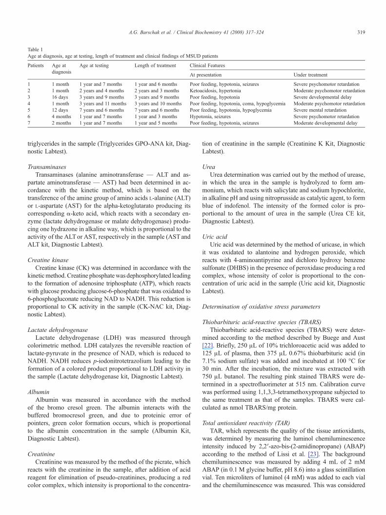
Table 1Age at diagnosis, age at testing, length of treatment and clinical findings of MSUD patients
Patients Age atdiagnosis
Age at testing Length of treatment Clinical Features
At presentation Under treatment
1 1 month 1 year and 7 months 1 year and 6 months Poor feeding, hypotonia, seizures Severe psychomotor retardation2 1 month 2 years and 4 months 2 years and 3 months Ketoacidosis, hypertonia Moderate psychomotor retardation3 16 days 3 years and 9 months 3 years and 9 months Poor feeding, hypotonia Severe developmental delay4 1 month 3 years and 11 months 3 years and 10 months Poor feeding, hypotonia, coma, hypoglycemia Moderate psychomotor retardation5 12 days 7 years and 6 months 7 years and 6 months Poor feeding, hypotonia, hypoglycemia Severe mental retardation6 4 months 1 year and 7 months 1 year and 3 months Hypotonia, seizures Severe psychomotor retardation7 2 months 1 year and 7 months 1 year and 5 months Poor feeding, hypotonia, seizures Moderate developmental delay
319A.G. Barschak et al. / Clinical Biochemistry 41 (2008) 317–324
triglycerides in the sample (Triglycerides GPO-ANA kit, Diag-nostic Labtest).
TransaminasesTransaminases (alanine aminotransferase — ALT and as-
partate aminotransferase — AST) had been determined in ac-cordance with the kinetic method, which is based on thetransference of the amine group of amino acids L-alanine (ALT)or L-aspartate (AST) for the alpha-ketoglutarato producing itscorresponding α-keto acid, which reacts with a secondary en-zyme (lactate dehydrogenase or malate dehydrogenase) produ-cing one hydrazone in alkaline way, which is proportional to theactivity of the ALTor AST, respectively in the sample (AST andALT kit, Diagnostic Labtest).
Creatine kinaseCreatine kinase (CK) was determined in accordance with the
kineticmethod.Creatine phosphatewas dephosphorylated leadingto the formation of adenosine triphosphate (ATP), which reactswith glucose producing glucose-6-phosphate that was oxidated to6-phosphogluconate reducing NAD to NADH. This reduction isproportional to CK activity in the sample (CK-NAC kit, Diag-nostic Labtest).
Lactate dehydrogenaseLactate dehydrogenase (LDH) was measured through
colorimetric method. LDH catalyzes the reversible reaction oflactate-pyruvate in the presence of NAD, which is reduced toNADH. NADH reduces p-iodonitrotetrazolium leading to theformation of a colored product proportional to LDH activity inthe sample (Lactate dehydrogenase kit, Diagnostic Labtest).
AlbuminAlbumin was measured in accordance with the method
of the bromo cresol green. The albumin interacts with thebuffered bromocresol green, and due to proteinic error ofpointers, green color formation occurs, which is proportionalto the albumin concentration in the sample (Albumin Kit,Diagnostic Labtest).
CreatinineCreatinine was measured by the method of the picrate, which
reacts with the creatinine in the sample, after addition of acidreagent for elimination of pseudo-creatinines, producing a redcolor complex, which intensity is proportional to the concentra-
tion of creatinine in the sample (Creatinine K Kit, DiagnosticLabtest).
UreaUrea determination was carried out by the method of urease,
in which the urea in the sample is hydrolyzed to form am-monium, which reacts with salicylate and sodium hypochlorite,in alkaline pH and using nitroprusside as catalytic agent, to formblue of indofenol. The intensity of the formed color is pro-portional to the amount of urea in the sample (Urea CE kit,Diagnostic Labtest).
Uric acidUric acid was determined by the method of uricase, in which
it was oxidated to alantoine and hydrogen peroxide, whichreacts with 4-aminoantipyrine and dichloro hydroxy benzenesulfonate (DHBS) in the presence of peroxidase producing a redcomplex, whose intensity of color is proportional to the con-centration of uric acid in the sample (Uric acid kit, DiagnosticLabtest).
Determination of oxidative stress parameters
Thiobarbituric acid-reactive species (TBARS)Thiobarbituric acid-reactive species (TBARS) were deter-
mined according to the method described by Buege and Aust[22]. Briefly, 250 μL of 10% trichloroacetic acid was added to125 μL of plasma, then 375 μL 0.67% thiobarbituric acid (in7.1% sodium sulfate) was added and incubated at 100 °C for30 min. After the incubation, the mixture was extracted with750 μL butanol. The resulting pink stained TBARS were de-termined in a spectrofluorimeter at 515 nm. Calibration curvewas performed using 1,1,3,3-tetramethoxypropane subjected tothe same treatment as that of the samples. TBARS were cal-culated as nmol TBARS/mg protein.
Total antioxidant reactivity (TAR)TAR, which represents the quality of the tissue antioxidants,
was determined by measuring the luminol chemiluminescenceintensity induced by 2,2′-azo-bis-(2-amidinopropane) (ABAP)according to the method of Lissi et al. [23]. The backgroundchemiluminescence was measured by adding 4 mL of 2 mMABAP (in 0.1 M glycine buffer, pH 8.6) into a glass scintillationvial. Ten microliters of luminol (4 mM) was added to each vialand the chemiluminescence was measured. This was considered

Fig. 1. Plasma glucose from MSUD patients and controls. Data represent themean±SD (n=6–7). ⁎pb0.05 (Student's t test for unpaired samples) comparedto control.
320 A.G. Barschak et al. / Clinical Biochemistry 41 (2008) 317–324
to be the basal value. Tenmicroliters of 25–200μMTrolox (curvecalibration) or plasmawas then added and the chemiluminescencewas measured during 60 s. The Trolox and plasma additionreduces the chemiluminescence. The rapid reduction in luminolintensity is considered as a measure of the TAR capacity. TARmeasurement was calculated as nmol Trolox/mg protein.
Total proteinsProtein concentrations were determined by the Biuret
method (Total protein kit, Diagnostic Labtest), using albuminas standard.
Fig. 2. Lipidic profile in plasma from MSUD patients and controls [total cholesData represent the mean±SD (n=7–8). ⁎pb0.05 (Student's t test for unpaired samp
Statistical analysis
The Student's t test for unpaired samples was used tocompare means between controls and MSUD patients. Correla-tions were carried out using the Pearson correlation coefficient.Only significant values are shown in the text. A p value less than0.05 was considered significant. All analyses were performedusing the Statistical Package for the Social Sciences (SPSS)software in a PC-compatible computer.
Results
Fig. 1 shows glycemia measurement in controls and MSUDpatients under dietary treatment. It can be observed that MSUDpatients presented a significant reduction in the plasma levels ofglucose [t(11)=2.508, pb0.05] as compared to the controls.The glucose levels presented by these patients are below refe-rence values (70–105 mg/dL).
Fig. 2 shows the lipidic profile in controls and treated MSUDpatients. It can be seen that the concentrations of total cho-lesterol (A) [t(13)=2.894, pb0.05] were significantly reducedin MSUD patients as compared to the control group. How-ever, both groups presented values in the reference range (112–205 mg/dL). The plasma levels of HDL cholesterol (B) [t(12)=0.591, pN0.05], LDL cholesterol (C) [t(11)=0.140, pN0.05]and triglycerides (D) [t(13)=0.988, pN0.05] in theMSUDpatients
terol (A), HDL cholesterol (B), LDL cholesterol (C) and triglycerides (D)].les) compared to control.

Fig. 3. Plasma transaminases [aspartate aminotransferase (AST) (A) and alanine aminotransferase (ALT) (B)], creatine kinase (CK) (C) and lactate dehydrogenase (D)from MSUD patients and controls. Data represent the mean±SD (n=5–7). ⁎pb0.05 (Student's t test for unpaired samples) compared to control.
321A.G. Barschak et al. / Clinical Biochemistry 41 (2008) 317–324
were not significantly different when compared to the controlgroup.
Fig. 3 shows enzymatic activities of AST, ALT, CK and LDHin plasma of MSUD patients. These patients presented an in-crease of AST activity (A) [t(12)=−2.281, pb0.05] when com-pared to the control group, lightly increased in relation to thereference range (10–40 UI/L). The levels of ALT (B) had notbeen significantly different from the controls [t(12)=−0.107,pN0.05]. We can also observe that LDH activity (D) wasincreased in plasma of MSUD patients [t(10)=−2.364, pb0.05]when compared to the control group, both groups presented
Fig. 4. Plasma albumin from MSUD patients and controls. Data represent themean±SD (n=5–7). ⁎⁎pb0.01 (Student's t test for unpaired samples) comparedto control.
values in the reference range (110–295 UI/L). However, CKactivity (C) was not altered in these patients [t(9)=2.470,pN0.05].
It can be observed in Fig. 4 that MSUD patients presented areduction in the plasma levels of albumin [t(10)=5.685, pb0.01]when compared to the control group, these levels being belowreference range (3.8–5.4 g/dL).
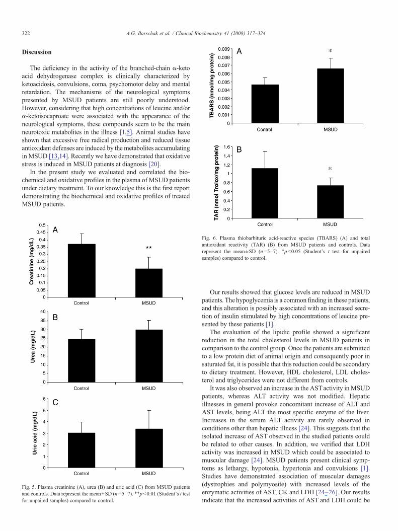
The concentrations of creatinine, urea and uric acid in theplasma of controls andMSUD patients were determined. As it canbe observed, in Fig. 5, MSUD patients presented a significantreduction in plasma levels of creatinine (A) [t(12)=4.076, pb0.01]relatively to the control group. The plasma levels of creatinine inthese patients were below the reference values (0.3–0.7 mg/dL).We did not observe alterations in the urea (B) [t(12)=−1.813,pN0.05] and uric acid levels (C) [t(10)=−0.391, pN0.05] whencompared to the control group.
Fig. 6 shows the measure of thiobarbituric acid-reactivespecies (TBARS) and total antioxidant reactivity (TAR) inplasma from controls and MSUD patients. It was observed asignificant increase of TBARS (A) [t(11)=−3.112, pb0.05],while TAR was markedly reduced (B) [t(9)=2.273, pb0.05],when compared to the controls.
A significant positive correlation was observed between theplasma levels of triglycerides and TBARS measurement[r=0.838, pb0.05]. Correlation between the other biochemicalparameters and the oxidative stress parameters was notobserved.
322 A.G. Barschak et al. / Clinical Biochemistry 41 (2008) 317–324
Discussion
The deficiency in the activity of the branched-chain α-ketoacid dehydrogenase complex is clinically characterized byketoacidosis, convulsions, coma, psychomotor delay and mentalretardation. The mechanisms of the neurological symptomspresented by MSUD patients are still poorly understood.However, considering that high concentrations of leucine and/orα-ketoisocaproate were associated with the appearance of theneurological symptoms, these compounds seem to be the mainneurotoxic metabolites in the illness [1,5]. Animal studies haveshown that excessive free radical production and reduced tissueantioxidant defenses are induced by the metabolites accumulatingin MSUD [13,14]. Recently we have demonstrated that oxidativestress is induced in MSUD patients at diagnosis [20].
In the present study we evaluated and correlated the bio-chemical and oxidative profiles in the plasma of MSUD patientsunder dietary treatment. To our knowledge this is the first reportdemonstrating the biochemical and oxidative profiles of treatedMSUD patients.
Fig. 5. Plasma creatinine (A), urea (B) and uric acid (C) from MSUD patientsand controls. Data represent the mean±SD (n=5–7). ⁎⁎pb0.01 (Student's t testfor unpaired samples) compared to control.
Fig. 6. Plasma thiobarbituric acid-reactive species (TBARS) (A) and totalantioxidant reactivity (TAR) (B) from MSUD patients and controls. Datarepresent the mean±SD (n=5–7). ⁎pb0.05 (Student's t test for unpairedsamples) compared to control.
Our results showed that glucose levels are reduced in MSUDpatients. The hypoglycemia is a common finding in these patients,and this alteration is possibly associated with an increased secre-tion of insulin stimulated by high concentrations of leucine pre-sented by these patients [1].
The evaluation of the lipidic profile showed a significantreduction in the total cholesterol levels in MSUD patients incomparison to the control group. Once the patients are submittedto a low protein diet of animal origin and consequently poor insaturated fat, it is possible that this reduction could be secondaryto dietary treatment. However, HDL cholesterol, LDL choles-terol and triglycerides were not different from controls.
It was also observed an increase in the ASTactivity inMSUDpatients, whereas ALT activity was not modified. Hepaticillnesses in general provoke concomitant increase of ALT andAST levels, being ALT the most specific enzyme of the liver.Increases in the serum ALT activity are rarely observed inconditions other than hepatic illness [24]. This suggests that theisolated increase of AST observed in the studied patients couldbe related to other causes. In addition, we verified that LDHactivity was increased in MSUD which could be associated tomuscular damage [24]. MSUD patients present clinical symp-toms as lethargy, hypotonia, hypertonia and convulsions [1].Studies have demonstrated association of muscular damages(dystrophies and polymyosite) with increased levels of theenzymatic activities of AST, CK and LDH [24–26]. Our resultsindicate that the increased activities of AST and LDH could be

323A.G. Barschak et al. / Clinical Biochemistry 41 (2008) 317–324
associated to the characteristic muscular alterations presented byMSUD patients. However, total CK activity was not altered inMSUD suggesting that additional studies evaluating the activityof CK-MM (muscular isoenzyme) could contribute in betterunderstanding these findings.
It was also evaluated the plasma concentrations of creatinine,urea and uric acid. The MSUD patients presented a significantreduction in plasma creatinine levels, but urea and uric acidconcentrations were not altered. The low concentration of creat-inine could be associated to muscular alterations presented bypatients in the course of the illness. It is important to emphasizethat creatinine is formed through free creatine in the muscle, sothe produced amount of endogenous creatinine is proportional tomuscular mass [24].
MSUD treatment consists of a restricted ingestion of proteins,supplemented with a semi-synthetic formula of essential aminoacids except leucine, isoleucine and valine. The aim of thetreatment is to normalize the plasma levels of the BCAA, mini-mizing the neurological damage associated mainly to increase inleucine levels [1]. It was observed that MSUD patients presenteda reduction in plasma albumin levels, which probably reflectsdeficiencies associated to low protein diet that these patients aresubmitted to.
It was observed a significant increase of TBARS in plasmafrom MSUD patients. Considering that TBARS reflects theamount of malondialdehyde, an end product of lipid peroxidation[6,27], our data indicate that lipid peroxidation is stimulated inMSUD patients, probably secondary to free radical generation.Already, TAR measurement, which reflects the quality of anti-oxidant substances [28], wasmarkedly reduced inMSUDpatients,suggesting a deficient capacity of plasma to modulate the damageassociated with the increased production of reactive species. Theincrease of lipid peroxidation and the reduction of the antioxidantdefenses suggest that oxidative stress occurs in treatedMSUD andcan explain at least in part the pathophysiology of this illness. Itwas previously verified an increase in TBARS and a decrease inTAR in MSUD patients at diagnosis [20].
A positive correlation was observed between plasma tri-glycerides levels and TBARS measurement. Triglycerides arecomposed by the union of three fat acids and glycerol. Fat acidsare easily oxidated by free radicals, leading to lipid peroxidation.Considering that malondialdehyde (MDA) is an end product oflipid breakdown due to lipid peroxidation, and TBARS reflectsthe content of MDA (27), it is possible that lipid peroxidationcould be related to triglycerides amount.
In conclusion, our results show that glucose, total cholesterol,albumin and creatinine are reduced and that AST and LDHactivities are increased in plasma from MSUD patients underdietary treatment. It is probable that these findings are secondaryto low protein diet and/or to the characteristic clinical mani-festations of the illness. These results suggest that serum bio-chemical profile should be evaluated in all MSUD patients duringtreatment. However, our results must be taken with caution sinceour experiments were conducted with samples collected at asingle time point during treatment. Therefore, these samplesmight not represent patients' metabolic control as a whole. Theincreased TBARS and the reduction of the TAR are in accordance
with our previous studies, suggesting that oxidative stress occursalso in MSUD patients under treatment. In this context, it ispossible that dietary antioxidant supplementation could beimportant as an adjuvant therapy in MSUD.
Acknowledgments
This work was supported by grants from Brazilian NationalResearch Council (CNPq), FAPERGS, and FIPE/HCPA —Brazil.
References
[1] Chuang DT, Shih VE. Maple syrup urine disease (branched-chainketoaciduria). In: Scriver CR, Beaudt AL, Sly WL, Valle D, editors. TheMetabolic and Molecular Bases of Inherited Disease. 8ed. New York:McGraw-Hill; 2001. p. 1971–2005.
[2] Schadewaldt P, Wendel U. Metabolism of branched-chain amino acids inmaple syrup urine disease. Eur J Pediatr 1997;156(Suppl 1):S62–6.
[3] Schadewaldt P, Wendel U. Variant maple syrup urine disease (MSUD)—the entire spectrum. J Inherit Metab Dis 2006;29(6):716–24.
[4] Schönberger S, Schweiger B, Schwahn B, Schwarz M, Wendel U.Dysmyelination in the brain of adolescents and young adults with maplesyrup urine disease. Mol Genet Metab 2004;82(1):69–75.
[5] Strauss KA, Morton DH. Branched-chain ketoacyl dehydrogenase defi-ciency: maple syrup disease. Curr Treat Option Neurol 2003;5(4): 329–41.
[6] Halliwell B, Gutteridge JC. Free radicals in biology and medicine. 3th ed.New York: Oxford; 2001.
[7] Harris ED. Regulation of antioxidant enzymes. Faseb J 1992;6(9): 2675–83.[8] Pawlak W, Kedziora J, Zolynski K, et al. Effect of long term bed rest in
man on enzymatic antioxidative defence and lipid peroxidation inerythrocytes. J Gravit Physiol 1998;1:163–4.
[9] Beckman KB, Ames BN. The free theory of aging matures. Phisiol Rev1998;78(2):547–81.
[10] Colome C, Sierra C, Vilaseca MA. Congenital errors of metabolism: causeof oxidative stress? Méd Clin 2000;115:111–7.
[11] Fontella FU, Pulrolnik V, Wannmacher CMD, et al. Propionic andL-methylmalonic acids induce oxidative stress in brain of young rats. NeuroReport 2000;11:541–4.
[12] de Oliveira Marques F, Hagen ME, Pederzolli CD. Glutaric acid inducesoxidative stress in brain of young rats. Brain Res 2003;964: 153–8.
[13] Fontella FU, Gassen E, Pulrolnik V, et al. Stimulation of lipid peroxidationin vitro in rat brain by metabolites accumulating in maple syrup urinedisease. Metab Brain Dis 2002;17:47–54.
[14] Bridi R, Araldi J, Sgarbi MB, et al. Induction of oxidative stress in rat brainby the metabolites accumulating in maple syrup urine disease. Int J DevlNeuroscience 2003;21:327–32.
[15] Wajner M, Latini A, Wyse ATS, Dutra-Filho CS. The role of oxidativedamage in the neuropathology of organic acidurias: insights from animalstudies. J Inherit Metab Dis 2004;27:427–48.
[16] Sirtori LR, Dutra-Filho CS, Fitarelli D, et al. Oxidative stress in patientwith phenylketonuria. Biochim Biophys Acta 2005;1740: 68–73.
[17] Sitta A, Barschak AG, Deon M, et al. Investigation of oxidative stressparameters in treated phenylketonuric patients. Metab Brain Dis 2006;20:287–96.
[18] Vargas CR, Wajner M, Sirtori LR, et al. Evidence that oxidative stress isincreased in patients with X-linked adrenoleukodystrophy. BiochimBiophys Acta 2004;1688:26–32.
[19] DeonM,WajnerM, Sirtori LR, et al. The effect of Lorenzo's oil on oxidativestress in X-linked adrenoleukodystrophy. J Neurol Sci 2006;247:157–64.
[20] Barschak AG, Sitta A, Deon M, et al. Evidence that oxidative stress isincreased in plasma from patients with maple syrup urine disease. MetabBrain Dis 2006;20:279–86.
[21] Joseph MH, Marsden CA. Amino acids and small peptides. In: Lim CF,editor. HPLC of Small Peptides. Oxford: IRL Press; 1986. p. 13–27.
324 A.G. Barschak et al. / Clinical Biochemistry 41 (2008) 317–324
[22] Buege JA, Aust SD. Microsomal lipid peroxidation. Methods Enzymol1978;52:302–9.
[23] Lissi E, Pascual C, Del Castillo MD. Luminol luminescence induced by2,2′-azo-bis-(2-amidinopropane) thermolysis. Free Radic Res Commun1992;17:299–311.
[24] Burtis CA, Ashwood ER, editors. Tietz: Fundamentos de Química Clínica.4°edição. Rio de Janeiro: Guanabara-Koogan; 1998.
[25] Lott JA, Landesman PW. The enzymology of skeletal muscle disorders.Crit Rev Clin Lab Sci 1984;20(2):153–90.
[26] Nathwani RA, Pais S, Reynolds TB, Kaplowitz N. Serum alanineaminotransferase in skeletal muscle diseases. Hepatology 2005;41(2):380–2.
[27] Esterbauer H, Cheeseman KH. Determination of aldehydic lipid pero-xidation products: malonaldehyde and 4-hydroxynonenal. MethodsEnzymol 1990;186:407–21.
[28] Lissi E, Salim-Hanna M, Pascual C, Del Castillo MD. Evaluation of totalantioxidant potential (TRAP) and total antioxidant reactivity from luminol-enhanced chemiluminescence measurements. Free Radic Biol Med1995;18:153–8.


