Embed Size (px)
Citation preview
Universidade Federal do Rio Grande do Sul
Instituto de Ciências Básicas da Saúde
Programa de Pós-Graduação em Ciências Biológicas: Bioquímica
Investigação de estresse oxidativo em pacientes
tratados e não tratados com Doença do Xarope
do Bordo
Alethéa Gatto Barschak
Orientadora: Profa Dra Carmen Regla Vargas
Co-orientador: Prof. Dr. Moacir Wajner
Porto Alegre, 2008

I
Universidade Federal do Rio Grande do Sul
Instituto de Ciências Básicas da Saúde
Programa de Pós-Graduação em Ciências Biológicas: Bioquímica
Investigação de estresse oxidativo em pacientes
tratados e não tratados com Doença do Xarope
do Bordo
Alethéa Gatto Barschak
Orientadora: Profa Dra Carmen Regla Vargas
Co-orientador: Prof. Dr. Moacir Wajner
Tese apresentada ao Programa de Pós-Graduação em Ciências Biológicas:
Bioquímica da Universidade Federal do Rio Grande do Sul como requisito à
obtenção do grau de Doutor em Bioquímica
Porto Alegre, 2008

II
Ao meu marido, Válter, pelo amor e pelo apoio.
Aos meus pais, Nelson e Zulmira,
por tudo que sempre fizeram por mim.

III
Agradecimentos
À minha orientadora Carmen pelo incentivo e amizade, sua dedicação e
objetividade permitiram a realização deste trabalho de forma tranqüila e agradável. Ao Moacir, suas sugestões e críticas enriqueceram este trabalho.
A Ângela, Marion e Franciele, amigas e colegas, sua amizade tornou este
trabalho mais fácil e suas sugestões o tornaram melhor.
Às bolsistas de iniciação científica: Amanda, Giovana, Camila, Roberta,
Marcella, Thatiana e Maiara, sem a sua ajuda este trabalho não poderia ter sido
realizado.
Aos colegas do Laboratório de Análise de Metabólitos do Serviço de
Genética Médica do HCPA: Daniella, Anderson, Graziela, Estela, Anelise e
Rafaela, pelos ótimos e divertidos momentos que passamos juntos.
Ao Departamento de Bioquímica da UFRGS, e em especial aos amigos do
laboratório 38, sempre prontos para ajudar.
Ao Serviço de Genética Médica do HCPA, que possibilitou a realização
deste trabalho, e em especial aos colegas do Laboratório de Erros Inatos do
Metabolismo.
Aos pacientes que participaram deste estudo, sinceramente, muito obrigada,
tenho certeza de que ciência lhes proporcionará um futuro melhor.
Ao CNPq pela bolsa concedida.
Aos meus pais, Nelson e Zulmira, meus grandes orientadores, muito
obrigada por tudo.
Ao Válter por seu amor e por estar sempre ao meu lado, apoiando minhas
decisões e me ajudando a alcançar meus objetivos.

IV
ÍNDICE
RESUMO.................................................................................................... 1
ABSTRACT................................................................................................. 4
LISTA DE ABREVIATURAS....................................................................... 7
I. INTRODUÇÃO......................................................................................... 9
I.1 Erros inatos do metabolismo............................................................ 9
I.2 Doença do Xarope do Bordo............................................................ 11
I.3 Radicais livres................................................................................... 21
I.4 Defesas antioxidantes...................................................................... 23
I.5 Estresse oxidativo............................................................................ 26
II. OBJETIVOS............................................................................................ 29
II.1 Objetivo geral................................................................................... 29
II.2 Objetivos específicos....................................................................... 29
III. RESULTADOS....................................................................................... 31
III.1 Capítulo I – Artigo 01...................................................................... 31
Evidence that oxidative stress is increased in plasma from patients with
maple syrup urine disease..................................................................... 31
III.2 Capítulo II – Artigo 02..................................................................... 40
Oxidative stress in plasma from maple syrup urine disease patients during
treatment............................................................................................... 40
III.3 Capítulo III – Artigo 03.................................................................... 51
Erythrocyte glutathione peroxidase activity and plasma selenium
concentration are reduced in maple syrup urine disease patients during
treatment............................................................................................... 51

V
III.4 Capítulo IV – Artigo 04................................................................... 56
Maple Syrup Urine Disease in treated patients: Biochemical and oxidative
stress profiles ................................................................................................. 56
IV. DISCUSSÃO......................................................................................... 65
V. CONCLUSÕES...................................................................................... 78
VI. PERSPECTIVAS................................................................................... 82
VII. REFERÊNCIAS.................................................................................... 83
ANEXO 1 – Lista de Figuras....................................................................... 94
ANEXO 2 – Lista de Tabelas ..................................................................... 95
ANEXO 3 – Parecer da Comissão Científica e Comissão de Pesquisa e Ética
em Saúde do HCPA ................................................................................... 96
ANEXO 4 – Termo de Consentimento ...................................................... 97

1
RESUMO
A Doença do Xarope do Bordo (DXB) é um erro inato do metabolismo
causado pela deficiência na atividade do complexo da desidrogenase dos α-
cetoácidos de cadeia ramificada. Como conseqüência deste bloqueio ocorre o
acúmulo dos aminoácidos de cadeia ramificada leucina, isoleucina e valina,
bem como de seus respectivos α-cetoácidos e α-hidroxiácidos de cadeia
ramificada. A manifestação clínica da DXB varia da forma clássica severa a
formas variantes moderadas. Os principais sinais clínicos laboratoriais
apresentados pelos pacientes com DXB incluem cetoacidose, hipoglicemia,
recusa alimentar, opistótono, apnéia, ataxia, convulsões, coma, atraso no
desenvolvimento psicomotor e retardo mental. No entanto, os mecanismos
responsáveis pelos sintomas neurológicos apresentados pelos pacientes com
DXB ainda são pouco conhecidos. O tratamento para DXB consiste na
restrição da ingestão de proteínas, bem como dos aminoácidos leucina
isoleucina e valina, suplementada com uma fórmula semi-sintética contendo
aminoácidos essenciais, minerais e vitaminas. Estudos in vitro têm
demonstrado que os aminoácidos de cadeia ramificada e seus respectivos α-
cetoácidos acumulados na DXB estimulam a lipoperoxidação e reduzem as
defesas antioxidantes em córtex cerebral de ratos. O objetivo deste estudo foi
investigar se o estresse oxidativo ocorre em pacientes com DXB em tratamento
ou ainda não tratados. Inicialmente, foram analisados parâmetros de estresse
oxidativo em amostras de plasma de pacientes com DXB obtidas antes do
início do tratamento (no diagnóstico). Foi observado um aumento significativo
das espécies reativas ao ácido tiobarbitúrico (TBARS) e uma diminuição
significativa da reatividade antioxidante total (TAR). Em seguida foram

2
avaliados parâmetros de estresse oxidativo em plasma de dois grupos de
pacientes com DXB em tratamento dietético com níveis plasmáticos baixos e
altos de leucina. Verificou-se um marcado aumento do TBARS e uma
diminuição significativa do TAR em ambos os grupos de pacientes tratados,
porém a redução do TAR foi mais pronunciada nos pacientes com baixos níveis
sanguíneos de leucina. Além disso, não foi observada correlação entre os
níveis de leucina, isoleucina ou valina com os parâmetros de estresse
oxidativo. Em continuidade ao trabalho, foram estudados os níveis de selênio
no plasma de pacientes com DXB antes e durante o tratamento dietético, bem
como a atividade de enzimas antioxidantes em eritrócitos de pacientes em
tratamento. Foi verificado que os pacientes com DXB apresentam uma
deficiência moderada de selênio no momento do diagnóstico (antes do
tratamento) da doença e que essa deficiência se agrava com o tratamento
dietético. Ainda, verificou-se que a atividade da enzima glutationa peroxidase
está diminuída em eritrócitos de pacientes em tratamento. Posteriormente,
foram medidos vários parâmetros bioquímicos em plasma de pacientes com
DXB tratados, com o objetivo de verificar se havia correlação destes com
parâmetros de estresse oxidativo. Foi demonstrado que os pacientes com DXB
em tratamento apresentam redução nos níveis séricos de glicose, colesterol
total, creatinina sérica e albumina e aumento na atividade enzimática da
aspartato aminotransferase e da lactato desidrogenase. Entretanto, não foram
observadas alterações nos níveis de colesterol HDL, colesterol LDL,
triglicerídeos, alanina aminotransferase, creatina quinase, uréia e ácido úrico.
Foi ainda verificado um aumento significativo de TBARS e uma diminuição
significativa de TAR, bem como uma correlação positiva entre a medida de

3
TBARS e os níveis de triglicerídeos. Concluindo, nossos resultados indicam
que existe um desequilíbrio entre a produção de radicais livres (aumento) e as
defesas antioxidantes (diminuição) gerando estresse oxidativo em pacientes
com DXB não tratados e em tratamento e que esse processo não está
correlacionado com os níveis plasmáticos dos aminoácidos leucina, isoleucina
e valina acumulados nesta doença. Além disso, verificamos que a deficiência
de selênio e a concomitante diminuição da atividade da glutationa peroxidase
podem estar associados ao desenvolvimento do estresse oxidativo nesta
doença. É, portanto, possível inferir que o dano oxidativo contribua, pelo menos
em parte, para a fisiopatologia desta doença, explicando o dano neurológico
que ocorre nos pacientes afetados. Portanto, é possível que a administração de
antioxidantes possa ser útil na DXB.
4
ABSTRACT
Maple Syrup Urine Disease (MSUD) is an inborn error of metabolism
caused by a deficiency of the branched-chain α-keto acid dehydrogenase
complex activity. This blockage leads to accumulation of the branched-chain
amino acids leucine, isoleucine and valine, as well as their corresponding α-
keto acids and α-hydroxy acids. Clinical manifestations are variable, pending on
the variants classical severe form to milder variants. The major clinical features
presented by MSUD patients include ketoacidosis, hypoglycemia, poor feeding,
opisthotonos, apnea, ataxia, convulsions, coma, psychomotor delay and mental
retardation. However, the mechanisms of the neurological symptoms presented
by MSUD patients are still poorly understood. MSUD treatment consists of a low
protein diet and a semi-synthetic formula poor in leucine, isoleucine, and valina
and supplemented by essential amino acids minerals and vitamins. Studies
have been showed that the branched-chain amino acids and their
corresponding brached-chain α-keto acids accumulating in MSUD stimulate in
vitro lipid peroxidation and reduce antioxidant defences in cerebral cortex of
rats. The objective of the present study was to investigate oxidative stress
parameters in MSUD patients treated and non-treated. First, it was analyzed
parameters of oxidative stress in plasma from non-treated MSUD patients. It
was observed a significant increase of thiobarbituric acid-reactive species
(TBARS) and a marked reduction in total antioxidant reactivity (TAR).
Parameters of oxidative stress in plasma from two groups of MSUD patients
under dietetic treatment with low and high blood leucine levels were also
evaluated. It was verified an increase of TBARS and a significant decrease of
TAR in both groups of MSUD patients. However, TAR reduction was higher in

5
the group presenting low leucine levels. On the other hand, it was not found a
correlation between leucine, valine and isoleucine levels and oxidative stress
parameters. Selenium levels in plasma from MSUD patients at diagnosis (non-
treated) and under treatment, as well as the activities of antioxidant enzymes in
erythrocytes from treated patients were also determined. MSUD patients
presented a significant selenium deficiency at diagnosis which becomes more
pronounced during treatment and a decrease of erythrocyte glutathione
peroxidase activity during treatment. Subsequently, it was evaluated various
biochemical parameters in plasma from MSUD patients during treatment in
order to verify if there was correlation between these parameters and oxidative
stress. It was demonstrated that MSUD patients under treatment present a
reduction of glucose, total cholesterol, albumin and creatinine levels and an
increased activity of aspartate aminotransferase and lactate dehydrogenase.
However, HDL cholesterol, LDL cholesterol, triglycerides, alanine
aminotransferase, creatine kinase, urea and uric acid measurements were not
altered. Besides, it was verified a significant increase of TBARS and a
significant decrease of TAR, as well as a positive correlation between TBARS
and triglycerides levels. In summary, our results suggest an imbalance between
the production of free radicals and the antioxidant defenses in MSUD patients
leading to oxidative stress at diagnosis and during treatment and that this is not
correlated with the amino acids leucine, isoleucine and valine accumulating in
this disease. Besides, the selenium deficiency and the decrease of erythrocyte
glutathione peroxidase activity could be associated to the development of
oxidative stress. Finally, it is possible that oxidative damage may contribute, at

6
least in part, to the pathophysiology of this disorder. If that is the case,
antioxidants may serve as an adjuvant therapy in MSUD

7
LISTA DE ABREVIATURAS
AACR aminoácidos de cadeia ramificada
AloIleu aloisoleucina
ALT alanina aminotransferase
AST aspartato aminotransferase
CACR α-cetoácidos de cadeia ramificada
CAT catalase
CDCR desidrogenase dos α-cetoácido de cadeia ramificada
CIC ácido α-cetoisocapróico
CIV ácido α-cetoisovalérico
CK creatina quinase
CK-MM isoenzima muscular da creatina quinase
CMV ácido α-ceto-β-metilvalérico
DXB doença do xarope do bordo
EIM erros inatos do metabolismo
ERN espécies reativas de nitrogênio
ERO espécies reativas de oxigênio
GR glutationa redutase
GSH glutationa reduzida
GSH-Px glutationa peroxidase
GSSG glutationa oxidada
HACR α-hidroxiácidos de cadeia ramificada
HIC ácido α-hidroxiisocapróico
HIV ácido α-hidroxiisovalérico
8
HMV ácido α-hidroxi-β-metilvalérico
Ileu isoleucina
LDH lactato desidrogenase
Leu leucina
MDA malondialdeído
NADPH nicotinamida adenina dinucleotídeo fosfato
SNC sistema nervoso central
SOD superóxido dismutase
TAR reatividade antioxidante total
TAS status antioxidante total
TBARS espécies reativas ao ácido tiobarbitúrico
Val valina
9
I. INTRODUÇAO
I.1 Erros inatos do metabolismo
O termo erros inatos do metabolismo (EIM) foi proposto pela primeira
vez por Archibald Garrod em 1908, a partir de seus estudos em pacientes com
alcaptonúria. Garrod observou que os indivíduos afetados por esta doença
excretavam na urina quantidades aumentadas de ácido homogentísico e que
esta peculiaridade era encontrada em diversos membros de uma mesma
família. Além disso, os pais dos afetados tinham, geralmente, parentesco
consangüíneo próximo (Waber, 1990). Baseado nessas observações, Garrod,
juntamente com o geneticista inglês Bateson, sugeriu um modelo de herança
autossômica recessiva para esse distúrbio, e, ainda, que a alcaptonúria e
outras anormalidades metabólicas herdadas eram raras e incomuns (Waber,
1990; Scriver et al., 2001).
Desde os estudos de Garrod, muitos pesquisadores têm detectado
novas doenças metabólicas hereditárias e os erros inatos do metabolismo já
foram descritos em todas as áreas do metabolismo humano (aminoácidos,
lipídios, ácidos orgânicos, carboidratos etc.) (Scriver et al., 2001).
Os EIM são individualmente raros, porém são freqüentes quando
analisados em conjunto, atingindo um a cada mil nascidos vivos (Gimenez-
Sanchez et al., 2001). Estas doenças correspondem a cerca de 10% de todas
as doenças genéticas e, atualmente, já foram descritos aproximadamente 500
distúrbios envolvendo defeitos na síntese, degradação, transporte e
armazenamento de moléculas no organismo (Gimenez-Sanchez et al., 2001).

10
Esses defeitos hereditários do metabolismo devem-se a anormalidades
na síntese de uma proteína, geralmente um enzima, alterando suas funções. A
ausência ou a deficiência severa de atividade enzimática leva a um bloqueio
metabólico com acúmulo de substratos e seus derivados. Dependendo da
importância da rota afetada, este bloqueio repercute de forma clínica variável,
geralmente provocando sintomatologia grave, que na maioria das vezes afeta o
sistema nervoso central (SNC) (Scriver et al, 2001).
Os EIM podem ser classificados de diversas maneiras, como pela idade
de apresentação ou pela área do metabolismo afetada. A classificação descrita
por Saudubray e Charpentier (2001) estabelece três grandes grupos de EIM:
distúrbios de síntese ou degradação de macromoléculas complexas, incluindo
as doenças lisossômicas de depósito (ex: doença de Gaucher, doença de
Fabry etc.) e as desordens peroxissomais (ex: adrenoleucodistrofia ligada ao X,
doença de Refsum etc.); doenças com deficiência de energia, incluindo
doenças de depósito de glicogênio, defeitos de gliconeogênese, acidemias
lácticas congênitas, defeitos de oxidação de ácidos graxos e doenças
mitocondriais de cadeia respiratória; e erros inatos do metabolismo
intermediário, que incluem as aminoacidopatias (ex: doença do xarope do
bordo, fenilcetonúria etc.), as acidemias orgânicas (ex: acidemia propiônica,
acidemia metilmalônica etc.), os defeitos no ciclo da uréia (ex: citrulinemia,
argininemia etc.) e as intolerâncias a açúcares (ex: galactosemia etc.). Neste
último grupo de doenças pode ocorrer intoxicação aguda ou crônica, causada
pelo acúmulo de componentes tóxicos e de metabólitos produzidos devido ao
bloqueio de rotas metabólicas. Estudos revelam que aproximadamente um

11
terço dos EIM correspondem a aminoacidopatias, outro terço a acidemias
orgânicas e o terço final a todos os outros EIM (Hoffmann, 1994).
I.2 Doença do xarope do bordo
Menkes e colaboradores descreveram em 1954 quatro pacientes com
uma doença neurológica degenerativa, caracterizada por edema cerebral,
convulsões, espasticidade e sofrimento respiratório, com início na primeira
semana de vida e morte dentro de três meses. A característica mais
proeminente foi o forte odor de açúcar queimado na urina que deu origem ao
nome Doença do Xarope do Bordo (DXB). No final dos anos 1950, Dancis e
colaboradores, identificaram os compostos acumulados na urina e plasma dos
pacientes como os aminoácidos de cadeia ramificada leucina (Leu), isoleucina
(Ileu) e valina (Val) e seus correspondentes α-cetoácidos. Dessa forma, a
doença também é chamada cetoacidúria de cadeia ramificada (Dancis, 1959;
Dancis et al., 1959). Posteriormente, Dancis e colaboradores, demonstraram
que o bloqueio metabólico na DXB ocorre na descarboxilação dos α-cetoácidos
de cadeia ramificada (Dancis et al., 1960).
A DXB é, portanto, um erro inato do metabolismo causado pela
deficiência na atividade do complexo da desidrogenase dos α-cetoácidos de
cadeia ramificada (CDCR). Como conseqüência deste bloqueio ocorre o
acúmulo dos aminoácidos de cadeia ramificada (AACR) leucina, isoleucina e
valina, bem como de seus respectivos α-cetoácidos de cadeia ramificada
(CACR) α-cetoisocapróico (CIC), α-ceto-β-metilvalérico (CMV) e α-

12
cetoisovalérico (CIV) e α-hidroxiácidos de cadeia ramificada (HACR) α-
hidroxiisocapróico (HIC), α-hidroxi-β-metilvalérico (HMV) e α-hidroxiisovalérico
(HIV) (Figura 1). A DXB é uma desordem autossômica recessiva e apresenta
freqüência mundial estimada em 1:185000 nascidos vivos (Chuang e Shih,
2001).

13
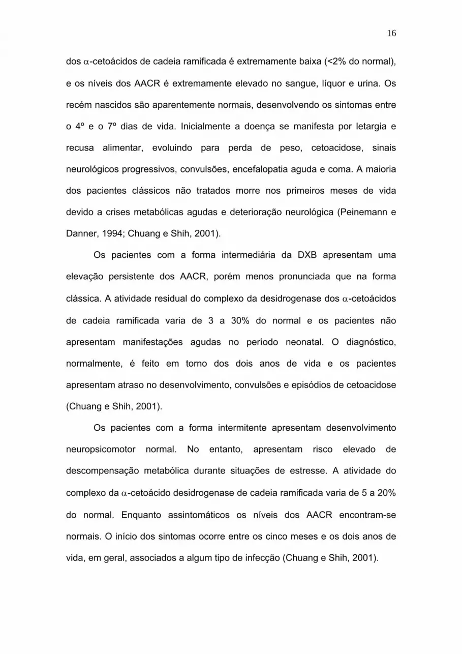
Figura 1. Rota metabólica dos aminoácidos de cadeia ramificada leucina, isoleucina e
valina, indicando o bloqueio metabólico que ocorre na Doença do Xarope do Bordo. (Adaptado
de Scriver et al., 2001).
Ácido α-cetoisocapróico Ácido α-ceto-β-metilvalérico Ácido α-cetoisovalérico
DXB
Isovaleril-CoA α-metilbutiril-CoA Isobutiril-CoA
β-metilcrotonil-CoA Tiglil-CoA Metilcrilil-CoA
Acetil-CoA Acetil-CoA Succinil-CoA
Ácido acetoacético Succinil-CoA
Leucina Isoleucina Valina
Complexo da desidrogenase dos α-cetoácidos de cadeia
ramificada

14
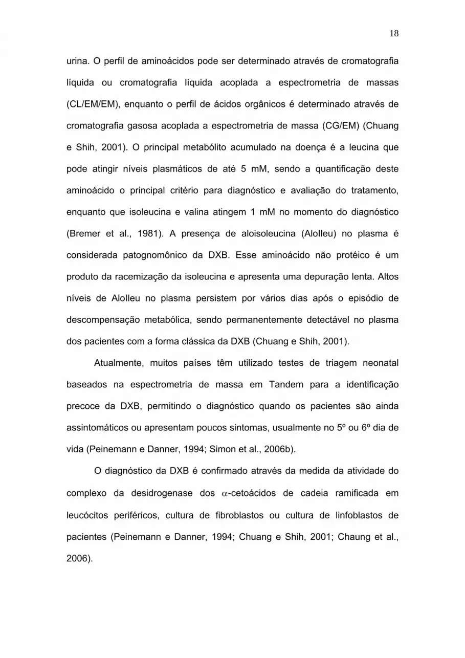
O complexo multienzimático da desidrogenase dos α-cetoácido de
cadeia ramificada está localizado no lado interno da membrana mitocondrial
interna. Esse complexo enzimático compreende três sítios catalíticos: uma
descarboxilase dependente de tiamina pirofosfato, ou E1; uma transacilase, ou
E2; e uma desidrogenase, ou E3. Esse complexo possui duas enzimas
reguladoras: uma quinase (quinase da desidrogenase dos α-cetoácidos de
cadeia ramificada) e uma fosfatase (fosfatase da desidrogenase dos α-
cetoácidos de cadeia ramificada) (Figura 2) (Peinemann e Danner, 1994;
Chuang, 1998; Chuang e Shih, 2001).
Figura 2. Subunidades e organização do complexo da desidrogenase dos α-cetoácido
de cadeia ramificada (CDCR). O complexo da CDCR mitocondrial é organizado em torno de um
centro cúbico que compreende a subunidade transacilase (E2). A descarboxilase dependente
de tiamina pirofosfato (E1) e a desidrogenase ligada ao FAD (E3) estão ligadas de forma não
covalente ao domínio de ligação E1/E3 da subunidade E2. A quinase específica se liga ao
domínio do ácido lipóico da E2. O sítio de ligação específico para a fosfatase é desconhecido.
Os três domínios da E2 são ligados por regiões flexíveis. Os sítios ativos ou de ligação ao
cofator em cada componente da enzima estão circulados. (Adaptado de Chuang, 1998).
E1 Descarboxilase
Fosfatase da CdCR
quinase da CdCR
E2 Domínio do Ácido lipóico E2 e1/e3
domínio de ligação
E2 Transacilase
E3 desidrogenase

15
O catabolismo dos AACR inicia por sua transaminação reversível
catalisada pela aminotransferase de cadeia ramificada formando os α-
cetoácidos de cadeia ramificada que são internalizados na mitocôndria através
de uma proteína transportadora específica (sistema L de transporte não
dependente de sódio). No interior da mitocôndria os CACR sofrem
descarboxilação oxidativa irreversível pelo complexo da desidrogenase dos α-
cetoácidos de cadeia ramificada. A reação produz respectivos acil CoA de
cadeia ramificada que são metabolizados por vias específicas. Os produtos
finais do catabolismo da leucina são acetil-CoA e acetoacetato, da valina
succinil-CoA e da isoleucina acetil-CoA e succinil-CoA (Schadewaldt e Wendel,
1997; Chuang e Shih, 2001).
A manifestação clínica da DXB é variável, dependendo da variante da
doença (forma clássica severa a formas variantes moderadas). O tratamento
envolve restrição na ingestão de proteínas e utilização de fórmulas específicas
com redução dos AACR, suplementada por aminoácidos essenciais e
associada ou não a administração do cofator tiamina. A classificação dos
pacientes é baseada em parâmetros como idade de início dos sintomas,
tolerância à leucina, atividade residual do complexo da desidrogenase dos α-
cetoácidos de cadeia ramificada e resposta a administração de tiamina. Os
pacientes com DXB podem ser classificados em cinco fenótipos: clássica,
intermediária, intermitente, responsiva à tiamina e deficiência de lipoamida
desidrogenase (Chuang e Shih, 2001; Simon et al., 2006a).
A forma clássica da doença se caracteriza por início neonatal e é a
forma mais severa e freqüente da doença, representando aproximadamente
80% dos casos de DXB. A atividade residual do complexo da desidrogenase
16
dos α-cetoácidos de cadeia ramificada é extremamente baixa (<2% do normal),
e os níveis dos AACR é extremamente elevado no sangue, líquor e urina. Os
recém nascidos são aparentemente normais, desenvolvendo os sintomas entre
o 4º e o 7º dias de vida. Inicialmente a doença se manifesta por letargia e
recusa alimentar, evoluindo para perda de peso, cetoacidose, sinais
neurológicos progressivos, convulsões, encefalopatia aguda e coma. A maioria
dos pacientes clássicos não tratados morre nos primeiros meses de vida
devido a crises metabólicas agudas e deterioração neurológica (Peinemann e
Danner, 1994; Chuang e Shih, 2001).
Os pacientes com a forma intermediária da DXB apresentam uma
elevação persistente dos AACR, porém menos pronunciada que na forma
clássica. A atividade residual do complexo da desidrogenase dos α-cetoácidos
de cadeia ramificada varia de 3 a 30% do normal e os pacientes não
apresentam manifestações agudas no período neonatal. O diagnóstico,
normalmente, é feito em torno dos dois anos de vida e os pacientes
apresentam atraso no desenvolvimento, convulsões e episódios de cetoacidose
(Chuang e Shih, 2001).
Os pacientes com a forma intermitente apresentam desenvolvimento
neuropsicomotor normal. No entanto, apresentam risco elevado de
descompensação metabólica durante situações de estresse. A atividade do
complexo da α-cetoácido desidrogenase de cadeia ramificada varia de 5 a 20%
do normal. Enquanto assintomáticos os níveis dos AACR encontram-se
normais. O início dos sintomas ocorre entre os cinco meses e os dois anos de
vida, em geral, associados a algum tipo de infecção (Chuang e Shih, 2001).

17
Em geral os pacientes com a forma responsiva à tiamina não
apresentam doença neonatal aguda, e o curso inicial da doença se caracteriza
por atraso no desenvolvimento psicomotor. Nesses pacientes a administração
de tiamina associada à dieta restrita em proteínas pode reduzir os altos níveis
dos AACR para valores normais. As doses de tiamina utilizadas variam de 10 a
1000 mg/dia (Chuang e Shih, 2001).
A deficiência de lipoamida desidrogenase (E3 - deficiente) é uma
desordem rara, caracterizada por atraso no desenvolvimento psicomotor e
acidose lática. Os AACR no plasma estão levemente ou moderadamente
aumentados. Esses pacientes apresentam uma deficiência combinada das
desidrogenases dos α-cetoácidos de cadeia ramificada, do piruvato e do α-
cetoglutarato. O curso da doença é caracterizado por deterioração neurológica
progressiva, incluindo hipotonia, atraso no desenvolvimento e desmielinização
(Chuang e Shih, 2001).
Os principais sinais clínicos apresentados pelos pacientes com a forma
clássica da DXB incluem cetoacidose, hipoglicemia, recusa alimentar,
opistótono, apnéia, ataxia, convulsões, coma, atraso no desenvolvimento
psicomotor e retardo mental. A encefalopatia também é um achado bastante
importante nos pacientes com DXB. Além disso, pode ser observada a
presença de edema generalizado e hipomielinização/desmielinização no
sistema nervoso central desses pacientes, principalmente durante as crises de
descompensação metabólica (Chuang e Shih, 2001; Schönberger et al., 2004;
Sener, 2007).
A DXB pode ser diagnosticada pela detecção de altas concentrações de
leucina, isoleucina e valina no plasma, e de níveis elevados dos CACR na
18
urina. O perfil de aminoácidos pode ser determinado através de cromatografia
líquida ou cromatografia líquida acoplada a espectrometria de massas
(CL/EM/EM), enquanto o perfil de ácidos orgânicos é determinado através de
cromatografia gasosa acoplada a espectrometria de massa (CG/EM) (Chuang
e Shih, 2001). O principal metabólito acumulado na doença é a leucina que
pode atingir níveis plasmáticos de até 5 mM, sendo a quantificação deste
aminoácido o principal critério para diagnóstico e avaliação do tratamento,
enquanto que isoleucina e valina atingem 1 mM no momento do diagnóstico
(Bremer et al., 1981). A presença de aloisoleucina (AloIleu) no plasma é
considerada patognomônico da DXB. Esse aminoácido não protéico é um
produto da racemização da isoleucina e apresenta uma depuração lenta. Altos
níveis de AloIleu no plasma persistem por vários dias após o episódio de
descompensação metabólica, sendo permanentemente detectável no plasma
dos pacientes com a forma clássica da DXB (Chuang e Shih, 2001).
Atualmente, muitos países têm utilizado testes de triagem neonatal
baseados na espectrometria de massa em Tandem para a identificação
precoce da DXB, permitindo o diagnóstico quando os pacientes são ainda
assintomáticos ou apresentam poucos sintomas, usualmente no 5º ou 6º dia de
vida (Peinemann e Danner, 1994; Simon et al., 2006b).
O diagnóstico da DXB é confirmado através da medida da atividade do
complexo da desidrogenase dos α-cetoácidos de cadeia ramificada em
leucócitos periféricos, cultura de fibroblastos ou cultura de linfoblastos de
pacientes (Peinemann e Danner, 1994; Chuang e Shih, 2001; Chaung et al.,
2006).

19
O diagnóstico pré-natal da DXB pode ser realizado através da análise
enzimática direta do tecido de vilosidades coriônicas ou através de cultura de
células do fluído amniótico ou das vilosidades coriônicas, coletados entre a 14º
e 18º semanas de gestação (Chuang e Shih, 2001).
O tratamento para DXB consiste na restrição da ingestão de proteínas, o
que minimiza o acúmulo dos AACR e, consequentemente, seus efeitos tóxicos,
principalmente ao sistema nervoso central. O objetivo do tratamento é
normalizar as concentrações dos AACR, sem prejudicar o crescimento e
desenvolvimento dos pacientes. O tratamento deve ser iniciado o mais
precocemente possível, ainda no período neonatal, e deve ser mantido por
toda a vida do paciente. A terapia com tiamina (50-300mg/dia) é empregada
por 3 semanas no início do tratamento para detecção de pacientes com a
forma responsiva a tiamina. A dieta prescrita aos pacientes com DXB deve ser
hipoproteica e hipercalórica, praticamente isenta de proteínas de origem animal
combinada com uma fórmula semi-sintética contendo aminoácidos essenciais,
exceto os AACR, e suplementada com minerais (molibdênio, manganês...) e
vitaminas (vitaminas C, D, E...) (Chuang e Shih, 2001). Sem o diagnóstico
precoce e o tratamento efetivo as crianças desenvolvem problemas cerebrais
severos e permanentes, podendo até mesmo morrer nos primeiros meses de
vida (Morton et al., 2002).
Durante a fase aguda da doença medidas mais agressivas precisam ser
empregadas para reduzir rapidamente os níveis dos AACR, que podem levar a
deterioração das funções cerebrais. A descompensação metabólica aguda
pode ser desencadeada por períodos de estresse como, infecções, febre ou
outras doenças. Dois aspectos são importantes no manejo das crises

20
metabólicas: a rápida remoção dos metabólitos tóxicos e a supressão do
catabolismo e/ou a promoção do anabolismo (Chuang e Shih, 2001; Yoshino et
al., 1999).
Entre as medidas utilizadas para a rápida remoção dos metabólitos
acumulados estão a diálise peritoneal, a transfusão exsangüínea e a
hemodiálise, as quais apresentam bons resultados, aumentando
significativamente a depuração desses compostos. Entre os tratamentos extra
corporais, a hemodiálise apresenta melhor desempenho que a hemofiltração,
devendo ser considerada a primeira opção de tratamento. Quando esta medida
não é disponível, a transfusão exsangüínea pode ser considerada a melhor
escolha. A administração parenteral de uma solução contendo eletrólitos,
glicose, lipídios, vitaminas e uma mistura de aminoácidos isenta de AACR,
sozinha ou combinada com a administração de insulina, tem sido utilizada
como terapia durante a descompensação metabólica visando suprimir o
catabolismo e/ou promover o anabolismo (Chuang e Shih, 2001; Yoshino et al.,
1999).
A maioria das crianças afetadas que são prospectivamente monitoradas
e controladas apresentam bons resultados de desenvolvimento mental;
contudo, a intoxicação metabólica aguda e a deterioração neurológica podem
se desenvolver rapidamente em qualquer idade. Cada episódio de
descompensação metabólica está associado com um risco para edema e
comprometimento cerebral (Chuang e Shih, 2001; Strauss e Morton, 2003).
A leucina e/ou seu respectivo α-cetoácido (α-cetoisocapróico) tem sido
considerados os principais metabólitos neurotóxicos na DXB, uma vez que o
aumento na concentração plasmática desses compostos tem sido associado ao

21
aparecimento dos sintomas neurológicos (Chuang e Shih, 2001; Snyderman et
al., 1964). No entanto, os mecanismos responsáveis pelos sintomas
neurológicos apresentados pelos pacientes com DXB ainda são pouco
conhecidos.
I.3 Radicais livres
Radicais livres são estruturas químicas que possuem um elétron
desemparelhado, ou seja, ocupando um orbital atômico ou molecular sozinho.
Isso os torna muito instáveis, extraordinariamente reativos e com uma enorme
capacidade para combinar-se inespecificamente com diversas moléculas
integrantes da estrutura celular (Halliwell e Gutteridge, 2007).
Existem diversas fontes geradoras de radicais livres nos sistemas
biológicos. Os radicais livres podem ser formados endogenamente (subprodutos
do metabolismo aeróbico) ou por influências externas (dieta inadequada,
consumo exagerado de álcool, fumo, uso de quimioterápicos e outras drogas,
exposição às radiações ionizante e eletromagnética, poluição atmosférica, etc.)
(Ames et al., 1993; Halliwell, 1994; Dröge, 2002).
O termo espécies reativas de oxigênio (ERO) inclui não apenas os
radicais formados pela redução do oxigênio (superóxido (O2●-) e hidroxila
(OH●)), mas também alguns não radicais derivados do oxigênio, como o
peróxido de hidrogênio (H2O2) e o oxigênio singlet (1O2). Além das espécies
reativas de oxigênio, existem ainda as espécies reativas de nitrogênio (ERN),

22
representadas principalmente pelo óxido nítrico (NO●) e peroxinitrito (ONOO-)
(Halliwell e Gutteridge, 2007) (tabela 1).
Tabela 1. Algumas espécies reativas de oxigênio (ERO) e de nitrogênio
(ERN).
Radicais Não Radicais
ERO Superóxido, O2●- Peróxido de hidrogênio, H2O2
Hidroxila, OH● Oxigênio singlet, 1O2
Peroxila, RO2● Ácido hipocloroso, HOCl
Hidroperoxila, HO2● Peroxinitrito, ONOO-
Ozônio, O3
ERN Óxido nítrico, NO● Ácido nitroso, HNO2
Dióxido de nitrogênio, NO●2 Peroxinitrito, ONOO-
Peroxinitrato, O2NOO-
(Adaptado de Halliwell e Gutteridge, 2007)
O radical superóxido é formado normalmente no organismo,
principalmente através da cadeia transportadora de elétrons mitocondrial ou
por ação de células fagocitárias durante o processo de defesa. Apesar do
nome, este radical é fracamente reativo. O radical de oxigênio mais reativo é o
radical hidroxila (OH●), que uma vez formado reage rápida e inespecificamente,
podendo atacar e lesar qualquer biomolécula. O radical hidroxila é formado
pela reação entre o radical superóxido e o H2O2 ou pela reação entre o H2O2 e
metais de transição como o ferro (Reação de Fenton). O H2O2 é formado em

23
praticamente todos os tecidos do organismo e apesar de fracamente reativo
sua importância está relacionada à sua capacidade de formar o radical hidroxila
(Halliwell, 1996; Halliwell, 2001).
O oxido nítrico é um radical pouco reativo que apresenta grande
importância biológica atuando na vasorregulação e neurotransmissão, porém
em excesso pode ser citotóxico. A reação entre os radicais óxido nítrico e
superóxido leva a formação de peroxinitrito, o qual apresenta maior reatividade
podendo oxidar lipídios, DNA e aminoácidos (Halliwell, 1996; Halliwell, 2001).
As ERO e as ERN, em baixos níveis, são indispensáveis em muitos
processos bioquímicos, incluindo a comunicação intracelular, a apoptose, a
defesa do organismo contra agentes infecciosos, entre outros. Entretanto, uma
produção excessiva dessas espécies ou uma deficiência na sua remoção
podem gerar um estado pró-oxidante que favorece a ocorrência de lesões
oxidativas em macromoléculas e estruturas celulares como proteínas, lipídios e
DNA. Os radicais livres podem promover a lipoperoxidação, causar a oxidação
de proteínas levando a sua inativação e podem também reagir com DNA e
RNA causando mutações ou distúrbios de transcrição (Halliwell, 1996; Delanty
e Dichter, 1998; Halliwell e Gutteridge, 2007).
I.4 Defesas antioxidantes
Os antioxidantes são substâncias endógenas ou exógenas que reduzem
a formação de radicais livres ou reagem promovendo sua inativação. Para
evitar o dano celular que pode ser causado pela presença de radicais livres, o

24
organismo possui defesas antioxidantes enzimáticas e não-enzimáticas
(Halliwell e Gutteridge, 2007).
O organismo sintetiza uma série de compostos não-enzimáticos que
apresentam grande capacidade de defesa antioxidante, atuando para manter o
equilíbrio celular. Dentre esses compostos podemos citar, a bilirrubina, a
melatonina, o ácido lipóico, a coenzima Q, o acido úrico, a glutationa e os
estrógenos. Esses antioxidantes atuam de diferentes maneiras combatendo
diretamente os radicais livres ou indiretamente, ligando íons como ferro e
cobre, tornando-os menos reativos ou mesmo estimulando a produção de
outras defesas antioxidantes (Halliwell e Gutteridge, 2007). Além dos
antioxidantes sintetizados endogenamente, alguns importantes antioxidantes
são obtidos através da dieta, incluindo as vitamina A, C e E, e polifenóis
(Salvador e Henriques, 2004; Halliwell e Gutteridge, 2007).
Os antioxidantes enzimáticos também são importantes na detoxificação
celular dos radicais livres. Dentre os antioxidantes enzimáticos podemos
destacar a enzima superóxido dismutase (SOD) que catalisa a dismutação do
radical superóxido a H2O2, a enzima catalase (CAT) que é responsável pela
decomposição direta do H2O2 formando água (H2O) e oxigênio (O2) e a enzima
glutationa peroxidase (GSH-Px) que catalisa a decomposição de peróxidos
através da oxidação da glutationa reduzida (GSH) formando glutationa oxidada
(GSSG). Fisiologicamente a GSH-Px atua acoplada a enzima glutationa
redutase (GR) que, por sua vez, catalisa a redução da GSSG, usando NADPH
como coenzima (Halliwell, 2001; Bonnefoy et al., 2002; Salvador e Henriques
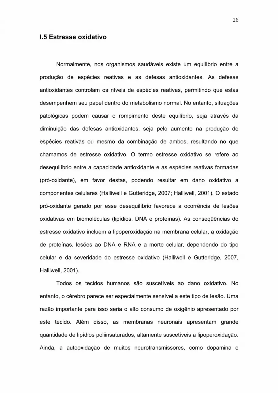
2004). A figura 3 mostra os sítios de formação de radicais livres de oxigênio e
as correspondentes defesas antioxidantes.

25
Figura 3. Geração de espécies reativas de oxigênio e as correspondentes defesas
antioxidantes. O2●-, radical superóxido; H2O2, peróxido de hidrogênio; OH●, radical hidroxila;
GSH, glutationa reduzida; GSSG, glutationa oxidada; GSH-Px, glutationa peroxidase; GR,
glutationa redutase; SOD, superóxido dismutase; CAT, catalase (Adaptado de Wajnet et al.,
2004).
O2●- H2O2
OH●
O2●- O2
O2
Cadeia transportadora
de elétrons mitocondrial
O2
O2
Xantina oxidase
O2
SOD
H2O
CAT
GSH-Px
GSSG
GSH
NADPH oxidase
NADPH
NADP+ Fe2+
Fe3+
Reação de Fenton
GR
26
I.5 Estresse oxidativo
Normalmente, nos organismos saudáveis existe um equilíbrio entre a
produção de espécies reativas e as defesas antioxidantes. As defesas
antioxidantes controlam os níveis de espécies reativas, permitindo que estas
desempenhem seu papel dentro do metabolismo normal. No entanto, situações
patológicas podem causar o rompimento deste equilíbrio, seja através da
diminuição das defesas antioxidantes, seja pelo aumento na produção de
espécies reativas ou mesmo da combinação de ambos, resultando no que
chamamos de estresse oxidativo. O termo estresse oxidativo se refere ao
desequilíbrio entre a capacidade antioxidante e as espécies reativas formadas
(pró-oxidante), em favor destas, podendo resultar em dano oxidativo a
componentes celulares (Halliwell e Gutteridge, 2007; Halliwell, 2001). O estado
pró-oxidante gerado por esse desequilíbrio favorece a ocorrência de lesões
oxidativas em biomoléculas (lipídios, DNA e proteínas). As conseqüências do
estresse oxidativo incluem a lipoperoxidação na membrana celular, a oxidação
de proteínas, lesões ao DNA e RNA e a morte celular, dependendo do tipo
celular e da severidade do estresse oxidativo (Halliwell e Gutteridge, 2007,
Halliwell, 2001).
Todos os tecidos humanos são suscetíveis ao dano oxidativo. No
entanto, o cérebro parece ser especialmente sensível a este tipo de lesão. Uma
razão importante para isso seria o alto consumo de oxigênio apresentado por
este tecido. Além disso, as membranas neuronais apresentam grande
quantidade de lipídios poliinsaturados, altamente suscetíveis a lipoperoxidação.
Ainda, a autooxidação de muitos neurotransmissores, como dopamina e

27
noradrenalina, gera espécies reativas, sendo que esta autooxidação pode ser
acelerada pela presença de ferro, amplamente distribuído no cérebro. Por fim,
o tecido cerebral apresenta um baixo nível de defesas antioxidantes (Halliwell e
Gutteridge, 2007).
Existem evidências crescentes sugerindo que as espécies reativas
desempenham um papel importante na patogênese de muitas doenças, como
diabetes, neoplasias, aterosclerose, doenças inflamatórias e doenças
neurodegenerativas, em particular a doença de Alzheimer, a doença de
Parkinson e a esclerose lateral amiotrófica (Reznick e Packer, 1993;
Przedborski et al., 1996; Bem–Menachem et al., 2000).
Tem também sido recentemente demonstrado que o estresse oxidativo
atua em vários erros inatos do metabolismo (Colomé et al., 2000, Wajner et al.,
2004). A produção excessiva de radicais livres e a redução das defesas
antioxidantes ocorrem em algumas acidemias orgânicas, como nas acidemias
glutárica (Kolker et al., 2001; Latini et al., 2005; Latini et al., 2007), propiônica e
metilmalônica (Fontella et al., 2000), bem como em aminoacidopatias como na
homocistinúria (Streck et al., 2003; Stefanello et al., 2005), tirosinemia tipo I
(Bird et al., 1995) e fenilcetonúria (Sierra et al., 1998; Artuch et al., 2001; Hagen
et al., 2002; Artuch et al., 2004; Sirtori et al., 2005; Sitta et al., 2006) e também
na doença peroxissomal adrenoleucodistrofia ligada ao X (Vargas et al., 2004;
Deon et al., 2006), sugerindo que o estresse oxidativo possa estar envolvido no
dano neurológico observado nessas doenças.
Estudos em animais demonstraram que os aminoácidos de cadeia
ramificada (leucina, isoleucina e valina) e seus respectivos α-cetoácidos
acumulados na DXB estimulam a lipoperoxidação em homogeneizado de

28
cérebro de ratos. (Fontella et al., 2002). Ainda, foi demonstrado que estes
compostos, principalmente a leucina e o ácido α-cetoisocapróico, reduzem a
capacidade do cérebro em modular o dano associado ao aumento na produção
de radicais livres e que a lipoperoxidação estimulada pela leucina pode ser
atenuada por antioxidantes como vitaminas C e E, glutationa reduzida e
superóxido dismutase (Bridi et al., 2003; Bridi et al., 2005a; Bridi et al., 2005b).
Embora o mecanismo responsável pelo estresse oxidativo nos erros
inatos do metabolismo não seja totalmente compreendido, é possível que o
acúmulo de metabólitos tóxicos induza a formação excessiva de radicais livres.
Além disso, é provável que a restrição dietética a qual muitos dos pacientes
com erros inatos do metabolismo são submetidos, cause redução nas defesas
antioxidantes devido à deficiência de nutrientes essenciais, como vitaminas e
minerais (Artuch et al., 2004).

29
II. OBJETIVOS
II.1 Objetivo geral
Considerando que os mecanismos envolvidos no dano neurológico
apresentado pelos pacientes com DXB ainda não estão totalmente
esclarecidos e que estudos em animais têm demonstrado que o estresse
oxidativo é induzido pelos metabólitos acumulados na DXB, o objetivo deste
estudo foi investigar vários parâmetros de estresse oxidativo em amostras de
plasma e eritrócitos de pacientes com DXB, antes e durante o tratamento, no
intuito de melhor entender a fisiopatologia e o efeito do tratamento nestes
indivíduos.
II.2 Objetivos específicos
Capitulo I - Avaliar parâmetros de estresse oxidativo, como a medida
das espécies reativas ao ácido tiobarbitúrico (TBARS), a medida da reatividade
antioxidante total (TAR) e a medida do status antioxidante total (TAS) em
plasma de pacientes não tratados (no diagnóstico) com doença do xarope do
bordo.
Capitulo II - Avaliar parâmetros de estresse oxidativo, como a medida
das espécies reativas ao ácido tiobarbitúrico (TBARS), a medida da reatividade
antioxidante total (TAR) e a medida do status antioxidante total (TAS) em

30
plasma de pacientes com doença do xarope do bordo sob tratamento dietético
com níveis baixos ou níveis altos de leucina.
Capitulo III – Avaliar os níveis de selênio no plasma de pacientes com
doença do xarope do bordo não tratados (no diagnóstico) e durante o
tratamento dietético, bem como a atividade das enzimas glutationa peroxidase
(GSH-Px), catalase (CAT) e superóxido dismutase (SOD) em eritrócitos de
pacientes em tratamento.
Capitulo IV – Avaliar parâmetros de estresse oxidativo, como a medida
das espécies reativas ao ácido tiobarbitúrico (TBARS) e a medida da
reatividade antioxidante total (TAR), e outros parâmetros bioquímicos (glicose,
colesterol total e frações, triglicerídeos, albumina, transaminases, creatina
quinase, lactato desidrogenase, uréia, creatinina e ácido úrico) em plasma de
pacientes com doença do xarope do bordo em tratamento dietético, a fim de
verificar se há correlação entre os parâmetros de estresse oxidativo e os
parâmetros bioquímicos.

31
III. RESULTADOS
Os resultados estão apresentados na forma de artigos científicos.
III.1 Capítulo I – Artigo 01
Evidence that oxidative stress is increased in plasma from patients with
maple syrup urine disease
Alethéa G. Barschak, Angela Sitta, Marion Deon, Marcella H. de Oliveira,
Alexsandro Haeser, Carlos S. Dutra-Filho, Moacir Wajner, Carmen R. Vargas
Este trabalho foi desenvolvido no Programa de Pós-Graduação em Ciências
Biológicas: Bioquímica, ICBS, UFRGS.
Periódico: Metabolic Brain disease
Status: Publicado

Metab Brain Dis (2006) 21:279–286DOI 10.1007/s11011-006-9030-5
ORIGINAL PAPER
Evidence that oxidative stress is increased in plasmafrom patients with maple syrup urine disease
Alethea G. Barschak · Angela Sitta · Marion Deon ·Marcella H. de Oliveira · Alexsandro Haeser ·Carlos S. Dutra-Filho · Moacir Wajner ·Carmen R. Vargas
Received: 17 January 2006 / Accepted: 8 May 2006 /Published online: 8 November 2006C© Springer Science+Business Media, Inc. 2006
Abstract Maple syrup urine disease (MSUD) or branched-chain α-keto aciduria (BCKA) isan inherited disorder caused by a deficiency of the branched-chain α-keto acid dehydrogenasecomplex (BCKAD) activity. The blockage of this pathway leads to tissue accumulation of thebranched-chain amino acids (BCAA) leucine, isoleucine and valine and their respective keto-acids. The clinical features presented by MSUD patients include ketoacidosis, convulsions,coma, psychomotor delay and mental retardation. The mechanism of brain damage in thisdisease is still poorly understood. However, an increase in lipid peroxidation in vitro incerebral cortex of young rats as well as a decrease in the antioxidant defenses has beenpreviously observed. In the present work we evaluated different oxidative stress parameters,named reactive species of thiobarbituric acid (TBARS), total antioxidant reactivity (TAR)and total antioxidant status (TAS) in plasma of MSUD patients in order to evaluate whetheroxidative stress is involved in this disorder. We verified a marked increase of plasma TBARSmeasurements, which is indicative of increased lipid peroxidation, as well as a decrease onplasma TAR reflecting a deficient capacity to efficiently modulate the damage associatedwith an increased production of reactive species. In contrast, TAS was not changed indicatingthat the total content of antioxidants in plasma of patients affected by MSUD was not altered.
A. G. Barschak · A. Sitta · M. Deon · C. S. Dutra-Filho · M. Wajner · C. R. VargasDepartamento de Bioquımica, Instituto de Ciencias Basicas da Saude, Universidade Federal do RioGrande do Sul, Porto Alegre, RS, Brazil
A. G. Barschak · A. Sitta · M. Deon · M. H. de Oliveira · M. Wajner · C. R. Vargas (�)Servico de Genetica Medica, Hospital de Clınicas de Porto Alegre, Rua Ramiro Barcelos, 2350 CEP90.035–903, Porto Alegre, RS, Brazile-mail: [email protected]
A. Haeser · C. R. VargasPrograma de Pos-Graduacao em Ciencias Farmaceuticas, Universidade Federal do Rio Grande do Sul,Porto Alegre, RS, Brazil
C. R. VargasDepartamento de Analises, Faculdade de Farmacia, Universidade Federal do Rio Grande do Sul, PortoAlegre, RS, Brazil
Springer

280 Metab Brain Dis (2006) 21:279–286
These results suggest that free radical generation is elicited in MSUD and is possibly involvedin the pathophysiology of the tissue damage found in this disorder.
Keywords Maple syrup urine disease . Oxidative stress . Lipid peroxidation . Antioxidantdefenses
Introduction
Maple syrup urine disease (MSUD) or branched-chain α-keto aciduria (BCKA) is an autoso-mal recessive metabolic disorder caused by a severe deficiency of the branched-chain α-ketoacid dehydrogenase complex (BCKAD) activity. The blockage in this enzyme complex leadsto tissue accumulation of the branched-chain amino acids (BCAA) leucine, isoleucine and va-line as well as their corresponding branched chain α-keto acids (BCKA) α-ketoisocaproate,α-keto-β-methylvalerate and α-ketoisovalerate, respectively (Chuang and Shih, 2001; Treacyet al., 1992).
The major clinical features presented by MSUD patients include ketoacidosis, hypo-glycemia, poor feeding, apnea, ataxia, convulsions, coma, psychomotor delay and mentalretardation. Magnetic resonance imaging studies have demonstrated generalized edema andhypomyelination/demyelination in central nervous system (CNS) of MSUD patients (Chuangand Shih, 2001; Schonberger et al., 2004).
MSUD presents heterogeneous molecular and clinical phenotypes range from a severeclassic form with neonatal onset to milder variant forms with later onset, and presentingdifferent residual enzyme activity (Chuang and Shih, 2001; Schadewaldt and Wendel, 1997).
The aim of MSUD treatment is to keep the BCAA plasma concentrations in the normalrange, protecting the brain from injury. The treatment consists of a low protein diet and asemi-synthetic formula restricted in BCAA and supplemented with essential amino acids.Metabolic intoxication may cause a fatal outcome in untreated patients (Chuang and Shih,2001; Danner and Elsas, 1989).
The mechanisms of the neurological symptoms presented by MSUD patients are stillpoorly understood. However, considering that increased concentrations of leucine and/or α-ketoisocaproate were associated with the appearance of neurological symptoms, these com-pounds seem to be the main important neurotoxic metabolites in MSUD (Chuang and Shih,2001; Snyderman et al., 1964). Furthermore, it has been demonstrated that the metabolitiesaccumulating in MSUD cause impairment of energy metabolism by inhibiting the electrontransport chain (Sgaravatti et al., 2003) and creatine kinase activity in rat brain (Pilla et al.,2003). Other investigators demonstrated that the BCAA and/or BCKA that accumulate inMSUD provoke neuronal apoptosis (Jouvet et al., 2000), as well as convulsions (Coitinhoet al., 2001), impairment of neurotransmitter synthesis and function (Zielke et al., 1996;Tavares et al., 2000), myelin alteration (Treacy et al., 1992; Tribble and Shapira, 1983;Taketomi et al., 1983) and reduced uptake of essential amino acids by the brain (Araujoet al., 2001).
Free radicals seem to be involved in a large number of human diseases. Increasingevidence has shown that damage caused by free radicals is an important contributing factorin chronic-inflammatory, vasculary, neoplastic and neurodegenerative diseases (Halliwell,1994; Reznick and Packer, 1993; Przedborski et al., 1996; Bem-Menachem et al., 2000).
Oxidative stress has been observed in some inborn errors of intermediary metabolismowing to the accumulation of toxic metabolites which leads to excessive free radical produc-tion (Colome et al., 2000). Restricted diets utilized to treat patients affected by metabolicdisorders may result in a low antioxidant status (Colome et al., 2000).
Springer

Metab Brain Dis (2006) 21:279–286 281
Recently, it was demonstrated that the BCAA and their respective BCKA that accumu-late in MSUD stimulate in vitro lipid peroxidation in brain homogenates of rats (Fontellaet al., 2002). It was later demonstrated that these compounds, particularly leucine and α-ketoisocaproate, not only stimulate in vitro lipid peroxidation but also reduce the cerebralcapacity to modulate the damage associated with the increased free radical production (Bridiet al., 2003, 2005a). Furthermore, it was shown that the increased lipid peroxidation inducedby leucine could be attenuated by the free radicals scavengers ascorbic acid, α-tocopherol,gluthatione and superoxide dismutase (Bridi et al., 2005b).
The aim of the present work was to evaluate some parameters of oxidative stress, namelythiobarbituric acid-reactive substances (TBARS), total antioxidant reactivity (TAR) and totalantioxidant status (TAS), in plasma of MSUD patients at the time of diagnosis in order toverify whether free radicals could be involved in the pathophysiology of this disease.
Material and methods
Patients and controls
Plasma from five MSUD patients (classic form) aged between 15 days and 4 months atdiagnosis were used to evaluate the parameters of oxidative stress. The most commonclinical features presented by these patients were convulsions, hypoglycemia, poor feeding,ketoacidosis and psychomotor delay. Samples were obtained at the time of the diagnosis,which was made by increased plasma levels of leucine (2,346.1 ± 810.7 µmol/L), isoleucine(304.8 ± 185.2 µmol/L) and valine (456.5 ± 275.1 µmol/L) by HPLC method (Joseph andMarsden, 1986). Control group was composed of healthy age matched individuals (leucine158.33 ± 37.63 µmol/L; isoleucine 76.54 ± 18.02 µmol/L; valine 260.73 ± 39.79 µmol/L).
Reagents
All chemicals were of PA purity and were purchased from Sigma (St. Louis, MO, USA)except for thiobarbituric acid, which was purchased from Merck (Darmstadt, Germany) anda kit for TAS measurement that was purchased from Randox Laboratories (Antrim, UnitedKingdom). TAR was assayed using a beta liquid scintillation spectrometer (Wallac model1409) and TBARS was measured with a spectrofluorimeter (Hitachi F2000).
Plasma preparation
Plasma was prepared from whole blood samples obtained from fasting individuals (controlsand MSUD patients) by venous puncture with heparinized vials. Whole blood was centrifugedat 1,000g. Plasma was removed by aspiration and frozen at −80◦C until determination.
Thiobarbituric Acid-Reactive Species (TBARS)
Thiobarbituric acid-reactive substances (TBARS) were determined according to the methoddescribed by Buege and Aust (1978). Briefly, 250 µL of 10% trichloroacetic acid were addedto 125 µL of plasma, then 375 µl 0.67% thiobarbituric acid (in 7.1% sodium sulphate) wereadded and incubated at 100◦C for 30 min. After the incubation, the mixture was extracted with750 µL butanol. The resulting pink stained TBARS were determined in a spectrofluorimeterat 515 nm. Calibration curve was performed using 1,1,3,3-tetramethoxypropane subjected
Springer

282 Metab Brain Dis (2006) 21:279–286
to the same treatment as that of the samples. TBARS were calculated as nmol TBARS/mgprotein.
Total Antioxidant Reactivity (TAR)
TAR, which represents the quality of the tissue antioxidants, was determined by measur-ing the luminol chemiluminescence intensity induced by 2,2′-azo-bis-(2-amidinopropane)(ABAP) according to the method of Lissi et al. (1992). The background chemiluminescencewas measured by adding 4 mL of 2 mM ABAP (in 0.1 M glycine buffer, pH 8.6) into a glassscintillation vial. Ten microliters of luminol (4 mM) were added to each vial and the chemi-luminescence was measured. This was considered to be the basal value. Ten microliters of25–200 µM Trolox (curve calibration) or plasma was then added and the chemiluminescencewas measured during 60 s. The Trolox and plasma addition reduces the chemiluminescence.The rapid reduction in luminol intensity is considered as a measure of the TAR capacity.TAR measurement was calculated as nmol Trolox/mg protein.
Total Antioxidant Status (TAS)
TAS, which represents the quantity of the tissue antioxidants, was determined by using a kitfrom RANDOX Laboratories. The plasma sample was incubated with ABTS (2,2′-azino-di-[3-ethylbenzthiazoline sulphonate]) plus a peroxidase (metmyoglobin) and H2O2 to producethe cation ABTS+. A relatively stable blue–green color occurred and was measured at 37◦Cat 600 nm. Antioxidants in the added sample cause suppression of this color production toa degree which was proportional to their concentration (Miller et al., 1993; Yu and Ong,1999). The results were expressed in mmol/L plasma.
Protein determination
Protein concentrations were determined by the biuret method from Labtest r© (Gornall et al.,1949), using albumin as standard.
Statistical analysis
The Student t test was used to compare results from controls and MSUD patients. A p valueless than 0.05 was considered significant. All analyses were performed using the StatisticalPackage for the Social Sciences (SPSS) software in a PC-compatible computer.
Results
Figure 1 shows that TBARS was significantly increased in plasma of MSUD patients whencompared to control group [t(8) = −4.552, p < 0.01], indicating that lipid peroxidation isstimulated in MSUD patients.
TAR measurement, which is a measure of the tissue capacity to react with free radicals,was markedly reduced [t(8) = 3.021, p < 0.05] in plasma of MSUD patients (Fig. 2). Theseresults suggest a deficient capacity of plasma to modulate the damage associated withthe enhanced production of reactive species in these MSUD patients. Finally, it was alsoobserved that TAS measurement, which represents the quantity of the tissue antioxidants,
Springer

Metab Brain Dis (2006) 21:279–286 283
TBARS
0
0.002
0.004
0.006
0.008
0.01
0.012
Control MSUD
nm
ol T
BA
RS
/ m
g p
rote
in **
Fig. 1 Plasma thiobarbituricacid reactive species (TBARS)from MSUD patients andcontrols. Data represent the mean± SD (n = 5). ∗∗p < 0.01(Student’ t test for unpairedsamples) compared to control
was not altered in plasma of MSUD patients [t(6) = −0.713, p > 0.05] (Fig. 3), suggestingthat the total nonenzymatic antioxidant defenses were not altered.
Discussion
Neurological symptoms are frequent in MSUD patients and untreated patients normallyhave a fatal outcome (Chuang and Shih, 2001; Danner and Elsas, 1989). Leucine and/orα-ketoisocaproate are considered the main neurotoxic metabolites in these disease (Chuangand Shih, 2001; Snyderman et al., 1964). However, the mechanisms underlying the sequelaepresented by these patients are not well understood.
It was previously demonstrated that the BCAA and BCKA accumulating in MSUD stim-ulate in vitro lipid peroxidation and reduce the antioxidant defenses in cerebral homogenatesof young rats (Fontella et al., 2002; Bridi et al., 2003, 2005a). However, to our knowledgethere is no report in the literature assessing whether oxidative stress occurs in tissues fromMSUD patients. Therefore, in the present study we investigated some parameters of oxida-tive stress in plasma from these patients which were not under any therapy. So our resultscannot be attributed to any medication.
TAR
0
2
4
6
8
10
12
Control MSUD
nm
ol T
rolo
x / m
g p
rote
in
*
Fig. 2 Plasma total antioxidant reactivity (TAR) from MSUD patients and controls. Data represent the mean± SD (n = 5). ∗p < 0.05 (Student’ t test for unpaired samples) compared to control
Springer

284 Metab Brain Dis (2006) 21:279–286
TAS
0
0.5
1
1.5
2
Control MSUD
mm
ol/L
pla
sma
Fig. 3 Plasma total antioxidant status (TAS) from MSUD patients and controls. Data represent the mean ±SD (n = 4). No significant differences between means were found (Student’ t test for unpaired samples)
We demonstrated a significant increase of TBARS in plasma from MSUD patients. Con-sidering that TBARS reflects the content of malondialdehyde, an end product of lipid break-down due to lipid peroxidation (Halliwell and Gutteridge, 2001; Esterbauer and Cheeseman,1990), our data indicate that lipid peroxidation is induced in MSUD patients, probably sec-ondary to free radical generation. Despite the fact that we did not find any decrease of totalantioxidant defenses in plasma from these patients as indicated by TAS values TAR, whichcorresponds to a useful index of the capacity of a given tissue to modulate the damageassociated with an increased production of free radicals and mainly reflects the quality ofantioxidants (Lissi et al., 1995), was significantly decreased in the patients studied. Theseresults indicate a deficient capacity of plasma from MSUD patients to rapidly handle anincrease of reactive species. Considering that an imbalance between the total antioxidantdefenses and the reactive species formed in the tissues are indicative of oxidative stress(Halliwell and Gutteridge, 2001), it is proposed that free radical generation is involved inthe pathophysiology of the tissue damage found in MSUD.
Oxidative stress has been considered an important contributor to brain damage in neu-rodegenerative diseases, seizures and demyelination (Halliwell, 2001; Mendez-Alvarez et al.,2001; Karelson et al., 2001), probably because brain has relatively low levels of antioxidantdefenses (Halliwell and Gutteridge, 2001), as well as high lipid content, specially unsaturatedfatty acids, and iron that stimulates the Fenton reaction being therefore highly susceptible toreactive species attack. Taken together our present in vivo results and those demonstrating instudies in vitro with rats that the metabolities accumulated in MSUD cause a stimulation oflipid peroxidation and a reduction of brain antioxidant defenses, (Fontella et al., 2002; Bridiet al., 2003, 2005a), suggest that oxidative stress is probably involved in the pathophysiologyof MSUD.
To our knowledge this is the first report demonstrating increased oxidative stress in patientsaffected by MSUD. Our results should, however, be taken with caution and confirmed witha higher number of patients and with other techniques to measure oxidative stress. In thiscontext, CSF specimens may be useful in order to evaluate if the brain is also a target forreactive species. In case the present results are confirmed, we may conclude that oxidativestress contributes at least in part to the severe neurological dysfunction found in MSUD.
Acknowledgements This work was supported by grants from Brazilian National Research Council (CNPq),FAPERGS, and FIPE/HCPA—Brazil.
Springer

Metab Brain Dis (2006) 21:279–286 285
References
Araujo P, Wassermann GF, Tallini K, Furlanetto V, Vargas CR, Wannmacher CMD, Dutra-Filho CS, WyseATS, Wajner M (2001) Reduction of large neutral amino acid level in plasma and brain of hyperleucinemicrats. Neurochem Int 38:529–537
Bem-Menachem E, Kyllerman R, Markleind S (2000) Superoxide dismutase and glutathione peroxidasefunction in progressive myoclonus epilepsies. Epilepsy Res 40:33–39
Bridi R, Araldi J, Sgarbi MB, Testa CG, Durigon K, Wajner M, Dutra-Filho CS (2003) Induction of oxidativestress in rat brain by the metabolites accumulating in maple syrup urine disease. Int J Devl Neuroscience21:327–332
Bridi R, Braun CA, Zorzi GK, Wannmacher CM, Wajner M, Lissi EG, Dutra-Filho CS (2005a) Alpha-ketoacids accumulating in maple syrup urine disease stimulate lipid peroxidation and reduce antioxidantdefences in cerebral cortex from young rats. Metab Brain Dis Jun 20(2):155–167
Bridi R, Latini A, Braum CA, Zorzi GK, Wajner M, Lissi E, Dutra-Filho CS (2005b) Evaluation of themechanism involved in leucine-induced oxidative damage in cerebral cortex of young rats. Free RadicRes Jan 39(1):71–79
Buege JA, Aust SD (1978) Microssomal lipid peroxidation. Methods Enzymol 52:302–309Chuang DT, Shih VE (2001) Maple syrup urine disease (branche-chain ketoaciduria). In: Scriver CR, Beaudt
AL, Sly WL, Valle D (eds) The metabolic and molecular bases of inherited disease. McGraw-Hill,New York, pp 1971–2005
Coitinho AS, de Mello CF, Lima TT, de Bastiani J, Fighera MR, Wajner M (2001) Pharmacological evidencethat alpha-keto isovaleric acid induces convulsions through GABAergic and glutamatergic mechanismsin rats. Brain Res 894:68–73
Colome C, SIerra C, Vilaseca MA (2000) Congenital errors of metabolism: Cause of oxidative stress? MedClin 115(3):111–117
Danner DJ, Elsas JL II (1989) Disorders of branched chain amino acid and keto acid metabolism. In: ScriverCR, Beaudt AL, Sly WL, Valle D (eds) The metabolic and molecular bases of inherited disease. McGraw-Hill, New York, pp 671–692
Esterbauer H, Cheeseman KH (1990) Determination of aldehydic lipid peroxidation products: Malonaldehydeand 4-hydroxynonenal. Methods Enzymol 186:407–421
Fontella FU, Gassen E, Pulrolnik V, Wannmacher CMD, Klein AB, Wajner M, Dutra CS (2002) Stimulationof lipid peroxidation in vitro in rat brain by metabolites accumulating in maple syrup urine disease. MetabBrain Dis 17:47–54
Gornall AG, Bardawill CJ, David MM (1949) Determination of serum proteins by means of the biuret reaction.J Biol Chem 177:751–766
Halliwell B (1994) Free radicals, antioxidants and human disease: Curiosity, cause or consequence? Lancet344:721–724
Halliwell B (2001) Role of free radicals in the neurodegenerative diseases. Drugs Aging 18:685–716Halliwell B, Gutteridge JMC (2001) Detection of free radicals and others reactive species: Trapping and
fingerprinting. In: Halliwell B, Gutteridge JMC (eds) Free radicals in biology and medicine. OxfordUniversity Press, Oxford, UK, pp 351–425
Joseph MH, Marsden CA (1986) Amino acids and small peptides. In: Lim CF (ed) HPLC of small peptides.IRL Press, Oxford, pp 13–27
Jouvet P, Rustin P, Taylor DL, Pocock JM, Felderhoff-Mueser U, Mazarakis ND, Sarraf C, Joashi U, KoszmaM, Greewood K, Edwards AD, Mehmet H (2000) Branched chain amino acids induce apoptosis inneural cells without mitochondrial membrane despolarization or cytochrome c release: Implications forneurological impairment associated with maple syrup urine disease. Mol Biol Cell 11:1919–1932
Karelson E, Bogdanovic N, Garlind A, Winblad B, Zilmer K, Kullisaar T, Vihalemm T, Kairane C, ZilmerM (2001) The cerebrocortical areas in normal brain aging and in the Alzheimer’s disease: Noticeabledifference in the lipid peroxidation level and in antioxidant defense. Neurochem Res 26:353–361
Lissi E, Pascual C, Del Castillo MD (1992) Luminol luminescence induced by 2,2′-azo-bis-(2-amidinopropane) thermolysis. Free Rad Res Commun 17:299–311
Lissi E, Salim-Hanna M, Pascual C, Del Castillo MD (1995) Evaluation of total antioxidant potential (TRAP)and total antioxidant reactivity from luminol-enhanced chemiluminescence measurements. Free RadicBiol Med 18:153–158
Mendez-Alvarez E, Soto-Otero R, Hermida-Aeijeiras A, Lopez-Real AM, Labandeira-Garcıa JL (2001)Effects of aluminium and zinc on the oxidative stress caused by 6-hydroxydopamine autoxida-tion: Relevance for the pathogenesis of Parkinson’s disease. Biochim Biophys Acta 1586:155–168
Springer

286 Metab Brain Dis (2006) 21:279–286
Miller NJ, Rice-Evans C, Davies MJ, Gopinathan V, Milner A (1993) A novel method for measuring antiox-idant capacity and its application to monitoring the antioxidant status in premature neonates. Clin Sci84:407–412
Pilla C, Cardozo RFD, Dutra CS, Wyze ATS, Wajner M, Wannmacher CMD (2003) Effect of leucineadministration on creatine kinase activity in rat brain. Metab Brain Dis 18:17–25
Przedborski S, Donaldson DBS, Jakowec M, Kish JS, Guttman M, Rosoklija G, Hays AP (1996) Brainsuperoxide dismutase, catalase and glutathione peroxidase activities in amyotrophic lateral sclerosis. AnnNeurol 39:158–165
Reznick AZ, Packer L (1993) Free radicals and antioxidants in muscular neurological diseases and disorders.In: Poli G, Albano E, Dianzani MU (eds) Free radicals: From basic science to medicine. BirkhauserVerlag, Basel, pp 425–437
Schadewaldt P, Wendel U (1997) Metabolism of branched-chain amino acids in maple syrup urine disease.Eur J Pediatr 156(Suppl 1):S62–S66
Schonberger S, Schweiger B, Schwahn B, Schwarz M, Wendel U (2004) Dysmyelination in the brain ofadolescents and young adults with maple syrup urine disease. Mol Genet Metab 82:69–75
Sgaravatti AM, Rosa RB, Schuck PF, Ribeiro CAJ, Wannacher CMD,Wyse ATS, Dutra-Filho CS, WajnerM (2003) Inhibition of brain energy metabolism by the α-keto acids accumulating in maple syrup urinedisease. Biochim Biophys Acta 1639:232–238
Snyderman SE, Norton PM, Roitman E (1964) Maple syrup urine disease with particular reference to diettherapy. Pediatrics 34:454–472
Taketomi T, Kunishita T, Hara A, Mizushima S (1983) Abnormal protein and lipid compositions of thecerebral myelin in patient with maple syrup urine disease. Jpn J Exp Med 53:109–116
Tavares RG, Santos CES, Tasca C, Wajner M, Souza DO, Dutra-Filho CS (2000) Inhibition of glutamateuptake into synaptic vesicles of rat brain by the metabolites accumulating in maple syrup urine disease. JNeurol Sci 181:44–49
Treacy E, Clow CL, Reade TR, Chitayat D, Mamer OA, Scriver CR (1992) Maple syrup urine disease:Interrelationship between branched chain amino-, oxo- and hydroxyacids; implications for treatment;association with CNS dysmelination. J Inherit Metab Dis 15:121–135
Tribble D, Shapira R (1983) Myelin proteins: Degradation in rat brain initiated by metabolites causative ofmaple syrup urine disease. Biochem Biophys Res Commun 114:440–446
Yu T-W, Ong ChN (1999) Lag-time measurement of antioxidant capacity using myoglobin and 2,29-azino-bis(3-ethyl-benzthiazoline-6-sulfonic acid): Rationale, application and limitation. Anal Biochem275:217–223
Zielke HR, Huang Y, Tildon JT, Zielke CL, Baab PJ (1996) Elevation of amino acids in the interstitial space ofthe rat brain following infusion of large neutral amino and keto acids by microdialysis: Leucine infusion.Dev Neurosci 18:420–425
Springer
40
III.2 Capítulo II – Artigo 02
Oxidative stress in plasma from maple syrup urine disease patients
during treatment
Alethéa G. Barschak, Angela Sitta, Marion Deon, Amanda T. Barden, Carlos S.
Dutra-Filho, Moacir Wajner, Carmen R. Vargas
Este trabalho foi desenvolvido no Programa de Pós-Graduação em Ciências
Biológicas: Bioquímica, ICBS, UFRGS.
Periódico: Metabolic Brain Disease
Status: Publicado
ORIGINAL PAPER
Oxidative stress in plasma from maple syrup urine diseasepatients during treatment
Alethéa G. Barschak & Angela Sitta & Marion Deon &
Amanda T. Barden & Carlos S. Dutra-Filho &
Moacir Wajner & Carmen R. Vargas
Received: 6 December 2006 /Accepted: 22 August 2007 /Published online: 17 November 2007# Springer Science + Business Media, LLC 2007
Abstract Maple Syrup Urine Disease (MSUD) is an autossomal recessive metabolicdisorder caused by a deficiency of branched-chain α-keto acid dehydrogenasecomplex activity leading to accumulation of the branched-chain amino acids leucine,isoleucine and valine and their corresponding branched-chain α-keto acids. Affectedpatients usually present hypoglycemia, ketoacidosis, convulsions, poor feeding,coma, psychomotor delay and mental retardation. Considering that the pathophys-iology of MSUD is still poorly understood, in this study we evaluated someparameters of oxidative stress, namely thiobarbituric acid-reactive substances(TBARS), total antioxidant reactivity (TAR) and total antioxidant status (TAS) inplasma from treated MSUD patients presenting high and low plasma leucine levels.We verified a significant increase of TBARS (lipid peroxidation) and a decrease ofTAR (capacity to rapidly react with free radicals) in plasma from treated MSUDpatients with low and with high plasma levels of leucine compared to the controlgroup. It was also verified that TAS (quantity of tissue antioxidants) was not alteredin plasma from treated MSUD patients with low and high blood leucine levels.Finally, we found no correlation between leucine, valine and isoleucine levels withthe various parameters of oxidative stress. These results are indicative that increasedlipid oxidative damage and decreased antioxidant defenses occur in plasma ofMSUD patients and that the accumulating branched-chain amino acids are probablynot directly associated to oxidative stress in this disorder.
Metab Brain Dis (2008) 23:71–80DOI 10.1007/s11011-007-9077-y
A. G. Barschak : A. Sitta :M. Deon :M. Wajner : C. R. Vargas (*)Serviço de Genética Médica, HCPA, Rua Ramiro Barcelos, 2350, CEP 90.035-903 Porto Alegre,Rio Grande do Sul, Brazile-mail: [email protected]
A. G. Barschak : A. Sitta :M. Deon : C. S. Dutra-Filho :M. Wajner : C. R. VargasDepartamento de Bioquímica, Instituto de Ciências Básicas da Saúde,Universidade Federal do Rio Grande do Sul, Porto Alegre, Rio Grande do Sul, Brazil
A. T. Barden : C. R. VargasDepartamento de Análises, Faculdade de Farmácia, Universidade Federal do Rio Grande do Sul,Porto Alegre, Rio Grande do Sul, Brazil
Keywords MSUD . Oxidative stress . Free radicals . Leucine
Introduction
Maple Syrup Urine Disease (MSUD) is an inherited disorder affecting the metabolismof branched-chain amino acids (BCAA) leucine (Leu), isoleucine (Ile) and valine(Val). The activity of the branched-chain α-keto acid dehydrogenase complex(BCKAD) is deficient in MSUD leading to tissue accumulation of BCAA (Leu, Ile eVal), as well as their corresponding transaminated branched-chainα-keto acids (BCKA)α-ketoisocaproate, α-keto-β-methylvalerate and α-ketoisovalerate (Chuang and Shih2001; Treacy et al. 1992).
The clinical and biochemical phenotypes of MSUD patients are heterogeneous.Patients can be divided into five phenotypes ranging from the classical form with aneonatal onset to milder variants with later onset (Chuang and Shih 2001;Schadewaldt and Wendel 1997). Individuals with MSUD usually present poorfeeding, convulsions, ketoacidosis, apnea, hypoglycemia, coma, ataxia, psychomotordelay and mental retardation, as well as generalized edema and hypomyelination/demyelination on magnetic resonance imaging studies of the central nervous system(CNS) (Chuang and Shih 2001; Schönberger et al. 2004). Therapy for this disease isbased on a natural protein restricted diet with low BCAA supplemented with a semi-synthetic formula of essential amino acids, vitamins and minerals. This treatmentminimizes the accumulation of the toxic metabolites and contributes to the survivalof the affected individuals, but do not prevent a variable degree of neurologicaldysfunction evidenced by developmental delay and mental retardation whosepathogenesis is poorly known (Chuang and Shih 2001). Leucine and/or α-ketoisocaproate are thought to be the main neurotoxic metabolites in this disorderonce their increased concentrations have been associated to the appearance ofneurological symptoms (Chuang and Shih 2001; Snyderman et al. 1964).
Free radicals and oxidative stress have been associated with a large number ofdiseases including various neurodegenerative disorders, epileptic seizures, demyelin-ation and dementia (Halliwell 1994; Reznick and Packer 1993; Przedborski et al. 1996;Ben-Menachem et al. 2000). This may occur because the CNS is highly susceptible tooxidative damage due to the relatively low activity of antioxidant defenses, high ironcontent, high lipid content, specially unsaturated fatty acids, and high oxygenconsumption (Halliwell and Gutteridge 2001). Oxidative stress has been also observedin some inborn errors of intermediary metabolism, and this has been attributed to theaccumulation of toxic metabolites which are able to induce excessive free radicalgeneration (Colome et al. 2000; Wajner et al. 2004). We cannot also exclude thepossibility that the restricted diets used to treat patients with metabolic disorders maydecrease the tissue antioxidant defenses since they potentially deplete essentialnutrients involved in the antioxidant defenses (Halliwell and Gutteridge 2001).
With regards to MSUD, experimental animal studies have demonstrated that lipidperoxidation is stimulated in vitro by the BCAA and their respective BCKA in brainhomogenates of rats (Fontella et al. 2002). It was also verified that these metabolitesreduce in vitro the cerebral capacity to modulate the damage associated to increasedfree radical production (Bridi et al. 2003; Bridi et al. 2005a) and that the increased
72 Metab Brain Dis (2008) 23:71–80

lipid peroxidation induced in vitro in cerebral cortex of rats by leucine is attenuatedby the free radicals scavengers ascorbic acid, α-tocopherol, gluthatione andsuperoxide dismutase (Bridi et al. 2005b).
On the other hand, we recently verified that the antioxidant status and lipidperoxidation were significantly altered in plasma of MSUD patients at diagnosis,suggesting the involvement of oxidative stress in the pathogenesis of this disease(Barschak et al. 2006). In the present study, we evaluated various parameters ofoxidative stress in blood from treated MSUD patients showing high and low bloodleucine levels, in order to test whether this amino acid could be involved in theoxidative stress. The parameters analyzed were thiobarbituric acid-reactive substances(TBARS), total antioxidant reactivity (TAR) and total antioxidant status (TAS).
Materials and methods
Reagents
All chemicals were of PA purity and were purchased from Sigma (St. Louis, MO,USA) except thiobarbituric acid that was purchased from Merck (Darmstadt,Germany) and a kit for TAS measurement that was purchased from RandoxLaboratories (Antrim, United Kingdom). TAR was assayed using a beta liquidscintillation spectrometer (Wallac model 1409), TBARS was measured in aspectrofluorimeter (Hitachi F2000) and TAS in a double-bean spectrophotometerwith temperature control (Hitachi U-2001).
Subjects
Plasma specimens from ten treated MSUD patients with the classic form and fiveage matched controls were used to evaluate the parameters of oxidative stress. Thepatients were aged between 15 days and 4 months at diagnosis and followed atreatment that consisted of a natural protein restricted diet with low BCAA andsupplemented with a semi-synthetic formula of essential amino acids, vitamins andminerals. The patients ingested the following amounts of Leu (before 12 months ofage: 40–80 mg kg−1 day−1; after 1 year of age: 275–535 mg/day), Ile (before12 months of age: 20–50 mg kg−1 day−1; after 1 year of age: 165–325 mg/day) andVal (before 12 months of age: 20–60 mg kg−1 day−1; after 1 year of age: 190–375 mg/day). Plasma samples obtained from MSUD patients under treatment weredivided into two groups depending on blood Leu levels. In group I plasma Leulevels were lower than 100 μmol/l (36.3±17.1 μmol/l, treatment duration was 18.6±12.9 months), whereas in group II plasma Leu levels were higher than 600 μmol/l(1,314±914 μmol/l, treatment duration was 17.2±20.1 months). The control groupcorresponded to healthy age matched individuals (leucine 158±37.6 μmol/l;isoleucine 76.5±18.0 μmol/l; valine 260±39.8 μmol/l) (Table 1).
The present study was approved by the Ethical Committee of Hospital de Clínicasde Porto Alegre, RS, Brazil (protocol number 04-256). The parents of the patientsincluded in the present study gave informed consent.
Metab Brain Dis (2008) 23:71–80 73

Plasma preparation and amino acids determination
Plasma was prepared from whole blood samples obtained from fasting individuals(controls and MSUD patients) by venous puncture with heparinized vials. Wholeblood was centrifuged at 1,000×g and plasma was removed by aspiration and frozenat −80°C until analysis. Blood amino acids levels were measured by HPLC (Josephand Marsden 1986), with slight modifications (Wajner et al. 2000).
Thiobarbituric acid-reactive substances (TBARS)
Thiobarbituric acid-reactive substances (TBARS) were determined according to themethod described by Buege and Aust 1978. Briefly, 250 μl of 10% trichloroacetic acidwere added to 125 μl of plasma. Then 375 μl 0.67% thiobarbituric acid (in 7.1%sodium sulphate) were added and incubated at 100°C for 30 min. After the incubation,the mixture was extracted with 750 μl butanol. The resulting pink stained TBARSwere determined in a spectrofluorimeter at 515 nm. Calibration curve was performedusing 1,1,3,3-tetramethoxypropane subjected to the same treatment as that of thesamples. TBARS were calculated as nmol TBARS/mg protein.
Total antioxidant reactivity (TAR)
TAR, which represents the quality of the tissue antioxidants, was determined bymeasuringthe luminol chemiluminescence intensity induced by 2,2′-azo-bis-(2-amidinopropane)(ABAP) according to the method of Lissi et al. (1992). The background chemilumines-cence was measured by adding 4 ml of 2 mM ABAP (in 0.1 M glycine buffer, pH 8.6)into a glass scintillation vial. Ten microliters of luminol (4 mM) were added to each vialand the chemiluminescence was measured. This was considered to be the basal value.Ten microliters of 25–200 μM Trolox (curve calibration) or plasma was then added andthe chemiluminescence was measured during 60 s. The Trolox and plasma additionreduce the chemiluminescence. The rapid reduction in luminol intensity is considered asa measure of the TAR capacity. TAR measurement was calculated as nmol Trolox/mgprotein.
Total antioxidant status (TAS)
TAS, which represents the quantity of the tissue antioxidants, was determined by using akit from RANDOX Laboratories. The plasma sample was incubated with ABTS (2,2′-
Table 1 Plasma concentrations of leucine (Leu), isoleucine (Ile) and valine (Val) in controls and MSUDpatients during treatment
Leu (μmol/l) Ile (μmol/l) Val (μmol/l)
Controls 158±16.8 76.5±8.0 260±17.7MSUD patients group I 36.3±7.66 112±42.2 341±169MSUD patients group II 1,314±408* 198±62.6 384±154
Values represent mean±SE (n=5)*p<0.01, different from controls (ANOVA followed by the Duncan multiple range test)
74 Metab Brain Dis (2008) 23:71–80

azino-di-[3-ethylbenzthiazoline sulphonate]) plus a peroxidase (metmyoglobin) andH2O2 to produce the cation ABTS+. A relatively stable blue–green color occurred andwas measured at 37°C at 600 nm. Antioxidants in the added sample cause suppressionof this color production to a degree which is proportional to their concentration (Milleret al. 1993; Yu and Ong 1999). The results were expressed in millimole per liter.
Protein determination
Protein concentrations were determined by the Biuret method from Labtest®(Gornall et al. 1949), using bovine serum albumin as standard.
Statistical analysis
Results were analyzed by one-way ANOVA, followed by the Duncan multiple range testwhen appropriated (Altman 1991). Only significant F values are shown in the text.Correlations of plasma Leu, Val and Ile levels with TBARS, TAR or TAS were carriedout using the Pearson correlation coefficient. A p value less than 0.05 was consideredsignificant. All analyses were performed using the Statistical Package for the SocialSciences (SPSS) software in a PC-compatible computer.
Results
Figure 1 shows that TBARS measurement was significantly increased in plasmafrom MSUD patients both with low (group I) and high (group II) Leu levels, ascompared to the control group [F(2,12)=4.289, p<0.05]. These data indicate thatlipid peroxidation is stimulated in plasma from MSUD patients.
0
0.001
0.002
0.003
0.004
0.005
0.006
0.007
0.008
0.009
Control Group I Group II
TB
A (
nm
ol T
BA
. m
g p
rote
in -1
)
* *
Fig. 1 Comparison between thiobarbituric acid reactive substances (TBARS) in plasma from MSUDpatients during treatment and controls. Data represent the mean±SE (n=5). Single asterisk p<0.05,compared to controls (ANOVA, followed by the Duncan multiple range test). Group I: low plasma leucinelevels; group II: high plasma leucine levels
Metab Brain Dis (2008) 23:71–80 75

Furthermore, TAR measurement was significantly reduced in plasma of MSUDpatients independently of the concentration of Leu (high and low Leu levels), ascompared to the control group [F(2,12)=30.196, p<0.001] (Fig. 2). It can be alsoobserved in the figure that group I is different from group II. Considering that TARmeasurement reflects the tissue capacity to react with free radicals, these results
0
2
4
6
8
10
12
Control Group I Group II
TA
R
(nm
ol T
rolo
x . m
g p
rote
in -1
)
* #
*
Fig. 2 Comparison between total antioxidant reactivity (TAR) in plasma from MSUD patients duringtreatment and controls. Data represent the mean±SE (n=5). Single asterisk p<0.001, compared to controls(ANOVA, followed by the Duncan multiple range test). Pound sign p<0.001, compared to group II(ANOVA, followed by the Duncan multiple range test). Group I: low plasma leucine levels; group II: highplasma leucine levels
0
0.2
0.4
0.6
0.8
1
1.2
1.4
1.6
1.8
Control Group I Group II
TA
S (
mm
ol .
L p
lasm
a -1)
Fig. 3 Comparison between total antioxidant status (TAS) in plasma from MSUD patients duringtreatment and controls. Data represent the mean±SE (n=5). No significant differences between meanswere found (ANOVA). Group I: low plasma leucine levels; group II: high plasma leucine levels
76 Metab Brain Dis (2008) 23:71–80

suggest a deficient capacity of plasma from treated MSUD patients to modulate thedamage associated with the enhanced production of reactive species.
We also observed that TAS measurement, which represents the quantity of tissueantioxidants, was not altered in plasma from treated MSUD patients (group I andgroup II), as compared to the control group [F(2,12)=3.352, p>0.05] (Fig. 3).
Finally we found no correlation between Leu, Ile and Val blood levels andTBARS, TAR and TAS measurements (results not shown).
Discussion
MSUD treatment is directed to minimize the accumulation of the toxic metabolitesBCAA and BCKA and decisively contributes to the survival of affected individuals.However, some patients present a variable degree of developmental delay andmental retardation that are attributed to a poor adherence to the restricted diet(Chuang and Shih 2001).
Although the mechanisms underlying the pathophysiology of the brain damage inMSUD are still poorly understood, high plasma levels of Leu and its ketoacidderivative α-ketoisocaproate have been correlated with the appearance of neurologicalsymptoms (Chuang and Shih 2001). Furthermore, it has been demonstrated that themetabolites accumulating in MSUD cause impairment of energy metabolism byinhibiting the electron transport chain (Sgaravatti et al. 2003) and creatine kinaseactivity (Pilla et al. 2003), induce neuronal apoptosis (Jouvet et al. 2000), convulsions(Coitinho et al. 2001), impairment of neurotransmitter synthesis and function (Zielkeet al. 1996; Tavares et al. 2000), induce alterations of myelin synthesis or degradation(Treacy et al. 1992; Tribble and Shapira 1983; Taketomi et al. 1983) and also reducethe uptake of essential amino acids by the brain (Araújo et al. 2001).
Human and animal studies have indicated that metabolites accumulating invarious inborn errors of metabolism induce excessive free radical production andreduce the tissue antioxidant defenses (Colome et al. 2000; Wajner et al. 2004). Inthis context, oxidative stress has been demonstrated in patients with phenylketonuria(Sirtori et al. 2005) and adrenoleukodystrophy (Vargas et al. 2004) and leucine andα-ketoisocaproate were shown to stimulate in vitro lipid peroxidation and reduce thecerebral antioxidant reactivity in cerebral homogenates from young rats (Fontellaet al. 2002; Bridi et al. 2003; Bridi et al. 2005a).
We have recently verified that plasma from MSUD patients at diagnosis presentincreased lipid peroxidation and decreased antioxidant reactivity, indicating theinvolvement of oxidative stress in the pathogenesis of this disease (Barschak et al.2006). In order to extend this investigation and better understand the involvement ofoxidative stress in the pathophysiology of MSUD, in the present study we measuredTBARS, TAR and TAS in plasma from MSUD patients with high and low levels ofleucine and compared to plasma from normal age matched individuals. We alsoinvestigated whether alterations of those parameters were correlated with plasmaleucine concentrations, as well as, isoleucine and valine.
We demonstrated that TBARS measurement was significantly increased in plasmafrom treated MSUD patients with both high and low leucine levels. Since TBARSreflects the formation of malondialdehyde, an end product of membrane fatty acid
Metab Brain Dis (2008) 23:71–80 77

peroxidation (Halliwell and Gutteridge 2001; Esterbauer and Cheeseman 1990),these data suggest that patients under treatment present increased lipid oxidativedamage (lipid peroxidation) independently of plasma Leu levels. Furthermore, wedid not find a correlation between plasma concentrations of leucine, valine andisoleucine with the lipid peroxidation parameter TBARS. Taken together theseobservations, it may be presumed that leucine and the other BCAA are not directlyassociated to free radical production in MSUD.
We also observed that TARmeasurement, which represents the capacity of a tissue tomodulate the damage associated with an increased production of free radicals, reflectingthe quality of non enzymatic antioxidants (Lissi et al. 1995), was significantlydecreased in plasma from treated MSUD patients and that this decrease was notdependent on the plasma concentrations of leucine. In addition, TAS (total antioxidantstatus) measurement, which corresponds to the total quantity of tissue non enzymaticantioxidants, was not altered plasma of MSUD patients presenting low and highleucine levels. It may be therefore presumed that the dietetic treatment (low proteindiet) contributed to reduce the tissue antioxidants in these patients since strict proteiningest may secondarily deplete essential substances involved in the antioxidant system,like minerals, vitamins and selenium. We believe that lack of nutrients may beinvolved in the reduction of antioxidant defenses observed in our present study sincepatients with low blood leucine levels (more adherent to treatment) would theoreticallyhave greater nutrient deficiency than those with high plasma leucine concentrationsand consequently much lower TAR, what was observed in ours patients. Theseobservations, allied to the fact that no correlation was found between leucine, valineand isoleucine with TBARS, TAR and TAS values, indicate that other factors than theBCAA should be investigated to explain our present results.
In summary, taken together the present and previous findings (Barschak et al.2006), it can be concluded that MSUD patients present increased lipid peroxidationand decreased antioxidant defenses at diagnosis and after treatment, which is stronglyindicative that oxidative stress may be an underlying mechanism of tissue damage inthis disorder. We also observed that the alterations observed were not associated to theplasma levels of leucine, suggesting that other factors, including the BCKA, which areprimarily accumulated in these patients, are potentially responsible for the oxidativedamage in this disorder. It could be therefore presumed that oxidative stress maycontribute at least in part to the chronic progressive neurological damage observed inMSUD patients. Thus, it is desirable that more studies involving other oxidative stressparameters and a larger number of treated patients are carried out in order to betterunderstand the contribution of oxidative stress in MSUD pathophysiology.
Acknowledgements This work was supported by grants from Brazilian National Research Council(CNPq), CAPES, FAPERGS, and FIPE/HCPA—Brazil.
References
Altman DG (1991) Practical statistics for medical research. Chapman & Hall, LondonAraújo PR, Wassermann GF, Tallini K, Furlanetto V, Vargas CR, Wannmacher CMD, Dutra-Filho CS,
Wyse ATS, Wajner M (2001) Reduction of large neutral amino acid level in plasma and brain ofhyperleucinemic rats. Neurochem Int 38:529–537
78 Metab Brain Dis (2008) 23:71–80

Barschak AG, Sitta A, Deon M, Oliveira MH, Haeser A, Dutra-Filho CS, Wajner M, Vargas CR (2006)Evidence that oxidative stress is increased in plasma from patients with Maple Syrup Urine Disease.Metab Brain Dis 21:279–286
Ben–Menachem E, Kyllerman R, Markleind S (2000) Superoxide dismutase and glutathione peroxidasefunction in progressive myoclonus epilepsies. Epilepsy Res 40:33–39
Bridi R, Araldi J, Sgarbi MB, Testa CG, Durigon K, Wajner M, Dutra-Filho CS (2003) Induction ofoxidative stress in rat brain by the metabolites accumulating in maple syrup urine disease. Int J DevNeurosci 21:327–332
Bridi R, Braun CA, Zorzi GK, Wannmacher CMD, Wajner M, Lissi EG, Dutra-Filho CS (2005a) Alpha-keto acids accumulating in maple syrup urine disease stimulate lipid peroxidation and reduceantioxidant defences in cerebral cortex from young rats. Metab Brain Dis 20:155–167
Bridi R, Latini A, Braum CA, Zorzi GK, Wajner M, Lissi E, Dutra-Filho CS (2005b) Evaluation of themechanism involved in leucine-induced oxidative damage in cerebral córtex of young rats. Free RadicRes 39:71–79
Buege JA, Aust SD (1978) Microssomal lipid peroxidation. Methods Enzymol 52:302–309Chuang DT, Shih VE (2001) Maple syrup urine disease (branched-chain ketoaciduria). In: Scriver CR,
Beaudt AL, Sly WL, Valle D (eds) The metabolic and molecular bases of inherited disease. McGraw-Hill, New York, pp 1971–2005
Coitinho AS, de Mello CF, Lima TT, de Bastiani J, Fighera MR, Wajner M (2001) Pharmacologicalevidence that alpha-keto isovaleric acid induces convulsions through GABAergic and glutamatergicmechanisms in rats. Brain Res 894:68–73
Colome C, Sierra C, Vilaseca MA (2000) Congenital errors of metabolism: cause of oxidative stress? MedClin 115:111–117
Esterbauer H, Cheeseman KH (1990) Determination of aldehydic lipid peroxidation products:malonaldehyde and 4-hydroxynonenal. Methods Enzymol 186:407–421
Fontella FU, Gassen E, Pulrolnik V, Wannmacher CMD, Klein AB, Wajner M, Dutra CS (2002)Stimulation of lipid peroxidation in vitro in rat brain by metabolites accumulating in maple syrupurine disease. Metab Brain Dis 17:47–54
Gornall AG, Bardawill CJ, David MM (1949) Determination of serum proteins by means of the biuretreaction. J Biol Chem 177:751–766
Halliwell B (1994) Free radicals, antioxidants and human disease: curiosity, cause or consequence? Lancet344:721–724
Halliwell B, Gutteridge JMC (eds) (2001) Free radicals in biology and medicine. Oxford University Press,Oxford
Joseph MH, Marsden CA (1986) Amino acids and small peptides. In: Lim CF (ed) HPLC of smallpeptides. IRL Press, Oxford, pp 13–27
Jouvet P, Rustin P, Taylor DL, Pocock JM, Felderhoff-Mueser U, Mazarakis ND, Sarraf C, Joashi U,Koszma M, Greewood K, Edwards AD, Mehmet H (2000) Branched chain amino acids induceapoptosis in neural cells without mitochondrial membrane despolarization or cytochrome c release:implications for neurological impairment associated with maple syrup urine disease. Mol Biol Cell11:1919–1932
Lissi E, Pascual C, Del CastilloMD (1992) Luminol luminescence induced by 2,2′-azo-bis-(2-amidinopropane)thermolysis. Free Radic Res Commun 17:299–311
Lissi E, Salim-Hanna M, Pascual C, Del Castillo MD (1995) Evaluation of total antioxidant potential(TRAP) and total antioxidant reactivity from luminol-enhanced chemiluminescence measurements.Free Radic Biol Med 18:153–158
Miller NJ, Rice-Evans C, Davies MJ, Gopinathan V, Milner A (1993) A novel method for measuringantioxidant capacity and its application to monitoring the antioxidant status in premature neonates.Clin Sci 84:407–412
Pilla C, de Oliveira Cardozo RF, Dutra-Filho CS, Wyze ATS, Wajner M, Wannmacher CMD (2003) Effectof leucine administration on creatine kinase activity in rat brain. Metab Brain Dis 18:17–25
Przedborski S, Donaldson DBS, Jakowec M, Kish JS, Guttman M, Rosoklija G, Hays AP (1996) Brainsuperoxide dismutase, catalase and glutathione peroxidase activities in amyotrophic lateral sclerosis.Ann Neurol 39:158–165
Reznick AZ, Packer L (1993) Free radicals and antioxidants in muscular neurological diseases anddisorders. In: Poli G, Albano E, Dianzani MU (eds) Free radicals: from basic science to medicine.Birkhäuser Verlag, Basel, pp 425–437
Schadewaldt P, Wendel U (1997) Metabolism of branched-chain amino acids in maple syrup urine disease.Eur J Pediatr 156(suppl. 1):S62–S66
Metab Brain Dis (2008) 23:71–80 79

Schönberger S, Schweiger B, Schwahn B, Schwarz M, Wendel U (2004) Dysmyelination in the brain ofadolescents and young adults with maple syrup urine disease. Mol Genet Metab 82:69–75
Sgaravatti AM, Rosa RB, Schuck PF, Ribeiro CAJ, Wannmacher CMD, Wyse ATS, Dutra-Filho CS,Wajner M (2003) Inhibition of brain energy metabolism by the α-keto acids accumulating in maplesyrup urine disease. Biochim Biophys Acta 1639:232–238
Sirtori LR, Dutra-Filho CS, Fitarelli D, Sitta A, Haeser A, Barschak AG, Wajner M, Coelho DM, LlesuyS, Belló-Klein A, Giugliani R, Deon M, Vargas CR (2005) Oxidative stress in patient withphenylketonuria. Biochim Biophys Acta 1740:68–73
Snyderman SE, Norton PM, Roitman E, Holt LE Jr (1964) Maple syrup urine disease with particularreference to dietotherapy. Pediatrics 34:454–472
Taketomi T, Kunishita T, Hara A, Mizushima S (1983) Abnormal protein and lipid compositions of thecerebral myelin in patient with maple syrup urine disease. Jpn J Exp Med 53:109–116
Tavares RG, Santos CE, Tasca CI, Wajner M, Souza DO, Dutra-Filho CS (2000) Inhibition of glutamateuptake into synaptic vesicles of rat brain by the metabolites accumulating in maple syrup urinedisease. J Neurol Sci 181:44–49
Treacy E, Clow CL, Reade TR, Chitayat D, Mamer OA, Scriver CR (1992) Maple syrup urine disease:interrelationship between branched-chain amino-, oxo- and hydroxyacids; implications for treatment;associations with CNS dysmyelination. J Inherit Metab Dis 15:121–135
Tribble D, Shapira R (1983) Myelin proteins: degradation in rat brain initiated by metabolites causative ofmaple syrup urine disease. Biochem Biophys Res Commun 114:440–446
Vargas CR, Wajner M, Sirtori LR, Goulart L, Chiochetta M, Coelho D, Latini A, Llesuy S, Belló-Klein A,Giugliani R, Deon M, Mello CF (2004) Evidence that oxidative stress is increased in patients with X-linked adrenoleukodystrophy. Biochim Biophys Acta 1688:26–32
Wajner M, Coelho DM, Barschak AG, Araujo PR, Pires RF, Lulhier FL, Vargas CR (2000) Reduction oflarge neutral amino acid concentrations in plasma and CSF of patients with maple syrup urine diseaseduring crises. J Inherit Metab Dis 23:505–512
Wajner M, Latini A, Wyse ATS, Dutra-Filho CS (2004) The role of oxidative damage in theneuropathology of organic acidurias: insights from animal studies. J Inherit Metab Dis 27:427–448
Yu TW, Ong CN (1999) Lag-time measurement of antioxidant capacity using myoglobin and 2,29-azino-bis(3-ethyl-benzthiazoline-6-sulfonic acid): rationale, application and limitation. Anal Biochem275:217–223
Zielke HR, Huang Y, Tildon JT, Zielke CL, Baab PJ (1996) Elevation of amino acids in the interstitialspace of the rat brain following infusion of large neutral amino and keto acids by microdialysis:leucine infusion. Dev Neurosci 18:420–425
80 Metab Brain Dis (2008) 23:71–80

51
51
III.3 Capítulo III – Artigo 03
Erythrocyte glutathione peroxidase activity and plasma selenium
concentration are reduced in maple syrup urine disease patients during
treatment
Alethéa G. Barschak, Angela Sitta, Marion Deon, Amanda T. Barden, Graziela
O. Schmitt, Carlos S. Dutra-Filho, Moacir Wajner, Carmen R. Vargas
Este trabalho foi desenvolvido no Programa de Pós-Graduação em Ciências
Biológicas: Bioquímica, ICBS, UFRGS.
Periódico: International Journal of Developmental Neuroscience
Status: Publicado
www.elsevier.com/locate/ijdevneu
Int. J. Devl Neuroscience 25 (2007) 335–338
Short communication
Erythrocyte glutathione peroxidase activity and plasma selenium
concentration are reduced in maple syrup urine
disease patients during treatment
Alethea G. Barschak a,b,*, Angela Sitta a,b, Marion Deon a,b, Amanda T. Barden c,Graziela O. Schmitt b, Carlos S. Dutra-Filho a, Moacir Wajner a,b,
Carmen R. Vargas a,b,c,*a Departamento de Bioquımica, Instituto de Ciencias Basicas da Saude, Universidade
Federal do Rio Grande do Sul, Porto Alegre, RS, Brazilb Servico de Genetica Medica, Hospital de Clınicas de Porto Alegre, Porto Alegre, RS, Brazil
c Departamento de Analises, Faculdade de Farmacia, Universidade Federal do Rio Grande do Sul, Porto Alegre, RS, Brazil
Received 6 February 2007; received in revised form 27 April 2007; accepted 4 May 2007
Abstract
Maple syrup urine disease (MSUD) is an inherited disorder caused by a deficiency of the branched-chain a-keto acid dehydrogenase complex
activity. In the present study we evaluated selenium levels in plasma from MSUD patients at diagnosis and under treatment and the activities of
glutathione peroxidase, catalase and superoxide dismutase in erythrocytes from treated patients. We verified that MSUD patients present a
significant selenium deficiency at diagnosis, which becomes more pronounced during treatment, as well as a decrease of erythrocyte glutathione
peroxidase activity during treatment. In contrast, erythrocyte catalase and superoxide dismutase activities were not altered in these patients. Our
present results suggest that the reduction of an important antioxidant enzyme activity may be partially involved in the pathomechanisms of this
disorder and that plasma selenium levels must be corrected through dietary supplementation in MSUD patients.
# 2007 ISDN. Published by Elsevier Ltd. All rights reserved.
Keywords: MSUD; Selenium; Glutathione peroxidase; Oxidative stress
Maple syrup urine disease (MSUD) is an inherited disorder
caused by a deficiency of the branched-chain a-keto acid
dehydrogenase complex (BCKAD) activity. The blockage of
this pathway leads to tissue accumulation of the branched-chain
amino acids (BCAA) leucine (Leu), isoleucine (Ile) and valine
(Val) and their, respective a-keto-acids (Chuang and Shih,
2001; Treacy et al., 1992). MSUD treatment consists of a low
protein diet and a semi-synthetic formula restricted in BCAA
and supplemented with essential amino acids. This treatment
reduces the BCAA plasma concentrations and minimizes the
toxic effect of the accumulating metabolites (Chuang and Shih,
2001).
* Corresponding authors at: Servico de Genetica Medica, HCPA, Rua Ramiro
Barcelos, 2350 CEP 90.035-903, Porto Alegre, RS, Brazil.
Tel.: +55 51 21018011; fax: +55 51 21018010.
E-mail addresses: [email protected] (A.G. Barschak),
[email protected] (C.R. Vargas).
0736-5748/$30.00 # 2007 ISDN. Published by Elsevier Ltd. All rights reserved.
doi:10.1016/j.ijdevneu.2007.05.004
Animal studies have shown that excessive free radical
production and reduced tissue antioxidant defenses are induced
by the metabolites accumulating in various inborn errors of
metabolism (Colome et al., 2000; Fontella et al., 2000; Latini
et al., 2002; de Oliveira Marques et al., 2003; Fontella et al., 2002;
Fighera et al., 2003; Bridi et al., 2003; Wajner et al., 2004; Bridi
et al., 2005). Furthermore, human studies have demonstrated that
oxidative stress occurs in patients with phenylketonuria (Sierra
et al., 1998; Sirtori et al., 2005) and adrenoleukodystrophy
(Vargas et al., 2004). Furthermore, we have recently demon-
strated that plasma from MSUD patients at diagnosis present
increased lipid peroxidation and decreased antioxidant defenses
(Barschak et al., 2006), indicating the involvement of oxidative
stress in the pathogenesis of this disease.
In the present study we evaluated selenium (Se) levels in
plasma from MSUD patients at diagnosis and under treatment
and the activities of glutathione peroxidase (GSH-Px), catalase
(CAT) and superoxide dismutase (SOD) in erythrocytes from
treated patients.
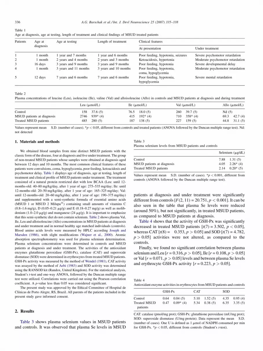
Table 1
Age at diagnosis, age at testing, length of treatment and clinical findings of MSUD treated patients
Patients Age at
diagnosis
Age at testing Length of treatment Clinical features
At presentation Under treatment
1 1 month 1 year and 7 months 1 year and 6 months Poor feeding, hypotonia, seizures Severe psychomotor retardation
2 1 month 2 years and 4 months 2 years and 3 months Ketoacidosis, hypertonia Moderate psychomotor retardation
3 16 days 3 years and 9 months 3 years and 9 months Poor feeding, hypotonia Severe developmental delay
4 1 month 3 years and 11 months 3 years and 10 months Poor feeding, hypotonia,
coma, hypoglycemia
Moderate psychomotor retardation
5 12 days 7 years and 6 months 7 years and 6 months Poor feeding, hypotonia,
hypoglycemia
Severe mental retardation
Table 2
Plasma concentrations of leucine (Leu), isoleucine (Ile), valine (Val) and alloisoleucine (Allo) in controls and MSUD patients at diagnosis and during treatment
Leu (mmol/L) Ile (mmol/L) Val (mmol/L) Allo (mmol/L)
Control 158 � 37.6 (5) 76.5 � 18.0 (5) 260 � 39.7 (5) Nd (5)
MSUD patients at diagnosis 2746 � 939* (4) 415 � 192* (4) 710 � 358* (4) 69.3 � 42.7 (4)
Treated MSUD patients 485 � 280 (5) 187 � 138 (5) 227 � 159 (5) 44.8 � 31.1 (5)
Values represent mean � S.D. (number of cases). *p < 0.05, different from controls and treated patients (ANOVA followed by the Duncan multiple range test). Nd:
not detected
Table 3
Plasma selenium levels from MSUD patients and controls
Selenium (mg/dL)
Control 7.88 � 1.31 (5)
MSUD patients at diagnosis 4.05 � 2.26* (4)
Treated MSUD patients 2.14 � 0.20* (5)
Values represent mean � S.D. (number of cases). *p < 0.001, different from
controls (ANOVA followed by the Duncan multiple range test).
Table 4
Antioxidant enzyme activities in erythrocytes from MSUD patients and controls
GSH-Px CAT SOD
Control 0.64 � 0.04 (5) 5.10 � 1.52 (5) 4.35 � 0.95 (4)
Treated MSUD
patients
0.47 � 0.09* (4) 5.34 � 0.38 (5) 6.35 � 3.35 (5)
CAT: catalase (pmol/mg prot); GSH-Px: glutathione peroxidase (mU/mg prot);
SOD: superoxide dismutase (U/mg protein). Data represent the mean � S.D.
(number of cases). One U is defined as 1 mmol of NADPH consumed per min
for GSH-Px. *p < 0.05, different from controls (Student’s t-test).
A.G. Barschak et al. / Int. J. Devl Neuroscience 25 (2007) 335–338336
1. Materials and methods
We obtained blood samples from nine distinct MSUD patients with the
classic form of the disease, four at diagnosis and five under treatment. The group
of non-treated MSUD patients whose samples were obtained at diagnosis aged
between 12 days and 10 months. The most common clinical features of these
patients were convulsions, coma, hypoglycemia, poor feeding, ketoacidosis and
psychomotor delay. Table 1 displays age of diagnosis, age at testing, length of
treatment and clinical profile of MSUD patients under treatment. The treatment
consisted of a natural protein restricted diet with low BCAA (Leu: until 12-
months-old: 40–80 mg/kg/day, after 1 year of age: 275–535 mg/day; Ile: until
12-months-old: 20–50 mg/kg/day, after 1 year of age: 165–325 mg/day; Val:
until 12-months-old: 20–60 mg/kg/day, after 1 year of age: 190–375 mg/day)
and supplemented with a semi-synthetic formula of essential amino acids
(MSUD 1 or MSUD 2 Milupa1) containing small amounts of vitamins C
(0.8–1.6 mg/g), D (0.05–0.21 mg/g) and E (0.18–0.27 mg/g) as well as molib-
denium (1.0–2.0 mg/g) and manganese (24 mg/g). It is important to emphasize
that this semi-synthetic diet do not contain selenium. Table 2 shows plasma Val,
Ile, Leu and alloisoleucine (Allo) concentrations in MSUD patients at diagnosis
and under treatment and in normal healthy age matched individuals (controls).
Blood amino acids levels were measured by HPLC according Joseph and
Marsden (1986), with slight modifications (Wajner et al., 2000). Atomic
absorption spectrophotometry was used for plasma selenium determination.
Plasma selenium concentrations were determined in controls and MSUD
patients at diagnosis and under treatment. The activities of the antioxidant
enzymes glutathione peroxidase (GSH-Px), catalase (CAT) and superoxide
dismutase (SOD) were determined in erythrocytes from treated MSUD patients.
GSH-Px activity was measured by the method of Wendel (1981), CAT activity
was assayed by the method of Aebi (1983) and SOD activity was determined
using the RANSOD kit (Randox, United Kingdom). For the statistical analysis,
Student’s t-test and one-way ANOVA, followed by the Duncan multiple range
test were utilized. Correlations were carried out using the Pearson correlation
coefficient. A p-value less than 0.05 was considered significant.
The present study was approved by the Ethical Committee of Hospital de
Clınicas de Porto Alegre, RS, Brazil. All parents of the patients included in the
present study gave informed consent.
2. Results
Table 3 shows plasma selenium values in MSUD patients
and controls. It was observed that plasma Se levels in MSUD
patients at diagnosis and under treatment were significantly
different from controls [F(2, 11) = 20.751, p < 0.001]. It can be
also seen in the table that plasma Se levels were reduced
(around 50%), but not significantly, in treated MSUD patients,
as compared to MSUD patients at diagnosis.
Table 4 shows that the activity of GSH-Px was significantly
decreased in treated MSUD patients [t(7) = 3.502, p < 0.05],
whereas CAT [t(8) = �0.353, p > 0.05] and SOD [t(7) = 4.782,
p > 0.05] activities were not altered, as compared to the
controls.
Finally, we found no significant correlation between plasma
selenium and Leu [r = 0.316, p > 0.05], Ile [r = 0.108, p > 0.05]
or Val [r = 0.071, p > 0.05] levels and between plasma Se levels
and erythrocyte GSH-Px activity [r = 0.223, p > 0.05].

A.G. Barschak et al. / Int. J. Devl Neuroscience 25 (2007) 335–338 337
3. Discussion
Our objective in the present study was to evaluate the
selenium levels in plasma from MSUD patients at diagnosis and
under treatment, as well as to verify the activities of glutathione
peroxidase (GSH-Px), catalase (CAT) and superoxide dis-
mutase (SOD) in erythrocytes from treated patients, trying to
correlate GSH-Px activity with selenium levels. GSH-Px
functions as a part of the antioxidant system that protects
membranes and essential proteins from the potentially
damaging effects of reactive oxygen and lipid peroxides
(Lombeck et al., 1996; Schulz et al., 2000). Selenium is an
important trace element and diet (predominantly grains, cereals
and meat) represents the only source of selenium in humans
(Brenneisen et al., 2005). Dietary selenium is important to
provide the selenium-containing cofactor for the glutathione
peroxidase enzyme (Halliwell and Gutteridge, 2001).
CAT is a ferric heme protein that directly catalyses the
decomposition of hydrogen peroxide, whereas GSH-Px
presents a similar function as to CAT breaking predominantly
lipid peroxides (Halliwell and Gutteridge, 2001). SOD removes
the anion superoxide by accelerating the rate of its dismutation
to hydrogen peroxide (Halliwell and Gutteridge, 2001).
We verified that MSUD patients present a moderate
selenium deficiency at diagnosis and a marked deficiency of
this element under treatment. This may possibly be secondary
to a protein restricted diet that implies less amount of dietary
selenium. In this context, patients presenting inborn errors of
amino acid metabolism are potentially at risk for selenium
deficiency once their usual treatment consists of a protein
restricted diet (Chuang and Shih, 2001).
However, there was no correlation between the plasma
branched-chain amino acids (BCAA) leucine, isoleucine and
valine with plasma selenium levels, suggesting that the major
accumulating metabolites in MSUD are not directly associated to
selenium deficiency. These findings do not necessarily rule out a
causative association, so that the inclusion of a larger sample size
would be important to better evaluate this association.
We also observed in the present study a significant decrease
of erythrocyte glutathione peroxidase (GSH-Px) activity in
treated MSUD patients, but we did not find a correlation
between erythrocyte GSH-Px activity and plasma Se concen-
trations. Thus, it is presumed that Se deficiency was not
responsible for the deficient activity of this enzyme, implying
that other factors may be involved in this effect. We also
verified that erythrocyte CAT and SOD activities were not
altered in treated MSUD patients, indicating that GSH-Px
deficient activity was a specific finding in these MSUD patients.
In summary, our data shows that the plasma BCAA and Allo
concentrations were decreased in treated MSUD patients, but
not to near normal levels indicating that the patients were not
strictly adherent to the therapy. Furthermore, plasma Se levels
were decreased in non-treated and particularly in patients under
treatment. These findings, allied to a poor adherence to the
protein restricted diet with low BCAA, may possibly explain
why all treated patients presented a variable degree of
psychomotor delay/mental retardation. However, it must be
emphasized that the overall clinical manifestations of MSUD
patients at diagnosis were more severe, presenting mainly
hypotonia, poor feeding, hypoglycemia ketoacidosis, convul-
sions and coma.
Recently, we demonstrated an increased lipid peroxidation
and decreased antioxidant reactivity in plasma of MSUD
patients at diagnosis (Barschak et al., 2006). Our present results
therefore indicate that an important antioxidant activity (GSH-
Px) is decreased in MSUD patients under treatment, further
indicating that oxidative stress may be considered as one of the
pathomechanisms involved in this disorder. Furthermore, since
Se levels were markedly deficient in MSUD treated patients, it
is proposed that dietary Se supplementation should be
incorporated as an adjuvant therapy in MSUD.
Acknowledgements
This work was supported by grants from Brazilian National
Research Council (CNPq), CAPES, FAPERGS, and FIPE/
HCPA—Brazil.
References
Aebi, H., 1983. In: Bergmeyer, H.U., Bergmeyer, J., Grabl, M. (Eds.), Methods
of Enzymatic Analysis. 3rd ed. pp. 273–296.
Barschak, A.G., Sitta, A., Deon, M., Oliveira, M.H., Haeser, A., Dutra-Filho,
C.S., Wajner, M., Vargas, C.R., 2006. Evidence that oxidative stress is
increased in plasma from patients with maple syrup urine disease. Metab.
Brain Dis. 20, 279–286.
Brenneisen, P., Steinbrenner, H., Sies, H., 2005. Selenium, oxidative stress, and
health aspects. Mol. Aspect Med. 26, 256–267.
Bridi, R., Araldi, J., Sgarbi, M.B., Testa, C.G., Durigon, K., Wajner, M., Dutra-
Filho, C.S., 2003. Induction of oxidative stress in rat brain by the meta-
bolites accumulating in maple syrup urine disease. Int. J. Dev. Neurosci. 21,
327–332.
Bridi, R., Braun, C.A., Zorzi, G.K., Wannmacher, C.M.D., Wajner, M., Lissi,
E.G., Dutra-Filho, C.S., 2005. Alpha-keto acids accumulating in maple
syrup urine disease stimulate lipid peroxidation and reduce antioxidant
defences in cerebral cortex from young rats. Metab. Brain Dis. 20, 155–167.
Chuang, D.T., Shih, V.E., 2001. Maple syrup urine disease (branched-chain
ketoaciduria). In: Scriver, C.R., Beaudt, A.L., Sly, W.L., Valle, D.
(Eds.), The Metabolic and Molecular Bases of Inherited Disease.
McGraw-Hill, New York, pp. 1971–2005.
Colome, C., Sierra, C., Vilaseca, M.A., 2000. Congenital errors of metabolism:
cause of oxidative stress? Med. Clin. 115, 111–117.
de Oliveira Marques, F., Hagen, M.E., Pederzolli, C.D., 2003. Glutaric acid
induces oxidative stress in brain of young rats. Brain Res. 964, 153–158.
Fighera, M.R., Bonini, J.S., de Oliveira, T.G., Frussa-Filho, R., Rocha, J.B.,
Dutra-Filho, C.S., Rubin, M.A., Mello, C.F., 2003. GM1 ganglioside
attenuates convulsions and thiobarbituric acid reactive substances produc-
tion induced by the intrastriatal administration of methylmalonicacid. Int. J.
Biochem. Cell Biol. 35, 465–473.
Fontella, F.U., Pulrolnik, V., Gassen, E., Wannmacher, C.M.D., Klein, A.B.,
Wajner, M., Dutra, C.S., 2000. Propionic and L-methylmalonic acids induce
oxidative stress in brain of young rats. Neuroreport 11, 541–544.
Fontella, F.U., Gassen, E., Pulrolnik, V., Wannmacher, C.M.D., Klein, A.B.,
Wajner, M., Dutra, C.S., 2002. Stimulation of lipid peroxidation in vitro in
rat brain by metabolites accumulating in maple syrup urine disease. Metab.
Brain Dis. 17, 47–54.
Halliwell, B., Gutteridge, J.M.C. (Eds.), 2001. Free Radicals in Biology and
Medicine. Oxford University Press, Oxford.
Joseph, M.H., Marsden, C.A., 1986. Amino acids and small peptides. In: Lim,
C.F. (Ed.), HPLC of Small Peptides. IRL Press, Oxford, pp. 13–27.
A.G. Barschak et al. / Int. J. Devl Neuroscience 25 (2007) 335–338338
Latini, A., Borba Rosa, R., Scussiato, K., Llesuy, S., Bello-Klein, A., Wajner,
M., 2002. 3-Hydroxyglutaric acid induces oxidative stress and decreases
the antioxidant defences in cerebral cortex of young rats. Brain Res. 956,
367–373.
Lombeck, I., Jochum, F., Terwolbeck, K., 1996. Selenium status in infants and
children with phenylketonuria and in maternal phenylketonuria. Eur. J.
Pediatr. 155, 140–144.
Schulz, J.B., Lindenau, J., Seyfried, J., Dichgans, J., 2000. Glutathione,
oxidative stress and neurodegeneration. Eur. J. Biochem. 267, 4904–4911.
Sierra, C., Vilaseca, M.A., Moyano, D., Brandi, N., Campistol, J., Lam-
bruschini, N., Cambra, F.J., Deulofeu, R., Mira, A., 1998. Antioxidant
status in hyperphenylalaninemia. Clin. Chim. Acta 276, 1–9.
Sirtori, L.R., Dutra-Filho, C.S., Fitarelli, D., Sitta, A., Haeser, A., Barschak,
A.G., Wajner, M., Coelho, D.M., Llesuy, S., Bello-Klein, A., Giugliani, R.,
Deon, M., Vargas, C.R., 2005. Oxidative stress in patient with phenylk-
etonuria. Biochim. Biophys. Acta 1740, 68–73.
Treacy, E., Clow, C.L., Reade, T.R., Chitayat, D., Mamer, O.A., Scriver, C.R.,
1992. Maple syrup urine disease: interrelationship between branched-chain
amino-, oxo- and hydroxyacids; implications for treatment; associations
with CNS dysmyelination. J. Inherit. Metab. Dis. 15, 121–135.
Vargas, C.R., Wajner, M., Sirtori, L.R., Goulart, L., Chiochetta, M., Coelho, D.,
Latini, A., Llesuy, S., Bello-Klein, A., Giugliani, R., Deon, M., Mello, C.F.,
2004. Evidence that oxidative stress is increased in patients with X-linked
adrenoleukodystrophy. Biochim. Biophys. Acta 1688, 26–32.
Wajner, M., Coelho, D.M., Barschak, A.G., Araujo, P.R., Pires, R.F., Lulhier,
F.L., Vargas, C.R., 2000. Reduction of large neutral amino acid concentra-
tions in plasma and CSF of patients with maple syrup urine disease during
crises. J. Inherit. Metab. Dis. 23, 505–512.
Wajner, M., Latini, A., Wyse, A.T.S., Dutra-Filho, C.S., 2004. The role of
oxidative damage in the neuropathology of organic acidurias: insights from
animal studies. J. Inherit. Metab. Dis. 27, 427–448.
Wendel, A., 1981. Glutathione peroxidase. Methods Enzymol. 77, 325–332.

56
III.4 Capítulo IV – Artigo 04
Maple Syrup Urine Disease in treated patients: Biochemical and oxidative
stress profiles
Alethéa G. Barschak, Christiane Marchesan, Angela Sitta, Marion Deon,
Roberto Giugliani, Moacir Wajner, Carmen R. Vargas
Este trabalho foi desenvolvido no Programa de Pós-Graduação em Ciências
Biológicas: Bioquímica, ICBS, UFRGS.
Periódico: Clinical Biochemistry
Status: publicado

Available online at www.sciencedirect.com
(2008) 317–324
Clinical Biochemistry 41Maple syrup urine disease in treated patients: Biochemical andoxidative stress profiles
Alethéa G. Barschak a,b,⁎, Christiane Marchesan c, Angela Sitta a,b, Marion Deon a,b,Roberto Giugliani b, Moacir Wajner a,b, Carmen Regla Vargas a,b,c,⁎
a Programa de Pós-Graduação em Ciências Biológicas: Bioquímica, ICBS, UFRGS, Porto Alegre, RS, Brazilb Serviço de Genética Médica, HCPA, Rua Ramiro Barcelos, 2350 CEP 90.035-903, Porto Alegre, RS, Brazil
c Departamento de Análises, Faculdade de Farmácia, PPGCF, UFRGS, Porto Alegre, RS, Brazil
Received 12 September 2007; received in revised form 22 November 2007; accepted 27 November 2007Available online 5 December 2007
Abstract
Objective: The objective of this study was to evaluate and correlate the biochemical and oxidative stress profiles in MSUD patients during thedietary treatment.
Design and methods: Plasma samples from treated MSUD patients were used to evaluate the biochemical profile and oxidative stressparameters.
Results: It was observed that glucose, total cholesterol, albumin and creatinine are reduced and that aspartate aminotransferase and lactatedehydrogenase activities are increased in plasma from MSUD patients under treatment. Besides, it was verified an increase of thiobarbituric acid-reactive species (TBARS) and a decrease of total antioxidant reactivity (TAR).
Conclusions: Our results suggest that oxidative stress occurs in treated MSUD patients and that dietary treatment and clinical conditionsassociated to the disease can lead to biochemical alterations in these patients.© 2007 The Canadian Society of Clinical Chemists. Published by Elsevier Inc. All rights reserved.
Keywords: Maple syrup urine disease; Biochemical profile; Lipid peroxidation; Antioxidant reactivity
Introduction
Maple syrup urine disease (MSUD) or branched-chain keto-aciduria is an inborn error ofmetabolism caused by a deficiency ofthe branched-chain α-keto acid dehydrogenase complex(BCKAD) activity. This blockage leads to accumulation of thebranched-chain amino acids (BCAA) leucine, isoleucine, andvaline, and their correspondingα-keto acids (α-ketoisocaproic,α-keto-β-methylvaleric and α-ketoisovaleric, respectively). MSUDis an autosomal recessive metabolic disorder with a worldfrequency estimated in approximately 1 in 185,000 newborns [1].
Based on the clinical presentation and biochemical responses tothiamine administration, MSUD can be divided into five phe-
⁎ Corresponding authors. Serviço de Genética Médica, HCPA, Rua RamiroBarcelos, 2350 CEP 90.035-903, Porto Alegre, RS, Brazil. Fax: +55 51 21018010.
E-mail addresses: [email protected] (A.G. Barschak),[email protected] (C.R. Vargas).
0009-9120/$ - see front matter © 2007 The Canadian Society of Clinical Chemistsdoi:10.1016/j.clinbiochem.2007.11.015
notypes: classic, intermediate, intermittent, thiamine-responsiveand dihydrolipoyl dehydrogenase (E3)-deficient [1,2]. The classicform has a neonatal onset of encephalopathy and is the mostsevere and common form. The main clinical signals presented byMSUD patients include ketoacidosis, hypoglycemia, opisthoto-nos, poor feeding, apnea, ataxia, convulsions, coma, psychomo-tor delay and mental retardation. Severe brain edema is usuallyseen in MSUD patients who died during acute metabolic crisis[1,3,4].
The levels of the BCAAs leucine, isoleucine and valine aregreatly increased in tissues and biological fluids of the patients, andwith the presence of alloisoleucine, they are diagnostic of MSUD.High levels of leucine and α-ketoisocaproic acid seem to be themain important neurotoxic metabolites in MSUD [1,5]. Thetreatment involves restriction in the protein ingestion and a specificformula of essential amino acids except those accumulated in theDXB, as well as the aggressive intervention during acute metabolicdecompensation. The majority of untreated classic patients die
. Published by Elsevier Inc. All rights reserved.

318 A.G. Barschak et al. / Clinical Biochemistry 41 (2008) 317–324
within the early months of life from recurrent metabolic crisis andneurologic deterioration. The age of diagnosis and the subsequentmetabolic control are the most important determinants ofprognostic.
Free radicals are molecules or atoms presenting unpairedelectrons in their outer orbitals, which make them very unstableand reactive. Free radicals present an enormous capacity to reactwith diverse integrant molecules of the cellular structureand derived from each one of them [6]. The free radicals canbe produced by exogenous (radiation, tobacco and stress, forexample) and endogenous sources [7]. In vivo they are producedin all cells as by-products of normal metabolism, and endo-genous mechanisms exist to reduce their formation or increasetheir inactivation [8]. The enzymatic antioxidant defenses in-volve mainly the superoxide dismutase, catalase and glutathioneperoxidase enzymes, whereas the nonenzymatic antioxidantsystems involve small molecules as vitamins, glutathione, anduric acid [6].
The imbalance between the formation and the removal of thefree radicals in the organism, decurrent of the reduction of theendogenous antioxidants and/or of the increase of the generationof oxidant species generates a prooxidant state defined as oxi-dative stress [6]. Increasing evidences show that the oxidativestress is involved in a large number of diseases, for example,arterioscleroses, diabetes, neurodegenerative disorders, such asAlzheimer's disease and Parkinson's disease, cancer and aging,among others [8,9]. In this context, studies in animals haveshowed that the excessive production of free radicals and thedecrease of antioxidant defenses are induced by metabolitesaccumulated in several inborn errors of metabolism [10–15].Besides, studies in humans showed that oxidative stress occursin patients with phenylketonuria [16,17] and X-linked adre-noleukodystrophy [18,19]. Recently, it was demonstrated anincrease of the lipid peroxidation and a decrease of the anti-oxidant defenses in MSUD untreated patients [20], indicatingthat oxidative stress is involved in the pathophysiology of thisdisease.
Thus, the aim of the present study was to evaluate andcorrelate the biochemical profile and oxidative stress parametersin plasma from MSUD patients during the dietary treatment.
Material and methods
Patients and controls
Plasma samples obtained from seven treated MSUD patients(classic form) were used to evaluate the biochemical profile andoxidative stress parameters. The treatment consisted of a naturalprotein restricted diet with low BCAA and supplemented witha semi-synthetic formula of essential amino acids containingsmall amounts of vitamins andminerals. The average duration oftreatment at the test time was 3 years and 5 months. Table 1displays age of diagnosis, age at testing, length of treatment andclinical profile ofMSUD patients under treatment. Plasma levelsof leucine, isoleucine, valine and alloisoleucine at the test were365.9±287.9 μmol/L, 99.0±65.3 μmol/L, 197.7±184.2 μmol/Land 25.6±14.0 μmol/L, respectively. Blood amino acids were
determined by HPLC method [21]. Control group was composedof eighth healthy individuals with similar age to the patients (1 to8 years) (leucine 158.3±37.6μmol/L, isoleucine 76.5±18.0μmol/L, valine 260.7±39.7 μmol/L and alloisoleucine were notdetected).
The present study was approved by the Ethical Committee ofHospital de Clínicas de Porto Alegre, RS, Brazil. All parentsof the patients included in the present study gave informedconsent.
Plasma preparation
Plasma was prepared from whole blood samples obtainedfrom fasting individuals (controls and MSUD patients) byvenous puncture using vials with EDTA. Whole blood wascentrifuged at 1000×g and plasma was removed by aspirationand frozen at −80 °C until determinations.
Determination of the biochemical parameters
GlucoseGlycemia measurement was carried out using the method of
the glucose oxidase that catalyzes the oxidation of the glucose andhydrogen peroxide formation, which reacts with 4-aminoantipyr-ine and phenol under the catalytic action of peroxidase, to form acolored complex whose intensity of color is proportional to theglucose concentration in the sample (Glucose PAP kit, DiagnosticLabtest).
Total cholesterolTotal cholesterol was determined in accordance with the
method of the cholesterol esterase. The formed free choles-terol is oxidated producing hydrogen peroxide that reacts with4-aminoantipyrine to form a red complex, whose intensity ofcolor is proportional to the cholesterol concentration in thesample (Cholesterol kit, Diagnostic Labtest).
HDL cholesterolCholesterol HDL was measured through cholesterol esterase
method for dosage of described total cholesterol above, after theprecipitation of lipoproteins of low and very low density withphosphotungstic acid and magnesium chloride (CholesterolHDL kit, Diagnostic Labtest).
LDL cholesterolCholesterol LDL was determined through calculation, using
the Friedewald formula (cholesterol LDL=total cholesterol−(cholesterol HDL+triglycerides /5)) for values of triglyceridesuntil 400 mg/dL.
TriglyceridesTriglycerides had been measured in accordance with
the method of lipoprotein lipase, where glycerol set free isconverted to glycerol-3-phosphate by the action of glycerolk-inase, and it is oxidated to form hydrogen peroxide. This reactswith the 4-aminoantipyrine producing a violet complex whoseintensity of color is proportional to the concentration of

Table 1Age at diagnosis, age at testing, length of treatment and clinical findings of MSUD patients
Patients Age atdiagnosis
Age at testing Length of treatment Clinical Features
At presentation Under treatment
1 1 month 1 year and 7 months 1 year and 6 months Poor feeding, hypotonia, seizures Severe psychomotor retardation2 1 month 2 years and 4 months 2 years and 3 months Ketoacidosis, hypertonia Moderate psychomotor retardation3 16 days 3 years and 9 months 3 years and 9 months Poor feeding, hypotonia Severe developmental delay4 1 month 3 years and 11 months 3 years and 10 months Poor feeding, hypotonia, coma, hypoglycemia Moderate psychomotor retardation5 12 days 7 years and 6 months 7 years and 6 months Poor feeding, hypotonia, hypoglycemia Severe mental retardation6 4 months 1 year and 7 months 1 year and 3 months Hypotonia, seizures Severe psychomotor retardation7 2 months 1 year and 7 months 1 year and 5 months Poor feeding, hypotonia, seizures Moderate developmental delay
319A.G. Barschak et al. / Clinical Biochemistry 41 (2008) 317–324
triglycerides in the sample (Triglycerides GPO-ANA kit, Diag-nostic Labtest).
TransaminasesTransaminases (alanine aminotransferase — ALT and as-
partate aminotransferase — AST) had been determined in ac-cordance with the kinetic method, which is based on thetransference of the amine group of amino acids L-alanine (ALT)or L-aspartate (AST) for the alpha-ketoglutarato producing itscorresponding α-keto acid, which reacts with a secondary en-zyme (lactate dehydrogenase or malate dehydrogenase) produ-cing one hydrazone in alkaline way, which is proportional to theactivity of the ALTor AST, respectively in the sample (AST andALT kit, Diagnostic Labtest).
Creatine kinaseCreatine kinase (CK) was determined in accordance with the
kineticmethod.Creatine phosphatewas dephosphorylated leadingto the formation of adenosine triphosphate (ATP), which reactswith glucose producing glucose-6-phosphate that was oxidated to6-phosphogluconate reducing NAD to NADH. This reduction isproportional to CK activity in the sample (CK-NAC kit, Diag-nostic Labtest).
Lactate dehydrogenaseLactate dehydrogenase (LDH) was measured through
colorimetric method. LDH catalyzes the reversible reaction oflactate-pyruvate in the presence of NAD, which is reduced toNADH. NADH reduces p-iodonitrotetrazolium leading to theformation of a colored product proportional to LDH activity inthe sample (Lactate dehydrogenase kit, Diagnostic Labtest).
AlbuminAlbumin was measured in accordance with the method
of the bromo cresol green. The albumin interacts with thebuffered bromocresol green, and due to proteinic error ofpointers, green color formation occurs, which is proportionalto the albumin concentration in the sample (Albumin Kit,Diagnostic Labtest).
CreatinineCreatinine was measured by the method of the picrate, which
reacts with the creatinine in the sample, after addition of acidreagent for elimination of pseudo-creatinines, producing a redcolor complex, which intensity is proportional to the concentra-
tion of creatinine in the sample (Creatinine K Kit, DiagnosticLabtest).
UreaUrea determination was carried out by the method of urease,
in which the urea in the sample is hydrolyzed to form am-monium, which reacts with salicylate and sodium hypochlorite,in alkaline pH and using nitroprusside as catalytic agent, to formblue of indofenol. The intensity of the formed color is pro-portional to the amount of urea in the sample (Urea CE kit,Diagnostic Labtest).
Uric acidUric acid was determined by the method of uricase, in which
it was oxidated to alantoine and hydrogen peroxide, whichreacts with 4-aminoantipyrine and dichloro hydroxy benzenesulfonate (DHBS) in the presence of peroxidase producing a redcomplex, whose intensity of color is proportional to the con-centration of uric acid in the sample (Uric acid kit, DiagnosticLabtest).
Determination of oxidative stress parameters
Thiobarbituric acid-reactive species (TBARS)Thiobarbituric acid-reactive species (TBARS) were deter-
mined according to the method described by Buege and Aust[22]. Briefly, 250 μL of 10% trichloroacetic acid was added to125 μL of plasma, then 375 μL 0.67% thiobarbituric acid (in7.1% sodium sulfate) was added and incubated at 100 °C for30 min. After the incubation, the mixture was extracted with750 μL butanol. The resulting pink stained TBARS were de-termined in a spectrofluorimeter at 515 nm. Calibration curvewas performed using 1,1,3,3-tetramethoxypropane subjected tothe same treatment as that of the samples. TBARS were cal-culated as nmol TBARS/mg protein.
Total antioxidant reactivity (TAR)TAR, which represents the quality of the tissue antioxidants,
was determined by measuring the luminol chemiluminescenceintensity induced by 2,2′-azo-bis-(2-amidinopropane) (ABAP)according to the method of Lissi et al. [23]. The backgroundchemiluminescence was measured by adding 4 mL of 2 mMABAP (in 0.1 M glycine buffer, pH 8.6) into a glass scintillationvial. Ten microliters of luminol (4 mM) was added to each vialand the chemiluminescence was measured. This was considered

Fig. 1. Plasma glucose from MSUD patients and controls. Data represent themean±SD (n=6–7). ⁎pb0.05 (Student's t test for unpaired samples) comparedto control.
320 A.G. Barschak et al. / Clinical Biochemistry 41 (2008) 317–324
to be the basal value. Tenmicroliters of 25–200μMTrolox (curvecalibration) or plasmawas then added and the chemiluminescencewas measured during 60 s. The Trolox and plasma additionreduces the chemiluminescence. The rapid reduction in luminolintensity is considered as a measure of the TAR capacity. TARmeasurement was calculated as nmol Trolox/mg protein.
Total proteinsProtein concentrations were determined by the Biuret
method (Total protein kit, Diagnostic Labtest), using albuminas standard.
Fig. 2. Lipidic profile in plasma from MSUD patients and controls [total cholesData represent the mean±SD (n=7–8). ⁎pb0.05 (Student's t test for unpaired samp
Statistical analysis
The Student's t test for unpaired samples was used tocompare means between controls and MSUD patients. Correla-tions were carried out using the Pearson correlation coefficient.Only significant values are shown in the text. A p value less than0.05 was considered significant. All analyses were performedusing the Statistical Package for the Social Sciences (SPSS)software in a PC-compatible computer.
Results
Fig. 1 shows glycemia measurement in controls and MSUDpatients under dietary treatment. It can be observed that MSUDpatients presented a significant reduction in the plasma levels ofglucose [t(11)=2.508, pb0.05] as compared to the controls.The glucose levels presented by these patients are below refe-rence values (70–105 mg/dL).
Fig. 2 shows the lipidic profile in controls and treated MSUDpatients. It can be seen that the concentrations of total cho-lesterol (A) [t(13)=2.894, pb0.05] were significantly reducedin MSUD patients as compared to the control group. How-ever, both groups presented values in the reference range (112–205 mg/dL). The plasma levels of HDL cholesterol (B) [t(12)=0.591, pN0.05], LDL cholesterol (C) [t(11)=0.140, pN0.05]and triglycerides (D) [t(13)=0.988, pN0.05] in theMSUDpatients
terol (A), HDL cholesterol (B), LDL cholesterol (C) and triglycerides (D)].les) compared to control.
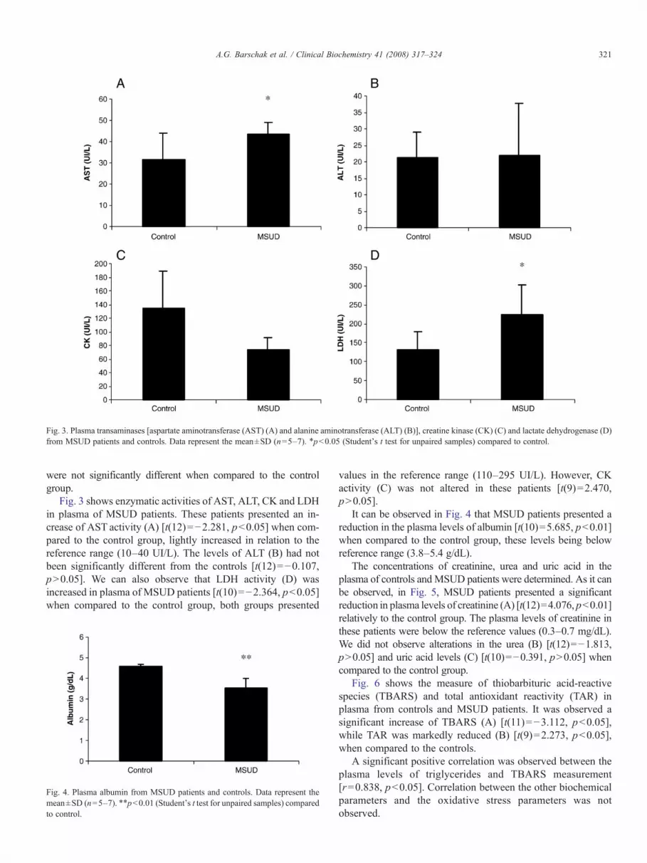
Fig. 3. Plasma transaminases [aspartate aminotransferase (AST) (A) and alanine aminotransferase (ALT) (B)], creatine kinase (CK) (C) and lactate dehydrogenase (D)from MSUD patients and controls. Data represent the mean±SD (n=5–7). ⁎pb0.05 (Student's t test for unpaired samples) compared to control.
321A.G. Barschak et al. / Clinical Biochemistry 41 (2008) 317–324
were not significantly different when compared to the controlgroup.
Fig. 3 shows enzymatic activities of AST, ALT, CK and LDHin plasma of MSUD patients. These patients presented an in-crease of AST activity (A) [t(12)=−2.281, pb0.05] when com-pared to the control group, lightly increased in relation to thereference range (10–40 UI/L). The levels of ALT (B) had notbeen significantly different from the controls [t(12)=−0.107,pN0.05]. We can also observe that LDH activity (D) wasincreased in plasma of MSUD patients [t(10)=−2.364, pb0.05]when compared to the control group, both groups presented
Fig. 4. Plasma albumin from MSUD patients and controls. Data represent themean±SD (n=5–7). ⁎⁎pb0.01 (Student's t test for unpaired samples) comparedto control.
values in the reference range (110–295 UI/L). However, CKactivity (C) was not altered in these patients [t(9)=2.470,pN0.05].
It can be observed in Fig. 4 that MSUD patients presented areduction in the plasma levels of albumin [t(10)=5.685, pb0.01]when compared to the control group, these levels being belowreference range (3.8–5.4 g/dL).
The concentrations of creatinine, urea and uric acid in theplasma of controls andMSUD patients were determined. As it canbe observed, in Fig. 5, MSUD patients presented a significantreduction in plasma levels of creatinine (A) [t(12)=4.076, pb0.01]relatively to the control group. The plasma levels of creatinine inthese patients were below the reference values (0.3–0.7 mg/dL).We did not observe alterations in the urea (B) [t(12)=−1.813,pN0.05] and uric acid levels (C) [t(10)=−0.391, pN0.05] whencompared to the control group.
Fig. 6 shows the measure of thiobarbituric acid-reactivespecies (TBARS) and total antioxidant reactivity (TAR) inplasma from controls and MSUD patients. It was observed asignificant increase of TBARS (A) [t(11)=−3.112, pb0.05],while TAR was markedly reduced (B) [t(9)=2.273, pb0.05],when compared to the controls.
A significant positive correlation was observed between theplasma levels of triglycerides and TBARS measurement[r=0.838, pb0.05]. Correlation between the other biochemicalparameters and the oxidative stress parameters was notobserved.

322 A.G. Barschak et al. / Clinical Biochemistry 41 (2008) 317–324
Discussion
The deficiency in the activity of the branched-chain α-ketoacid dehydrogenase complex is clinically characterized byketoacidosis, convulsions, coma, psychomotor delay and mentalretardation. The mechanisms of the neurological symptomspresented by MSUD patients are still poorly understood.However, considering that high concentrations of leucine and/orα-ketoisocaproate were associated with the appearance of theneurological symptoms, these compounds seem to be the mainneurotoxic metabolites in the illness [1,5]. Animal studies haveshown that excessive free radical production and reduced tissueantioxidant defenses are induced by the metabolites accumulatingin MSUD [13,14]. Recently we have demonstrated that oxidativestress is induced in MSUD patients at diagnosis [20].
In the present study we evaluated and correlated the bio-chemical and oxidative profiles in the plasma of MSUD patientsunder dietary treatment. To our knowledge this is the first reportdemonstrating the biochemical and oxidative profiles of treatedMSUD patients.
Fig. 5. Plasma creatinine (A), urea (B) and uric acid (C) from MSUD patientsand controls. Data represent the mean±SD (n=5–7). ⁎⁎pb0.01 (Student's t testfor unpaired samples) compared to control.
Fig. 6. Plasma thiobarbituric acid-reactive species (TBARS) (A) and totalantioxidant reactivity (TAR) (B) from MSUD patients and controls. Datarepresent the mean±SD (n=5–7). ⁎pb0.05 (Student's t test for unpairedsamples) compared to control.
Our results showed that glucose levels are reduced in MSUDpatients. The hypoglycemia is a common finding in these patients,and this alteration is possibly associated with an increased secre-tion of insulin stimulated by high concentrations of leucine pre-sented by these patients [1].
The evaluation of the lipidic profile showed a significantreduction in the total cholesterol levels in MSUD patients incomparison to the control group. Once the patients are submittedto a low protein diet of animal origin and consequently poor insaturated fat, it is possible that this reduction could be secondaryto dietary treatment. However, HDL cholesterol, LDL choles-terol and triglycerides were not different from controls.
It was also observed an increase in the ASTactivity inMSUDpatients, whereas ALT activity was not modified. Hepaticillnesses in general provoke concomitant increase of ALT andAST levels, being ALT the most specific enzyme of the liver.Increases in the serum ALT activity are rarely observed inconditions other than hepatic illness [24]. This suggests that theisolated increase of AST observed in the studied patients couldbe related to other causes. In addition, we verified that LDHactivity was increased in MSUD which could be associated tomuscular damage [24]. MSUD patients present clinical symp-toms as lethargy, hypotonia, hypertonia and convulsions [1].Studies have demonstrated association of muscular damages(dystrophies and polymyosite) with increased levels of theenzymatic activities of AST, CK and LDH [24–26]. Our resultsindicate that the increased activities of AST and LDH could be

323A.G. Barschak et al. / Clinical Biochemistry 41 (2008) 317–324
associated to the characteristic muscular alterations presented byMSUD patients. However, total CK activity was not altered inMSUD suggesting that additional studies evaluating the activityof CK-MM (muscular isoenzyme) could contribute in betterunderstanding these findings.
It was also evaluated the plasma concentrations of creatinine,urea and uric acid. The MSUD patients presented a significantreduction in plasma creatinine levels, but urea and uric acidconcentrations were not altered. The low concentration of creat-inine could be associated to muscular alterations presented bypatients in the course of the illness. It is important to emphasizethat creatinine is formed through free creatine in the muscle, sothe produced amount of endogenous creatinine is proportional tomuscular mass [24].
MSUD treatment consists of a restricted ingestion of proteins,supplemented with a semi-synthetic formula of essential aminoacids except leucine, isoleucine and valine. The aim of thetreatment is to normalize the plasma levels of the BCAA, mini-mizing the neurological damage associated mainly to increase inleucine levels [1]. It was observed that MSUD patients presenteda reduction in plasma albumin levels, which probably reflectsdeficiencies associated to low protein diet that these patients aresubmitted to.
It was observed a significant increase of TBARS in plasmafrom MSUD patients. Considering that TBARS reflects theamount of malondialdehyde, an end product of lipid peroxidation[6,27], our data indicate that lipid peroxidation is stimulated inMSUD patients, probably secondary to free radical generation.Already, TAR measurement, which reflects the quality of anti-oxidant substances [28], wasmarkedly reduced inMSUDpatients,suggesting a deficient capacity of plasma to modulate the damageassociated with the increased production of reactive species. Theincrease of lipid peroxidation and the reduction of the antioxidantdefenses suggest that oxidative stress occurs in treatedMSUD andcan explain at least in part the pathophysiology of this illness. Itwas previously verified an increase in TBARS and a decrease inTAR in MSUD patients at diagnosis [20].
A positive correlation was observed between plasma tri-glycerides levels and TBARS measurement. Triglycerides arecomposed by the union of three fat acids and glycerol. Fat acidsare easily oxidated by free radicals, leading to lipid peroxidation.Considering that malondialdehyde (MDA) is an end product oflipid breakdown due to lipid peroxidation, and TBARS reflectsthe content of MDA (27), it is possible that lipid peroxidationcould be related to triglycerides amount.
In conclusion, our results show that glucose, total cholesterol,albumin and creatinine are reduced and that AST and LDHactivities are increased in plasma from MSUD patients underdietary treatment. It is probable that these findings are secondaryto low protein diet and/or to the characteristic clinical mani-festations of the illness. These results suggest that serum bio-chemical profile should be evaluated in all MSUD patients duringtreatment. However, our results must be taken with caution sinceour experiments were conducted with samples collected at asingle time point during treatment. Therefore, these samplesmight not represent patients' metabolic control as a whole. Theincreased TBARS and the reduction of the TAR are in accordance
with our previous studies, suggesting that oxidative stress occursalso in MSUD patients under treatment. In this context, it ispossible that dietary antioxidant supplementation could beimportant as an adjuvant therapy in MSUD.
Acknowledgments
This work was supported by grants from Brazilian NationalResearch Council (CNPq), FAPERGS, and FIPE/HCPA —Brazil.
References
[1] Chuang DT, Shih VE. Maple syrup urine disease (branched-chainketoaciduria). In: Scriver CR, Beaudt AL, Sly WL, Valle D, editors. TheMetabolic and Molecular Bases of Inherited Disease. 8ed. New York:McGraw-Hill; 2001. p. 1971–2005.
[2] Schadewaldt P, Wendel U. Metabolism of branched-chain amino acids inmaple syrup urine disease. Eur J Pediatr 1997;156(Suppl 1):S62–6.
[3] Schadewaldt P, Wendel U. Variant maple syrup urine disease (MSUD)—the entire spectrum. J Inherit Metab Dis 2006;29(6):716–24.
[4] Schönberger S, Schweiger B, Schwahn B, Schwarz M, Wendel U.Dysmyelination in the brain of adolescents and young adults with maplesyrup urine disease. Mol Genet Metab 2004;82(1):69–75.
[5] Strauss KA, Morton DH. Branched-chain ketoacyl dehydrogenase defi-ciency: maple syrup disease. Curr Treat Option Neurol 2003;5(4): 329–41.
[6] Halliwell B, Gutteridge JC. Free radicals in biology and medicine. 3th ed.New York: Oxford; 2001.
[7] Harris ED. Regulation of antioxidant enzymes. Faseb J 1992;6(9): 2675–83.[8] Pawlak W, Kedziora J, Zolynski K, et al. Effect of long term bed rest in
man on enzymatic antioxidative defence and lipid peroxidation inerythrocytes. J Gravit Physiol 1998;1:163–4.
[9] Beckman KB, Ames BN. The free theory of aging matures. Phisiol Rev1998;78(2):547–81.
[10] Colome C, Sierra C, Vilaseca MA. Congenital errors of metabolism: causeof oxidative stress? Méd Clin 2000;115:111–7.
[11] Fontella FU, Pulrolnik V, Wannmacher CMD, et al. Propionic andL-methylmalonic acids induce oxidative stress in brain of young rats. NeuroReport 2000;11:541–4.
[12] de Oliveira Marques F, Hagen ME, Pederzolli CD. Glutaric acid inducesoxidative stress in brain of young rats. Brain Res 2003;964: 153–8.
[13] Fontella FU, Gassen E, Pulrolnik V, et al. Stimulation of lipid peroxidationin vitro in rat brain by metabolites accumulating in maple syrup urinedisease. Metab Brain Dis 2002;17:47–54.
[14] Bridi R, Araldi J, Sgarbi MB, et al. Induction of oxidative stress in rat brainby the metabolites accumulating in maple syrup urine disease. Int J DevlNeuroscience 2003;21:327–32.
[15] Wajner M, Latini A, Wyse ATS, Dutra-Filho CS. The role of oxidativedamage in the neuropathology of organic acidurias: insights from animalstudies. J Inherit Metab Dis 2004;27:427–48.
[16] Sirtori LR, Dutra-Filho CS, Fitarelli D, et al. Oxidative stress in patientwith phenylketonuria. Biochim Biophys Acta 2005;1740: 68–73.
[17] Sitta A, Barschak AG, Deon M, et al. Investigation of oxidative stressparameters in treated phenylketonuric patients. Metab Brain Dis 2006;20:287–96.
[18] Vargas CR, Wajner M, Sirtori LR, et al. Evidence that oxidative stress isincreased in patients with X-linked adrenoleukodystrophy. BiochimBiophys Acta 2004;1688:26–32.
[19] DeonM,WajnerM, Sirtori LR, et al. The effect of Lorenzo's oil on oxidativestress in X-linked adrenoleukodystrophy. J Neurol Sci 2006;247:157–64.
[20] Barschak AG, Sitta A, Deon M, et al. Evidence that oxidative stress isincreased in plasma from patients with maple syrup urine disease. MetabBrain Dis 2006;20:279–86.
[21] Joseph MH, Marsden CA. Amino acids and small peptides. In: Lim CF,editor. HPLC of Small Peptides. Oxford: IRL Press; 1986. p. 13–27.

324 A.G. Barschak et al. / Clinical Biochemistry 41 (2008) 317–324
[22] Buege JA, Aust SD. Microsomal lipid peroxidation. Methods Enzymol1978;52:302–9.
[23] Lissi E, Pascual C, Del Castillo MD. Luminol luminescence induced by2,2′-azo-bis-(2-amidinopropane) thermolysis. Free Radic Res Commun1992;17:299–311.
[24] Burtis CA, Ashwood ER, editors. Tietz: Fundamentos de Química Clínica.4°edição. Rio de Janeiro: Guanabara-Koogan; 1998.
[25] Lott JA, Landesman PW. The enzymology of skeletal muscle disorders.Crit Rev Clin Lab Sci 1984;20(2):153–90.
[26] Nathwani RA, Pais S, Reynolds TB, Kaplowitz N. Serum alanineaminotransferase in skeletal muscle diseases. Hepatology 2005;41(2):380–2.
[27] Esterbauer H, Cheeseman KH. Determination of aldehydic lipid pero-xidation products: malonaldehyde and 4-hydroxynonenal. MethodsEnzymol 1990;186:407–21.
[28] Lissi E, Salim-Hanna M, Pascual C, Del Castillo MD. Evaluation of totalantioxidant potential (TRAP) and total antioxidant reactivity from luminol-enhanced chemiluminescence measurements. Free Radic Biol Med1995;18:153–8.

65
IV. DISCUSSÃO
Os mecanismos responsáveis pelos sintomas neurológicos
apresentados pelos pacientes com DXB ainda são pouco conhecidos. Nesse
sentido, muitos estudos vêm sendo desenvolvidos no intuito de melhor
entender a fisiopatologia da DXB. Existem muitas evidências associando a
deficiência no metabolismo da leucina e os sintomas neurológicos nesses
pacientes. Estudos mostraram que a administração crônica de leucina a ratos
jovens induz a déficit no aprendizado/memória na idade adulta, indicando que
os altos níveis de leucina podem contribuir para a deterioração neurológica
observada na doença (Mello et al., 1999). Foi também demonstrado que o
ácido α-cetoisovalérico apresenta propriedades convulsivantes, sugerindo que
este metabólito esteja envolvido na gênese das convulsões características da
DXB (Coitinho et al., 2001). Ainda, altas concentrações dos AACR e dos CACR
causam déficit no metabolismo energético através da inibição da cadeia
transportadora de elétrons (Sgaravatti et al., 2003) e da enzima creatina
quinase (Pilla et al., 2003), sugerindo que o déficit energético possa estar
envolvido nos sintomas neurológicos apresentados na doença. Outros autores
demonstraram que os AACR e/ou CACR acumulados na DXB provocam
apoptose neuronal (Jouvet et al., 2000), redução na síntese e função de
neurotransmissores (Zielke et al., 1997; Tavares et al., 2000), alterações na
mielina (Schönberger, 2004; Treacy et al., 1992; Tribble e Shapira, 1983;
Taketomi et al., 1983) e redução na captação de aminoácidos essenciais pelo
cérebro (Araújo et al., 2001).
66
Nos últimos anos tem crescido o número de evidências associando
estresse oxidativo e erros inatos do metabolismo com manifestação
neurológica, sugerindo que o dano oxidativo possa contribuir para a disfunção
neurológica nessas doenças (Colomé et al., 2000; Wajner et al., 2004). Estudos
in vivo e in vitro mostraram que os ácidos metilmalônico e propiônico, principais
metabólitos acumulados nas acidemias metilmalônica e propiônica,
respectivamente, estimulam a lipoperoxidação e reduzem as defesas
antioxidantes em cérebro de ratos (Figuera et al., 2003; Fontella et al., 2000).
Além disso, estudos têm demonstrado que o ácido 3-hidroxiglutárico estimula o
estresse oxidativo em cérebro de ratos (Latini et al., 2002; Latini et al., 2005).
Estudos em humanos mostraram que as defesas antioxidantes estão
diminuídas no plasma de pacientes com acidemias propiônica e metilmalônica
(Moyano et al., 1997). Ainda, estudos recentes têm demonstrado que o
estresse oxidativo está induzido em pacientes com fenilcetonúria (Artuch et al.,
2004). Foi verificado que a lipoperoxidação está induzida e as defesas
antioxidantes não–enzimáticas estão diminuídas no plasma, bem como a
atividade da enzima glutationa peroxidase está diminuída em eritrócitos de
pacientes fenilcetonúricos ao diagnóstico e durante o tratamento dietético
(Sirtori et al., 2005; Sitta et al., 2006). Ainda, estes autores verificaram que os
níveis plasmáticos de fenilalanina não se correlacionam com o estresse
oxidativo.
Vários estudos em animais têm demonstrado o envolvimento do
estresse oxidativo na DXB. Fontella e colaboradores (2002) mostraram que os
aminoácidos, α-cetoácidos e α-hidroxiácidos acumulados na DXB estimulam a
lipoperoxidação em cérebro de ratos jovens. Corroborando com esses dados,

67
Bridi e colaboradores (2003; 2005a; 2005b) verificaram que os AACR e os
CACR estimulam a lipoperoxidação e reduzem as defesas antioxidantes, e,
ainda, que o ácido α-cetoisocapróico inibe a atividade da enzima glutationa
peroxidase no córtex cerebral de ratos. No entanto, não encontramos na
literatura relatos de estudos sobre estresse oxidativo em pacientes com DXB.
Dessa forma, o presente trabalho teve por objetivo investigar vários
parâmetros de estresse oxidativo em pacientes com DXB antes e durante o
tratamento dietético a fim de melhor entender a fisiopatologia da DXB, bem
como o efeito do tratamento atualmente preconizado para estes pacientes.
Esse projeto de pesquisa foi aprovado pelo Comitê de Ética em Pesquisa do
Hospital de Clínicas de Porto Alegre, cujo parecer encontra-se em anexo
(anexo 3).
Inicialmente, foram analisadas amostras de plasma de pacientes com
DXB obtidas antes da instituição do tratamento (no diagnóstico). Foi
demonstrado um aumento significativo do TBARS nos pacientes com DXB
quando comparado ao grupo controle. Uma vez que o TBARS reflete a
quantidade de malondialdeído formado, um produto final da peroxidação de
lipídios de membrana (Halliwell e Gutteridge, 2007), este resultado sugere que
a lipoperoxidação está induzida em plasma de pacientes com DXB,
provavelmente devido ao aumento da formação de radicais livres. Apesar de
não ter sido observado redução no conteúdo total de antioxidantes no plasma
dos pacientes, como indicado pelos valores de TAS, foi observado uma
diminuição significativa nos níveis de TAR. O TAR corresponde a um índice da
capacidade de um determinado tecido em modular o dano associado à
produção aumentada de radicais livres e reflete não só a quantidade dos

68
antioxidantes, mas principalmente a qualidade desses antioxidantes
(demonstrada por sua reatividade) (Lissi et al., 1995).
Estudos em animais mostraram que os aminoácidos leucina, isoleucina
e valina acumulados na DXB reduzem as defesas antioxidantes e estimulam a
lipoperoxidação em córtex cerebral de ratos (Bridi et al., 2003). Ainda, foi
demonstrado que os α-cetoácidos e os α-hidroxiácidos acumulados na DXB
estimulam a lipoperoxidação em cérebro de ratos (Fontella et al., 2002; Bridi et
al., 2005a).
Assim, os resultados observados nos pacientes com DXB antes do
tratamento (aumento da lipoperoxidação e a diminuição da capacidade de lidar
com os radicais livres) sugerem que existe um aumento na produção de
radicais livres e uma diminuição da reatividade antioxidante, gerando o
processo conhecido com o estresse oxidativo.
O tratamento preconizado para a DXB consiste em uma dieta pobre em
proteínas, com baixo conteúdo de AACR e suplementada com uma fórmula
semi-sintética contendo aminoácidos essenciais (exceto os AACR), vitaminas e
minerais. Esse tratamento diminui o acúmulo de metabólitos tóxicos,
principalmente do aminoácido leucina e seu respectivo α-cetoácido (ácido α-
cetoisocapróico), minimizando seus efeitos deletérios ao SNC. O objetivo do
tratamento é manter os níveis plasmáticos de leucina o mais próximo possível
dos valores de referência, que se encontram entre 77 e 153 µmol/L (Lepage et
al., 1997) ou, preferencialmente, entre 100 e 300 µmol/L que são limites
aceitáveis no sentido de evitar danos (Morton et al., 2002). No entanto, a
disfunção neurológica pode ser observada em muitos pacientes, uma vez que o
desenvolvimento mental está relacionado à idade de início da terapia (que deve

69
ser o mais precoce possível), à duração dos períodos de descompensação
metabólica e, ainda, ao estrito cumprimento da dieta a longo prazo (Chuang e
Shih, 2001; Morton et al., 2002).
Considerando os resultados obtidos inicialmente, indicando o
envolvimento do estresse oxidativo na DXB, buscamos estender essas
investigações para melhor entender a participação do estresse oxidativo na
DXB. Determinamos os mesmos parâmetros de estresse oxidativo (TBARS,
TAR e TAS) em pacientes com DXB em tratamento dietético. Os pacientes
tratados foram divididos em dois grupos, um grupo com níveis de leucina muito
elevados e outro com níveis de leucina baixos para verificar o efeito da terapia
sobre o estresse oxidativo e também se existe correlação entre o estresse
oxidativo nesta doença e os níveis de leucina, isoleucina e valina, os
aminoácidos que se encontram acumulados nestes pacientes.
A medida do TBARS mostrou-se igualmente aumentada nos dois grupos
de pacientes com DXB tratados, indicando que a terapia dietética não altera a
lipoperoxidação induzida na doença. Além disso, não foi observada correlação
entre os níveis plasmáticos de leucina, isoleucina ou valina e a medida de
TBARS. Foi observada ainda uma diminuição significativa na medida do TAR
em ambos os grupos de pacientes tratados comparativamente aos controles.
No entanto, os pacientes com baixos níveis de leucina apresentaram níveis de
TAR mais baixos que do grupo de pacientes com níveis elevados de leucina.
Além disso, não foi verificada associação entre essa medida antioxidante e os
níveis séricos de leucina, isoleucina ou valina. Por outro lado, não observamos
alteração da medida de TAS nos dois grupos de pacientes tratados, o que
70
sugere que o conteúdo de antioxidantes não esteja alterado nos pacientes sob
tratamento, mas apenas a reatividade antioxidante.
O organismo faz uso de uma série de compostos para neutralizar os
radicais livres formados durante o metabolismo aeróbico normal. Esses
compostos em conjunto formam o sistema antioxidante que inclui enzimas,
vitaminas e oligoelementos. Para que o sistema antioxidante seja eficiente e
evite o dano oxidativo, é necessário que esses elementos atuem de forma
sinérgica neutralizando os radicais livres formados. A deficiência em qualquer
parte do sistema antioxidante pode levar ao desequilíbrio gerando estresse
oxidativo (Davies, 1995).
Considerando que muitas substâncias antioxidantes são provenientes da
dieta, é possível que a restrição dietética a qual os pacientes com DXB são
submetidos possa causar a deficiência de vitaminas e micronutrientes
envolvidos nas defesas antioxidantes, como observado na fenilcetonúria
(Acosta, 1996; Artuch et al., 2004; Sitta et al., 2006). Dessa forma, é possível
que o grupo de pacientes com baixos níveis de leucina, o qual parece ser mais
aderente ao tratamento, apresente uma maior deficiência nutricional que
aquele grupo com altos níveis de leucina e, como conseqüência, tenha
atividade antioxidante (TAR) menor. Nos pacientes estudados verificou-se
diminuição mais acentuada da reatividade antioxidante no grupo com baixos
níveis de leucina.
O selênio é um importante micronutriente, essencial para a atividade da
enzima glutationa peroxidase e a dieta (principalmente grãos, cereais e carnes)
representa a única fonte de selênio para o ser humano (Brenneisen et al.,
2005). Sendo assim, em continuidade a nossa investigação medimos as

71
concentrações de selênio no plasma de pacientes com DXB antes e durante o
tratamento dietético e também avaliamos as atividades das enzimas
antioxidantes glutationa peroxidase (GSH-Px), catalase (CAT) e superóxido
dismutase (SOD) em eritrócitos de pacientes com DXB em tratamento.
Verificamos que os pacientes com DXB apresentam uma deficiência
moderada de selênio no momento do diagnóstico da doença, ou seja, antes do
tratamento, e que essa deficiência se agrava com o tratamento dietético.
Estudos com pacientes fenilcetonúricos em tratamento dietético têm
demonstrado que a deficiência de selênio ocorre na fenilcetonúria (Lombeck et
al., 1996). Como a dieta representa a única fonte de selênio nos seres
humanos, é provável que a deficiência desse micronutriente possa ser
secundária à restrição protéica e que essa deficiência possa contribuir para a
ocorrência de estresse oxidativo nesse pacientes. Verificamos também que não
foi observada correlação entre os níveis plasmáticos dos AACR leucina,
isoleucina e valina e a concentração plasmática de selênio, sugerindo que os
principais metabólitos acumulados na DXB não estão diretamente associados à
deficiência de selênio.
Paralelamente, mostramos que a enzima GSH-Px apresentou atividade
significativamente diminuída nos eritrócitos de pacientes com DXB em
tratamento, porém não foi observada correlação entre a atividade GSH-Px em
eritrócitos e os níveis de selênio no plasma. As atividades das enzimas CAT e
SOD em eritrócitos não estavam alteradas em pacientes com DXB em
tratamento. De forma semelhante foi observado em pacientes fenilcetonúricos
ao diagnóstico e durante o tratamento uma diminuição significativa na atividade
da GSH-Px em eritrócitos (Sirtori et al., 2005; Sitta et al., 2006).

72
A enzima GSH-Px funciona como parte de um sistema antioxidante que
protege membranas e proteínas essenciais dos efeitos potencialmente danosos
das espécies reativas e de lipoperóxidos (Schulz et al., 2000). Nos eritrócitos, a
ação desse sistema é particularmente importante, já que essas células são
altamente suscetíveis ao estresse oxidativo por suas membranas serem ricas
em ácidos graxos poliinsaturados e pelo seu conteúdo ser rico em ferro e
oxigênio. O selênio está presente no sítio ativo da GSH-Px e desempenha um
papel importante na atividade dessa enzima. Estudos mostraram que somente
existe uma boa correlação entre os níveis de selênio e a atividade da GSH-Px
em sangue total quando as concentrações de selênio são muito baixas
(Halliwell e Gutteridge, 2007). Dessa forma, apesar de não ter sido observada
correlação entre os níveis de selênio e a atividade da GSH-Px, é provável que
além do selênio outros fatores estejam associados à deficiência na atividade
dessa enzima.
Não foi observado nos pacientes com DXB em tratamento, por nós
estudados, correlação entre os parâmetros de estresse oxidativo (TBARS e
TAR) e as concentrações plasmáticas dos AACR leucina, isoleucina e valina, o
que sugere que esses aminoácidos não estejam diretamente envolvidos no
dano oxidativo observado nos pacientes DXB.
Os pacientes com DXB apresentam sintomas graves incluindo
cetoacidose, hipoglicemia, opistótono, dificuldade de alimentação, apnéia,
ataxia, hipotonia/hipertonia, convulsões, encefalopatia e coma (Chuang e Shih,
2001; Schönberger et al., 2004). A instituição do tratamento permite uma
melhora significativa, atenuando as manifestações clínicas. No entanto, o bom

73
controle metabólico a longo prazo é essencial para o desenvolvimento
neuropsicomotor dos pacientes (Hoffmann et al., 2006; Simon et al., 2006b).
Não existem relatos na literatura mostrando o perfil bioquímico em
pacientes com DXB e sua correlação com parâmetros de estresse oxidativo.
Assim, estudamos o perfil bioquímico em pacientes com DXB sob tratamento
dietético. Avaliamos os níveis plasmáticos de glicose, colesterol total, colesterol
HDL, colesterol LDL, triglicerídeos, alanina aminotransferase, aspartato
aminotransferase, creatina quinase, lactato desidrogenase, albumina,
creatinina, uréia e ácido úrico, objetivando verificar a existência de uma
possível correlação entre essas medidas e os parâmetros de estresse oxidativo
(TBARS e TAR).
Nossos resultados mostraram que os níveis glicêmicos estão diminuídos
nos pacientes com DXB. A hipoglicemia é um achado comum nestes pacientes,
e essa alteração está possivelmente associada a um aumento na secreção de
insulina estimulada pela alta concentração de leucina apresentada pelos
pacientes (Chuang e Shih, 2001). Ainda, é possível que a diminuição da
glicemia destes pacientes possa ser explicada por uma baixa aderência ao
tratamento, uma vez que eles apresentam níveis plasmáticos de leucina
aumentados.
Na avaliação do perfil lipídico foi possível observar uma redução
significativa na concentração de colesterol total, porém não verificamos
alteração nos níveis plasmáticos de triglicerídeos e de colesterol HDL e LDL.
Os pacientes estudados foram submetidos a uma dieta pobre em proteínas de
origem animal e, consequentemente, pobre em gorduras. Dessa forma é

74
possível que a redução de colesterol observada seja secundária ao tratamento
dietético.
Foi verificado, ainda, um aumento na atividade enzimática da aspartato
aminotransferase (AST) nos pacientes com DXB em tratamento, sem alteração
na atividade da alanina aminotransferase (ALT). As doenças hepáticas de uma
forma geral promovem o aumento concomitante dos níveis de AST e ALT. A
ALT é considerada a enzima mais específica do tecido hepático e raramente
são observadas elevações na sua atividade que não tenham como causa
doenças hepáticas. Isso nos permite sugerir que o aumento isolado da AST
apresentado pelos pacientes estudados possa estar relacionado a outras
causas (Burtis e Ashwood, 1998). Também, verificamos que a atividade da
lactato desidrogenase (LDH) está aumentada nos pacientes DXB sob
tratamento. Os pacientes com DXB apresentam sinais clínicos como letargia,
hipotonia/hipertonia e convulsões (Chuang e Shih, 2001). Estudos têm
demonstrado a associação de dano muscular (doenças distróficas e
polimiosite) com aumento na atividade da AST, da creatina quinase (CK) e da
lactato desidrogenase (LDH) (Lott e Landesman, 1984; Burtis e Ashwood,
1998; Nathwani et al., 2005). Assim, pode-se supor que o aumento na atividade
da AST e da LDH nos pacientes por nós estudados poderia estar relacionado
às alterações musculares características da DXB. Entretanto, não foi verificada
alteração na atividade da creatina quinase total (CK) no plasma desses
pacientes, sugerindo que estudos adicionais avaliando a atividade da
isoenzima muscular da CK (CK-MM) poderiam contribuir para o esclarecimento
desses achados.

75
Foram também avaliadas as concentrações plasmáticas de creatinina,
uréia e ácido úrico. Os pacientes DXB apresentaram uma redução nos níveis
de creatinina sérica, porém os níveis de uréia e ácido úrico não estavam
alterados, sugerindo que a função renal está normal nesses pacientes. A
diminuição da concentração sérica de creatinina, por sua vez, pode também
estar associada às alterações musculares manifestadas pelos pacientes no
curso da doença, assim como as atividades alteradas das enzimas AST e LDH.
A creatinina endógena é formada através da creatina livre no músculo, ou seja,
a creatinina produzida é proporcional à massa muscular (Burtis e Ashwood,
1998).
Foi demonstrado ainda que os níveis plasmáticos de albumina estão
diminuídos nos pacientes com DXB. Essa redução poderia estar relacionada a
dieta hipoproteica a que eles estão submetidos. Na ausência de doença
hepática, a diminuição de concentração de albumina plasmática indica uma
disponibilidade insuficiente de aminoácidos para a síntese protéica endógena
(Marks et al., 1996).
Uma correlação positiva entre a medida de TBARS e os níveis
plasmáticos de triglicerídeos, foi observada nos pacientes DXB sob tratamento
dietético. Entretanto, não foi observada correlação entre os demais parâmetros
bioquímicos e os parâmetros de estresse oxidativo. Os triglicerídeos são
compostos formados pela união de três ácidos graxos com glicerol. Os ácidos
graxos, principalmente os poliinsaturados, são facilmente oxidados por radicais
livres, levando a peroxidação lipídica. Entre os produtos finais da
lipoperoxidação está o malondialdeído (MDA), que é determinado através da
medida do TBARS. Sendo assim, o TBARS reflete a quantidade de ácidos

76
graxos oxidados por radicais livres, podendo ser influenciado pela
concentração de triglicerídeos.
Enfim, nossos resultados mostrando aumento da lipoperoxidação e
redução da reatividade antioxidante e da atividade da GSH-Px, indicam que
existe um desequilíbrio entre a produção de radicais livres e as defesas
antioxidantes na DXB, gerando estresse oxidativo antes e durante o
tratamento. Esses resultados permitem supor que o dano oxidativo pode estar
contribuindo, pelo menos em parte, para a fisiopatologia desta doença.
Os resultados obtidos em pacientes com DXB antes e durante o
tratamento dietético indicam que os aminoácidos acumulados na DXB (leucina,
isoleucina e valina) não estão diretamente envolvidos no estresse oxidativo,
uma vez que não foi observada correlação entre esses compostos e os
parâmetros de estresse oxidativo estudados. No entanto, essas observações
devem ser analisadas com cuidado uma vez que os experimentos foram
realizados com amostras coletadas em um único momento durante o
tratamento e as concentrações dos metabólitos acumulados podem não ser
representativas do real controle metabólico dos pacientes.
Por outro lado, é possível supor que a deficiência de micronutrientes
importantes ao sistema antioxidante possa contribuir para o estresse oxidativo
observado nos pacientes. Como foi verificado, a deficiência de selênio se torna
mais pronunciada nos pacientes durante o tratamento dietético, que envolve
uma dieta restrita em proteínas naturais. Além desse, é possível que outros
micronutrientes e vitaminas estejam deficientes nos pacientes tratados,
permitindo sugerir que a suplementação com antioxidantes e oligoelementos
deva ser considerada como uma terapia adjuvante para os pacientes com DXB.

77
Ainda, é possível que a investigação de parâmetros de estresse
oxidativo em líquor de pacientes com DXB poderia ser mais apropriado para
melhor entender a relação entre os metabólitos envolvidos na DXB, o estresse
oxidativo e a disfunção neurológica apresentada pelos pacientes. É possível
que o início tardio do tratamento, a baixa aderência ao tratamento e as
deficiências causadas pela dieta, em conjunto com o estresse oxidativo
possam ajudar a explicar o variável grau de retardo mental e/ou atraso
psicomotor apresentados pelos pacientes estudados. Por fim, cabe salientar a
importância da monitorização do perfil bioquímico durante a terapêutica de
pacientes com DXB, no intuito de bem adequar o tratamento destes pacientes
incluindo, se necessário, suplementação dietética.

78
V. CONCLUSÕES
As conclusões abaixo descritas estão agrupadas conforme os objetivos
específicos propostos neste trabalho, seguido a seqüência dos capítulos
apresentados.
Capítulo I
• A medida de lipoperoxidação TBARS está significativamente
aumentada no plasma de pacientes com DXB antes do início do
tratamento.
• A medida da reatividade antioxidante total (TAR) está
significativamente diminuída no plasma de pacientes com DXB antes
do início do tratamento.
• A medida do status antioxidante total (TAS) nos pacientes DXB antes
do início do tratamento não diferiu do grupo controle.
Capítulo II
• A medida de TBARS está significativamente aumentada no plasma
de pacientes com DXB durante o tratamento dietético preconizado
para a doença.
• Não foi observada correlação entre os níveis plasmáticos de leucina,
isoleucina ou valina e a medida de TBARS nos pacientes durante o
tratamento.
• A medida do TAR está significativamente diminuída no plasma de
pacientes com DXB durante o tratamento dietético preconizado para

79
a doença, sendo mais pronunciada nos pacientes com níveis de
leucina mais baixos.
• Não foi observada correlação entre os níveis plasmáticos de leucina,
isoleucina ou valina e a medida de TAR nos pacientes durante o
tratamento.
• A medida do status antioxidante total (TAS) nos pacientes DXB
durante o tratamento dietético preconizado para a doença não diferiu
do grupo controle.
Capítulo III
• Os níveis de selênio estão significativamente diminuídos no plasma
dos pacientes com DXB antes e durante o tratamento dietético,
sendo está redução mais pronunciada durante o tratamento.
• Não foi observada correlação entre os níveis plasmáticos de leucina,
isoleucina e valina e a concentração plasmática de selênio nos
pacientes DXB.
• A atividade da enzima antioxidante GSH-Px em eritrócitos está
diminuída em pacientes com DXB durante o tratamento dietético, e
não mostrou correlação com os níveis de selênio.
• As atividades das enzimas SOD e CAT em eritrócitos de pacientes
com DXB em tratamento não diferiram do grupo controle.
80
Capítulo IV
• A medida da glicemia está significativamente diminuída nos
pacientes com DXB durante o tratamento.
• A concentração de colesterol total está significativamente diminuída
nos pacientes com DXB durante o tratamento, enquanto os níveis de
colesterol HDL, colesterol LDL e triglicerídeos não diferem do grupo
controle.
• As atividades da AST e da LDH estão significativamente aumentadas
no plasma de pacientes com DXB durante o tratamento, enquanto
que as atividades da ALT e da CK não diferem do grupo controle.
• A concentração plasmática de creatinina está diminuída nos
pacientes com DXB durante o tratamento, enquanto as
concentrações plasmáticas de uréia e ácido úrico não diferem do
grupo controle.
• A concentração de albumina plasmática está significativamente
diminuída nos pacientes DXB durante o tratamento.
• A medida de TBARS está significativamente aumentada e a medida
do TAR está significativamente diminuída no plasma de pacientes
com DXB durante o tratamento.
• Foi observada uma correlação positiva entre a medida de TBARS e
os níveis plasmáticos de triglicerídeos, enquanto que não foi
observada correlação entre os demais parâmetros bioquímicos e as
medidas de TBARS ou TAR.
81
Conclusão Geral
Enfim, podemos concluir que ocorre estresse oxidativo na doença do
xarope do bordo antes e durante o tratamento dietético, porém este processo
não está diretamente associado com os níveis plasmáticos dos principais
metabólitos acumulados na doença (Leu, Ileu e Val). Ainda, os pacientes com
doença do xarope do bordo apresentam deficiência de selênio antes e durante
o tratamento e diminuição da atividade da glutationa peroxidase durante o
tratamento, sugerindo que a suplementação com antioxidantes e
oligoelementos poderia ser benéfica como um adjuvante na terapêutica da
doença.

82
VI. PERSPECTIVAS
Pretendemos dar continuidade a este trabalho, através dos seguintes
estudos:
1. Avaliar, nos pacientes com DXB, outros parâmetros de estresse
oxidativo como medida de sulfidrilas e carbonilas em plasma, bem como
a medida de glutationa em eritrócitos.
2. Avaliar os níveis séricos de outros micronutrientes e oligoelementos,
como ubiquinona-10, vitamina E, vitamina C e ferro, em pacientes com
DXB.
3. Avaliar em pacientes com DXB os níveis séricos de carnitina e
homocisteína.
4. Avaliar os níveis dos α-cetoácidos e α-hidroxiácidos de cadeia
ramificada em plasma de pacientes com DXB.
5. Avaliar o estresse oxidativo em outros líquidos biológicos de pacientes
com DXB como urina e líquor.
6. Avaliar, se possível, a terapêutica com antioxidantes (selênio, vitaminas,
carnitina) em pacientes com DXB.

83
VII. REFERÊNCIAS
Acosta, P.B. (1996) Nutrition studies in treated infants and children with
phenylketonuria: vitamins, mineral, trace elements. Eur J Pediatr 155: 136-
139.
Ames, B.N., Shigenaga, M.K., Hagen, T.M. (1993) Oxidants, antioxidants, and
the degenerative diseases of aging. Proc Natl Acad Sci U S A., 90: 7915-
7922.
Araújo, P., Wassermann, G.F., Tallini, K., Furlanetto, V., Vargas, C.R.,
Wannmacher, C.M., Dutra-Filho, C.S., Wyse, A.T., Wajner, M. (2001)
Reduction of large neutral amino acid level in plasma and brain of
hyperleucinemic rats. Neurochem Int 38: 529-537.
Artuch, R., Colome, C., Sierra, C., Brandi, N., Lambruschini, N., Campistol, J.,
Ugarte, D. and Vilaseca, M.A. (2004). A longitudinal study of antioxidant
status in phenylketonuric patients. Clin Biochem 37:198-203.
Artuch, R., Colome, C., Vilaseca, M.A., Sierra, C., Cambra, F.J., Lambruschini,
N., Campistol, J. (2001) Plasma phenylalanine is associated with
decreased serum ubiquinone-10 concentrations in phenylketonuria. J
Inherit Metab Dis 24: 359-366.
Bem–Menachem, E., Kyllerman, R., Markleind, S. (2000). Superoxide
dismutase and glutathione peroxidase function in progressive myoclonus
epilepsies. Epilepsy Res 40: 33-39.
Bird, S., Miller, N.J., Collins, J.E., Rice-Evans, A. (1995) Plasma antioxidant
capacity in two cases of tirosinaemia type 1: one case treated with NTBC.
J Inher Metab Dis 18: 123-126.

84
Bonnefoy, M., Dray, J., Kostka, T. (2002) Antioxidants to slow aging, facts and
perspectives. Presse Med 31:1174-1184.
Bremer, H.J., Duran, M., Kamerling, J.P., Przyrembel, H., Wadman, S.K. (1981)
Disturbances of amino acids metabolism: Clinical chemistry and diagnosis.
Munchen: Urban & Schwarzenberg.
Brenneisen, P., Steinbrenner, H., Sies, H. (2005) Selenium, oxidative stress,
and health aspects. Molec Aspec of Med 26: 256-267.
Bridi, R., Araldi, J., Sgarbi, M.B., Testa, C.G., Durigon, K., Wajner, M., Dutra-
Filho, C.S. (2003) Induction of oxidative stress in rat brain by the
metabolites accumulating in maple syrup urine disease. Int J Devl
Neuroscience 21: 327-332.
Bridi, R., Braun, C.A., Zorzi, G.K., Wannmacher, C.M.D., Wajner, M., Lissi,
E.G., Dutra-Filho, C.S. (2005a) Alpha-keto acids accumulating in maple
syrup urine disease stimulate lipid peroxidation and reduce antioxidant
defences in cerebral cortex from young rats. Metab Brain Dis 20: 155-67.
Bridi, R., Latini, A., Braum, C.A., Zorzi, G.K., Wajner, M., Lissi, E., Dutra-Filho,
C.S. (2005b) Evaluation of the mechanism involved in leucine-induced
oxidative damage in cerebral córtex of young rats. Free Radic Res 39: 71-
79.
Burtis, C.A., Ashwood, E.R. (Eds) (1998) Tietz: Fundamentos de Química
Clínica. 4°edição. Rio de Janeiro: Guanabara-Koogan.
Chuang, D.T., Shih, V.E. (2001) Maple syrup urine disease (branched-chain
ketoaciduria). In: Scriver, C.R., Beaudt, A.L., Sly, W.L., Valle, D. (Eds.).
The Metabolic and Molecular Bases of Inherited Disease. 8ºedição. New
York: McGraw-Hill.

85
Chuang, D.T. (1998) Maple syrup urine disease: It has come a long way. J
Pediatr 132: S17-S23.
Chuang, D.T., Chuang, J.L., Wynn, R.M. (2006) Branched-chain amino acids:
metabolism, physiological function and application. J Nutr 136: 243S-
249S.
Coitinho, A.S., de Mello, C.F., Lima, T.T., de Bastiani, J., Fighera, M.R., and
Wajner, M. (2001) Pharmacological evidence that alpha-keto isovaleric
acid induces convulsions through GABAergic and glutamatergic
mechanisms in rats. Brain Res 894: 68-73.
Colome, C., Sierra, C., Vilaseca, M.A. (2000) Congenital errors of metabolism:
cause of oxidative stress? Med Clin 115: 111-117.
Dancis, J. (1959) Phenylketonuria and maple sugar urine disease. Bull N Y
Acad Med 35: 427-432.
Dancis, J., Levitz, M., Miller, S., Westall, R.G. (1959) Maple syrup urine
disease. Br Med J 1: 91-93.
Dancis, J., Hutzler, J., Levitz, M. (1960) Metabolism of the white blood cells in
maple syrup urine disease. Biochem Biophys Acta 43: 342-345.
Davies, K.J.A. (1995) Oxidative stress: the paradox of aerobic life. In: Rice-
Evans, C., Halliwell, B., Lunt, C.G. (Eds) Free radicals and oxidative
stress: environment, drugs and foods additives. London: Portland Press.
Delanty, M., Dichter, N.A. (1998) Oxidative injury in nervous system. Acta
Neurol Scand 98: 145-153.
Deon, M., Wajner, M., Sirtori, L.R., Fitarelli, D., Coelho, D., Sitta, A., Barschak,
A.G., Ferreira, G.C., Haeser, A., Giugliani, R., Vargas, C.R. (2006) the

86
effect of Lorenzo’s oil on oxidative stress in X-linked adrenoleukodistrophy.
J Neurol Sci 247:157-164.
Dröge, W. (2002) Free radicals in the physiological control of cell function.
Physiol Rev 82: 47-95.
Fighera, M.R., Bonini, J.S., de Oliveira, T.G., Frussa-Filho, R., Rocha, J.B.,
Dutra-Filho, C.S., Rubin, M.A., Mello, C.F. (2003) GM1 ganglioside
attenuates convulsions and thiobarbituric acid reactive substances
production induced by the intrastriatal administration of methylmalonicacid.
Int J Biochem Cell Biol 35: 465-473.
Fontella, F.U., Gassen, E., Pulrolnik, V., Wannmacher, C.M.D., Klein, A.B.,
Wajner, M., Dutra, C.S., (2002). Stimulation of lipid peroxidation in vitro in
rat brain by metabolites accumulating in maple syrup urine disease. Metab
Brain Dis 17: 47-54.
Fontella, F.U., Pulrolnik, V., Gasse, E., Wanmacher, C.M.D., Klein, A.B.,
Wajner, M., Dutra-Filho, C.S. (2000) Propionic and L-methylmalonic acids
induce oxidative stress in brain of young rats. Neurochemistry 11: 541-
544.
Gimenez-Sanchez, G., Childs, B., Valle, D. (2001) The effect of mendelian
disease on human health. In: Scriver, C.R., Beaudet, A.L., Sly, W.S., Valle,
D. (Eds). The metabolic and molecular basis of inherited disease.
8ºedição. New York: McGraw-Hill.
Hagen, M.E.K., Pederezolli, C.D., Sgaravatti, A.M., Bridi, R., Wajner, M.,
Wanmacher, C.M.D., Wyse, A.T.S., Dutra-Filho, C.S. (2002) Experimental
hyperphenylalaninemia provokes oxidative stress in rat brain. Biochim
Biophys Acta 1586: 344-352.

87
Halliwell, B., Gutteridge, J.M. (2007) Free radicals in biology and medicine. New
York: Oxford University Press.
Halliwell, B. (1994). Free radicals, antioxidants and human disease: curiosity,
cause or consequence ? Lancet 344: 721-724.
Halliwell, B. (1996) Antioxidant in human health and disease. Annu Rev Nutr 16:
33-50.
Halliwell, B. (2001). Role of free radicals in the neurodegenerative diseases.
Drugs Aging 18: 685-716.
Hoffman, G.F. (1994) Selective screening for inborn errors of metabolism: past,
present and future. Eur J Pediatr 153: 2-8.
Hoffmann, B., Helbling, C., Schadewaldt, P., Wendel, U. (2006) Impact of
longitudinal plasma leucine levels on the intellectual outcome in patients
with classic MSUD. Pediatr Res 59: 17-20.
Jouvet, P., Rustin, P., Taylor, D.L., Pocock, J.M., Felderhoff-Mueser, U.,
Mazarakis, N.D., Sarraf, C., Joashi,U., Koszma, M., Greewood, K.,
Edwards, A.D., and Mehmet, H. (2000) Branched chain amino acids
induce apoptosis in neural cells without mitochondrial membrane
despolarization or cytochrome c release: Implications for neurological
impairment associated with maple syrup urine disease. Mol Biol Cell
11:1919–1932.
Kolker, S., Ahlemeyer, B., Krieglstein, J., Hoffmann, G.F. (2001) Contribution of
reactive oxygen species to 3-hydroxyglutarate neurotoxicity in primary
neuronal cultures from chick embryo telencephalons. Pediatr Res 50: 76-
82.
88
Latini, A., Borba Rosa, R., Scussiato, K., Llesuy, S., Bello-Klein, A., Wajner, M.,
(2002) 3-Hydroxyglutaric acid induces oxidative stress and decreases the
antioxidant defences in cerebral cortex of young rats. Brain Res 956, 367-
373.
Latini, A., Ferreira, C.G., Scussiato, K., Schuck, P.F., Dutra-Filho, C.S., Vargas,
C.R., Wajner, M. (2007) Induction of oxidative stress by chronic and acute
glutaric acid administration to rats. Cell Mol Neurobiol 27: 423-438.
Latini, A., Scussiato, K., Leipnitz, G., Dutra-Filho, C.S., Wajner, M. (2005)
Promotion of oxidative stress by 3-hydroxyglutaric acid in rat striatum. J
Inherit Metab Dis 28: 57-67.
Lepage, N., McDonald, N., Dallaire L., Lambert, M. (1997) Age-specific
distribution of plasma amino acid concentration in healthy pediatric
population. Clin Chem 43: 2397- 2402.
Lissi, E., Salim-Hanna, M., Pascual, C., Del Castillo, M.D., (1995) Evaluation of
total antioxidant potential (TRAP) and total antioxidant reactivity from
luminol-enhanced chemiluminescence measurements. Free Radic Biol
Med 18: 153-158.
Lombeck, I., Jochum, F., Terwolbeck, K., (1996) Selenium status in infants and
children with phenylketonuria and in maternal phenylketonuria, Eur J
Pediatr 155: 140-144.
Lott, J.A., Landesman, P.W. (1984) The enzymology of skeletal muscle
disorders. Crit Rev Clin Lab Sci 20: 153-190.
Marks, D.B, Marks, A.D., Smith, C.M. (1996) Intertissue relationships in the
metabolism of amino acids. In: Basic Medical Biochemistry. Baltimore:
Lippincott Williams & Wilkins.
89
Mello, C.F., Feksa, L., Brusque, A.M., Wannmacher, C.M., Wajner, M. (1999)
Chronic early leucine administration induces behavioral deficits in rats. Life
Sci 8: 747-755.
Morton, D.H., Strauss, K.A., Robinson, D.L., Puffenberger, E.G., Kelley, R.I.
(2002) Diagnosis and treatment of maple syrup urine disease: a study of
36 patients. Pediatrics 109: 999-1008.
Moyano, D., Vilaseca, M.A., Pineda, M., Campistol, J., Vernet, A., Póo, P.,
Artuch, R., Sierra, C. (1997) Tocopherol in inborn errors of intermediary
metabolism. Clin Chim Acta 263:147-55.
Nathwani, R.A., Pais, S., Reynolds, T.B., Kaplowitz, N. (2005) Serum alanina
aminotransferase in skeletal muscle diseases. Hepatology 41: 380-382.
Peinemann, F., Danner, D.J. (1994) Maple syrup urine disease 1954 to 1993. J
Inherit Metab Dis 17: 3-15.
Pilla, C., Cardozo, R.F.D, Dutra, C.S., Wyze, A.T.S., Wajner, M., and
Wannmacher, C.M.D. (2003). Effect of leucine administration on creatine
kinase activity in rat brain. Metab Brain Dis 18: 17-25.
Przedborski, S., Donaldson, D.B.S., Jakowec, M., Kish, J.S., Guttman, M.,
Rosoklija, G., and Hays, A.P. (1996) Brain Superoxide Dismutase,
Catalase and Glutathione Peroxidase Activities in Amyotrophic Lateral
Sclerosis. Ann Neurol 39: 158-165.
Reznick, A. Z., Packer, L. (1993) Free radicals and antioxidants in muscular
neurological diseases and disorders. In: Poli, G., Albano, E., Dianzani,
M.U. (Eds). Free Radicals: from Basic Science to Medicine. Basel:
Birkhäuser Verlag, pp. 425-437.
90
Salvador, M., Henriques, J.A.P. (2004) Radicais livres e a resposta cellular ao
estresse oxidativo. 1ºedição. Canoas: Editora da Ulbra.
Saudubray, J.M., Charpentier, C. (2001) Clinical phenotypes:
diagnosis/algorithms. In: Scriver, C.R., Beaudet, A.L., Sly, W.S., Valle, D.
(Eds). The metabolic and molecular basis of inherited disease. 8ºedição.
New York: McGraw-Hill.
Schadewaldt, P., Wendel, U. (1997) Metabolism of branched-chain amino acids
in maple syrup urine disease. Eur J Pediatr 156: S62-S66.
Schönberger, S., Schweiger, B., Schwahn, B., Schwarz, M., Wendel, U. (2004)
Dysmyelination in the brain of adolescents and young adults with maple
syrup urine disease. Mol Genet Metab 82: 69-75.
Schulz, J. B., Lindenau, J., Seyfried, J., Dichgans, J., (2000) Glutathione,
oxidative stress and neurodegeneration, Eur J Biochem 267: 4904-4911.
Scriver, C.R., Beaudet, A.L., Sly, W.S., Valle, D. eds (2001) The metabolic and
molecular basis of inherited disease. 8ºedição. New York: McGraw-Hill.
Sener, R.N. (2007) Maple syrup urine disease: diffusion MRI, and proton MR
spectroscopy findings. Comput Med Imaging Graph 31: 106-110.
Sgaravatti, A.M., Rosa, R.B., Schuck, P.F., Ribeiro, C.A.J.,Wannacher,
C.M.D.,Wyse, A.T.S., Dutra-Filho, C.S., and Wajner, M. (2003) Inhibition of
brain energy metabolism by the α-keto acids accumulating in maple syrup
urine disease. Biochim Biophys Acta 1639:232-238.
Sierra, C., Vilaseca, M.A., Moyano, D., Brandi, N., Campistol, J., Lambruschini,
N., Cambra, F.J., Deulofeu, R., Mira, A. (1998) Antioxidant status in
hyperphenylalaninemia. Clin Chim Acta 276:1-9.

91
Simon, E., Flaschker, N., Schadewaldt, P., Langenbeck, U., Wendel, U. (2006a)
Variant maple syrup urine disease (MSUD) – The entire spectrum. J Inherit
Metab Dis 29: 716-724.
Simon, E., Fingerhut, R., Baumkötter, J., Konstantopoulou, V., Ratschmann, R.,
Wendel, U. (2006b) Maple syrup urine disease: favourable effect of early
diagnosis by newborn screening on the neonatal course of the disease. J
Inherit Metab Dis 29: 532-537.
Sirtori, L.R., Dutra-Filho, C.S., Fitarelli, D., Sitta, A., Haeser, A., Barschak, A.G.,
Wajner, M., Coelho, D.M., Llesuy, S., Belló-Klein, A., Giugliani, R., Deon,
M., Vargas, C.R., (2005) Oxidative stress in patient with phenylketonuria.
Biochim Biophys Acta 1740: 68-73.
Sitta A., Barschak A.G., Deon M., Terroso, T., Pires, R., Giugliani, R., Dutra-
Filho, C.S., Wajner, M., Vargas, C.R. (2006) Investigation of oxidative
stress parameters in treated phenylketonuric patients. Metab Brain Dis 20:
287-296.
Snyderman, S.E., Norton, P.M., and Roitman, E. (1964). Maple syrup urine
disease with particular reference to diet therapy. Pediatrics 34: 454-472.
Stefanello, F.M., Franzon, R., Tagliari, B., Wannmacher, C., Wajner, M., Wyse,
A.T. (2005) Reduction of butyrylcholinesterase activity in rat serum
subjected to hyperhomocysteinemia. Metab Brain Dis 20: 97-103.
Strauss, K.A., Morton, D.H. (2003) Branched-chain ketoacyl dehydrogenase
deficiency: maple syrup disease. Curr Treat Options Neurol 5: 329-341.
Streck, E.L., Vieira, P.S., Wanmacher, C.M., Dutra-Filho, C.S., Wajner, M.,
Wyse, A.T. (2003) In vitro effect of homocysteine on some parameters of
oxidative stress in rat hippocampus. Metab Brain Dis 18: 147-154.

92
Taketomi, T., Kunishita, T., Hara, A., Mizushima, S. (1983) Abnormal protein
and lipid compositions of the cerebral myelin in patient with maple syrup
urine disease. Jpn J Exp Med 53: 109-116.
Tavares, R.G., Santos, C.E.S, Tasca, C., Wajner, M., Souza, D.O., Dutra-Filho,
C.S. (2000) Inhibition of glutamate uptake into synaptic vesicles of rat
brain by the metabolites accumulating in maple syrup urine disease. J
Neurol Sci 181: 44-49.
Treacy, E., Clow, C.L., Reade, T.R., Chitayat, D., Mamer, O.A., and Scriver,
C.R. (1992) Maple syrup urine disease: interrelationship betweed
branched chain amino-, oxo- and hydroxyacids implications for treatment
association with CNS dysmelination. J Inherit Metab Dis 15: 121-135.
Tribble, D., Shapira, R. (1983) Myelin proteins: degradation in rat brain initiated
by metabolites causative of maple syrup urine disease. Biochem Biophys
Res Commun 114: 440-446.
Vargas, C.R., Wajner, M., Sirtori, L.R., Goulart, L., Chiochetta, M., Coelho, D.,
Latini, A., Llesuy, S., Belló-Klein, A., Giugliani, R., Deon, M., Mello, C.F.,
(2004) Evidence that oxidative stress is increased in patients with X-linked
adrenoleukodystrophy. Biochim Biophys Acta 1688: 26-32.
Waber, L. (1990) Inborn errors of metabolism. Ped Ann 19: 115-118.
Wajner, M., Latini, A., Wyse, A.T.S., Dutra-Filho, C.S., (2004) The role of
oxidative damage in the neuropathology of organic acidurias: Insights from
animal studies. J Inherit Metab Dis 27: 427-448.
Yoshino, M., Aoki, K., Akeda, H., Hashimoto, K., Ikeda, T., Inoue, F., Ito, M.,
Kawamura, M., Kohno, Y., Koga, Y., Kuroda, Y., Maesaka, H.,
Murakamisoda, H., Sugiyama, N., Suzuki, Y., Yano, S., Yoshioka, A.

93
(1999) Management of acute metabolic decompensation in maple syrup
urine disease: a multi-center study. Pediatr Int 41: 132-137.
Zielke, H.R., Huang, Y., Baab, J.P., Collins Jr., R.M., Zielke, C.L., Tildon, J.T.
(1997) Effect of α-ketoisocaproate and leucine on the in vivo oxidation of
glutamate and glutamine in rat brain. Neurochem Res 22: 1159-1164.

94
ANEXO 1 – Lista de Figuras
Pág
Figura 1. Rota metabólica dos aminoácidos de cadeia ramificada............ 13
leucina, isoleucina e valina
Figura 2. Sub-unidades e organização do complexo da............................. 14
desidrogenase dos α-cetoácidode cadeia ramificada.
Figura 3. Geração de espécies reativas de oxigênio e as.......................... 25
correspondentes defesas antioxidantes.

95
ANEXO 2 – Lista de Tabelas
Pág.
Tabela 1. Algumas espécies reativas de oxigênio (ERO) e de................... 22
nitrogênio (ERN).

96
ANEXO 3 – Parecer da Comissão Científica e Comissão de Pesquisa e Ética em Saúde do HCPA

97
ANEXO 4 – Termo de Consentimento TERMO DE CONSENTIMENTO LIVRE E ESCLARECIDO – PACIENTES COM DOENÇA DA URINA DO XAROPE DO BORDO
O presente projeto de pesquisa tem por objetivo verificar os efeitos de
substâncias danosas (radicais livres) ao organismo em pacientes portadores da
Doença da Urina do Xarope do Bordo. Esta doença é caracterizada pelo
aumento das concentrações dos aminoácidos leucina, isoleucina e valina no
sangue.
Os dados necessários para a realização do projeto serão obtidos através
da análise de aminoácidos e dos parâmetros de avaliação de estresse
oxidativo em plasma e eritrócitos obtidos pela coleta de sangue periférico.
Estes dados serão coletados nos dias de consultas rotineiras dos pacientes,
não sendo necessário comparecimento dos pacientes em consultas extras.
Os riscos e desconfortos causados pela coleta de sangue para o estudo
são semelhantes aos envolvidos na coleta de sangue para exames de
laboratoriais de rotina. O material coletado será utilizado única e
exclusivamente para fins do projeto de pesquisa, sendo garantido o sigilo dos
dados de identificação dos participantes e que os mesmos terão acesso aos
resultados obtidos.
Pelo presente Consentimento, declaro que fui informado, de forma clara
e detalhada, sobre o presente Projeto de Pesquisa. Os pesquisadores
responsáveis pelo projeto são a Prof.Dra. Carmen Regla Vargas e a
farmacêutica-bioquimica Alethéa Gatto Barschak.
Fui igualmente informado da garantia de receber resposta a qualquer
pergunta ou esclarecimento a qualquer dúvida acerca da pesquisa; da
liberdade de não participar do estudo, da segurança da preservação da
privacidade .
Data:
Indivíduo:
Responsável Legal:
Pesquisador:
Telefone para contato: 2101 8309


