Embed Size (px)
Citation preview

Lysis of Endogenously Infected CD4+ T Cell Blasts byrIL-2 Activated Autologous Natural Killer Cells from HIV-Infected Viremic IndividualsManuela Fogli1., Domenico Mavilio1,2.*, Enrico Brunetta1,2, Stefania Varchetta1,2,3, Khaled Ata1, Gregg
Roby1, Colin Kovacs4, Dean Follmann5, Daniela Pende6, Jeffrey Ward7, Edward Barker8, Emanuela
Marcenaro9, Alessandro Moretta9, Anthony S. Fauci1
1 Laboratory of Immunoregulation, National Institute of Allergy and Infectious Diseases, National Institutes of Health, Bethesda, Maryland, United States of America,
2 Laboratory of Experimental Immunology, Istituto Clinico Humanitas, Rozzano, Milano, Italy, 3 Centre for Hepatology and Infectious Diseases, Policlinico San Matteo and
University of Pavia, Italy, 4 Department of Medicine, University of Toronto, Toronto, Ontario, Canada, 5 Biostatistics Research Branch, National Institute of Allergy and
Infectious Diseases, National Institutes of Health, Bethesda, Maryland, United States of America, 6 Istituto Nazionale per la Ricerca sul Cancro, Genova, Italy, 7 Department
of Microbiology and Immunology, State University of New York, Upstate Medical University, Syracuse, New York, United States of America, 8 Department of Immunology
and Microbiology, Rush University Medical Center, Chicago, Illinois, United States of America, 9 Dipartimento di Medicina Sperimentale, University of Genova, Genova, Italy
Abstract
Understanding the cellular mechanisms that ensure an appropriate innate immune response against viral pathogens is animportant challenge of biomedical research. In vitro studies have shown that natural killer (NK) cells purified from healthydonors can kill heterologous cell lines or autologous CD4+ T cell blasts exogenously infected with several strains of HIV-1.However, it is not known whether the deleterious effects of high HIV-1 viremia interferes with the NK cell-mediated cytolysisof autologous, endogenously HIV-1-infected CD4+ T cells. Here, we stimulate primary CD4+ T cells, purified ex vivo from HIV-1-infected viremic patients, with PHA and rIL2 (with or without rIL-7). This experimental procedure allows for the significantexpansion and isolation of endogenously infected CD4+ T cell blasts detected by intracellular staining of p24 HIV-1 coreantigen. We show that, subsequent to the selective down-modulation of MHC class-I (MHC-I) molecules, HIV-1-infectedp24pos blasts become partially susceptible to lysis by rIL-2-activated NK cells, while uninfected p24neg blasts are spared fromkilling. This NK cell-mediated killing occurs mainly through the NKG2D activation pathway. However, the degree of NK cellcytolytic activity against autologous, endogenously HIV-1-infected CD4+ T cell blasts that down-modulate HLA-A and –Balleles and against heterologous MHC-Ineg cell lines is particularly low. This phenomenon is associated with the defectivesurface expression and engagement of natural cytotoxicity receptors (NCRs) and with the high frequency of the anergicCD56neg/CD16pos subsets of highly dysfunctional NK cells from HIV-1-infected viremic patients. Collectively, our datademonstrate that the chronic viral replication of HIV-1 in infected individuals results in several phenotypic and functionalaberrancies that interfere with the NK cell-mediated killing of autologous p24pos blasts derived from primary T cells.
Citation: Fogli M, Mavilio D, Brunetta E, Varchetta S, Ata K, et al. (2008) Lysis of Endogenously Infected CD4+ T Cell Blasts by rIL-2 Activated Autologous NaturalKiller Cells from HIV-Infected Viremic Individuals. PLoS Pathog 4(7): e1000101. doi:10.1371/journal.ppat.1000101
Editor: Richard A. Koup, National Institutes of Health-NIAID, United States of America
Received February 8, 2008; Accepted June 11, 2008; Published July 11, 2008
This is an open-access article distributed under the terms of the Creative Commons Public Domain declaration which stipulates that, once placed in the publicdomain, this work may be freely reproduced, distributed, transmitted, modified, built upon, or otherwise used by anyone for any lawful purpose.
Funding: This research was supported by the intramural Research Program of NIAID, NIH.
Competing Interests: The authors have declared that no competing interests exist.
* E-mail: [email protected]
. These authors contributed equally to this work
Introduction
Natural killer (NK) cells are important effectors of innate
immune responses and are capable of providing cellular immunity
against tumor-transformed and virally-infected cells, without prior
antigen sensitization [1,2]. Among the several NK cell effector-
functions, spontaneous killing of non-self targets was the first to be
described and is the reason they were named ‘‘natural killer’’ cells
[3]. NK cell cytolytic machinery is modulated by a delicate
balance between opposing signals delivered by two heterogeneous
families of inhibitory and activating NK cell receptors. Under
physiological conditions, cytotoxicity against normal autologous
cells is blocked by the specific recognition of different MHC class –
I (MHC-I) molecules by inhibitory NK cell receptors (iNKRs).
Interactions between iNKRs and MHC-I, tolerance to self, and
determination of the extent of cytolytic activity are achieved
through a complex process that educates NK cells to ensure self-
recognition [4]. Diminution or absence of expression of HLA-I
alleles on a cell surface following viral infection or tumor
transformation results in the reduced engagement of iNKRs and
allows activating NK receptors and co-receptors to trigger NK
cell-mediated cytolysis [5].
Several studies have already described numerous aberrancies of
NK cell phenotype and function in chronically HIV-1 infected
patients with high levels of ongoing viral replication. These
abnormalities include aberrant expression and function of several
iNKRs and natural cytotoxicity receptors (NCRs), markedly
impaired cytolytic activity against tumor cell targets, defective
production of important antiviral cytokines [6,7] and defective
interactions with autologous dendritic cells (DCs) [8]. All of these
PLoS Pathogens | www.plospathogens.org 1 July 2008 | Volume 4 | Issue 7 | e1000101

phenotypic and functional perturbations are particularly pro-
nounced in an unusual CD56neg/CD16pos (CD56neg) NK cell
subset that is preferentially expanded in HIV-1 infected viremic
patients [9,10,11].
Because the frequency of peripheral blood CD4+ T cells that
harbor replication-competent virus is extremely low in HIV-1
infected patients [12,13], it remains to be determined whether
highly dysfunctional NK cells from patients with high levels of
ongoing viral replication are able to eliminate autologous and
endogenously HIV-1 infected CD4+ T cells.
In order to further understand the direct effects of HIV-1 on
CD4+ T cells and other cell types, several models of in vitro infection
with different HIV-1 strains have been developed. Through these
experimental methods, several reports show that HIV-1 selectively
down-modulates HLA-A and -B alleles in both cell lines and CD4+T cell-derived blasts [14,15,16]. It has been also demonstrated that
the ability of NK cells from healthy donors to kill autologous and
exogenously infected CD4+ T cell blasts is influenced by
modulation by the exogenous virus of ligands for inhibitory and
activating NK cell receptors on these primary T cell blasts [15,17].
Even though these approaches using in vitro infection have
significantly contributed to understanding the cellular interactions
between NK cells and autologous HIV-1 infected CD4+ T cell
blasts, it is still unclear what role, if any, NK cells obtained from
HIV-1 infected viremic patients play in the clearance of
endogenously infected autologous CD4+ T cells ex vivo.
In the present study, we describe the killing of endogenously
infected CD4+ T cell blasts by autologous NK cells from HIV-1
infected viremic individuals. In addition, we describe several
mechanisms involved in the regulation of this NK cell-mediated
killing. Finally, we characterize phenotypically the endogenously
HIV-1 infected CD4+ T cell blasts used as targets in this
experimental system.
Results
Establishment of CD4+ T cell blasts endogenouslyinfected with HIV-1
It has been reported previously that activation in vitro with
different stimuli of either total PBMCs or purified CD4+ T cells
from HIV-1 infected individuals induces viral expression and
replication [18,19,20,21]. The number of HIV-1 virions produced
by these endogenously infected activated blasts was detected
through real-time PCR, while the amount of p24 HIV-1 core
antigen released in culture supernatant was determined by ELISA.
Using an experimental approach similar to that for activating
primary T cells in vitro (Figure S1), we sought to isolate and
characterize these productively infected cells starting from highly
enriched CD4+ T cells purified from HIV-1 infected viremic
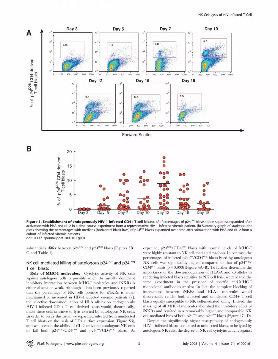
patients. As shown in Figure 1A, 5–7 days of stimulation with
phytohemoagglutinin (PHA) and recombinant IL-2 (rIL2) were
required to observe a variable but consistent percentage of
endogenously HIV-1 infected CD4+ T cell blasts, characterized by
the presence of intracellular viral p24 core antigen. In order to
determine the peak of maximal expansion of these infected cells,
we tested the amount of p24 antigen in CD4+ T cell blasts every 3
days for 3 weeks. Within our cohort of HIV-1 infected viremic
donors, the highest percentages of p24pos expression in activated
CD4+ T cell blasts were detected, on average, after 12 days of
activation (median: 9.43%; SD = 66.6) and started to decrease
progressively after this time point (Figure 1B).
Of the other several stimuli used to expand HIV-1 infected
CD4+ T cell blasts, only PHA plus rIL-2 and rIL-7 achieved
similar and sometimes better results after 12 days in culture
compared to stimulation with PHA and rIL-2 (Figure S2).
Physiological status and phenotype of p24pos CD4+ T cellblasts
We then analyzed whether p24pos CD4+ T cell blasts were able
to proliferate during the period of maximal expansion. As
expected, the ability of unfractionated CD4+ T cell blasts from
HIV-1 infected patients to undergo proliferation was significantly
lower compared to that of unfractionated blasts from healthy
donors (Figure 2A). Even though the positive expression of Ki67
nuclear antigen by p24pos fractions of CD4+ T cell blasts indicated
that these endogenously infected cells were able to enter the cell
cycle (Figure 2B), it has been shown both in vitro and ex vivo that
they are arrested in G2/M stage and do not complete the cell cycle
[22,23]. In order to correlate the kinetics of expansion of these
endogenously infected blasts with cell proliferation and CD4
expression, we analyzed the dilution of the vital dye carboxy-
fluorescein diacetate succinimidyl ester (CFSE) in CD4+ T cell
blasts using a multicolor flow cytometric approach. After 12 days
of stimulation, a subset of proliferating CFSE-labeled blasts
showed active intracellular viral replication (p24pos cells) with a
simultaneous down-modulation of cell surface CD4 (Figure 2C).
Therefore, the loss of CD4 is associated with a productive infection
in either endogenously (Figure 3A) or exogenously infected CD4+T cell-derived blasts [17,24,25]. We also visualized the intracel-
lular HIV-1 p24 core antigens in endogenously infected CD4+ T
cell blasts by fluorescence microscopy (Figure 2D).
It has also been reported that HIV-1 infection in vitro results in a
selective down-modulation of MHC-I molecules in cell lines and in
exogenously infected primary CD4+ T cells [14,15,16]. In order to
determine whether this phenomenon also occurs in endogenously
infected CD4+ T cell blasts expanded from HIV-1 infected
individuals, we analyzed the expression of classic and non-classic
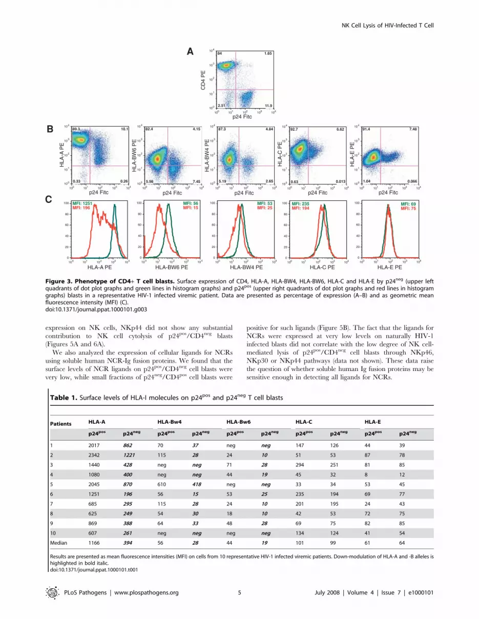
HLA molecules on p24pos and p24neg blasts. We found that surface
levels of HLA-A and -B alleles, calculated as mean fluorescence
intensity, were significantly down modulated on p24pos/CD4neg
blasts compared to p24neg/CD4pos blasts (p = 0.004 for HLA-A
alleles; p = 0.018 for HLA-BW4 and –BW6 alleles). In contrast,
the expression of HLA-C and HLA-E molecules did not
Author Summary
Natural killer (NK) cells represent an important line ofdefense against viral infections. In vitro studies withexogenously infected CD4+ T cell blasts from healthydonors have demonstrated that NK cells can kill autolo-gous HIV-1-infected target cells. However, the ability of NKcells from HIV-1-infected viremic patients to kill autolo-gous, endogenously infected CD4+ T cells had never beenexamined and remains uncertain. Given the reportedabnormalities in phenotype and functions of NK cells fromHIV-infected viremic individuals, we determined thefunction of NK cells in killing HIV-1-infected target cellsunder conditions that more closely mimic the in vivoenvironment in HIV-infected individuals. We show that NKcells from HIV-1-infected viremic patients display a variablealthough generally low ability to selectively eliminateautologous and endogenously HIV-1-infected CD4+ T cellblasts expanded ex vivo from peripheral blood. Variousfactors, including the markedly defective engagement ofimportant NK cell activation pathways and high frequen-cies of the pathologic CD56neg/CD16pos NK cell subset inHIV-1-infected viremic patients, influenced NK cell–medi-ated cytolysis of endogenously infected CD4+ T cell blasts.
NK Cell Lysis of HIV-Infected T Cell
PLoS Pathogens | www.plospathogens.org 2 July 2008 | Volume 4 | Issue 7 | e1000101
substantially differ between p24neg and p24neg blasts (Figures 3B–
C and Table 1).
NK cell-mediated killing of autologous p24pos and p24neg
T cell blastsRole of MHC-I molecules. Cytolytic activity of NK cells
against autologous cells is possible when the usually dominant
inhibitory interaction between MHC-I molecules and iNKRs is
either absent or weak. Although it has been previously reported
that the percentage of NK cells positive for iNKRs is either
maintained or increased in HIV-1 infected viremic patients [7],
the selective down-modulation of HLA alleles on endogenously
HIV-1 infected CD4+ T cell-derived blasts would, theoretically,
make these cells sensitive to lysis exerted by autologous NK cells.
In order to verify this issue, we separated infected from uninfected
T cell blasts on the basis of CD4 surface expression (Figure S3),
and we assessed the ability of rIL-2 activated autologous NK cells
to kill both p24neg/CD4pos and p24pos/CD4neg blasts. As
expected, p24neg/CD4pos blasts with normal levels of MHC-I
were highly resistant to NK cell-mediated cytolysis. In contrast, the
percentages of infected p24pos/CD4neg blasts lysed by autologous
NK cells was significantly higher compared to that of p24neg/
CD4pos blasts (p = 0.003) (Figure 4A–B). To further determine the
importance of the down-modulation of HLA-A and -B alleles in
rendering infected blasts sensitive to NK cell lysis, we repeated the
same experiment in the presence of specific anti-MHC-I
monoclonal antibodies (mAbs). In fact, the complete blocking of
interactions between iNKRs and HLA-I molecules would
theoretically render both infected and uninfected CD4+ T cell
blasts equally susceptible to NK cell-mediated killing. Indeed, the
masking of all MHC-I molecules abolished the inhibitory effect of
iNKRs and resulted in a remarkably higher and comparable NK
cell-mediated lysis of both p24neg and p24pos blasts (Figure 4C–D).
Despite the significantly higher susceptibility of endogenously
HIV-1 infected blasts, compared to uninfected blasts, to be lysed by
autologous NK cells, the degree of NK cell cytolytic activity against
Figure 1. Establishment of endogenously HIV-1 infected CD4+ T cell blasts. (A) Percentages of p24pos blasts (open squares) expanded afteractivation with PHA and rIL-2 in a time-course experiment from a representative HIV-1 infected viremic patient. (B) Summary graph of statistical dotplots showing the percentages with medians (horizontal black bars) of p24pos blasts expanded over time after stimulation with PHA and rIL-2 from acohort of infected viremic patients.doi:10.1371/journal.ppat.1000101.g001
NK Cell Lysis of HIV-Infected T Cell
PLoS Pathogens | www.plospathogens.org 3 July 2008 | Volume 4 | Issue 7 | e1000101

p24pos/CD4neg cell blasts was still relatively low and variable from
patient to patient (range: 4.2%–23%; median: 9.6%). Even when
interactions between iNKRs and MHC-I were completely blocked,
NK cells only partially eliminated both p24pos and p24neg
autologous CD4+ T cell blasts (range: 13%–48%; median: 38.4%)
(Figure 4). In order to understand whether NK cells from HIV-1
infected viremic patients, compared to healthy donors, were
defective in killing cells with low or no expression of MHC-I
molecules, we analyzed the NK cell-mediated cytolysis of cell lines
that do not express classic and non-classic HLA-I alleles. In line with
data previously reported [6,7], we confirmed that the ability of NK
cells from HIV-1 infected viremic patients to lyse K562 and 221 cell
lines was significantly lower compared with that of uninfected
individuals (Figure S4). A possible explanation for this low cytolytic
ability by NK cells from HIV-1 infected viremic individuals against
both MHC-Ineg cell lines or HIV-1 endogenously infected CD4+ T
cell blasts that down-modulate HLA-A and –B alleles might be the
defective engagement of activating NK cell receptors.
Role of NCRs and CD56neg NK cell subset. If the
dominant and negative effect of interactions between iNKRs
and MHC-I molecules is partially overcome by the HIV-1 induced
selective down-modulation of HLA-A and –B alleles, NK cell
killing of autologous and endogenously infected CD4+ T cell blasts
must occur through activating NK cell receptors. In order to
identify those NK receptor(s) that trigger this NK cell-mediated
lysis, we analyzed the phenotype and functions of NCRs and
NKG2D that, under physiological conditions, represent the major
activating receptors that regulate NK cell cytotoxicity.
We performed masking experiments to address the role of
NCRs in the NK cell lysis of autologous and endogenously HIV-1
infected CD4+ T cell blasts. The blocking of NKp46, NKp30 and
NKp44 did not result in a significant decrease in NK cell-mediated
lysis of p24pos/CD4neg cell blasts, indicating that the NCRs were
not playing a substantial role in this killing (Figure 5A). The lack of
significant NK cell killing of endogenously HIV-1 infected CD4+T cell blasts through the NCR activation pathway was also
consistent with previously reported data showing that the
percentage of NK cells expressing NCRs was significantly
decreased on fresh and rIL-2 activated NK cells from HIV-1
viremic patients compared to those from healthy donors
(Figure 5C) [6,7]. Nevertheless, among those few HIV-1 infected
viremic patients with higher levels of NKp46pos and NKp30pos NK
cells, we could detect a more pronounced inhibition of NK cell-
mediated killing especially after the simultaneous masking of three
NCRs (Figure 5A; Patients 4 and 6). In this context, we found a
highly significant correlation between the low degrees of NK cell-
mediated lysis of p24pos/CD4neg blasts and the decreased NK cell
surface levels of NKp46 and NKp30, indicating that the low levels
of expression of these two activating NK cell receptors negatively
contribute to the killing of HIV-1 infected cells. Regardless of its
Figure 2. Physiological status of CD4+ T cell blasts at the day of maximal expansion. (A) Proliferation of total CD4+ T cell blasts obtainedfrom uninfected and HIV-1 infected viremic individuals at different cell numbers in culture. Data are presented as median of experiments performedin triplicate on 5 healthy donors and 5 HIV-1 infected viremic individuals. (* p,0.0001; # p = 0.009). (B) Percentages of p24neg (upper left quadrant ofdot plot graph) and p24pos (upper right quadrant of dot plot graph) blasts undergoing cell cycling (Ki67pos cells) in a representative HIV-1 infectedviremic patient. (C) Multicolor flow cytometric analysis showing the down-modulation of CD4 in a subset of p24pos blasts (open squares) withinproliferating CD4+ T cell blasts analyzed by CFSE dye dilution in a representative HIV-1 infected viremic patient. (D) Fluorescence microscopic imageof two endogenously HIV-1 infected CD4+ T cell blasts. The HIV-1 p24 core antigen is stained blue within the cytoplasmic compartment of the cells.doi:10.1371/journal.ppat.1000101.g002
NK Cell Lysis of HIV-Infected T Cell
PLoS Pathogens | www.plospathogens.org 4 July 2008 | Volume 4 | Issue 7 | e1000101
expression on NK cells, NKp44 did not show any substantial
contribution to NK cell cytolysis of p24pos/CD4neg blasts
(Figures 5A and 6A).
We also analyzed the expression of cellular ligands for NCRs
using soluble human NCR-Ig fusion proteins. We found that the
surface levels of NCR ligands on p24pos/CD4neg cell blasts were
very low, while small fractions of p24neg/CD4pos cell blasts were
positive for such ligands (Figure 5B). The fact that the ligands for
NCRs were expressed at very low levels on naturally HIV-1
infected blasts did not correlate with the low degree of NK cell-
mediated lysis of p24pos/CD4neg cell blasts through NKp46,
NKp30 or NKp44 pathways (data not shown). These data raise
the question of whether soluble human Ig fusion proteins may be
sensitive enough in detecting all ligands for NCRs.
Figure 3. Phenotype of CD4+ T cell blasts. Surface expression of CD4, HLA-A, HLA-BW4, HLA-BW6, HLA-C and HLA-E by p24neg (upper leftquadrants of dot plot graphs and green lines in histogram graphs) and p24pos (upper right quadrants of dot plot graphs and red lines in histogramgraphs) blasts in a representative HIV-1 infected viremic patient. Data are presented as percentage of expression (A–B) and as geometric meanfluorescence intensity (MFI) (C).doi:10.1371/journal.ppat.1000101.g003
Table 1. Surface levels of HLA-I molecules on p24pos and p24neg T cell blasts
Patients HLA-A HLA-Bw4 HLA-Bw6 HLA-C HLA-E
p24pos p24neg p24pos p24neg p24pos p24neg p24pos p24neg p24pos p24neg
1 2017 862 70 37 neg neg 147 126 44 39
2 2342 1221 115 28 24 10 51 53 87 78
3 1440 428 neg neg 71 28 294 251 81 85
4 1080 400 neg neg 44 19 45 32 8 12
5 2045 870 610 418 neg neg 33 34 53 45
6 1251 196 56 15 53 25 235 194 69 77
7 685 295 115 28 24 10 201 195 24 43
8 625 249 54 30 18 10 42 53 72 75
9 869 388 64 33 48 28 69 75 82 85
10 607 261 neg neg neg neg 134 124 41 54
Median 1166 394 56 28 44 19 101 99 61 64
Results are presented as mean fluorescence intensities (MFI) on cells from 10 representative HIV-1 infected viremic patients. Down-modulation of HLA-A and -B alleles ishighlighted in bold italic.doi:10.1371/journal.ppat.1000101.t001
NK Cell Lysis of HIV-Infected T Cell
PLoS Pathogens | www.plospathogens.org 5 July 2008 | Volume 4 | Issue 7 | e1000101

We previously reported that among HIV-1 infected viremic
individuals there is a high frequency of a markedly dysfunctional
CD56neg subset of NK cells in which surface expression of NKp46
and NKp30 and cytolytic activitiy against tumor cell line targets
were found to be very low [11]. In this regard, we found that the
degree of NK cell baseline cytolysis of autologous p24pos blasts is
inversely correlated with the frequencies of anergic CD56neg NK
cells (Figure 6B).
Role of NKG2D. As we previously showed [7], there were no
differences in NK cell surface levels of NKG2D between infected
and uninfected individuals (Figure 7A). We also found that
NKG2D ligands, detected through a soluble human NKG2D-Ig
fusion protein, were expressed at high levels on endogenously
HIV-1-infected CD4+ T cell-derived blasts. In particular, among
the previously described NKG2D ligands [1], ULBP molecules (in
particular ULBP-2) were clearly expressed on p24pos blasts
(Figure 7B), while the levels of MIC -A (Figure 7B) and –B (data
not shown) were almost undetectable.
As previously demonstrated for NCRs, we then performed
masking experiments to understand the extent of the contribution
of NKG2D to the NK cell-mediated killing of infected p24pos/
CD4neg cell blasts. In line with the phenotypic profiles described
and with our previously published results obtained with the in vitro
infection [17], the masking of NKG2D induced a substantial
reduction of NK cell cytolysis of p24pos cell blasts in all patients
analyzed (p,0.0001) (Figure 7C). Moreover, we found a
significant direct correlation between the expression of ULB-2
on p24pos/CD4neg cell blasts and the NK cell-mediated lysis of
autologous HIV-1 endogenously infected T cell blasts (Figure 7D).
Consistent with results previously published using an in vitro
experimental system, our data indicate that the pathway activated
by interactions of NKG2D with its ULBP ligands (in particular
ULBP-2) plays an important role in the NK cell killing of
endogenously HIV-1 infected CD4+ T cell blasts [17].
We did not find any functional contribution mediated by the
activating NK cell co-receptors 2B4 and NTB-A to lysis of
autologous and endogenously HIV-1 infected CD4+ T cell blasts
in line with the down-modulation of their cellular ligands, CD48
and NTB-A, on p24pos CD4+ T cell blasts (p = 0.0051 for CD48;
p = 0.0049 for NTBA) (Figure S5).
Discussion
In the present study we demonstrate the ability of NK cells from
HIV-1 infected viremic patients to kill endogenously HIV-1-
infected autologous CD4+ T cell blasts derived from viremic
patients. We show that, subsequent to the selective down-
modulation of MHC-I molecules, infected p24pos blasts become
partially susceptible to lysis by rIL-2 activated NK cells, while
p24neg blasts are spared from killing. This NK cell-mediated killing
occurs mainly through the NKG2D activation pathway. However,
decreased NK cell expressions of NCRs contribute to the low level
of NK cell cytolytic activity. In addition, the unusually high
frequency of dysfunctional CD56neg NK cell subsets among HIV-1
infected viremic patients was shown to strongly correlate with the
low degree of NK cell cytolytic responses against infected p24pos
cell blasts.
The ability of NK cells to kill autologous HIV-1 infected target
cells mainly through the NKG2D pathway has been previously
demonstrated in vitro with exogenously infected CD4+ T cell blasts
from healthy donors [15,17]. In contrast, we examined NK cells
from HIV-1 viremic individuals and autologous target cells that
were expanded from endogenously infected CD4+ T cells. Given
the reported abnormalities in phenotype and functions of NK cells
from HIV-infected viremic individuals [26,27,28], the present
Figure 4. NK cell-mediated killing of autologous CD4+ T cell blasts: role of MHC-I molecules. Cytolysis of autologous p24neg/CD4pos andp24pos/CD4neg blasts by rIL-2 activated NK cells purified from HIV-1 infected viremic patients. Cells were incubated either in the absence (baselinelysis in A and B) or in the presence (C and D) of specific mAbs masking classic HLA-A/-B/-C and non classic HLA-E molecules. Data shown are fromexperiments performed in triplicate (6SD) using cells from 8 (A–B) and 2 (C–D) representative HIV-1 infected viremic individuals. The NK cell:CD4-derived blast ratio in all experiments was 10:1.doi:10.1371/journal.ppat.1000101.g004
NK Cell Lysis of HIV-Infected T Cell
PLoS Pathogens | www.plospathogens.org 6 July 2008 | Volume 4 | Issue 7 | e1000101

study was designed to determine the function of NK cells in killing
HIV-1 infected target cells under conditions that more closely
mimic the in vivo situation in HIV-infected individuals. Our
experimental system relied on rIL-2 activated NK cells instead of
freshly purified NK cells. We previously reported that prolonged
activation with rIL-2 did not restore phenotype and function of
highly dysfunctional NK cells from HIV-1 infected viremic
individuals. Only CD56 expression was recovered upon activation
with rIL-2 after 3 weeks of culture. Despite the reversion of the
CD56neg to a CD56pos phenotype, rIL-2 stimulated CD56neg-
derived NK cell populations still expressed very high percentages
of iNKRs and extremely low levels of NCRs, similar to results
from experiments performed with freshly purified CD56neg NK
cell subsets. Even after 21 days of activation with rIL-2 the
cytolytic potential of these highly dysfunctional NK cells from
HIV-1 infected viremic patients did not improve and remained
significantly lower compared to that of NK cells from uninfected
individuals[7,8,11]. Given the fact that stimulation with rIL-2 does
not substantially reverse the pathologic characteristics of freshly
purified NK cells from HIV-1 infected viremic donors, we
designed an experimental system using rIL-2 activated NK cells.
This yielded NK cells as effector cells against autologous and
endogenously HIV-1 infected CD4+ T blasts at the day of their
maximal expansion.
Circulating CD4+ T cells from HIV-infected individuals harbor
very low frequencies of replication-competent virus [12,13]. This
has made it very difficult to adequately characterize endogenously
infected CD4+ T cells from HIV-infected individuals. Several
studies showed that activation in vitro with several and multiple
stimuli enhanced viral replication in CD4+ T cells from HIV-1
Figure 5. NK cell-mediated killing of autologous CD4+ T cell blasts: role of NCRs. (A) Cytolysis (in triplicate 6SD) of autologous p24pos/CD4neg blasts by rIL-2 activated NK cells purified from HIV-1 infected viremic patients. Cells were incubated either in the absence (baseline lysis) or inthe presence of specific mAbs masking NKp46, NKp30 and NKp44. Experiments were also performed masking the effector with anti NKp46, NKp30and NKp44 (NCRs) simultaneously. Data shown are from experiments performed in triplicate (6SD) using cells from 3 representative HIV-1 infectedviremic individuals. We used an anti-human CD56 IgM mAb as an isotype control for masking experiments. The NK cell:CD4-derived blast ratio in allexperiments was 10:1. (B) Surface expression of ligands for NKp44, NKp30 and NKp44 by p24neg (upper left quadrants of dot plot graphs) and p24pos
(upper right quadrants of dot plot graphs) blasts derived from a representative HIV-1 infected viremic patient. The relative percentage of expressionand geometric mean fluorescence intensity (MFI) for each ligand is indicated in the upper left (for p24neg CD4-derived T cell blasts) and upper right(p24pos CD4-derived T cell blasts) quadrants of each dot plot. (C) Summary graphs of statistical dot plots with medians (horizontal black bars) showingthe percentages of total rIL-2 activated NKp46pos, NKp30pos, and NKp44pos NK cells purified from healthy donors (blue circles) and HIV-1 infectedviremic individual (red circles). Yellow, green and purple circles represent the three representative HIV-1 patients whose NK cell cytolytic functions areshown in Figures 4 and 5.doi:10.1371/journal.ppat.1000101.g005
NK Cell Lysis of HIV-Infected T Cell
PLoS Pathogens | www.plospathogens.org 7 July 2008 | Volume 4 | Issue 7 | e1000101

infected patients. The in vitro induced replication of HIV-1 was
measured by the release of p24 HIV-1 core antigen in culture-
supernatant [18,19,20,21]. Using a similar approach for cell
activation, we stimulated freshly purified CD4+ T cells from HIV-
1 infected viremic patients in order to expand and isolate these
endogenously infected T cell blasts detected by intracellular p24
staining. Activation with PHA plus rIL2 (with or without rIL-7)
was the most effective in expanding a population of endogenously
infected CD4+ T cell blasts. The peak maximal expansion of HIV-
1 productively infected blasts was reached, on average, 12 days
after activation and the rate of expansion of p24pos cell blasts
differed among samples from the 30 HIV-1 infected viremic
patients analyzed in the present study. The reasons for such
heterogeneous results are unclear and many variables such as
cellular or soluble suppressive factors, different numbers of
circulating latently HIV-1 infected cells, different viral strains,
culture conditions or other factors could contribute to this
variability. Further investigations are needed to extensively
characterize the kinetics of HIV-1 in endogenously infected cells
and the replication cycle of these p24pos/CD4neg cell blasts. Our
aim in the present study was restricted to an examination of the
interactions between endogenously HIV-1 infected CD4+ T cell
blasts and autologous NK cells from HIV-1 infected viremic
patients.
In the preparation of target cells, we separated infected from
uninfected cells on the basis of the lack of expression of CD4
together with intracellular expression of p24 on certain cells
(infected) and the expression of CD4 and lack of intracellular
expression of p24 on other cells (uninfected). It has been reported
that HIV-1 is able to down-modulate the expression of CD4 on T
cell surfaces, a phenomenon induced either by Nef, which
enhances the internalization and degradation of CD4, or by
Vpu and Env, which interfere with the transport of newly
synthesized CD4 to cell surface [24,25]. Even though the
physiological relevance of CD4 down-regulation is not fully
understood, the absence of CD4 on cell surfaces represents
another marker of cells productively infected with HIV-1. We
confirmed that endogenously infected CD4+ T cell blasts
harboring replication-competent virus down-modulate CD4
expression and, on the basis of surface levels of this molecule,
Figure 6. NCRs and CD56neg cell impact on baseline lysis of endogenously infected CD4+ T cell blasts. Statistical analyses showing thecorrelation between the NK-cell mediated baseline lysis of autologous p24neg/CD4pos blasts and the NK cell surface levels of NKp46, NKp30, NKp44(MFI on IL-2 activated NK cells) (Panel A) and CD56 (percentages on freshly purified NK cells) (Panel B). Orange, brown, gray, blue, yellow, red, greenand purple circles represent the eight representative HIV-1 patients whose NK cell cytolytic functions are also shown in Figure 4.doi:10.1371/journal.ppat.1000101.g006
NK Cell Lysis of HIV-Infected T Cell
PLoS Pathogens | www.plospathogens.org 8 July 2008 | Volume 4 | Issue 7 | e1000101

we were able to separate infected p24pos from uninfected p24neg T
cell blasts.
In line with results previously reported with HIV-1 infection in
vitro [15], endogenously HIV-1 infected CD4+ T cell blasts
selectively down-modulated HLA-A and –B alleles while the
expression of HLA-C and HLA-E molecules was conserved. The
selective down-regulation of these MHC-I molecules should
render p24pos/CD4neg cell blasts susceptible to NK cell-mediated
killing. In fact, although the surface levels of HLA-C and -E may
still protect infected cell blasts from the cytolysis exerted by
autologous NK cells expressing iNKRs specific for these conserved
alleles of MHC-I [14,29], this is not the case for NK cells that
express iNKRs specific for HLA-A and –B [15]. We show that the
degree of NK cell-mediated lysis of p24pos/CD4neg blasts was
significantly higher compared with that of p24neg/CD4pos blasts.
Moreover, masking experiments highlighted the important role of
the selective down-modulation of HLA-I molecules, because only
the complete blocking of all MHC-I alleles rendered infected
p24pos and uninfected p24neg cell blasts equally susceptible to NK
cell-mediated lysis.
Other studies reported that conserved or even up-regulated
levels of HLA-E on HIV-1 infected cells are able to inhibit NK
cell-mediated cytolysis of HIV-1 infected cells through binding to
its specific inhibitory receptor NKG2A [14,30]. In our study, we
used two different mAbs (3D12 and 4D12) in order to detect the
surface levels of HLA-E. Despite the fact that there was some
variability among different donors, we detected no significant
differences in the high levels of HLA-E expression between HIV-1
infected and uninfected CD4+ T cell blasts from HIV-1 infected
viremic patients. Moreover, given that the frequency of the
NKG2Apos NK cell subset is greatly decreased in chronic HIV-1
infected viremic patients compared to that of healthy donors
[7,31], it is unlikely that the interaction between HLA-E and
NKG2A can explain the decreased NK cell mediated killing of
HIV-1 infected blasts. In fact, our masking experiments demon-
strated that the complete blocking of NKG2A did not increase NK
100 101 102 103 104100
101
102
103
104
NK
G2D
Lig
and
PE
12%MFI: 10
51%MFI: 53
p24 Fitc
100 101 102 103 104100
101
102
103
104
ULB
P-1
PE
3%MFI: 5.6
12%MFI: 4.9
p24 Fitc100 101 102 103 104
100
101
102
103
104
ULB
P-2
PE
11%MFI: 13
32%MFI: 19
p24 Fitc
100 101 102 103 104100
101
102
103
104
ULB
P-3
PE
3%MFI: 7
p24 Fitc100 101 102 103 104
100
101
102
103
104
MIC
-A P
E
5%MFI: 8.1
0.2%MFI: 4.4
p24 Fitc
CPatient 3
% C
r51 S
peci
fic R
elea
se
Baseline Lysis0.0
2.5
5.0
7.5
10.0
12.5
Baseline Lysis Masking ofNKG2D
0.0
2.5
5.0
7.5
10.0
12.5
15.0
17.5
20.0
Patient 4
Baseline Lysis0.02.55.07.5
10.012.515.017.520.022.525.0
Patient 6
Masking ofNKG2D
Masking ofNKG2D
BA
0
25
50
75
100
% N
KG
2Dp
os
NK
Cel
ls
ViremicPatients
p = ns
HealthyDonors
Isot
ype
Con
trol
PE
100 101 102 103 104100
101
102
103
104
0.11%MFI: 4.2
0.05%MFI: 4.3
p24 Fitc
8%MFI: 5.2
Isotype Isotype Isotype
D
% o
f ULB
P-2
pos
p24po
s T c
ell b
last
s
Baseline Lysis0 10 20 30
0
10
20
30
40
p < 0.0001r = 0.87
Figure 7. NK cell-mediated killing of autologous CD4+ T cell blasts: role of NKG2D. (A) Summary graphs of statistical dot plots withmedians (horizontal black bars) showing the percentages of total rIL-2 activated NKG2Dpos NK cells purified from healthy donors (blue circles) andHIV-1 infected viremic individual (red circles). Yellow, green and purple circles represent the three representative HIV-1 patients whose NK cellcytolytic functions are shown in Figures 4 and 5. The NK cell:CD4-derived blast ratio in all experiments was 10:1. (B) Surface expression of ligands forNKG2D by p24neg (upper left quadrants of dot plot graphs) and p24pos (upper right quadrants of dot plot graphs) blasts derived from a representativeHIV-1 infected viremic patient. The relative percentage of expression and geometric mean fluorescence intensity (MFI) for each ligand is indicated inthe upper left (for p24neg CD4-derived T cell blasts) and upper right (p24pos CD4-derived T cell blasts) quadrants of each dot plot. (C) Cytolysis (intriplicate 6SD) of autologous p24pos/CD4neg blasts by rIL-2 activated NK cells purified from HIV-1 infected viremic patients. Cells were incubatedeither in the absence (baseline lysis) or in the presence of specific mAbs masking NKG2D. Data shown are from experiments performed in triplicate(6SD) using cells from 3 representative HIV-1 infected viremic individuals. We used an anti-human CD56 IgM mAb as an isotype control for maskingexperiments. The NK cell:CD4-derived blast ratio in all experiments was 10:1. (D) Statistical analyses showing the correlation between the NK-cellmediated baseline lysis of autologous HIV-1 infected blasts and the percentages of ULBP-2 ligand for NKG2D on p24pos/CD4neg cell blasts. Orange,brown, gray, blue, yellow, red, green and purple circles correspond to the eight representative HIV-1 patients whose NK cell cytolytic functions arealso shown in Figures 4 and 6.doi:10.1371/journal.ppat.1000101.g007
NK Cell Lysis of HIV-Infected T Cell
PLoS Pathogens | www.plospathogens.org 9 July 2008 | Volume 4 | Issue 7 | e1000101

cell cytolysis of autologous p24pos blasts (data not shown). The
reason for the discrepancy in the role of NKG2A/HLA-E
interactions between these previous studies and our data may be
the fact that the effector cells used in those previous studies were
heterologous NK cell lines expressing high levels of NKG2A
against HLA-E transfected target cell lines.
As mentioned above, the engagement of activating NK cell
receptors should be able to trigger the cytolytic activity in NK cells
expressing iNKRs specific for HLA-A and -B and lacking iNKRs
for HLA-C and-E. In this regard, NK cell-mediated killing of
infected p24pos/CD4neg cell blasts was found to be mainly
NKG2D-dependent. These results are in line with the highly
conserved expression of NKG2D on NK cells from HIV-1
infected viremic individuals and with the relatively high percent-
ages of p24pos blasts expressing NKG2D ligands. The direct effect
of HIV-1 on the positive or negative modulation of NKG2D
ligands on the surfaces of primary CD4+ T cells infected in vitro
with HIV-1 is controversial [17,32]. We show that masking the
binding of NKG2D to its ligands clearly resulted in a marked
reduction of the NK cell-mediated lysis of infected blasts. These
results suggest that the NKG2D ligands, expressed at high levels in
p24pos/CD4neg blasts, play an important role in NK cell-mediated
killing of autologous infected cells. Several groups previously
reported that HIV-1 viremia affects several functions of NK cells
and dramatically influences their phenotype [26,27,28]. Interest-
ingly, the surface expression and activation pathway of NKG2D
are among the few NK cell characteristics spared from the
deleterious effects of HIV-1 infection. In order to understand
better how HIV-1 affects NK cell cytolytic responses, it would be
important for future investigations to address the molecular
mechanism(s) underlying the resistance of NKG2D, compared to
other important activating and inhibitory NK receptor pathways,
to the dysfunction associated with HIV viremia.
Although defective in HLA-A and –B expression, p24pos/
CD4neg blasts remain still poorly sensitive to killing exerted by
autologous NK cells. This is partly the result of inhibitory
interactions between iNKRs and conserved HLA-C molecules, as
demonstrated by the relatively low levels of killing of both infected
and uninfected autologous blasts even in the presence of anti-
MHC-I mAbs which completely block the interactions between
MHC-I molecules and iNKRs. The relatively low degree of NK
cell cytolytic activity against endogenously HIV-1 infected CD4+T cell blasts might be secondary to aberrancies in NK cell
triggering through important activating receptors other than
NKG2D. This concept is further supported by the finding that
NK cells from HIV-1 infected viremic patients were markedly
impaired, compared to that from healthy donors, in their ability to
kill highly susceptible target cells such as K562 and 221 tumor cell
lines that do not express MHC-I molecules. Moreover, if we
compare these experimental data with our previously reported
results obtained with HIV-1 infection in vitro [17], the degree of
killing of autologous, exogenously HIV-1 infected CD4+ cell blasts
by NK cells from healthy donors appears to be markedly higher
compared to killing by of highly dysfunctional NK cells obtained
from HIV-1 infected viremic individuals. In this context, the low
levels of NKp46 and NKp30 on NK cells from HIV-infected
individuals significantly correlated with NK cell-mediated killing
of MHC-Ineg K562 and 221 cell lines (data not shown) and of
endogenously HIV-1 infected autologous CD4+ T cell blasts.
These data suggest that the negative effect of HIV-1 viremia on
NKp46 and NKp30 expression interfere with the NK cell lysis of
endogenously HIV-1 infected autologous CD4+ T cell blasts.
HIV-1 infected autologous CD4+ T cell blasts. Our finding of the
negative contribution of NCRs in the killing of endogenously
infected targets in HIV-infected viremic individuals differs from
the findings of a previous study in which we described that
NKG2D was important in NK lysis of infected targets, but that
NCRs played no demonstrable role [17]. This discrepancy may
result from the fact that the study in question used NK cells from
normal individuals and target cells that were infected in vitro with
several viral strains, whereas the present study employed NK cells
from HIV-infected viremic individuals and endogenously infected
target cells.
It is well known that HIV-1 viremia induces a CD4+ T cell
depletion that leads to immunodeficiency and correlates with
disease progression. However, it has also been reported that the
majority of CD4+ T cells dying during the infection are not
productively infected with HIV-1[33]. One possible explanation is
that these uninfected CD4+ T cells are eliminated through a
mechanism not directly linked to viral replication. It has been
demonstrated both in vitro[17] and ex vivo (Figure 5B) that HIV-1
replication can modulate the expression of ligands for NKp46,
NKp30 and NKp44 on uninfected p24neg/CD4pos T cell blasts. In
particular, an highly conserved motif of HIV-1 gp41 envelope
protein can induce the expression of NKp44 ligand on uninfected
CD4+ T cell blasts and render these cells susceptible to NK cell-
mediated killing via NKp44 activation pathway[34]. A recent
report showed that is possible to prevent the expression of NKp44
ligand on CD4+ T cells, thus providing new insight for both
preventive and therapeutic HIV-1 vaccine strategies[35].
In conclusion, the present study shows that NK cells from HIV-
1 infected viremic patients display a variable although generally
low ability to lyse endogenously HIV-1 infected autologous CD4+T cell blasts derived from peripheral blood. The selective down-
modulation of HLA-A and -B molecules makes p24pos/CD4neg
cell blasts susceptible, at least in part, to autologous NK cell-
mediated lysis mainly through the NKG2D activation pathway.
Several other factors including the decreased NK cell expression of
NCRs, low levels of NCR-specific ligands on p24pos CD4+ T cell
blasts and the high frequency of the dysfunctional CD56neg NK
cell subset also contribute to the low levels of NK cell-mediated
killing of HIV-1 endogenously infected autologous CD4+ T cell
blasts. In fact, the defective killing through the NCR activation
pathways and the presence at very high levels of a markedly
anergic CD56neg NK cell population substantially impair the
ability of NK cell to kill endogenously HIV-1 infected autologous
CD4+ T cell blasts. Understanding the mechanisms by which
HIV-1 is able to negatively modulate the expression and function
of NCRs on NK cell and of their ligands on HIV-1 infected CD4+T cells will certainly give us new insights for improving the NK
cell-mediated lysis of infected cells and for enforcing the innate
immune control of HIV-1 infection.
Materials and Methods
Study SubjectsThirty HIV-1 infected viremic individuals were studied. The
median CD4+ T cell count was 373 cell per ml (SD = 6193) and
the median viremia was 32,677 HIV-1 RNA copies
(SD = 680,734) per ml of plasma as detected by an ultrasensitive
branched DNA (bDNA) assay (Chiron) with a lower limit of
detection of 50 copies per ml. Patients were either naı̈ve to
antiretroviral therpay (ART) or had formerly been receiving ART,
but were not receiving therapy at the time of the study.
Leukapheresis was conducted in accordance with protocols
approved by the Institutional Review Boards (IRBs) of the
University of Toronto, Ontario, Canada and the National Institute
of Allergy and Infectious Diseases (NIAID), National Institutes of
NK Cell Lysis of HIV-Infected T Cell
PLoS Pathogens | www.plospathogens.org 10 July 2008 | Volume 4 | Issue 7 | e1000101

Health (NIH), Bethesda, Maryland, USA. Each patient signed a
consent form that was approved by the above IRBs. As negative
controls, cells from 30 healthy donors seronegative for HIV-1 were
obtained by apheresis generously provided by the Transfusion
Medicine Department of the Mark O. Hatfied Clinical Research
Center of the NIH as a part of IRB approved clinical studies.
Isolation and culture of CD4+ T cells and NK cellsPBMCs were obtained from leukapheresis packs by Ficoll-
Hypaque density gradient centrigugation (LSM, MP Biomedicals).
CD4+ T cells and NK cells were freshly isolated by negative
selection (Stem Cell Technologies) according to the protocol
provided by the manufacturer. The purity of CD3+/CD4+ T cells
was $97%. Purified NK cells contained # 3% contamination
with other PBMC subsets, as determined by expression of CD3,
TCR-a/b, TCR-g/d, CD19 or CD14.
In order to expand CD4+ T cell blasts productively and
endogenously infected with HIV-1, we activated freshly purified
CD4+ T cells (26106/ml) with different stimuli, as shown in Figure
S1. Briefly, cells were cultured with RPMI medium 1640
supplemented with antibiotics (Gibco) and FCS (HyClone) as
previously described[7] and stimulated with phytohemoagglutinin
(PHA) (Sigma-Aldrich) at 3 mg/ml for 24 hours plus recombinant
IL-2 (rIL-2) (Roche) at 50 IU/ml with or without recombinant IL-7
(rIL-7) (R&D Systems) at 10ng/ml for 21 days. We also activated
freshly purified CD4+ T cells with rIL-7 with or without rIL-2 or
with soluble anti-CD28 mAbs at 5 mg/ml on tissue culture plates
coated with anti-CD3 mAbs at 10 mg/ml (BD-Pharmingen) for 21
days in the presence of rIL-2. Freshly purified NK cells were
activated in vitro for 12 days with rIL-2 at 200 IU/ml at 2*106/ml.
mAbsThe following panel of anti-human monoclonal antibodies
(mAbs) were used in this study: mAbs 289 (IgG2a anti-CD3), C218
and A6-220 (IgG1and IgM anti-CD56, respectively), KD1 (IgG2a
anti-CD16), AZZ20 and F252 (IgG1 and IgM anti-NKp30,
respectively), BAB281 and KL247 (IgG1 and IgM anti-NKp46,
respectively), Z231 and KS38 (IgG1 and IgM anti-NKp44,
respectively), ON72 and Bat221 (IgG1 anti-NKG2D), MA127
and ON56 (IgG1 and IgG2b anti-NTBA, respectively), pp35 and
Co54 (IgG1 and IgM anti-2B4, respectively), Ma152 and CER1
(IgG1 and IgM anti-NKp80, respectively), KRA236 and F5 (IgG1
and IgM anti-DNAM-1, respectively), L14 (IgG2a anti-Nectin 2),
L95 (IgG1 anti-poliovirus receptor), Z270 (IgG1 anti-NKG2A), Y9
(IgM anti-CD94), EB6 (IgG1 anti-p58.1/KIR2DL1), Gl183 (IgG1
anti p58.2/KIR2DL2), Z276 (IgG1 anti-p70/KIR3DL1), F278
(IgG1 anti-LIR-1ILT2) and A6.136 (anti-MHC class I molecules,
IgM). FITC-, PE- or APC-labeled anti-CD3, anti-CD4, anti-CD8,
anti-TCRa/b, anti-TCRc/d, anti-CD14, anti-CD19, anti-CD56,
anti-MICA/B and anti-CD48 mAbs were purchased from BD
Biosciences. Soluble fusion proteins for NKp30, NKp46, NKp44
and NKG2D with the Fc portion of human IgG and anti-human
ULBP-1,-2 and -3 mAbs were purchased from R&D Systems. PE-
labeled anti-human Fc fragment mAb was purchased from Jackson
ImmunoResearch Laboratories. FITC- and PE- anti human anti-
HIV-1 p24 mAb (clone KC57) used for intracellular flow
cytometry staining was purchased from Coulter Clone. PE-labeled
anti-HLA-A mAbs were purchased from Lab Vision Corporation.
Anti-human HLA-C mAb (clone L31) was kindly provided by Dr.
Patrizio Giacomini (Regina Elena Cancer Institute, Rome, Italy)
and used in flow cytometry as previously described[36]. Anti-
human HLA-Bw4 (clone 116.5.28) and HLA-BW6 (clone 126.39)
mAbs were kindly provided by Dr. Keith Gelsthorpe (National
Blood Transfusion Service, Sheffield, UK). Anti-human HLA-E
mAbs (clones 3D12 and 4D12) were kindly provided by Dr. Dan
Gerarthy (Fred Hutchinson Cancer Research, Seattle, WA, USA).
Flow cytometryFor one-, two- or three-color cytofluorimetric analysis (FACS
Calibur, BD), cells were stained with the appropriate FITC-, PE-
or APC-labeled mAbs. For indirect staining, cells were stained
with appropriate unlabeled mAbs followed by FITC- or PE-
conjugated isotype-specific goat anti-mouse second reagent
(Southern Biotechnology Associates). Second appropriate anti-
isotypic mAbs stained with FITC and/or PE and/or APC were
used as negative controls. For intracellular staining, samples were
fixed and permeabilized by cytofix/cytoperm solution and washed
with perm-wash solution 1X (BD-Pharmigen) according to the
protocol provided from the manufacturer. The data were analyzed
using FlowJo software (Tree Star Inc.).
Detection, proliferation and fluorescence microscopy ofendogenously HIV-1 infected CD4+ T cell blasts
The percentages of HIV-1 infected CD4+ T cell blasts were
detected by intracellular flow cytometry with an anti-p24 core
virus antigen mAb.
Cells undergo cell cycling were evaluated by detecting the intra-
nuclear expression of Ki67 (BD-Pharmigen).
CD4-derived T cell blast proliferation was detected by3[H]thymidine uptake assay (16 hours). Cellular proliferation
was also evaluated by dilution of the vital dye CFSE (Molecular
Probes) according to the supplier’s instructions.
After 10–12 days of activation with PHA and rIL-2,
unfractionated CD4+ T cell blasts were permeabilized by
cytofix/cytoperm solution and stained with an PE-labeled anti-
p24 HIV-1 core antigen mAb (Coulter Clone) followed by a biotin
conjugated mouse anti R-PE mAb (BD). Infected p24pos CD4+ T
cell blasts were detected by a Pacific Blue fluorescent-dye
conjugate of streptavidin (Molecular probes) according to the
supplier’s instructions. Fluorescent cells were then washed in PBS,
suspended in medium, and sealed on the slides with cover slips.
Images were collected on a Leica TCS-NT/SP confocal
microscope (Leica) using a 63x oil immersion objective NA 1.32.
Pacific Blue was excited using an Argon laser at 364 nm. DIC
(differential interference contrast) images were collected simulta-
neously with the fluorescence images using the transmitted light
detector. Images were processed using Leica TCS-NT/SP
software (version 1.6.587), Imaris 3.3.2 (Bitplane AG), and Adobe
Photoshop 7.0 (Adobe systems).
Cytolytic activityIn line with the timeframe of maximal expansion of p24pos/
CD4neg blasts, after 12 days of stimulation we removed by negative
selection all contaminant cells from CD4+ T cell blast cultures
(MACS, Milteny Biotec). As a result, we obtained a highly purified
population of CD4+ T cell-derived blasts containing #5%
contamination of other lymphocyte subsets (TCRg/d+, CD8+,
CD56+, CD16+ and CD19+ cells). HIV-1 infected CD4+ T cell
blasts were then separated from uninfected blasts on the basis of
CD4 surface expression through magnetic microbeads conjugated
with an anti-human CD4 mAb (MACS, Milteny Biotec),
according to the protocol provided by the manufacturer. The
purities of fractions of uninfected CD4pos and infected CD4neg
blasts , as assessed by intracellular staining with HIV-1 p24 core
antigens, was $97% and $70%, respectively. p24neg/CD4pos and
p24pos/CD4neg cell blasts were then used as target cells against
autologous rIL-2 activated NK cells in a 4-hour 51Cr release assay
NK Cell Lysis of HIV-Infected T Cell
PLoS Pathogens | www.plospathogens.org 11 July 2008 | Volume 4 | Issue 7 | e1000101

as described previously[37]. Saturating concentration (10 mg/ml)
of specific mAbs blocking NK cell receptors or MHC-I molecules
were added for the masking experiments. The NK cell:T cell blast
ratio was 10:1 (Figure S1).
Polyclonal NK cells were also tested in a 4-hour 51Cr release
assay against MHC-Ineg erythroleukemia K562 and MHC-Ineg B-
EBV cell line 721.221 (thereafter termed 221). E/T ratios are
indicated in the figures.51Cr release cytolytic assay were performed on cells from 15
HIV-1 infected viremic patients.
Statistical AnalysisImmune response distributions between healthy donors and
HIV-1 infected viremic patients were compared using the Mann-
Whitney test. The phenotypic and functional differences between
p24pos and p24neg blasts from HIV-1 infected individuals were
evaluated using the Wilcoxon signed ranks test. The functional
differences between NK cell-mediated baseline lysis and lysis in
masking experiments were evaluated using the Wilcoxon signed
ranks test. All p-values are 2-sided and unadjusted. All statistical
associations between different immune parameters were deter-
mined by the Spearman rank test for correlation. To estimate the
time of maximal infection, the mean outcome of infected p24pos
over time was modeled as a polynomial function of time and
estimated using least squares. The maximum of this function was
then determined and a bootstrap procedure was used to provide a
confidence interval $95% for the maximum.
Supporting Information
Figure S1 Methodology. p24pos blasts were expanded from total
PBMCs obtained from HIV-1 infected viremic patients and used
as targets for autologous rIL-2 activated NK cells
Found at: doi:10.1371/journal.ppat.1000101.s001 (0.05 MB PDF)
Figure S2 Expansion ex vivo of HIV-1 infected CD4+ T cell-
derived blasts by using different stimuli. Percentages of p24pos
blasts (open squares) expanded at day 12 after activation with rIL-
2 or rIL7 alone, with PHA plus rIL-7 or rIL-26rIL-7 and with
anti-CD3 plus anti CD28 mAbs from a representative HIV-1
infected viremic patient.
Found at: doi:10.1371/journal.ppat.1000101.s002 (0.04 MB PDF)
Figure S3 Sorting of HIV-1 infected and uninfected CD4+ T
cell-derived blasts. Representative example of separation of p24pos
from p24neg blasts through magnetic microbeads conjugated with
an anti-CD4 mAb. Purities of sorted uninfected CD4pos and
infected CD4neg T cell blast fractions were assessed by intracellular
staining with HIV-1 p24 core antigens in a double color flow
cytometric analysis.
Found at: doi:10.1371/journal.ppat.1000101.s003 (0.04 MB PDF)
Figure S4 Cytolytic Activity of NK cells against HLA-Ineg tumor
target cell lines. Spontaneous killing of K562 (A) and 221 (B)
tumor cell lines by rIL-2 activated NK cells-. Data are presented as
the average of experiments conducted on 15 healthy donors (black
squares) and 15 HIV-1 infected viremic patients (red diamonds).
Found at: doi:10.1371/journal.ppat.1000101.s004 (0.01 MB PDF)
Figure S5 NK cell-mediated killing of autologous HIV-1
infected CD4+ T cell-derived blasts: role of 2B4 and NTBA and
expression of their ligands on cell targets. (A-B) Surface expression
of CD48 and NTBA in p24neg (upper left quadrants of dot plot
graphs and green lines in histogram graphs) and p24pos (upper
right quadrants of dot plot graphs and red lines in histogram
graphs) blasts derived from a representative HIV-1 infected
viremic patient. Data are indicated as percentage of expression
(A) and as MFI (b). (C) Cytolysis (in triplicate 6SD) of autologous
p24neg/CD4pos blasts exerted by rIL-2 activated NK cells purified
from a representative HIV-1 infected viremic patient. Cells were
incubated either in the absence (baseline lysis) or in the presence of
specific mAbs masking 2B4 and NTBA. We used an anti-human
CD56 IgM mAb as an isotype control for masking experiments.
The NK cell:CD4-derived blast ratio in all experiments was 10:1.
Found at: doi:10.1371/journal.ppat.1000101.s005 (0.04 MB PDF)
Acknowledgments
We thank the patients for their generosity and participation in this study.
We also thank Nancy Touchette for her invaluable editorial assistance.
Author Contributions
Conceived and designed the experiments: D. Mavilio. Performed the
experiments: M. Fogli, E. Brunetta, S. Varchetta, K. Ata, J. Ward.
Analyzed the data: M. Fogli, D. Mavilio, E. Brunetta, K. Ata, E. Barker,
A. Moretta, A. Fauci. Wrote the paper: M. Fogli, D. Mavilio, E. Barker, A.
Moretta, A. Fauci. Purified and titrated mAbs: D. Pende, E. Marcenaro.
Performed statistical analysis: D. Follmann. Enrolled patients: G. Roby, C.
Kovacs.
References
1. Cerwenka A, Lanier LL (2001) Natural killer cells, viruses and cancer. Nat Rev
Immunol 1: 41–49.
2. Cooper MA, Fehniger TA, Caligiuri MA (2001) The biology of human natural
killer-cell subsets. Trends Immunol 22: 633–640.
3. Karre K, Ljunggren HG, Piontek G, Kiessling R (1986) Selective rejection of H-
2-deficient lymphoma variants suggests alternative immune defence strategy.
Nature 319: 675–678.
4. Anfossi N, Andre P, Guia S, Falk CS, Roetynck S, et al. (2006) Human NK cell
education by inhibitory receptors for MHC class I. Immunity 25: 331–342.
5. Moretta A, Bottino C, Mingari MC, Biassoni R, Moretta L (2002) What is a
natural killer cell? Nat Immunol 3: 6–8.
6. De Maria A, Fogli M, Costa P, Murdaca G, Puppo F, et al. (2003) The impaired
NK cell cytolytic function in viremic HIV-1 infection is associated with a
reduced surface expression of natural cytotoxicity receptors (NKp46, NKp30
and NKp44). Eur J Immunol 33: 2410–2418.
7. Mavilio D, Benjamin J, Daucher M, Lombardo G, Kottilil S, et al. (2003)
Natural killer cells in HIV-1 infection: dichotomous effects of viremia on
inhibitory and activating receptors and their functional correlates. Proc Natl
Acad Sci U S A 100: 15011–15016.
8. Mavilio D, Lombardo G, Kinter A, Fogli M, La Sala A, et al. (2006)
Characterization of the defective interaction between a subset of natural killer
cells and dendritic cells in HIV-1 infection. J Exp Med 203: 2339–2350.
9. Alter G, Teigen N, Davis BT, Addo MM, Suscovich TJ, et al. (2005) Sequential
deregulation of NK cell subset distribution and function starting in acute HIV-1
infection. Blood 106: 3366–3369.
10. Hu PF, Hultin LE, Hultin P, Hausner MA, Hirji K, et al. (1995) Natural killer
cell immunodeficiency in HIV disease is manifest by profoundly decreased
numbers of CD16+CD56+ cells and expansion of a population of
CD16dimCD56- cells with low lytic activity. J Acquir Immune Defic Syndr
Hum Retrovirol 10: 331–340.
11. Mavilio D, Lombardo G, Benjamin J, Kim D, Follman D, et al. (2005)
Characterization of CD56-/CD16+ natural killer (NK) cells: A highly
dysfunctional NK subset expanded in HIV-infected viremic individuals. Proc
Natl Acad Sci U S A.
12. Chun TW, Carruth L, Finzi D, Shen X, DiGiuseppe JA, et al. (1997)
Quantification of latent tissue reservoirs and total body viral load in HIV-1
infection. Nature 387: 183–188.
13. Pierson T, McArthur J, Siliciano RF (2000) Reservoirs for HIV-1: mechanisms
for viral persistence in the presence of antiviral immune responses and
antiretroviral therapy. Annual review of immunology 18: 665–708.
14. Cohen GB, Gandhi RT, Davis DM, Mandelboim O, Chen BK, et al. (1999)
The selective downregulation of class I major histocompatibility complex
proteins by HIV-1 protects HIV-infected cells from NK cells. Immunity 10:
661–671.
NK Cell Lysis of HIV-Infected T Cell
PLoS Pathogens | www.plospathogens.org 12 July 2008 | Volume 4 | Issue 7 | e1000101

15. Bonaparte MI, Barker E (2004) Killing of human immunodeficiency virus-
infected primary T-cell blasts by autologous natural killer cells is dependent onthe ability of the virus to alter the expression of major histocompatibility
complex class I molecules. Blood 104: 2087–2094.
16. Noraz N, Verrier B, Fraisier C, Desgranges C (1995) Cell surface phenotypicchanges induced in H9 T cells chronically infected with HTLV type I or HIV
type 1 or coinfected with the two viruses. AIDS research and human retroviruses11: 145–154.
17. Ward J, Bonaparte M, Sacks J, Guterman J, Fogli M, et al. (2007) HIV
modulates the expression of ligands important in triggering natural killer cellcytotoxic responses on infected primary T-cell blasts. Blood 110: 1207–1214.
18. Asjo B, Cefai D, Debre P, Dudoit Y, Autran B (1993) A novel mode of humanimmunodeficiency virus type 1 (HIV-1) activation: ligation of CD28 alone
induces HIV-1 replication in naturally infected lymphocytes. Journal of virology67: 4395–4398.
19. Chun TW, Engel D, Mizell SB, Ehler LA, Fauci AS (1998) Induction of HIV-1
replication in latently infected CD4+ T cells using a combination of cytokines.The Journal of experimental medicine 188: 83–91.
20. Smithgall MD, Wong JG, Critchett KE, Haffar OK (1996) IL-7 up-regulatesHIV-1 replication in naturally infected peripheral blood mononuclear cells.
Journal of immunology (Baltimore, Md 156: 2324–2330.
21. Wang FX, Xu Y, Sullivan J, Souder E, Argyris EG, et al. (2005) IL-7 is a potentand proviral strain-specific inducer of latent HIV-1 cellular reservoirs of infected
individuals on virally suppressive HAART. The Journal of clinical investigation115: 128–137.
22. Bahbouhi B, Landay A, Al-Harthi L (2004) Dynamics of cytokine expression inHIV productively infected primary CD4+ T cells. Blood 103: 4581–4587.
23. Zimmerman ES, Sherman MP, Blackett JL, Neidleman JA, Kreis C, et al. (2006)
Human immunodeficiency virus type 1 Vpr induces DNA replication stress invitro and in vivo. J Virol 80: 10407–10418.
24. Lundquist CA, Tobiume M, Zhou J, Unutmaz D, Aiken C (2002) Nef-mediateddownregulation of CD4 enhances human immunodeficiency virus type 1
replication in primary T lymphocytes. Journal of virology 76: 4625–4633.
25. Wildum S, Schindler M, Munch J, Kirchhoff F (2006) Contribution of Vpu,Env, and Nef to CD4 down-modulation and resistance of human immunode-
ficiency virus type 1-infected T cells to superinfection. Journal of virology 80:8047–8059.
26. Scott-Algara D, Paul P (2002) NK cells and HIV infection: lessons from other
viruses. Curr Mol Med 2: 757–768.
27. Fauci AS, Mavilio D, Kottilil S (2005) NK cells in HIV infection: Paradigm for
protection or targets for ambush. Nat Rev Immunol 5: 835–843.
28. Alter G, Altfeld M (2006) NK cell function in HIV-1 infection. Curr Mol Med 6:
621–629.
29. Ward JP, Bonaparte MI, Barker E (2004) HLA-C and HLA-E reduce antibody-
dependent natural killer cell-mediated cytotoxicity of HIV-infected primary T
cell blasts. AIDS (London, England) 18: 1769–1779.
30. Nattermann J, Nischalke HD, Hofmeister V, Kupfer B, Ahlenstiel G, et al.
(2005) HIV-1 infection leads to increased HLA-E expression resulting in
impaired function of natural killer cells. Antiviral therapy 10: 95–107.
31. Mela CM, Burton CT, Imami N, Nelson M, Steel A, et al. (2005) Switch from
inhibitory to activating NKG2 receptor expression in HIV-1 infection: lack of
reversion with highly active antiretroviral therapy. Aids 19: 1761–1769.
32. Cerboni C, Neri F, Casartelli N, Zingoni A, Cosman D, et al. (2007) Human
immunodeficiency virus 1 Nef protein downmodulates the ligands of the
activating receptor NKG2D and inhibits natural killer cell-mediated cytotox-
icity. J Gen Virol 88: 242–250.
33. Alimonti JB, Ball TB, Fowke KR (2003) Mechanisms of CD4+ T lymphocyte
cell death in human immunodeficiency virus infection and AIDS. J Gen Virol
84: 1649–1661.
34. Vieillard V, Strominger JL, Debre P (2005) NK cytotoxicity against CD4+ T
cells during HIV-1 infection: A gp41 peptide induces the expression of an
NKp44 ligand. Proc Natl Acad Sci U S A 102: 10981–10986.
35. Vieillard V, Le Grand R, Dausset J, Debre P (2008) A vaccine strategy against
AIDS: an HIV gp41 peptide immunization prevents NKp44L expression and
CD4+ T cell depletion in SHIV-infected macaques. Proc Natl Acad Sci U S A
105: 2100–2104.
36. Setini A, Beretta A, De Santis C, Meneveri R, Martayan A, et al. (1996)
Distinctive features of the alpha 1-domain alpha helix of HLA-C heavy chains
free of beta 2-microglobulin. Hum Immunol 46: 69–81.
37. Mavilio D, Benjamin J, Kim D, Lombardo G, Daucher M, et al. (2005)
Identification of NKG2A and NKp80 as specific natural killer cell markers in
rhesus and pigtailed monkeys. Blood 106: 1718–1725.
NK Cell Lysis of HIV-Infected T Cell
PLoS Pathogens | www.plospathogens.org 13 July 2008 | Volume 4 | Issue 7 | e1000101


![Pharmacokinetics, bioavailability and tissue residues of [14C]isometamidium in non-infected and Trypanosoma congolense-infected Boran cattle](https://img.dokumen.tips/doc/110x75/635d3d81a3fa66b45c0e4ca7/pharmacokinetics-bioavailability-and-tissue-residues-of-14cisometamidium-in-non-infected.jpg)


![[Renal abnormalities in HIV infected patients]](https://img.dokumen.tips/doc/110x75/635b0f8fcbcde0bb73073a9a/renal-abnormalities-in-hiv-infected-patients.jpg)


