Embed Size (px)
Citation preview
A Viral, Transporter Associated with Antigen Processing(TAP)-independent, High Affinity Ligand with AlternativeInteractions Endogenously Presented by the NonclassicalHuman Leukocyte Antigen E Class I Molecule*
Received for publication, March 14, 2012, and in revised form, August 10, 2012 Published, JBC Papers in Press, August 27, 2012, DOI 10.1074/jbc.M112.362293
Elena Lorente‡, Susana Infantes‡, David Abia§, Eilon Barnea¶, Ilan Beer�, Ruth García‡, Fatima Lasala‡,Mercedes Jimenez‡, Carmen Mir‡, Antonio Morreale§, Arie Admon¶, and Daniel Lopez‡1
From the ‡Centro Nacional de Microbiología, Instituto de Salud Carlos III, 28220 Majadahonda, Madrid, Spain, the §Centro deBiología Molecular Severo Ochoa, Consejo Superior de Investigaciones Cientıficas (CSIC)/Universidad Autonoma de Madrid, 28049Madrid, Spain, the ¶Department of Biology, Technion-Israel Institute of Technology, Haifa 32000, Israel, and the �IBM HaifaResearch Lab, Haifa 31905, Israel
Background: Individuals with nonfunctional transporter associated with antigen processing (TAP) present ligands gener-ated by TAP-independent processing pathways associated with classical HLA class I molecules.Results: A vaccinia virus ligand is efficiently presented by nonclassical HLA-E using alternative interactions.Conclusion: Nonclassical HLA-E presents viral ligands.Significance: This expands the role of HLA-E as an antigen-presenting molecule.
The transporter associated with antigen processing (TAP)enables the flow of viral peptides generated in the cytosol by theproteasome and other proteases to the endoplasmic reticulum,where they complex with nascent human leukocyte antigen(HLA) class I. Later, these peptide-HLA class I complexes can berecognized by CD8� lymphocytes. Cancerous cells and infectedcells in which TAP is blocked, as well as individuals with unus-able TAP complexes, are able to present peptides onHLA class Iby generating them throughTAP-independent processingpath-ways. Here, we identify a physiologically processed HLA-Eligand derived from the D8L protein in TAP-deficient vacciniavirus-infected cells. This natural high affinity HLA-E class Iligand uses alternative interactions to the anchor motifs previ-ously described to be presented onnonclassicalHLAclass Imol-ecules. This octameric peptide was also presented on HLA-Cw1with similar binding affinity on both classical and nonclassicalclass I molecules. In addition, this viral peptide inhibits HLA-E-mediated cytolysis by natural killer cells. Comparison betweenthe amino acid sequences of the presenting HLA-E and HLA-Cw1 alleles revealed a shared structural motif in bothHLA classmolecules, which could be related to their observed similarcross-reactivity affinities. Thismotif consists of several residueslocated on the floor of the peptide-binding site. These dataexpand the role of HLA-E as an antigen-presenting molecule.
CD8� cytolyticT lymphocyte-mediated recognition and kill-ing of virally infected cells first requires proteolytic degradation
of viral proteins by the proteasome and other cytosolic pro-teases (1). This degradation generates short peptides of 8–11amino acids, which are then translocated to the endoplasmicreticulum lumen by transporter associated with antigen proc-essing (TAP),2 where they assemble with newly synthesizedHLA class I heavy chain and �2-microglobulin.
Humans and mice with mutations in the TAP gene that gen-erate nonfunctional TAP complexes have been described (2, 3).This TAP deficiency implies reduced functionality of the CD8�
population, but TAP-deficient patients are not particularly sus-ceptible to viral infections or neoplasms. Thus, TAP-indepen-dent HLA class I loading pathways may be sufficient to controlthese diseases and allow these individuals to live with only aincreased susceptibility to chronic respiratory bacterial infec-tions. In addition, several strains of viruses have specific mech-anisms to block TAP expression or to prevent CD8� lympho-cytes from identifying infected cells (reviewed in Ref. 4);therefore, the TAP-independent pathways must also be impor-tant for killing cells infected with these viruses.Early administration of the cowpox virus, which encodes for
a TAP-blocking protein (5), was the inspiration for the massiveworldwide cross-protective vaccination by vaccinia virus(VACV) that eradicated pandemic smallpox, a disease causedby the variola major virus (6). The Orthopoxvirus vaccinia is awidely used tool for research and vaccine development (7). Cur-rently, bioterrorism and emerging infectious diseases have elic-ited renewed interest inVACVand other poxviruses (8). VACVadministration generates a strong humoral response leading toviral clearance, and the role of cytotoxic T lymphocyteresponses in this cross-protection is well documented (9, 10).During the last several years, studies in both HLA-transgenicmouse models and vaccinated humans have identified more
* This work was supported by grants from the Ministerio de Ciencia e Inno-vacion and the Fundacion para la Investigacion y Prevencion del SIDA enEspana (FIPSE) Foundation (to D. L.) and Israel Science Foundation Grant916/05 (to A. A.).
1 To whom correspondence should be addressed: Unidad de ProcesamientoAntigenico, Centro Nacional de Microbiología, Instituto de Salud Carlos III,28220 Majadahonda (Madrid), Spain. Tel.: 34-91-822-37-08; Fax: 34-91-509-79-19; E-mail: [email protected].
2 The abbreviations used are: TAP, transporter associated with antigen proc-essing; HLA, human leukocyte antigen; VACV, vaccinia virus; Ab, antibody.
THE JOURNAL OF BIOLOGICAL CHEMISTRY VOL. 287, NO. 42, pp. 34895–34903, October 12, 2012© 2012 by The American Society for Biochemistry and Molecular Biology, Inc. Published in the U.S.A.
OCTOBER 12, 2012 • VOLUME 287 • NUMBER 42 JOURNAL OF BIOLOGICAL CHEMISTRY 34895
at BIB
LIOT
EC
A N
AC
CS
SA
LUD
, on October 17, 2012
ww
w.jbc.org
Dow
nloaded from
than 70 VACV-derived epitopes presented by various HLAmolecules (11, 12).HLA-E is a nonclassical class I molecule that binds mono-
morphic signal peptides derived from classical HLA class I pro-teins. This complex is the ligand of innate receptors expressedmainly by natural killer cells and thereby regulates lymphocyteactivity (13). Several recent studies have indicated that HLA-Ecomplexed with pathogen-derived peptides could be recog-nized by CD8� T cells (14). In addition, binding to HLA-E hasbeen demonstrated for some viral peptides that were previouslythought to bind the classical HLA-A2 class I molecule (15, 16).In a previous study usingmass spectrometry to analyzeHLA-
bound peptide pools isolated from large numbers of TAP-defi-cient VACV-infected cells, we identified eleven ligands thatwere naturally presented by four different HLA-A, -B, and -Cclass I molecules (17). Of these, six were obtained by immuno-precipitation with the mAb W6/32, which is specific for amonomorphic HLA class I determinant (18). Later, to identifytheHLA restriction of these ligands,HLA-peptide complex sta-bility assays were performed using the TAP-deficient T2 cellswith specific anti-HLAmAbs (17). Two of these VACV ligandswere endogenously presented by HLA-B51 in human TAP-de-ficient cells, and another three were presented by HLA-Cw1class I molecules. In addition, one VACV ligand, C11R101–110,was presented by both classical HLA-B51 and -Cw1 class Imol-ecules in infected cells. Because the mAb W6/32 used in theHLA immunoprecipitation recognizes a conformationalepitope on human HLA class I molecules, including the non-classical HLA-E allele (19), some of these six VACV ligandscould also have additional binding ability and could be pre-sented by theHLA-E allele inT2TAP-deficientVACV-infectedcells. In the present study, we explore a possible role for HLA-Ein presenting some of the previously described TAP-indepen-dent VACV ligands.
EXPERIMENTAL PROCEDURES
Cell Lines—T2 cells are a line of TAP-deficient human cellsthat expressHLA-A2, -B51, and -Cw1 class Imolecules on theirsurface (20). The 721.221 cells are a HLA-A, -B, and -C nullhuman line that express HLA-E on their surface (21). Both celllines were cultured in RPMI 1640 supplemented with 10%fetal bovine serum and 5 �M �-mercaptoethanol. The NK3.3natural killer cells were cultured in �-minimal essentialmedium supplemented with 100 units/ml recombinant humanIL-2 and 25% FBS (22). Recombinant human IL-2 was gener-ously provided by Hoffmann-La Roche for the long term prop-agation of NK3.3 cell line.Synthetic Peptides—Peptideswere synthesizedwith a peptide
synthesizer (model 433A; Applied Biosystems, Foster City, CA)and were purified by reverse phase HPLC. The monosubsti-tuted Ala analogues of VACV D8L peptide (DGLIIISI) werenamed according to the position of the substituted residue.Thus, A3 refers to the octamer of sequence DGAIIISI. The cor-rectmolecularmass of the peptideswas established byMALDI-TOF MS, and their correct composition was determined byMS/MS on a quadrupole ion trap micro-HPLC.HLA-Peptide Stability Assays—The following synthetic pep-
tides were used as controls in HLA-peptide complex stability
assays: KPNA2 (GLVPFLVSV, HLA-A2-restricted) (23), HBVHBc19–27 (LPSDFFPSV, HLA-B51-restricted) (24), CMVpp657–15 (RCPEMISVL, HLA-Cw1-restricted) (25), the leaderpeptide of HLA (VMAPRALLL, HLA-E-restricted), andC4CON (QYDDAVYLK, HLA-Cw4-restricted) (26).The T2 line of TAP-deficient cells expresses low amounts of
classical MHC class I on the cell surface. For classical HLA-A2,-B51, and -Cw1 class I stability assays, T2 cells were incubatedat 26 °C for 16 h in RPMI 1640medium supplementedwith 10%heat-inactivated FBS. This allows the expression of emptyMHC class I molecules that lack antigenic peptide and are onlystable on the cell membrane at 26 °C and not at 37 °C. Later, thecells were washed and incubated for 2 h at 26 °C with variousconcentrations of peptide in the same medium. The cells werethen kept at 37 °C and collected for flow cytometry after 4 h(27). This assay allows for the internalization of empty HLAclass I molecules and can therefore discriminate betweenbound and unbound peptides.ForHLA-E stability assays, T2 cells were incubatedwith pep-
tides for 6 h at 37 °C in culturemedium before immunofluores-cent staining as described previously (15). This treatmentenhances cell surface expression of HLA-E class I moleculesbearing specific HLA-E-bound peptides (15).HLA expression levels were measured using the following
Abs: monoclonal 3D12 (anti-HLA-E) (28), monoclonal PA2.1(anti-HLA-A2) (29), polyclonal H00003106-B01P (specific forHLA-B class I molecules) (Abnova, Taipei, Taiwan), and poly-clonal SC-19438 (specific for HLA-C class I molecules) (SantaCruz Biotechnology, Santa Cruz, CA) as previously described(30). The samples were assayed on a FACSCanto flow cytome-ter (BD Biosciences) and analyzed using CellQuest Pro 2.0 soft-ware (BD Bioscience). The cells incubated without peptide hadpeak fluorescence intensities similar to the background stainingobserved with the secondary Ab alone or isotypic controls. Thefluorescence indexwas calculated as the ratio of themean chan-nel fluorescence of the sample to that of control cells incubatedwithout the peptides. Peptide binding was also expressed asEC50, which is defined as the molar concentration of the pep-tides producing 50% of the maximum fluorescence obtained ata concentration range between 0.001 and 100 �M.T Cell Line and Cytotoxicity Assays—Cytotoxicity assays
were performed using the 721.221 cell line as target (T) cellsand NK3.3 cell line as effector (E) cells. Before performing theassay 1 � 105 target cells were incubated overnight at 26 °Ceither in the absence or in the presence of the indicated pep-tides at 100 �M. A 2-h 51Cr release assay was performed aspreviously described (21). Specific lysis was calculated as[(experimental release � spontaneous release)/(maximumrelease � spontaneous release)] � 100. The spontaneousrelease never exceeded 20%.Statistical Analysis—To analyze statistical significance, an
unpaired Student t test was used. p values� 0.001 were consid-ered to be significant.Molecular Dynamics: Starting Structures—The native leader
peptide HLA-E-binding peptide was taken from chains A, B,and C of the 3bzf Protein Data Bank file. The D8L112–119 pep-tide bound toHLA-Emodel was built with theMODELLER9v7program using the 3bzf Protein Data Bank file as template. The
Natural HLA-E Ligand in TAP� Vaccinia-infected Cells
34896 JOURNAL OF BIOLOGICAL CHEMISTRY VOLUME 287 • NUMBER 42 • OCTOBER 12, 2012
at BIB
LIOT
EC
A N
AC
CS
SA
LUD
, on October 17, 2012
ww
w.jbc.org
Dow
nloaded from
protonation states of the ionizable groups for the three systemwere calculated using the H�� server (31) (32). The positionsof hydrogen atoms, standard atomic charges, and radii for allthe atoms were assigned according to the ff03 force field (33).The complexes were immersed in cubic boxes of TIP3P watermolecules that were large enough to guarantee that the shortestdistance between the solute and the edge of the box was morethan 13 Å (34). Counter ions were also added to maintain elec-troneutrality. Three consecutive minimizations were per-formed: the first involved only hydrogen atoms, the secondinvolved only the water molecules and ions, and the thirdinvolved the entire system.Simulation Details—The initial minimized structures, pre-
pared as stated before, were simulated in the NPT ensembleusing Periodic Boundary Conditions and Particle Mesh Ewaldto treat long range electrostatic interactions. The systems werethen heated and equilibrated in two steps. The first stepinvolved 200 ps of MD heating the whole system from 100 to300 K, and the second involved equilibration of the entire sys-temduring 1.0 ns at 300K. The equilibrated structures were thestarting points for the 10-ns MD simulations at constant tem-perature (300 K) and pressure (1 atm). The SHAKE algorithmwas used to keep bonds involving hydrogen atoms at their equi-librium length, allowing a 2-fs time step for the integration ofNewton’s equations of motion. ff03 and TIP3P force fields, asimplemented in AMBER 10 package, were used to describe theproteins, the peptides, and the water molecules, respectively.Sample frames at 20-ps intervals from the molecular dynamicstrajectory were subsequently used for analysis.Interaction Energies Analysis—Effective binding free ener-
gies between the peptides and HLA-E were estimated using theMM-GB-SA approach as implemented in the AMBER10 pack-age (35). The MM-GB-SA method approaches the free energyof binding as a sum of amolecularmechanics (MM) interactionterm, a solvation contribution thorough a generalized Born(GB)model, and a surface area (SA) contribution to account forthe nonpolar part of solvation. In addition, to better character-ize peptide-protein interactions, an energy decompositionanalysis in a pairwise fashion (between the peptides residuesand HLA-E residues) was performed using a cutoff of 5 Å fromthe peptides. Polar contribution to solvation free energies werecalculated with GB, whereas nonpolar were estimated to beproportional to the area lost upon binding using the linear com-binations of pairwise overlaps (LCPO) method to calculateaccessible surface areas (36). These calculations were per-formed for each snapshot from the simulations using theappropriate module within AMBER 10 package.
RESULTS
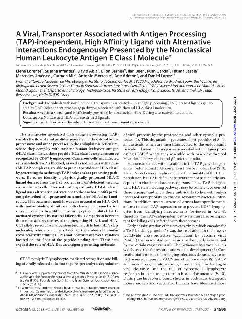
VACV D8L112–119 Is a Noncanonical HLA-E Ligand—Toexplore the potential role of HLA-E as an antigen-presentingmolecule of TAP-independent VACV ligands, HLA-peptidecomplex stability assays were performed using TAP-deficientT2 cells with an anti-HLA-E Ab. Fig. 1 shows that in contrastwith a control HLA-E ligand, the leader peptide of HLA, theinduction of complexes with five of the six VACV peptidestested were not detected. Thus, these viral ligands do not bindto HLA-E. In contrast, the D8L112–119 synthetic peptide
induced similar numbers of HLA-peptide surface complexes asthe positive control HLA-E ligand (Fig. 1). The consensus pep-tide-binding motif for HLA-E is Met, Leu, or Gln at peptideposition 2 (P2); Leu, Ile, Val, or Pro at P7; and Leu, Glu, or PheC-terminal residues (37, 38). Thus, the D8L112–119 octamerDGLIIISI is an unusual VACV ligand presented byHLA-E classI molecules.Identical Binding Affinity to Classical HLA-Cw1 and Non-
classical HLA-E Class I Molecules for the Viral D8L112–119Peptide—TheD8L112–119 peptide was previously described as aHLA-Cw1-restricted ligand (17). Because the mAb 3D12 usedin the current study for HLA-E binding cross-reacts with someHLA-C class I molecules, although not with HLA-Cw1 (39),HLA-peptide complex stability assayswere performedusingT2cells incubated with a natural high affinity HLA-Cw1 ligand,the CMV pp65 peptide (25), and stained with the anti-HLA-EmAb 3D12 to exclude HLA-Cw1 cross-reactivity. In this case,induction of HLA complexes with the CMV pp65 peptide wasnot detected (data not shown). Thus, the mAb 3D12 does notbind to HLA-Cw1.In addition, the relative affinity of D8L112–119 to both HLA-E
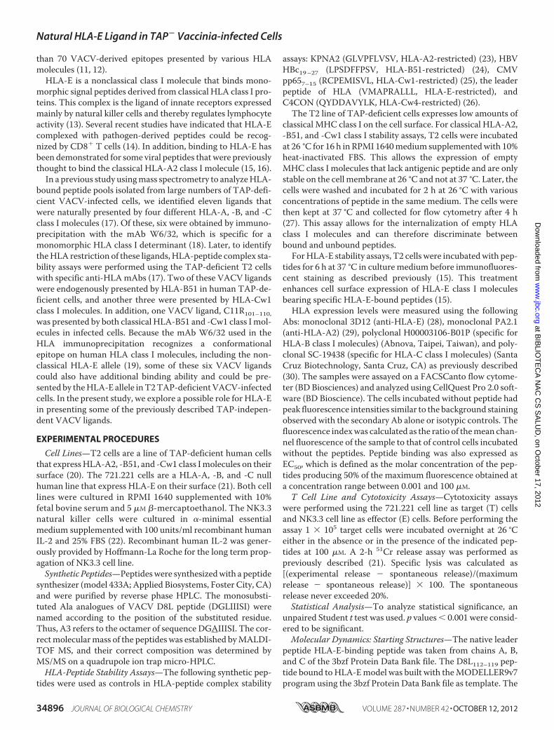
and -Cw1 class I molecules was evaluated. This peptide boundto HLA class I molecules in the range commonly found amongnatural ligands (Fig. 2). This octamer efficiently stabilized
0 100 200 300anti-HLA-E
Cel
l num
ber
Isotype
KPNA2
D8L 112-119
Leader peptide HLA
FIGURE 1. HLA-E stabilization with synthetic VACV ligands. The stability ofHLA-E-peptide complexes on the cell surface of T2 TAP-deficient cells wasmeasured by flow cytometry. The indicated peptides were used at 200 �M.The KPNA2 peptide and the leader peptide of HLA were used as negative andpositive controls, respectively. The mAb 3D12 was used for staining. Theresults, calculated as fluorescence index values � S.D., are the means of fourto five independent experiments. ***, significant p values (p � 0.001). A rep-resentative experiment was depicted in the bottom panel. Shaded histogram,isotypic control; thin line, KPNA2 peptide; medium line, D8L 112–119; thick line,leader peptide of HLA.
Natural HLA-E Ligand in TAP� Vaccinia-infected Cells
OCTOBER 12, 2012 • VOLUME 287 • NUMBER 42 JOURNAL OF BIOLOGICAL CHEMISTRY 34897
at BIB
LIOT
EC
A N
AC
CS
SA
LUD
, on October 17, 2012
ww
w.jbc.org
Dow
nloaded from
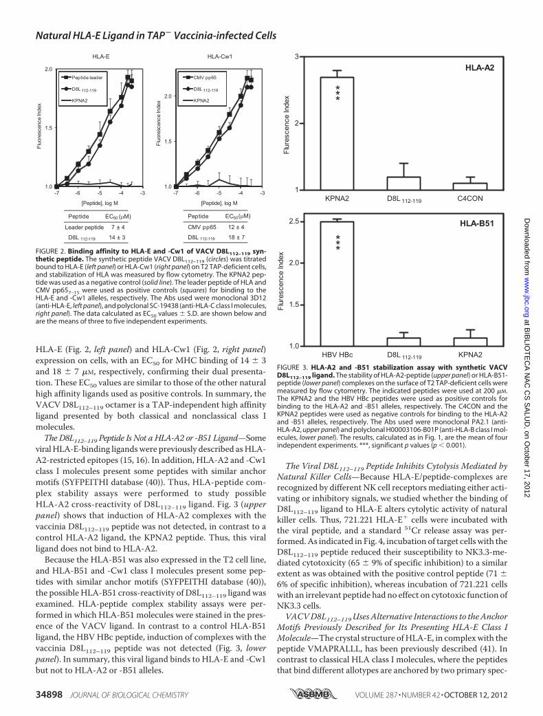
HLA-E (Fig. 2, left panel) and HLA-Cw1 (Fig. 2, right panel)expression on cells, with an EC50 for MHC binding of 14 � 3and 18 � 7 �M, respectively, confirming their dual presenta-tion. These EC50 values are similar to those of the other naturalhigh affinity ligands used as positive controls. In summary, theVACV D8L112–119 octamer is a TAP-independent high affinityligand presented by both classical and nonclassical class Imolecules.The D8L112–119 Peptide Is Not a HLA-A2 or -B51 Ligand—Some
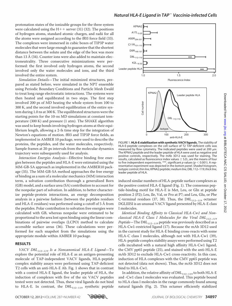
viralHLA-E-binding ligandswere previously described asHLA-A2-restricted epitopes (15, 16). In addition, HLA-A2 and -Cw1class I molecules present some peptides with similar anchormotifs (SYFPEITHI database (40)). Thus, HLA-peptide com-plex stability assays were performed to study possibleHLA-A2 cross-reactivity of D8L112–119 ligand. Fig. 3 (upperpanel) shows that induction of HLA-A2 complexes with thevaccinia D8L112–119 peptide was not detected, in contrast to acontrol HLA-A2 ligand, the KPNA2 peptide. Thus, this viralligand does not bind to HLA-A2.Because the HLA-B51 was also expressed in the T2 cell line,
and HLA-B51 and -Cw1 class I molecules present some pep-tides with similar anchor motifs (SYFPEITHI database (40)),the possible HLA-B51 cross-reactivity of D8L112–119 ligandwasexamined. HLA-peptide complex stability assays were per-formed in which HLA-B51 molecules were stained in the pres-ence of the VACV ligand. In contrast to a control HLA-B51ligand, the HBV HBc peptide, induction of complexes with thevaccinia D8L112–119 peptide was not detected (Fig. 3, lowerpanel). In summary, this viral ligand binds to HLA-E and -Cw1but not to HLA-A2 or -B51 alleles.
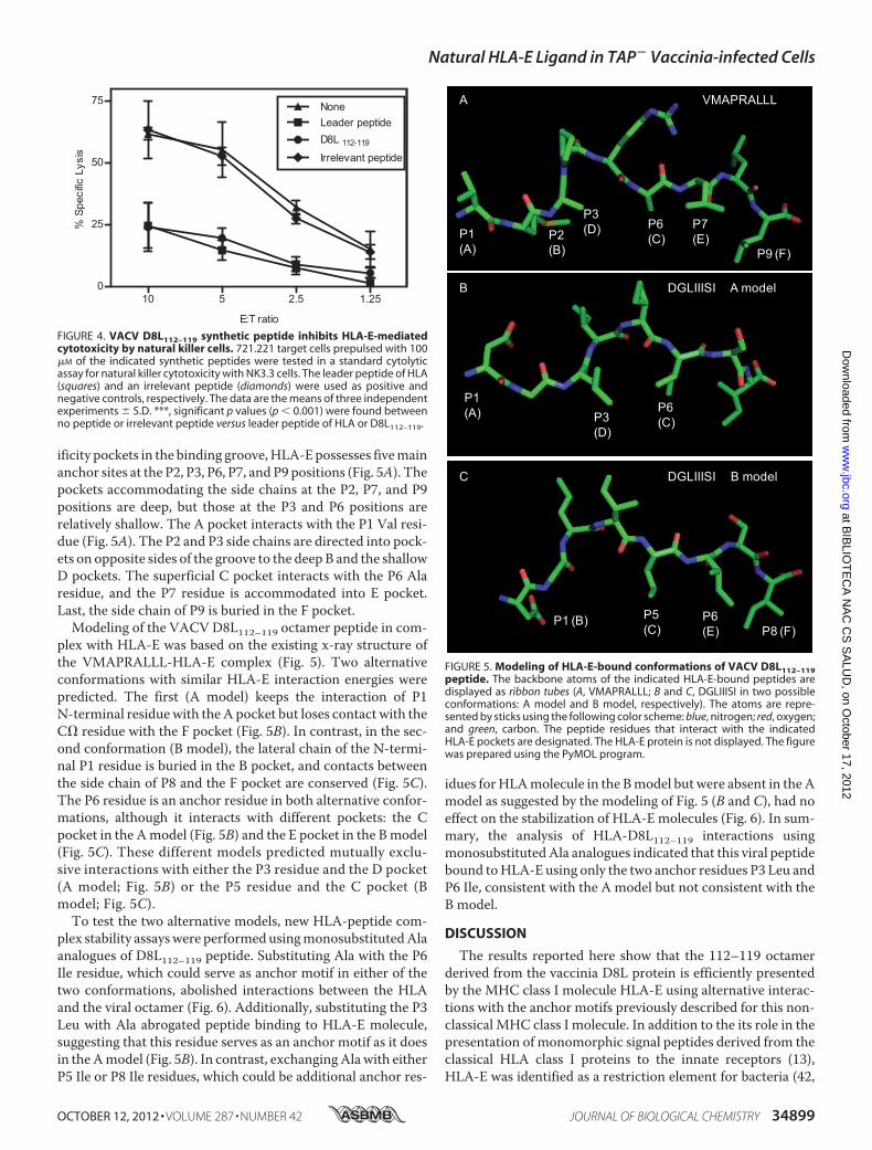
The Viral D8L112–119 Peptide Inhibits Cytolysis Mediated byNatural Killer Cells—Because HLA-E/peptide-complexes arerecognized by different NK cell receptorsmediating either acti-vating or inhibitory signals, we studied whether the binding ofD8L112–119 ligand to HLA-E alters cytolytic activity of naturalkiller cells. Thus, 721.221 HLA-E� cells were incubated withthe viral peptide, and a standard 51Cr release assay was per-formed.As indicated in Fig. 4, incubation of target cells with theD8L112–119 peptide reduced their susceptibility to NK3.3-me-diated cytotoxicity (65 � 9% of specific inhibition) to a similarextent as was obtained with the positive control peptide (71 �6% of specific inhibition), whereas incubation of 721.221 cellswith an irrelevant peptide had no effect on cytotoxic function ofNK3.3 cells.VACVD8L112–119UsesAlternative Interactions to theAnchor
Motifs Previously Described for Its Presenting HLA-E Class IMolecule—The crystal structure ofHLA-E, in complexwith thepeptide VMAPRALLL, has been previously described (41). Incontrast to classical HLA class I molecules, where the peptidesthat bind different allotypes are anchored by two primary spec-
FIGURE 2. Binding affinity to HLA-E and -Cw1 of VACV D8L112–119 syn-thetic peptide. The synthetic peptide VACV D8L112–119 (circles) was titratedbound to HLA-E (left panel) or HLA-Cw1 (right panel) on T2 TAP-deficient cells,and stabilization of HLA was measured by flow cytometry. The KPNA2 pep-tide was used as a negative control (solid line). The leader peptide of HLA andCMV pp657–15 were used as positive controls (squares) for binding to theHLA-E and -Cw1 alleles, respectively. The Abs used were monoclonal 3D12(anti-HLA-E, left panel), and polyclonal SC-19438 (anti-HLA-C class I molecules,right panel). The data calculated as EC50 values � S.D. are shown below andare the means of three to five independent experiments.
FIGURE 3. HLA-A2 and -B51 stabilization assay with synthetic VACVD8L112–119 ligand. The stability of HLA-A2-peptide (upper panel) or HLA-B51-peptide (lower panel) complexes on the surface of T2 TAP-deficient cells weremeasured by flow cytometry. The indicated peptides were used at 200 �M.The KPNA2 and the HBV HBc peptides were used as positive controls forbinding to the HLA-A2 and -B51 alleles, respectively. The C4CON and theKPNA2 peptides were used as negative controls for binding to the HLA-A2and -B51 alleles, respectively. The Abs used were monoclonal PA2.1 (anti-HLA-A2, upper panel) and polyclonal H00003106-B01P (anti-HLA-B class I mol-ecules, lower panel). The results, calculated as in Fig. 1, are the mean of fourindependent experiments. ***, significant p values (p � 0.001).
Natural HLA-E Ligand in TAP� Vaccinia-infected Cells
34898 JOURNAL OF BIOLOGICAL CHEMISTRY VOLUME 287 • NUMBER 42 • OCTOBER 12, 2012
at BIB
LIOT
EC
A N
AC
CS
SA
LUD
, on October 17, 2012
ww
w.jbc.org
Dow
nloaded from
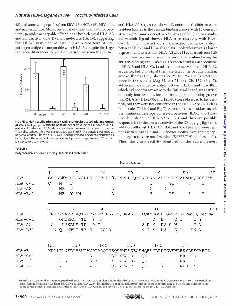
ificity pockets in the binding groove,HLA-Epossesses fivemainanchor sites at the P2, P3, P6, P7, and P9 positions (Fig. 5A). Thepockets accommodating the side chains at the P2, P7, and P9positions are deep, but those at the P3 and P6 positions arerelatively shallow. The A pocket interacts with the P1 Val resi-due (Fig. 5A). The P2 and P3 side chains are directed into pock-ets on opposite sides of the groove to the deep B and the shallowD pockets. The superficial C pocket interacts with the P6 Alaresidue, and the P7 residue is accommodated into E pocket.Last, the side chain of P9 is buried in the F pocket.Modeling of the VACV D8L112–119 octamer peptide in com-
plex with HLA-E was based on the existing x-ray structure ofthe VMAPRALLL-HLA-E complex (Fig. 5). Two alternativeconformations with similar HLA-E interaction energies werepredicted. The first (A model) keeps the interaction of P1N-terminal residuewith theA pocket but loses contact with theC� residue with the F pocket (Fig. 5B). In contrast, in the sec-ond conformation (B model), the lateral chain of the N-termi-nal P1 residue is buried in the B pocket, and contacts betweenthe side chain of P8 and the F pocket are conserved (Fig. 5C).The P6 residue is an anchor residue in both alternative confor-mations, although it interacts with different pockets: the Cpocket in the Amodel (Fig. 5B) and the E pocket in the Bmodel(Fig. 5C). These different models predicted mutually exclu-sive interactions with either the P3 residue and the D pocket(A model; Fig. 5B) or the P5 residue and the C pocket (Bmodel; Fig. 5C).To test the two alternative models, new HLA-peptide com-
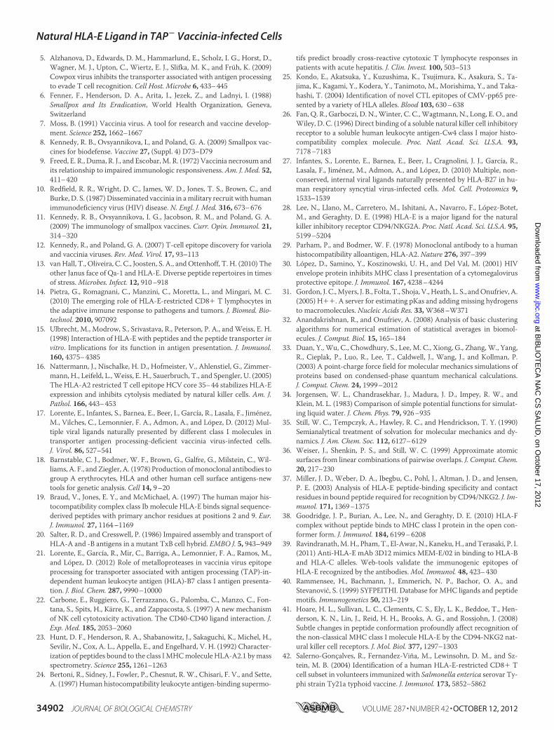
plex stability assayswere performedusingmonosubstitutedAlaanalogues of D8L112–119 peptide. Substituting Ala with the P6Ile residue, which could serve as anchor motif in either of thetwo conformations, abolished interactions between the HLAand the viral octamer (Fig. 6). Additionally, substituting the P3Leu with Ala abrogated peptide binding to HLA-E molecule,suggesting that this residue serves as an anchor motif as it doesin theAmodel (Fig. 5B). In contrast, exchangingAlawith eitherP5 Ile or P8 Ile residues, which could be additional anchor res-
idues forHLAmolecule in the Bmodel but were absent in theAmodel as suggested by the modeling of Fig. 5 (B and C), had noeffect on the stabilization of HLA-E molecules (Fig. 6). In sum-mary, the analysis of HLA-D8L112–119 interactions usingmonosubstitutedAla analogues indicated that this viral peptidebound toHLA-E using only the two anchor residues P3 Leu andP6 Ile, consistent with the A model but not consistent with theB model.
DISCUSSION
The results reported here show that the 112–119 octamerderived from the vaccinia D8L protein is efficiently presentedby the MHC class I molecule HLA-E using alternative interac-tions with the anchor motifs previously described for this non-classical MHC class I molecule. In addition to the its role in thepresentation of monomorphic signal peptides derived from theclassical HLA class I proteins to the innate receptors (13),HLA-E was identified as a restriction element for bacteria (42,
FIGURE 4. VACV D8L112–119 synthetic peptide inhibits HLA-E-mediatedcytotoxicity by natural killer cells. 721.221 target cells prepulsed with 100�M of the indicated synthetic peptides were tested in a standard cytolyticassay for natural killer cytotoxicity with NK3.3 cells. The leader peptide of HLA(squares) and an irrelevant peptide (diamonds) were used as positive andnegative controls, respectively. The data are the means of three independentexperiments � S.D. ***, significant p values (p � 0.001) were found betweenno peptide or irrelevant peptide versus leader peptide of HLA or D8L112–119.
FIGURE 5. Modeling of HLA-E-bound conformations of VACV D8L112–119peptide. The backbone atoms of the indicated HLA-E-bound peptides aredisplayed as ribbon tubes (A, VMAPRALLL; B and C, DGLIIISI in two possibleconformations: A model and B model, respectively). The atoms are repre-sented by sticks using the following color scheme: blue, nitrogen; red, oxygen;and green, carbon. The peptide residues that interact with the indicatedHLA-E pockets are designated. The HLA-E protein is not displayed. The figurewas prepared using the PyMOL program.
Natural HLA-E Ligand in TAP� Vaccinia-infected Cells
OCTOBER 12, 2012 • VOLUME 287 • NUMBER 42 JOURNAL OF BIOLOGICAL CHEMISTRY 34899
at BIB
LIOT
EC
A N
AC
CS
SA
LUD
, on October 17, 2012
ww
w.jbc.org
Dow
nloaded from
43) and some viral peptides fromEBV (15), HCV (16), HIV (44),and influenza (15). Moreover, most of these viral, but not bac-terial, peptides are capable of binding to both classical HLA-A2and nonclassical HLA-E class I molecules (15, 16), suggestingthat HLA-E may bind, at least in part, a repertoire of viralpathogen antigens comparable with HLA-A2 despite the largesequence differences found. Comparison between the HLA-E
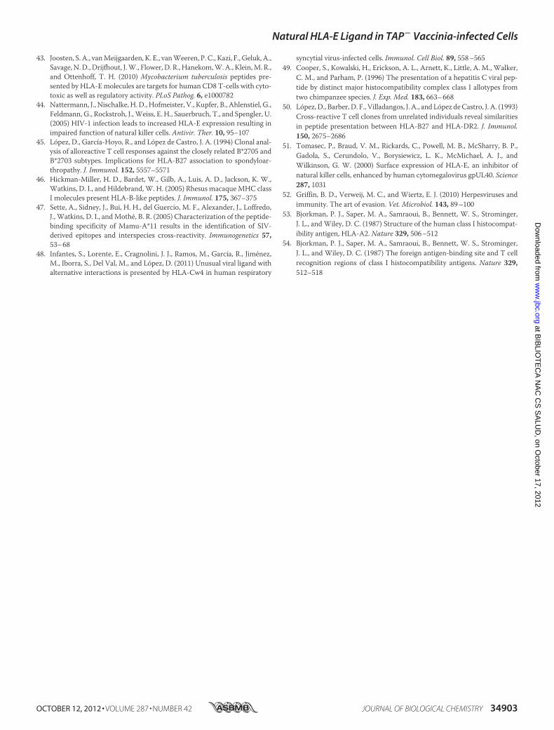
and HLA-A2 sequences shows 42 amino acid differences inresidues located in the peptide binding groove,with 15 conserv-ative and 27 nonconservative changes (Table 1). In our study,the vaccinia ligand showed HLA cross-reactivity with HLA-Cw1 but not HLA-A2 class I molecules. Sequence analysisbetweenHLA-E andHLA-Cw1 class Imolecules reveals a lowerdegree of differences thanHLA-A2with 14 conservative and 20nonconservative amino acid changes in the residues facing theantigen-binding site (Table 1). Fourteen residues are identicalin HLA-E andHLA-Cw1 and are not conserved in the HLA-A2sequence, but only six of them are facing the peptide bindinggroove: three in the � sheets (Ser-24, Leu-95, and Trp-97) andthree in the � helix (Arg-62, Ala-71, and Glu-152) (Fig. 7).When similar sequence analysis betweenHLA-E andHLA-B51,which did not cross-react with the D8L viral ligand, was carriedout, only four residues located in the peptide binding groove(Ser-24, Ala-71, Leu-95, and Trp-97) were observed to be iden-tical, but they were not conserved in the HLA-A2 or -B51 classImolecules (Table 1 and Fig. 7). All four of these residuesmatchthe minimum desetope conserved between HLA-E and HLA-Cw1 but absent in HLA-A2 or -B51 and thus are possiblyresponsible for the cross-reactivity of the D8L112–119 ligand. Inaddition, althoughHLA-A2, -B51, and -Cw1 present some pep-tides with similar P2 and P� anchor motifs, overlapping pep-tide repertoires are not described (SYFPEITHI database (40)).Thus, the cross-reactivity identified in the current report
FIGURE 6. HLA stabilization assay with monosubstituted Ala analoguesof VACV D8L112–119 synthetic peptide. Stability at the cell surface of HLA-Eon the cell surface of T2 TAP-deficient cells was measured by flow cytometry.The indicated peptides were used at 200 �M. The KPNA2 peptide was used asnegative control. The mAb 3D12 was used for staining. The data, calculated asin Fig. 1, are the means of three or four independent experiments. ***, signif-icant p values (p � 0.001).
TABLE 1Polymorphic residues among HLA class I molecules
a �1 and �2 HLA-E residues were compared with HLA-Cw1, -A2, or -B51 class I Molecules. Blanks indicate identity with the HLA-E-reference sequence. The identical resi-dues identified between HLA-E and HLA-Cw1 but not HLA-A2 or -B51 molecules (minimum desetope) and proposed as contributing to a shared structural motif thatcould confer peptide presenting similarities to HLA-E and HLA-Cw1 are in bold type. The sequences are from the IMGT/HLA Database.
Natural HLA-E Ligand in TAP� Vaccinia-infected Cells
34900 JOURNAL OF BIOLOGICAL CHEMISTRY VOLUME 287 • NUMBER 42 • OCTOBER 12, 2012
at BIB
LIOT
EC
A N
AC
CS
SA
LUD
, on October 17, 2012
ww
w.jbc.org
Dow
nloaded from

betweenHLA-E and -Cw1, an allele not clustered into HLA-A2supertype, expands the range of possible HLA-E cross-reactiv-ity and indicates that several other HLA class I viral ligandsdifferent from HLA-A2-restricted epitopes could be presentedin association with HLA-E. Thus, future studies analyzing theHLA-E peptide repertoire under infection conditions with dif-ferent viruses are needed.Some studies have shown cross-reactivity of epitopes
between very differentMHC class I molecules. Cross-reactivitybetween multiple HLA-B alleles (HLA-B7, -B27, -B40, -B54,-B55, and -B56) that differ by �20 residues facing the antigen-binding site has been widely reported (45). Additionally, inter-species cross-reactivity of viral ligands, shared by a human anda rhesusmacaque, a rhesusmacaque and amouse, a human anda mouse, and two different chimpanzeeMHC class I moleculeshave been described (46–49). These pairs of cross-reactiveMHCmolecules are very different and havemarked differencesin the sequence and structure of the peptide-binding groove.Dual reactivity of CD8� T cell clones reflected presentation ofstructurally related peptides by two HLA class I and II mole-cules: HLA-B27 and HLA-DR2 (50). These findings and theresults reported in the current study show the complexity andplasticity of interactions in MHC-peptide complexes.Our study includes one distinct difference from the previous
viral classical andnonclassicalHLAcross-reactivity reports (15,16, 44); the D8L112–119 ligand was isolated from TAP-deficientvaccinia virus-infected cells, and thus this viral ligand was nat-urally processed by a TAP-independent pathway previous to itspresentation by HLA-E. Only the HCMV gpUL-40-derivedligand is currently known to assemble with HLA-E via a TAP-independentmechanism (51). This peptide exactlymatches theleader sequence peptides of various HLA class I alleles and isable to substitute for the natural leader peptides from HLA-E
produced by TAP that are blocked by the protein US6 inHCMV-infected cells (52). Therefore, this TAP-independentantigen presentation was previously reported as a viral mecha-nism to bypass the normal HLA-E loading system that evolvedto occlude NK cell recognition of infected cells, whereas mostHCMVepitopes remain in the cytosolwithout any possibility ofentering the endoplasmic reticulum. Unlike this previouslydescribed tolerogenic peptide, D8L112–119 could be recognizedby CD8� T cells in the same manner as vaccinia virus-encodedHLA-A2-restricted epitopes generated in the same TAP-defi-cient infected cells (17), allowing it to contribute to host defenseagainst viral infection. Lastly, the lack of polymorphism of theHLA-E gene in humans suggests that D8L112–119 could be auniversal epitope, requiring future studies to understand theHLA-E-restricted response of this viral peptide.
Acknowledgments—We thank Drs. Jose A. Lopez de Castro (Centro deBiología Molecular Severo Ochoa, Madrid, Spain), Mar Valdes (CentroNacional de Biotecnología, Madrid, Spain), and Isidoro Gonzalez (Hos-pital de la Princesa, Madrid, Spain) for the cell lines. Recombinanthuman interleukin-2 was generously provided by Hoffmann-LaRoche.
REFERENCES1. Kloetzel, P. M., and Ossendorp, F. (2004) Proteasome and peptidase func-
tion in MHC-class-I-mediated antigen presentation. Curr. Opin. Immu-nol. 16, 76–81
2. Cerundolo, V., and de la Salle, H. (2006) Description of HLA class I- andCD8-deficient patients. Insights into the function of cytotoxic T lympho-cytes and NK cells in host defense. Semin. Immunol. 18, 330–336
3. Van Kaer, L., Ashton-Rickardt, P. G., Ploegh, H. L., and Tonegawa, S.(1992) TAP1 mutant mice are deficient in antigen presentation, surfaceclass I molecules, and CD4–8� T cells. Cell 71, 1205–1214
4. Voldby Larsen, M., Nielsen, M., Weinzierl, A., and Lund, O. (2006) TAP-independent MHC class I presentation. Curr. Immunol. Rev. 2, 233–245
FIGURE 7. Structural similarities between HLA-E and HLA-Cw1 but not with HLA-A2 or-B51. The amino acid sequence of the �1 and �2 domains of HLA-E(white backbone) was compared with the sequence of the equivalent domains of HLA-A2, -B51, and -Cw1 class I molecules using the same alignment used byBjorkmann et al. (53, 54). The identical residues identified between HLA-E and HLA-Cw1 but not HLA-A2 or -B51 molecules (minimum desetope) and proposedas contributing to a shared structural motif that could confer peptide presenting similarities between HLA-E and HLA-Cw1 are depicted. The atoms of thesefour residues are represented by sticks using the following color scheme: blue, nitrogen; red, oxygen; and green, carbon. The viral peptide is not displayed. Thefigure was prepared using the PyMOL program.
Natural HLA-E Ligand in TAP� Vaccinia-infected Cells
OCTOBER 12, 2012 • VOLUME 287 • NUMBER 42 JOURNAL OF BIOLOGICAL CHEMISTRY 34901
at BIB
LIOT
EC
A N
AC
CS
SA
LUD
, on October 17, 2012
ww
w.jbc.org
Dow
nloaded from
5. Alzhanova, D., Edwards, D. M., Hammarlund, E., Scholz, I. G., Horst, D.,Wagner, M. J., Upton, C., Wiertz, E. J., Slifka, M. K., and Fruh, K. (2009)Cowpox virus inhibits the transporter associated with antigen processingto evade T cell recognition. Cell Host. Microbe 6, 433–445
6. Fenner, F., Henderson, D. A., Arita, I., Jezek, Z., and Ladnyi, I. (1988)Smallpox and Its Eradication, World Health Organization, Geneva,Switzerland
7. Moss, B. (1991) Vaccinia virus. A tool for research and vaccine develop-ment. Science 252, 1662–1667
8. Kennedy, R. B., Ovsyannikova, I., and Poland, G. A. (2009) Smallpox vac-cines for biodefense. Vaccine 27, (Suppl. 4) D73–D79
9. Freed, E. R., Duma, R. J., and Escobar,M. R. (1972) Vaccinia necrosum andits relationship to impaired immunologic responsiveness. Am. J. Med. 52,411–420
10. Redfield, R. R., Wright, D. C., James, W. D., Jones, T. S., Brown, C., andBurke, D. S. (1987) Disseminated vaccinia in amilitary recruit with humanimmunodeficiency virus (HIV) disease. N. Engl. J. Med. 316, 673–676
11. Kennedy, R. B., Ovsyannikova, I. G., Jacobson, R. M., and Poland, G. A.(2009) The immunology of smallpox vaccines. Curr. Opin. Immunol. 21,314–320
12. Kennedy, R., and Poland, G. A. (2007) T-cell epitope discovery for variolaand vaccinia viruses. Rev. Med. Virol. 17, 93–113
13. vanHall, T., Oliveira, C. C., Joosten, S. A., andOttenhoff, T. H. (2010) Theother Janus face of Qa-1 and HLA-E. Diverse peptide repertoires in timesof stress.Microbes. Infect. 12, 910–918
14. Pietra, G., Romagnani, C., Manzini, C., Moretta, L., and Mingari, M. C.(2010) The emerging role of HLA-E-restricted CD8� T lymphocytes inthe adaptive immune response to pathogens and tumors. J. Biomed. Bio-technol. 2010, 907092
15. Ulbrecht, M., Modrow, S., Srivastava, R., Peterson, P. A., andWeiss, E. H.(1998) Interaction of HLA-E with peptides and the peptide transporter invitro. Implications for its function in antigen presentation. J. Immunol.160, 4375–4385
16. Nattermann, J., Nischalke, H. D., Hofmeister, V., Ahlenstiel, G., Zimmer-mann, H., Leifeld, L.,Weiss, E. H., Sauerbruch, T., and Spengler, U. (2005)The HLA-A2 restricted T cell epitope HCV core 35–44 stabilizes HLA-Eexpression and inhibits cytolysis mediated by natural killer cells. Am. J.Pathol. 166, 443–453
17. Lorente, E., Infantes, S., Barnea, E., Beer, I., García, R., Lasala, F., Jimenez,M., Vilches, C., Lemonnier, F. A., Admon, A., and Lopez, D. (2012) Mul-tiple viral ligands naturally presented by different class I molecules intransporter antigen processing-deficient vaccinia virus-infected cells.J. Virol. 86, 527–541
18. Barnstable, C. J., Bodmer, W. F., Brown, G., Galfre, G., Milstein, C., Wil-liams, A. F., and Ziegler, A. (1978) Production ofmonoclonal antibodies togroup A erythrocytes, HLA and other human cell surface antigens-newtools for genetic analysis. Cell 14, 9–20
19. Braud, V., Jones, E. Y., and McMichael, A. (1997) The human major his-tocompatibility complex class Ib molecule HLA-E binds signal sequence-derived peptides with primary anchor residues at positions 2 and 9. Eur.J. Immunol. 27, 1164–1169
20. Salter, R. D., and Cresswell, P. (1986) Impaired assembly and transport ofHLA-A and -B antigens in a mutant TxB cell hybrid. EMBO J. 5, 943–949
21. Lorente, E., García, R., Mir, C., Barriga, A., Lemonnier, F. A., Ramos, M.,and Lopez, D. (2012) Role of metalloproteases in vaccinia virus epitopeprocessing for transporter associated with antigen processing (TAP)-in-dependent human leukocyte antigen (HLA)-B7 class I antigen presenta-tion. J. Biol. Chem. 287, 9990–10000
22. Carbone, E., Ruggiero, G., Terrazzano, G., Palomba, C., Manzo, C., Fon-tana, S., Spits, H., Karre, K., and Zappacosta, S. (1997) A new mechanismof NK cell cytotoxicity activation. The CD40-CD40 ligand interaction. J.Exp. Med. 185, 2053–2060
23. Hunt, D. F., Henderson, R. A., Shabanowitz, J., Sakaguchi, K., Michel, H.,Sevilir, N., Cox, A. L., Appella, E., and Engelhard, V. H. (1992) Character-ization of peptides bound to the class IMHCmolecule HLA-A2.1 bymassspectrometry. Science 255, 1261–1263
24. Bertoni, R., Sidney, J., Fowler, P., Chesnut, R. W., Chisari, F. V., and Sette,A. (1997) Human histocompatibility leukocyte antigen-binding supermo-
tifs predict broadly cross-reactive cytotoxic T lymphocyte responses inpatients with acute hepatitis. J. Clin. Invest. 100, 503–513
25. Kondo, E., Akatsuka, Y., Kuzushima, K., Tsujimura, K., Asakura, S., Ta-jima, K., Kagami, Y., Kodera, Y., Tanimoto, M., Morishima, Y., and Taka-hashi, T. (2004) Identification of novel CTL epitopes of CMV-pp65 pre-sented by a variety of HLA alleles. Blood 103, 630–638
26. Fan, Q. R., Garboczi, D. N.,Winter, C. C.,Wagtmann, N., Long, E. O., andWiley, D. C. (1996) Direct binding of a soluble natural killer cell inhibitoryreceptor to a soluble human leukocyte antigen-Cw4 class I major histo-compatibility complex molecule. Proc. Natl. Acad. Sci. U.S.A. 93,7178–7183
27. Infantes, S., Lorente, E., Barnea, E., Beer, I., Cragnolini, J. J., García, R.,Lasala, F., Jimenez, M., Admon, A., and Lopez, D. (2010) Multiple, non-conserved, internal viral ligands naturally presented by HLA-B27 in hu-man respiratory syncytial virus-infected cells. Mol. Cell. Proteomics 9,1533–1539
28. Lee, N., Llano, M., Carretero, M., Ishitani, A., Navarro, F., Lopez-Botet,M., and Geraghty, D. E. (1998) HLA-E is a major ligand for the naturalkiller inhibitory receptor CD94/NKG2A. Proc. Natl. Acad. Sci. U.S.A. 95,5199–5204
29. Parham, P., and Bodmer, W. F. (1978) Monoclonal antibody to a humanhistocompatibility alloantigen, HLA-A2. Nature 276, 397–399
30. Lopez, D., Samino, Y., Koszinowski, U. H., and Del Val, M. (2001) HIVenvelope protein inhibits MHC class I presentation of a cytomegalovirusprotective epitope. J. Immunol. 167, 4238–4244
31. Gordon, J. C.,Myers, J. B., Folta, T., Shoja, V., Heath, L. S., andOnufriev, A.(2005) H��. A server for estimating pKas and adding missing hydrogensto macromolecules. Nucleic Acids Res. 33,W368–W371
32. Anandakrishnan, R., and Onufriev, A. (2008) Analysis of basic clusteringalgorithms for numerical estimation of statistical averages in biomol-ecules. J. Comput. Biol. 15, 165–184
33. Duan, Y., Wu, C., Chowdhury, S., Lee, M. C., Xiong, G., Zhang, W., Yang,R., Cieplak, P., Luo, R., Lee, T., Caldwell, J., Wang, J., and Kollman, P.(2003) A point-charge force field for molecular mechanics simulations ofproteins based on condensed-phase quantum mechanical calculations.J. Comput. Chem. 24, 1999–2012
34. Jorgensen, W. L., Chandrasekhar, J., Madura, J. D., Impey, R. W., andKlein, M. L. (1983) Comparison of simple potential functions for simulat-ing liquid water. J. Chem. Phys. 79, 926–935
35. Still, W. C., Tempczyk, A., Hawley, R. C., and Hendrickson, T. Y. (1990)Semianalytical treatment of solvation for molecular mechanics and dy-namics. J. Am. Chem. Soc. 112, 6127–6129
36. Weiser, J., Shenkin, P. S., and Still, W. C. (1999) Approximate atomicsurfaces from linear combinations of pairwise overlaps. J. Comput. Chem.20, 217–230
37. Miller, J. D., Weber, D. A., Ibegbu, C., Pohl, J., Altman, J. D., and Jensen,P. E. (2003) Analysis of HLA-E peptide-binding specificity and contactresidues in bound peptide required for recognition by CD94/NKG2. J. Im-munol. 171, 1369–1375
38. Goodridge, J. P., Burian, A., Lee, N., and Geraghty, D. E. (2010) HLA-Fcomplex without peptide binds to MHC class I protein in the open con-former form. J. Immunol. 184, 6199–6208
39. Ravindranath,M.H., Pham,T., El-Awar,N., Kaneku,H., andTerasaki, P. I.(2011) Anti-HLA-E mAb 3D12 mimics MEM-E/02 in binding to HLA-Band HLA-C alleles. Web-tools validate the immunogenic epitopes ofHLA-E recognized by the antibodies.Mol. Immunol. 48, 423–430
40. Rammensee, H., Bachmann, J., Emmerich, N. P., Bachor, O. A., andStevanovic, S. (1999) SYFPEITHI. Database for MHC ligands and peptidemotifs. Immunogenetics 50, 213–219
41. Hoare, H. L., Sullivan, L. C., Clements, C. S., Ely, L. K., Beddoe, T., Hen-derson, K. N., Lin, J., Reid, H. H., Brooks, A. G., and Rossjohn, J. (2008)Subtle changes in peptide conformation profoundly affect recognition ofthe non-classical MHC class I molecule HLA-E by the CD94-NKG2 nat-ural killer cell receptors. J. Mol. Biol. 377, 1297–1303
42. Salerno-Goncalves, R., Fernandez-Vina, M., Lewinsohn, D. M., and Sz-tein, M. B. (2004) Identification of a human HLA-E-restricted CD8� Tcell subset in volunteers immunized with Salmonella enterica serovar Ty-phi strain Ty21a typhoid vaccine. J. Immunol. 173, 5852–5862
Natural HLA-E Ligand in TAP� Vaccinia-infected Cells
34902 JOURNAL OF BIOLOGICAL CHEMISTRY VOLUME 287 • NUMBER 42 • OCTOBER 12, 2012
at BIB
LIOT
EC
A N
AC
CS
SA
LUD
, on October 17, 2012
ww
w.jbc.org
Dow
nloaded from
43. Joosten, S. A., vanMeijgaarden, K. E., vanWeeren, P. C., Kazi, F., Geluk, A.,Savage,N.D., Drijfhout, J.W., Flower, D. R., Hanekom,W.A., Klein,M. R.,and Ottenhoff, T. H. (2010) Mycobacterium tuberculosis peptides pre-sented by HLA-Emolecules are targets for human CD8 T-cells with cyto-toxic as well as regulatory activity. PLoS Pathog. 6, e1000782
44. Nattermann, J., Nischalke, H.D., Hofmeister, V., Kupfer, B., Ahlenstiel, G.,Feldmann, G., Rockstroh, J.,Weiss, E. H., Sauerbruch, T., and Spengler, U.(2005) HIV-1 infection leads to increased HLA-E expression resulting inimpaired function of natural killer cells. Antivir. Ther. 10, 95–107
45. Lopez, D., García-Hoyo, R., and Lopez de Castro, J. A. (1994) Clonal anal-ysis of alloreactive T cell responses against the closely related B*2705 andB*2703 subtypes. Implications for HLA-B27 association to spondyloar-thropathy. J. Immunol. 152, 5557–5571
46. Hickman-Miller, H. D., Bardet, W., Gilb, A., Luis, A. D., Jackson, K. W.,Watkins, D. I., and Hildebrand,W. H. (2005) Rhesus macaqueMHC classI molecules present HLA-B-like peptides. J. Immunol. 175, 367–375
47. Sette, A., Sidney, J., Bui, H. H., del Guercio, M. F., Alexander, J., Loffredo,J., Watkins, D. I., andMothe, B. R. (2005) Characterization of the peptide-binding specificity of Mamu-A*11 results in the identification of SIV-derived epitopes and interspecies cross-reactivity. Immunogenetics 57,53–68
48. Infantes, S., Lorente, E., Cragnolini, J. J., Ramos, M., García, R., Jimenez,M., Iborra, S., Del Val, M., and Lopez, D. (2011) Unusual viral ligand withalternative interactions is presented by HLA-Cw4 in human respiratory
syncytial virus-infected cells. Immunol. Cell Biol. 89, 558–56549. Cooper, S., Kowalski, H., Erickson, A. L., Arnett, K., Little, A. M., Walker,
C. M., and Parham, P. (1996) The presentation of a hepatitis C viral pep-tide by distinct major histocompatibility complex class I allotypes fromtwo chimpanzee species. J. Exp. Med. 183, 663–668
50. Lopez, D., Barber, D. F., Villadangos, J. A., and Lopez deCastro, J. A. (1993)Cross-reactive T cell clones from unrelated individuals reveal similaritiesin peptide presentation between HLA-B27 and HLA-DR2. J. Immunol.150, 2675–2686
51. Tomasec, P., Braud, V. M., Rickards, C., Powell, M. B., McSharry, B. P.,Gadola, S., Cerundolo, V., Borysiewicz, L. K., McMichael, A. J., andWilkinson, G. W. (2000) Surface expression of HLA-E, an inhibitor ofnatural killer cells, enhanced by human cytomegalovirus gpUL40. Science287, 1031
52. Griffin, B. D., Verweij, M. C., and Wiertz, E. J. (2010) Herpesviruses andimmunity. The art of evasion. Vet. Microbiol. 143, 89–100
53. Bjorkman, P. J., Saper, M. A., Samraoui, B., Bennett, W. S., Strominger,J. L., andWiley, D. C. (1987) Structure of the human class I histocompat-ibility antigen, HLA-A2. Nature 329, 506–512
54. Bjorkman, P. J., Saper, M. A., Samraoui, B., Bennett, W. S., Strominger,J. L., and Wiley, D. C. (1987) The foreign antigen-binding site and T cellrecognition regions of class I histocompatibility antigens. Nature 329,512–518
Natural HLA-E Ligand in TAP� Vaccinia-infected Cells
OCTOBER 12, 2012 • VOLUME 287 • NUMBER 42 JOURNAL OF BIOLOGICAL CHEMISTRY 34903
at BIB
LIOT
EC
A N
AC
CS
SA
LUD
, on October 17, 2012
ww
w.jbc.org
Dow
nloaded from


