Embed Size (px)
Citation preview
Neurobiology of Disease
Loss of Dendritic HCN1 Subunits Enhances CorticalExcitability and Epileptogenesis
Zhuo Huang,1 Matthew C. Walker,2 and Mala M. Shah1
1Department of Pharmacology, The School of Pharmacy, University of London, London WC1N 1AX, United Kingdom, and 2UCL Institute of Neurology,University College London, London WC1N 3BG, United Kingdom
Hyperpolarization-activated cation nonselective 1 (HCN1) plasticity in entorhinal cortical (EC) and hippocampal pyramidal cell den-drites is a salient feature of temporal lobe epilepsy. However, the significance remains undetermined. We demonstrate that adult HCN1null mice are more susceptible to kainic acid-induced seizures. After termination of these with an anticonvulsant, the mice also developedspontaneous behavioral seizures at a significantly more rapid rate than their wild-type littermates. This greater seizure susceptibility wasaccompanied by increased spontaneous activity in HCN1�/� EC layer III neurons. Dendritic Ih in these neurons was ablated, too.Consequentially, HCN1�/� dendrites were more excitable, despite having significantly more hyperpolarized resting membrane poten-tials (RMPs). In addition, the integration of EPSPs was enhanced considerably such that, at normal RMP, a 50 Hz train of EPSPs producedaction potentials in HCN1�/� neurons. As a result of this enhanced pyramidal cell excitability, spontaneous EPSC frequency ontoHCN1�/� neurons was considerably greater than that onto wild types, causing an imbalance between normal excitatory and inhibitorysynaptic activity. These results suggest that dendritic HCN channels are likely to play a critical role in regulating cortical pyramidal cellexcitability. Furthermore, these findings suggest that the reduction in dendritic HCN1 subunit expression during epileptogenesis is likelyto facilitate the disorder.
IntroductionTemporal lobe epilepsy (TLE) is the most common, drug-resistant form of the human condition (Engel, 1996; Herman,2002). There is substantial evidence to suggest that the ento-rhinal cortex (EC) and hippocampus play pivotal roles in theinduction and maintenance of TLE (Spencer and Spencer,1994). EC layer III pyramidal neurons, particularly, may play acritical role (Du et al., 1993, 1995; Jones, 1993; Barbarosie et al.,2000; Avoli et al., 2002; Wu and Leung, 2003; Shah et al., 2004;Dawodu and Thom, 2005; Wozny et al., 2005). Indeed, theyhave been shown to be spontaneously hyperactive in vivo afterthe induction of TLE (Shah et al., 2004). Furthermore, theiraxons (the temporoammonic pathway), which innervate thestratum lacunosum-moleculare of CA1 and the molecular layerof the subiculum, have been suggested to provide the majorexcitatory drive to the hippocampus during chronic TLE(Barbarosie et al., 2000; Avoli et al., 2002; Wu and Leung,2003; Wozny et al., 2005; Ang et al., 2006). Thus, altered EClayer III pyramidal cell excitability is likely to have a largeimpact on the development of TLE.
Interestingly, enhanced EC layer III neuronal excitability duringTLE is accompanied by a decrease in the hyperpolarization-activated cation current, Ih (Shah et al., 2004). Hyperpolarization-activated cation nonselective (HCN) subunits underlie Ih
(Robinson and Siegelbaum, 2003). The HCN1 subunit is pre-dominantly expressed in the cortex and hippocampus, in which itis primarily located in pyramidal cell dendrites (Lorincz et al.,2002; Notomi and Shigemoto, 2004). Indeed, HCN1 expressionis significantly reduced in the EC after TLE (Shah et al., 2004;Powell et al., 2008). Similar HCN1 channel plasticity has alsobeen shown to occur in neocortical and hippocampal neurons inmultiple animal models (Brewster et al., 2002; Bender et al., 2003;Dugladze et al., 2007; Jung et al., 2007; Shin et al., 2008; Marcelinet al., 2009) as well as humans (Brewster et al., 2002). Thisis surprising because the current depolarizes the resting mem-brane potential (RMP) (Pape, 1996; Robinson and Siegelbaum,2003), and, hence, a decline in Ih might be expected to reduceexcitability. However, Ih inhibition has been suggested to en-hance pyramidal cell dendritic excitability by increasing the avail-ability of Ca 2� channels (Tsay et al., 2007), as well as byamplifying the membrane resistance (RN) (Magee, 1998; Stuartand Spruston, 1998), thereby modifying synaptic signal integra-tion (Magee, 1999). Because Ih and HCN1 plasticity is a prevalenthallmark of TLE (Chen et al., 2001; Brewster et al., 2002; Benderet al., 2003; Shah et al., 2004; Dugladze et al., 2007; Jung et al.,2007; Dyhrfjeld-Johnsen et al., 2008; Powell et al., 2008; Shin etal., 2008; Marcelin et al., 2009), it is crucial to determine whetheralterations in Ih and HCN1 protein expression during TLE areconsequential to neural network adaptation or critically influ-ence the disorder. In this study, we have used HCN1 null
Received March 26, 2009; revised July 2, 2009; accepted July 18, 2009.This work was supported by a Medical Research Council New Investigator Award and Epilepsy Research Founda-
tion UK and Royal Society project grants (M.M.S.). We are grateful to S. Martin (University College London Sequenc-ing and Genotyping Facility, London, UK) for genotyping the HCN transgenic mice. We also thank Prof. D. Johnston(University of Texas at Austin, Austin, TX), Prof. D. A. Brown (University College London, London, UK), and Dr. M. F.Nolan (Edinburgh University, Edinburgh, UK) for useful discussions and critically reading our manuscript.
Correspondence should be addressed to Dr. Mala M. Shah, Department of Pharmacology, The School of Phar-macy, University of London, 29-39 Brunswick Square, London WC1N 1AX, UK. E-mail: [email protected].
DOI:10.1523/JNEUROSCI.1531-09.2009Copyright © 2009 Society for Neuroscience 0270-6474/09/2910979-10$15.00/0
The Journal of Neuroscience, September 2, 2009 • 29(35):10979 –10988 • 10979
mice together with in vivo electroen-cephalographic recordings, in vitro elec-trophysiological analysis, and selectivepharmacological tools to address this im-portant question.
Materials and MethodsHCN1 null mice. HCN1 heterozygote breedingpairs were a kind gift from Prof. E. R. Kandel(Columbia University, New York, NY). Hybrid(HCN1�/�) male and female progeny weremaintained on a 129SVEV background andcrossed to obtain mixtures of HCN1 null mice,heterozygotes, and wild-type (WT) littermatesas described previously (Nolan et al., 2003,2004). These mice had been backcrossed for 10generations. The mouse genotype was deter-mined using PCR. Briefly, genomic DNA wasextracted from 0.2 cm tail snips using the HOt-Shot protocol as detailed by Truett et al.(2000). A PCR reaction was then performedusing TAQ polymerase (Bioline) and the fol-lowing primers: oMR3410, 5�-CAC CTG CTACGC AAT GTT TG-3�; oMR3411, 5�-ATTGGG CAC TAC ACG CTA GG-3�; oMR3412,5�-AGA GAA ATC ATT CCC CGT GA-3�.
The PCR reaction consisted of an initial 2 minat 94°C, followed by 35 cycles of 30 s at 94°C, 30 sat 55°C, and 45 s at 72°C. After the last cycle, thereaction is kept at 72°C for 4 min and then held at10°C. A 359 bp band was observed for mice con-taining the wild-type allele, whereas a 450 bpband was seen for mice containing the mutantallele. Heterozygotes contained both alleles.
Surgical procedure and electroencephalogra-phy. Six- to 8-week-old HCN1 null mice andwild-type littermate controls were anesthetizedusing a ketamine/xylamine mixture (Sigma) andpositioned in a stereotaxic frame. Depth elec-trodes (Plastics One) were surgically implantedinto the EC area using the following stereotaxiccoordinates: 4.2 mm lateral to lambda and 4.0mm below the cortical surface, with the nose barset at 3.0 mm and the electrode holder at an angleof 17° posterior to the sagittal plane.
All mice were kept on 12 h light dark cycles.Electroencephalographic (EEG) recordings wereobtained 7 d later using a neurolog amplifier.Video recordings were obtained using a wiredday/night camera with audio and infrared (Na-ture Cameras Ltd.). These together with theEEG data were simultaneously acquired on acomputer using the micro1401 analog-to-digital converter and spike 2 software (Cam-bridge Electronic Design). For baseline EEG,we did 6 h recordings during light cycle and 6 hduring dark cycle for a minimum period of 5 d.After kainate injections, 4 h EEG recordingswere made during the light and dark cycles. Allrecordings were referenced to a frontal surfaceelectrode. Recordings were visually inspectedfor electrographic seizure activity and interictalspikes as defined previously (Shah et al., 2004).Interictal spikes were defined as high-amplitude (at least three times baseline ampli-tude) sharp transients lasting �70 ms. Electrographic seizure activity wasdefined as the appearance of high-amplitude (more than three times baselineactivity), high-frequency, rhythmic activity with an evolution in spike fre-
quency that lasted a minimum of 10 s. In addition, after each experiment, thelocation of the electrode was confirmed to be in the EC.
Kainic acid-induced status epilepticus. Kainic acid (KA) (Tocris Bio-science) was administered to HCN1 null mice and wild-type littermates
0.5 mV1 s
0.5 mV10 s
A Enhanced susceptibility of HCN1-/- mice to KA induced seizures(i) Control wildtype EEG
(iii) Wt + 10 mg/kg KA
(ii) Control HCN1-/- EEG
(iv) HCN1-/- + 10 mg/kg KA
(v) Wt + 20 mg/kg KA
B Significantly shorter latent period duration in HCN1-/- mice
1 s
1 s
0.5 mV
0.5 mV
(i) EEG from HCN1-/- mouse 2 days following SE termination
(ii) EEG from wildtype mouse 2 days following SE termination
5040302010
0
(vi)
Tim
e ta
ken
to
reac
h S
E (m
in)
n=8
n=3
n=5*
20 m
g/kg
KA
Wt
20 m
g/kg
KA
H
CN
1-/-
10 m
g/kg
KA
H
CN
1-/-
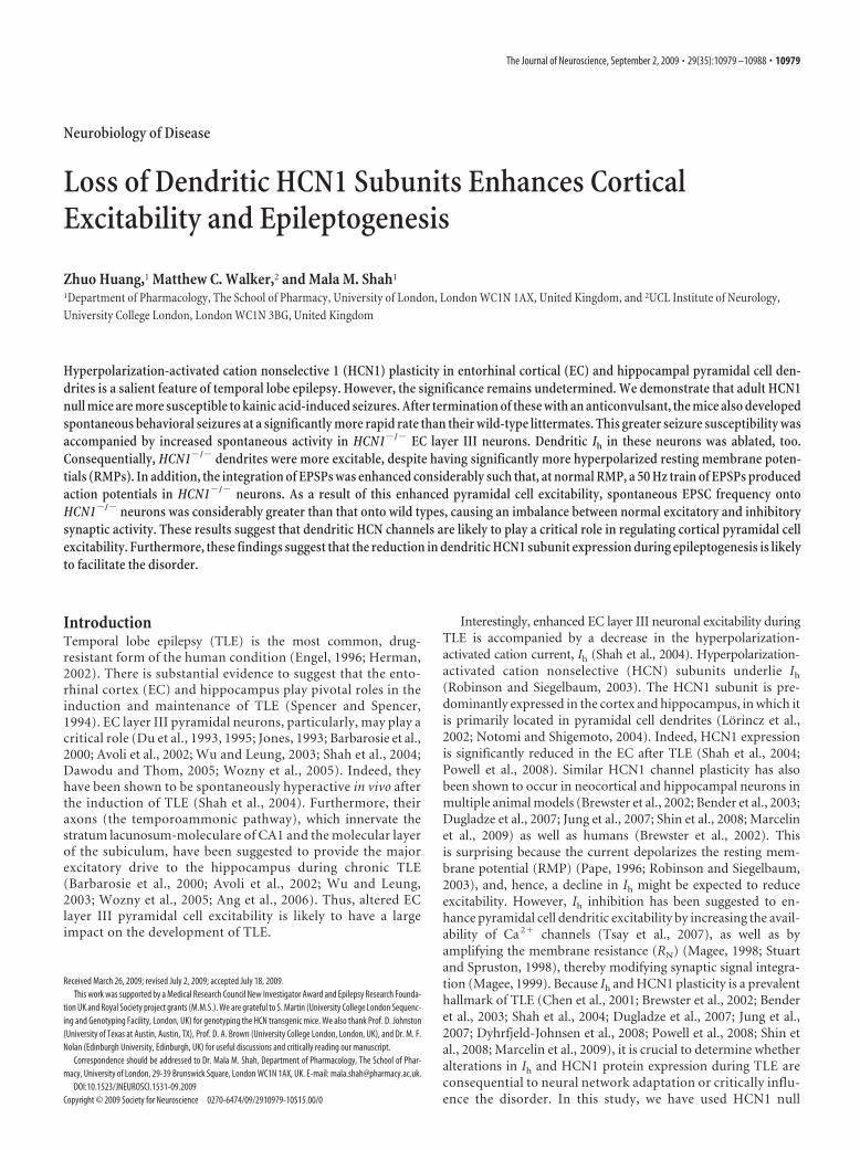
Figure 1. Enhanced seizure susceptibility and shorter latent period duration in HCN1�/� mice. Ai, Aii, Example 10-s-long EEGrecordings from untreated 8-week-old wild-type (WT or HCN1�/�) and HCN1�/� mice. Calibration in Ai applies to Aii. Aiii–Av,One hundred second sections of EEG obtained �1 h after treatment with KA. Calibration in Aiii is applicable to Aiv and Av, too. Avi,Graph to depict the time taken to reach SE after administration of KA. The numbers of observations are indicated above each bar.*p � 0.05. Bi, Example EEG recording of seizure activity obtained from an HCN1�/� mice�2 d after termination of KA-induced SE. Bii,EEG traces to show that only interictal spikes (indicated by arrows) were present in a WT mouse �2 d after cessation of KA-induced SE.
10980 • J. Neurosci., September 2, 2009 • 29(35):10979 –10988 Huang et al. • Dendritic HCN1 Channels and Epileptogenesis
to induce class V seizures [as defined by the Racine scale (Racine, 1972)].These were terminated 1 h after onset using sodium pentobarbital (30mg/kg, s.c.; Sigma-Aldrich). Control groups were mice that had beentreated SP only (30 mg/kg, s.c.). In some mice, EEG recordings wereobtained after kainic acid administration. These were quantified as describedby Lehmkuhle et al. (2009). Briefly, EEG raw data was initially bandpassfiltered in the gamma band range (20–70 Hz) and the ratio of the gammaband power (the square of the average amplitude root mean square value)before and after kainic acid-induced seizures was calculated and expressed asa percentage. As described by Williams et al. (2009), the latent period dura-tion was calculated as the time taken for the onset of motor seizures [typicallyclass III, as defined by Racine (1972)]. All procedures concerning animalswere approved by the United Kingdom Home Office.
Electrophysiological studies. Entorhinal– hippocampal slices were ob-tained from 6- to 9-week-old HCN1�/� and HCN1�/� (wild type) miceas described previously (Shah et al., 2004). Whole-cell and cell-attachedrecordings were obtained from both the soma and dendrites of EC layerIII pyramidal neurons. For recording purposes, slices were placed in achamber containing external recording solution maintained at 34 –36°Cand viewed using an Olympus BX51W1 equipped with differential infra-red optics. The external solution (unless otherwise noted) was supple-mented with 0.05 mM APV, 0.01 mM CNQX, 0.01 mM bicuculline, and0.001 mM CGP 55845 [(2S)-3-[(1S)-1-(3,4-dichlorophenyl)ethyl]amino-2-hydroxypropyl)(phenylmethyl)phosphinic acid]. The internal recordingpipette solution for whole-cell current-clamp and EPSC voltage-clamprecordings was composed of the following (in mM): 120 KMeSO4, 20 KCl,10 HEPES, 2 MgCl2, 0.2 EGTA, 4 Na2-ATP, 0.3 Tris-GTP, and 14 Tris-phosphocreatine, pH was adjusted to 7.3 with KOH. To record sponta-neous IPSCs, KMeSO4 was replaced with KCl. Furthermore, ZD7288(4-ethylphenylamino-1,2-dimethyl-6-methylaminopyrimidinium chlo-ride) (15 �M) was added to the intracellular solution when EPSC andIPSC recordings were made. In addition, for cell-attached recordings, theinternal pipette solution contained the following (mM): 120 KCl, 20tetraethylammonium-Cl, 5 4-AP, 1 BaCl2, 10 HEPES, 1 MgCl2, 2 CaCl2,0.001 tetrodotoxin, and 0.1 NiCl2, pH adjusted to 7.3. Pipettes contain-ing any of these internal solutions had resistances of 5–12 M�.Whole-cell current-clamp recordings were obtained using a bridge-mode amplifier (AxoClamp 2B; Molecular Devices), filtered at 10 kHz,and sampled at 50 kHz. Series resistance was usually in the order of 10 –30M� and was �70% compensated for the whole-cell voltage-clamp re-cordings. Cell-attached recordings were obtained using the Axopatch200B (Molecular Devices), filtered 2 kHz, and sampled at 3.5 kHz. Datawere acquired using pClamp 8.2 (Molecular Devices).
�EPSPs were generated by current injection of the order: A � (t/�) *exp(1 � (t/�), where A is the amplitude of the current injected, and � isthe rise time constant. Tungsten electrodes (A-M Systems) were placed inEC layer I to elicit EPSPs. All drugs were bath applied. The effects ofZD7288 (15 �M) occurred within 15 min, and recordings were usuallymade within 25 min of application.
Data analysis. pClamp software was used to analyze whole-cellcurrent-clamp and cell-attached voltage-clamp recordings. The RN wascalculated from 400 ms hyperpolarizing pulses of 100 pA applied from aholding potential of �70 mV. The �EPSP decay time constants wereobtained by fitting the double-exponential function: A1e (�t / � 1) �A2e (�t / � 2), where �1 and �2 represent time constants of the initial andfalling phase of the �EPSPs. Because Ih is activated during the fallingphase of the �EPSP, only �2 was used. The summation ratio of EPSPs wascalculated as the ratio of the peak of the fifth EPSP to that of the firstEPSP. Action potential threshold was determined as the point beforethe first derivative of the trace was no longer equal to zero. For cell-attached recordings, the steady-state current after the 2 s, hyperpo-larizing step was used as an indication of the amount of Ih. EPSCs andIPSCs were analyzed using the Mini-analysis program (version 6.07;Synaptosoft). Events �3 pA in amplitude were detected and used foranalysis. Decay times and amplitudes of these events were obtained byfitting the averaged EPSC or IPSC with a single-exponential equation:I(t) � A exp(�t/�), where I is the current amplitude at any given time
(t), A is the peak amplitude of the EPSC or IPSC, and � is the decaytime constant. Group data are expressed as mean SEM. Statisticalsignificance was determined using either paired or unpaired Student’st tests as appropriate. Statistical significance of p � 0.05 is indicated as* in all figures.
Materials. All chemicals were obtained from Sigma-Aldrich apart fromZD 7288, CGP 55848, CNQX, TTX, bicuculline, and APV, which werepurchased from Ascent Scientific Ltd. Stock solutions of bicuculline andCGP 55848 were made in DMSO and stored at �20°C until use. Thesewere then dissolved in the external solution such that the final DMSOconcentration was �0.1%. Aqueous stock solutions of ZD7288, CNQX,TTX, and APV were also kept at �20°C until use.
A Dendritic Cell-attached Voltage Clamp Recordings
B Dendritic Whole-cell Current Clamp Recordings
-80 mV
HCN1-/-(ii)
10
8
6
4
2
0200150100500
Act
ion
Pot
entia
l No.
Current Injection (pA)
*
**
*
Wt (n=10)HCN1-/- (n=17)
(iii)
-71 mV250 ms
20 mV
-300 pA
200 pA
Wt(i)
10 pA1 s
-140 mV
-40 mV
HCN1-/-
Wt
(i)
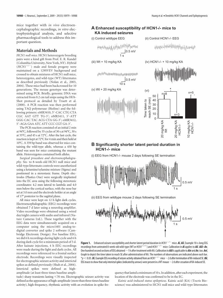
Figure 2. HCN1 deletion is accompanied by dendritic Ih ablation as well as increased den-dritic excitability and dendritic RN in EC layer III pyramidal neurons. Ai, Recordings of Ih obtainedin the cell-attached mode from the apical dendrites of HCN1�/� (red) and WT (black) EC layerIII pyramids. The traces were obtained by applying a voltage step from �40 to �140 mV andthen leak subtracted (leak current was obtained by applying a step from �40 to �30 mV). Thetraces have also been superimposed to illustrate the difference in current amplitude. Bi, Bii,Example recordings obtained from �150 �m along the apical dendrite of a WT and HCN1�/�
neuron after application of a series of 400 ms steps from �300 to �200 pA. The cells wereheld at the normal resting membrane potential. Calibration in Bi also applies Bii. Biii,Graph to show the average action potential numbers in response to varying current injec-tions in WT and HCN1�/� dendrites. In all cases, the n values indicate the numbers ofobservations. *p � 0.05.
Huang et al. • Dendritic HCN1 Channels and Epileptogenesis J. Neurosci., September 2, 2009 • 29(35):10979 –10988 • 10981
ResultsHCN1 null mice have a lowerseizure threshold and shorterlatent period durationTo determine how HCN1 subunits influ-ence epileptogenesis, HCN1 null micewere used. These mice have proved to beuseful in exploring the role of HCN1channels, because expression of HCN2–HCN4 is minimally affected (Nolan et al.,2003, 2004, 2007; Tsay et al., 2007). To testthe role of HCN1 subunits in influencingseizure susceptibility, depth electrodeswere stereotaxically implanted in the ECof adult (7- to 9-week-old) HCN1 nullmice and their wild-type littermates (seeMaterials and Methods). EEG recordingswere obtained 7–10 d later from awake,freely moving mice present in their nor-mal environment. As observed in thehippocampus (Nolan et al., 2004), no ep-ileptiform abnormalities, interictal spikes,or spontaneous seizures were detected inthe EEG recorded daily from HCN1�/�
(n � 7) or wild type (n � 8) for 6 h lightand 6 h dark cycles over a period of 1–3weeks (Fig. 1Ai,Aii) (total period of re-cording from seven HCN1�/� was 900 h;total recording time from eight WT micewas 840 h), indicating that these mice arenot innately epileptic.
To investigate whether there were dif-ferences in seizure threshold and manifes-tation of TLE between HCN1�/� andwild-type mice, we used the commonlyused so-called “kainate” model (Ben-Ariand Cossart, 2000; Dudek et al., 2002;White, 2002). In this model, a single epi-sode of class V seizures [as defined by the Racine scale (Racine,1972)] or status epilepticus (SE) is induced in rodents by admin-istering kainic acid and then terminated �1 h later with an anti-convulsant such as sodium pentobarbital (see Materials andMethods). After a delay of a few weeks, known as the latent period(during which animals appear to be normal), spontaneous overtbehavioral seizures occur (defined as the onset of chronic TLE)(Ben-Ari and Cossart, 2000; Dudek et al., 2002; White, 2002).This model is widely used because many of the clinical and patho-logical features of the human disorder (including the latent pe-riod) can be reproduced (Ben-Ari and Cossart, 2000; Dudek etal., 2002; White, 2002). As demonstrated previously (He et al.,2004), administration of 20 mg/kg KA intraperitoneally elicitedSE in wild-type mice in 43.1 9.6 min (n � 8) (Fig. 1Av). All wildtypes treated with 20 mg/kg KA survived the treatment. Thisconcentration, however, caused SE within 5 min in HCN1�/�
mice and was lethal (n � 3) (Fig. 1Avi). Instead, half the amountof KA, 10 mg/kg delivered intraperitoneally, was required to in-duce SE in a similar time frame (Fig. 1Aiv,Avi) and was not fatal.This lower dose had no effect in wild-type mice for up to 4 h (n �4) (Fig. 1Aiii). EEG recordings showed that, despite the differ-ences in dose, the intensity of KA-induced SE in HCN1�/� miceand wild types was comparable [percentage change in gammaband power during SE from baseline of HCN1�/� and wild type,
143.75 18.75% (n � 4) and 168.75 23.66% (n � 4), respec-tively]. Because this was the case and the time taken to reach SEno different, we terminated the kainic acid-induced SE in bothHCN1�/� and wild types with sodium pentobarbital (30 mg/kgs.c.) and used EEG recordings together with video monitoring todetermine whether there were differences in the latent periodduration (see Materials and Methods) as well. HCN1�/� micedisplayed overt motor convulsions [class III forelimb clonus sei-zures as defined by Racine (1972)] within 72 h of halting SE(latent period duration, 60.0 7.3 h; n � 4) (Fig. 1B). Con-versely, wild types displayed similar spontaneous seizures �2weeks later (wild-type latent period duration, 386.8 11.1 h; n �4) (Fig. 1B). Hence, these mice appeared normal at 72 h, and onlyinterictal spikes were detected in EEG recordings (Fig. 1Bii).These results, therefore, indicate that the decrease in HCN1 sub-unit expression that occurs in animal models and humans afterTLE initiation (Brewster et al., 2002; Bender et al., 2003; Shah etal., 2004; Dugladze et al., 2007; Jung et al., 2007; Powell et al.,2008; Shin et al., 2008; Marcelin et al., 2009) is likely to have asubstantial impact on the induction (seizure threshold) and ex-pression of the disorder.
Loss of Ih in HCN1�/� EC layer III pyramid dendritesBecause the EC (Jones, 1993; Spencer and Spencer, 1994; Avoli etal., 2002; Wu and Leung, 2003; Wozny et al., 2005) and EC layer
C RMP change in dendrites with ZD
-85-80-75
-70
-65-60
-55
Wt (n=6) *
Con + ZD Con + ZD
HCN1-/- (n=6) ns
RM
P (m
V)
-70mV -79
mV
-70mV
20 mV250 ms
Control, NRMP +ZD, HRMP +ZD, ORMPA Wt Dendrite
AP
No.
Current Injection (pA)
76543210
200150100500
*
**
**
*Control (n=6)ZD, HRMPZD, ORMP
-79mV
-79mV
B HCN1-/- Dendrite76543210
200150100500Current Injection (pA)
Control (n=6)ZD, HRMP
AP
No.
-300 pA
+200 pA
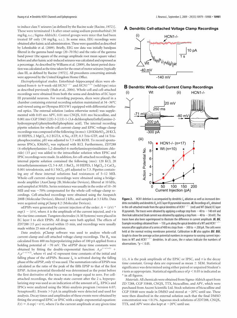
Figure 3. Effects of the Ih inhibitor ZD7288 on dendritic HCN1�/� and wild-type excitability. A, B, Example recordings obtainedfrom HCN1�/� or WT (control) dendrites of EC layer III neurons in the absence and presence ZD7288 (15 �M). A series of 400 mshyperpolarizing and depolarizing steps in 50 pA increments were applied to produce the traces. In all cases, the cells were held atthe normal RMP (NRMP). Application of ZD7288 hyperpolarized WT dendrites, and, therefore, traces were obtained at the hyper-polarized RMP (HRMP) and, for comparison purposes, at the original RMP (ORMP). Calibration: 20 mV, 250 ms. The mean numberof action potentials generated in response to depolarizing current pulses before (control) and after ZD7288 at the hyperpolarizedRMP or original RMP are shown on the far right of each panel. C, Average changes in RMP produced by application of ZD7288 (ZD)on WT and HCN1�/� soma. *p � 0.05. Con, Control.
10982 • J. Neurosci., September 2, 2009 • 29(35):10979 –10988 Huang et al. • Dendritic HCN1 Channels and Epileptogenesis

III neurons in particular (Du et al., 1993, 1995; Barbarosie andAvoli, 1997; Wu and Leung, 2003; Shah et al., 2004; Wozny et al.,2005; Ang et al., 2006) play a significant role during TLE, it isessential to determine how HCN1 ablation affects EC layer IIIcellular excitability. However, given that HCN2 subunits are alsoexpressed in the EC, albeit to a much lower level than HCN1subunits (Notomi and Shigemoto, 2004), it was first important todetermine the extent to which Ih is reduced in HCN1�/� neurons.To investigate this, we made cell-attached voltage-clamp record-ings from the soma and apical dendrite, �150 �m from the soma[total EC layer III pyramidal neuron apical dendrite length, �250�m (Tahvildari and Alonso, 2005)]. Ih was activated by applying3 s hyperpolarizing pulses from �40 to �140 mV in the presenceof Na�, K�, and Ca 2� channel blockers (see Materials and Meth-ods). In agreement with the reported predominant dendritic ex-pression of HCN channels (Notomi and Shigemoto, 2004; Shahet al., 2004), a slowly activating current with an average steady-state magnitude of 19.25 3.3 pA (n � 8) (Fig. 2A) was observedonly in wild-type dendrites. The same protocol elicited no signif-icant current (0.86 1.43 pA; n � 6) (Fig. 2A) from HCN1�/�
dendrites. Because Western blot analysishas shown that HCN2 levels are unalteredin these mice (Nolan et al., 2003), theseresults indicate that HCN1 subunits areessential for the generation of Ih in theseneurons.
Spontaneous activity and pyramidalcell dendritic excitability enhancedin HCN1�/� neuronsTo test how HCN1 deletion affected EClayer III pyramidal cell intrinsic mem-brane properties and excitability, we madewhole-cell current-clamp recordings fromthe soma and dendrites of these neurons. Inthe absence of both GABA and glutamatereceptor blockers, HCN1�/� neurons hadsignificantly more hyperpolarized RMPsas well as more spontaneous activity thanwild types (supplemental Fig. 1, availableat www.jneurosci.org as supplementalmaterial). The greater spontaneous post-synaptic potential frequency was detectedin HCN1�/� neurons, even when the so-matic RMP was artificially adjusted to�70 mV (supplemental Fig. 1, available atwww.jneurosci.org as supplemental ma-terial), suggesting that EC neural networkexcitability was enhanced considerablymore in HCN1�/� mice compared withthat of wild types. This may, at least partly,explain why the HCN1�/� mice weremore susceptible to kainic acid-inducedseizures.
The enhanced spontaneous postsyn-aptic potential frequency could be attrib-utable to greater action potential-drivensynaptic release resulting from altered in-trinsic excitability of neurons. To assessthis, spontaneous postsynaptic potentialactivity was first suppressed with gluta-mate and GABA receptor blockers (seeMaterials and Methods). Under these
conditions, significantly larger numbers of action potentialscould be recorded in HCN1�/� dendrites compared with wild-type neurons when depolarizing current pulses were applied de-spite a more hyperpolarized RMP (HCN1�/� dendritic RMP,�74.1 0.8 mV, n � 17; wild-type dendritic RMP, �68.9 0.9mV, n � 10; p � 0.05) (Fig. 2B). A comparable effect could beproduced by in wild-type dendrites if Ih was suppressed using theinhibitor ZD7288 [15 �M, a maximal concentration (BoSmith etal., 1993)] (Fig. 3A,C). This inhibitor, however, did not affectHCN1�/� dendrite excitability (Fig. 3).
In accordance with the predominant dendritic location ofHCN channels (see above), the differences in somatic action po-tentials elicited in response to depolarizing current steps weremuch smaller between HCN1�/� and wild-type neurons at theirnormal RMPs (HCN1�/� somatic RMP, �75.3 0.6 mV, n �33; wild-type somatic RMP, �69.3 0.4 mV, n � 22; p � 0.5)(Fig. 4). If, however, all cells were artificially held at fixed poten-tials of �70 mV, depolarizing current steps in HCN1�/� neuronsproduced significantly more action potentials (Fig. 4A,B,E). Ap-plication of 15 �M ZD7288 affected wild-type somatic excitability
A Somatic recordings at NRMP
B Somatic recordings at -70 mV
20 mV250 ms
-68 mV -79
mV
Wt (HCN1+/+) KO (HCN1-/-)
Wt (HCN1+/+) KO (HCN1-/-)
-70 mV
-70 mV
100 pA
-150 pA
121086420
200150100500
10
8
6
4
2
0200150100500
Wt (n=20)KO (n=29)
Wt (n=18)KO (n=18)
Current Injection (pA) Current Injection (pA)
*
*
*
*
Act
ion
Pot
entia
l No.
Act
ion
Pot
entia
l No.
C Soma, NRMP D Soma, -70 mV
Figure 4. Effects of HCN1 deletion on somatic intrinsic excitability. A, B, Example traces obtained from WT or HCN1�/� (KO)neurons in response to a series of 400 ms current steps from �150 to �100 pA in increments of 50 pA. The recordings have beenobtained either at the normal resting membrane potential (NRMP) or at the fixed potential of �70 mV. Calibration in A applies toall traces in A and B. C, D, Graphs to demonstrate average numbers of action potentials obtained in response to varying depolarizingcurrent pulses when the soma was held at the NRMP or at �70 mV. *p � 0.05.
Huang et al. • Dendritic HCN1 Channels and Epileptogenesis J. Neurosci., September 2, 2009 • 29(35):10979 –10988 • 10983
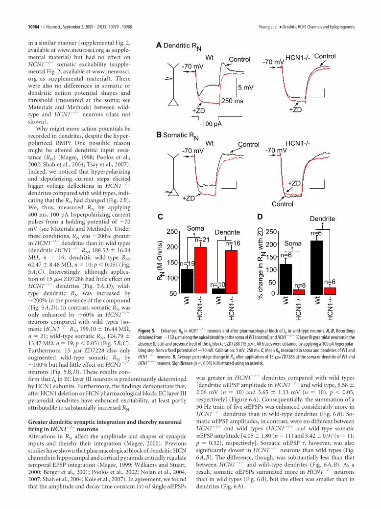
in a similar manner (supplemental Fig. 2,available at www.jneurosci.org as supple-mental material) but had no effect onHCN1�/� somatic excitability (supple-mental Fig. 2, available at www.jneurosci.org as supplemental material). Therewere also no differences in somatic ordendritic action potential shapes andthreshold (measured at the soma; seeMaterials and Methods) between wild-type and HCN1�/� neurons (data notshown).
Why might more action potentials berecorded in dendrites, despite the hyper-polarized RMP? One possible reasonmight be altered dendritic input resis-tance (RN) (Magee, 1998; Poolos et al.,2002; Shah et al., 2004; Tsay et al., 2007).Indeed, we noticed that hyperpolarizingand depolarizing current steps elicitedbigger voltage deflections in HCN1�/�
dendrites compared with wild types, indi-cating that the RN had changed (Fig. 2B).We, thus, measured RN by applying400 ms, 100 pA hyperpolarizing currentpulses from a holding potential of �70mV (see Materials and Methods). Underthese conditions, RN was �200% greaterin HCN1�/� dendrites than in wild types(dendritic HCN1�/� RN, 188.52 16.04M�, n � 16; dendritic wild-type RN,62.47 8.48 M�, n � 10; p � 0.05) (Fig.5A,C). Interestingly, although applica-tion of 15 �M ZD7288 had little effect onHCN1�/� dendrites (Fig. 5A,D), wild-type dendritic RN was increased by�200% in the presence of the compound(Fig. 5A,D). In contrast, somatic RN wasonly enhanced by �60% in HCN1�/�
neurons compared with wild types (so-matic HCN1�/� RN, 199.10 16.44 M�,n � 21; wild-type somatic RN, 124.79 13.47 M�, n � 19; p � 0.05) (Fig. 5B,C).Furthermore, 15 �M ZD7228 also onlyaugmented wild-type somatic RN by�100% but had little effect on HCN1�/�
neurons (Fig. 5B,D). These results con-firm that Ih in EC layer III neurons is predominantly determinedby HCN1 subunits. Furthermore, the findings demonstrate that,after HCN1 deletion or HCN pharmacological block, EC layer IIIpyramidal dendrites have enhanced excitability, at least partlyattributable to substantially increased RN.
Greater dendritic synaptic integration and thereby neuronalfiring in HCN1�/� neuronsAlterations in RN affect the amplitude and shapes of synapticinputs and thereby their integration (Magee, 2000). Previousstudies have shown that pharmacological block of dendritic HCNchannels in hippocampal and cortical pyramids critically regulatetemporal EPSP integration (Magee, 1999; Williams and Stuart,2000; Berger et al., 2001; Poolos et al., 2002; Nolan et al., 2004,2007; Shah et al., 2004; Kole et al., 2007). In agreement, we foundthat the amplitude and decay time constant (�) of single �EPSPs
was greater in HCN1�/� dendrites compared with wild types[dendritic �EPSP amplitude in HCN1�/� and wild type, 5.58 2.06 mV (n � 10) and 3.65 1.13 mV (n � 10), p � 0.05,respectively] (Figure 6A). Consequentially, the summation of a50 Hz train of five �EPSPs was enhanced considerably more inHCN1�/� dendrites than in wild-type dendrites (Fig. 6B). So-matic �EPSP amplitudes, in contrast, were no different betweenHCN1�/� and wild types (HCN1�/� and wild-type somatic�EPSP amplitude [4.05 1.80 (n � 11) and 3.42 0.97 (n � 11;p � 0.32), respectively]. Somatic �EPSP �, however, was alsosignificantly slower in HCN1�/� neurons than wild types (Fig.6A,B). The difference, though, was substantially less than thatbetween HCN1�/� and wild-type dendrites (Fig. 6A,B). As aresult, somatic �EPSPs summated more in HCN1�/� neuronsthan in wild types (Fig. 6B), but the effect was smaller than indendrites (Fig. 6A).
-100 pA
-70 mV -70 mV
5 mV
250 ms
B Somatic RNWt HCN1-/-
+ZD
Control
+ZD
Control
n=19n=6
*
R N (M
Ohm
s)
250
200
150
100
50
Soma Dendriten=16
n=10
C
**
*250
200
150
100
500
n=6
n=8 n=6
D Dendrite
Soman=21
n=19n=6
-70 mV-70 mV
A Dendritic RNWt HCN1-/-
+ZD
Control
+ZD
Control
% c
hang
e in
RN
with
ZD
Wt
Wt
Wt
Wt
HC
N1-
/-
HC
N1-
/-
HC
N1-
/-
HC
N1-
/-
Figure 5. Enhanced RN in HCN1�/� neurons and after pharmacological block of Ih in wild-type neurons. A, B, Recordingsobtained from �150 �m along the apical dendrite or the soma of WT (control) and HCN1�/� EC layer III pyramidal neurons in theabsence (black) and presence (red) of the Ih blocker, ZD7288 (15 �M). All traces were obtained by applying a 100 pA hyperpolar-izing step from a fixed potential of �70 mV. Calibration: 5 mV, 250 ms. C, Mean RN measured in soma and dendrites of WT andHCN1�/� neurons. D, Average percentage change in RN after application of 15 �M ZD7288 at the soma or dendrite of WT andHCN1�/� neurons. Significance (p � 0.05) is illustrated using an asterisk.
10984 • J. Neurosci., September 2, 2009 • 29(35):10979 –10988 Huang et al. • Dendritic HCN1 Channels and Epileptogenesis
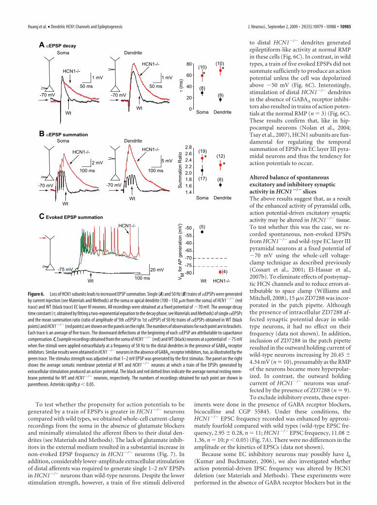
To test whether the propensity for action potentials to begenerated by a train of EPSPs is greater in HCN1�/� neuronscompared with wild types, we obtained whole-cell current-clamprecordings from the soma in the absence of glutamate blockersand minimally stimulated the afferent fibers to their distal den-drites (see Materials and Methods). The lack of glutamate inhib-itors in the external medium resulted in a substantial increase innon-evoked EPSP frequency in HCN1�/� neurons (Fig. 7). Inaddition, considerably lower-amplitude extracellular stimulationof distal afferents was required to generate single 1–2 mV EPSPsin HCN1�/� neurons than wild-type neurons. Despite the lowerstimulation strength, however, a train of five stimuli delivered
to distal HCN1�/� dendrites generatedepileptiform-like activity at normal RMPin these cells (Fig. 6C). In contrast, in wildtypes, a train of five evoked EPSPs did notsummate sufficiently to produce an actionpotential unless the cell was depolarizedabove �50 mV (Fig. 6C). Interestingly,stimulation of distal HCN1�/� dendritesin the absence of GABAA receptor inhibi-tors also resulted in trains of action poten-tials at the normal RMP (n � 3) (Fig. 6C).These results confirm that, like in hip-pocampal neurons (Nolan et al., 2004;Tsay et al., 2007), HCN1 subunits are fun-damental for regulating the temporalsummation of EPSPs in EC layer III pyra-midal neurons and thus the tendency foraction potentials to occur.
Altered balance of spontaneousexcitatory and inhibitory synapticactivity in HCN1 �/� slicesThe above results suggest that, as a resultof the enhanced activity of pyramidal cells,action potential-driven excitatory synapticactivity may be altered in HCN1�/� tissue.To test whether this was the case, we re-corded spontaneous, non-evoked EPSPsfrom HCN1�/� and wild-type EC layer IIIpyramidal neurons at a fixed potential of�70 mV using the whole-cell voltage-clamp technique as described previously(Cossart et al., 2001; El-Hassar et al.,2007b). To eliminate effects of postsynap-tic HCN channels and to reduce errors at-tributable to space clamp (Williams andMitchell, 2008), 15 �M ZD7288 was incor-porated in the patch pipette. Althoughthe presence of intracellular ZD7288 af-fected synaptic potential decay in wild-type neurons, it had no effect on theirfrequency (data not shown). In addition,inclusion of ZD7288 in the patch pipetteresulted in the outward holding current ofwild-type neurons increasing by 20.45 4.34 mV (n � 10), presumably as the RMPof the neurons became more hyperpolar-ized. In contrast, the outward holdingcurrent of HCN1�/� neurons was unaf-fected by the presence of ZD7288 (n � 9).To exclude inhibitory events, these exper-
iments were done in the presence of GABA receptor blockers,bicuculline and CGP 55845. Under these conditions, theHCN1�/� EPSC frequency recorded was enhanced by approxi-mately fourfold compared with wild types (wild-type EPSC fre-quency, 2.95 0.28, n � 11; HCN1�/� EPSC frequency, 11.08 1.36, n � 10; p � 0.05) (Fig. 7A). There were no differences in theamplitude or the kinetics of EPSCs (data not shown).
Because some EC inhibitory neurons may possibly have Ih
(Kumar and Buckmaster, 2006), we also investigated whetheraction potential-driven IPSC frequency was altered by HCN1deletion (see Materials and Methods). These experiments wereperformed in the absence of GABA receptor blockers but in the
A αEPSP decay
80
60
40
20
0
B αEPSP summation
2.82.62.42.22.01.81.61.4
τ (m
s)
Soma Dendrite
(10)
(8)
(10)
(9)
**
Sum
mat
ion
Rat
io
Soma Dendrite
(12)
(8)
(19)
(17)
**
-70 mV -70 mV
WtWt
HCN1-/-HCN1-/-
50 ms 50 ms
1 mV 1 mV
Soma Dendrite
-70 mV -70 mV
WtWt
HCN1-/- HCN1-/-
100 ms 100 ms
2 mV 5 mV
Soma Dendrite
20 mV
100 ms
-75 mV
HCN1-/-
Wt
-80-75-70-65-60-55-50
VM
for A
P ge
nera
tion
(mV
)
Wt HCN1-/-
(4)
C Evoked EPSP summation
(5)
Figure 6. Loss of HCN1 subunits leads to increased EPSP summation. Single (A) and 50 Hz (B) trains of �EPSPs were generatedby current injection (see Materials and Methods) at the soma or apical dendrite (100 –150 �m from the soma) of HCN1�/� (redtrace) and WT (black trace) EC layer III neurons. All recordings were obtained at a fixed potential of �70 mV. The average decaytime constant (�, obtained by fitting a two-exponential equation to the decay phase; see Materials and Methods) of single �EPSPsand the mean summation ratio (ratio of amplitude of 5th �EPSP to 1st �EPSP) of 50 Hz trains of �EPSPs obtained in WT (blackpoints) and HCN1�/� (red points) are shown on the panels on the right. The numbers of observations for each point are in brackets.Each trace is an average of five traces. The downward deflections at the beginning of each �EPSP are attributable to capacitancecompensation. C, Example recordings obtained from the soma of HCN1�/� (red) and WT (black) neurons at a potential of�75 mVwhen five stimuli were applied extracellularly at a frequency of 50 Hz to the distal dendrites in the presence of GABAA receptorinhibitors. Similar results were obtained in HCN1�/� neurons in the absence of GABAA receptor inhibitors, too, as illustrated by thegreen trace. The stimulus strength was adjusted so that 1–2 mV EPSP was generated by the first stimulus. The panel on the rightshows the average somatic membrane potential of WT and HCN1�/� neurons at which a train of five EPSPs generated byextracellular stimulation produced an action potential. The black and red dotted lines indicate the average normal resting mem-brane potential for WT and HCN1�/� neurons, respectively. The numbers of recordings obtained for each point are shown inparentheses. Asterisks signify p � 0.05.
Huang et al. • Dendritic HCN1 Channels and Epileptogenesis J. Neurosci., September 2, 2009 • 29(35):10979 –10988 • 10985
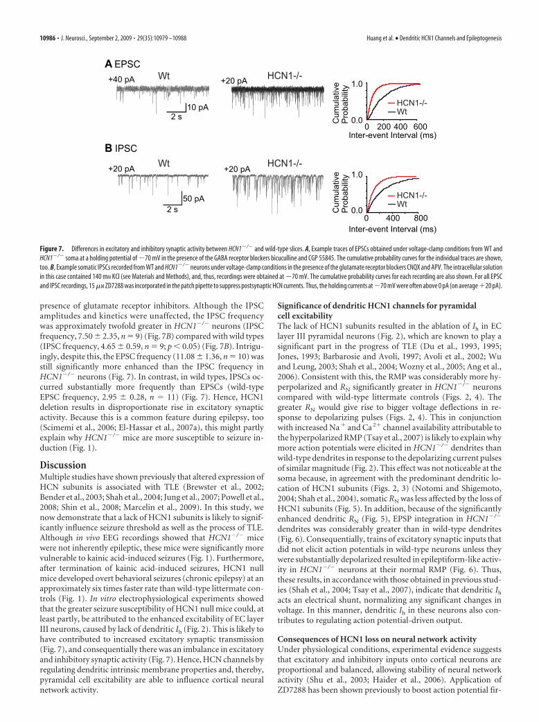
presence of glutamate receptor inhibitors. Although the IPSCamplitudes and kinetics were unaffected, the IPSC frequencywas approximately twofold greater in HCN1�/� neurons (IPSCfrequency, 7.50 2.35, n � 9) (Fig. 7B) compared with wild types(IPSC frequency, 4.65 0.59, n � 9; p � 0.05) (Fig. 7B). Intrigu-ingly, despite this, the EPSC frequency (11.08 1.36, n � 10) wasstill significantly more enhanced than the IPSC frequency inHCN1�/� neurons (Fig. 7). In contrast, in wild types, IPSCs oc-curred substantially more frequently than EPSCs (wild-typeEPSC frequency, 2.95 0.28, n � 11) (Fig. 7). Hence, HCN1deletion results in disproportionate rise in excitatory synapticactivity. Because this is a common feature during epilepsy, too(Scimemi et al., 2006; El-Hassar et al., 2007a), this might partlyexplain why HCN1�/� mice are more susceptible to seizure in-duction (Fig. 1).
DiscussionMultiple studies have shown previously that altered expression ofHCN subunits is associated with TLE (Brewster et al., 2002;Bender et al., 2003; Shah et al., 2004; Jung et al., 2007; Powell et al.,2008; Shin et al., 2008; Marcelin et al., 2009). In this study, wenow demonstrate that a lack of HCN1 subunits is likely to signif-icantly influence seizure threshold as well as the process of TLE.Although in vivo EEG recordings showed that HCN1�/� micewere not inherently epileptic, these mice were significantly morevulnerable to kainic acid-induced seizures (Fig. 1). Furthermore,after termination of kainic acid-induced seizures, HCN1 nullmice developed overt behavioral seizures (chronic epilepsy) at anapproximately six times faster rate than wild-type littermate con-trols (Fig. 1). In vitro electrophysiological experiments showedthat the greater seizure susceptibility of HCN1 null mice could, atleast partly, be attributed to the enhanced excitability of EC layerIII neurons, caused by lack of dendritic Ih (Fig. 2). This is likely tohave contributed to increased excitatory synaptic transmission(Fig. 7), and consequentially there was an imbalance in excitatoryand inhibitory synaptic activity (Fig. 7). Hence, HCN channels byregulating dendritic intrinsic membrane properties and, thereby,pyramidal cell excitability are able to influence cortical neuralnetwork activity.
Significance of dendritic HCN1 channels for pyramidalcell excitabilityThe lack of HCN1 subunits resulted in the ablation of Ih in EClayer III pyramidal neurons (Fig. 2), which are known to play asignificant part in the progress of TLE (Du et al., 1993, 1995;Jones, 1993; Barbarosie and Avoli, 1997; Avoli et al., 2002; Wuand Leung, 2003; Shah et al., 2004; Wozny et al., 2005; Ang et al.,2006). Consistent with this, the RMP was considerably more hy-perpolarized and RN significantly greater in HCN1�/� neuronscompared with wild-type littermate controls (Figs. 2, 4). Thegreater RN would give rise to bigger voltage deflections in re-sponse to depolarizing pulses (Figs. 2, 4). This in conjunctionwith increased Na� and Ca 2� channel availability attributable tothe hyperpolarized RMP (Tsay et al., 2007) is likely to explain whymore action potentials were elicited in HCN1�/� dendrites thanwild-type dendrites in response to the depolarizing current pulsesof similar magnitude (Fig. 2). This effect was not noticeable at thesoma because, in agreement with the predominant dendritic lo-cation of HCN1 subunits (Figs. 2, 3) (Notomi and Shigemoto,2004; Shah et al., 2004), somatic RN was less affected by the loss ofHCN1 subunits (Fig. 5). In addition, because of the significantlyenhanced dendritic RN (Fig. 5), EPSP integration in HCN1�/�
dendrites was considerably greater than in wild-type dendrites(Fig. 6). Consequentially, trains of excitatory synaptic inputs thatdid not elicit action potentials in wild-type neurons unless theywere substantially depolarized resulted in epileptiform-like activ-ity in HCN1�/� neurons at their normal RMP (Fig. 6). Thus,these results, in accordance with those obtained in previous stud-ies (Shah et al., 2004; Tsay et al., 2007), indicate that dendritic Ih
acts an electrical shunt, normalizing any significant changes involtage. In this manner, dendritic Ih in these neurons also con-tributes to regulating action potential-driven output.
Consequences of HCN1 loss on neural network activityUnder physiological conditions, experimental evidence suggeststhat excitatory and inhibitory inputs onto cortical neurons areproportional and balanced, allowing stability of neural networkactivity (Shu et al., 2003; Haider et al., 2006). Application ofZD7288 has been shown previously to boost action potential fir-
Inter-event Interval (ms)
0.0
1.0
0 400 800
HCN1-/-Wt
Cum
ulat
ive
Pro
babi
lity
Inter-event Interval (ms)
0.0
1.0
0 200 400 600
Wt
Wt HCN1-/-A EPSC
B IPSCHCN1-/-
Cum
ulat
ive
Pro
babi
lity
50 pA2 s
10 pA2 s
+40 pA +20 pA
HCN1-/-Wt
+20 pA +20 pA
Figure 7. Differences in excitatory and inhibitory synaptic activity between HCN1�/� and wild-type slices. A, Example traces of EPSCs obtained under voltage-clamp conditions from WT andHCN1�/� soma at a holding potential of �70 mV in the presence of the GABA receptor blockers bicuculline and CGP 55845. The cumulative probability curves for the individual traces are shown,too. B, Example somatic IPSCs recorded from WT and HCN1�/� neurons under voltage-clamp conditions in the presence of the glutamate receptor blockers CNQX and APV. The intracellular solutionin this case contained 140 mM KCl (see Materials and Methods), and, thus, recordings were obtained at �70 mV. The cumulative probability curves for each recording are also shown. For all EPSCand IPSC recordings, 15 �M ZD7288 was incorporated in the patch pipette to suppress postsynaptic HCN currents. Thus, the holding currents at �70 mV were often above 0 pA (on average �20 pA).
10986 • J. Neurosci., September 2, 2009 • 29(35):10979 –10988 Huang et al. • Dendritic HCN1 Channels and Epileptogenesis
ing and thereby recurrent network activity in prefrontal cortex(Wang et al., 2007). In agreement, our findings also suggest that aloss of HCN1, by increasing the propensity for action potentialsto occur in pyramidal cells with any given synaptic input (Fig.6C), would lead to greater neuronal output and hence substan-tially more synaptic activity. Indeed, action potential-drivenHCN1�/� EPSC frequency was enhanced fourfold (Fig. 7A).However, this might also be predicted to augment interneuronactivity and thereby raise inhibition (Dugladze et al., 2007). Fur-thermore, some interneurons have been suggested to have Ih
(McBain and Fisahn, 2001; Aponte et al., 2006; Kumar andBuckmaster, 2006; Dugladze et al., 2007), and a loss of Ih maycontribute to greater activity of these (Dugladze et al., 2007).Although we did not test whether interneuron activity per se wasaltered as a consequence of either enhanced synaptic activation ora loss of Ih, our findings suggested that this might be the casebecause IPSC frequency onto EC layer III pyramidal neurons wasamplified (Fig. 7B). Despite this, the EPSC/IPSC ratio changedfrom 0.63 in wild types to 1.47 in HCN1�/� neurons (Fig. 7).Hence, loss of HCN1 subunits leads to a disproportionate in-crease in excitatory synaptic activity.
Implications of HCN1 plasticity for epilepsyWe have shown that a lack of HCN1 subunits results in EC layerIII pyramidal cell hyperexcitability, enhanced action potential-driven spontaneous HCN1�/� EPSC frequency and, thus, alteredEC neural network activity. Previous studies have also demon-strated that HCN1 deletion results in greater dendritic excit-ability and EPSP summation in hippocampal pyramidal CA1neurons (Nolan et al., 2004; Tsay et al., 2007), which are involvedin seizure generation during TLE too (Spencer and Spencer,1994). It is thus surprising that interictal spikes or electrographicseizures were not observed in HCN1�/� mice in vivo (Fig. 1)(Nolan et al., 2004). However, the amplified IPSC frequency,although this was to a much lower extent than excitatory synaptictransmission (Fig. 7), together with increased IPSC summationcaused by loss of Ih (Chen et al., 2001) might serve to offset theheightened neural network activity and thereby prevent the oc-currence of inherent interictal spikes or seizures. Hence, althoughthe reduction of HCN1 subunit clearly favors neural networkexcitability (supplemental Fig. 1, available at www.jneurosci.orgas supplemental material) (Fig. 7), the imbalance between exci-tation and inhibition may be insufficient to render HCN1�/�
mice spontaneously epileptic.The HCN1�/� mice, though, were clearly more susceptible to
seizures and developed chronic TLE at a much faster rate thanwild-type littermates (Fig. 1). Multiple studies have demon-strated that HCN1 subunit expression is reduced after status epi-lepticus (Brewster et al., 2002; Shah et al., 2004; Dugladze et al.,2007; Jung et al., 2007; Powell et al., 2008; Shin et al., 2008;Marcelin et al., 2009). Hence, the latent period duration might beexpected to be comparable between wild types and HCN1�/�
mice after TLE induction. However, it should be noted thatHCN1 expression in wild types is not ablated in the hippocam-pus or cortex after TLE (Brewster et al., 2002; Shah et al., 2004;Dugladze et al., 2007; Jung et al., 2007; Powell et al., 2008; Shin etal., 2008; Marcelin et al., 2009). Indeed, in hippocampal CA1dendrites, depending on the time point of measurement andmodel used, the decrease in Ih after status epilepticus in wild typescan vary between 30 and 50% (Jung et al., 2007; Shin et al., 2008;Marcelin et al., 2009). Furthermore, somatic HCN1 subunit ex-pression may also be transiently enhanced in CA1 pyramidal cellsafter TLE in wild types (Shin et al., 2008). Moreover, it is not
known whether status epilepticus results in altered HCN1 pro-tein levels in all wild-type neurons expressing HCN1 subunits. Incontrast, in HCN1�/� mice, Ih is persistently reduced by �70%in CA1 neurons (Nolan et al., 2004). Thus, variations in HCN1levels may explain the difference in latent period duration aftertermination of status epilepticus in wild-type and HCN1�/�
mice. Nonetheless, our results show that the decline in HCNsubunit expression after TLE induction is likely to contribute tothe condition.
ReferencesAng CW, Carlson GC, Coulter DA (2006) Massive and specific dysregula-
tion of direct cortical input to the hippocampus in temporal lobe epilepsy.J Neurosci 26:11850 –11856.
Aponte Y, Lien CC, Reisinger E, Jonas P (2006) Hyperpolarization-activated cation channels in fast-spiking interneurons of rat hippocam-pus. J Physiol 574:229 –243.
Avoli M, D’Antuono M, Louvel J, Kohling R, Biagini G, Pumain R,D’Arcangelo G, Tancredi V (2002) Network and pharmacologicalmechanisms leading to epileptiform synchronization in the limbic systemin vitro. Prog Neurobiol 68:167–207.
Barbarosie M, Avoli M (1997) CA3-driven hippocampal-entorhinal loopcontrols rather than sustains in vitro limbic seizures. J Neurosci17:9308 –9314.
Barbarosie M, Louvel J, Kurcewicz I, Avoli M (2000) CA3-Released entorhi-nal seizures disclose dentate gyrus epileptogenicity and unmask a tem-poroammonic pathway. J Neurophysiol 83:1115–1124.
Ben-Ari Y, Cossart R (2000) Kainate, a double agent that generates seizures:two decades of progress. Trends Neurosci 23:580 –587.
Bender RA, Soleymani SV, Brewster AL, Nguyen ST, Beck H, Mathern GW,Baram TZ (2003) Enhanced expression of a specific hyperpolarization-activated cyclic nucleotide-gated cation channel (HCN) in surviving dentategyrus granule cells of human and experimental epileptic hippocampus.J Neurosci 23:6826–6836.
Berger T, Larkum ME, Luscher HR (2001) High I(h) channel density in thedistal apical dendrite of layer V pyramidal cells increases bidirectionalattenuation of EPSPs. J Neurophysiol 85:855– 868.
BoSmith RE, Briggs I, Sturgess NC (1993) Inhibitory actions of ZENECAZD7288 on whole-cell hyperpolarization activated inward current (If) inguinea-pig dissociated sinoatrial node cells. Br J Pharmacol 110:343–349.
Brewster A, Bender RA, Chen Y, Dube C, Eghbal-Ahmadi M, Baram TZ(2002) Developmental febrile seizures modulate hippocampal gene ex-pression of hyperpolarization-activated channels in an isoform- and cell-specific manner. J Neurosci 22:4591– 4599.
Chen K, Aradi I, Thon N, Eghbal-Ahmadi M, Baram TZ, Soltesz I (2001)Persistently modified h-channels after complex febrile seizures convertthe seizure-induced enhancement of inhibition to hyperexcitability. NatMed 7:331–337.
Cossart R, Dinocourt C, Hirsch JC, Merchan-Perez A, De Felipe J, Ben-Ari Y,Esclapez M, Bernard C (2001) Dendritic but not somatic GABAergicinhibition is decreased in experimental epilepsy. Nat Neurosci 4:52– 62.
Dawodu S, Thom M (2005) Quantitative neuropathology of the entorhinalcortex region in patients with hippocampal sclerosis and temporal lobeepilepsy. Epilepsia 46:23–30.
Du F, Whetsell WO Jr, Abou-Khalil B, Blumenkopf B, Lothman EW,Schwarcz R (1993) Preferential neuronal loss in layer III of the entorhi-nal cortex in patients with temporal lobe epilepsy. Epilepsy Res16:223–233.
Du F, Eid T, Lothman EW, Kohler C, Schwarcz R (1995) Preferential neu-ronal loss in layer III of the medial entorhinal cortex in rat models oftemporal lobe epilepsy. J Neurosci 15:6301– 6313.
Dudek FE, Hellier JL, Williams PA, Ferraro DJ, Staley KJ (2002) The courseof cellular alterations associated with the development of spontaneousseizures after status epilepticus. Prog Brain Res 135:53– 65.
Dugladze T, Vida I, Tort AB, Gross A, Otahal J, Heinemann U, Kopell NJ,Gloveli T (2007) Impaired hippocampal rhythmogenesis in a mousemodel of mesial temporal lobe epilepsy. Proc Natl Acad Sci U S A104:17530 –17535.
Dyhrfjeld-Johnsen J, Morgan RJ, Foldy C, Soltesz I (2008) UpregulatedH-current in hyperexcitable CA1 dendrites after febrile seizures. FrontCell Neurosci 2:2.
Huang et al. • Dendritic HCN1 Channels and Epileptogenesis J. Neurosci., September 2, 2009 • 29(35):10979 –10988 • 10987
El-Hassar L, Esclapez M, Bernard C (2007a) Hyperexcitability of the CA1 hip-pocampal region during epileptogenesis. Epilepsia 48 [Suppl 5]:131–139.
El-Hassar L, Milh M, Wendling F, Ferrand N, Esclapez M, Bernard C (2007b)Cell domain-dependent changes in the glutamatergic and GABAergicdrives during epileptogenesis in the rat CA1 region. J Physiol578:193–211.
Engel J Jr (1996) Introduction to temporal lobe epilepsy. Epilepsy Res26:141–150.
Haider B, Duque A, Hasenstaub AR, McCormick DA (2006) Neocorticalnetwork activity in vivo is generated through a dynamic balance of exci-tation and inhibition. J Neurosci 26:4535– 4545.
He XP, Kotloski R, Nef S, Luikart BW, Parada LF, McNamara JO (2004)Conditional deletion of TrkB but not BDNF prevents epileptogenesis inthe kindling model. Neuron 43:31– 42.
Herman ST (2002) Epilepsy after brain insult: targeting epileptogenesis.Neurology 59:S21–S26.
Jones RS (1993) Entorhinal-hippocampal connections: a speculative view oftheir function. Trends Neurosci 16:58 – 64.
Jung S, Jones TD, Lugo JN Jr, Sheerin AH, Miller JW, D’Ambrosio R, AndersonAE, Poolos NP (2007) Progressive dendritic HCN channelopathy duringepileptogenesis in the rat pilocarpine model of epilepsy. J Neurosci27:13012–13021.
Kole MH, Brauer AU, Stuart GJ (2007) Inherited cortical HCN1 channelloss amplifies dendritic calcium electrogenesis and burst firing in a ratabsence epilepsy model. J Physiol 578:507–525.
Kumar SS, Buckmaster PS (2006) Hyperexcitability, interneurons, and lossof GABAergic synapses in entorhinal cortex in a model of temporal lobeepilepsy. J Neurosci 26:4613– 4623.
Lehmkuhle MJ, Thomson KE, Scheerlinck P, Pouliot W, Greger B, Dudek FE(2009) A simple quantitative method for analyzing electrographic statusepilepticus in rats. J Neurophysiol 101:1660 –1670.
Lorincz A, Notomi T, Tamas G, Shigemoto R, Nusser Z (2002) Polarizedand compartment-dependent distribution of HCN1 in pyramidal celldendrites. Nat Neurosci 5:1185–1193.
Magee JC (1998) Dendritic hyperpolarization-activated currents modifythe integrative properties of hippocampal CA1 pyramidal neurons. J Neu-rosci 18:7613–7624.
Magee JC (1999) Dendritic lh normalizes temporal summation in hip-pocampal CA1 neurons. Nat Neurosci 2:508 –514.
Magee JC (2000) Dendritic integration of excitatory synaptic input. Nat RevNeurosci 1:181–190.
Marcelin B, Chauviere L, Becker A, Migliore M, Esclapez M, Bernard C(2009) h channel-dependent deficit of theta oscillation resonance andphase shift in temporal lobe epilepsy. Neurobiol Dis 33:436 – 447.
McBain CJ, Fisahn A (2001) Interneurons unbound. Nat Rev Neurosci2:11–23.
Nolan MF, Malleret G, Lee KH, Gibbs E, Dudman JT, Santoro B, Yin D,Thompson RF, Siegelbaum SA, Kandel ER, Morozov A (2003) Thehyperpolarization-activated HCN1 channel is important for motor learn-ing and neuronal integration by cerebellar Purkinje cells. Cell115:551–564.
Nolan MF, Malleret G, Dudman JT, Buhl DL, Santoro B, Gibbs E, VronskayaS, Buzsaki G, Siegelbaum SA, Kandel ER, Morozov A (2004) A behav-ioral role for dendritic integration: HCN1 channels constrain spatialmemory and plasticity at inputs to distal dendrites of CA1 pyramidalneurons. Cell 119:719 –732.
Nolan MF, Dudman JT, Dodson PD, Santoro B (2007) HCN1 channelscontrol resting and active integrative properties of stellate cells from layerII of the entorhinal cortex. J Neurosci 27:12440 –12451.
Notomi T, Shigemoto R (2004) Immunohistochemical localization of Ihchannel subunits, HCN1-4, in the rat brain. J Comp Neurol 471:241–276.
Pape HC (1996) Queer current and pacemaker: the hyperpolarization-activated cation current in neurons. Annu Rev Physiol 58:299 –327.
Poolos NP, Migliore M, Johnston D (2002) Pharmacological upregulationof h-channels reduces the excitability of pyramidal neuron dendrites. NatNeurosci 5:767–774.
Powell KL, Ng C, O’Brien TJ, Xu SH, Williams DA, Foote SJ, Reid CA (2008)Decreases in HCN mRNA expression in the hippocampus after kindlingand status epilepticus in adult rats. Epilepsia 49:1686 –1695.
Racine RJ (1972) Modification of seizure activity by electrical stimula-tion. I. After-discharge threshold. Electroencephalogr Clin Neuro-physiol 32:269 –279.
Robinson RB, Siegelbaum SA (2003) Hyperpolarization-activated cationcurrents: from molecules to physiological function. Annu Rev Physiol65:453– 480.
Scimemi A, Schorge S, Kullmann DM, Walker MC (2006) Epileptogenesis isassociated with enhanced glutamatergic transmission in the perforantpath. J Neurophysiol 95:1213–1220.
Shah MM, Anderson AE, Leung V, Lin X, Johnston D (2004) Seizure-induced plasticity of h channels in entorhinal cortical layer III pyramidalneurons. Neuron 44:495–508.
Shin M, Brager D, Jaramillo TC, Johnston D, Chetkovich DM (2008) Mis-localization of h channel subunits underlies h channelopathy in temporallobe epilepsy. Neurobiol Dis 32:26 –36.
Shu Y, Hasenstaub A, McCormick DA (2003) Turning on and off recurrentbalanced cortical activity. Nature 423:288 –293.
Spencer SS, Spencer DD (1994) Entorhinal-hippocampal interactions inmedial temporal lobe epilepsy. Epilepsia 35:721–727.
Stuart G, Spruston N (1998) Determinants of voltage attenuation in neo-cortical pyramidal neuron dendrites. J Neurosci 18:3501–3510.
Tahvildari B, Alonso A (2005) Morphological and electrophysiologicalproperties of lateral entorhinal cortex layers II and III principal neurons.J Comp Neurol 491:123–140.
Truett GE, Heeger P, Mynatt RL, Truett AA, Walker JA, Warman ML (2000)Preparation of PCR-quality mouse genomic DNA with hot sodium hy-droxide and tris (HotSHOT). Biotechniques 29:52:54.
Tsay D, Dudman JT, Siegelbaum SA (2007) HCN1 channels constrain syn-aptically evoked Ca 2� spikes in distal dendrites of CA1 pyramidal neu-rons. Neuron 56:1076 –1089.
Wang M, Ramos BP, Paspalas CD, Shu Y, Simen A, Duque A, VijayraghavanS, Brennan A, Dudley A, Nou E, Mazer JA, McCormick DA, Arnsten AF(2007) Alpha2A-adrenoceptors strengthen working memory networksby inhibiting cAMP-HCN channel signaling in prefrontal cortex. Cell129:397– 410.
White HS (2002) Animal models of epileptogenesis. Neurology 59:S7–S14.Williams PA, White AM, Clark S, Ferraro DJ, Swiercz W, Staley KJ, Dudek FE
(2009) Development of spontaneous recurrent seizures after kainate-induced status epilepticus. J Neurosci 29:2103–2112.
Williams SR, Mitchell SJ (2008) Direct measurement of somatic voltageclamp errors in central neurons. Nat Neurosci 11:790 –798.
Williams SR, Stuart GJ (2000) Site independence of EPSP time course ismediated by dendritic I(h) in neocortical pyramidal neurons. J Neuro-physiol 83:3177–3182.
Wozny C, Gabriel S, Jandova K, Schulze K, Heinemann U, Behr J (2005)Entorhinal cortex entrains epileptiform activity in CA1 in pilocarpine-treated rats. Neurobiol Dis 19:451– 460.
Wu K, Leung LS (2003) Increased dendritic excitability in hippocampal ca1in vivo in the kainic acid model of temporal lobe epilepsy: a study usingcurrent source density analysis. Neuroscience 116:599 – 616.
10988 • J. Neurosci., September 2, 2009 • 29(35):10979 –10988 Huang et al. • Dendritic HCN1 Channels and Epileptogenesis


