Embed Size (px)
Citation preview
Behavioral/Systems/Cognitive
Local Connections of Excitatory Neurons to CorticothalamicNeurons in the Rat Barrel Cortex
Yasuhiro R. Tanaka,1 Yasuyo H. Tanaka,1 Michiteru Konno,1 Fumino Fujiyama,1,2 Takahiro Sonomura,3
Keiko Okamoto-Furuta,1 Hiroshi Kameda,1 Hiroyuki Hioki,1 Takahiro Furuta,1 Kouichi C. Nakamura,1
and Takeshi Kaneko1
1Department of Morphological Brain Science, Graduate School of Medicine, Kyoto University, Kyoto 606-8501, Japan, 2Japan Science and TechnologyAgency (JST), Core Research for Evolutional Science and Technology (CREST), Tokyo 102-0075, Japan, and 3Department of Anatomy for Oral Sciences,Graduate School of Medical and Dental Sciences, Kagoshima University, Kagoshima 890-8544, Japan
Corticothalamic projection neurons in the cerebral cortex constitute an important component of the thalamocortical reciprocalcircuit, an essential input/output organization for cortical information processing. However, the spatial organization of localexcitatory connections to corticothalamic neurons is only partially understood. In the present study, we first developed anadenovirus vector expressing somatodendritic membrane-targeted green fluorescent protein. After injection of the adenovirusvector into the ventrobasal thalamic complex, a band of layer (L) 6 corticothalamic neurons in the rat barrel cortex were retro-gradely labeled. In addition to their cell bodies, fine dendritic spines of corticothalamic neurons were well visualized without thelabeling of their axon collaterals or thalamocortical axons. In cortical slices containing retrogradely labeled L6 corticothalamicneurons, we intracellularly stained single pyramidal/spiny neurons of L2– 6. We examined the spatial distribution of contact sitesbetween the local axon collaterals of each pyramidal neuron and the dendrites of corticothalamic neurons. We found that corti-cothalamic neurons received strong and focused connections from L4 neurons just above them, and that the most numerousnearby and distant sources of local excitatory connections to corticothalamic neurons were corticothalamic neurons themselvesand L6 putative corticocortical neurons, respectively. These results suggest that L4 neurons may serve as an important source oflocal excitatory inputs in shaping the cortical modulation of thalamic activity.
IntroductionThe primary sensory cortex, consisting of six layers, receivesthalamocortical projections principally in layer (L) 4 and sendscorticothalamic projections mainly from L6, constituting thethalamocortical reciprocal circuit with the corresponding tha-lamic nucleus (Jones, 1984; Deschenes et al., 1998; Douglas et al.,2004; Sherman and Guillery, 2006; Shipp, 2007; Fox, 2008;Thomson, 2010). In this reciprocal circuit, L6 corticothalamicprojection neurons (CTNs) are considered to enhance and tune
thalamic responses to peripheral stimuli (Yuan et al., 1986; Yanand Suga, 1996; Przybyszewski et al., 2000; Alitto and Usrey,2003; Temereanca and Simons, 2004; Thomson, 2010). Localconnections between cortical layers are well developed (Briggsand Callaway, 2001; Mercer et al., 2005; Zarrinpar and Callaway,2006; West et al., 2006; Lefort et al., 2009; Llano and Sherman,2009; Lam and Sherman, 2010; Hooks et al., 2011), and in addi-tion to external inputs, local translaminar inputs are involved inshaping CTN activity. Therefore, the laminar organization of lo-cal excitatory inputs to CTNs may be important for cortical mod-ulation of thalamic activity.
It is widely accepted that columnar modules serve as elemen-tary units of information processing in the cerebral cortex, espe-cially in the primary sensory cortices (Mountcastle, 1997). Therodent primary somatosensory (S1) barrel cortex, where L4 neu-rons assemble barrel-like structures, processes sensory informa-tion from facial whiskers (Fox, 2008). Neurons in the L4 barrel(300 – 400 �m in diameter) and those in the other layers respondprimarily to a single whisker, thus congregating in a vertical col-umn extending across the cortical layers. Furthermore, recentstudies have revealed that a single barrel includes several neuro-nal clusters (�100 �m in diameter) that contain neurons show-ing similar preferences for the direction of whisker deflection,suggesting that a barrel column is further divided into functionalsubstructures (Bruno et al., 2003; Andermann and Moore, 2006).Because cortical local connections often spread tangentially or
Received June 20, 2011; revised Oct. 14, 2011; accepted Oct. 24, 2011.Author contributions: Y.R.T. and T.K. designed research; Y.R.T., Y.H.T., M.K., and K.O.-F. performed research; T.S.,
H.K., H.H., T.F., and K.C.N. contributed unpublished reagents/analytic tools; Y.R.T. and F.F. analyzed data; Y.R.T. andT.K. wrote the paper.
This work was supported by The Ministry of Education, Culture, Sports, Science and Technology of Japan (T.K.,Grants 22300113, 23650175, 23115101; H.H, 18700341; Y.R.T., 08J03974). We are grateful to Mr. H. Kohda fortechnical assistance in electron microscopy; to Drs. Ryohei Tomioka and Nobuaki Tamamaki for advice on theproduction of adenovirus; and to Drs. Shuzo Sakata and Roberto Gavinio for comments on the manuscript.
Correspondence should be addressed to Dr. Takeshi Kaneko, Department of Morphological Brain Science, Grad-uate School of Medicine, Kyoto University, Kyoto 606-8501, Japan. E-mail: [email protected].
Y.R. Tanaka’s and Y.H. Tanaka’s present address: Division of Brain Circuits, National Institute for Basic Biology,Okazaki 444-8585, Japan.
H. Kameda’s present address: Department of Physiology, Teikyo University School of Medicine, Tokyo 173-8605,Japan.
K.C. Nakamura’s present address: Medical Research Council Anatomical Neuropharmacology Unit, University ofOxford, Oxford OX1 3TH, UK.
DOI:10.1523/JNEUROSCI.3139-11.2011Copyright © 2011 the authors 0270-6474/11/3118223-14$15.00/0
The Journal of Neuroscience, December 14, 2011 • 31(50):18223–18236 • 18223

horizontally beyond these structures, it is important to examinethe horizontal organization of local excitatory inputs to CTNs, inaddition to laminar organization.
Here, we retrogradely labeled CTNs with a newly developedadenoviral vector expressing a somatodendritic membrane-targeted green fluorescent protein (GFP), which was useful invisualizing CTNs from their cell bodies to fine dendritic spines.Combining the retrograde labeling method with an intracellularrecording/staining technique in brain slices, we examined thedistribution of contact sites between the axons of single pyrami-dal/spiny neurons and the dendrites of a multitude of CTNs inthe rat barrel cortex. Finally, we made a morphological estima-tion of the interlaminar and horizontal organization of local ex-citatory connections to CTNs.
Materials and MethodsAll procedures in the experiments were conducted in accordance with theCommittee for Animal Care and Use and that for Recombinant DNAStudy in Kyoto University. One hundred and two male Wister rats(weight, 250 –350 g; age, 8 –12 postnatal weeks; Japan SLC) were used inthe present study. All efforts were made to minimize the number ofanimals used and their suffering.
Adenoviral vectorThe adenoviral vector plasmid (Fig. 1 A) was constructed and producedaccording to Virapower adenoviral promoterless gateway expression kit(Invitrogen). Enhanced human synapsin I promoter (E/SYN) (Hioki etal., 2007), GFP with an N-terminal myristoylation site of Fyn protein andwith a C-terminal portion of low-density lipoprotein receptor (myrGFP-LDLRct) (Kameda et al., 2008), woodchuck hepatitis virus posttranscrip-tional regulatory element (WPRE; nucleotides 1093–1684 of gb: U57609;a gift from Dr. Hope, Department of Cell and Molecular Biology, North-western University Medical School, Chicago, IL) (Zufferey et al., 1999),and polyadenylation signal from bovine growth hormone (BGHpA)were subcloned into pENTR1 vector (Invitrogen). The insert (E/SYN-myrGFP-LDLRct-WPRE-BGHpA) was transferred to the adenoviralbackbone ( pAd/PL-DEST) by homologous recombination by LR clon-ase (Invitrogen), resulting pAd-E/SYN-myrGFP-LDLRct-BGHpA. Ad-enoviral vector was produced according to the manufacture’s protocoland purified as described previously (Tomioka and Rockland, 2006).Obtained viral stocks in 0.6 M NaCl were stored in �80°C until use. Thefinal viral stocks typically have the titer of 10 10–10 11 gene transferunit (GTU)/ml. For titration of the viral stocks, we disseminatedserially diluted viral solutions to 90% confluent HEK cells in 6 wellplates. Two days after dissemination, cells were fixed and immuno-stained with anti-GFP antibody. The titer was determined by count-ing GFP-immunopositive cells.
SurgeryRats were anesthetized through an intraperitoneal injection of 7%(w/v) chloral hydrate. For the analysis of the connection of singlepyramidal neurons to corticothalamic neurons, 2 �l of 0.6 M NaClcontaining adenoviral vector (Tomioka and Rockland, 2006) was in-jected very slowly for 30 min into the right ventrobasal thalamiccomplex by pressure through a glass micropipette equipped with apicospritzer III (Parker Hannifin Corporation, General Valve Divi-sion). Air pressure, duration, and frequency were set at 40 psi, 5–10ms, and 0.1–1.0 Hz, respectively. For the assessment of the labelingefficiency by retrograde tracers, 1 �l of 0.6 M NaCl containing adeno-viral vector (Tomioka and Rockland, 2006), 0.2 �l of 1% (w/v) Chol-era toxin B subunit (List Biological Laboratories) in 10 mM PBS, pH7.4, or 0.2 �l of 4% (w/v) Fluorogold (Fluochrome) in distilled waterwas injected.
Assessment of the efficiency of retrograde labelingMethods for fixation and immunofluorescence were described previ-ously (Kuramoto et al., 2007) and antibodies/reagents/filter sets used inthe detection of retrograde tracers and NeuN, a neuron-specific marker,
are summarized in Table 1. Under a confocal laser-scanning microscope(LSM 5 Pascal; Carl Zeiss), images were serially taken through the thicknessof sections (30 �m) with the optical thickness of 1.8 �m (corresponding topinhole of 1 Airy unit), using an oil-immersion �63 objective lens (Plan-NEOFLUAR, numerical aperture 1.40). We divided the number of NeuN/GFP (or other tracers) double-positive cells by the number of NeuN-positivecells, to obtain the rate of the retrograde labeling. Cells were counted stereo-logically in all cases (Howard and Reed, 1998).
In vitro intracellular recordingRats were allowed to survive for 6 –14 d after operation, again anesthe-tized deeply by ether inhalation, and decapitated. Slices were prepared asdescribed previously (Cho et al., 2004a) with slight modifications. Toretain neuronal viability in slices, we used N-methyl-D-glucamine-basedcutting solution (Tanaka et al., 2008), which contained (in mM) 147N-methyl-D-glucamine, 20 HEPES, 1 KCl, 1.3 KH2PO4, 2.5 MgSO4, 1CaCl2, 10 glucose ( pH was adjusted to 7.4 by HCl). The brains wereremoved quickly and cut frontally into 500-�m-thick slices in the cuttingsolution saturated with 95% O2 and 5% CO2. The cutting direction wasoptimized in the preliminary experiments to be parallel to the apicaldendrites of pyramidal neurons.
Cortical slices were placed at 34 –35°C in an interface chamber andperfused with ACSF, which was composed of (in mM) 124 NaCl, 3.3 KCl,1.3 KH2PO4, 26 NaHCO3, 1 MgSO4, 2.5 CaCl2, and 10 glucose (pH was7.4 when saturated with 95% O2 and 5% CO2 gas). Glass microelectrodeswere made with a puller (P-97; Sutter) and filled with 3% (w/v) biocytin(Sigma) dissolved in 2 M KCH3SO4 and 50 mM Tris-HCl, pH 7.4. Theresistance of the electrodes was typically 80 –100 M�. To maximize themorphological recovery of neuronal processes, we recorded neurons inthe middle two fifth of the slice thickness (150 –350 �m from the slice cutsurface) in the cortical slices where successful retrograde labeling of L6was checked by an epifluorescence microscopy.
With the help of the fluorescence microscope and stereomicro-scope, we moved the recording electrode into the region containingmany retrogradely labeled neurons. After impalement, the responseof the pyramidal neuron to current injection was recorded with acurrent-clamp amplifier (IR-183; Cygnus Technology) and stored ina computer through an analog-digital converter (PowerLab; ADInstru-ments). Before releasing the impaled neuron, biocytin was injected bypassing 200-ms-long, 0.1– 0.5 nA positive pulses at 2.0 Hz. In most cases,only one neuron was impaled in a slice to avoid an overlap of dendritic oraxonal arbors of two or more neurons. After recording, the slices werefurther incubated for 1– 4 h and fixed for 20 h at 24 –25°C in 0.1 M sodiumphosphate, pH 7.4, containing 3% (w/v) formaldehyde, 0.01% (w/v)glutaraldehyde, and 75%-saturated picric acid.
Double peroxidase stainingAfter cryoprotection with 30% (w/v) sucrose in PBS, the slices werefurther cut into 25-�m-thick sections on a freezing microtome. In thefollowing procedures, each section was separately incubated in a well atroom temperature. All the reagents of the following incubations wereresolved in PBS containing 0.3% (v/v) Triton X-100 and 0.02% (w/v)sodium merthiolate unless otherwise stated, and an incubation was fol-lowed by several rinses with PBS containing 0.3% (v/v) Triton X-100.The sections were: (1) soaked for 30 min in 1% (v/v) H2O2 in PBS tosuppress endogenous peroxidase activity in the tissue, (2) incubated for30 min with 10% (v/v) normal donkey serum (Millipore), (3) incubatedfor at least 12 h with a mixture of avidin-biotinylated peroxidase complex(ABC; 1:50; PK-6100, Vector Laboratories), 0.5 �g/ml Alexa Fluor 594-conjugated StreptAvidin (Invitrogen) and 0.5 �g/ml affinity-purifiedanti-GFP rabbit antibody (Tamamaki et al., 2000; Nakamura et al.,2008), (4) observed under an epifluorescence microscope (Axiophot;Carl Zeiss) to determine which section contained the cell body of thebiocytin-injected neuron, (5) incubated for 30 – 60 min with 0.02% (w/v)diaminobenzidine-4HCl (DAB; Dojindo Laboratories), 10 mM nickelammonium sulfate, and 0.0001% (v/v) H2O2 in 50 mM Tris-HCl, pH 7.6,for visualization of the intracellularly labeled neuron followed by rinseswith 50 mM Tris-HCl, (6) incubated for 30 min with 2% (w/v) NaN3 in 50mM Tris-HCl to inactivate peroxidase attached to biocytin, (7) incubated
18224 • J. Neurosci., December 14, 2011 • 31(50):18223–18236 Tanaka et al. • Local Excitatory Inputs to Corticothalamic Neurons

for 1 h with 10 �g/ml biotinylated anti-rabbit IgG goat antibody (VectorLaboratories), (8) incubated for 1 h with ABC (1:150), (9) incubated for30 min with 0.6 �M biotinylated tyramine in 50 mM sodium phosphatebuffer, pH 7.4, containing 3 �g/ml glucose oxidase (259 U/mg; NacalaiTesque) and 2 mg/ml �-D-glucose (Nacalai Tesque) for further signalenhancement of GFP immunoreactivity (Furuta et al., 2009; Kuramotoet al., 2009), (10) incubated for 1 h with ABC (1:10,000) again, (11)
incubated for 30 – 60 min with 0.02% DAB and 0.001% (v/v) H2O2 in 50mM Tris-HCl, pH 7.6 for visualization of retrogradely labeled neurons,and (12) mounted on gelatin-coated glass slides, dehydrated with anethanol series, cleared in xylene, and coverslipped with organic mount-ing medium MX (Matsunami).
After the step (4), the section containing the cell body of the impaledneuron was incubated with 5 �g/ml Alexa Fluor 488-conjugated anti-
Figure 1. Retrograde labeling of corticothalamic neurons with adenoviral vector. A, Construction of the adenoviral vector expressing somatodendritic membrane-targeted GFP. ITR, Invertedterminal repeat; �, packaging signal; E1–5, early regions 1–5; L1–5, late regions 1–5; pA, poly-adenylation signal. B, C, L6a neurons of the barrel cortex were retrogradely immunolabeled 1 weekafter injection of adenoviral vector expressing somatodendritic membrane-targeted GFP into the ventrobasal thalamic nucleus. GFP immunoreactivity was visualized either with Alexa Fluor 488 (B)or with DAB (C). NeuN immunoreactivity was also visualized with Alexa Fluor 647 (B�,B�) to clarify the cortical cytoarchitecture. D–H, At higher magnification, immunolabeling of GFP successfullyvisualized cell bodies (D), basal dendritic branches (E, F ), apical dendritic shafts (G), and apical dendritic tufts (H ) of CTNs. Note that no obvious axonal fibers were found. (I,I�) Dense neuronal andglial processes were found in and around the injection site (I ) and cells in reticular thalamic nucleus (Rt) were also labeled (I,I�). The photograph in I� was taken 300 �m anterior to that in I. Po,Posterior thalamic nuclei; VPM, ventral posteromedial thalamic nucleus; VPL, ventral posterolateral thalamic nucleus; Rt, thalamic reticular nucleus. Scale bars: (in B�) B–B�, 500 �m; C, 500 �m;D, 10 �m; (in H ), E–H, 5 �m; (in I�) I, I�, 500 �m.
Tanaka et al. • Local Excitatory Inputs to Corticothalamic Neurons J. Neurosci., December 14, 2011 • 31(50):18223–18236 • 18225
rabbit IgG goat antibody (Invitrogen) and 1 �g/ml 4�,6-diamidino-2-phenylindole (DAPI; Invitrogen) for 1 h. This section was observed againunder the epifluorescence microscope with filter sets for DAPI (359 –371nm excitation and 397– 490 nm emission), Alexa Fluor 488 (450 – 490 nmexcitation and 515–565 nm emission), and Alexa Fluor 594 (530 –585 nmexcitation and �615 nm emission) to determine which layer the impaledneuron was located at, whether it expressed GFP or not, and how densethe retrograde labeling was in this slice. The section was then incubatedfor 1 h with 0.5 �g/ml anti-GFP rabbit antibody again, to recover GFPimmunoreactivity that had been reduced by the incubation with thefluorescent-conjugated secondary antibody. After these additional steps,the section returned to the step (5).
Data analysisAnalyses of neuronal processes and bouton distribution. Slices collected foranalyses met all the following criteria: (1) CTNs in the posteromedialbarrel subfield were sufficiently labeled (�50% of L6a neurons), (2)stained axons of the intracellularly labeled cell did not fade, and (3) slicesdid not show clear damage, except at the slice cut surface. Because �60%of L6a neurons were labeled after massive injections of retrograde tracersinto the thalamus (see Results), the condition (1) ensures that �80% ofL6 CTNs were labeled in the analyzed region (50/60 � 0.83). Five neu-rons from each of L2/3, L4, L5b, L6 GFP-positive (L6�), and L6 GFP-negative (L6�) groups and three neurons from L5a group were analyzed(see also Results). First, neuronal processes of an intracellularly stainedneuron projected onto the frontal plane were reconstructed using thecamera lucida method with a light microscope and an attached drawingtube. After 2D-reconstruction of neuronal processes, we plotted the cellbody and boutons of intracellularly labeled neurons by using Neurolu-cida (MBF Bioscience) installed on a microscope (VANOX; Olympus)with an oil-immersion �100 objective lens (PLAN Apo, numerical ap-erture 1.35) and 30 inch monitor (resultant magnification was �5000).When axon varicosities were �1.5-fold thicker than intervaricose fibers,they were presumed to be presynaptic axon boutons in the present study.During plotting, we carefully examined whether or not each bouton wasapposed to a CTN dendrite, while moving the plane of focus up anddown.
The data of three-dimensional distribution of axon boutons were an-alyzed using IgorPro 5.05 (WaveMetrics). Because z-axis, which is per-pendicular to the section surface, was shrunken through the poststainingprocedure (i.e. mounting, dehydration, and clearing), we corrected thez-axis depths by dividing them with the shrinkage factors (� [the actualthickness of mounted sections in �m]/[25 �m]). We measured horizon-tal distance (x) between each axon bouton and the cell body of its origin.Horizontal distance was measured after projection onto the L4/5 bound-ary plane, compensating for the curvature of the rat cerebral cortex.Relative frequency histograms for x of apposed boutons were fitted witha mixture of gamma distributions, x�exp(�x/�). Fitting was performedby maximum likelihood estimation and the model (i.e., how many
gamma distributions were mixed) was selected by the Bayesian informa-tion criterion (Schwarz, 1978). All histograms were best fitted with asingle gamma distribution, except in the case of three L4 neurons andthree L5b neurons. The vertical location of boutons was transformed in alinear normalized scale in which the pia mater was set to 0.0 and the whitematter border was to 1.0.
For analysis of the distance between descending axons and dendriticbundles, we plotted descending axons of L2/3 and L4 neurons as well asdendritic bundles around those descending axons with Neurolucida. Thethree-dimensional Euclidian distance between the center of the nearestdendritic bundle and the axon was measured and the minimal value wasused as the distance to the nearest dendritic bundle center.
Morphological estimation of local excitatory inputs to L6 CTNs. Wecounted apposed boutons located in the hollow cylindrical interval be-tween horizontal distances x and x � 1 (�m) from the cell body of a singlepyramidal/spiny neuron and divided this number by the area of thiscylindrical interval, �{(x � 1) 2 � x 2}. The obtained density was definedas a(x, y), where y is the normalized vertical location at which the pyra-midal/spiny neuron’s cell body was located. The density a(x, y) can alsobe seen as a density of apposed boutons originating from a pyramidal/spiny neuron at (x, y), where horizontal distance x is measured from CTNdendrites receiving these apposed boutons (see also Results for underly-ing suppositions). Multiplying a(x, y) by n( y), the density of pyramidal/spiny neurons at y, we obtained i(x, y), which indicates how manynumbers of apposed boutons on CTN dendrites in a slender square prism(1 � 1 �m 2 base; located at x � 0) are originating from neurons withina small cube at (x, y) as shown in Figure 8 B. To compensate the effect ofslice cutting, when x is �250 (�m), we corrected a(x, y) by dividing it
with the correction factor, 2
sin�1�250
x �x
. n( y) was determined by stereo-
logical counting (Howard and Reed, 1998) of vesicular glutamate trans-porter 1-positive cells in each layer that were labeled by in situhybridization (Nakamura et al., 2007) with the counterstain of neuronalcell bodies. n( y) was 52,340 4831/mm 3 for 0.06 y 0.23 (L2/3),78,364 2680 for 0.23 y 0.38 (L4), 26,706 6198 for 0.38 y 0.45 (L5a), 37,350 2145 for 0.45 y 0.62 (L5b), and 51,755 5840for 0.62 y 0.91 (L6a) (mean SD, N � 3). We discretized y into 15segments and allocated a(x, y) obtained from 25 neurons. For segmentswith two or more neurons, a(x, y) was obtained by averaging the sets ofa(x, y) of contained neurons.
IL[x1, x2] represents how many apposed boutons on CTN dendritesin a slender prism (1 � 1 �m 2 base) are originating from a group ofpyramidal/spiny neurons (L � L2/3, L4, L5a, L5b, L6�, or L6�)located in the hollow cylinder whose internal and external radii are x1
and x2, respectively (see Fig. 9A). IL[x1, x2] could be obtained as IL[x1,x2] � �Vi(x, y)dv, where V was the volume of the hollow cylinder
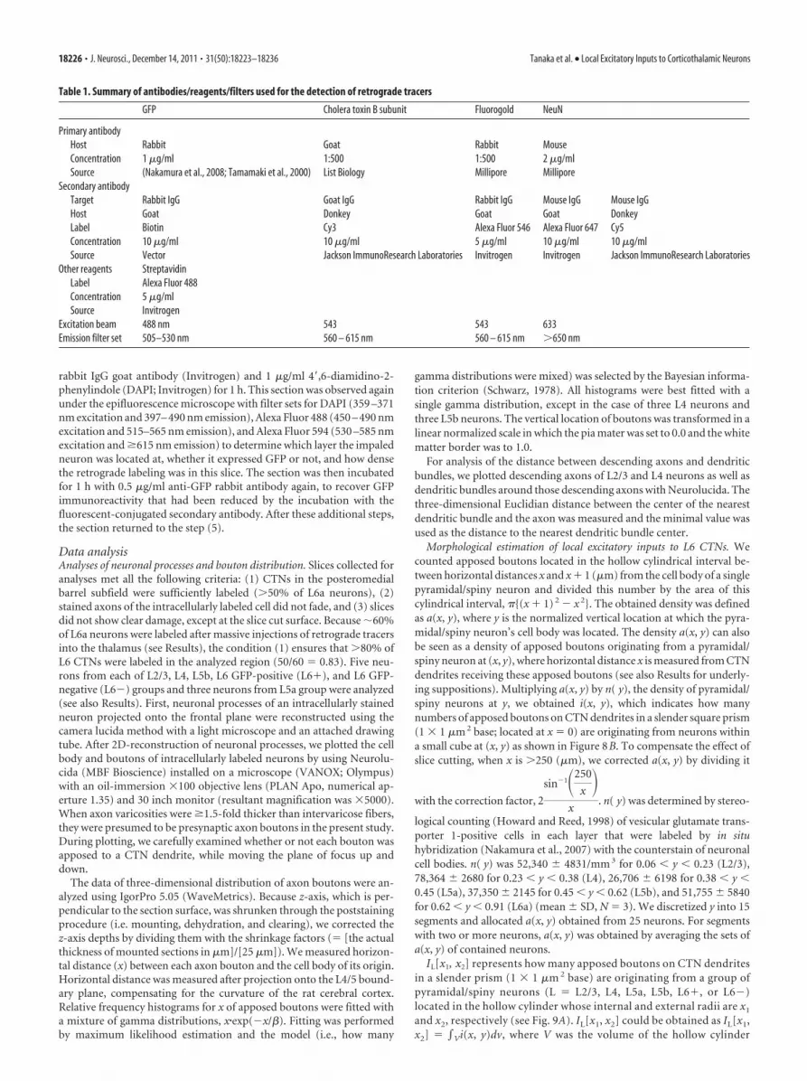
Table 1. Summary of antibodies/reagents/filters used for the detection of retrograde tracers
GFP Cholera toxin B subunit Fluorogold NeuN
Primary antibodyHost Rabbit Goat Rabbit MouseConcentration 1 �g/ml 1:500 1:500 2 �g/mlSource (Nakamura et al., 2008; Tamamaki et al., 2000) List Biology Millipore Millipore
Secondary antibodyTarget Rabbit IgG Goat IgG Rabbit IgG Mouse IgG Mouse IgGHost Goat Donkey Goat Goat DonkeyLabel Biotin Cy3 Alexa Fluor 546 Alexa Fluor 647 Cy5Concentration 10 �g/ml 10 �g/ml 5 �g/ml 10 �g/ml 10 �g/mlSource Vector Jackson ImmunoResearch Laboratories Invitrogen Invitrogen Jackson ImmunoResearch Laboratories
Other reagents StreptavidinLabel Alexa Fluor 488Concentration 5 �g/mlSource Invitrogen
Excitation beam 488 nm 543 543 633Emission filter set 505–530 nm 560 – 615 nm 560 – 615 nm �650 nm
18226 • J. Neurosci., December 14, 2011 • 31(50):18223–18236 Tanaka et al. • Local Excitatory Inputs to Corticothalamic Neurons
between distances x1 and x2 in each layer or sublayer. However, toassess the variability of the data, we first integrated a(x, y)�n( y) ob-tained from single pyramidal/spiny neurons and then averaged withineach group. To obtain n( y) for L6� and L6� neurons, we estimatedthat 69% of excitatory neurons in L6a were corticothalamic neurons
because 59% of L6a neurons were retrogradely labeled following amassive injection of retrograde tracer, and because 85% of neuronswere positive for vesicular glutamate transporter 1 in L6a. On theother hand, 31% of L6a excitatory neurons were considered as corti-cocortical neurons (see also Results).
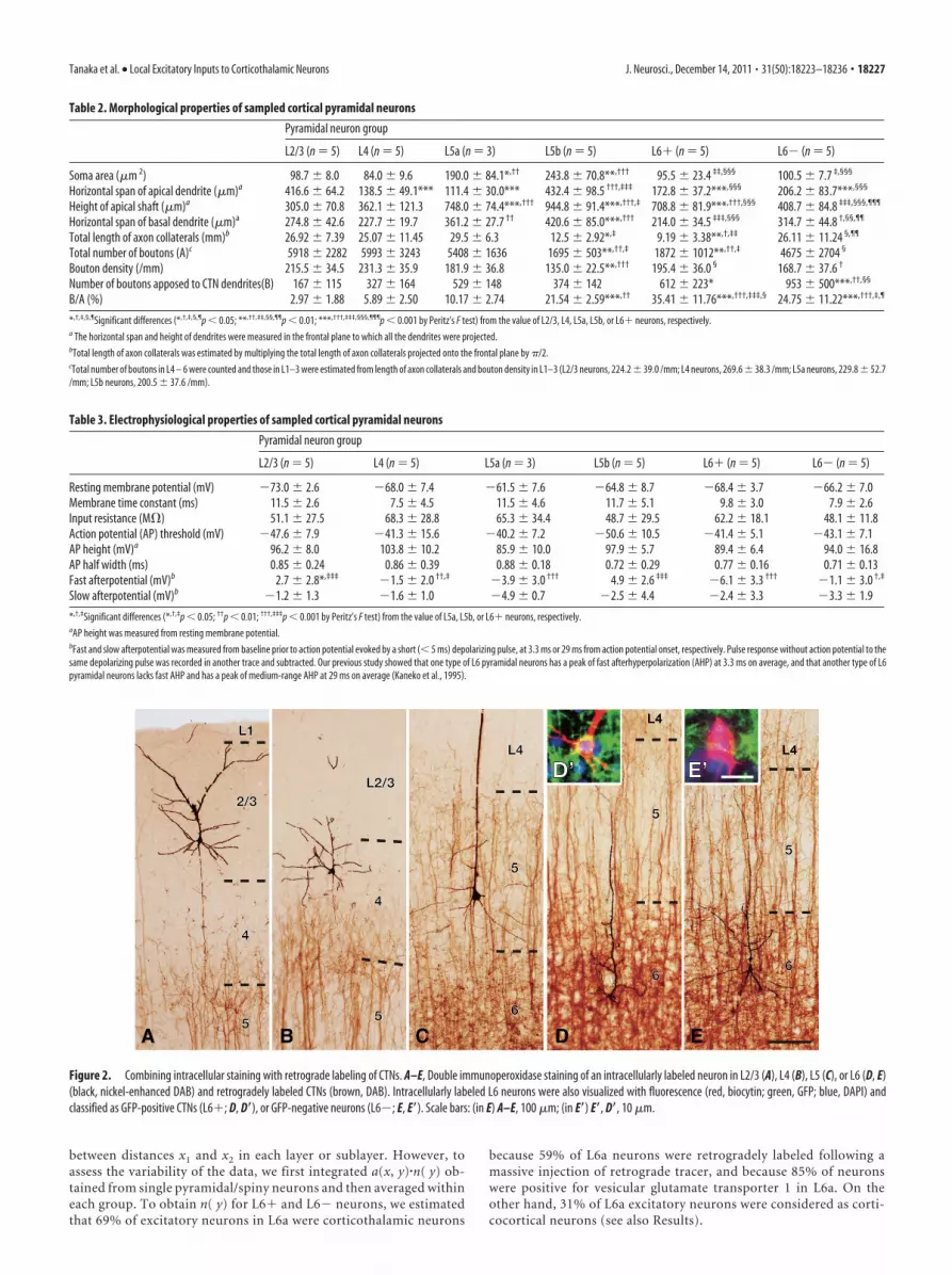
Figure 2. Combining intracellular staining with retrograde labeling of CTNs. A–E, Double immunoperoxidase staining of an intracellularly labeled neuron in L2/3 (A), L4 (B), L5 (C), or L6 (D, E)(black, nickel-enhanced DAB) and retrogradely labeled CTNs (brown, DAB). Intracellularly labeled L6 neurons were also visualized with fluorescence (red, biocytin; green, GFP; blue, DAPI) andclassified as GFP-positive CTNs (L6�; D, D�), or GFP-negative neurons (L6�; E, E�). Scale bars: (in E) A–E, 100 �m; (in E�) E�, D�, 10 �m.
Table 2. Morphological properties of sampled cortical pyramidal neurons
Pyramidal neuron group
L2/3 (n � 5) L4 (n � 5) L5a (n � 3) L5b (n � 5) L6� (n � 5) L6� (n � 5)
Soma area (�m 2) 98.7 8.0 84.0 9.6 190.0 84.1*,†† 243.8 70.8**,††† 95.5 23.4 ‡‡,§§§ 100.5 7.7 ‡,§§§
Horizontal span of apical dendrite (�m)a 416.6 64.2 138.5 49.1*** 111.4 30.0*** 432.4 98.5 †††,‡‡‡ 172.8 37.2***,§§§ 206.2 83.7***,§§§
Height of apical shaft (�m)a 305.0 70.8 362.1 121.3 748.0 74.4***,††† 944.8 91.4***,†††,‡ 708.8 81.9***,†††,§§§ 408.7 84.8 ‡‡‡,§§§,¶¶¶
Horizontal span of basal dendrite (�m)a 274.8 42.6 227.7 19.7 361.2 27.7 †† 420.6 85.0***,††† 214.0 34.5 ‡‡‡,§§§ 314.7 44.8 †,§§,¶¶
Total length of axon collaterals (mm)b 26.92 7.39 25.07 11.45 29.5 6.3 12.5 2.92*,‡ 9.19 3.38**,†,‡‡ 26.11 11.24 §,¶¶
Total number of boutons (A)c 5918 2282 5993 3243 5408 1636 1695 503**,††,‡ 1872 1012**,††,‡ 4675 2704 §
Bouton density (/mm) 215.5 34.5 231.3 35.9 181.9 36.8 135.0 22.5**,††† 195.4 36.0 § 168.7 37.6 †
Number of boutons apposed to CTN dendrites(B) 167 115 327 164 529 148 374 142 612 223* 953 500***,††,§§
B/A (%) 2.97 1.88 5.89 2.50 10.17 2.74 21.54 2.59***,†† 35.41 11.76***,†††,‡‡‡,§ 24.75 11.22***,†††,‡,¶
*,†,‡,§,¶Significant differences (*,†,‡,§,¶p 0.05; **,††,‡‡,§§,¶¶p 0.01; ***,†††,‡‡‡,§§§,¶¶¶p 0.001 by Peritz’s F test) from the value of L2/3, L4, L5a, L5b, or L6� neurons, respectively.a The horizontal span and height of dendrites were measured in the frontal plane to which all the dendrites were projected.bTotal length of axon collaterals was estimated by multiplying the total length of axon collaterals projected onto the frontal plane by �/2.cTotal number of boutons in L4 – 6 were counted and those in L1–3 were estimated from length of axon collaterals and bouton density in L1–3 (L2/3 neurons, 224.2 39.0 /mm; L4 neurons, 269.6 38.3 /mm; L5a neurons, 229.8 52.7/mm; L5b neurons, 200.5 37.6 /mm).
Table 3. Electrophysiological properties of sampled cortical pyramidal neurons
Pyramidal neuron group
L2/3 (n � 5) L4 (n � 5) L5a (n � 3) L5b (n � 5) L6� (n � 5) L6� (n � 5)
Resting membrane potential (mV) �73.0 2.6 �68.0 7.4 �61.5 7.6 �64.8 8.7 �68.4 3.7 �66.2 7.0Membrane time constant (ms) 11.5 2.6 7.5 4.5 11.5 4.6 11.7 5.1 9.8 3.0 7.9 2.6Input resistance (M�) 51.1 27.5 68.3 28.8 65.3 34.4 48.7 29.5 62.2 18.1 48.1 11.8Action potential (AP) threshold (mV) �47.6 7.9 �41.3 15.6 �40.2 7.2 �50.6 10.5 �41.4 5.1 �43.1 7.1AP height (mV)a 96.2 8.0 103.8 10.2 85.9 10.0 97.9 5.7 89.4 6.4 94.0 16.8AP half width (ms) 0.85 0.24 0.86 0.39 0.88 0.18 0.72 0.29 0.77 0.16 0.71 0.13Fast afterpotential (mV)b 2.7 2.8*,‡‡‡ �1.5 2.0 ††,‡ �3.9 3.0 ††† 4.9 2.6 ‡‡‡ �6.1 3.3 ††† �1.1 3.0 †,‡
Slow afterpotential (mV)b �1.2 1.3 �1.6 1.0 �4.9 0.7 �2.5 4.4 �2.4 3.3 �3.3 1.9
*,†,‡Significant differences (*,†,‡p 0.05; ††p 0.01; †††,‡‡‡p 0.001 by Peritz’s F test) from the value of L5a, L5b, or L6� neurons, respectively.aAP height was measured from resting membrane potential.bFast and slow afterpotential was measured from baseline prior to action potential evoked by a short ( 5 ms) depolarizing pulse, at 3.3 ms or 29 ms from action potential onset, respectively. Pulse response without action potential to thesame depolarizing pulse was recorded in another trace and subtracted. Our previous study showed that one type of L6 pyramidal neurons has a peak of fast afterhyperpolarization (AHP) at 3.3 ms on average, and that another type of L6pyramidal neurons lacks fast AHP and has a peak of medium-range AHP at 29 ms on average (Kaneko et al., 1995).
Tanaka et al. • Local Excitatory Inputs to Corticothalamic Neurons J. Neurosci., December 14, 2011 • 31(50):18223–18236 • 18227

Statistics. When we compared all pairs betweenthe six pyramidal/spiny neuron groups (15 com-parisons) we used Peritz’s F test (Harper, 1984), amultiple-comparison test (Tables 2, 3; see Figs. 6,9). For pairwise comparison in each neurongroup, we used two-sided t test with Bonferronicorrection (see Fig. 7). Differences were acceptedas significant if the family-wise type I error prob-ability was 0.05. Each statistic and p value wascalculated with Prism 5.0 or programs written inIgor 5.05.
ResultsRetrograde labeling of CTNs withadenoviral vectorTo visualize CTN dendrites, we developedan adenoviral vector expressing myrGFP-LDLRct (Kameda et al., 2008), a somato-dendritic membrane-targeted GFP, underthe control of an enhanced human synap-sin I promoter (Hioki et al., 2007) (Fig.1A). After the adenoviral vector was in-jected with the vehicle containing 0.6 M
NaCl (Tomioka and Rockland, 2006) intothe ventrobasal thalamic complex of rats,a band of L6 neurons in the S1 cortex wasretrogradely labeled (Fig. 1B,C). As re-ported previously (Killackey and Sher-man, 2003), labeled cells were mostlyfound in L6a, the upper part of L6 sepa-rated from L6b by cell-sparse fibrous zone(Fig. 1B) (Valverde et al., 1989). L5 corti-cothalamic neurons mainly projecting toposterior thalamic nuclei (Bourassa et al.,1995; Killackey and Sherman, 2003) wererarely labeled (1% in all GFP-positiveneurons) and were negligible in subse-quent analyses when the injection wasconfined to the ventrobasal complex.With this retrograde labeling method, notonly the cell bodies and dendritic shafts ofCTNs but the fine dendritic structuressuch as spines were visualized (Fig. 1D–H). In contrast, almost no axon collateralsof retrogradely labeled neurons or an-terogradely labeled thalamocortical ax-ons were observed in the S1 cortex.Moreover, �60% of L6a neurons (i.e., 70%of L6a excitatory neurons) were labeled fol-lowing vector injection �3 � 106 GTU.Since this saturation level (59.0 3.9%;mean SD, N � 8) was similar to thelabeling efficiency of other potent retro-grade tracers such as cholera toxin B subunit and Fluorogold(58.6 11.0%, N � 4 and 57.6 8.9%, N � 3; p � 0.96 byone-way ANOVA), almost all CTNs were considered to be visu-alized at least in the center of labeling. The labeling efficiency ofadenovirus vector (1 � 10 7 GTU) did not significantly differbetween postinjection survival times of 4, 8, and 16 d (56.1 2.6%, 58.1 5.6%, and 59.8 1.1%, N � 3 each; p � 0.50 byone-way ANOVA). Furthermore, thalamic neurons in andaround the injection site in the ventrobasal thalamic complexwere labeled, and some neurons in the thalamic reticular nucleuswas labeled retrogradely (Fig. 1 I).
Intracellular labeling of pyramidal/spiny neurons and theirlocal connections to CTNsIn 500-�m-thick cortical slices containing retrogradely labeledCTNs, 11, 9, 29, and 41 pyramidal/spiny neurons were sampled inL2/3, L4, L5, and L6, respectively, for intracellular recording/staining in the posteromedial barrel subfield of the rat S1 cortex.All the stained neurons were pyramidal neurons, except for fourspiny stellate cells and one star pyramid of Lorente de No (1938)sampled in L4. We collected five pyramidal/spiny neurons fromL2/3 and L4 and eight from L5 (Fig. 2A–C) that met the criteriafor sufficient retrograde labeling and intracellular staining (see
Figure 3. Two-dimensional reconstruction of intracellularly labeled neurons. Dendrites and axons originating from an L2/3, L4,L5a, L5b, L6�, or L6� neuron are shown with red and blue lines, respectively. Thick blue lines denote the thick straight axons.
18228 • J. Neurosci., December 14, 2011 • 31(50):18223–18236 Tanaka et al. • Local Excitatory Inputs to Corticothalamic Neurons
also Materials and Methods). We also accumulated five each ofGFP-positive and GFP-negative pyramidal neurons located in L6(Fig. 2D,E). We further divided eight L5 neurons into three L5aneurons and five L5b neurons according to their cell body loca-tions (Schubert et al., 2006). As a result, each of the L2/3, L5b, L6GFP-positive (L6�), or L6 GFP-negative (L6�) pyramidal neu-ron groups examined in the present study was thus comprised offive pyramidal neurons and the L5a neuron group containedthree pyramidal neurons. The L4 neuron group involved twopyramidal neurons, two spiny stellate cells, and one star pyramid.Finally, all the 28 neurons were located in barrel columns, but notin septal columns of the posteromedial barrel subfield.
L6� pyramidal neurons were different from thalamus-projecting L6� neurons in several morphological and electro-physiological properties, and may represent a type of pyramidalneuron different from the CTN. All L6� neurons showed tonicfiring responses to a strong current injection, whereas four of thefive L6� neurons displayed phasic firing responses. The fast hy-perpolarizing afterpotentials of L6� neurons were relatively deepand significantly different from those of L6� neurons (Table 3).Morphologically, L6� neurons had narrow basal dendriticarbors and tufted apical dendrites that extended to L4, whereasL6� neurons had wide basal dendritic arbors and short apicaldendrites that ended without entering L4. The axon collaterals ofL6� neurons took a direction toward the pia mater and enteredL4 without spreading widely, while those of L6� neurons mainlyspread horizontally through L5/6 (Table 2; Fig. 3). Because theseelectrophysiological and morphological properties of L6� neu-rons were similar to those of L6 corticocortical neurons identifiedby single axon tracing (Zhang and Deschenes, 1997) or by the lackof retrograde labeling after massive injection of retrograde tracer
into the thalamus (Brumberg et al., 2003; Mercer et al., 2005;Kumar and Ohana, 2008), at least a subset of L6� neurons wereconsidered corticocortical neurons.
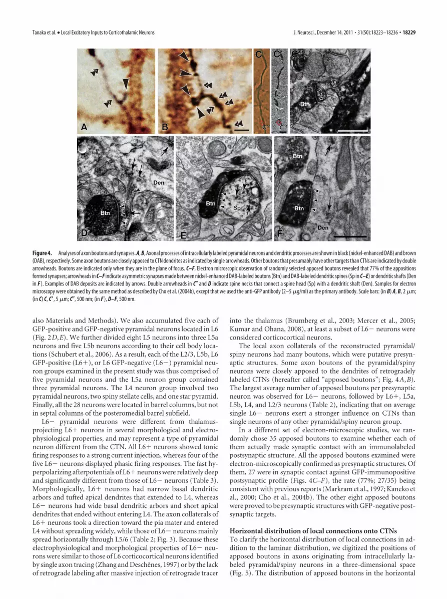
The local axon collaterals of the reconstructed pyramidal/spiny neurons had many boutons, which were putative presyn-aptic structures. Some axon boutons of the pyramidal/spinyneurons were closely apposed to the dendrites of retrogradelylabeled CTNs (hereafter called “apposed boutons”; Fig. 4A,B).The largest average number of apposed boutons per presynapticneuron was observed for L6� neurons, followed by L6�, L5a,L5b, L4, and L2/3 neurons (Table 2), indicating that on averagesingle L6� neurons exert a stronger influence on CTNs thansingle neurons of any other pyramidal/spiny neuron group.
In a different set of electron-microscopic studies, we ran-domly chose 35 apposed boutons to examine whether each ofthem actually made synaptic contact with an immunolabeledpostsynaptic structure. All the apposed boutons examined wereelectron-microscopically confirmed as presynaptic structures. Ofthem, 27 were in synaptic contact against GFP-immunopositivepostsynaptic profile (Figs. 4C–F), the rate (77%; 27/35) beingconsistent with previous reports (Markram et al., 1997; Kaneko etal., 2000; Cho et al., 2004b). The other eight apposed boutonswere proved to be presynaptic structures with GFP-negative post-synaptic targets.
Horizontal distribution of local connections onto CTNsTo clarify the horizontal distribution of local connections in ad-dition to the laminar distribution, we digitized the positions ofapposed boutons in axons originating from intracellularly la-beled pyramidal/spiny neurons in a three-dimensional space(Fig. 5). The distribution of apposed boutons in the horizontal
Figure 4. Analyses of axon boutons and synapses. A, B, Axonal processes of intracellularly labeled pyramidal neurons and dendritic processes are shown in black (nickel-enhanced DAB) and brown(DAB), respectively. Some axon boutons are closely apposed to CTN dendrites as indicated by single arrowheads. Other boutons that presumably have other targets than CTNs are indicated by doublearrowheads. Boutons are indicated only when they are in the plane of focus. C–F, Electron microscopic observation of randomly selected apposed boutons revealed that 77% of the appositionsformed synapses; arrowheads in C–F indicate asymmetric synapses made between nickel-enhanced DAB-labeled boutons (Btn) and DAB-labeled dendritic spines (Sp in C–E) or dendritic shafts (Denin F ). Examples of DAB deposits are indicated by arrows. Double arrowheads in C� and D indicate spine necks that connect a spine head (Sp) with a dendritic shaft (Den). Samples for electronmicroscopy were obtained by the same method as described by Cho et al. (2004b), except that we used the anti-GFP antibody (2–5 �g/ml) as the primary antibody. Scale bars: (in B) A, B, 2 �m;(in C) C, C�, 5 �m; C�, 500 nm; (in F ), D–F, 500 nm.
Tanaka et al. • Local Excitatory Inputs to Corticothalamic Neurons J. Neurosci., December 14, 2011 • 31(50):18223–18236 • 18229

plane perpendicular to the cortical colum-nar structures differed between the pyrami-dal/spiny neuron groups; for example, inFigure 6, A1 and A2, L6� and L4 neuronsdisplayed much narrower distributionsthan L5 neurons shown in Figure 6A3.
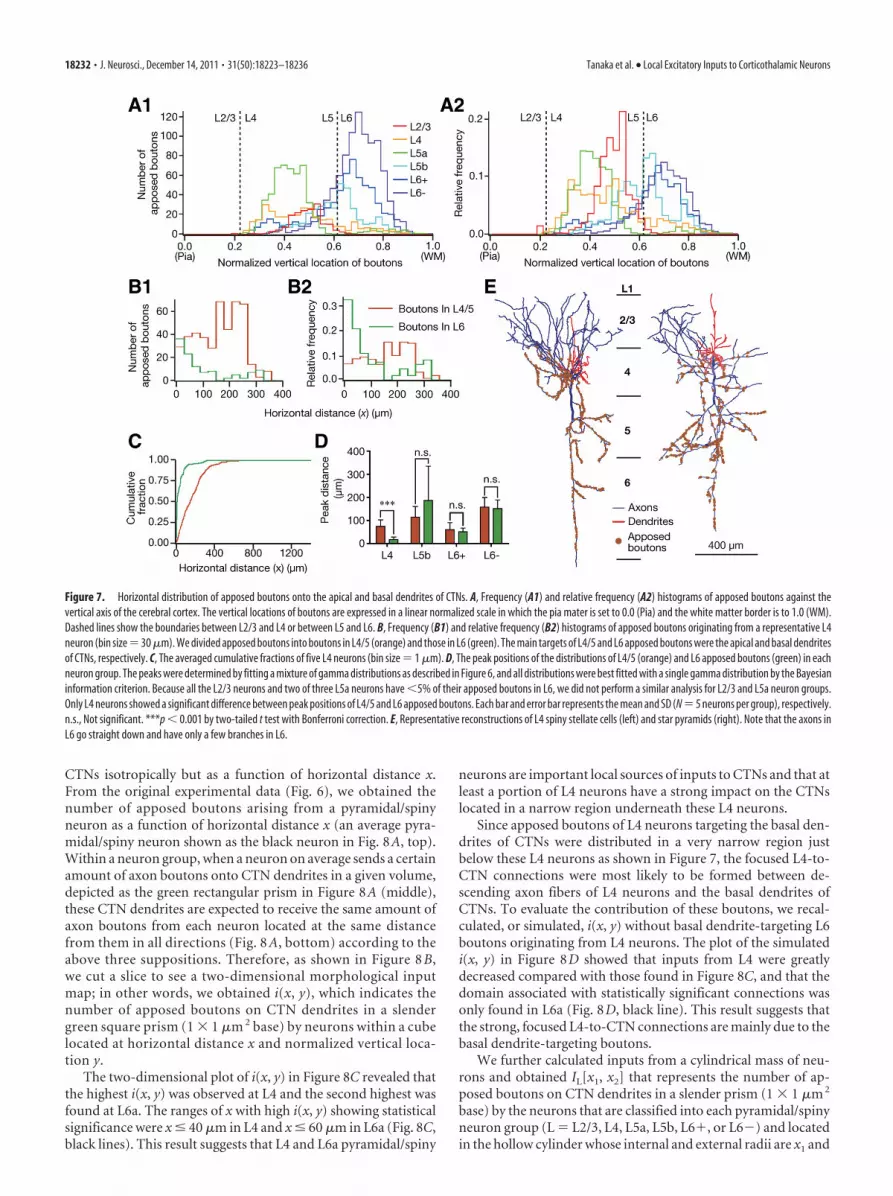
The “horizontal distance” betweeneach apposed bouton and the cell body ofits origin was normalized by measuring,after projection of the bouton and the cellbody onto the L4/5 boundary plane tocompensate for the effect of cortical cur-vature on the horizontal distance (Fig.6B). In Figure 6C, the horizontal distribu-tion of the apposed boutons arising fromeach neuron was summarized in a relativefrequency histogram showing a single peak(blue lines and histograms are sample distri-butions, and black lines are fitting curves fordetermining peak positions; see Materialsand Methods). The apposed boutons of L4and L6� neurons showed peak positionscloser to their cell bodies and narrower halfwidths than those of the other neurongroups. Differences in the peak distance andhalf width between L4 neuron group andL2/3, L5b, or L6� group, or those betweenL6� group and L2/3 or L6� group showedstatistical significance (Fig. 6D,E). These re-sults suggest that L4 and L6� neurons aremore likely to project to nearby CTNs thanare neurons of the other groups. In contrast,L6� neurons exhibited the most distantpeak position and the broadest half width(Fig. 6D,E). Consistent with these findings,the averaged cumulative distributions of theL4 and L6� neuron groups increased rap-idly, whereas that of the L6� neuron grouprose slowly as x increased (Fig. 6F).
Connections to apical and basaldendrites of CTNsSince apical and basal dendrites mayseparately integrate their inputs and dis-tinctly affect the output of a cortical pyra-midal neuron (Mel, 2008; Sjostrom et al.,2008; Ledergerber and Larkum, 2010), we further tried to exam-ine the difference between the horizontal distributions of bou-tons targeting the apical dendrites of CTNs and those targetingthe basal dendrites. We first examined the histograms of fre-quency and relative frequency against the vertical location of ap-posed boutons (Fig. 7A). L2/3, L4, and L5a neurons allocated themost of their apposed boutons within L4/5, while L6� and L6�neurons distributed the majority of their apposed boutons in L6.L5b neurons dispersed their apposed boutons rather evenly inL4/5 or in L6. For further analyses, we compared the horizontaldistributions of apposed boutons in L4/5 and those in L6 for eachpyramidal/spiny neuron group, as the majority of apical or basaldendrites of L6 CTNs spread in L4/5 or L6, respectively (Fig. 1B).In Figure 7B, the histograms show that L6 apposed boutons orig-inating from a representative L4 spiny stellate cell were concen-trated near the cell body (green), whereas its L4/5 boutons hadthe peak of the distribution at �200 �m from the cell body (or-
ange). A similar distribution was observed in all subtypes of L4pyramidal/spiny neurons and thus the average cumulative distri-bution of L6 apposed boutons arising from L4 pyramidal/spinyneurons was saturated at a clearly smaller x than that of L4/5apposed boutons (Figs. 7C). Significant differences between thepeak positions of L4/5 and L6 apposed boutons were only foundin the L4 neuron group (p � 0.0008 by t tests with Bonferronicorrection, Fig. 7D), but not in the other neuron groups. Thischaracteristic distribution of apposed boutons originating fromL4 neurons was mainly ascribed to their axonal arborization. Asshown in Figure 7E, the axon collaterals of L4 neurons were wide-spread in L2–5 but highly restricted in L6.
Morphological estimation of local excitatory inputs toL6 CTNsWe next reexamined the present data so that we could estimateinputs from a mass of neurons to CTN dendrites and obtain a
Figure 5. Three-dimensional distribution of axon boutons. The axon boutons of an L6� pyramidal neuron were plottedthree-dimensionally, and shown in three different views.
18230 • J. Neurosci., December 14, 2011 • 31(50):18223–18236 Tanaka et al. • Local Excitatory Inputs to Corticothalamic Neurons

morphological input map that is comparable to the maps ob-served in previous photostimulation studies on L6 pyramidalneurons (Briggs and Callaway, 2001; Zarrinpar and Callaway,2006; Llano and Sherman, 2009; Lam and Sherman, 2010; Hookset al., 2011). For this reexamination, we made the following three
suppositions: (1) The density of CTN dendrites is uniform at agiven cortical depth, (2) the distribution and density of cell bod-ies of pyramidal/spiny neurons are horizontally constant at agiven cortical depth, and (3) as a group, pyramidal/spiny neuronsat a given cortical depth distribute their apposed boutons to
Figure 6. Horizontal distribution of apposed boutons. A1–3, The density map of apposed boutons in a horizontal plane (Fig. 5, the plane corresponding to “Top view”) is shown after beingsmoothed by 2D convolution with a Gaussian kernel (� � 4 �m; a comparable value with the interbouton interval) and normalized (bottom of A3). B, We plotted the boundary of L4 and L5 (L4/5boundary) and the directions of apical dendrites of corticothalamic neurons every 50 �m (lines PmWm) and determined Om as the intersection of lines PmWm and Pm�1Wm�1. These intersectionswere always located in the white matter because of the convexity of the rat cerebral cortex. The locations of the cell body and boutons were then projected onto L4/5 boundary; for examples, S wasprojected along line SO2 to S’ and B along line BO1 to B’. Horizontal distance x between S and B is to be measured as the distance between S’ and B’ along the L4/5 boundary (red curve). Because therat barrel cortex along the rostrocaudal axis is much flatter, no correction for rostrocaudal direction was performed. C1– 6, Relative frequency histograms (blue histogram, bin size � 30 �m) andcumulative fractions (insets, blue lines, bin size � 1 �m) for the horizontal distance of apposed boutons originating from a representative cell in each layer. Each distribution was fitted with amixture of gamma distributions (black lines). Most distributions were best fitted with a single gamma distribution in terms of the Bayesian information criterion. D, E), The peak position and the halfwidth of the curves are compared between the neuron groups. F, The cumulative fraction averaged within each neuron group (bin size � 1 �m). Scale bar: (in A1) A1–3, 100 �m. Each bar and errorbar in D, E represents the mean and SD, respectively. *p 0.05; **p 0.01; ***p 0.001 by Peritz’s F test.
Tanaka et al. • Local Excitatory Inputs to Corticothalamic Neurons J. Neurosci., December 14, 2011 • 31(50):18223–18236 • 18231
CTNs isotropically but as a function of horizontal distance x.From the original experimental data (Fig. 6), we obtained thenumber of apposed boutons arising from a pyramidal/spinyneuron as a function of horizontal distance x (an average pyra-midal/spiny neuron shown as the black neuron in Fig. 8A, top).Within a neuron group, when a neuron on average sends a certainamount of axon boutons onto CTN dendrites in a given volume,depicted as the green rectangular prism in Figure 8A (middle),these CTN dendrites are expected to receive the same amount ofaxon boutons from each neuron located at the same distancefrom them in all directions (Fig. 8 A, bottom) according to theabove three suppositions. Therefore, as shown in Figure 8 B,we cut a slice to see a two-dimensional morphological inputmap; in other words, we obtained i(x, y), which indicates thenumber of apposed boutons on CTN dendrites in a slendergreen square prism (1 � 1 �m 2 base) by neurons within a cubelocated at horizontal distance x and normalized vertical loca-tion y.
The two-dimensional plot of i(x, y) in Figure 8C revealed thatthe highest i(x, y) was observed at L4 and the second highest wasfound at L6a. The ranges of x with high i(x, y) showing statisticalsignificance were x � 40 �m in L4 and x � 60 �m in L6a (Fig. 8C,black lines). This result suggests that L4 and L6a pyramidal/spiny
neurons are important local sources of inputs to CTNs and that atleast a portion of L4 neurons have a strong impact on the CTNslocated in a narrow region underneath these L4 neurons.
Since apposed boutons of L4 neurons targeting the basal den-drites of CTNs were distributed in a very narrow region justbelow these L4 neurons as shown in Figure 7, the focused L4-to-CTN connections were most likely to be formed between de-scending axon fibers of L4 neurons and the basal dendrites ofCTNs. To evaluate the contribution of these boutons, we recal-culated, or simulated, i(x, y) without basal dendrite-targeting L6boutons originating from L4 neurons. The plot of the simulatedi(x, y) in Figure 8D showed that inputs from L4 were greatlydecreased compared with those found in Figure 8C, and that thedomain associated with statistically significant connections wasonly found in L6a (Fig. 8D, black line). This result suggests thatthe strong, focused L4-to-CTN connections are mainly due to thebasal dendrite-targeting boutons.
We further calculated inputs from a cylindrical mass of neu-rons and obtained IL[x1, x2] that represents the number of ap-posed boutons on CTN dendrites in a slender prism (1 � 1 �m 2
base) by the neurons that are classified into each pyramidal/spinyneuron group (L � L2/3, L4, L5a, L5b, L6�, or L6�) and locatedin the hollow cylinder whose internal and external radii are x1 and
Figure 7. Horizontal distribution of apposed boutons onto the apical and basal dendrites of CTNs. A, Frequency (A1) and relative frequency (A2) histograms of apposed boutons against thevertical axis of the cerebral cortex. The vertical locations of boutons are expressed in a linear normalized scale in which the pia mater is set to 0.0 (Pia) and the white matter border is to 1.0 (WM).Dashed lines show the boundaries between L2/3 and L4 or between L5 and L6. B, Frequency (B1) and relative frequency (B2) histograms of apposed boutons originating from a representative L4neuron (bin size � 30 �m). We divided apposed boutons into boutons in L4/5 (orange) and those in L6 (green). The main targets of L4/5 and L6 apposed boutons were the apical and basal dendritesof CTNs, respectively. C, The averaged cumulative fractions of five L4 neurons (bin size � 1 �m). D, The peak positions of the distributions of L4/5 (orange) and L6 apposed boutons (green) in eachneuron group. The peaks were determined by fitting a mixture of gamma distributions as described in Figure 6, and all distributions were best fitted with a single gamma distribution by the Bayesianinformation criterion. Because all the L2/3 neurons and two of three L5a neurons have 5% of their apposed boutons in L6, we did not perform a similar analysis for L2/3 and L5a neuron groups.Only L4 neurons showed a significant difference between peak positions of L4/5 and L6 apposed boutons. Each bar and error bar represents the mean and SD (N � 5 neurons per group), respectively.n.s., Not significant. ***p 0.001 by two-tailed t test with Bonferroni correction. E, Representative reconstructions of L4 spiny stellate cells (left) and star pyramids (right). Note that the axons inL6 go straight down and have only a few branches in L6.
18232 • J. Neurosci., December 14, 2011 • 31(50):18223–18236 Tanaka et al. • Local Excitatory Inputs to Corticothalamic Neurons

x2, respectively (Fig. 9A). Considering the size of the barrel in theposteromedial barrel subfield (200 �m in radius) (Welker andWoolsey, 1974; Fox, 2008) and the maximum horizontal distancefound in our samples (1492 �m), we compared I[0, 200] andI[200, 1500] between the five pyramidal/spiny neuron groups.IL6�[0, 200] was higher than the IL[0, 200] of any other neurongroup, while IL6�[200, 1500] displayed larger value than anyother IL[200, 1500]. This suggests that CTNs themselves are themost important nearby/columnar source of connections,whereas L6 corticocortical neurons are the most crucial distant/extracolumnar source.
Correlative location of L4 axons and CTN dendritic bundlesThe horizontally focused L4-to-CTN connection (Fig. 8) may beorganized with certain vertical structures in the cerebral cortex,such as apical dendritic bundles of CTNs. The bundles typicallyconsisted of 2–5 apical dendrites and their interbundle distancewas 56 14 �m (mean SD, N � 7 slices). IL4[0, 28] and IL6�[0,28] were significantly higher than those of the other neurongroups (Fig. 9D). This result confirms the importance of L4 neu-rons as a source of inputs to CTNs within a narrow horizontalrange. We further examined the relationship of descending thickaxons of L4 neurons with CTN dendritic bundles, compared withthose of L2/3 neurons. The thick axons of L4 neurons approached
CTN dendritic bundles in the middle of L5 and descended inclose association with CTN bundles (Fig. 9F), whereas those ofL2/3 neurons descended straight to the white matter and did notcome into contact with CTN bundles (Fig. 9E). Consistently, thethick axons of L4 neurons descended at a closer distance (�7�m) to the center of the nearest dendritic bundle than axonsoriginating from L2/3 neurons (p � 0.045 by two-tailed t test;Fig. 9G). As the diameter of CTN dendritic bundles was �10 �m,the thick axons of L4 neurons ran alongside the bundles (Fig. 9F).
DiscussionHere, we estimated local excitatory inputs to CTNs (Figs. 8, 9) onthe basis of the distribution of apposed boutons of cortical pyra-midal/spiny neurons to CTNs (Table 2; Fig. 6). L6 CTNs them-selves and L4 neurons were the important nearby sources ofconnections to CTNs, whereas L6 corticocortical neurons werethe main distant source within the local (1500 �m) connec-tions. We further found that CTNs received strong and focusedinputs from L4 neurons just above them and that this focusedL4-to-CTN connectivity was formed between descending axonfibers of L4 neurons and the basal dendrites of CTNs. (Figs. 7,8D). These results illustrate which excitatory neurons of the S1cortical column send information through L6 CTNs to the tha-lamic compartment corresponding to the column.
Figure 8. Estimation of input from pyramidal/spiny neurons in a cube to CTN dendrites. A, Reexamination of the data under the three suppositions described in the text. An average neuron in eachpyramidal/spiny neuron group distributed its apposed boutons isotropically to CTN dendrites located at a horizontal distance x (green cylinder, top). The red cylinder (middle) represents anotherhollow cylinder filled with pyramidal/spiny neurons around a green prism. CTN dendrites in the central prism would then receive the same amount of projections from each average neuron locatedat the same distance from these dendrites in all directions (bottom). B, As shown on top, i(x, y) represents the number of apposed boutons projected to CTN dendrites in the prism by neurons in acube located at (x, y). The scheme of two-dimensional input map is shown on bottom. C, i(x, y) is displayed in the x–y plane (smoothed with Gaussian kernel; � � 10 �m for x). Color code showsthe magnitude of i(x, y) in normalized value (maximum � 1), and regions encircled by black borders show significantly high i(x, y) (� mean � 2SD; calculated within the range of x � 0 –200 �m).D, Simulated i(x, y) without basal dendrite-targeting L6 boutons originating from L4 neurons. Magnitude of i(x, y) in D is normalized with the maximum value in C. Note that the signal in L4 isremarkably reduced.
Tanaka et al. • Local Excitatory Inputs to Corticothalamic Neurons J. Neurosci., December 14, 2011 • 31(50):18223–18236 • 18233

Strong local excitatory inputs to L6CTNs from their own layer were foundin previous studies using laser-scanningphotostimulation by glutamate uncaging(Zarrinpar and Callaway, 2006; Llano andSherman, 2009; Lam and Sherman, 2010;Hooks et al., 2011). Because photostimu-lation with a resolution of 50 –75 �mprobably would activate several CTNssimultaneously (Hooks et al., 2011), largeEPSPs might be evoked in the recorded neu-ron through dense nearby connections be-tween CTNs. Similar results were obtainedby our morphological estimation of inputsto CTNs from neurons in a cube (Fig. 8C) orin a cylindrical volume (Fig. 9D).
The present results further suggest thata small population of L4 neurons locatedjust above a CTN appear to be a mainsource of connections to this CTN (Fig. 8).Interestingly, a photostimulation study re-ported a case of strong L4 inputs to a puta-tive L6 CTN [Zarrinpar and Callaway(2006), their Fig. 4A]. Furthermore, a recentmultiple whole-cell recording study of themouse barrel cortex has reported cases ofL4-to-L6 connections (3/93 pairs) and hasalso shown that EPSP amplitudes of theseconnections are sometimes much larger(0.17, 0.96, and 5.67 mV) than the typicalEPSP amplitude in postsynaptic L6 neurons(�0.2 mV) (Lefort et al., 2009). These largeEPSP amplitudes of L4-to-L6 connectionsmight be well represented by the strong, fo-cused L4-to-CTN connection revealed inthe present study (Fig. 8C).
Figure 10. Anatomical organization of local excitatory connections to L6 CTNs and its relationship to thalamic barreloid. Withina barrel column, the L6 CTNs receive strong, focused connections from L4 neurons located just above them (red). L6 CTNs havedense recurrent connections of a subcolumnar size within themselves (green), whereas L6 corticocortical neurons provide CTNswith a considerable amount of horizontal connection beyond the barrel column (black). The projecting axons of L6 CTNs spread outin the corresponding thalamic compartment (barreloid) and thus show a multitude of overlaps within the barreloid.
Figure 9. Horizontal organization of local excitatory inputs to CTNs. A, An example of IL[x1, x2]. As the scheme shows, IL[x1, x2] represents the number of apposed boutons on CTN dendrites in thecentral slender prism (1 � 1 �m 2 base) by the neurons that are classified into each pyramidal/spiny neuron group (L � L2/3, L4, L5a, L5b, L6�, or L6�) and located in the hollow cylinder whoseinternal and external radii are x1 and x2, respectively. B–D, Inputs from neurons located at x � 0 –200 �m (B), x � 200 –1500 �m (C), and x � 0 –28 �m (D). E, F, Descending thick axons of arepresentative L2/3 neuron (E) and L4 neuron (F ) are indicated by arrows and dendritic bundles of CTNs are indicated by arrowheads. Note that the axon of the L4 neuron changes course andapproaches a CTN dendritic bundle in the middle of L5 and descends along the bundle. G, The minimal distance between the descending axon and the center of the nearest apical dendritic bundleshowed that the descending axons of L4 neurons ran at closer range to the dendritic bundle than those of L2/3 neurons. Scale bar, (in F ) E, F, 50 �m. Each bar and error bar represents the mean andSD, respectively. *p 0.05; **p 0.01; ***p 0.001 by Peritz’s F test. †p 0.05 by two-tailed t test.
18234 • J. Neurosci., December 14, 2011 • 31(50):18223–18236 Tanaka et al. • Local Excitatory Inputs to Corticothalamic Neurons
Although strong L4-to-L6 connections were found in a few casesin photostimulation and multiple whole-cell recording studies (Zar-rinpar and Callaway, 2006; Lefort et al., 2009; Hooks et al., 2011),these previous studies generally showed infrequency of L4-to-L6 ex-citatory connections (Briggs and Callaway, 2001; Zarrinpar and Cal-laway, 2006; Lefort et al., 2009; Llano and Sherman, 2009; Lam andSherman, 2010; Hooks et al., 2011). This seems contradictory to thepresent findings that strong morphological connections were con-stantly found from L4 neurons to CTNs. This contradiction mightbe explained by differences in animal age, species, or cortical areaexamined, but technical differences should be considered whencomparing present and previous methods. We recorded neurons ata deep position from the slice cut surface and plotted the apposedboutons throughout the thickness (500 �m) of the cortical slices.This might be helpful in observing the focused L4-to-CTN connec-tions. On the other hand, in laser-scanning photostimulation and inwhole-cell recordings, because neurons near the slice cut surface arepreferentially stimulated by laser beams and recorded by patch pi-pettes, a vertically aligned, focused connection could be found onlywhen the focused connection was set appropriately near the surfaceof a slice. This may be one of the reasons why strong L4-to-CTNconnections have been reported only infrequently by previous pho-tostimulation or multiple whole-cell recording studies.
The presence of strong, focused L4-to-CTN connections raisesthe possibility that there are functional substructures in a barrel col-umn. In the measurement of i(x, y), the L4 neurons forming strongconnections to a portion of CTN dendrites were located within 40�m in horizontal distance from these CTN dendrites (Figs. 8C, 9D).Recent physiological studies have pointed out that single barrels in-clude several neuronal clusters (�100 �m in diameter) that containneurons showing similar preferences for the direction of whiskerdeflection, suggesting that barrel columns are divided into func-tional substructures (Bruno et al., 2003; Andermann and Moore,2006) such as orientation columns in the ocular dominance columnof the cat and monkey primary visual cortex (Mountcastle, 1997).One of the potential anatomical substrates of these functional sub-structures in the barrel column is dendritic bundles, which are com-monly found in the mammalian neocortex (Rockland and Ichinohe,2004). Visualizing CTN dendrites, we observed that CTNs formedapical dendritic bundles (Fig. 9E,F) and that the interbundle inter-val was 56 14 �m (mean SD). Interestingly, this interval wasvery close to that of dendritic bundles formed by L5 pyramidal neu-rons in the rat S1 cortex [�50 �m in the study by Feldman andPeters (1974) and 49 16 �m in the study by Skoglund et al.(2004)]. More interestingly, the thick axons of L4 neurons de-scended in close proximity with CTN dendritic bundles (Fig. 9F,G),suggesting that L4 neurons around the CTN dendritic bundle focustheir descending axons on CTNs forming the dendritic bundle. Be-cause more than half of the dendritic length of CTNs (62.1 8.2%;measured from the present intracellularly labeled samples) was in-cluded within a 56 �m radius cylinder around an apical dendriticshaft, both axons of L4 neurons and dendrites of L6 CTNs are ar-ranged in a subcolumnar structure around CTN dendritic bundles.
Previous studies have shown that inactivation of the primaryvisual (Przybyszewski et al., 2000), auditory (Yan and Suga, 1996), orsomatosensory (Yuan et al., 1986) cortices generally decreases tha-lamic responses to sensory stimuli (Alitto and Usrey, 2003). A recentstudy has further reported that activation of the barrel column en-hances the sensory responses of relay neurons in the correspondingthalamic compartment, or barreloid (Temereanca and Simons,2004). L6 CTN activation in a barrel column thus has an excitatory/facilitatory influence on the activity of thalamic relay neurons, whichtransfer peripheral information to the cerebral cortex. The facili-
tatory cortical control of thalamic activity by a single L6 CTN pre-sumably spreads beyond a single barreloid, because a single L6 CTNin a barrel column spreads its axons throughout the corre-sponding barreloid (Bourassa et al., 1995) and because the den-drites of barreloid neurons extend into surrounding barreloids[33% on average (Varga et al., 2002)]. Together with these struc-tural and functional properties of CTNs, the local excitatory con-nections revealed in the present study suggest how finely thebarrel column controls the activity of relay neurons in the corre-sponding barreloid and its surrounding structures (Fig. 10). Inrecent in vivo juxtacellular recordings and whole-cell recordings,it has been revealed that a single whisker deflection activates onlya small fraction of L4 neurons (Brecht and Sakmann, 2002; deKock et al., 2007), which are most likely organized into subco-lumnar structures (Bruno et al., 2003; Andermann and Moore,2006). When L4 neurons in a subcolumnar structure are acti-vated, a limited number of CTNs therein might be activated viafocused L4-to-CTN connections. Because a CTN at least inner-vates the whole extent of the corresponding barreloid (Bourassaet al., 1995; Varga et al., 2002), the subcolumnar structures mightprovide thalamic relay neurons with an additive or summative en-hancement mechanism. In other words, when CTNs in several sub-columnar structures are activated in a barrel column, the activity ofrelay neurons in the corresponding barreloid might be facilitated ina finely additive manner that depends on the number of subcolum-nar structures containing activated CTNs. With this additive mech-anism, even on a weak, limited sensory input, the barrel cortex couldprovide a commensurate strength of excitatory/facilitatory feedbackto all relay neurons in the corresponding barreloid, and finely con-trol the thalamic activity to efficiently transfer information from thecorresponding whisker to the cortex.
In conclusion, we have shown a detailed map of local excit-atory inputs to L6 CTNs based on the morphological data. Thedata indicate that L4 pyramidal/spiny neurons have an importantrole in shaping the cortical modulation of thalamic relay neuronsthrough CTNs. To examine whether local connectivity actuallyfunctions as discussed above, it will be of primary importance toexplore the dynamics of neurons in the thalamocortical recipro-cal circuit in alert animals.
ReferencesAlitto HJ, Usrey WM (2003) Corticothalamic feedback and sensory pro-
cessing. Curr Opin Neurobiol 13:440 – 445.Andermann ML, Moore CI (2006) A somatotopic map of vibrissa motion
direction within a barrel column. Nat Neurosci 9:543–551.Bourassa J, Pinault D, Deschenes M (1995) Corticothalamic projections from
the cortical barrel field to the somatosensory thalamus in rats: a single-fibrestudy using biocytin as an anterograde tracer. Eur J Neurosci 7:19–30.
Brecht M, Sakmann B (2002) Dynamic representation of whisker deflectionby synaptic potentials in spiny stellate and pyramidal cells in the barrelsand septa of layer 4 rat somatosensory cortex. J Physiol 543:49 –70.
Briggs F, Callaway EM (2001) Layer-specific input to distinct cell types inlayer 6 of monkey primary visual cortex. J Neurosci 21:3600 –3608.
Brumberg JC, Hamzei-Sichani F, Yuste R (2003) Morphological and phys-iological characterization of layer VI corticofugal neurons of mouse pri-mary visual cortex. J Neurophysiol 89:2854 –2867.
Bruno RM, Khatri V, Land PW, Simons DJ (2003) Thalamocortical angulartuning domains within individual barrels of rat somatosensory cortex.J Neurosci 23:9565–9574.
Cho RH, Segawa S, Mizuno A, Kaneko T (2004a) Intracellularly labeled pyra-midal neurons in the cortical areas projecting to the spinal cord. I. Electro-physiological properties of pyramidal neurons. Neurosci Res 50:381–394.
Cho RH, Segawa S, Okamoto K, Mizuno A, Kaneko T (2004b) Intracellu-larly labeled pyramidal neurons in the cortical areas projecting to thespinal cord. II. Intra- and juxta-columnar projection of pyramidal neu-rons to corticospinal neurons. Neurosci Res 50:395– 410.
Tanaka et al. • Local Excitatory Inputs to Corticothalamic Neurons J. Neurosci., December 14, 2011 • 31(50):18223–18236 • 18235
de Kock CP, Bruno RM, Spors H, Sakmann B (2007) Layer- and cell-type-specific suprathreshold stimulus representation in rat primary somato-sensory cortex. J Physiol 581:139 –154.
Deschenes M, Veinante P, Zhang ZW (1998) The organization of cortico-thalamic projections: reciprocity versus parity. Brain Res Brain Res Rev28:286 –308.
Douglas R, Markram H, Martin K (2004) Neocortex. In: The synaptic orga-nization of the brain, Ed 5. (Sherherd GM., ed), pp 499 –558. New York:Oxford UP.
Feldman ML, Peters A (1974) A study of barrels and pyramidal dendriticclusters in the cerebral cortex. Brain Res 77:55–76.
Fox K (2008) Barrel cortex. New York: Cambridge UP.Furuta T, Kaneko T, Deschenes M (2009) Septal neurons in barrel cortex
derive their receptive field input from the lemniscal pathway. J Neurosci29:4089 – 4095.
Harper JF (1984) Peritz’ F test: basic program of a robust multiple compar-ison test for statistical analysis of all differences among group means.Comput Biol Med 14:437– 445.
Hioki H, Kameda H, Nakamura H, Okunomiya T, Ohira K, Nakamura K,Kuroda M, Furuta T, Kaneko T (2007) Efficient gene transduction ofneurons by lentivirus with enhanced neuron-specific promoters. GeneTher 14:872– 882.
Hooks BM, Hires SA, Zhang YX, Huber D, Petreanu L, Svoboda K, ShepherdGM (2011) Laminar analysis of excitatory local circuits in vibrissal mo-tor and sensory cortical areas. PLoS Biol 9: e1000572.
Howard CV, Reed MG (1998) Unbiased stereology: three-dimensionalmeasurement in microscopy. Oxford: Bios Scientific Publishers.
Jones EG (1984) Laminar distribution of cortical efferent cells. In: Cellularcomponents of cerebral cortex (Peters A, Jones EG, eds), pp 521–553.New York: Plenum.
Kameda H, Furuta T, Matsuda W, Ohira K, Nakamura K, Hioki H, Kaneko T(2008) Targeting green fluorescent protein to dendritic membrane incentral neurons. Neurosci Res 61:79 –91.
Kaneko T, Kang Y, Mizuno N (1995) Glutaminase-positive and glutaminase-negative pyramidal cells in layer VI of the primary motor and somatosensorycortices: a combined analysis by intracellular staining and immunocyto-chemistry in the rat. J Neurosci 15:8362–8377.
Kaneko T, Cho R, Li Y, Nomura S, Mizuno N (2000) Predominant infor-mation transfer from layer III pyramidal neurons to corticospinal neu-rons. J Comp Neurol 423:52– 65.
Killackey HP, Sherman SM (2003) Corticothalamic projections from the ratprimary somatosensory cortex. J Neurosci 23:7381–7384.
Kumar P, Ohana O (2008) Inter- and intralaminar subcircuits of excitatoryand inhibitory neurons in layer 6a of the rat barrel cortex. J Neurophysiol100:1909 –1922.
Kuramoto E, Fujiyama F, Unzai T, Nakamura K, Hioki H, Furuta T, Shige-moto R, Ferraguti F, Kaneko T (2007) Metabotropic glutamate receptor4-immunopositive terminals of medium-sized spiny neurons selectivelyform synapses with cholinergic interneurons in the rat neostriatum.J Comp Neurol 500:908 –922.
Kuramoto E, Furuta T, Nakamura KC, Unzai T, Hioki H, Kaneko T (2009) Twotypes of thalamocortical projections from the motor thalamic nuclei of the rat: asingle neuron-tracing study using viral vectors. Cereb Cortex 19:2065–2077.
Lam YW, Sherman SM (2010) Functional organization of the somatosen-sory cortical layer 6 feedback to the thalamus. Cereb Cortex 20:13–24.
Ledergerber D, Larkum ME (2010) Properties of layer 6 pyramidal neuronapical dendrites. J Neurosci 30:13031–13044.
Lefort S, Tomm C, Floyd Sarria JC, Petersen CC (2009) The excitatory neu-ronal network of the C2 barrel column in mouse primary somatosensorycortex. Neuron 61:301–316.
Llano DA, Sherman SM (2009) Differences in intrinsic properties and localnetwork connectivity of identified layer 5 and layer 6 adult mouse audi-tory corticothalamic neurons support a dual corticothalamic projectionhypothesis. Cereb Cortex 19:2810 –2826.
Lorente de No R (1938) Architectonics and the structure of the cerebralcortex. In: Physiology of the nervous system (Fulton JF, ed), pp 291–330.New York: Oxford UP.
Markram H, Lubke J, Frotscher M, Roth A, Sakmann B (1997) Physiologyand anatomy of synaptic connections between thick tufted pyramidalneurones in the developing rat neocortex. J Physiol 500:409 – 440.
Mel BW (2008) Why have dendrites? A computational perspective. In: Den-
drites, Ed 2. (Stuart G, Spruston N, Hausser M, eds), pp 421– 440. NewYork: Oxford UP.
Mercer A, West DC, Morris OT, Kirchhecker S, Kerkhoff JE, Thomson AM(2005) Excitatory connections made by presynaptic cortico-cortical py-ramidal cells in layer 6 of the neocortex. Cereb Cortex 15:1485–1496.
Mountcastle VB (1997) The columnar organization of the neocortex. Brain120:701–722.
Nakamura K, Watakabe A, Hioki H, Fujiyama F, Tanaka Y, Yamamori T,Kaneko T (2007) Transiently increased colocalization of vesicular glu-tamate transporters 1 and 2 at single axon terminals during postnataldevelopment of mouse neocortex: a quantitative analysis with correlationcoefficient. Eur J Neurosci 26:3054 –3067.
Nakamura KC, Kameda H, Koshimizu Y, Yanagawa Y, Kaneko T (2008) Pro-duction and histological application of affinity-purified antibodies to heat-denatured green fluorescent protein. J Histochem Cytochem 56:647–657.
Przybyszewski AW, Gaska JP, Foote W, Pollen DA (2000) Striate cortex in-creases contrast gain of macaque LGN neurons. Vis Neurosci 17:485–494.
Rockland KS, Ichinohe N (2004) Some thoughts on cortical minicolumns.Exp Brain Res 158:265–277.
Schubert D, Kotter R, Luhmann HJ, Staiger JF (2006) Morphology, electro-physiology and functional input connectivity of pyramidal neurons char-acterizes a genuine layer va in the primary somatosensory cortex. CerebCortex 16:223–236.
Schwarz G (1978) Estimating the dimension of a model. Ann Statist 6:461–464.
Sherman SM, Guillery RW (2006) Exploring the thalamus and its role incortical function, Ed 2. Cambridge, MA: MIT press.
Shipp S (2007) Structure and function of the cerebral cortex. Curr Biol17:R443–R449.
Sjostrom PJ, Rancz EA, Roth A, Hausser M (2008) Dendritic excitability andsynaptic plasticity. Physiol Rev 88:769 – 840.
Skoglund TS, Pascher R, Berthold CH (2004) Aspects of the organization ofneurons and dendritic bundles in primary somatosensory cortex of therat. Neurosci Res 50:189 –198.
Tamamaki N, Nakamura K, Furuta T, Asamoto K, Kaneko T (2000) Neu-rons in Golgi-stain-like images revealed by GFP-adenovirus infection invivo. Neurosci Res 38:231–236.
Tanaka Y, Tanaka Y, Furuta T, Yanagawa Y, Kaneko T (2008) The effects ofcutting solutions on the viability of GABAergic interneurons in cerebralcortical slices of adult mice. J Neurosci Methods 171:118 –125.
Temereanca S, Simons DJ (2004) Functional topography of corticothalamicfeedback enhances thalamic spatial response tuning in the somatosensorywhisker/barrel system. Neuron 41:639 – 651.
Thomson AM (2010) Neocortical layer 6, a review. Front Neuroanat 4:13.Tomioka R, Rockland KS (2006) Improved Golgi-like visualization in ret-
rogradely projecting neurons after EGFP-adenovirus infection in adultrat and monkey. J Histochem Cytochem 54:539 –548.
Valverde F, Facal-Valverde MV, Santacana M, Heredia M (1989) Develop-ment and differentiation of early generated cells of sublayer VIb in thesomatosensory cortex of the rat: a correlated Golgi and autoradiographicstudy. J Comp Neurol 290:118 –140.
Varga C, Sík A, Lavallee P, Deschenes M (2002) Dendroarchitecture of relaycells in thalamic barreloids: a substrate for cross-whisker modulation.J Neurosci 22:6186 – 6194.
Welker C, Woolsey TA (1974) Structure of layer IV in the somatosensoryneocortex of the rat: description and comparison with the mouse. J CompNeurol 158:437– 453.
West DC, Mercer A, Kirchhecker S, Morris OT, Thomson AM (2006) Layer6 cortico-thalamic pyramidal cells preferentially innervate interneuronsand generate facilitating EPSPs. Cereb Cortex 16:200 –211.
Yan J, Suga N (1996) Corticofugal modulation of time-domain processingof biosonar information in bats. Science 273:1100 –1103.
Yuan B, Morrow TJ, Casey KL (1986) Corticofugal influences of S1 cortex onventrobasal thalamic neurons in the awake rat. J Neurosci 6:3611–3617.
Zarrinpar A, Callaway EM (2006) Local connections to specific types oflayer 6 neurons in the rat visual cortex. J Neurophysiol 95:1751–1761.
Zhang ZW, Deschenes M (1997) Intracortical axonal projections of laminaVI cells of the primary somatosensory cortex in the rat: a single-cell label-ing study. J Neurosci 17:6365– 6379.
Zufferey R, Donello JE, Trono D, Hope TJ (1999) Woodchuck hepatitisvirus posttranscriptional regulatory element enhances expression oftransgenes delivered by retroviral vectors. J Virol 73:2886 –2892.
18236 • J. Neurosci., December 14, 2011 • 31(50):18223–18236 Tanaka et al. • Local Excitatory Inputs to Corticothalamic Neurons