Embed Size (px)
Citation preview

REVIEW ARTICLEpublished: 04 May 2012
doi: 10.3389/fnins.2012.00053
Development of the corticothalamic projectionsEleanor Grant , Anna Hoerder-Suabedissen and Zoltán Molnár*
Department of Physiology, Anatomy and Genetics, University of Oxford, Oxford, UK
Edited by:
Steffen Scholpp, Karlsruhe Institute ofTechnology, Germany
Reviewed by:
Tomomi Shimogori, RIKEN BrainScience Institute, JapanAndre Goffinet, Catholic University ofLeuven, Belgium
*Correspondence:
Zoltán Molnár , Department ofPhysiology, Anatomy and Genetics,University of Oxford, Le Gros ClarkBuilding, South Parks Road, OxfordOX1 3QX, UK.e-mail: [email protected]
In this review we discuss recent advances in the understanding of corticothalamic axonguidance; patterning of the early telencephalon, the sequence and choreography of thedevelopment of projections from subplate, layers 5 and 6. These cortical subpopulationsdisplay different axonal outgrowth kinetics and innervate distinct thalamic nuclei in a tem-poral pattern determined by cortical layer identity and subclass specificity. Guidance bymolecular cues, structural cues, and activity-dependent mechanisms contribute to thisdevelopment.There is a substantial rearrangement of the corticofugal connectivity outsidethe thalamus at the border of and within the reticular thalamic nucleus, a region that sharessome of the characteristics of the cortical subplate during development.The early transientcircuits are not well understood, nor the extent to which this developmental pattern may bedriven by peripheral sensory activity. We hypothesize that transient circuits during embry-onic and early postnatal development are critical in the matching of the cortical and thalamicrepresentations and forming the cortical circuits in the mature brain.
Keywords: subplate, layer 6, layer 5, cerebral cortex,VB, LGN, reticular thalamic nucleus
INTRODUCTIONThe elaborations and changes in cortical representation duringevolution have been accompanied by equally impressive changesin the structure of the thalamus (Kaas, 2007). The thalamus is notmerely a relay station passing on verbatim information to the cor-tex, rather the thalamus and cortex represent a highly integratedprocessing unit that dynamically regulates thalamic transmissionof peripherally derived data for cortical processing (Shermanand Guillery, 1998). Layer 6 corticothalamic connectivity largelyoutnumbers the sensory input to the thalamus (Mitrofanis andGuillery, 1993) providing the feedforward and feedback mecha-nisms essential in this processing unit. Furthermore the thalamusrelays layer 5 cortical output to other distal cortical areas (Guilleryand Sherman, 2002), thus distributing cortico-cortical informa-tion and integrating disparate cortical areas into a global network.This network provides a substrate for the widespread synchroniza-tion of cortical and thalamic cell populations. The high frequencyoscillations associated with this synchrony are suggested to under-lie discrete conscious events (Steriade, 2000), highlighting theimportance of layer 5-derived cortical innervation of the thalamus.As such cortical innervation of the thalamus is highly importantyet its development has received little attention.
THE ADULT CORTICOTHALAMIC RELATIONSHIPAll cortical areas receive thalamic input and send projections tothe thalamus (Caviness and Frost, 1980). The circuit involvesthree cortical cell populations and two orders of thalamic nuclei(Figure 1A). The cortical component consists of glutamatergicprojection neurons residing in layers 4, 5, and 6. First order thal-amic nuclei include dorsal lateral geniculate nucleus (dLGN),ventrobasal nucleus (VB), medial geniculate nucleus (MGN), theventrolateral nucleus (VL), and the anterior thalamic group. Thesenuclei contain thalamic relay cells with specific projections thatprocess peripheral sensory information and relay it to the cortex.
Higher order nuclei include the pulvinar group, mediodorsal thal-amic group, and lateral posterior nucleus. These nuclei containthalamic matrix cells with diffuse projections that relate cortico-cortical information between different cortical areas (Jones, 2002;Sherman and Guillery, 2002).
NUCLEAR AND LAMINAR SPECIFICITY OF THE THALAMOCORTICALAND CORTICOTHALAMIC CIRCUITThalamic relay cells in first order thalamic nuclei receive modalityspecific sensory information from peripheral nerves. All periph-eral sensory information is represented in the thalamus with theexception of olfaction (which is represented indirectly via piriformcortex projection to mediodorsal thalamic nucleus; Jones, 1985).Ascending projections from thalamic nuclei are primarily directedto modality matched cortical areas, i.e., dLGN projects to primaryvisual cortex (V1). The target cells of first order nuclei are situatedlargely in layers 4 and 6 (Frost and Caviness, 1980). Collateralsfrom these thalamocortical axons synapse onto the GABAergicneurons residing in the reticular thalamic nucleus (RTN). TheseRTN neurons project to the thalamus, connecting with thala-mic relay cells thus closing an inhibitory feedback loop which isinvolved in modulating the activity of thalamic relay cells (Jones,2002; Cruikshank et al., 2010).
Cortical innervation of thalamic nuclei depends on the laminaridentity of the cortical neurons. Layer 6 projects to the first orderthalamic nuclei from which it receives input, continuing modalityspecificity (Figure 1B); from V1 they project to dLGN (Guillery,1967), from primary somatosensory cortex (S1) to VB (Jones andPowell, 1968; Hoogland et al., 1987), and from auditory cortex(A1) to MGN (Diamond et al., 1969). The layer 6 axons terminatein small but numerous glutamatergic synapses on the distal den-drites of the relay cells (Guillery, 1995; Rouiller and Welker, 2000;Jones, 2002). These axons provide modulator input, modifying
www.frontiersin.org May 2012 | Volume 6 | Article 53 | 1

Grant et al. Corticothalamic development
FIGURE 1 |Thalamocortical circuits in the adult on an idealized section
containing somatosensory cortical connections (A) and schematic
representation of the two major sets of thalamic projection neurons (B).
(A) Inset : outline of the mouse brain with the line indicating the plane ofsection to obtain thalamocortical slice containing S1 with intactthalamocortical projections. For clarity S2 cortex connectivity is also indicatedin the idealized section, although a different plane of section would berequired to maintain connections. Main image: coronal schematicdemonstrating the specificity of the connections between the cortex andthalamus using the somatosensory system as an example. The first order VBthalamic nucleus receives somatosensory peripheral input (pink). The VB thenprojects axons (red) to layer 4 of the primary somatosensory cortex (S1; lightblue). Layer 6 “modulator” neurons (light green) in S1 project back to the VB.Layer 5 neurons (dark green) in S1 project to subcerebral structures and makea collateral branch to a higher order thalamic nucleus, e.g., posterior thalamicnucleus (Po). The higher order nuclei then project (dark blue) to an area ofcortex that is different from the one they received input from (for example S2;
light pink). This projection pattern generates an open loop. (B) Schematicillustration of the possible functional circuits generated by this reoccurringopen loop connectivity. Sensory information is relayed through the first orderthalamic nucleus to the cortex (red). This cortical area then projects from layer6 reciprocally back to the first order nucleus (light green). Each area is alsonon-reciprocally connected to a higher order thalamic nucleus. The layer 5input to the thalamus (dark green) is an “efference copy” of the layer 5 outputto the motor system in the brainstem and spinal cord. This copy is forwardedto a higher cortical area (blue). Direct cortico-cortical connections are alsodepicted between cortical layers and cortical areas (pale gray lines). Thesecircuits enable cortical areas to act with other cortical areas and motorapparatus in a coordinated manner. Modified from Sherman and Guillery(2002). CP, cerebral peduncle; FO, first order thalamic nuclei; GP, globuspallidus; HO, higher order thalamic nuclei; ic, internal capsule; RTN, reticularthalamic nuclei; SP, subplate; Str, striatum; S1, primary somatosensory cortex;S2, secondary association somatosensory cortex; Po, posterior thalamicnuclei; VB, ventrobasal thalamic nuclei; wm, white matter.
thalamic relay cell activity and thus gating pathways which trans-mit peripheral information (Sherman and Guillery, 1998). Layer 6axons also provide collateral projections to the RTN, generating aninhibitory feedforward circuit thus modifying thalamic relay cellactivity by at least two mechanisms (Guillery, 1995; Jones, 2002).
Higher order thalamic nuclei receive the majority of their driverinputs from collaterals of layer 5 corticobulbar and corticospinalneurons (Sherman and Guillery, 2002). These layer 5 “driver” neu-rons synapse in large glutamatergic terminals on the matrix cells(Sherman and Guillery, 1998). The higher order thalamic nucleithen project excitatory fibers to a different cortical area than theone they received input from. These projections do not aim forlayer 4, they mainly target the upper and lower layers of the cortex(Figure 1B).
CONNECTIVITY ANALYSIS REVEALS COMPLEX THALAMOCORTICALTRAJECTORY ARRANGEMENTS IN THE ADULTThe overall relationships between thalamus and cortex follow rel-atively simple principles (Caviness and Frost, 1980; Behrens et al.,2003), but the fine topography is complex and not fully under-stood. Retinal information is represented with different polarity
in the primary and secondary visual areas (Hubel and Wiesel, 1977;Rosa et al., 1997). Recording visual representations in the dLGNand primary visual cortex, Connolly and Van Essen (1984) arguedthat the two-dimensional visual representation has to undergo atransformation between the thalamus and the cortex in a fash-ion that requires the crossing of the projections in one, but notthe other dimension (Connolly and Van Essen, 1984). Indeed,tracing experiments by Nelson and LeVay (1985) demonstratedexactly this arrangement (Figure 2). Paired injections of tracersrevealed that thalamocortical afferent trajectories rotate in themedio-lateral,but not the antero-posterior dimension in the catV1in the white matter, close to their target cortex (Nelson and LeVay,1985). Adams et al. (1997) and Molnár (1998) argued that suchthalamocortical transformations are common in several corticalareas.
Corticothalamic axons also undergo temporary trajectorychanges, de- and re-fasciculating and rotating around one another(Bernardo and Woolsey, 1987; Lozsádi et al., 1996). These changesare visible at the RTN and the perireticular thalamic nucleus(PRN – a population of cells lateral to the RTN), close to theirthalamic target (Figure 3).
Frontiers in Neuroscience | Neurogenesis May 2012 | Volume 6 | Article 53 | 2

Grant et al. Corticothalamic development
FIGURE 2 | Complex thalamocortical fiber trajectory changes in adult
animals. Nelson and LeVay delivered paired injections of different tracers intothe cat thalamus in an anterior and posterior (A,B) or medio-lateralarrangement (C). The tracers revealed thalamocortical projections as theyleave the thalamus, traverse the optic radiation and white matter before theyenter the corresponding cortical regions. The antero-posterior pairs ofinjections revealed no crossing of the fibers at any sector of the trajectory
(D,E), whereas the medio-lateral pairs of thalamic injections revealed fibersthat crossed each other close to the primary visual cortex (F). Theseexperiments demonstrated that lateral geniculate nucleus (LGN) afferentsperform transformation in the medio-lateral, but not the antero-posteriorfashion in the cat V1 close to the cortex in the white matter (Nelson andLeVay, 1985). Adapted from Nelson and LeVay (1985). H, hippocampus; IC,internal capsule; Th, thalamus; V, ventricle.
DEVELOPMENTAL ESTABLISHMENT OF COMPLEX THALAMOCORTICALTRAJECTORY ARRANGEMENTSGuillery and colleagues suggested that the fiber crossings observedin the region of the thalamus in the adult brain arise byrearrangements of the corticofugal projections during develop-ment (Lozsádi et al., 1996; Adams et al., 1997). Mitrofanis andGuillery (1993) suggested that during development the subcor-tical subplate and PRN and RTN serve as compartments wheresuch rearrangements can occur. There are numerous similaritiesbetween these structures. Each compartment is more extensiveduring development than adulthood and contains largely transientcells that form part of the early circuits. Furthermore they sharegene-expression patterns as demonstrated by correlation datafrom Allan Brain Atlas (Figures 3C,D) and comparative expres-sion research (Montiel et al., 2011; Wang et al., 2011). Importantlyduring development they may act as accumulation compartmentsfor growing fibers; thalamocortical axons accumulate in the sub-plate, while corticothalamic axons accumulate at the PRN andRTN. According to this hypothesis, coarse reciprocal connectionsare established during early development while distances are mini-mal. Fine-tuning of representations occurs subsequently using thetwo stable platforms provided by the subplate and RTN. Thereis anatomical and electrophysiological evidence for connectionsfrom thalamic projections to subplate neurons before the formerinvade the cortex. We later discuss the role of transient circuitsthat assist the formation and maturation of the earliest corticalcircuits (Kostovic and Rakic, 1990; Allendoerfer and Shatz, 1994;Kanold and Luhmann, 2010). However, research into the corti-cofugal rearrangements and transient circuits at the thalamus isless established.
DEVELOPMENT OF CORTICOTHALAMIC PROJECTIONSThanks to improved labeling methods, time-lapse video-microscopy and new transgenic lines that express reporter genesthere has been some progress in the understanding of the earliestcorticofugal outgrowth in mice. After the preplate, the earliestpost-mitotic cortical neurons migrate along radial glia to thenascent preplate around embryonic day (E) 10. Before they haveeven left the intermediate zone (between the germinal zone andcortical plate) the cells begin extending neurites (Noctor et al.,2004; Lickiss et al., 2012). This extension continues and becomesdirected, laterally, medially, rostrally, or caudally, depending ontranscription factor expression. Ctip2 is highly expressed in lat-erally projecting corticofugals with complementary high Satb2expression in callosal projections (Molyneaux et al., 2007; Fishelland Hanashima, 2008). These corticofugal projections extendthrough the intermediate zone, deep to the cortex, until theyreach the lateral internal capsule between E13 and E15.5 (Auladellet al., 2000; Jacobs et al., 2007). The lateral fibers arrive first andbriefly pause until dorsally derived fibers have grown the extradistance (De Carlos and O’Leary, 1992; Molnár and Cordery,1999). At E15.5 these projections resume extension, crossing thepallial–subpallial boundary (PSPB) and entering the internal cap-sule. After traversing the internal capsule the axons arrive at thediencephalon–telencephalon boundary (DTB). Here the axonsenter the prethalamus where they encounter the cells of the PRNand RTN at E16. Here there is a second pause in corticofugal fiberfront progression until E17.5 (Molnár and Cordery, 1999; Jacobset al., 2007). Furthermore the heterogeneous corticofugal projec-tions are “sorted” and separated here, with some continuing tothe cerebral peduncle (layer 5), and others entering the thalamus
www.frontiersin.org May 2012 | Volume 6 | Article 53 | 3
Grant et al. Corticothalamic development
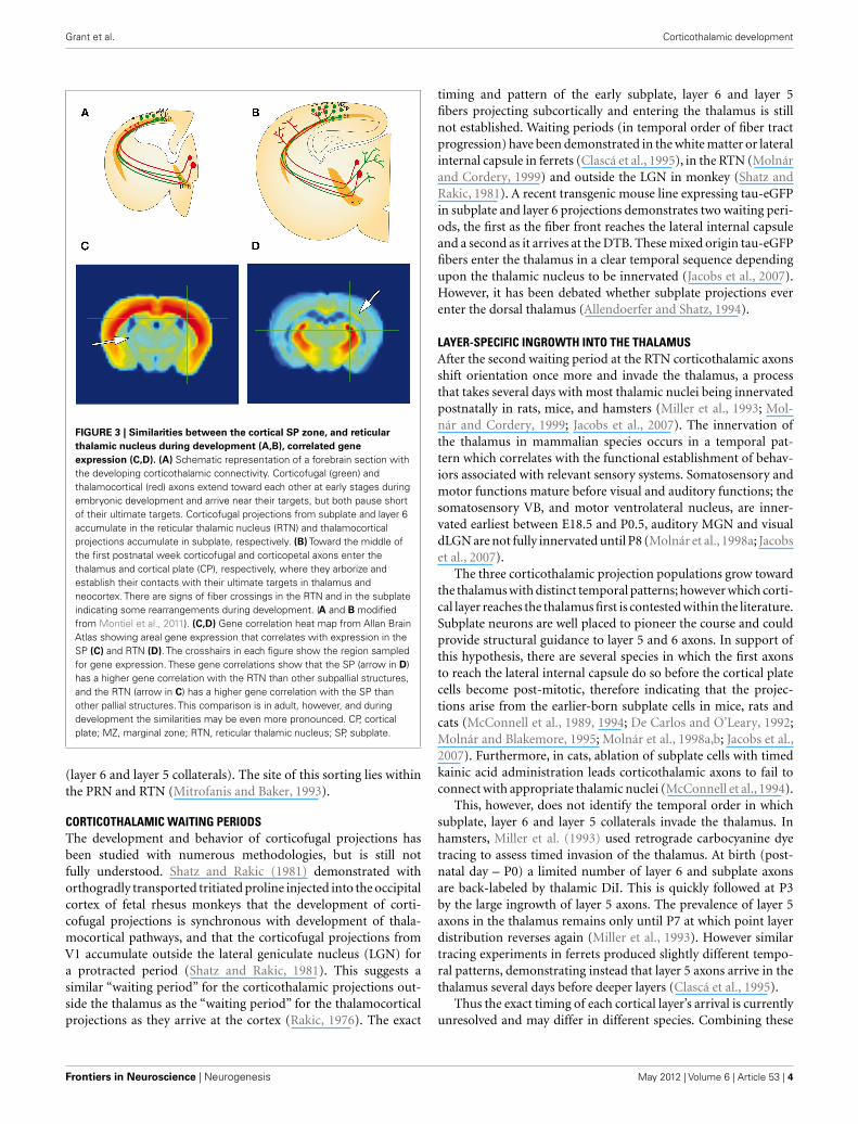
FIGURE 3 | Similarities between the cortical SP zone, and reticular
thalamic nucleus during development (A,B), correlated gene
expression (C,D). (A) Schematic representation of a forebrain section withthe developing corticothalamic connectivity. Corticofugal (green) andthalamocortical (red) axons extend toward each other at early stages duringembryonic development and arrive near their targets, but both pause shortof their ultimate targets. Corticofugal projections from subplate and layer 6accumulate in the reticular thalamic nucleus (RTN) and thalamocorticalprojections accumulate in subplate, respectively. (B) Toward the middle ofthe first postnatal week corticofugal and corticopetal axons enter thethalamus and cortical plate (CP), respectively, where they arborize andestablish their contacts with their ultimate targets in thalamus andneocortex. There are signs of fiber crossings in the RTN and in the subplateindicating some rearrangements during development. (A and B modifiedfrom Montiel et al., 2011). (C,D) Gene correlation heat map from Allan BrainAtlas showing areal gene expression that correlates with expression in theSP (C) and RTN (D). The crosshairs in each figure show the region sampledfor gene expression. These gene correlations show that the SP (arrow in D)has a higher gene correlation with the RTN than other subpallial structures,and the RTN (arrow in C) has a higher gene correlation with the SP thanother pallial structures. This comparison is in adult, however, and duringdevelopment the similarities may be even more pronounced. CP, corticalplate; MZ, marginal zone; RTN, reticular thalamic nucleus; SP, subplate.
(layer 6 and layer 5 collaterals). The site of this sorting lies withinthe PRN and RTN (Mitrofanis and Baker, 1993).
CORTICOTHALAMIC WAITING PERIODSThe development and behavior of corticofugal projections hasbeen studied with numerous methodologies, but is still notfully understood. Shatz and Rakic (1981) demonstrated withorthogradly transported tritiated proline injected into the occipitalcortex of fetal rhesus monkeys that the development of corti-cofugal projections is synchronous with development of thala-mocortical pathways, and that the corticofugal projections fromV1 accumulate outside the lateral geniculate nucleus (LGN) fora protracted period (Shatz and Rakic, 1981). This suggests asimilar “waiting period” for the corticothalamic projections out-side the thalamus as the “waiting period” for the thalamocorticalprojections as they arrive at the cortex (Rakic, 1976). The exact
timing and pattern of the early subplate, layer 6 and layer 5fibers projecting subcortically and entering the thalamus is stillnot established. Waiting periods (in temporal order of fiber tractprogression) have been demonstrated in the white matter or lateralinternal capsule in ferrets (Clascá et al., 1995), in the RTN (Molnárand Cordery, 1999) and outside the LGN in monkey (Shatz andRakic, 1981). A recent transgenic mouse line expressing tau-eGFPin subplate and layer 6 projections demonstrates two waiting peri-ods, the first as the fiber front reaches the lateral internal capsuleand a second as it arrives at the DTB. These mixed origin tau-eGFPfibers enter the thalamus in a clear temporal sequence dependingupon the thalamic nucleus to be innervated (Jacobs et al., 2007).However, it has been debated whether subplate projections everenter the dorsal thalamus (Allendoerfer and Shatz, 1994).
LAYER-SPECIFIC INGROWTH INTO THE THALAMUSAfter the second waiting period at the RTN corticothalamic axonsshift orientation once more and invade the thalamus, a processthat takes several days with most thalamic nuclei being innervatedpostnatally in rats, mice, and hamsters (Miller et al., 1993; Mol-nár and Cordery, 1999; Jacobs et al., 2007). The innervation ofthe thalamus in mammalian species occurs in a temporal pat-tern which correlates with the functional establishment of behav-iors associated with relevant sensory systems. Somatosensory andmotor functions mature before visual and auditory functions; thesomatosensory VB, and motor ventrolateral nucleus, are inner-vated earliest between E18.5 and P0.5, auditory MGN and visualdLGN are not fully innervated until P8 (Molnár et al., 1998a; Jacobset al., 2007).
The three corticothalamic projection populations grow towardthe thalamus with distinct temporal patterns; however which corti-cal layer reaches the thalamus first is contested within the literature.Subplate neurons are well placed to pioneer the course and couldprovide structural guidance to layer 5 and 6 axons. In support ofthis hypothesis, there are several species in which the first axonsto reach the lateral internal capsule do so before the cortical platecells become post-mitotic, therefore indicating that the projec-tions arise from the earlier-born subplate cells in mice, rats andcats (McConnell et al., 1989, 1994; De Carlos and O’Leary, 1992;Molnár and Blakemore, 1995; Molnár et al., 1998a,b; Jacobs et al.,2007). Furthermore, in cats, ablation of subplate cells with timedkainic acid administration leads corticothalamic axons to fail toconnect with appropriate thalamic nuclei (McConnell et al., 1994).
This, however, does not identify the temporal order in whichsubplate, layer 6 and layer 5 collaterals invade the thalamus. Inhamsters, Miller et al. (1993) used retrograde carbocyanine dyetracing to assess timed invasion of the thalamus. At birth (post-natal day – P0) a limited number of layer 6 and subplate axonsare back-labeled by thalamic DiI. This is quickly followed at P3by the large ingrowth of layer 5 axons. The prevalence of layer 5axons in the thalamus remains only until P7 at which point layerdistribution reverses again (Miller et al., 1993). However similartracing experiments in ferrets produced slightly different tempo-ral patterns, demonstrating instead that layer 5 axons arrive in thethalamus several days before deeper layers (Clascá et al., 1995).
Thus the exact timing of each cortical layer’s arrival is currentlyunresolved and may differ in different species. Combining these
Frontiers in Neuroscience | Neurogenesis May 2012 | Volume 6 | Article 53 | 4
Grant et al. Corticothalamic development
results suggest that whilst subplate projections leave the cortexfirst, layer 5 projections may be the first to innervate thalamus,followed by layer 6. It is tempting to speculate that the extratime taken by subplate axons is a result of some rearrangementof representation during this period. How this rearrangement iscontrolled during the waiting period, or whether it is modulatedby the input from the sensory periphery is not yet understood.The layer-specific timing of cortical innervation to the thalamussuggests future work should address questions of waiting perioddifferences; do both layer 5 and layer 6 undergo the same wait-ing periods or does one population wait whilst the other forgesahead?
RECENT ADVANCES USING REPORTER GENE EXPRESSING LINESThe inability to label subplate, layer 6 and layer 5 neurons and theirneurites selectively, hinders our understanding of the developmen-tal integration of these neurons into the intra- and extra-corticalcircuitry. This is now rapidly changing. Due to the advances inmolecular taxonomy of cortical neurons, we have more tools toanalyze circuits (Molnár and Cheung,2006; Molyneaux et al., 2007;Hoerder-Suabedissen et al., 2009). These tools include subplatespecific transgenic GFP animals, the Lpar1-eGFP (formerly Edg2-eGFP) mouse, the Golli-tau-eGFP mouse, and the CTGF-eGFPmouse (Jacobs et al., 2007; Hoerder-Suabedissen and Molnár,
2012b) (Figure 4). These mice express GFP primarily in the sub-plate. Below we present our work characterizing the subplate cellslabeled in these animals.
Co-localizing GFP with neuronal and subplate markersrevealed distinct, although overlapping, subpopulations within thesubplate (Hoerder-Suabedissen and Molnár, 2012b). These sub-populations display different patterns of growth into the thalamicnuclei (Figure 5). Lpar1-eGFP fibers have not reached the RTN byP2. By P6 GFP+ fibers have entered VB in a pattern which sug-gests they innervate the hollows of the barreloids. No fibers haveentered the LGN. By P14 the VB and LGN have been innervated.
In contrast Golli-tau-eGFP cortical fibers have different growthkinetics. At P2 the GFP+ fiber front is at the RTN and many fiberscan be seen clearly entering VB. By P6 the fibers have fully enteredVB and are patterned in the septa between barreloids. At this agethe dLGN is not innervated, however, the first fibers are accumu-lating between VB and the ventral edge of the dLGN. At P14 theGFP+ fibers have innervated VB in the hollows of barreloids. ThedLGN is now completely innervated although the vLGN is not(Figure 5).
Retrograde carbocyanine dye tracing at P8 demonstrates only7% of the cells back-labeled from the thalamus are GFP+ in theLpar1-eGFP mouse whereas 50% of back-labeled cells are GFP+in the Golli-tau-eGFP mouse (Figure 6).
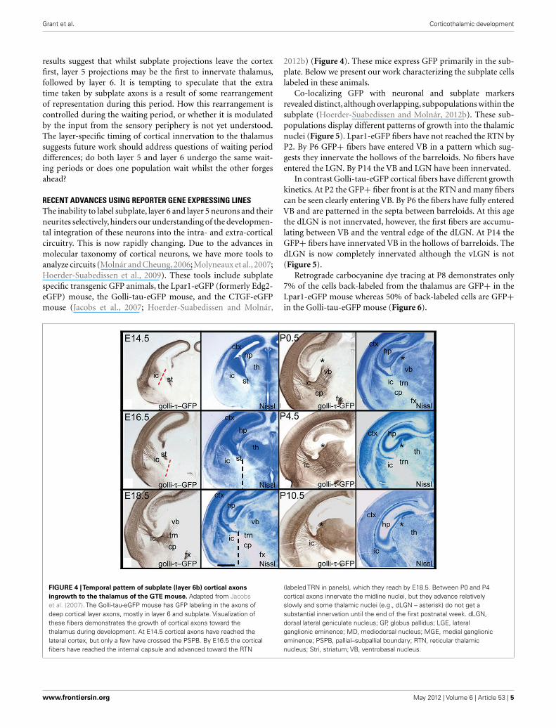
FIGURE 4 |Temporal pattern of subplate (layer 6b) cortical axons
ingrowth to the thalamus of the GTE mouse. Adapted from Jacobset al. (2007). The Golli-tau-eGFP mouse has GFP labeling in the axons ofdeep cortical layer axons, mostly in layer 6 and subplate. Visualization ofthese fibers demonstrates the growth of cortical axons toward thethalamus during development. At E14.5 cortical axons have reached thelateral cortex, but only a few have crossed the PSPB. By E16.5 the corticalfibers have reached the internal capsule and advanced toward the RTN
(labeled TRN in panels), which they reach by E18.5. Between P0 and P4cortical axons innervate the midline nuclei, but they advance relativelyslowly and some thalamic nuclei (e.g., dLGN – asterisk) do not get asubstantial innervation until the end of the first postnatal week. dLGN,dorsal lateral geniculate nucleus; GP, globus pallidus; LGE, lateralganglionic eminence; MD, mediodorsal nucleus; MGE, medial ganglioniceminence; PSPB, pallial–subpallial boundary; RTN, reticular thalamicnucleus; Stri, striatum; VB, ventrobasal nucleus.
www.frontiersin.org May 2012 | Volume 6 | Article 53 | 5
Grant et al. Corticothalamic development
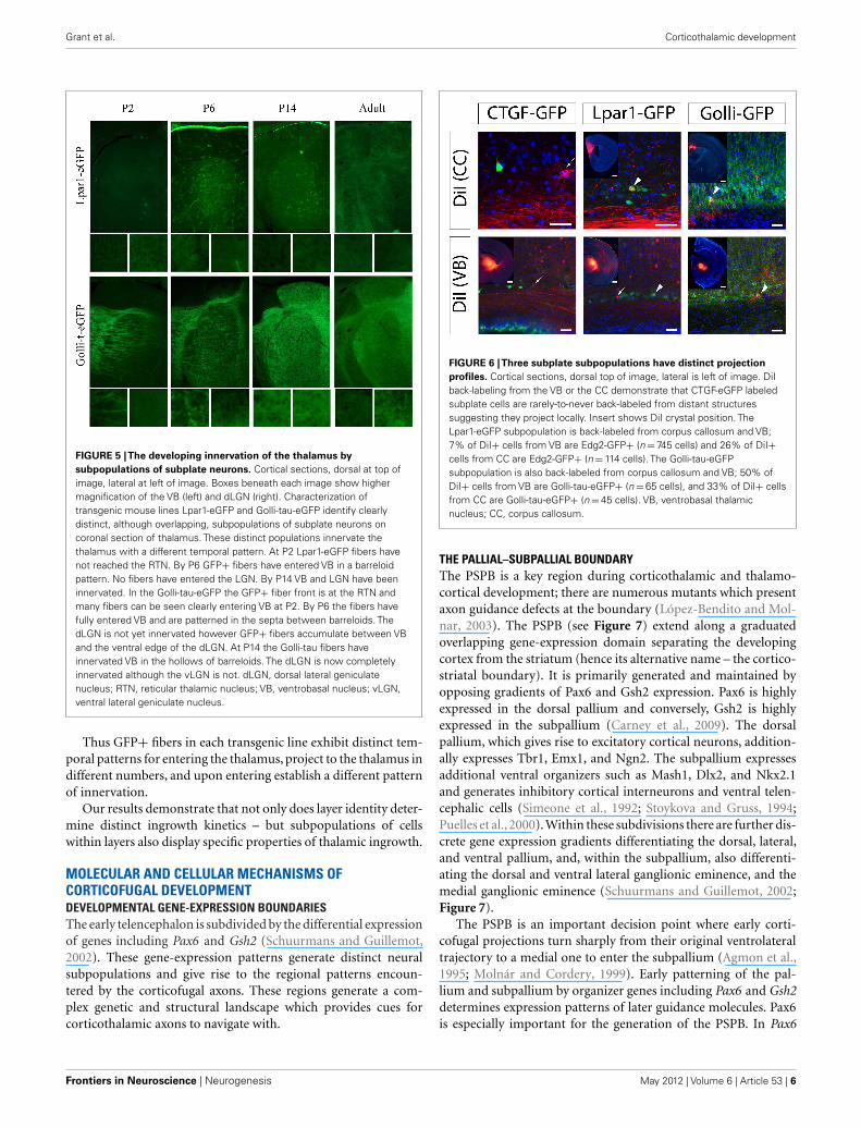
FIGURE 5 |The developing innervation of the thalamus by
subpopulations of subplate neurons. Cortical sections, dorsal at top ofimage, lateral at left of image. Boxes beneath each image show highermagnification of the VB (left) and dLGN (right). Characterization oftransgenic mouse lines Lpar1-eGFP and Golli-tau-eGFP identify clearlydistinct, although overlapping, subpopulations of subplate neurons oncoronal section of thalamus. These distinct populations innervate thethalamus with a different temporal pattern. At P2 Lpar1-eGFP fibers havenot reached the RTN. By P6 GFP+ fibers have entered VB in a barreloidpattern. No fibers have entered the LGN. By P14 VB and LGN have beeninnervated. In the Golli-tau-eGFP the GFP+ fiber front is at the RTN andmany fibers can be seen clearly entering VB at P2. By P6 the fibers havefully entered VB and are patterned in the septa between barreloids. ThedLGN is not yet innervated however GFP+ fibers accumulate between VBand the ventral edge of the dLGN. At P14 the Golli-tau fibers haveinnervated VB in the hollows of barreloids. The dLGN is now completelyinnervated although the vLGN is not. dLGN, dorsal lateral geniculatenucleus; RTN, reticular thalamic nucleus; VB, ventrobasal nucleus; vLGN,ventral lateral geniculate nucleus.
Thus GFP+ fibers in each transgenic line exhibit distinct tem-poral patterns for entering the thalamus, project to the thalamus indifferent numbers, and upon entering establish a different patternof innervation.
Our results demonstrate that not only does layer identity deter-mine distinct ingrowth kinetics – but subpopulations of cellswithin layers also display specific properties of thalamic ingrowth.
MOLECULAR AND CELLULAR MECHANISMS OFCORTICOFUGAL DEVELOPMENTDEVELOPMENTAL GENE-EXPRESSION BOUNDARIESThe early telencephalon is subdivided by the differential expressionof genes including Pax6 and Gsh2 (Schuurmans and Guillemot,2002). These gene-expression patterns generate distinct neuralsubpopulations and give rise to the regional patterns encoun-tered by the corticofugal axons. These regions generate a com-plex genetic and structural landscape which provides cues forcorticothalamic axons to navigate with.
FIGURE 6 |Three subplate subpopulations have distinct projection
profiles. Cortical sections, dorsal top of image, lateral is left of image. DiIback-labeling from the VB or the CC demonstrate that CTGF-eGFP labeledsubplate cells are rarely-to-never back-labeled from distant structuressuggesting they project locally. Insert shows DiI crystal position. TheLpar1-eGFP subpopulation is back-labeled from corpus callosum and VB;7% of DiI+ cells from VB are Edg2-GFP+ (n = 745 cells) and 26% of DiI+cells from CC are Edg2-GFP+ (n = 114 cells). The Golli-tau-eGFPsubpopulation is also back-labeled from corpus callosum and VB; 50% ofDiI+ cells from VB are Golli-tau-eGFP+ (n = 65 cells), and 33% of DiI+ cellsfrom CC are Golli-tau-eGFP+ (n = 45 cells). VB, ventrobasal thalamicnucleus; CC, corpus callosum.
THE PALLIAL–SUBPALLIAL BOUNDARYThe PSPB is a key region during corticothalamic and thalamo-cortical development; there are numerous mutants which presentaxon guidance defects at the boundary (López-Bendito and Mol-nar, 2003). The PSPB (see Figure 7) extend along a graduatedoverlapping gene-expression domain separating the developingcortex from the striatum (hence its alternative name – the cortico-striatal boundary). It is primarily generated and maintained byopposing gradients of Pax6 and Gsh2 expression. Pax6 is highlyexpressed in the dorsal pallium and conversely, Gsh2 is highlyexpressed in the subpallium (Carney et al., 2009). The dorsalpallium, which gives rise to excitatory cortical neurons, addition-ally expresses Tbr1, Emx1, and Ngn2. The subpallium expressesadditional ventral organizers such as Mash1, Dlx2, and Nkx2.1and generates inhibitory cortical interneurons and ventral telen-cephalic cells (Simeone et al., 1992; Stoykova and Gruss, 1994;Puelles et al., 2000). Within these subdivisions there are further dis-crete gene expression gradients differentiating the dorsal, lateral,and ventral pallium, and, within the subpallium, also differenti-ating the dorsal and ventral lateral ganglionic eminence, and themedial ganglionic eminence (Schuurmans and Guillemot, 2002;Figure 7).
The PSPB is an important decision point where early corti-cofugal projections turn sharply from their original ventrolateraltrajectory to a medial one to enter the subpallium (Agmon et al.,1995; Molnár and Cordery, 1999). Early patterning of the pal-lium and subpallium by organizer genes including Pax6 and Gsh2determines expression patterns of later guidance molecules. Pax6is especially important for the generation of the PSPB. In Pax6
Frontiers in Neuroscience | Neurogenesis May 2012 | Volume 6 | Article 53 | 6
Grant et al. Corticothalamic development
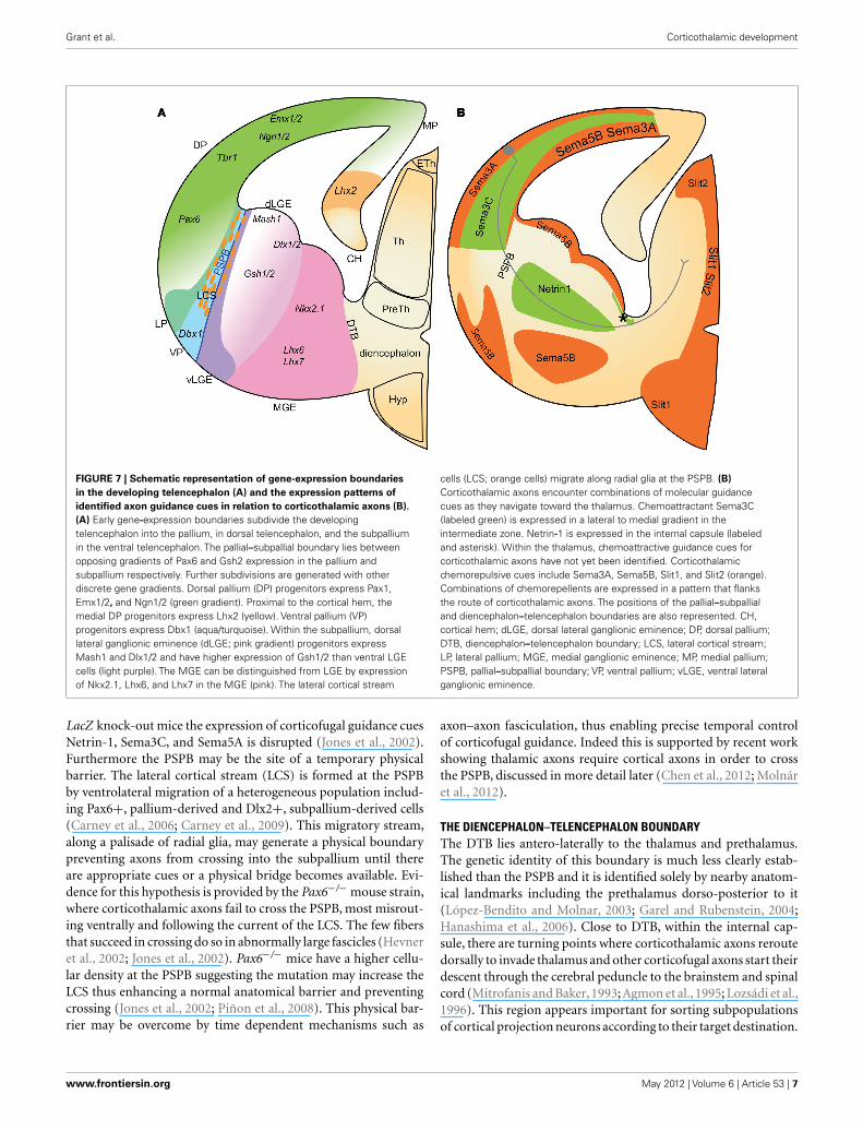
FIGURE 7 | Schematic representation of gene-expression boundaries
in the developing telencephalon (A) and the expression patterns of
identified axon guidance cues in relation to corticothalamic axons (B).
(A) Early gene-expression boundaries subdivide the developingtelencephalon into the pallium, in dorsal telencephalon, and the subpalliumin the ventral telencephalon. The pallial–subpallial boundary lies betweenopposing gradients of Pax6 and Gsh2 expression in the pallium andsubpallium respectively. Further subdivisions are generated with otherdiscrete gene gradients. Dorsal pallium (DP) progenitors express Pax1,Emx1/2, and Ngn1/2 (green gradient). Proximal to the cortical hem, themedial DP progenitors express Lhx2 (yellow). Ventral pallium (VP)progenitors express Dbx1 (aqua/turquoise). Within the subpallium, dorsallateral ganglionic eminence (dLGE; pink gradient) progenitors expressMash1 and Dlx1/2 and have higher expression of Gsh1/2 than ventral LGEcells (light purple). The MGE can be distinguished from LGE by expressionof Nkx2.1, Lhx6, and Lhx7 in the MGE (pink). The lateral cortical stream
cells (LCS; orange cells) migrate along radial glia at the PSPB. (B)
Corticothalamic axons encounter combinations of molecular guidancecues as they navigate toward the thalamus. Chemoattractant Sema3C(labeled green) is expressed in a lateral to medial gradient in theintermediate zone. Netrin-1 is expressed in the internal capsule (labeledand asterisk). Within the thalamus, chemoattractive guidance cues forcorticothalamic axons have not yet been identified. Corticothalamicchemorepulsive cues include Sema3A, Sema5B, Slit1, and Slit2 (orange).Combinations of chemorepellents are expressed in a pattern that flanksthe route of corticothalamic axons. The positions of the pallial–subpallialand diencephalon–telencephalon boundaries are also represented. CH,cortical hem; dLGE, dorsal lateral ganglionic eminence; DP, dorsal pallium;DTB, diencephalon–telencephalon boundary; LCS, lateral cortical stream;LP, lateral pallium; MGE, medial ganglionic eminence; MP, medial pallium;PSPB, pallial–subpallial boundary; VP, ventral pallium; vLGE, ventral lateralganglionic eminence.
LacZ knock-out mice the expression of corticofugal guidance cuesNetrin-1, Sema3C, and Sema5A is disrupted (Jones et al., 2002).Furthermore the PSPB may be the site of a temporary physicalbarrier. The lateral cortical stream (LCS) is formed at the PSPBby ventrolateral migration of a heterogeneous population includ-ing Pax6+, pallium-derived and Dlx2+, subpallium-derived cells(Carney et al., 2006; Carney et al., 2009). This migratory stream,along a palisade of radial glia, may generate a physical boundarypreventing axons from crossing into the subpallium until thereare appropriate cues or a physical bridge becomes available. Evi-dence for this hypothesis is provided by the Pax6−/− mouse strain,where corticothalamic axons fail to cross the PSPB, most misrout-ing ventrally and following the current of the LCS. The few fibersthat succeed in crossing do so in abnormally large fascicles (Hevneret al., 2002; Jones et al., 2002). Pax6−/− mice have a higher cellu-lar density at the PSPB suggesting the mutation may increase theLCS thus enhancing a normal anatomical barrier and preventingcrossing (Jones et al., 2002; Piñon et al., 2008). This physical bar-rier may be overcome by time dependent mechanisms such as
axon–axon fasciculation, thus enabling precise temporal controlof corticofugal guidance. Indeed this is supported by recent workshowing thalamic axons require cortical axons in order to crossthe PSPB, discussed in more detail later (Chen et al., 2012; Molnáret al., 2012).
THE DIENCEPHALON–TELENCEPHALON BOUNDARYThe DTB lies antero-laterally to the thalamus and prethalamus.The genetic identity of this boundary is much less clearly estab-lished than the PSPB and it is identified solely by nearby anatom-ical landmarks including the prethalamus dorso-posterior to it(López-Bendito and Molnar, 2003; Garel and Rubenstein, 2004;Hanashima et al., 2006). Close to DTB, within the internal cap-sule, there are turning points where corticothalamic axons reroutedorsally to invade thalamus and other corticofugal axons start theirdescent through the cerebral peduncle to the brainstem and spinalcord (Mitrofanis and Baker, 1993; Agmon et al., 1995; Lozsádi et al.,1996). This region appears important for sorting subpopulationsof cortical projection neurons according to their target destination.
www.frontiersin.org May 2012 | Volume 6 | Article 53 | 7

Grant et al. Corticothalamic development
The DTB may also have similar barrier functions to the PSPB.Disruption of pioneer axons or fasciculation with thalamic axonsprevents corticothalamic axons from entering the diencephalon orinvading the thalamus correctly (McConnell et al., 1994; Hevneret al., 2002).
These developmental gene-expression boundaries contributeto corticothalamic guidance in three ways. (1) Generating physi-cal boundaries that can be appropriately permissive or restrictivedepending on developmental stage. (2) Acting as decision pointsfor major trajectory changes. (3) Setting up important patterns ofguidance molecules. The full extent of the contribution to guid-ance that these developmental gene-expression boundaries makeis still under investigation. As is the importance of the temporalchoreography of the development of functional corticothalamiccircuits (Molnár et al., 2012).
STRUCTURAL GUIDANCE CUESSince the first description of the early corticofugal axon frontextending toward the thalamus (McConnell et al., 1989) a pos-sible role of structural guidance cues in thalamocortical axonguidance has been postulated. These ideas were formulated asthe “Handshake Hypothesis” (Blakemore and Molnar, 1990; Mol-nár and Blakemore, 1995; Molnár et al., 1998a,b), which suggeststhat early corticofugal and thalamocortical projections meet atthe PSPB and the early corticofugal projections aid the thalamicfibers to cross through this region toward the cortex. The relation-ship between early corticofugal and thalamic projections has beendebated. It has been suggested that they fasciculate with each otherin internal capsule and intermediate zone (Molnár and Blakemore,1995; Molnár et al., 1998a,b); but other studies suggest that theyrun in separate compartments (Miller et al., 1993) or interdigitateonly in a restricted portion of their path (Bicknese et al., 1994).Some of these issues are related to the difficulties of deliveringpaired tracers into the equivalent regions of the cortex and thethalamus. The Golli-tau-eGFP mouse model (see above) demon-strates the intimate association of early corticofugal projectionswith thalamic afferents (Piñon et al., 2005) from the intermediatezone, PSPB and also throughout the lateral sector of the internalcapsule.
Several mutants with thalamocortical guidance defects also dis-play aberrant development of corticofugal projections (Hevneret al., 2002; López-Bendito and Molnar, 2003). Mutation of thethalamic gene Gbx2 causes a reduced projection of thalamocorti-cal axons into the internal capsule. Subsequently, corticothalamicaxons fail to enter the diencephalon from the subpallium, thus sug-gesting that thalamic axons provide some cue to corticothalamicaxons that may include physical fasciculation to help them crossthe DTB (Hevner et al., 2002). Recent research has demonstratedthat structural support from populations of other axons can becrucial to crossing early developmental gene boundaries (Chenet al., 2012). When cortical fibers are absent and thus do not pro-vide structural support at the PSPB, thalamic axons in Emx1cre;Apcloxp/loxp mouse fail to reach the cortex although the mutantcortex remains attractive to the thalamic fibers (Chen et al., 2012).The authors demonstrate that the phenotype can be rescued bythe replacement of cortical fibers across the PSPB. As such theuse of structural support from other fiber populations to cross
gene boundaries is important and may be contributing to corticalcrossing at the DTB.
Due to a better understanding of forebrain patterning and theavailability of conditional knock-outs, there is a recent revival offocus on structural guidance cues to overcome physical barriersin the telencephalon. Zhou et al. (2008, 2009) have demonstratedthat region specific Celsr3 inactivation affects development of theinternal capsule in different ways. In Celsr3|Foxg1 mice the inter-nal capsule is defective and thalamic axons either cross to thecontralateral diencephalon or descend to the ventral surface of thetelencephalon. In Celsr3|Dlx5/6 mice, the internal capsule is alsoabnormal and thalamic fibers are misrouted to the amygdala. Fur-thermore, the early corticofugal axons fail to advance toward thethalamus, and instead stop at the PSPB, forming a mass resem-bling an amputational neuroma. In Celsr3|Emx1 mice, the internalcapsule is intact and there are normal thalamocortical connections(Zhou et al., 2008).
The molecular and cellular nature of these interactions is notunderstood. This limits the interpretation even in these condi-tional knockouts. The cell surface proteins involved in fascicula-tion have not been identified and it appears bidirectional signalingas well as just fasciculation may be important for axonal guidance(Hevner et al., 2002).
CORRIDOR CELLSEarly work on corticothalamic development identified a popu-lation of cells in the internal capsule, the perireticular cells ofthe PRN. Their position coincides with the point where corti-cothalamic axons deflect dorsally into the prethalamus and wherelayer 5 axon branches are sorted to project to thalamus or to sub-cerebral targets (Mitrofanis and Baker, 1993). These cells werepreviously suggested have a role in guiding the corticothalamicaxons given the major rearrangement behavior upon reachingthem (Mitrofanis and Guillery, 1993).
Since then a second population of cells named corridor cells,derived from the lateral ganglionic eminence, have been demon-strated to generate a critical permissive corridor, within the restric-tive medial ganglionic eminence (MGE) (López-Bendito et al.,2006). Without these cells thalamocortical axons are repelled byMGE and fail to properly navigate to the cortex. These cells mayalso be required to generate a permissive substrate for corticalaxons to grow across similar to their support of thalamic axons.Furthermore back-labeling has suggested these cells may overlapwith the perireticular neurons and so may contribute to guidingcorticothalamic axons and sorting layer 5 thalamic and subcerebralprojections. This will become apparent with further research.
MOLECULAR GUIDANCE CUESRecent advances have identified major families of well-knownguidance molecules in the guidance of corticothalamic axons(Figure 7B).
Members of the semaphorin family provide several early, con-text dependent cues, and mutations of these genes generate subtlephenotypes suggesting multiple cues collaborate at each stage ofguidance. Corticofugal axons are repelled from Sema3A expressingcortical plate and attracted toward the Sema3C expressing inter-mediate zone (Bagnard et al., 1998, 2001; Skaliora et al., 1998).
Frontiers in Neuroscience | Neurogenesis May 2012 | Volume 6 | Article 53 | 8
Grant et al. Corticothalamic development
The intermediate zone forms a permissive lane between the corticalplate and the ventricular zone as Sema3A, along with Sema5B, pre-vents cortical axons overshooting into the cortical germinal zone(Bagnard et al., 1998; Lett et al., 2009). Sema3A also attracts cor-tical dendrites. The asymmetric distribution of cellular guanylatecyclase enables different responses of cortical axons and dendritesto Sema3A (Polleux et al., 2000).
The lateral-to-medial gradient of Sema3C attracts corticothal-amic axons extending within the intermediate zone toward thelateral cortex (Bagnard et al., 1998, 2000). The complementarymedial-to-lateral gradient of expression of Sema3A in the ven-tricular zone may also repel corticofugal axons coming from themedial cortex.
Chemoattractant Netrin-1 is expressed in the internal cap-sule and ventral telencephalon. This is complementary to Dccexpression (Netrin-1 attraction receptor) in corticothalamic axons(Oeschger et al., 2011). In vitro, Netrin-1 mediates long rangeattraction to E12.5 and E13.5 corticothalamic axons (Métin et al.,1997; Richards et al., 1997). This attraction can induce turning andtherefore appears responsible for corticofugal growth cone reori-entation toward the ventral telencephalon (Métin et al., 1997).Chemorepulsion may also guide turning due to Sema5B expres-sion in the lateral cortex flanking the route of axons that cross thePSPB (Skaliora et al., 1998; Lett et al., 2009).
Ensuring that axons remain within the internal capsule involvesseveral chemorepulsive interactions. During development Sema5Bexpression in the germinal zones of the ganglionic eminences andthe globus pallidus borders the corticothalamic path through theinternal capsule (Skaliora et al., 1998; Lett et al., 2009). Corti-cothalamic explants are repelled by Sema5B expressing cells andectopic Sema5B in the internal capsule causes cortical axons tostall at the new Sema5B boundary. Furthermore RNA interfer-ence against Sema5B causes aberrant entry of cortical axons intothe germinal zones (Lett et al., 2009). Thus Sema5B restricts thegrowth of cortical axons to their appropriate trajectory.
Inhibitory cell surface molecules Slit1 and 2, and receptorsRobo1 and 2, also mediate the guidance of the corticothalamicaxons within the ventral telencephalon and diencephalon. Slit1and 2 are expressed in overlapping domains including the gan-glionic eminences, prethalamus, hypothalamus, and the germinalzone of the dorsal thalamus (Braisted et al., 2000). Robo1 and 2 areexpressed in complementary patterns in the cortical plate, inter-mediate zone, and dorsal thalamus (López-Bendito et al., 2007). InSlit2 mutants, Slit1 and 2 double mutants, and Robo1 and 2 doublemutants corticothalamic guidance is disrupted with the majorityof corticofugal fibers continuing ventrally instead of turning at thePSPB, some reaching the basal telencephalic surface. Those whichdo correctly enter the ventral telencephalon then aberrantly crossthe ventral midline (Bagri et al., 2002; López-Bendito et al., 2007).
Slit pathway components therefore ensure containment of cor-ticothalamic axons within the internal capsule and direct corti-cothalamic axons dorsally upon reaching the DTB in order toenter the thalamus rather than crossing the midline (Bagri et al.,2002; López-Bendito et al., 2007; Braisted et al., 2009).
Guidance cues, which direct axons from specific cortical regionsto the thalamic nuclei that they connect to in adulthood, areyet to be elucidated; however candidate cues are beginning tobe identified. Using microarrays, Sur and colleagues identify
gene-expression differences between the LGN and MGN. Axonguidance molecules including Ephs and ephrins, semaphorins, slitsand netrin pathways were differentially expressed between the twonuclei (Horng et al., 2009). Furthermore cues EphA7 and Ntrk2expression is up-regulated in both the LGN and the rewired MGN,in which after peripheral ablation of auditory nerves, the ingrow-ing retinal axons invade the MGN and the LGN. Thus distinctguidance cue expression may contribute to the specific neural con-nectivity between thalamic nuclei. Indeed a review in this researchtopic proposes the hypothesis that overlapping molecular expres-sion in the thalamus may be responsible for the determinationof areal axon guidance from thalamus to the cortex (Price et al.,2012). These overlapping and combinatorial gene-expression pat-terns may also be responsible for organizing cortical axon guidanceinto specific thalamic nuclei.
Molecular control of temporal dynamics such as the waitingperiods has proved harder to elucidate although recent discoveriesare beginning to suggest answers. Robo1 is expressed by corti-cothalamic neurons. It appears to act as a molecular slowing signalas Robo1−/− corticothalamic axons reach their targets a day early(Andrews et al., 2006). It appears this slowing signal is not medi-ated by the canonical Slit–Robo interaction as Slit2 and Slit 1and 2 double mutants have a different phenotype in which mostcorticothalamic axons fail to reach the thalamus rather than beingdelayed and Slit1 mutants do not have corticothalamic phenotypes(Bagri et al., 2002).
Signals that might cause different waiting periods for dis-tinct populations of corticothalamic axons are currently unclear.Guidance pathway molecules, including Unc5c, differ between thesubplate and lower cortical plate (Oeschger et al., 2011). Unc5c isa receptor which mediates repulsion to soluble Netrin-1. It is tran-siently up-regulated in subplate cells compared to layer 5/layer6. This up-regulation coincides temporally with the first waitingperiod that corticofugal fibers undergo (E14.5 in mouse). Unc5chas been demonstrated to produce a waiting period during theguidance of primary sensory axons to the spinal cord (Watanabeet al., 2006).
Switching responses to Netrin 1 requires cortical axons to altertheir molecular expression after crossing the PSPB. Fluctuatingexpression of guidance cue receptors over time allows a pop-ulation of axons to grow through different compartments andonly respond to relevant cues. We suggest a temporal pattern ofreceptor expression as seen with Unc5c may generate the wait-ing periods and specific temporal growth patterns. Furthermoredifferential expression of guidance cue receptors between popula-tions of corticothalamic axons generates specific responses of eachpopulation to each cue. We propose such molecular differencesmay generate different waiting behavior in corticothalamic axonpopulations. Once layer-specific waiting periods are recognizedthis may provide insight into how temporal control helps corticalneurons path-find to the correct thalamic nucleus.
Many major molecular cues guiding corticothalamic axonshave been identified. Complex combinations of various cuesgenerate the specific and detailed connectivity patterns thatcharacterize the connections between cortex and thalamus.
There are likely to be many more, and subtler, cues involvedin the precise details of the developmental events. For exam-ple cortical layer-specific competency to respond to guidance
www.frontiersin.org May 2012 | Volume 6 | Article 53 | 9
Grant et al. Corticothalamic development
cues has not been addressed. Future work should determine cor-tical layer-specific or even neuronal subtype specific ingrowthto understand the relevance of the stage and subtype specificinnervation of different thalamic nuclei by axons from partic-ular cortical areas. Selective subplate, layer 6 and layer 5 geneexpression profiling at the time of major decisions in axongrowth would help to resolve some of these issues. Further-more greater understanding of the sequence of circuit forma-tion might give insight into self-organizing mechanisms dur-ing development. In addition to molecular cues there may bean activity related component although so far no phenotypehas been described in various SNARE complex knock-out micemutant (Washbourne et al., 2002; Molnár et al., 2002) or cocul-tures (Blakey et al., 2012). Furthermore evidence so far hasshown no phenotype of early corticothalamic innervation in theGolli-taueGFP mouse after peripheral manipulations (Grant andMolnár, 2012).
UNDERSTANDING TRANSIENT CIRCUITS IN CORTEXThe role of transient circuits involving subplate neurons has beendemonstrated in the developing visual cortex during ocular dom-inance formation (Ghosh et al., 1990) and orientation columnformation (Kanold et al., 2003) in carnivores. Similar mecha-nisms might operate in the barrel field of the mouse primarysomatosensory cortex (Piñon et al., 2009; Tolner et al., 2012).Thalamic axons reach the intermediate zone and subplate sev-eral days prior to innervating the cortex. During this accumu-lation of thalamic axons, layer 4 exhibits di-synaptic activationin response to thalamic fiber excitation suggesting that thalamicaxons synapse with the subplate neurons that synapse onto layer4 neurons. After 2 days thalamic activation of cortical layer 4 neu-rons is monosynaptic reflecting direct thalamic innervation (Zhaoet al., 2009). Furthermore subplate ablation in cats stops normalup-regulation of glutamate receptor subunit, GluR1, in corticaldendrites thus leading to reduced strength at the thalamocorti-cal synapse (Kanold et al., 2003). Selective ablation of subplatebeneath limb or barrel cortex in rat confirms these results; abla-tion abolishes spontaneous and evoked spindle burst activity inlimb cortex in vivo and thalamocortical connections to layer 4 areweaker than controls in vitro (Tolner et al., 2012). Therefore thisearly developmental feedforward innervation via the subplate isproposed to strengthen and stabilize the developing thalamic tolayer 4 synapses.
This transient circuit also regulates maturation of cortical inhi-bition and ocular dominance columns. Subplate ablation disruptsneuronal receptor profile maturation. Cortical neurons fail toup-regulate KCC2 channel expression. Without KCC2 neuronsmaintain high internal chloride concentrations and GABA recep-tor activation continues to depolarize, rather than hyperpolarize,the membrane (Kanold et al., 2003). The loss of proper cor-tical inhibition causes paradoxical effects on ocular dominancecolumns; subplate ablation causes monocular deprivation to favorthe deprived eye (Kanold et al., 2003; Kanold and Shatz, 2006).By adulthood the subplate circuit is transformed or dismantled,a large proportion of the early born subplate neurons have died(Price et al., 1997; Hoerder-Suabedissen and Molnár, 2012b), butsome survive into adulthood as layer 6b.
We have little understanding of the mode of integration ofsubplate neurites into the cortical plate prior, during, and afterthalamic innervation. Axonal and/or dendritic remodeling asso-ciated with thalamocortical ingrowth and periphery related pat-terning has recently been demonstrated by studies of single cellmorphology (Hoerder-Suabedissen and Molnár, 2012a,b). Theestablishment of area-specific thalamocortical connections is alsoconsidered to be dependent on early circuits involving subplateneurons (Molnár and Blakemore, 1995; Catalano and Shatz, 1998;Shimogori and Grove,2005). Shimogori and Grove (2005) demon-strated that thalamocortical projections could be shifted to differ-ent cortical areas by manipulating cortical gene-expression pat-terns. The site of these shifts was identified in subplate and whitematter.
UNDERSTANDING TRANSIENT CIRCUITS IN THALAMUSThe interactions between the early corticofugal projections andthe PRN and RTN are even less understood. A critical questionwhich remains to be addressed is how corticothalamic axons inte-grate into functional circuits with thalamic and reticular (RTN)neurons.
The potential role of transient circuits in cortical connectionsto the thalamus has not been probed. As discussed previously thesubplate shares many similarities with the PRN and RTN. Theselargely transient populations of cells, subplate and PRN, containearly born neurons, which display early mature synaptic connec-tions (Mitrofanis and Guillery, 1993; Molnár and Cordery, 1999;Cruikshank et al., 2010). Given the importance of transient circuitsinvolving the subplate, we propose transient circuits comprisingcorticothalamic axons, PRN and RTN neurons and the thalamusmay shape mature corticothalamic connections.
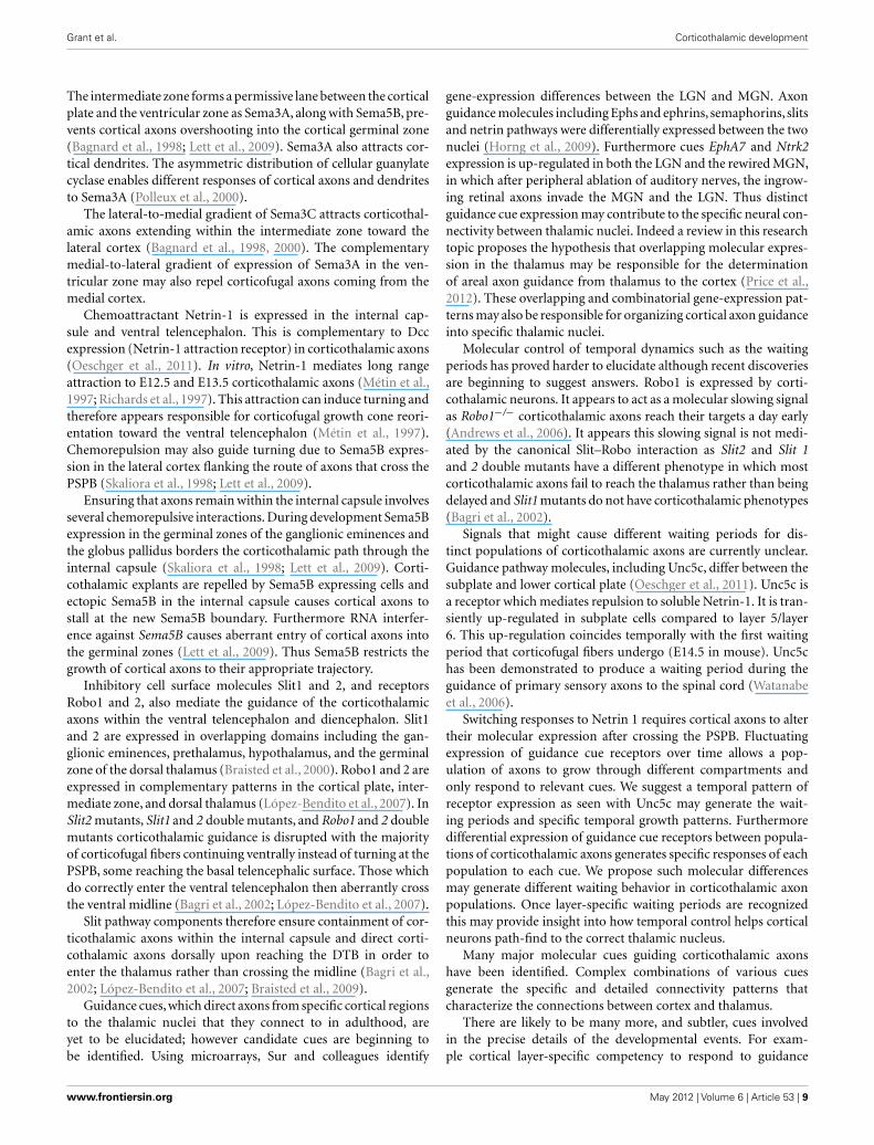
Anatomical tracing confirms that corticofugal axons project tothe RTN and reticular neurons project to the thalamus prior tocorticothalamic axons invading thalamic nuclei (Mitrofanis andBaker, 1993; Molnár et al., 1998a; Molnár and Cordery, 1999).Optical recording using voltage sensitive dyes demonstrate func-tional excitatory synapses between the cortex and RTN in earlypostnatal rat (Figure 8). This circuit may also contribute to thedepolarization seen in the ventroposterior lateral thalamic nucleus(VPL) at this age, although with this method it is difficult to dis-tinguish between direct cortical and indirect RTN activation of theVPL. In the adult the RTN to thalamus circuit, unlike the subplatecircuit, is a substrate for feedforward and feedback inhibition tothe relay cells of the thalamus (Guillery and Sherman, 2002; Jones,2002). Immediately after birth GABAergic IPSCs are recorded inmouse thalamic relay cells in response to reticular neuron activity.This inhibitory innervation increases over early postnatal weeksalthough adult properties are not fully established until P9 (War-ren and Jones, 1997; Warren et al., 1997; Evrard and Ropert, 2009).Thus at birth the circuit is already exhibiting adult features.
However the embryonic connectivity has not been studied andmay mimic the subplate’s feedforward excitation to strengthencorticothalamic connections. Indeed the RTN axons are the firstto innervate the thalamus – at E14 in rats. This is days beforeperipheral and cortical inputs (cortical innervation arrives 3 dayslater at E17) thus suggesting an important developmental role ofthe RTN-thalamic connectivity (Mitrofanis and Guillery, 1993).
Frontiers in Neuroscience | Neurogenesis May 2012 | Volume 6 | Article 53 | 10
Grant et al. Corticothalamic development
FIGURE 8 | Functional synapses revealed in the reticular thalamic
nucleus after cortical stimulation in a thalamocortical slice from a P2 rat.
A thalamocortical slice was prepared and stained with voltage sensitive dyeRH482 according to the protocols previously described in Higashi et al. (2002,2005). The slice was positioned to be able to observe the internal capsule,reticular thalamic nucleus (labeled RT in panels), part of the thalamus (VPL)and hippocampus (HIP). A stimulating electrode was placed into the whitematter below the primary somatosensory cortex. Stimulus-induced changesin the intensity of transmitted light (700 ± 30 nm) were collected with a128 × 128 pixel array of photosensors (70 μm2/pixel) every 0.6 ms (Fujifilm HRDeltaron 1700, Japan) for up to 300 ms (each pixel measured the change in
transmitted light intensity relative to a prestimulus reference image acquiredjust before the recording trial). The selected frames on the left record theresponse to stimulation after 0.6, 1.8, 3, 4.2, 5.4, 6.6, 7.8, 9, 10.2, 11.4, 17.4,and 23.4 ms; on the right the same frames are presented after the applicationof 40 μM 6,7-dinitroquinoxaline-2,3-dione (DNQX, Tocris, UK) and 50 μM2-amino-5-phosphonopentanoic acid (APV, Sigma, USA) for 20–30 min.Sustained depolarization was observed in RTN and VPL in controls which wasreduced after the DNQX and APV application, activity in RTN and VPLindicated by arrows in frames recorded at 9 and 17.4 ms in left and rightframes. HIP, hippocampus; RTN, reticular thalamic nucleus; VPL,ventro-posterior lateral thalamic nucleus.
Such questions concerning how cortical axons initially integrateinto thalamic circuits and what role the RTN contributes will beimportant in the coming years. The answers may be critical in ourunderstanding of how the developmental process may go awry inpathologies of connectivity.
CONCLUSIONClassic anatomical research has elucidated the tightly scheduledtiming and specificity of the development of corticothalamicaxons. This research demonstrates how the complexity of thecorticothalamic connection requires highly specific and combi-natorial use of guidance mechanisms during development. Fromthe start of their journey corticothalamic axons are encounter-ing cell type specific molecular cues which guide them out ofthe cortex, along the intermediate zone, across the internal cap-sule and into the thalamus. They must respond to structural cuesas they traverse developmental compartments at the PSPB andDTB which may direct gene-expression changes. Then as they
reach the prethalamus the axons interact with intermediate cellularpopulations including the perireticular/corridor cells and the RTNcells perhaps gaining both guidance instructions and integratinginto transient developmental circuits.
However there are still unresolved questions which we pro-pose lie in three key areas. Firstly cortical cell subpopulationsmust be regarded separately, distinguishing layers 5, 6 and sub-plate and subpopulations within layers, rather than gatheringfibers into heterogeneous groups which will likely have differentcues. This will enable the next level of detail in understanding thedevelopment of highly complex circuits. Secondly research mustprobe the cellular and molecular identities of the telencephalicand diencephalic regions that corticothalamic axons encounterand how this compartmental environment is important for cor-ticothalamic guidance. Thirdly work must look more closely atdevelopmental circuitry including the details of transient cir-cuitry and the balance between intrinsic guidance factors andexternal inputs. Modern techniques are now being harnessed and
www.frontiersin.org May 2012 | Volume 6 | Article 53 | 11
Grant et al. Corticothalamic development
are starting to yield results, including reporter gene expressingtransgenic mouse lines and conditional knock-out mice, in uteroelectroporation and the recent availability of population specificmarkers. These models could be further exploited after sen-sory alterations and during cross-modal plasticity in order toprobe the role of external input in generating highly specific cor-ticothalamic circuits. Understanding the logic of developmentof the cortical input to thalamus is integral to the compre-
hension of the function of the thalamus and corticothalamiccircuits.
ACKNOWLEDGMENTSWe thank Ray Guillery and John Mitrofanis for their thoughtfulcomments on earlier drafts of this review. Work in Zoltán Mol-nár’s laboratory is supported by Medical Research Council, TheWellcome Trust, EU and BBSRC.
REFERENCESAdams, N. C., Lozsádi, D. A., and
Guillery, R. W. (1997). Complexi-ties in the thalamocortical and cor-ticothalamic pathways. Eur. J. Neu-rosci. 9, 204–209.
Agmon, A., Yang, L. T., Jones, E. G., andO’Dowd, D. K. (1995). Topologicalprecision in the thalamic projectionto neonatal mouse barrel cortex. J.Neurosci. 15, 549–561.
Allendoerfer, K. L., and Shatz, C. J.(1994). The subplate, a transientneocortical structure: its role inthe development of connectionsbetween thalamus and cortex. Annu.Rev. Neurosci. 17, 185–218.
Andrews, W., Liapi, A., Plachez, C.,Camurri, L., Zhang, J., Mori, S.,Murakami, F., Parnavelas, J. G., Sun-daresan, V., and Richards, L. J.(2006). Robo1 regulates the devel-opment of major axon tracts andinterneuron migration in the fore-brain. Development 133, 2243–2252.
Auladell, C., Perez-Sust, P., Super, H.,and Soriano, E. (2000). The earlydevelopment of thalamocortical andcorticothalamic projections in themouse. Anat. Embryol. 201, 169–179.
Bagnard, D., Chounlamountri, N.,Puschel, A. W., and Bolz, J. (2001).Axonal surface molecules act incombination with semaphorin 3aduring the establishment of corti-cothalamic projections. Cereb. Cor-tex 11, 278–285.
Bagnard, D., Lohrum, M., Uziel, D.,Puschel, A. W., and Bolz, J. (1998).Semaphorins act as attractive andrepulsive guidance signals during thedevelopment of cortical projections.Development 125, 5043–5053.
Bagnard, D., Thomasset, N., Lohrum,M., Puschel, A. W., and Bolz, J.(2000). Spatial distributions of guid-ance molecules regulate chemore-pulsion and chemoattraction ofgrowth cones. J. Neurosci. 20,1030–1035.
Bagri, A., Mari´n, O., Plump, A. S., Mak,J., Pleasure, S. J., Rubenstein, J. L.R., and Tessier-Lavigne, M. (2002).Slit proteins prevent midline cross-ing and determine the dorsoventralposition of major axonal pathwaysin the mammalian forebrain. Neuron33, 233–248.
Behrens, T. E., Johansen-Berg, H.,Wool-rich, M. W., Smith, S. M., Wheeler-Kingshott, C. A., Boulby, P. A.,Barker, G. J., Sillery, E. L., Shee-han, K., Ciccarelli, O., Thompson,A. J., Brady, J. M., and Matthews,P. M. (2003). Non-invasive map-ping of connections between humanthalamus and cortex using dif-fusion imaging. Nat. Neurosci. 6,750–757.
Bernardo, K. L., and Woolsey, T. A.(1987). Axonal trajectories betweenmouse somatosensory thalamus andcortex. J. Comp. Neurol. 258,542–564.
Bicknese, A. R., Sheppard, A. M.,O’Leary, D. D., and Pearlman, A.L. (1994). Thalamocortical axonsextend along a chondroitin sul-fate proteoglycan-enriched pathwaycoincident with the neocortical sub-plate and distinct from the efferentpath. J. Neurosci. 14, 3500–3510.
Blakemore, C., and Molnar, Z. (1990).Factors involved in the establish-ment of specific interconnectionsbetween thalamus and cerebral cor-tex. Cold Spring Harb. Symp. Quant.Biol. 55, 491–504.
Blakey, D., Wilson, C., Molnár, Z.(2012). Development of thalamo-cortical arbors in the absence ofregulated synaptic vesicle release,a study in thalamocortical co-culture of Snap25 KO mouse. Eur.J. Neurosci. doi: 10.1111/j.1460-9568.2012.08120.x
Braisted, J. E., Catalano, S. M., Stimac,R., Kennedy, T. E., Tessier-Lavigne,M., Shatz, C. J., and O’Leary, D. D.M. (2000). Netrin-1 promotes thal-amic axon growth and is required forproper development of the thalam-ocortical projection. J. Neurosci. 20,5792–5801.
Braisted, J. E., Ringstedt, T., and O’Leary,D. D. M. (2009). Slits are chemore-pellents endogenous to hypothala-mus and steer thalamocortical axonsinto ventral telencephalon. Cereb.Cortex 19, i144–i151.
Carney, R. S., Alfonso, T. B., Cohen,D., Dai, H., Nery, S., Stoica, B.,Slotkin, J., Bregman, B. S., Fishell,G., and Corbin, J. G. (2006). Cellmigration along the lateral corti-cal stream to the developing basal
telencephalic limbic system. J. Neu-rosci. 26, 11562–11574.
Carney, R. S. E., Cocas, L. A., Hirata,T., Mansfield, K., and Corbin, J.G. (2009). Differential regulationof telencephalic pallial–subpallialboundary patterning by Pax6 andGsh2. Cereb. Cortex 19, 745–759.
Catalano, S. M., and Shatz, C. J. (1998).Activity-dependent cortical targetselection by thalamic axons. Science281, 559–562.
Caviness, V. S. Jr., and Frost, D. O.(1980). Tangential organization ofthalamic projections to the neocor-tex in the mouse. J. Comp. Neurol.194, 335–367.
Chen,Y., Magnani, D., Theil, T., Pratt, T.,and Price, D. J. (2012). Evidence thatdescending cortical axons are essen-tial for thalamocortical axons tocross the pallial–subpallial bound-ary in the embryonic forebrain. PLoSONE 7, e33105. doi:10.1371/jour-nal.pone.0033105
Clascá, F., Angelucci, A., and Sur, M.(1995). Layer-specific programs ofdevelopment in neocortical projec-tion neurons. Proc. Natil. Acad. Sci.U.S.A. 92, 11145–11149.
Connolly, M., and Van Essen, D. (1984).The representation of the visualfield in parvicellular and magnocel-lular layers of the lateral geniculatenucleus in the macaque monkey. J.Comp. Neurol. 226, 544–564.
Cruikshank, S. J., Urabe, H., Nurmikko,A. V., and Connors, B. W. (2010).Pathway-specific feedforward cir-cuits between thalamus and neo-cortex revealed by selective opticalstimulation of axons. Neuron 65,230–245.
De Carlos, J., and O’Leary, D. (1992).Growth and targeting of subplateaxons and establishment of majorcortical pathways [published erra-tum appears in J Neurosci 1993Mar;13(3):following table of con-tents]. J. Neurosci. 12, 1194–1211.
Diamond, I. T., Jones, E. G., and Pow-ell, T. P. (1969). The projection ofthe auditory cortex upon the dien-cephalon and brain stem in the cat.Brain Res. 15, 305–340.
Evrard, A., and Ropert, N. (2009).Early development of the thalamicinhibitory feedback loop in the
primary somatosensory system ofthe newborn mice. J. Neurosci. 29,9930–9940.
Fishell, G., and Hanashima, C. (2008).Pyramidal neurons grow up andchange their mind. Neuron 57,333–338.
Frost, D. O., and Caviness, V. S. Jr.(1980). Radial organization of thal-amic projections to the neocortexin the mouse. J. Comp. Neurol. 194,369–393.
Garel, S., and Rubenstein, J. L. R. (2004).Intermediate targets in formationof topographic projections: inputsfrom the thalamocortical system.Trends Neurosci. 27, 533–539.
Ghosh, A., Antonini, A., McConnell, S.K., and Shatz, C. J. (1990). Require-ment for subplate neurons in theformation of thalamocortical con-nections. Nature 347, 179–181.
Grant, E. G., and Molnar, Z. (2012).“Lack of effect of early sensory inputon development of the corticothal-amic connection,” in Poster presen-tation for The 8th FENS Forum ofNeuroscience, Barcelona.
Guillery, R. W. (1967). Patterns of fiberdegeneration in the dorsal lateralgeniculate nucleus of the cat follow-ing lesions in the visual cortex. J.Comp. Neurol. 130, 197–221.
Guillery, R. W. (1995). Anatomical evi-dence concerning the role of thethalamus in corticocortical commu-nication: a brief review. J. Anat.187(Pt 3), 583–592.
Guillery, R. W., and Sherman, S.M. (2002). Thalamic relay func-tions and their role in corticocorti-cal communication: generalizationsfrom the visual system. Neuron 33,163–175.
Hanashima, C., Molnár, Z., and Fishell,G. (2006). Building bridges to thecortex. Cell 125, 24–27.
Hevner, R. F., Miyashita-Lin, E., andRubenstein, J. L. R. (2002). Corti-cal and thalamic axon pathfindingdefects in Tbr1, Gbx2, and Pax6mutant mice: evidence that corti-cal and thalamic axons interact andguide each other. J. Comp. Neurol.447, 8–17.
Higashi, S., Hioki, K., Kurotani, T., andMolnár, Z. (2005). Functional thal-amocortical synapse reorganization
Frontiers in Neuroscience | Neurogenesis May 2012 | Volume 6 | Article 53 | 12

Grant et al. Corticothalamic development
from subplate to layer IV duringpostnatal development in thereeler-like mutant rat (Shaking ratKawasaki): an optical recordingstudy. J. Neurosci. 25, 1395–1406.
Higashi, S., Molnár, Z., Kurotani,T., Inokawa, H., and Toyama, K.(2002). Functional thalamocorticalconnections develop during embry-onic period in the rat: an opticalrecording study. Neuroscience 115,1231–1246.
Hoerder-Suabedissen, A., and Molnár,Z. (2012a). Morphology of mousesubplate cells with identified pro-jection targets changes with age. J.Comp. Neurol. 520, 174–185.
Hoerder-Suabedissen, A., and Molnár,Z. (2012b). Early-born subplateneurons are molecularly diverse.Cereb. Cortex (in press).
Hoerder-Suabedissen, A., Wang, W. Z.,Lee, S., Davies, K. E., Goffinet, A.M., Rakic, S., Parnavelas, J., Reim,K., Nicolic, M., Paulsen, O., andMolnar, Z. (2009). Novel markersreveal subpopulations of subplateneurons in the murine cerebralcortex. Cereb. Cortex 19, 1738–1750.
Hoogland, P. V., Welker, E., and Vander Loos, H. (1987). Organizationof the projections from barrel cor-tex to thalamus in mice studied withPhaseolus vulgaris-leucoagglutininand HRP. Exp. Brain Res. 68, 73–87.
Horng, S., Kreiman, G., Ellsworth, C.,Page, D., Blank, M., Millen, K., andSur, M. (2009). Differential geneexpression in the developing lateralgeniculate nucleus and medialgeniculate nucleus reveals novelroles for Zic4 and Foxp2 in visualand auditory pathway development.J. Neurosci. 29, 13672–13683.
Hubel, D. H., and Wiesel, T. N. (1977).Ferrier lecture. Functional archi-tecture of macaque monkey visualcortex. Proc. R. Soc. Lond. B Biol. Sci.198, 1–59.
Jacobs, E. C., Campagnoni, C., Kampf,K., Reyes, S. D., Kalra, V., Handley,V., Xie, Y. Y., Hong-Hu, Y., Spreur,V., Fisher, R. S., and Campagnoni,A. T. (2007). Visualization ofcorticofugal projections duringearly cortical development in atau-GFP-transgenic mouse. Eur. J.Neurosci. 25, 17–30.
Jones, E. G. (1985). The Thalamus. NewYork: Plenum Press.
Jones, E. G. (2002). Thalamic circuitryand thalamocortical synchrony.Philos. Trans. R. Soc. Lond. B Biol.Sci. 357, 1659–1673.
Jones, E. G., and Powell, T. P. (1968).The projection of the somaticsensory cortex upon the thalamusin the cat. Brain Res. 10, 369–391.
Jones, L., López-Bendito, G., Gruss, P.,Stoykova, A., and Molnár, Z. (2002).Pax6 is required for the normaldevelopment of the forebrain axonalconnections. Development 129,5041–5052.
Kaas, J. H. (2007). “The evolution ofthe dorsal thalamus in mammals,”in Evolution of Nervous Systems:A Comprehensive Reference, Vol. 3,Chap. 35, eds J. H. Kaas and L. A.Krubitzer (Amsterdam: ElsevierAcademic Press), 499–516.
Kanold, P. O., Kara, P., Reid, R. C., andShatz, C. J. (2003). Role of subplateneurons in functional maturationof visual cortical columns. Science301, 521–525.
Kanold, P. O., and Luhmann, H. J.(2010). The subplate and early cor-tical circuits. Annu. Rev. Neurosci.33, 23–48.
Kanold, P. O., and Shatz, C. J. (2006).Subplate neurons regulate matu-ration of cortical inhibition andoutcome of ocular dominanceplasticity. Neuron 51, 627–638.
Kostovic, I., and Rakic, P. (1990).Developmental history of the tran-sient subplate zone in the visualand somatosensory cortex of themacaque monkey and human brain.J. Comp. Neurol. 297, 441–470.
Lett, R. L. M., Wang, W., and O’Connor,T. P. (2009). Semaphorin 5B is anovel inhibitory cue for corticofugalaxons. Cereb. Cortex 19, 1408–1421.
Lickiss, T., Cheung, A. F., Hutchinson,C. E., Taylor, J. S., and Molnar, Z.(2012). Examining the relationshipbetween early axon growth andtranscription factor expression inthe developing cerebral cortex. J.Anat. 220, 201–211.
López-Bendito, G., Cautinat, A.,Sánchez, J. A., Bielle, F., Flames,N., Garratt, A. N., Talmage, D. A.,Role, L. W., Charnay, P., Marín, O.,and Garel, S. (2006). Tangentialneuronal migration controls axonguidance: a role for Neuregulin-1in thalamocortical axon navigation.Cell 125, 127–142.
López-Bendito, G., Flames, N., Ma,L., Fouquet, C., Di Meglio, T.,Chedotal, A., Tessier-Lavigne, M.,and Marín, O. (2007). Robo1 andRobo2 cooperate to control theguidance of major axonal tractsin the mammalian forebrain. J.Neurosci. 27, 3395–3407.
López-Bendito, G., and Molnar, Z.(2003). Thalamocortical develop-ment: how are we going to get there?Nat. Rev. Neurosci. 4, 276–289.
Lozsádi, D. A., Gonzalez-Soriano, J., andGuillery, R. W. (1996). The courseand termination of corticothalamic
fibres arising in the visual cortexof the rat. Eur. J. Neurosci. 8,2416–2427.
McConnell, S., Ghosh, A., and Shatz,C. (1994). Subplate pioneers andthe formation of descending con-nections from cerebral cortex. J.Neurosci. 14, 1892–1907.
McConnell, S. K., Ghosh, A., and Shatz,C. J. (1989). Subplate neuronspioneer the first axon pathway fromthe cerebral cortex. Science 245,978–982.
Métin, C., Deleglise, D., Serafini, T.,Kennedy, T. E., and Tessier-Lavigne,M. (1997). A role for netrin-1 inthe guidance of cortical efferents.Development 124, 5063–5074.
Miller, B., Chou, L., and Finlay, B. L.(1993). The early development ofthalamocortical and corticothala-mic projections. J. Comp. Neurol.335, 16–41.
Mitrofanis, J., and Baker, G. E. (1993).Development of the thalamicreticular and perireticular nuclei inrats and their relationship to thecourse of growing corticofugal andcorticopetal axons. J. Comp. Neurol.338, 575–587.
Mitrofanis, J., and Guillery, R. W.(1993). New views of the thalamicreticular nucleus in the adult and thedeveloping brain. Trends Neurosci.16, 240–245.
Molnár, Z. (1998). Developmentof Thalamocortical Connections.Heidelberg: Springer, 264.
Molnár, Z., Adams, R., and Blakemore,C. (1998a). Mechanisms underlyingthe early establishment of thalam-ocortical connections in the rat. J.Neurosci. 18, 5723–5745.
Molnár, Z., Adams, R., Goffinet, A., andBlakemore, C. (1998b). The role ofthe first postmitotic cortical cells inthe development of thalamocorticalinnervation in the Reeler mouse. J.Neurosci. 18, 5746–5765.
Molnár, Z., and Blakemore, C. (1995).How do thalamic axons find theirway to the cortex? Trends Neurosci.18, 389–397.
Molnár, Z., and Cheung, A. F. P. (2006).Towards the classification of sub-populations of layer V pyramidalprojection neurons. Neurosci. Res.55, 105–115.
Molnár, Z., and Cordery, P. (1999).Connections between cells of theinternal capsule, thalamus, andcerebral cortex in embryonic rat. J.Comp. Neurol. 413, 1–25.
Molnár, Z., Garel, S., López-Bendito,G., Maness, P., and Price, D. J.(2012). Mechanisms controllingthe guidance of thalamocorticalaxons through the embryonic
forebrain. Eur. J. Neurosci. doi:10.1111/j.1460-9568.2012.08119.x
Molnár, Z., Lopez-Bendito, G., Small,J., Partridge, L. D., Blakemore, C.,and Wilson, M. C. (2002). Nor-mal development of embryonicthalamocortical connectiv-ity in the absence of evokedsynaptic activity. J. Neurosci. 22,10313–10323.
Molyneaux, B. J., Arlotta, P., Menezes,J. R., and Macklis, J. D. (2007).Neuronal subtype specificationin the cerebral cortex. Nat. Rev.Neurosci. 8, 427–437.
Montiel, J. F., Wang, W. Z., Oeschger,F. M., Hoerder-Suabedissen, A.,Tung, W. L., Garcia Moreno, F.,Holm, I. E., Villalón, A., and Mol-nar, Z. (2011). Hypothesis on thedual origin of the mammaliansubplate. Front. Neuroanat. 5:25.doi:10.3389/fnana.2011.00025
Nelson, S. B., and LeVay, S. (1985).Topographic organization of theoptic radiation of the cat. J. Comp.Neurol. 240, 322–330.
Noctor, S. C., Martinez-Cerdeno, V.,Ivic, L., and Kriegstein, A. R. (2004).Cortical neurons arise in symmetricand asymmetric division zones andmigrate through specific phases.Nat. Neurosci. 7, 136–144.
Oeschger, F. M., Wang, W.-Z., Lee,S., García-Moreno, F., Goffinet, A.M., Arbonés, M. L., Rakic, S., andMolnár, Z. (2011). Gene expressionanalysis of the embryonic subplate.Cereb. Cortex. doi: 10.1093/cer-cor/bhr197. [Epub ahead of print].
Piñon, M. C., Jacobs, E., Campagnoni,A., and Molnar, Z. (2005). “Develop-ment of the cortical projections fromsubplate neurons to the thalamus inGolli-tau-eGFP transgenic mice”, inBNA, Brighton, Abstract 3.05.
Piñon, M. C., Jethwa, A., Jacobs, E.,Campagnoni, A., and Molnar, Z.(2009). Dynamic integration of sub-plate neurons into the cortical barrelfield circuitry during postnataldevelopment in the Golli-tau-eGFP(GTE) mouse. J. Physiol. 587,1903–1915.
Piñon, M. C., Tuoc, T. C., Ashery-Padan, R., Molnár, Z., and Stoykova,A. (2008). Altered molecularregionalization and normalthalamocortical connections incortex-specific Pax6 knock-outmice. J. Neurosci. 28, 8724–8734.
Polleux, F., Morrow, T., and Ghosh,A. (2000). Semaphorin 3A is achemoattractant for cortical apicaldendrites. Nature 404, 567–573.
Price, D. J., Aslam, S., Tasker, L., andGillies, K. (1997). Fates of the earli-est generated cells in the developing
www.frontiersin.org May 2012 | Volume 6 | Article 53 | 13
Grant et al. Corticothalamic development
murine neocortex. J. Comp. Neurol.377, 414–422.
Price, D. J., Clegg, J. M., Oliver Duo-castella, X., Willshaw, D. J., andPratt, T. (2012). The importanceof combinatorial gene expressionin early mammalian thalamic pat-terning and thalamocortical axonalguidance. Front. Neurosci. 6:37.doi:10.3389/fnins.2012.00037
Puelles, L., Kuwana, E., Puelles, E.,Bulfone, A., Shimamura, K., Keleher,J., Smiga, S., and Rubenstein, J. L.R. (2000). Pallial and subpallialderivatives in the embryonic chickand mouse telencephalon, tracedby the expression of the genesDlx-2, Emx-1, Nkx-2.1, Pax-6,and Tbr-1. J. Comp. Neurol. 424,409–438.
Rakic, P. (1976). Prenatal genesis ofconnections subserving oculardominance in the rhesus monkey.Nature 261, 467–471.
Richards, L. J., Koester, S. E., Tut-tle, R., and O’Leary, D. D. M.(1997). Directed growth of earlycortical axons is influenced by achemoattractant released from anintermediate target. J. Neurosci. 17,2445–2458.
Rosa, M. G., Casagrande, V. A., Preuss,T., and Kaas, J. H. (1997). Visual fieldrepresentation in striate and prestri-ate cortices of a prosimian primate(Galago garnetti). J. Neurophysiol.77, 3193–3217.
Rouiller, E. M., and Welker, E. (2000).A comparative analysis of themorphology of corticothalamicprojections in mammals. Brain Res.Bull. 53, 727–741.
Schuurmans, C., and Guillemot, F.(2002). Molecular mechanismsunderlying cell fate specification in
the developing telencephalon. Curr.Opin. Neurobiol. 12, 26–34.
Shatz, C. J., and Rakic, P. (1981). Thegenesis of efferent connectionsfrom the visual cortex of the fetalrhesus monkey. J. Comp. Neurol.196, 287–307.
Sherman, S. M., and Guillery, R. W.(1998). On the actions that onenerve cell can have on another:distinguishing “drivers” from “mod-ulators”. Proc. Natil. Acad. Sci. U.S.A.95, 7121–7126.
Sherman, S. M., and Guillery, R. W.(2002). The role of the thalamusin the flow of information to thecortex. Philos. Trans. R. Soc. Lond. BBiol. Sci. 357, 1695–1708.
Shimogori, T., and Grove, E. A. (2005).Fibroblast growth factor 8 regulatesneocortical guidance of area-specificthalamic innervation. J. Neurosci.25, 6550–6560.
Simeone, A., Acampora, D., Gulisano,M., Stornaiuolo, A., and Boncinelli,E. (1992). Nested expressiondomains of four homeobox genesin developing rostral brain. Nature358, 687–690.
Skaliora, I., Singer, W., Betz, H., andPuschel, A. W. (1998). Differentialpatterns of semaphorin expressionin the developing rat brain. Eur. J.Neurosci. 10, 1215–1229.
Steriade, M. (2000). Corticothala-mic resonance, states of vigilanceand mentation. Neuroscience 101,243–276.
Stoykova, A., and Gruss, P. (1994). Rolesof Pax-genes in developing and adultbrain as suggested by expressionpatterns. J. Neurosci. 14, 1395–1412.
Tolner, E. A., Sheikh, A., Yukin, A.Y., Kaila, K., and Kanold, P. O.(2012). Subplate neurons promote
spindle bursts and thalamocorticalpatterning in the neonatal rat soma-tosensory cortex. J. Neurosci. 32,692–702.
Wang, W. Z., Oeschger, F. M., Montiel,J. F., Garcia-Moreno, F., Hoerder-Suabedissen, A., Krubitzer, L., Ek,C. J., Saunders, N. R., Reim, K.,Villalon, A., and Molnar, Z. (2011).Comparative aspects of subplatezone studied with gene expressionin sauropsids and mammals. Cereb.Cortex 21, 2187–2203.
Warren, R. A., Golshani, P., andJones, E. G. (1997). GABAB-receptor-mediated inhibition indeveloping mouse ventral posteriorthalamic nucleus. J. Neurophysiol.78, 550–553.
Warren, R. A., and Jones, E. G. (1997).Maturation of neuronal form andfunction in a mouse thalamo-cortical circuit. J. Neurosci. 17,277–295.
Washbourne, P., Thompson, P. M.,Carta, M., Costa, E. T., Mathews,J. R., Lopez-Bendito, G., Molnar,Z., Becher, M. W., Valenzuela, C.F., Partridge, L. D., and Wilson,M. C. (2002). Genetic abla-tion of the t-SNARE SNAP-25distinguishes mechanisms ofneuroexocytosis. Nat. Neurosci. 5,19–26.
Watanabe, K., Tamamaki, N., Furuta,T., Ackerman, S. L., Ikenaka, K., andOno, K. (2006). Dorsally derivedNetrin 1 provides an inhibitory cueand elaborates the ‘waiting period’for primary sensory axons in thedeveloping spinal cord. Development133, 1379–1387.
Zhao, C., Kao, J. P. Y., and Kanold, P.O. (2009). Functional excitatorymicrocircuits in neonatal cortex
connect thalamus and layer 4. J.Neurosci. 29, 15479–15488.
Zhou, L., Bar, I., Achouri, Y., Campbell,K., De Backer, O., Hebert, J. M.,Jones, K., Kessaris, N., de Rouvroit,C. L., O’Leary, D., Richardson, W.D., Goffinet, A. M., and Tissir, F.(2008). Early forebrain wiring:genetic dissection using conditionalCelsr3 mutant mice. Science 320,946–949.
Zhou, L., Qu, Y., Tissir, F., and Goffinet,A. M. (2009). Role of the atypicalcadherin Celsr3 during develop-ment of the internal capsule. Cereb.Cortex 19, i114–i119.
Conflict of Interest Statement: Theauthors declare that the research wasconducted in the absence of anycommercial or financial relationshipsthat could be construed as a potentialconflict of interest.
Received: 01 February 2012; paperpending published: 16 February 2012;accepted: 29 March 2012; publishedonline: 04 May 2012.Citation: Grant E, Hoerder-SuabedissenA and Molnár Z (2012) Devel-opment of the corticothalamicprojections. Front. Neurosci. 6:53.doi: 10.3389/fnins.2012.00053This article was submitted to Frontiers inNeurogenesis, a specialty of Frontiers inNeuroscience.Copyright © 2012 Grant , Hoerder-Suabedissen and Molnár. This is anopen-access article distributed underthe terms of the Creative CommonsAttribution Non Commercial License,which permits non-commercial use,distribution, and reproduction in otherforums, provided the original authorsand source are credited.
Frontiers in Neuroscience | Neurogenesis May 2012 | Volume 6 | Article 53 | 14


