Embed Size (px)
Citation preview
Disulfiram targets cancer stem-like cells andreverses resistance and cross-resistance inacquired paclitaxel-resistant triple-negativebreast cancer cellsP Liu1, I S Kumar1, S Brown1, V Kannappan1, P E Tawari1, J Z Tang1, W Jiang2, A L Armesilla1, J L Darling1
and W Wang*,1
1Research Institute in Healthcare Science, School of Applied Sciences, University of Wolverhampton, Wolverhampton WV1 1LY,UK and 2Metastasis and Angiogenesis Research Group, University Department of Surgery, Cardiff University School of Medicine,Cardiff CF14 4XN, UK
Background: Triple-negative breast cancer (TNBC) has significantly worse prognosis. Acquired chemoresistance remains themajor cause of therapeutic failure of TNBC. In clinic, the relapsed TNBC is commonly pan-resistant to various drugs withcompletely different resistant mechanisms. Investigation of the mechanisms and development of new drugs to targetpan-chemoresistance will potentially improve the therapeutic outcomes of TNBC patients.
Methods: In this study, 1-(4,5-Dimethylthiazol-2-yl)-3,5-diphenylformazan (MTT), combination index (CI)–isobologram, westernblot, ALDEFLUOR analysis, clonogenic assay and immunocytochemistry were used.
Results: The chemoresistant MDA-MB-231PAC10 cells are highly cross-resistant to paclitaxel (PAC), cisplatin (CDDP), docetaxel anddoxorubicin. The MDA-MB-231PAC10 cells are quiescent with significantly longer doubling time (64.9 vs 31.7 h). This may be causedby high expression of p21Waf1. The MDA-MB-231PAC10 cells express high aldehyde dehydrogenase (ALDH) activity and a panel ofembryonic stem cell-related proteins, for example, Oct4, Sox2, Nanog and nuclealisation of HIF2a and NF-kBp65. We havepreviously reported that disulfiram (DS), an antialcoholism drug, targets cancer stem cells (CSCs) and enhances cytotoxicity ofanticancer drugs. Disulfiram abolished CSC characters and completely reversed PAC and CDDP resistance in MDA-MB-231PAC10
cells.
Conclusion: Cancer stem cells may be responsible for acquired pan-chemoresistance. As a drug used in clinic, DS may berepurposed as a CSC inhibitor to reverse the acquired pan-chemoresistance.
Triple-negative breast cancer (TNBC) is an aggressive variant ofbreast cancer. Because of lack of molecular target to be tackled,there are very few chemotherapeutic agents available for TNBCchemotherapy. Paclitaxel (PAC) is one of the first-line therapeuticagents in chemotherapy of the early-stage and metastatic TNBC.Paclitaxel targets cancer cells mainly by binding to and stabilisingmicrotubules (Schiff et al, 1979), arresting cancer cells in G2/M
mitotic checkpoint and subsequently inducing apoptosis via anintrinsic apoptotic pathway (Ferlini et al, 2009).
As with other anticancer drugs, TNBC can develop an acquiredresistance after repeated exposure to PAC. The acquired chemore-sistance remains a major hurdle for the PAC-based chemotherapy.The most recognised resistant mechanisms include overexpressionof P-glycoprotein (Pgp/MDR1) and alterations in microtubule
*Correspondence: Dr W Wang; E-mail: [email protected]
Revised 22 July 2013; accepted 13 August 2013
& 2013 Cancer Research UK. All rights reserved 0007 – 0920/13
FULL PAPER
Keywords: disulfiram; CSCs; paclitaxel; acquired resistance; breast cancer
British Journal of Cancer (2013), 1–10 | doi: 10.1038/bjc.2013.534
www.bjcancer.com | DOI:10.1038/bjc.2013.534 1Advance Online Publication: 5 September 2013
system (Trock et al, 1997; Kavallaris, 2010). The acquired PACresistance can also be introduced by mutations in tubulin thatmodulate the binding affinity of PAC to microtubules. Thefollowing molecular mechanisms are also related to PACresistance; for example, HER2 overexpression (Knuefermannet al, 2003) altered apoptotic and molecular signalling pathways(Takahashi et al, 2005). Chemotherapy would be benefited fromidentifying new compounds to target alternative chemoresistantpathways and sensitise cancer cells to classical anticancer drugs.
It has been suggested that human breast cancer contains a smallpopulation of cancer stem cells (CSCs) that can be detected by theexpression of stem cell markers (aldehyde dehydrogenases(ALDHs), CD24Low/CD44High) and activation of embryonic-related pathways (Sox2, Oct4, Nanog) (Tirino et al, 2013). Breastcancer stem cells (BCSCs) are slow-cycling and quiescentpopulation expressing high levels of Pgp (Dean, 2009). The TNBCcells with CSC phenotypes are resistant to a variety of conventionalanticancer drugs with poor prognosis (Dean, 2009; Ohi et al, 2011).Targeting CSCs may improve the outcomes of TNBC chemotherapy(Deng et al, 2012).
Disulfiram (DS), a commercially available antialcoholism drug(Schreck et al, 1992), shows anticancer activity in vitro and in vivo(Chen et al, 2006; Yip et al, 2011). It also potentiates cyclopho-sphamide, cisplatin and radiation in vitro and protects normal cellsin kidney, gut and bone marrow in vivo while increasing thetherapeutic index of cytotoxic drugs (Hacker et al, 1982; Bodenneret al, 1986). Our previous studies demonstrate that DS enhances5-fluorouracil (5-FU)-, PAC- and gemcitabine (dFdC)-inducedapoptosis in colon and breast cancer cell lines (Wang et al, 2003;Guo et al, 2010; Yip et al, 2011). The randomised clinical trialindicates that in combination with chemotherapy, ditiocarb, thederivative of DS, significantly improves the 5-year overall survivalof high-risk breast cancer patients (Dufour et al, 1993). Theanticancer activity of DS is copper (Cu) dependent (Cen et al, 2004;Chen et al, 2006). Copper plays a crucial role in redox reactionsand triggers the generation of reactive oxygen species (ROS) inhuman cells. The DS/Cu is a strong ROS inducer (Nobel et al,1995) and proteasome–NF-kB pathway inhibitor (Chen et al,2006). Disulfiram specifically inhibits the activity of ALDH, afunctional CSC marker and ROS scavenger (Estey et al, 2007;Ginestier et al, 2007). A combination of DS with Cu may targetcancer cells by simultaneous modulation of both ROS and NF-kB.Disulfiram and its metabolites can also covalently modify cysteineresidues within the nucleotide-binding domain of Pgp andpermanently inhibit Pgp activity (Loo et al, 2004). This willpotentially reverse multidrug resistance.
In clinic, the relapsed TNBC is commonly pan-resistant toanticancer drugs with completely different resistant mechanisms.In this study, we demonstrated that MDA-MB-231PAC10 cellsexpress various CSC markers and are cross-resistant to cisplatin(CDDP), docetaxel (DOC) and doxorubicin (DOX). Disulfirameradicates CSC characters and reverses PAC and CDDP resistancein MDA-MB-231PAC10 cells.
MATERIALS AND METHODS
Cell lines and reagents. The PAC-resistant cell line MDA-MB-231PAC10 (PAC10) was generated from MDA-MB-231 (MDA)(purchased from ATCC, Middlesex, UK) by continuously culturedin medium containing PAC (Sigma, Dorset, UK) in a stepwiseconcentration-increasing procedure. Cisplatin, DOC, DOX, DSand copper (II) chloride (CuCl2) were purchased from Sigma.
Cell culture and cytotoxicity analysis. All cell lines werecultured in DMEM (Lonza, Wokingham, UK) supplementedwith 10% FCS, 50 units ml� 1 penicillin and 50mg ml� 1 streptomycin.
The MDA-MB-231PAC10 cells were maintained in the mediumcontaining 10 nM of PAC. For in vitro cytotoxicity assay, theovernight cultured cells (5000 per well) in 96-well flat-bottomedmicrotiter plates were exposed to drugs for 72 h (PAC) or 120 h(CDDP) and subjected to a standard MTT assay (Plumb et al, 1989).
Analysis of the combinational effect of PACþDS/Cu andCDDPþDS/Cu by CI–isobologram. Overnight cultured cellswere exposed to various concentrations of PAC, CDDP, DS/Cu1 mM
or in combination of PAC/DS/Cu1 mM or CDDP/DS/Cu1 mM at aconstant ratio of PAC/DS (10 : 1) and CDDP/DS (500 : 1)determined by IC50 data generated from previous experiments.The cells were exposed to DS/Cu for 4 h and then cultured inDS/Cu-free fresh medium containing PAC or CDDP for another72 and 120 h, respectively, and subjected to MTT analysis asdescribed above. The combinational cytotoxicity of PAC/DS/Cu1 mM and CDDP/DS/Cu1 mM was analysed by combination index(CI)–isobologram analysis using CalcuSyn software (Biosoft,Cambridge, UK) (Chou and Talalay, 1984). The CI was determinedby mutually exclusive equations.
Growth curves and doubling time analysis. The cells (5� 103
cells per well) were cultured in 24-well plates in triplicate. The cellswere collected by trypsinisation and cell numbers in each of threewells were counted every 24 h for 120 h. The cell doubling time wascalculated using the program from the Doubling Time OnlineCalculator http://www.doubling-time.com/compute.php.
Clonogenic assay. Cells (5� 104 cells per well) were cultured insix-well plates overnight and then exposed to designatedconcentration of DS in combination with 1mM CuCl2 (DS/Cu1 mM)for 4 h or PAC (20 nM) for 72 h. The cells were collected andfurther cultured for 10 days in six-well plates containing drug-freemedium at a cell density of 2.5� 103 cells per well. Clonogenic cellswere determined as those able to form a colony consisting of atleast 50 cells.
Western blotting analysis. The protein expression levels weredetermined by staining with primary antibodies and relevant HRP-conjugated secondary antibodies. The primary antibodies (Bcl2,Bax, MDR1, p53, p21, p65, CDK2, cyclin D1 and cyclin E suppliedby Santa Cruz, Dallas, TX, USA; HIF2a, Sox2 and Oct4 by CellSignaling, Herts, UK) were diluted in a ratio of 1 : 1000 in 5%fat-free milk-TBST. Anti-a-tubulin (Amersham, Buckinghamshire,UK; 1 : 8000 diluted) and nucleolin (Sigma) were used as a loadingcontrol. The signal was detected using an ECL western blottingdetection kit (GeneFlow, Dallas, TX, USA, Staffordshire, UK).The strength of western blotting bands was determined by ImageJdensity measurement program (http://imagej.en.softonic.com).
Immunofluorescent flow cytometry and confocal microscopy.The expression of Nanog, Oct4 and Sox2 was determined byimmunofluorescent flow cytometry and confocal microscopy. Forimmunocytochemistry confocal microscopy analysis, the cells weregrown on culturing chamber slide (Sigma) overnight and fixed byacetone/methanol and permeabilised by 0.1% Triton X-100. Afterbeing blocked with 3% BSA for 1 h, the cells were stained withprimary antibodies (1 : 50 dilution) and FITC-conjugated second-ary antibody for 1 h at RT. The coverslips were mounted on glassslides with VectaShield mounting media containing the nucleicacid stain, 4,6-diamidino-2-phenylindole (DAPI; Vector Labora-tories Inc., Burlingame, CA, USA), and examined by laser scanningconfocal microscopy using a Zeiss Axiovert 200 microscope andZEN 2009 software (Carl Zeiss Canada Ltd, Mississauga, ON,Canada). For immunofluorescent flow cytometric analysis, the cellswere cultured in T25 flasks until 80% confluence and collected bytrypsinisation. The cells were stained in suspension using the sameconcentration of antibodies and procedure as immunocytochem-istry analysis. The positively stained population was detected using
BRITISH JOURNAL OF CANCER Disulfiram targets CSCs and reverses chemoresistance
2 www.bjcancer.com | DOI:10.1038/bjc.2013.534
a FACSCalibur flow cytometer with 488-nm blue laser andstandard FITC 530/30 nm bandpass filter.
Flow cytometric analysis of DNA content. The untreated anddrug-treated cells (1� 106) were harvested by trypsinisation. Thecells were fixed in 70% ethanol and then incubated with RNaseA (100 mg ml� 1) and propidium iodide (Sigma, 50 mg ml� 1) for30 min. The data from 10 000 cells of each sample were collected byFACS Scan (Becton Dickinson, NJ, USA) and the DNA contentswere analysed using CellQuest software (BD Biosciences,Oxford, UK).
Flow cytometric analysis of ALDH activity. The parental andPAC-resistant cells (2.5� 105) were stained for 30 min at 37 1Cusing ALDEFLUOR kit (StemCell Tech., Durham, NC, USA)following the manufacturer’s instructions. Cells treated withdiethylaminobenzaldehyde (DEAB), a specific ALDH inhibitor,were used as a control to determine the specificity of ALDEFLUORassay. The ALDHþ population was detected using a FACSCaliburflow cytometer with 488-nm blue laser and standard FITC530/30 nm bandpass filter. The ALDHþ cells were determinedby dot plot.
Statistical analysis. The data statistical analysis in this study wasperformed using Student’s t-test.
RESULTS
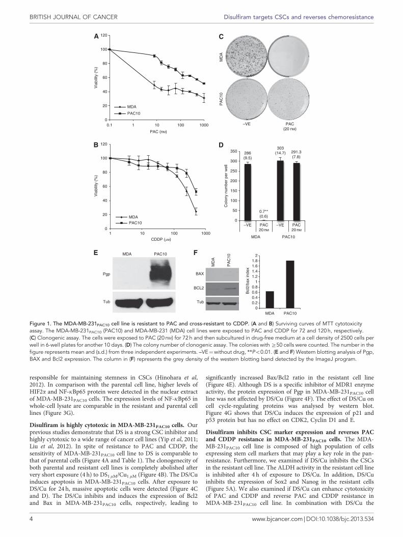
MDA-MB-231PAC10 cell line is pan-resistant to anticancerdrugs. First, the cytotoxic effect of PAC on both sensitive andresistant cell lines was compared by MTT assay (Table 1 andFigure 1A). The MDA-MB-231 cells are sensitive to thecytotoxicity of PAC with an IC50_72 h of 8.7 nM. In contrast, theMDA-MB-231PAC10 cell line is highly resistant to PAC with anIC50_72 h of over 1000 nM. The cytotoxic effect of CDDP, DOC andDOX on MDA-MB-231PAC10 cell line was also evaluated. Table 1and Figure 1B demonstrate that MDA-MB-231PAC10 cells are alsosignificantly cross-resistant to CDDP, DOC and DOX. In line withthe MTT data, PAC (20 nM) abolished the clonogenicity of theparental cell line but had no effect on MDA-MB-231PAC10 cells(Figure 1C and D). Because of the slower proliferation rate, thecolonies developed from the resistant cell line are smaller than thatfrom the parental cell line (Figure 1C). The overexpression ofMDR1 is the most common mechanism involved in multidrugresistance that includes PAC resistance. High expression of Pgp
was detected in the resistant cell line by western blot (Figure 1E).Paclitaxel induces apoptosis mainly via intrinsic apoptotic pathway(Ferlini et al, 2009). Therefore, the protein expression status of Baxand Bcl2, the two major components involved in intrinsicapoptotic pathway, was examined by western blot. Figure 1Fshows that MDA-MB-231PAC10 cell line expresses significantlyhigher background levels of Bcl2 protein than those in the parentalcells. The Bcl2/Bax ratio in the resistant cell line is markedly higherthan that in the parental cell line.
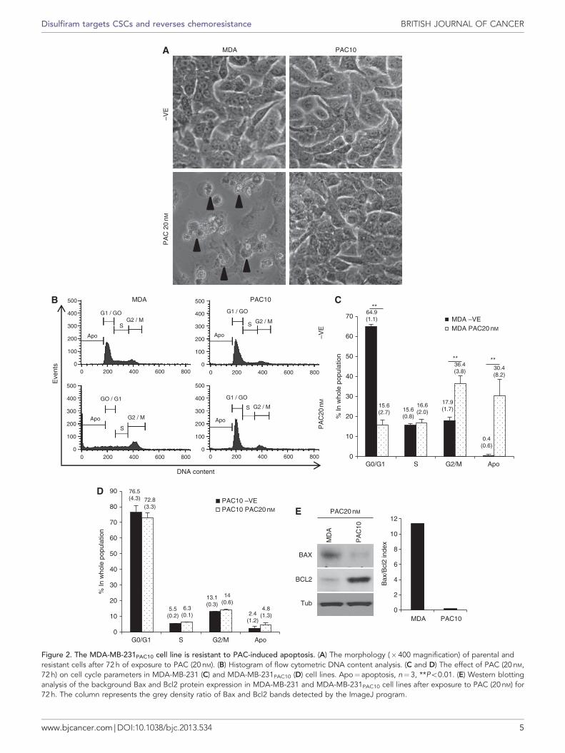
Resistance of MDA-MB-231PAC10 cell line to PAC-inducedapoptosis. After a 72-h exposure to 20 nM PAC, the phase-contrast microscopic images demonstrate apoptotic morphologies(cell blebbing and nuclear condensation and fragmentation) inMDA-MB-231 but not in the MDA-MB-231PAC10-resistant cells(Figure 2A). Flow cytometry DNA content analysis manifested thatPAC induced a significantly higher (Po0.01) apoptotic sub-G1population (30.4%) in the parental cell line than those in theuntreated cells (0.4%). Paclitaxel (20 nM, 72 h) also introducedG2/M-phase blockade leading to an increased G2/M population(untreated: 17.9%, treated: 36.4%; Po0.01) and a decreased G0/G1population (dropped from 64.9 to 15.6%, Po0.01; Figure 2B and C)in the parental cell line. In contrast, there is no significant effect ofPAC on the apoptotic status in the resistant cells. The cell cyclestatus in MDA-MB-231PAC10 cell line is also not affected by PACexposure (Figure 2D). Paclitaxel exposure induces Bax expressionleading to high Bax/Bcl2 ratio in the parental cells but not theresistant cells (Figure 2E).
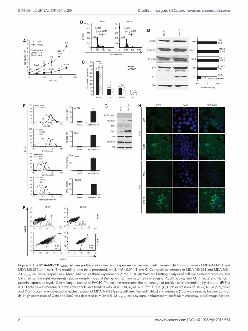
MDA-MB-231PAC10 has longer doubling time. In the cellculture, the MDA-MB-231PAC10 cells grow markedly slower thanMDA-MB-231 cells. Therefore, we compared the doubling timeand cell cycle parameters in these two cell lines. Figure 3A showsthe growth curves of both cell lines. The doubling time ofMDA-MB-231PAC10 cells (64.9 h) is significantly longer than thatof the sensitive cells (31.7 h; Po0.01). Flow cytometry analysisshows that in comparison with the parental cell line, the MDA-MB-231PAC10 cells have significantly higher G0/G1 and lowerS-phase population (Figure 3B and C). The expression levels ofcell cycle-determinant proteins were examined by western blot.Figure 3D shows the western blotting image and relative banddensity analysed by ImageJ program. The relative density ((Targetprotein/Tubulin)� 100) of p21 protein is markedly higher in theresistant cell line. The other moderately upregulated proteinsinclude p53, cyclin D1 and cyclin E.
MDA-MB-231PAC10 cells demonstrate CSC characteristics. Ithas been widely accepted that CSCs are responsible for chemo- andradio-resistance (Dean, 2009). The resistant cell line is slow cyclingwith high expression of p21 protein and expresses high levels ofPgp, which are the common features in CSCs (Tirino et al, 2013).Therefore, we examined CSC markers in the resistant and parentalcell lines. High ALDH activity is a functional marker of CSCsderived from different cancer types including breast cancer. Figures 3Eand G show that in comparison with the parental cells, theMDA-MB-231PAC10 cell line possesses higher ALDHþ populationthat also expresses higher levels of embryonic stem cell markers(Oct4, Sox2 and Nanog). The overexpression of Oct4 and Sox2protein was detected in nuclear protein by western blotting assay(Figure 3G). High expression of Oct4 and Sox2 in the resistant cellline was detected by immunofluorescent confocal microscopy(Figure 3H). The nuclear translocation of Oct4 was detected but forsome unknown reason Sox2 nuclear translocation was not detectedby immunocytochemistry. The specificity of ALDEFLUOR assaywas determined by treating the cells with DEAB, a specificinhibitor of ALDH (Figure 3F). The expression of NF-kB andHIF2a protein was also examined by western blotting analysisbecause emerging evidence indicates that hypoxia and NF-kB are
Table 1. Cytotoxicity of disulfiram and conventional anticancer drugs toMDA-MB-231 and MDA-MB-231PAC10 BC cell lines
PAC CDDP DOC DOX DS
IC50
MDA 8.7 (2.3) 256.7 (26.1) 4.6 (3.3) 27.6 (2.5) 151.9 (12.1)MDAPAC10 41000** 645.4* (127.3) 4250** 1575** (169.3) 116.4 (30.0)
CI value
IC50 0.61 0.64 NA NA NAIC75 0.64 0.41 NA NA NAIC90 0.72 0.28 NA NA NA
Abbreviations: CDDP¼ cisplatin; CI¼ combination index; DOC¼docetaxel; DOX¼doxorubicin; DS¼disulfiram; IC¼ inhibitory concentration; NA¼not available; PAC¼paclitaxel. The half-maximal inhibitory concentration (IC50) value (nM) from three experi-ments (mean (s.d.)) is shown. *Po0.05, **Po0.01 (n¼ 3). The CI value lower than 1.0:synergistic effect. The cells were exposed to drug for 72 or 120 h (CDDP). DS/Cu¼DS inmedium supplemented with 1mM CuCl2.
Disulfiram targets CSCs and reverses chemoresistance BRITISH JOURNAL OF CANCER
www.bjcancer.com | DOI:10.1038/bjc.2013.534 3
responsible for maintaining stemness in CSCs (Hinohara et al,2012). In comparison with the parental cell line, higher levels ofHIF2a and NF-kBp65 protein were detected in the nuclear extractof MDA-MB-231PAC10 cells. The expression levels of NF-kBp65 inwhole-cell lysate are comparable in the resistant and parental celllines (Figure 3G).
Disulfiram is highly cytotoxic in MDA-MB-231PAC10 cells. Ourprevious studies demonstrate that DS is a strong CSC inhibitor andhighly cytotoxic to a wide range of cancer cell lines (Yip et al, 2011;Liu et al, 2012). In spite of resistance to PAC and CDDP, thesensitivity of MDA-MB-231PAC10 cell line to DS is comparable tothat of parental cells (Figure 4A and Table 1). The clonogenecity ofboth parental and resistant cell lines is completely abolished aftervery short exposure (4 h) to DS1mM/Cu1 mM (Figure 4B). The DS/Cuinduces apoptosis in MDA-MB-231PAC10 cells. After exposure toDS/Cu for 24 h, massive apoptotic cells were detected (Figure 4Cand D). The DS/Cu inhibits and induces the expression of Bcl2and Bax in MDA-MB-231PAC10 cells, respectively, leading to
significantly increased Bax/Bcl2 ratio in the resistant cell line(Figure 4E). Although DS is a specific inhibitor of MDR1 enzymeactivity, the protein expression of Pgp in MDA-MB-231PAC10 cellline was not affected by DS/Cu (Figure 4F). The effect of DS/Cu oncell cycle-regulating proteins was analysed by western blot.Figure 4G shows that DS/Cu induces the expression of p21 andp53 protein but has no effect on CDK2, Cyclin D1 and E.
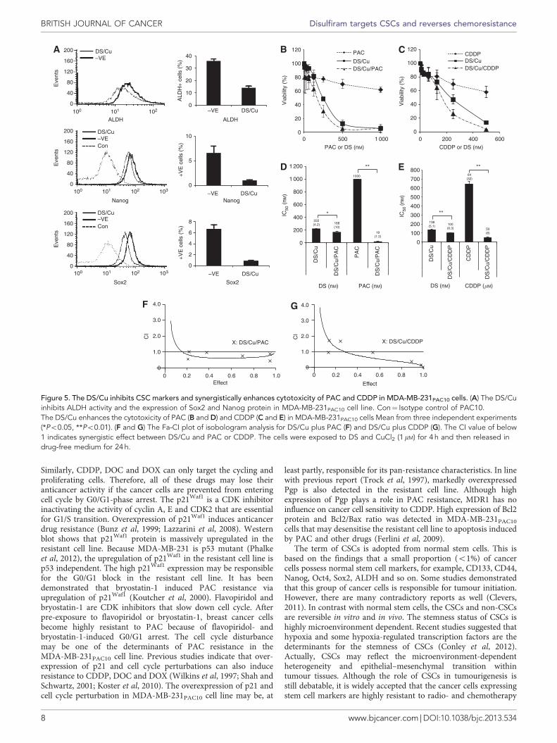
Disulfiram inhibits CSC marker expression and reverses PACand CDDP resistance in MDA-MB-231PAC10 cells. The MDA-MB-231PAC10 cell line is composed of high population of cellsexpressing stem cell markers that may play a key role in the pan-resistance. Furthermore, we examined if DS/Cu inhibits the CSCsin the resistant cell line. The ALDH activity in the resistant cell lineis inhibited after 4 h of exposure to DS/Cu. In addition, DS/Cuinhibits the expression of Sox2 and Nanog in the resistant cells(Figure 5A). We also examined if DS/Cu can enhance cytotoxicityof PAC and CDDP and reverse PAC and CDDP resistance inMDA-MB-231PAC10 cell line. In combination with DS/Cu the
MD
AP
AC
10
PAC(20 nM)
0
20
40
60
80
100
120
0.1 1 10 100 1000
Via
bilit
y (%
)
PAC (nM)
MDA
PAC10
286(9.5)
0.7**(0.6)
303(14.7) 291.3
(7.8)
0
50
100
150
200
250
300
350
PAC20 nM
–VE–VE PAC20 nM
MDA PAC10
Col
ony
num
ber
per
wel
l0
20
40
60
80
100
120
1 10 100 1000
Via
bilit
y (%
)
CDDP (�M)
MDAPAC10
MDA PAC10
Pgp
Tub
MD
A
PA
C10
BCL2
Tub
BAX
00.20.40.60.8
11.21.41.61.8
2
MDA PAC10
Bcl
2/ba
x in
dex
–VE
Figure 1. The MDA-MB-231PAC10 cell line is resistant to PAC and cross-resistant to CDDP. (A and B) Surviving curves of MTT cytotoxicityassay. The MDA-MB-231PAC10 (PAC10) and MDA-MB-231 (MDA) cell lines were exposed to PAC and CDDP for 72 and 120 h, respectively.(C) Clonogenic assay. The cells were exposed to PAC (20 nM) for 72 h and then subcultured in drug-free medium at a cell density of 2500 cells perwell in 6-well plates for another 10 days. (D) The colony number of clonogenic assay. The colonies with Z50 cells were counted. The number in thefigure represents mean and (s.d.) from three independent experiments. –VE¼without drug, **Po0.01. (E and F) Western blotting analysis of Pgp,BAX and Bcl2 expression. The column in (F) represents the grey density of the western blotting band detected by the ImageJ program.
BRITISH JOURNAL OF CANCER Disulfiram targets CSCs and reverses chemoresistance
4 www.bjcancer.com | DOI:10.1038/bjc.2013.534
PA
C 2
0nM
MDA PAC10
–VE
Eve
nts
DNA content
MDA PAC10
–VE
PA
C20
nM
76.5(4.3)
5.5(0.2)
13.1(0.3)
2.4(1.2)
72.8(3.3)
6.3(0.1)
14(0.6)
4.8(1.3)
0
10
20
30
40
50
60
70
80
90
G0/G1 S G2/M Apo
% In
who
le p
opul
atio
n
PAC10 –VEPAC10 PAC20 nM
64.9(1.1)
15.6(0.8)
17.9(1.7)
0.4(0.6)
15.6(2.7)
16.6(2.0)
36.4(3.8)
30.4(8.2)
0
10
20
30
40
50
60
70
G0/G1 S G2/M Apo
% In
who
le p
opul
atio
n
MDA –VEMDA PAC20 nM
**
** **
MD
A
PA
C10
PAC20 nM
BCL2
Tub
BAX
0
2
4
6
8
10
12
MDA PAC10
Bax
/Bcl
2 in
dex
00 200 400 600 800
100
200
300
400
500
Apo
G1 / GOG2 / M
S
Apo
G1 / GO
G2 / MS
Apo
GO / G1
G2 / M
S
Apo
G1 / GO
G2 / MS
00 200 400 600 800
100
200
300
400
500
00 200 400 600 800
100
200
300
400
500
00 200 400 600 800
100
200
300
400
500
Figure 2. The MDA-MB-231PAC10 cell line is resistant to PAC-induced apoptosis. (A) The morphology (� 400 magnification) of parental andresistant cells after 72 h of exposure to PAC (20 nM). (B) Histogram of flow cytometric DNA content analysis. (C and D) The effect of PAC (20 nM,72 h) on cell cycle parameters in MDA-MB-231 (C) and MDA-MB-231PAC10 (D) cell lines. Apo¼ apoptosis, n¼3, **Po0.01. (E) Western blottinganalysis of the background Bax and Bcl2 protein expression in MDA-MB-231 and MDA-MB-231PAC10 cell lines after exposure to PAC (20 nM) for72 h. The column represents the grey density ratio of Bax and Bcl2 bands detected by the ImageJ program.
Disulfiram targets CSCs and reverses chemoresistance BRITISH JOURNAL OF CANCER
www.bjcancer.com | DOI:10.1038/bjc.2013.534 5
0.5
1.325
2.5
3.3
4.725
7.5
0.30.5 0.625
1.1131.813
0
1
2
3
4
5
6
7
8
9
0 50 100 150
Cel
l cou
nt (
×10
4 )
Time (h)
MDA
PAC10
Doubling timeMDA: 31.7 hPAC10: 64.9 h **
Eve
nts
DNA content
MDA PAC10
**
**
64.9(1.1)
15.6(0.8)
17.9(1.7)
0.4(0.6)
76.5(1.3)
5.5(0.2)
13.1(3.2)
2.4(0.2)
0
10
20
30
40
50
60
70
80
90
G0/G1 S G2/M Apo
% In
who
le p
opul
atio
n
MDAPAC10
p21
Tub
p53
CDK2
Cyclin D1
Cyclin E
MD
A
PA
C10
0.04
79.7
146.0
178.9
69.6
73.7
132.7
201.4
190.3
78.5
0.01 1 100
p21
p53
CycE
CycD1
CDK2
Relative density
PAC10 MDA
MD
A
PA
C10
Oct4
Sox2
Nuc
Tub
p65_Cyto
p65_Nucl
HIF2α_Nucl
FITC DAPI FITC/DAPI
MD
AP
AC
10M
DA
PA
C10
Sox
2O
ct4
ALDH
Eve
nts
– DEAB + DEAB
MD
AP
AC
10
0.95(0.07)
1.04(0.1)
8.7(0.12)
5.04(0.2)
PAC10MDA
Eve
nt
Sox2 fluorescent
PAC10MDA
ALDH
0
5
10
15
MDA MDAPAC10
+V
E c
ells
(%
)
0
10
20
30
MDA MDAPAC10
+V
E c
ells
(%
)
0
10
20
MDA MDAPAC10
+V
E c
ells
(%
)
Oct4 fluorescent
ALDH
Oct4
Sox2
Nanog
Nanog fluorescent
0
10
20
30
MDA MDAPAC10
+V
E c
ells
(%
)
PAC10MDA
PAC10MDA
Con
Con
Con
00 200 400 600 0 200 400 600
100
200 Apo Apo
G1/G0 G1/G0
G2/M G2/MS S300
400
500
0
100
200
300
400
500
200
160
120
60
40
0
200
160
120
60
40
0
200
160
120
60
40
0
200
160
120
60
40
0
100 101 102 103
100 101 102 103
100 101 102 103
100 101 102 103
100 101 102 103 104 100 101 102 103 104
100 101 102 103 104100 101 102 103 104
100
101
102
103
104
100
101
102
103
104
100
101
102
103
104
100
101
102
103
104
Figure 3. The MDA-MB-231PAC10 cell line proliferates slower and expresses cancer stem cell markers. (A) Growth curves of MDA-MB-231 andMDA-MB-231PAC10 cells. The doubling time (h) is presented, n¼3, **Po0.01. (B and C) Cell cycle parameters in MDA-MB-231 and MDA-MB-231PAC10 cell lines, respectively. Mean and s.d. of three experiments (**Po0.01). (D) Western blotting analysis of cell cycle-related proteins. Thebar chart on the right represents relative density index of the bands. (E) Flow cytometry analysis of ALDH activity and Oct4, Sox2 and Nanogprotein expression levels. Con¼ Isotype control of PAC10. The column represents the percentage of positive cells determined by dot plot. (F) TheALDH activity was measured in the cancer cell lines treated with DEAB (30mM) at 37 1C for 30 min. (G) High expression of HIF2a, NF-kBp65, Sox2and Oct4 protein was detected in nuclear extract of MDA-MB-231PAC10 cell line. Nucleolin (Nuc) and a-tubulin (Tub) were used as loading control.(H) High expression of Oct4 and Sox2 was detected in MDA-MB-231PAC10 cells by immunofluorescent confocal microscopy � 400 magnification.
BRITISH JOURNAL OF CANCER Disulfiram targets CSCs and reverses chemoresistance
6 www.bjcancer.com | DOI:10.1038/bjc.2013.534
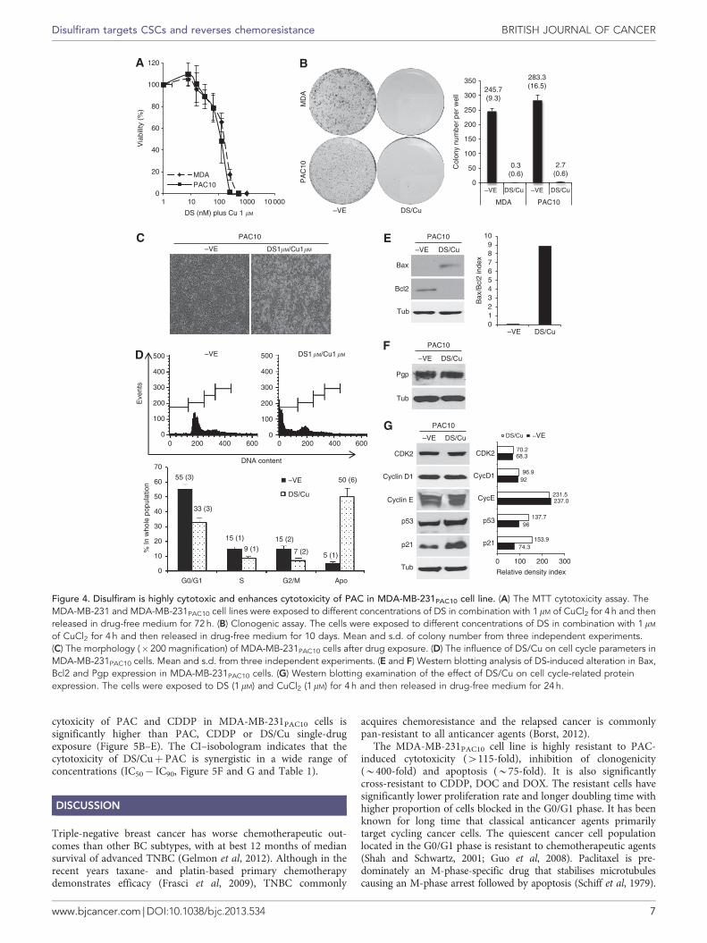
cytoxicity of PAC and CDDP in MDA-MB-231PAC10 cells issignificantly higher than PAC, CDDP or DS/Cu single-drugexposure (Figure 5B–E). The CI–isobologram indicates that thecytotoxicity of DS/Cuþ PAC is synergistic in a wide range ofconcentrations (IC50� IC90, Figure 5F and G and Table 1).
DISCUSSION
Triple-negative breast cancer has worse chemotherapeutic out-comes than other BC subtypes, with at best 12 months of mediansurvival of advanced TNBC (Gelmon et al, 2012). Although in therecent years taxane- and platin-based primary chemotherapydemonstrates efficacy (Frasci et al, 2009), TNBC commonly
acquires chemoresistance and the relapsed cancer is commonlypan-resistant to all anticancer agents (Borst, 2012).
The MDA-MB-231PAC10 cell line is highly resistant to PAC-induced cytotoxicity (4115-fold), inhibition of clonogenicity(B400-fold) and apoptosis (B75-fold). It is also significantlycross-resistant to CDDP, DOC and DOX. The resistant cells havesignificantly lower proliferation rate and longer doubling time withhigher proportion of cells blocked in the G0/G1 phase. It has beenknown for long time that classical anticancer agents primarilytarget cycling cancer cells. The quiescent cancer cell populationlocated in the G0/G1 phase is resistant to chemotherapeutic agents(Shah and Schwartz, 2001; Guo et al, 2008). Paclitaxel is pre-dominately an M-phase-specific drug that stabilises microtubulescausing an M-phase arrest followed by apoptosis (Schiff et al, 1979).
0
20
40
60
80
100
120
1 10 100 1000 10 000
Via
bilit
y (%
)
DS (nM) plus Cu 1 �M
MDAPAC10
MD
A
DS/Cu
PA
C10
245.7(9.3)
0.3(0.6)
283.3(16.5)
2.7(0.6)
0
50
100
150
200
250
300
350
–VE
–VE
–VE
–VE
–VEDS/Cu DS/Cu
MDA PAC10
Col
ony
num
ber
per
wel
l
DS1�M/Cu1�M
DS1 �M/Cu1 �M
PAC10
Eve
nts
DNA content
PAC10
DS/Cu
Pgp
Tub
PAC10
DS/Cu
Bax
Tub
Bcl2
0123456789
10
DS/Cu
Bax
/Bcl
2 in
dex
PAC10
–VE DS/Cu
Tub
p53
p21
CDK2
Cyclin D1
Cyclin E
74.3
96
237.0
92
68.3
153.9
137.7
231.5
96.9
70.2
0 100 200 300
p21
p53
CycE
CycD1
CDK2
Relative density index
DS/Cu –VE00
100
200 400 600 0 200 400 600
200
300
400
500
0
100
200
300
400
500
0
10
G0/G1 S G2/M Apo
20
30
% In
who
le p
opul
atio
n
40
50
6055 (3)
33 (3)
15 (1)
9 (1)15 (2)
50 (6)
7 (2) 5 (1)
70
–VE
DS/Cu
–VE
–VE
–VE
Figure 4. Disulfiram is highly cytotoxic and enhances cytotoxicity of PAC in MDA-MB-231PAC10 cell line. (A) The MTT cytotoxicity assay. TheMDA-MB-231 and MDA-MB-231PAC10 cell lines were exposed to different concentrations of DS in combination with 1mM of CuCl2 for 4 h and thenreleased in drug-free medium for 72 h. (B) Clonogenic assay. The cells were exposed to different concentrations of DS in combination with 1 mM
of CuCl2 for 4 h and then released in drug-free medium for 10 days. Mean and s.d. of colony number from three independent experiments.(C) The morphology (� 200 magnification) of MDA-MB-231PAC10 cells after drug exposure. (D) The influence of DS/Cu on cell cycle parameters inMDA-MB-231PAC10 cells. Mean and s.d. from three independent experiments. (E and F) Western blotting analysis of DS-induced alteration in Bax,Bcl2 and Pgp expression in MDA-MB-231PAC10 cells. (G) Western blotting examination of the effect of DS/Cu on cell cycle-related proteinexpression. The cells were exposed to DS (1 mM) and CuCl2 (1mM) for 4 h and then released in drug-free medium for 24 h.
Disulfiram targets CSCs and reverses chemoresistance BRITISH JOURNAL OF CANCER
www.bjcancer.com | DOI:10.1038/bjc.2013.534 7
Similarly, CDDP, DOC and DOX can only target the cycling andproliferating cells. Therefore, all of these drugs may lose theiranticancer activity if the cancer cells are prevented from enteringcell cycle by G0/G1-phase arrest. The p21Waf1 is a CDK inhibitorinactivating the activity of cyclin A, E and CDK2 that are essentialfor G1/S transition. Overexpression of p21Waf1 induces anticancerdrug resistance (Bunz et al, 1999; Lazzarini et al, 2008). Westernblot shows that p21Waf1 protein is massively upregulated in theresistant cell line. Because MDA-MB-231 is p53 mutant (Phalkeet al, 2012), the upregulation of p21Waf1 in the resistant cell line isp53 independent. The high p21Waf1 expression may be responsiblefor the G0/G1 block in the resistant cell line. It has beendemonstrated that bryostatin-1 induced PAC resistance viaupregulation of p21Waf1 (Koutcher et al, 2000). Flavopiridol andbryostatin-1 are CDK inhibitors that slow down cell cycle. Afterpre-exposure to flavopiridol or bryostatin-1, breast cancer cellsbecome highly resistant to PAC because of flavopiridol- andbryostatin-1-induced G0/G1 arrest. The cell cycle disturbancemay be one of the determinants of PAC resistance in theMDA-MB-231PAC10 cell line. Previous studies indicate that over-expression of p21 and cell cycle perturbations can also induceresistance to CDDP, DOC and DOX (Wilkins et al, 1997; Shah andSchwartz, 2001; Koster et al, 2010). The overexpression of p21 andcell cycle perturbation in MDA-MB-231PAC10 cell line may be, at
least partly, responsible for its pan-resistance characteristics. In linewith previous report (Trock et al, 1997), markedly overexpressedPgp is also detected in the resistant cell line. Although highexpression of Pgp plays a role in PAC resistance, MDR1 has noinfluence on cancer cell sensitivity to CDDP. High expression of Bcl2protein and Bcl2/Bax ratio was detected in MDA-MB-231PAC10
cells that may desensitise the resistant cell line to apoptosis inducedby PAC and other drugs (Ferlini et al, 2009).
The term of CSCs is adopted from normal stem cells. This isbased on the findings that a small proportion (o1%) of cancercells possess normal stem cell markers, for example, CD133, CD44,Nanog, Oct4, Sox2, ALDH and so on. Some studies demonstratedthat this group of cancer cells is responsible for tumour initiation.However, there are many contradictory reports as well (Clevers,2011). In contrast with normal stem cells, the CSCs and non-CSCsare reversible in vitro and in vivo. The stemness status of CSCs ishighly microenvironment dependent. Recent studies suggested thathypoxia and some hypoxia-regulated transcription factors are thedeterminants for the stemness of CSCs (Conley et al, 2012).Actually, CSCs may reflect the microenvironment-dependentheterogeneity and epithelial–mesenchymal transition withintumour tissues. Although the role of CSCs in tumourigenesis isstill debatable, it is widely accepted that the cancer cells expressingstem cell markers are highly resistant to radio- and chemotherapy
0
10
20
30
40
–VE DS/Cu
ALD
H+
cel
ls (
%)
Eve
nts
ALDH
Nanog
Sox2
Eve
nts
Eve
nts
0
2
4
6
8
DS/Cu
+V
E c
ells
(%
)0
5
10
DS/Cu
+V
E c
ells
(%
)
ALDH
Nanog
Sox2
–VEDS/Cu
DS/Cu
DS/Cu222(4.2) 168
(10)
1000
19(1.2)
0
200
400
600
800
1 000
1 200
DS
/Cu
DS
/Cu/
PA
C
PA
C
DS
/Cu/
PA
C
DS (nM) PAC (nM)IC
50 (
nM)
*
**
138(5.1) 100
(6.3)
64(32)
50(6)
0
100
200
300
400
500
600
700
800
DS
/Cu
DS
/Cu/
CD
DP
CD
DP
DS
/Cu/
CD
DP
DS (nM) CDDP (�M)
IC50
(nM
)
**
**
0
20
40
60
80
100
120
0 500 1 000
Via
bilit
y (%
)
PAC or DS (nM)
PAC
DS/CuDS/Cu/PAC
0
20
40
60
80
100
120
0 200 400 600
Via
bilit
y (%
)
CDDP or DS (nM)
CDDPDS/CuDS/Cu/CDDP
Effect
CI
X: DS/Cu/PAC
0 0.2 0.4 0.6 0.8 1.00
1.0
2.0
3.0
4.0
0 0.2 0.4 0.6 0.8 1.00
1.0
2.0
3.0
4.0
X: DS/Cu/CDDP
CI
Effect
0100 101 102
40
80
120
160
200
0
40
80
120
160
200
0
40
80
120
160
200
–VECon
–VECon
–VE
–VE
100 101 102 103
100 101 102 103
Figure 5. The DS/Cu inhibits CSC markers and synergistically enhances cytotoxicity of PAC and CDDP in MDA-MB-231PAC10 cells. (A) The DS/Cuinhibits ALDH activity and the expression of Sox2 and Nanog protein in MDA-MB-231PAC10 cell line. Con¼ Isotype control of PAC10.The DS/Cu enhances the cytotoxicity of PAC (B and D) and CDDP (C and E) in MDA-MB-231PAC10 cells Mean from three independent experiments(*Po0.05, **Po0.01). (F and G) The Fa-CI plot of isobologram analysis for DS/Cu plus PAC (F) and DS/Cu plus CDDP (G). The CI value of below1 indicates synergistic effect between DS/Cu and PAC or CDDP. The cells were exposed to DS and CuCl2 (1mM) for 4 h and then released indrug-free medium for 24 h.
BRITISH JOURNAL OF CANCER Disulfiram targets CSCs and reverses chemoresistance
8 www.bjcancer.com | DOI:10.1038/bjc.2013.534
and are the sources of cancer recurrence (Bjerkvig et al, 2005; Deanet al, 2005; Clevers, 2011). Also, the cells with CSC markers areresistant to all different anticancer drugs. Therefore, CSCs may bethe cause of pan-chemoresistance that is a common and a veryserious problem faced in cancer therapeutics. Elimination of thesecells may improve the outcomes of cancer chemotherapy. It hasrecently been reported that CSCs are involved in acquired taxaneresistance (Domingo-Domenech et al, 2012; McAuliffe et al, 2013).In contrast with the fast growing cancer mass, CSCs are slow-cycling dormant cells expressing stem cell markers. Highexpression of Pgp is also a common feature of CSCs (Dean,2009). Recent reports indicate that p21Waf1 is indispensable formaintaining the quiescent status, stemness and preventing excessDNA-damage accumulation in CSCs (Viale et al, 2009). Ourfindings in MDA-MB-231PAC10 cell line, for example, high p21expression, cell cycle slowing down and high expression of Pgp,indicate that the high population of CSCs in this cell line may playa crucial role for the pan-resistance. Based upon this hypothesis, weexamined several other CSC phenotypes. High levels of ALDH, afunctional CSC marker, were detected in the resistant cells. Theresistant cell line also expresses higher levels of CD44 (data notshown). The recent publications (Landen et al, 2010; Schafer et al,2012) and our unpublished data indicate that high ALDH activityconfers chemoresistance upon cancer cells that can be reversed bytargeting ALDH. High expression of the embryonic stem cell-associated genes Sox2, Oct4 and Nanog was also detected in theresistant cell line. Hypoxia-induced HIFs overexpression and NF-kB pathway activation is responsible for chemoresistance (Wanget al, 2004) and also the determinant factors for maintainingstemness of CSCs (Conley et al, 2012). Even cultured in normoxiccondition, the overexpression and nuclear translocation of HIF2aand NF-kBp65 were detected in the resistant cell line. Furtherstudies are being performed in our lab to elucidate the relationshipbetween these factors and CSC-related chemoresistance.
Disulfiram is a very efficacious ALDH inhibitor and CSC-targeting agent, demonstrating strong chemoresistance-reversingactivity (Yip et al, 2011; Hothi et al, 2012; Liu et al, 2012; Triscottet al, 2012). Previous clinical studies manifest that DS and itsderivative effectively improve survival of breast and other cancerpatients (Lewison, 1977; Dufour et al, 1993; Brar et al, 2004). Inthis study we examined its direct cytotoxicity and resistance-reversing effect on PAC and CDDP in MDA-MB-231PAC10 cells.Our results show that in contrast to its high resistance to PAC,DOC, DOX and CDDP, the MDA-MB-231PAC10 cell line remainsvery sensitive to DS-induced cytotoxicity. After exposure to DS foronly 4 h, the clonogenicity of the resistant cell line was completelyeradicated. The CI–isobologram analysis demonstrates that DSsynergistically enhances the cytotoxicity of PAC and CDDP inMDA-MB-231PAC10 cells. In combination with DS/Cu, the PACand CDDP resistance in MDA-MB-231PAC10 cell line is completelyreversed. The stem cell markers, for example, ALDH activity andthe expression of Sox2 and Nanog in the resistant cell line, aremarkedly inhibited by DS exposure. Therefore, DS may reversepan-chemoresistance in MDA-MB-231PAC10 cell line by targetingBCSCs. The simultaneous inhibition and induction of Bcl2 and Baxindicates that DS may induce apoptosis in the resistant cells via anintrinsic pathway (Guo et al, 2010; Yip et al, 2011; Liu et al, 2012).Although DS inhibits MDR1 activity (Loo et al, 2004), it has noeffect on the expression of Pgp. There is no effect of DS on cellcycle status in the resistant cell line. Similar to many other DNA-targeting agents, DS exposure further induces p21 expression in theresistant cells. Anticancer stem cell is a hot spot for anticancer drugdevelopment (Zhou et al, 2009). New drug development is a verytime-consuming and costly procedure. Disulfiram has been used asan antialcoholism drug for over 60 years with preclinical andclinical safety data available. Therefore, it is relatively easier forrepositioning of it into cancer indication (Cvek, 2012).
CONCLUSIONS
A newly developed PAC-resistant BC cell line, MDA-MB-231PAC10, is cross-resistant to a panel of different anticancer drugs,for example, DOC, DOX and CDDP. We first reported thatacquired BC cell line consists of high proportion of cells expressingCSC markers that may be, at least partly, responsible for itsacquired pan-chemoresistant characteristics. We also manifestedthat DS, an antialcoholism drug, abolishes the cancer stem-likepopulation and efficaciously reverses the PAC and CDDPresistance in MDA-MB-231PAC10 cell line.
ACKNOWLEDGEMENTS
This project was supported by Breast Cancer Campaign, UK.
REFERENCES
Bjerkvig R, Tysnes BB, Aboody KS, Najbauer J, Terzis AJ (2005) Opinion:the origin of the cancer stem cell: current controversies and new insights.Nat Rev Cancer 5: 899–904.
Bodenner DL, Dedon PC, Keng PC, Katz JC, Borch RF (1986) Selectiveprotection against cis-diamminedichloroplatinum(II)-induced toxicity inkidney, gut, and bone marrow by diethyldithiocarbamate. Cancer Res 46:2751–2755.
Borst P (2012) Cancer drug pan-resistance: pumps, cancer stem cells,quiescence, epithelial to mesenchymal transition, blocked cell deathpathways, persisters or what? Open Biol 2: 120066.
Brar SS, Grigg C, Wilson KS, Holder Jr WD, Dreau D, Austin C, Foster M,Ghio AJ, Whorton AR, Stowell GW, Whittall LB, Whittle RR, White DP,Kennedy TP (2004) Disulfiram inhibits activating transcription factor/cyclic AMP-responsive element binding protein and human melanomagrowth in a metal-dependent manner in vitro, in mice and in a patientwith metastatic disease. Mol Cancer Ther 3: 1049–1060.
Bunz F, Hwang PM, Torrance C, Waldman T, Zhang Y, Dillehay L,Williams J, Lengauer C, Kinzler KW, Vogelstein B (1999) Disruption ofp53 in human cancer cells alters the responses to therapeutic agents. J ClinInvest 104: 263–269.
Cen D, Brayton D, Shahandeh B, Meyskens Jr. FL, Farmer PJ (2004)Disulfiram facilitates intracellular Cu uptake and induces apoptosis inhuman melanoma cells. J Med Chem 47: 6914–6920.
Chen D, Cui QC, Yang H, Dou QP (2006) Disulfiram, a clinically used anti-alcoholism drug and copper-binding agent, induces apoptotic cell death inbreast cancer cultures and xenografts via inhibition of the proteasomeactivity. Cancer Res 66: 10425–10433.
Chou TC, Talalay P (1984) Quantitative analysis of dose-effect relationships:the combined effects of multiple drugs or enzyme inhibitors. Adv EnzymeRegul 22: 27–55.
Clevers H (2011) The cancer stem cell: premises, promises and challenges. NatMed 17: 313–319.
Conley SJ, Gheordunescu E, Kakarala P, Newman B, Korkaya H, Heath AN,Clouthier SG, Wicha MS (2012) Antiangiogenic agents increase breastcancer stem cells via the generation of tumor hypoxia. Proc Natl Acad SciUSA 109: 2784–2789.
Cvek B (2012) Nonprofit drugs as the salvation of the world’s healthcaresystems: the case of Antabuse (disulfiram). Drug Discov Today 17:409–412.
Dean M (2009) ABC transporters, drug resistance, and cancer stem cells.J Mammary Gland Biol Neoplasia 14: 3–9.
Dean M, Fojo T, Bates S (2005) Tumour stem cells and drug resistance.Nat Rev Cancer 5: 275–284.
Deng XS, Wang S, Deng A, Liu B, Edgerton SM, Lind SE, Wahdan-Alaswad R,Thor AD (2012) Metformin targets Stat3 to inhibit cell growth and induceapoptosis in triple-negative breast cancers. Cell Cycle 11: 367–376.
Domingo-Domenech J, Vidal SJ, Rodriguez-Bravo V, Castillo-Martin M,Quinn SA, Rodriguez-Barrueco R, Bonal DM, Charytonowicz E,Gladoun N, de la Iglesia-Vicente J, Petrylak DP, Benson MC, Silva JM,Cordon-Cardo C (2012) Suppression of acquired docetaxel resistance in
Disulfiram targets CSCs and reverses chemoresistance BRITISH JOURNAL OF CANCER
www.bjcancer.com | DOI:10.1038/bjc.2013.534 9
prostate cancer through depletion of notch- and hedgehog-dependenttumor-initiating cells. Cancer Cell 22: 373–388.
Dufour P, Lang JM, Giron C, Duclos B, Haehnel P, Jaeck D, Jung JM,Oberling F (1993) Sodium dithiocarb as adjuvant immunotherapy for highrisk breast cancer: a randomized study. Biotherapy 6: 9–12.
Estey T, Piatigorsky J, Lassen N, Vasiliou V (2007) ALDH3A1: a cornealcrystallin with diverse functions. Exp Eye Res 84: 3–12.
Ferlini C, Cicchillitti L, Raspaglio G, Bartollino S, Cimitan S, Bertucci C,Mozzetti S, Gallo D, Persico M, Fattorusso C, Campiani G, Scambia G(2009) Paclitaxel directly binds to Bcl-2 and functionally mimics activity ofNur77. Cancer Res 69: 6906–6914.
Frasci G, Comella P, Rinaldo M, Iodice G, Di Bonito M, D’Aiuto M, Petrillo A,Lastoria S, Siani C, Comella G, D’Aiuto G (2009) Preoperative weeklycisplatin-epirubicin-paclitaxel with G-CSF support in triple-negative largeoperable breast cancer. Ann Oncol 20: 1185–1192.
Gelmon K, Dent R, Mackey JR, Laing K, McLeod D, Verma S (2012)Targeting triple-negative breast cancer: optimising therapeutic outcomes.Ann Oncol 23: 2223–2234.
Ginestier C, Hur MH, Charafe-Jauffret E, Monville F, Dutcher J, Brown M,Jacquemier J, Viens P, Kleer CG, Liu S, Schott A, Hayes D, Birnbaum D,Wicha MS, Dontu G (2007) ALDH1 is a marker of normal and malignanthuman mammary stem cells and a predictor of poor clinical outcome. CellStem Cell 1: 555–567.
Guo X, Goessl E, Jin G, Collie-Duguid ES, Cassidy J, Wang W, O’Brien V(2008) Cell cycle perturbation and acquired 5-fluorouracilchemoresistance. Anticancer Res 28: 9–14.
Guo X, Xu B, Pandey S, Goessl E, Brown J, Armesilla AL, Darling JL, Wang W(2010) Disulfiram/copper complex inhibiting NFkappaB activity andpotentiating cytotoxic effect of gemcitabine on colon and breast cancer celllines. Cancer Lett 291: 104–113.
Hacker MP, Ershler WB, Newman RA, Gamelli RL (1982) Effect of disulfiram(tetraethylthiuram disulfide) and diethyldithiocarbamate on the bladdertoxicity and antitumor activity of cyclophosphamide in mice. Cancer Res42: 4490–4494.
Hinohara K, Kobayashi S, Kanauchi H, Shimizu S, Nishioka K, Tsuji E,Tada K, Umezawa K, Mori M, Ogawa T, Inoue J, Tojo A, Gotoh N (2012)ErbB receptor tyrosine kinase/NF-kappaB signaling controlsmammosphere formation in human breast cancer. Proc Natl Acad Sci USA109: 6584–6589.
Hothi P, Martins TJ, Chen LP, Deleyrolle L, Yoon JG, Reynolds B, Foltz G(2012) High-throughput chemical screens identify disulfiram as aninhibitor of human glioblastoma stem cells. Oncotarget 3: 1124–1136.
Kavallaris M (2010) Microtubules and resistance to tubulin-binding agents.Nat Rev Cancer 10: 194–204.
Knuefermann C, Lu Y, Liu B, Jin W, Liang K, Wu L, Schmidt M, Mills GB,Mendelsohn J, Fan Z (2003) HER2/PI-3K/Akt activation leads to amultidrug resistance in human breast adenocarcinoma cells. Oncogene22: 3205–3212.
Koster R, di Pietro A, Timmer-Bosscha H, Gibcus JH, van den Berg A,Suurmeijer AJ, Bischoff R, Gietema JA, De Jong S (2010) Cytoplasmic p21expression levels determine cisplatin resistance in human testicular cancer.J Clin Invest 120: 3594–3605.
Koutcher JA, Motwani M, Zakian KL, Li XK, Matei C, Dyke JP, Ballon D,Yoo HH, Schwartz GK (2000) The in vivo effect of bryostatin-1 onpaclitaxel-induced tumor growth, mitotic entry, and blood flow. ClinCancer Res 6: 1498–1507.
Landen Jr. CN, Goodman B, Katre AA, Steg AD, Nick AM, Stone RL,Miller LD, Mejia PV, Jennings NB, Gershenson DM, Bast Jr. RC,Coleman RL, Lopez-Berestein G, Sood AK (2010) Targeting aldehydedehydrogenase cancer stem cells in ovarian cancer. Mol Cancer Ther9: 3186–3199.
Lazzarini R, Moretti S, Orecchia S, Betta PG, Procopio A, Catalano A (2008)Enhanced antitumor therapy by inhibition of p21waf1 in humanmalignant mesothelioma. Clin Cancer Res 14: 5099–5107.
Lewison EF (1977) Spontaneous regression of breast cancer. Prog Clin Biol Res12: 47–53.
Liu P, Brown S, Goktug T, Channathodiyil P, Kannappan V, Hugnot JP,Guichet PO, Bian X, Armesilla AL, Darling JL, Wang W (2012) Cytotoxiceffect of disulfiram/copper on human glioblastoma cell lines andALDH-positive cancer-stem-like cells. Br J Cancer 107: 1488–1497.
Loo TW, Bartlett MC, Clarke DM (2004) Disulfiram metabolites permanentlyinactivate the human multidrug resistance P-glycoprotein. Mol Pharm 1:426–433.
McAuliffe SM, Morgan SL, Wyant GA, Tran LT, Muto KW, Chen YS,Chin KT, Partridge JC, Poole BB, Cheng KH, Daggett Jr. J, Cullen K,Kantoff E, Hasselbatt K, Berkowitz J, Muto MG, Berkowitz RS, Aster JC,Matulonis UA, Dinulescu DM (2013) Targeting Notch, a key pathway forovarian cancer stem cells, sensitizes tumors to platinum therapy. Proc NatlAcad Sci USA 109: E2939–E2948.
Nobel CI, Kimland M, Lind B, Orrenius S, Slater AF (1995) Dithiocarbamatesinduce apoptosis in thymocytes by raising the intracellular level of redox-active copper. J Biol Chem 270: 26202–26208.
Ohi Y, Umekita Y, Yoshioka T, Souda M, Rai Y, Sagara Y, Sagara Y, Sagara Y,Tanimoto A (2011) Aldehyde dehydrogenase 1 expression predicts poorprognosis in triple-negative breast cancer. Histopathology 59: 776–780.
Phalke S, Mzoughi S, Bezzi M, Jennifer N, Mok WC, Low DH, Thike AA,Kuznetsov VA, Tan PH, Voorhoeve PM, Guccione E (2012) p53-Independent regulation of p21Waf1/Cip1 expression and senescence byPRMT6. Nucleic Acids Res 40: 9534–9542.
Plumb JA, Milroy R, Kaye SB (1989) Effects of the pH dependence of 3-(4,5-dimethylthiazol-2-yl)-2,5-diphenyl-tetrazolium bromide-formazanabsorption on chemosensitivity determined by a novel tetrazolium-basedassay. Cancer Res 49: 4435–4440.
Schafer A, Teufel J, Ringel F, Bettstetter M, Hoepner I, Rasper M, Gempt J,Koeritzer J, Schmidt-Graf F, Meyer B, Beier CP, Schlegel J (2012)Aldehyde dehydrogenase 1A1–a new mediator of resistance totemozolomide in glioblastoma. Neuro Oncol 14: 1452–1464.
Schiff PB, Fant J, Horwitz SB (1979) Promotion of microtubule assemblyin vitro by taxol. Nature 277: 665–667.
Schreck R, Albermann K, Baeuerle PA (1992) Nuclear factor kappa B: anoxidative stress-responsive transcription factor of eukaryotic cells(a review). Free Radic Res Commun 17: 221–237.
Shah MA, Schwartz GK (2001) Cell cycle-mediated drug resistance: anemerging concept in cancer therapy. Clin Cancer Res 7: 2168–2181.
Takahashi T, Yamasaki F, Sudo T, Itamochi H, Adachi S, Tamamori-Adachi M,Ueno NT (2005) Cyclin A-associated kinase activity is needed for paclitaxelsensitivity. Mol Cancer Ther 4: 1039–1046.
Tirino V, Desiderio V, Paino F, De Rosa A, Papaccio F, La Noce M, Laino L,De Francesco F, Papaccio G (2013) Cancer stem cells in solid tumors: anoverview and new approaches for their isolation and characterization.FASEB J 27: 13–24.
Triscott J, Lee C, Hu K, Fotovati A, Berns R, Pambid M, Luk M, Kast RE,Kong E, Toyota E, Yip S, Toyota B, Dunn SE (2012) Disulfiram, a drugwidely used to control alcoholism, suppresses self-renewal of glioblastomaand overrides resistance to temozolomide. Oncotarget 3: 1112–1123.
Trock BJ, Leonessa F, Clarke R (1997) Multidrug resistance in breast cancer: ameta-analysis of MDR1/gp170 expression and its possible functionalsignificance. J Natl Cancer Inst 89: 917–931.
Viale A, De Franco F, Orleth A, Cambiaghi V, Giuliani V, Bossi D, Ronchini C,Ronzoni S, Muradore I, Monestiroli S, Gobbi A, Alcalay M, Minucci S,Pelicci PG (2009) Cell-cycle restriction limits DNA damage and maintainsself-renewal of leukaemia stem cells. Nature 457: 51–56.
Wang W, Cassidy J, O’Brien V, Ryan KM, Collie-Duguid E (2004)Mechanistic and predictive profiling of 5-Fluorouracil resistance in humancancer cells. Cancer Res 64: 8167–8176.
Wang W, McLeod HL, Cassidy J (2003) Disulfiram-mediated inhibition ofNF-kappaB activity enhances cytotoxicity of 5-fluorouracil in humancolorectal cancer cell lines. Int J Cancer 104: 504–511.
Wilkins DE, Ng CE, Raaphorst GP (1997) Cell cycle perturbations incisplatin-sensitive and resistant human ovarian carcinoma cells followingtreatment with cisplatin and low dose rate irradiation. Cancer ChemotherPharmacol 40: 159–166.
Yip NC, Fombon IS, Liu P, Brown S, Kannappan V, Armesilla AL, Xu B,Cassidy J, Darling JL, Wang W (2011) Disulfiram modulated ROS-MAPKand NFkB pathways and targeted breast cancer cells with cancer stem celllike properties. Br J Cancer 104: 1564–1574.
Zhou BB, Zhang H, Damelin M, Geles KG, Grindley JC, Dirks PB (2009)Tumour-initiating cells: challenges and opportunities for anticancer drugdiscovery. Nat Rev Drug Discov 8: 806–823.
This work is published under the standard license to publish agree-ment. After 12 months the work will become freely available andthe license terms will switch to a Creative Commons Attribution-NonCommercial-Share Alike 3.0 Unported License.
BRITISH JOURNAL OF CANCER Disulfiram targets CSCs and reverses chemoresistance
10 www.bjcancer.com | DOI:10.1038/bjc.2013.534


