Embed Size (px)
Citation preview

4269
Article HttP://DX.DOi.OrG/10.5504/BBeQ.2013.0067 A&eB
AGricUltUre AND eNVirONMeNtAl BiOtecHNOlOGY
© Biotechnol. & Biotechnol. eq. 27/2013/6
Biotechnol. & Biotechnol. eq. 2013, 27(6), 4269-4275
Keywords: lignin degradation, lignin peroxidase, laccase, decolorization, 18S rDnA
Introductionlignin is probably one of the most recalcitrant compounds synthesized by plants. it is degraded by only a few microorganisms. lignin is mostly abundant in trees and is the main contributor to wood strength (16). Bio-ligninolytic systems are employed in the paper industry (biopulping and biobleaching), degradation of xenobiotic compounds in industrial effluents, for enhancement of fodder digestibility, lignin bioconversion, desulphurization of coal, petroleum and dye decolorization.
Soft-rot Ascomycetes preferentially degrade the carbohydrates of the wood. Some of them are capable of mineralizing lignin (15). the mineralization of lignin involves the so-called lignin-modifying enzymes (lMes): lignin peroxidase (liP, ec. 1.11.1.14), Mn-dependant peroxidase, and laccase (ec. 1.10.3.2), a polyphenoloxidase. they catalyze the production of highly reactive radicals which oxidize phenolic and non-phenolic lignin components (6, 10). in addition, improvement of laccase activity for biofuel cell application has been achieved by coupling the enzyme with nanoparticles to create a cathode (3, 31).
Dyes are classified as azo, anthraquinone, triphenylmethane, heterocyclic, and polymeric dyes. Among them, the azo and triphenylmethane dyes have the largest share in the textile industry. hence, much attention is paid to the search for new dye-decolorizing bacterial and fungal strains, including white rot fungi, and to optimization of the cultivation conditions for known ones (5, 9).
therefore, the aim of this study was to investigate the extracellular ligninolytic system of egyptian fungal strains in view of the potential to decolorize and degrade a broad spectrum of reactive synthetic dyes rather than lignin degradation. the study was focused on lMes not only from the standpoint of comparative biology, but also with the expectation of finding better lignin-degrading systems for use as environmentally friendly systems in various biotechnological applications.
Materials and MethodsFungal strainstwenty-two fungal strains were isolated from different egyptian agriculture soils in the vicinity of some cultivated plants. the growth medium used in the method was PDA (Potato-dextrose-Agar). one gram of soil was suspended in 10 ml sterile distilled water. After soil sedimentation, 0.1 ml of the supernatant was spread on the surface of (PDA) plates. the plates were incubated at 30 °c for 7 days. out of them, 12
LIGNINOLYTIC OXIDATIVE SYSTEM OF FUNGAL EGYPTIAN ISOLATES AND THEIR APPLICATIONS IN THE DECOLORIZATION OF INDUSTRIAL DYES
Rania Mohamed Ahmed Abedin1, Amr A. el hanafy2,3, Sawsan Abd el-latif3, Samy A. el-Assar1, Mayada Sh. Fadel3
1Alexandria University, Botany and Microbiology Department, Alexandria, egypt2King Abdel Aziz University, Department of Biology, Jeddah, Saudi Arabia3City for Scientific Researches and Technology Application, Department of Nucleic Acid Research, Alexandria, Egyptcorrespondence to: Rania Mohamed Ahmed Abedine-mail: [email protected]
ABSTRACTThe aim of this study was to investigate the ligninolytic system of fungal strains isolated from Egyptian agricultural soil which are efficient in biodegradation and mineralization of lignin. They were identified morphologically, microscopically and confirmed by RAPD profiles. Based on PCR amplification and sequencing of the 18S rDNA gene an analytical phylogenetic tree was drawn for species confirmation. The screening experiment revealed that Emericella nidulans, Aspergillus fumigatus, Phoma betae, Penicillium oxalicum and Humicola grisea exhibited maximum potential for high lignin degradation. They showed higher lignin peroxidase and laccase activities. By process optimization, enhanced lignin degradation (94 %) was achieved within 7 days in lignin-glucose medium when compared with lignin degradation (70 %) obtained in glucose-free medium. The ligninolytic enzymatic activities had a great potential for decolorization of chemically different synthetic dyes (Azure B, Safranin, Crystal Violet and Malachite Green). The highest enzymatic activities and the highest decolorization rates were detected at a dye concentration of 0.2 g/L. The dye decolorization rate significantly increased with tryptophan addition as (1 mmol/L). Humicola grisea showed the highest decolorization rate (99 %) with azure B. Phoma beta also showed a high decolorization rate (99 % with crystal violet and 90 % with safranin). The observed activity enhancement resulted from the protective effect of tryptophan against H2O2 inactivation.
4270 © Biotechnol. & Biotechnol. eq. 27/2013/6
isolated fungal strains were tested for ligninolytic activity and were kindly identified at the Mycological Center, Faculty of Science, Assiut University, egypt, according to morphological and microscopic features as Rhizopus stolonifer, Trichoderma harzianum, Fusarium poae, Penicillium corylophilum, Mucor racemosus, Aspergillus versicolor, Emericella nidulans, Aspergillus fumigatus, Phoma betae, Aspergillus tamarii, Penicillium oxalicum and Humicola grisea. the strains were maintained on PDA slants at 30 °c, stored at 4 °c and subcultured monthly.
Basal culture mediumthe basal media used in all experiments contained the following:: 1 g/l of Kh2Po4, 0.5 g/l of (nh4)2So4, 0.01 g/lof Yeast extract, 0.001 g/l of cuSo4.5h2o, 0.5 g/l of MgSo4·7h2o, 0.001 g/l of Fe2(So4)3, 0.01 g/l of cacl2·2h2o, and 0.001 g/l of MnSo4.h2o, in distilled water. A stock solution of 20 g/L glucose was sterilized by syringe filter and added to the media after sterilization in all experiments. the basal medium may be conveniently stored as a 10x sterilized stock.
Screening of lignin-degrading microorganism
Utilization of lignin as a sole carbon sourceOne hundred milliliters of the medium in 250 mL flasks were autoclaved at 121 °c for 20 min. then, 0.2 g of lignin (Sigma, Germany) were added per flask, without glucose or any other additional carbon source, in order to test the ability of the selected isolates to degrade lignin as their sole carbon source. The pH was adjusted to 4–4.2 and the flasks were inoculated with 2 ml of spore suspension of each of the selected isolates (Emericella nidulans, Aspergillus fumigatus, Phoma betae, Penicillium oxalicum and Humicola grisea). A non-inoculated variant was used as a control. The flasks were then incubated at 30 °c in a qYc 211 incubator shaker (150 r/min) for 7 days. the absorbance of the samples was read at 280 nm and compared with that of the non-inoculated sample and then the percentage of lignin concentration was measured. this experiment was conducted along with another one in which 20 g/l of glucose was sterilized by syringe filter and was added to lignin in the sterilized media as a carbon source. each treatment was carried out in triplicate and the presented results are arithmetic means of at least two experiments.
Molecular identification
DNA extractionthe DnA extraction procedure was adopted with some modifications from the methods described by Jin et al. (8). the quantity of extracted DnA was measured spectrophotometrically based on the optical density values at 230 nm, 260 nm and 280 nm. Fungal mycelium was directly collected from culture plates and 200 mg to 500 mg of mycelium material, then snap frozen in liquid nitrogen, and the tissue was blended to powder. the DnA yields and quality were assessed by standard electrophoresis with a 1 % (w/v) ethidium bromide-stained agarose gel.
RAPD-PCRthe RAPD-PcR technique was used as described by liu et al. (12). Primers 1, 2, 3 and 4 in Table 1 were selected as they were recommended by the same author. Reaction solutions (25 μL) were prepared as follows: 2 μL of DNA, 2.5 μL of Mgcl2, 3 μL of primer, 1 U Dreem Taq polymerase, 0.5 μL of dNTP and 2.5 μL of Dreem Taq buffer.
PCR amplification and sequencing of 18S rRNAPCR amplification of fungal DNA was performed using universal primers (Table 1) targeting a highly conserved region of the fungal 18S small-subunit rRnA multicopy gene to generate a 625 bp PcR product of fungal DnA (14). the fungus-specific primers 5’ AActtAAggAAAttgAcggA 3’ and 5’ tccgcAggttcAcctAcggA 3’ were used to amplify a 750 bp fragment within the gene coding for the small ribosomal subunit 18S rDnA of fungi. two thermal amplification cycles were used to amplify the fungal 18S rDnA. the sequences were submitted to geneBank and deposited with the accession numbers presented in Table 2.
TABLE 1Primer sequences that were used in RAPD analysis and 18S rDnA PcR of the selected 12 isolates
Primers Sequence 5´→ 3´ Annealing (°C)
1 AtAtcgcccA402 gtAAAAgtcctggttcccc
3 tgAtgctgcttAcAtgtctcgA4 gtAAcccgttgAAccccAtt 35
Forward tccgcAggttcAcctAcggAReverse AActtAAggAAAttgAcggA
TABLE 2Accession numbers of the tested fungi
Accession number Isolate
Jq624869 Humicola grisea
Jq624870 Emericella nidulans
Jq624871 Phoma betae
Jq624872 Aspergillus fumigatusJq624873 Penicillium oxalicum
Sequencing of 18S rDNA gene (750 bp)PCR product purification. QIAquick PCR purification kit (Qiagen, Germany) was used to purify the amplified products of the 18S rRNA gene. The amplified fragment was eluted from the column using 50 µl Milli-q water.
Sequence reaction. ABi prism big dye terminator cycle sequencing ready reaction kit with ampli-Taq DnA polymerase FS was used in the sequencing of the 18S rRnA gene by using
4271© Biotechnol. & Biotechnol. eq. 27/2013/6
a chain-termination reaction (7). Forward primer was used to sequence 750 bp of the gene.
Sequence alignment and sequence analysis. DnA sequence analysis of the amplified 750 bp fragment as a partial sequence from the five tested fungi was performed using the clUStAlW program. the nucleotide sequences were analyzed with the BlASt database (11). the evolutionary distances were computed using the Maximum composite likelihood method (26) and are in the units of the number of base substitutions per site. Phylogenetic analysis was conducted in MegA4 (27).
Testing the selected isolates on media containing different dyes the selected isolates (Emericella nidulans, Aspergillus fumigatus, Phoma betae, Penicillium oxalicum and Humicola grisea) were tested them in submerged cultivation in basal media supplemented with glucose as a carbon source at 30 °±1°°c on an orbital shaker at 150 r/min. two different types of aromatic based dyes, heterocyclic (Azure B and Safranin), and triphenylmethane (crystal violet and Malachite green) dyes were used in order to measure the decolonization % and correlate it with laccase and peroxidase activities. the selected isolates were also tested on chemically different industrial dyes from two different textile factories, from the Borg el- Arab factory and from the StiA factory in Alexandria. these dyes were solochrome brown, cibacron brilliant red, terasil red, disulphin blue and neolan blue. two-hundred-and-fifty-milliliter Erlenmeyer flasks, each containing 100 mL of the basal culture medium and 0.2 g/l of the corresponding dye added were used. All media were sterilized at 121 °c for20 min and incubated for 7 days. each treatment was carried out in triplicate and the results obtained throughout the work are arithmetic means of at least two experiments.
Decolorization assay Decolorization was calculated by measuring the decrease in the absorbance, according to the following expression:
Decolorization (%) = (A0 – A)/A0 × 100 %where A0 is the initial absorbance and A is the final absorbance and the formula used (4, 21) for dye decolorization was determined spectrophotometrically by monitoring the absorbance at or near the wave length maximum for each dye. Decolorization of dyes was measured for each dye at its corresponding wave length: Azure B, crystal violet, malachite green and safranin were measured at 651 nm, 570 nm, 620 nm and 525 nm, respectively). Decolorization assays were performed in triplicate.
Ligninolytic enzyme assays Lignin peroxidase assay (LIP). liP activity was assayed using veratryl alcohol as a substrate. liP catalyzes the oxidation of veratryl alcohol by h2o2 to veratrylaldehyde. the alcohol exhibits no absorbance at 310 nm where the aldehyde was absorbed strongly after incubation for 1 h at 37 °c. the
reaction medium contained: 1 ml of the enzyme solution (culture filtrate), 0.2 mL of 2 mmol/L veratryl alcohol, 0.2 mL of 0.4 mmol/l h2o2 (daily prepared), 0.2 ml of 0.25 mmol/l tartaric acid (30).
Laccase assay. laccase activity was measured using syringaldazine as a substrate, based on the oxidation of 4,4’-[azinobis(methanylylidene)] bis (2,6-dimethoxyphenol) (syringaldazine) to the corresponding acetone, 4,4’-[azinobis(methanylylidene)]bis(2,6-dimethoxycyclohexa-2,5-diene-1-one). the increase in absorbance was measured at 530 nm and 37 °c after 1 h incubation to determine the laccase activity in international units. the reaction medium contained: 1 ml of the enzyme solution (culture filtrate), 1.5 mL of 0.2 mol/L, pH 5.7) sodium phosphate buffer, 0.2 ml of 1.6 mg/ml Syringaldazine dissolved in absolute ethanol (29).
Effects of different parameters on dye decolorization and ligninolytic enzyme production effects of various parameters, including different initial dye concentrations (0.2 g/l, 0.5 g/l, 0.75 g/l and 1 g/l) and adding tryptophan (1 mmol/l) as an amino acid, on dye decolorization and ligninolytic enzyme production were investigated. l-tryptophan (1.28 g) was dissolved in 10 ml of 0.5 mol/l sodium hydroxide solution on a shaker. When the solution becomes clear, it was incubated at 60 °c for 15 min. then, the solution was considered ready to use. each experiment was carried out in triplicate, and the average was recorded. Erlenmeyer flasks with a volume of 250 mL, each containing 100 ml of the basal culture medium sterilized at 121 °c for 20 min, were inoculated with the selected fungi and incubated for 7 days in a qYc 211 incubator shaker (150 r/min) at 30 °c.
Results and DiscussionScreening of lignin-degrading microorganism
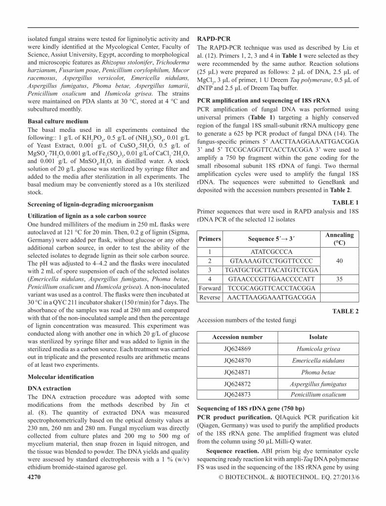
Utilization of lignin as a sole carbon sourcethe data in Fig. 1 indicated that Emericella nidulans and Humicola grisea showed high liP activity (176 U/l and175 U/l, respectively), followed by Phoma betae, Aspergillus fumigatus and Penicillium oxalicum (123 U/l, 103 U/l and89 U/l, respectively). Phoma betae showed the highest yield of laccase activity (165 U/l), followed by Penicillium oxalicum and Emericella nidulans (123 U/l and 111 U/l, respectively). Emericella nidulans, Phoma betae and Penicillium oxalicum showed maximum lignin degradation (70 %), followed by Aspergillus fumigatus and Humicola grisea (55 %), over all of the tested 12 fungal strains, which correlated with the highest lignin degradation percentage after the determined incubation period. As a result, Emericella nidulans, Aspergillus fumigatus, Phoma betae, Penicillium oxalicum and Humicola grisea were selected for their high lignin degradation, liP and laccase activities.
4272 © Biotechnol. & Biotechnol. eq. 27/2013/6
Fig. 1. testing the 12 fungal isolates on medium with lignin as a sole carbon source.
Effect of glucose addition to lignin as a carbon sourceThe activities of both enzymes (LIP and laccase) of the five selected fungal strains were increased in the glucose-containing medium as compared to those produced in the glucose-free medium. Humicola grisea showed an increasing in both enzyme activities: liP gave 543 U/l and laccase activity reached 201 U/l. the lignin degradation rate also increased in the glucose-containing medium, reaching its highest percentage (94 %) after 7 days of cultivation (data not shown). in this study, our strain required sugar, especially glucose, for lignin degradation and enzyme production.
our results were in agreement with the report of Rodríguez and toca (24) that lignin degradation did not occur without the presence of a readily metabolizable substrate such as glucose (24). Many other investigators have also reported that glucose enhances both liP and laccase activities (13, 17, 19). these results seem to suggest that the degradation activity
of this strain might be a sugar oxidase, as is the case with C. versicolour Ps4a and Bacillus cereus (4, 22).
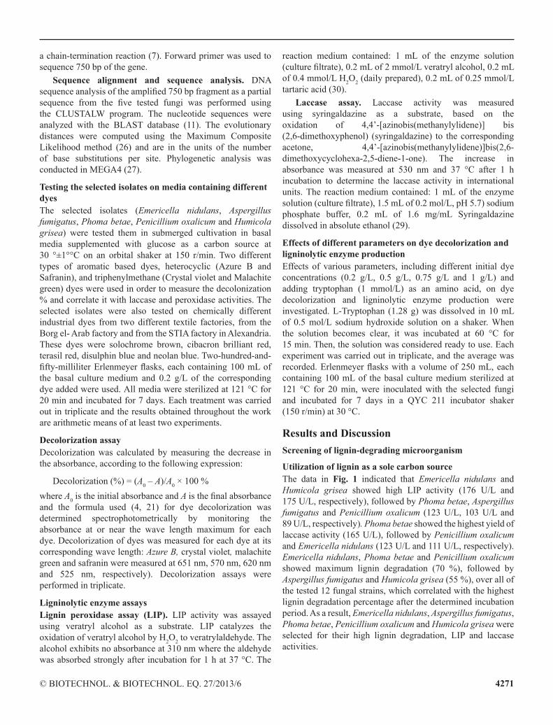
DNA extraction from the ligninolytic enzyme-producing isolates and RAPD–PCRDnA samples were subjected to both spectrophotometric analyses and ran in 1 % agarose gels. the RAPD analysis data for the fungal isolates were used for further molecular identification based on the 18S rDNA sequence. 18S rDNA sequences were obtained from the statistical analysis of the RAPD-PcR band pattern of 12 fungal isolates, using four RAPD primers given in Fig. 2.
The assessment of species confirmation by RAPD analysis involved the assumption that bands of similar size were homologous (28). obviously, data could be misinterpreted and faulty conclusions drawn if different DnA fragments have similar sizes (23). to minimize this effect, we used four different primers for one species, which collectively allowed comparisons between large numbers of bands. A similar approach was applied by liu et al. (12) for the differentiation of species and strains of fungi by RAPD.
PCR amplification and sequencing of 18S rDNAthe optimal tree with a sum of branch length of 12.61036518 is shown in Fig. 3. Fifteen fungi which have similar nucleotide sequence (according to BlASt) may be–on a molecular basics–related to the five fungi we selected. An analytical phylogenetic tree was drawn (Fig. 4) and the data showed that all of the selected isolates had a molecular relationship with other fungi from the same cluster of Ascomycota.
A B
C D Fig. 2. RAPD of 12 isolated fungal strains, using primer 1 (A), primer 2 (B), primer 3 (C) and primer 4 (D). M: 100 bp DnA marker.

4273© Biotechnol. & Biotechnol. eq. 27/2013/6
Fig. 3. Evolutionary distances between the five selected fungi based on the obtained 18S rDnA sequences.
Fig. 4. comparative phylogenetic analysis of the selected fungi with other fungal species.
Effects of different parameters on dye decolorization and ligninolytic enzyme production
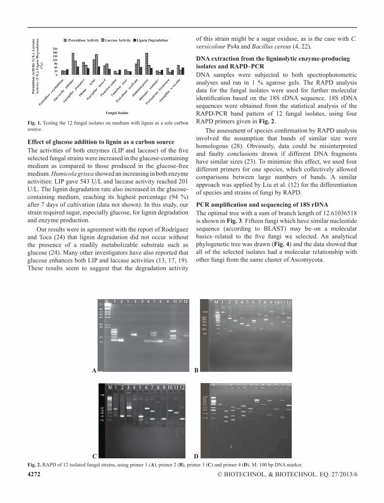
Effect of different dye concentrations in basal mediathis experiment was based on the decolorization of chemically different dyes used for various industrial applications, including textile dyestuffs. the highest enzymatic activities and the highest decolorization rates were obtained at a dye concentration of 0.2 g/l with all of the tested dyes (Table 3).At the highest dye concentration (1 g/l), the lowest decolorization rate was obtained. laccase activity was almost negligible for all the four dyes tested at a concentration of 1 g/l, while liP showed some activity compared with lower laccase activity at high dye concentrations. Humicola grisea, Phoma betae and Penicillium oxalicum were the most degradable fungal strains with respect to the four tested dyes. the results indicated that, Humicola grisea yielded 77 %, 65 %, 80 %, and 76 % decolorization rates for Azure B, Safranin, crystal violet, and Malachite green, respectively. Phoma betae yielded (31 %, 77 %, 60 %, 77 % decolorization rates for Azure B, Safranin, crystal violet, and Malachite green, respectively. Also, Penicillium oxalicum yielded 51 %, 59 %, 72 %, 89 %) decolorization rates for Azure B, Safranin, crystal violet, and Malachite green, respectively. it was observed that the higher dye concentrations had an inhibitory effect on the enzymatic activities of the tested fungi and that the dye concentration at which the tested isolates showed the highest enzymatic activities was 0.2 g/l.
A survey of the literature suggests that the decolorization rate is inversely related to the dye concentration, which could probably be attributed to toxic effects of dyes with regard to the individual bacteria and/or inadequate biomass concentration
A B
c D Fig. 5. lignolytic enzyme activities of the selected isolates and dye decolorization of azure B (A), crystal violet (B), safranin (C), and malachite green (D), in basal medium supplemented with tryptophan.
4274 © Biotechnol. & Biotechnol. eq. 27/2013/6
TABLE 3effect of different dye concentrations on dye decolorization and ligninolytic enzyme activity
Fungal Strain
Dye Concentration (g/L)
Dye0.2 (g/L) 0.5 (g/L) 0.75 (g/L) 1 (g/L)
liPa
(U/l)lac.b
(U/l)Dec.c
(%)liPa
(U/l)lac.b
(U/l)Dec.c
(%)liPa
(U/l)lac.b
(U/l)Dec.c
(%)liPa
(U/l)lac.b
(U/l)Dec.c
(%)
Emericella nidulans
Azure B 65 73 53 45 21 19 20 15 9 12 0 5Safranin 139 91 53 97 31 45 71 27 30 22 11 11
c.V.d 425 18 31 108 50 25 99 5 19 31 1 6M.g.e 53 67 38 32 15 20 32 8 13 18 1 2
Aspergillus umigatus
Azure B 51 57 44 66 10 23 36 5 12 11 0.5 9Safranin 118 35 20 71 21 12 62 12 11 19 3 7
c.V.d 103 91 56 85 68 53 45 34 25 26 1 5M.g.e 105 91 17 51 10 13 47 2 10 21 1 1
Phoma betae
Azure B 92 12 31 37 9 17 13 6 8 5 1 2Safranin 67 21 77 43 18 23 33 15 31 12 5 6
c.V.d 71 22 60 42 40 38 35 10 12 12 0 3M.g.e 130 109 77 33 28 35 26 1 27 12 0 0
Penicillium oxalicum
Azure B 89 21 51 48 12 20 38 4 10 9 2 6Safranin 83 32 59 63 25 25 40 19 23 24 13 10
c.V.d 291 35 72 91 22 56 89 23 30 32 3 14M.g.e 141 105 89 62 37 45 52 10 34 35 6 13
Humicola grisea
Azure B 108 36 77 42 12 56 23 10 26 15 6 7Safranin 103 74 65 79 59 43 65 53 41 21 17 15
c.V.d 376 65 80 309 26 78 111 13 53 43 1 20M.g.e 104 87 76 84 61 53 56 29 34 29 2 10
liPa: lignin peroxidase, lacb.: laccase, Decc.: Decolorization,c.V.d: crystal Violet, M.g.e: Malachite green.
(improper cell-to-dye ratio). other possible reasons could be blockage of azoreductase active sites by dye molecules with different structures (4, 25). there are other reports on white-rot fungi that the percentage of decolorization of the dye increased with increasing dye concentration up to 0.1 g/l, indicating substrate limitation at low dye concentrations (32).
Effect of adding Tryptophan in basal decolorization media the optimal dye concentration decolorized (0.2 g/l) was added to the basal medium with tryptophan as an additive and tween 20 to decrease the hyphal clamping. As shown in Fig. 5, LIP activity increased significantly, and laccase activity increased with a high rate. Humicola grisea showed the highest decolorization rate (99 %), also very high laccase and liP activities with azure B were obtained (3176 U/l and 1104 U/l, respectively). Phoma betae showed the highest liP and laccase for both crystal violet and safranin (3548 U/l and 3042 U/l for liP, and 1085 U/l and 749 U/l for laccase, respectively) and decolorization rate (99 % with crystal violet, and 90 % with safranin).
Supplementation of various cultures of white-rot fungi with tryptophan has been found to have a large stimulatory effect on liP activity levels (2). this enhancement was greater than that that observed in the presence of the liP-recycling agent veratryl alcohol. instead, the observed activity enhancement is likely to result from the protective effect of tryptophan against h2o2 inactivation. Furthermore, tryptophan was found to be a better substrate for liP than veratryl alcohol (2).
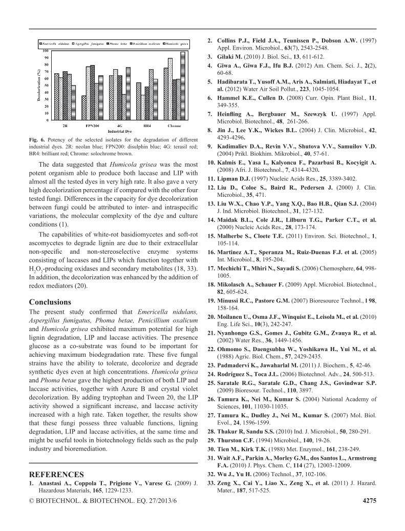
Potency of the selected fungal isolates for biodeclorization of chemically different industrial textile dyes the data illustrated in Fig. 6 showed that, Humicola grisea was the most potent organism able to produce both laccase and liP with almost all the tested dyes at a very high rate. it also showed a very high decolorization percentage if compared with the other four tested fungi. Emericella nidulans was the second active fungus in terms of the enzymatic activities and decolorization rate, followed by Aspergillus fumigatus. the selected isolates showed a high decolorization rate when tested on solochrome brown and disulphin blue.
4275© Biotechnol. & Biotechnol. eq. 27/2013/6
Fig. 6. Potency of the selected isolates for the degradation of different industrial dyes. 2R: neolan blue; FPn200: disulphin blue; 4g: terasil red; BR4: brilliant red; chrome: solochrome brown.
the data suggested that Humicola grisea was the most potent organism able to produce both laccase and liP with almost all the tested dyes in very high rate. it also gave a very high decolorization percentage if compared with the other four tested fungi. Differences in the capacity for dye decolorization between fungi could be attributed to inter- and intraspecific variations, the molecular complexity of the dye and culture conditions (1).
the capabilities of white-rot basidiomycetes and soft-rot ascomycetes to degrade lignin are due to their extracellular non-specific and non-stereoselective enzyme systems consisting of laccases and liPs which function together with h2o2-producing oxidases and secondary metabolites (18, 33). in addition, the decolorization was enhanced by the addition of redox mediators (20).
ConclusionsThe present study confirmed that Emericella nidulans, Aspergillus fumigatus, Phoma betae, Penicillium oxalicum and Humicola grisea exhibited maximum potential for high lignin degradation, liP and laccase activities. the presence glucose as a co-substrate was found to be important for achieving maximum biodegradation rate. These five fungal strains have the ability to tolerate, decolorize and degrade synthetic dyes even at high concentrations. Humicola grisea and Phoma betae gave the highest production of both liP and laccase activities, together with Azure B and crystal violet decolorization. By adding tryptophan and tween 20, the liP activity showed a significant increase, and laccase activity increased with a high rate. taken together, the results show that these fungi possess three valuable functions, ligning degradation, liP and laccase activities, at the same time and might be useful tools in biotechnology fields such as the pulp industry and bioremediation.
REFERENCES1. Anastasi A., Coppola T., Prigione V., Varese G. (2009) J.
hazardous Materials, 165, 1229-1233.
2. Collins P.J., Field J.A., Teunissen P., Dobson A.W. (1997) Appl. environ. Microbiol., 63(7), 2543-2548.
3. Gilaki M. (2010) J. Biol. Sci., 13, 611-612.4. Giwa A., Giwa F.J., Ifu B.J. (2012) Am. chem. Sci. J., 2(2),
60-68.5. Hadibarata T., Yusoff A.M., Aris A., Salmiati, Hiadayat T., et
al. (2012) Water Air Soil Pollut., 223, 1045-1054. 6. Hammel K.E., Cullen D. (2008) curr. opin. Plant Biol., 11,
349-355.7. Heinfling A., Bergbauer M., Szewzyk U. (1997) Appl.
Microbiol. Biotechnol., 48, 261-266.8. Jin J., Lee Y.K., Wickes B.L. (2004) J. clin. Microbiol., 42,
4293-4296.9. Kadimaliev D.A., Revin V.V., Shutova V.V., Samuilov V.D.
(2004) Prikl. Biokhim. Mikrobiol., 40, 57-61.10. Kalmis E., Yasa I., Kalyoncu F., Pazarbasi B., Kocyigit A.
(2008) Afri. J. Biotechnol., 7, 4314-4320.11. Lipman D.J. (1997) nucleic Acids Res., 25, 3389-3402.12. Liu D., Coloe S., Baird R., Pedersen J. (2000) J. clin.
Microbiol., 35, 471.13. Liu W.X., Chao Y.P., Yang X.Q., Bao H.B., Qian S.J. (2004)
J. ind. Microbiol. Biotechnol., 31, 127-132.14. Maidak B.L., Cole J.R., Lilburn T.G., Parker C.T., et al.
(2000) nucleic Acids Res., 28, 173-174.15. Malherbe S., Cloete T.E. (2011) environ. Sci. Biotechnol., 1,
105-114.16. Martinez A.T., Speranza M., Ruiz-Duenas F.J. et al. (2005)
int. Microbiol., 8, 195-204.17. Mechichi T., Mhiri N., Sayadi S. (2006) chemosphere, 64, 998-
1005.18. Mikolasch A., Schauer F. (2009) Appl. Microbiol. Biotechnol.,
82, 605-624.19. Minussi R.C., Pastore G.M. (2007) Bioresource technol., l 98,
158-164.20. Moilanen U., Osma J.F., Winquist E., Leisola M., et al. (2010)
eng. life Sci., 10(3), 242-247.21. Nyanhongo G.S., Gomes J., Gubitz G.M., Zvauya R., et al.
(2002) Water Res., 36, 1449-1456.22. Ohmomo S., Daengsubha W., Yoshikawa H., Yui M., et al.
(1988) Agric. Biol. chem., 57, 2429-2435.23. Padmadervi K., Jawaharlal M. (2011) J. Biochem., 5, 42-46.24. Rodríguez S., Toca J.L. (2006) Biotechnol. Adv., 24, 500-513.25. Saratale R.G., Saratale G.D., Chang J.S., Govindwar S.P.
(2009) Bioresour. technol., 110, 3897.26. Tamura K., Nei M., Kumar S. (2004) national Academy of
Sciences, 101, 11030-11035.27. Tamura K., Dudley J., Nei M., Kumar S. (2007) Mol. Biol.
evol., 24, 1596-1599.28. Thakur R, Sandu S.S. (2010) ind. J. Microbiol., 50, 280-291.29. Thurston C.F. (1994) Microbiol., 140, 19-26.30. Tien M., Kirk T.K. (1988) Met. enzymol., 161, 238-249.31. Wait A.F., Parkin A., Morley G.M., dos Santos L., Armstrong
F.A. (2010) J. Phys. chem. c, 114 (27), 12003-12009.32. Wu J., Yu H. (2006) technol., 37, 102-106.33. Zeng X., Cai Y., Liao X., Zeng X., et al. (2011) J. hazard.
Mater., 187, 517-525.


