Embed Size (px)
Citation preview
BIOCHIMICA ET BIOPHYSICA ACTA 47
~BA 755~8
LAMELLAR AND HEXAGONAL LIPID PHASES VISUALIZED BY
FREEZE-ETCHING
DAVID W. DEAMER, ROBERT LEONARD, ANNETTE TARDIEU* AND DANIEL BRANTON**
Department of Zoology, University of California, Davis, Calif. 956x6 (U.S.A .), *Centre G~n~tique Mol$culaire, Centre National de la Recherche Scientifique, Gif-sur-Yvette, 91 (France) and **Depart- ment of Botany, University of California, Berkeley, Calif. 94720 (U. S.A .)
(Received May 2Ist , 197 o)
SUMMARY
i. Rapid freezing of lipid-water preparations preserves the structure of the high temperature phases. Both lameUar and hexagonal phases can be readily demonstrated by electron microscopy and X-ray observations.
2. Without careful controls, contaminants are readily deposited on fresh fracture faces. The contamination can take the form of particles resembling those found on natural membranes.
3- Neither degree of saturation, degree of hydration, nor cholesterol admixture significantly affects the appearance of lamellar fracture faces which appear uniformly smooth. On uncontaminated specimens, no structures were found which resembled the particulate material of natural membranes.
INTRODUCTION
Many hypotheses of membrane structure are based, to some extent, on electron microscopic evidence. The electron microscope allows direct visualization at the near molecular level and makes it possible to examine specific areas within membra- nous structures. The freeze-fracture and freeze-etch methods of electron microscopy are particularly useful because they expose structure within the hydrophobic plane of biological membranes 1-7. In the freeze-fracture technique, a specimen is rapidly frozen to liquid nitrogen temperature, fractured in vacuo, shadowed and replicated with platinum and carbon. The specimen is dissolved away, and the replica may be examined in an electron microscope. In freeze-etching, ice is sublimed from the fractured specimen before it is shadowed. Etching sometimes reveals greater detail and may provide more information than fracture alone.
Since freeze-fracture planes and the resulting electron microscopic images depend on the physical state of membrane components, the significance of freeze-- fracture and freeze-etch results would be greatly enhanced if it could be shown that phase transitions are avoided during rapid freezing. One way to test the technique in this regard is to examine fracture faces in bulk-phase lipid systems. X-Ray diffraction studies of model systems involving lipids, proteins and wate#, 9 reveal
Biochim. Biophys. Acta, 219 (197o) 47--60
48 D.W. DEAMER et al.
a remarkable polymorphism which is highly sensitive to temperature and water concentration. I t is known that the low-temperature equilibrium conformation of lipid hydrocarbon chains is stiff, frequently with rotational disorder; the only structure compatible with stiff chains is lamellar. At higher temperatures the chains become more liquid and less ordered; structures compatible with these more liquid chains include hexagonal, cubic and other phases in which the chains fill volumes of odd shape 8. If rapid freezing could be demonstrated to preserve hexagonal phases, it would suggest that complex liquid-crystalline phases might also be preserved in frozen biological membranes.
In the present study, we addressed ourselves to the following questions. (I) Can the various lipid phases defined by X-ray methods be preserved and visualized by the freeze-etch technique? (2) What is the effect of hydration on the fracture faces? (3) What is the effect of unsaturation on the fracture faces? (4) Are there any structures in bulk-phase lipid systems that can account for the particulate appearance of biological membrane systems?
METHODS
Lipids Fat ty acids and cholesterol were purchased from Applied Science Laboratories,
Inc. (State College, Pa.). Lysolecithin was purchased from Calbiochem, Inc. (Los Angeles, Calif.). L-~-Dipalmitoyl phosphatidyl choline was purchased from Mann Research Laboratories (New York, N.Y.). Strontium laurate and strontium palmitate were prepared according to the method of SPEGT AND SKOULIOS 10, Egg lecithin was prepared by the method of LEA AND RHODES 11. Calcium cardiolipin (a gift of M. Faure) was precipitated from an aqueous solution of sodium cardiolipin prepared from beef heart TM.
Freeze-fracturing and freeze-etching Specimens were affixed to 3-mm flat copper discs or dish-shaped nickel or copper
discs and frozen in liquid Freon. Anhydrous lipids and fa t ty acids which tended to crumble during fracturing were either melted onto the discs, pressed into pellets or, in the case of calcium stearate and cholesterol, embedded in ethylene glycol prior to freezing. All materials with unsaturated bonds were handled in a dry nitrogen atmosphere. Specimens were fractured and replicated in a Balzers freeze-etch appa- ratus as described elsewhere1, TM. Etching conditions were varied and are specified in the figure legends. Shadows are white, and all figures are mounted with the shadow direction from bottom to top.
X-Ray diffraction The X-ray experiments were carried out in vacuo in a Guinier-type camera,
with bent quartz monochromator. The sample, I cm in diameter and I mm thick, was mounted between thin mica sheets in a 4o-g vacuum-tight brass holder. The sample holder was supported by a stand in which the temperature could be controlled f rom-3o to 300 °. In order to correlate freeze-etch and X-ray diffraction results, samples for X-ray diffraction were cooled as rapidly as possible by plunging the mounted specimens into liquid nitrogen and then remounting them in the diffraction
Biockim. Biophys. Acta, 219 (197 o) 47-60

LIPID PHASES VISUALIZED BY FREEZE-ETCHING 49
stand which had been precooled to - 3 0 °. Diffraction patterns were obtained before and after freezing.
Repeat distances Light diffraction patterns of selected regions in high contrast transparencies
of electron micrographs were produced with a o.5-mW gas laser beam (Electro-Optics Inc., Palo Alto, Calif.) or a modified light microscope system a4. The diffraction patterns were recorded on film, and spacings .were measured from densitometer tracings. The electron microscopic magnifications were individually measured on photographs of gratings (216o lines/mm) whose replicas were placed on the same electron microscope grid with the freeze-etch replica.
RESULTS
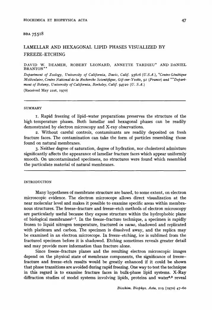
Freeze etching During preliminary experiments we noted occasional variability in the appear-
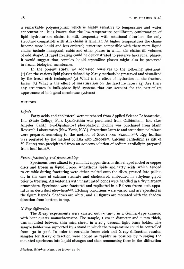
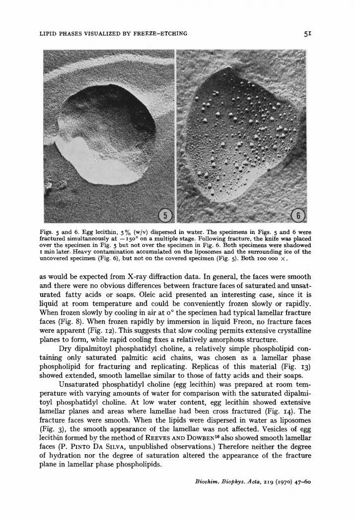
ance of identical lipids. For example, samples of phospholipid appeared either smooth (Fig. I) or particulate (Fig. 2) depending upon which of two freeze-etch machines was used. A thorough analysis of the possible variables led us to ascribe the particles seen in Fig. 2 to contamination of the fracture face in the vacuum of the freeze-etch device. Specifically, we found that if the cooling system of the knife was shut off for more than 3 min during final cutting, the specimen assumed an orange-peel appearance (Figs. 3 and 4) similar to that observed by FLUCK et al. 15. Condensable vapors trapped on the knife lead-in coils may have contaminated the specimen face when the knife cooling system was turned off. Specimens were also contaminated if the cutter was not positioned directly above the sample after the final fracture (Figs. 5 and 6). The pattern of contamination was specific and repro- ducible. Turning off the knife cooling resulted in contamination of the lipids but not the surrounding ice and always produced an orange-peel effect (Fig. 4). Improper positioning of the knife caused contamination of both lipid and ice, but the contam- inant was always more particulate and sharply outlined on the lipid faces than on the ice surfaces (Fig. 6). We found that many of the lipids were more sensitive to contamination than our previous experience with biological membranes had led us to expect. Fa t ty acids and their salts were particularly troublesome and sometimes showed particles (Fig. 9) or plaques IFig. II) in spite of efforts to prevent contami- nation.
As a result of these and other preliminary experiments we learned to reduce contamination to a minimum. In most cases proper knife cooling and positioning prevented detectable contamination. Nevertheless, subtle differences in the quality of the fracture faces were evident from day to day and these variations rather than resolution problems defined the limits within which it was possible to say whether a given lipid was "the same as" or "different than" another lipid in appearance.
Lamellar phases The simplest lipids were an obvious starting point for this study. Therefore,
stearic acid, oleic acid, calcium stearate, calcium oleate and strontium palmitate were fractured and replicated (Figs. 7-11). Each formed lamellar fracture faces,
Biochim. Biophys. Acta, 219 (197 o) 47-60
5 ° D . W . DEAMER el~ al.
Fig. I. Egg lec i th in-choles terol , I : I mole rat io, 5 0 % (w/v) d ispersed in water . No e tching. IO0 0 0 0 X •
Fig. 2. Egg lec i th in-choles terol , i : I mole ra t io , 5 ° % (w/v) dispersed in water . Compare wi th Fig. I. No e tching , ioo ooo × . Fig. 3. Egg leci thin, lO% (w/v) dispersed in water . Shadowing was carr ied ou t i m m e d i a t e l y following f rac ture a t - -15 o° (no etching) . C u t t i ng a r m and knife were cooled con t inuous ly for 3 ° m i n pr ior to f rac tu re and repl icat ion. F r a c t u r e d l iposomes show s m o o t h surfaces free of par- t icles, ioo ooo × . Fig. 4. Egg leci thin io % (w/v) dispersed in water . Liquid. n i t rogen . . . . to cu t t i ng a r m was t u r n e d off for 8 rain pr ior to fra~' turing a t -- 15 o°. Shadowing was ~arrxed ou t i m m e d i a t e l y following f rac tu re a9 in E ~ _ 3 l°/~rticu/at~ mmtezial ha s a c c u m u l a t e d on all f r ac tu red l iposome faces, b u t no t on s u r r o u n d i h g ' i c e - ' ~ . ~ o o × .
LIPID PHASES VISUALIZED BY FREEZE--ETCHING 51
Figs. 5 and 6. Egg lecithin, 5 % (w/v) dispersed in water. The specimens in Figs. 5 and 6 w e r e
fractured simultaneously at --I50 ° on a multiple stage. Following fracture, the knife was placed over the specimen in Fig. 5 but not over the specimen in Fig. 6. Both specimens were shadowed x rain later. Heavy contamination accumulated on the liposomes and the surrounding ice of the uncovered specimen (Fig. 6), but not on the covered specimen (Fig. 5). Both IOO ooo ×.
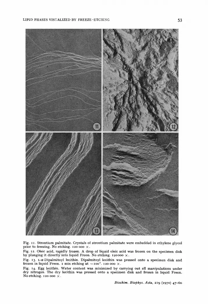
as would be expected from X-ray diffraction data. In general, the faces were smooth and there were no obvious differences between fracture faces of saturated and unsat- urated fa t ty acids or soaps. Oleic acid presented an interesting case, since it is liquid at room temperature and could be conveniently frozen slowly or rapidly. When frozen slowly by cooling in air at o ° the specimen had typical lamellar fracture faces (Fig. 8). When frozen rapidly by immersion in liquid Freon, no fracture faces were apparent (Fig. I2). This suggests that slow cooling permits extensive crystalline planes to form, while rapid cooling fixes a relatively amorphous structure.
Dry dipalmitoyl phosphatidyl choline, a relatively simple phospholipid con- talning only saturated palmitic acid chains, was chosen as a lamellar phase phospholipid for fracturing and replicating. Replicas of this material (Fig. I3) showed extended, smooth lamellae similar to those of fa t ty acids and their soaps.
Unsaturated phosphatidyl choline (egg lecithin) was prepared at room tem- perature with varying amounts of water for comparison with the saturated dipalmi- toyl phosphatidyl choline. At low water content, egg lecithin showed extensive lamellar planes and areas where lamellae had been cross fractured (Fig. I4). The fracture faces were smooth. When the lipids were dispersed in water as liposomes (Fig. 3), the smooth appearance of the lamellae was not affected. Vesicles of egg lecithin formed by the method of REEVES AND DOWBEN 16 also showed smooth lamellar faces (P. PINTO DA SILVA, unpublished observations.) Therefore neither the degree of hydration nor the degree of saturation altered the appearance of the fracture plane in lamellar phase phospholipids.
Biochim. Biophys. Acta, 219 (x97 o) 47-60

52 D . W . DEAMER et al.
Fig. 7- Crystalline stearic acid. Stearic acid was melted and allowed to recrystallize on the specimen disk. No etching. 12o ooo x .
Fig. 8. Oleic acid, slowly frozen. A drop of liquid oleic acid was crystallized on the specimen disk at 4 °. The disk was then plunged into liquid Freon. No etching. 12o ooo X •
Fig. 9. Calcium stearate. Crystals of calcium stearate were embedded in ethylene glycol prior to freezing in liquid Freon. No etching. 12o ooo x . Fig. io. Calcium oleate. Crystals of calcium oleate were pressed onto a specimen disk and frozen in liquid Freon. No etching. 12o ooo × .
Biochim. Biophys. ,4cta, 219 (197 o) 47-6o
LIPID PHASES VISUALIZED BY FREEZE-ETCHING 53
Fig. i i. S t r o n t i u m pa lmi ta te . Crys ta l s of s t r o n t i u m p a l m i t a t e were e m b e d d e d in e thy lene glycol prior to freezing. No etching. 12o ooo × . Fig. 12. Oleic acid, rap id ly frozen. A drop of l iquid oleic acid was frozen on t h e spec imen disk by p lung ing i t d i rec t ly into l iquid Freon. No etching. 12oo0o X.
Fig. 13. L-~-Dipalmitoyl lecithin. Dipa lmi toy l leci thin was pressed on to a spec imen disk a n d frozen in l iquid Freon. i min e tch ing a t -- IOO °. 12o ooo × .
Fig. 14 . Egg lecithin. W a t e r con t en t was min imized b y ca r ry ing ou t all m a n i p u l a t i o n s u n d e r d ry ni t rogen. The d ry leci thin was pressed on to a spec imen disk and frozen in l iquid Freon. No e tching . 12o ooo × .
Biochim. Biophys. Acta, 219 (197 o) 47-60
54 D . W . DEAMER et al.
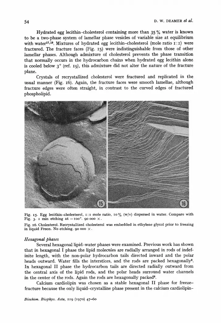
Hydrated egg lecithin-cholesterol containing more than 35 % water is known to be a two-phase system of lamellar phase vesicles of variable size at equilibrium with waterlL TM. Mixtures of hydrated egg lecithin-cholesterol (mole ratio I : I ) were fractured. The fracture faces (Fig. 15) were indistinguishable from those of other lamellar phases. Although admixture of cholesterol prevents the phase transition that normally occurs in the hydrocarbon chains when hydrated egg lecithin alone is cooled below 3 ° (ref. I9), this admixture did not alter the nature of the fracture plane.
Crystals of recrystallized cholesterol were fractured and replicated in the usual manner (Fig. 16). Again, the fracture faces were smooth lamellae, although fracture edges were often straight, in contrast to the curved edges of fractured phospholipid.
Fig. 15. Egg leci thin-cholesterol , i : i mole ratio, io % (w/v) dispersed in water . Compare wi th Fig. 3, i rain e tching a t - ioo °. 90 ooo × .
Fig. 16. Cholesterol. Recrystal l ized cholesterol was embedded in e thylene glycol prior to freezing in l iquid Freon. No etching. 90 ooo × .
Hexagonal phases Several hexagonal lipid-water phases were examined. Previous work has shown
that in hexagonal I phase the lipid molecules are radially arranged in rods of indef- inite length, with the non-polar hydrocarbon tails directed inward and the polar heads outward. Water fills the interstices, and the rods are packed hexagonally s. In hexagonal II phase the hydrocarbon tails are directed radially outward from the central axis of the lipid rods, and the polar heads surround water channels in the center of the rods. Again the rods are hexagonally packed 8.
Calcium cardiolipin was chosen as a stable hexagonal II phase for freeze- fracture because the only liquid-crystalline phase present in the calcium cardiolipin-
Biochim. Biophys. Acta, 219 (197 o) 47-60

LIPID PHASES VISUALIZED BY FREEZE-ETCHING 55
Fig. 17. Calcium cardiolipin. Calcium cardiolipin was precipitated f rom an aqueous suspension of sodium cardiolipin by addition of o. I M CaC12 .The precipitate was dried by filtration and pressed onto a specimen disk. i rain etching at - lOO% 90 ooo × .
Fig. i8. Egg lecithin-cholesterol, i : i mole ratio. Wate r content was minimized as described in
Fig. 14. No etching. 90 ooo × .
Fig. 19. S t ron t ium laurate. Melted s t ron t ium laurate at 3oo ° was rapidly frozen in liquid Freon. No etching. 90 ooo × .
Fig. 2o. Total spinach chloroplast lipids. 5o % (w/v) gel in water, i min etching at -- Ioo °. 90 ooo × .
Biochira. Biophys. Acta, 219 (197 o) 47-60

56 9. w. DEAMER et al.
water system from room temperature to - 3 °o is the hexagonal II phase (T. GOLIK- KRZYWlCKI, unpublished results). A replica fractured at -IOO ° is shown in Fig. 17. Calcium cardiolipin freeze-fractured at - 3 0 °, the lowest temperature at which our equipment permitted X-ray diffraction analysis, appeared similar to the material fractured at -lOO% In both cases, the fracture faces appear distinctly different from those of the lamellar phase. Although there were preferred fracture planes, the fracture faces were not smooth but were composed of long, parallel lines. The fracture faces thus took on a ribbed appearance. Furthermore, fractures occurred along at least two fracture planes at approx. 12o ° to each other.
Fracture faces resembling those of calcium cardiolipin were also found in anhydrous mixtures of egg lecithin-cholesterol (mole ratio I : I ) (Fig. 18). X-Ray diffraction studies of egg lecithin-cholesterol confirmed that it was in a hexagonal II phase. When water was added (50 % by weight), the hexagonal phase completely disappeared and was replaced by a lamellar phase (Fig. 15). A similar pattern of fracture planes was also found in strontium laurate heated to 30o ° and rapidly cooled by plunging into liquid Freon (Fig. 19). However, the spacings were too small to resolve the individual rods. X-Ray diffraction studies have shown that strontium laurate is in the hexagonal II phase at 300 ° (ref. 20).
The same ribbed pattern was also found in hydrated total lipid extracts of spinach chloroplasts (Fig. 2o), but it is unknown which lipids of this complex mixture are involved in the structures shown. The replicas of the spinach lipids also contained smooth lameUar areas and other unidentified configurations. X-Ray diffraction of such preparations suggests the existence of several different phases, none of which could be clearly resolved 11.
Lysolecithin-water mixtures form hexagonal I phases 2z and were prepared for freeze-etching by incubating a mixture of 60 % lipid-4o % water at 37 ° for several days. The incubation was carried out in a closed vessel with the lipid samples already on discs so that they could be rapidly frozen from the 37 ° incubation temperature. Replicas of this phase are shown in Figs. 21 and 22. As in the hexagonal II phase, the fracture faces are composed of indefinitely long, parallel lines (Fig. 21). However, it was not possible to determine whether the rods were fracturing through centers or along surfaces. If the samples were air-dried for 2 min or more before being frozen, extensive regions reverted to the lamellar phase.
Areas of replicas which apparently represented cross-fractures through the the hexagonal arrays of lipid rods (Fig. 22) were also found. If the reader will view Fig. 22 by sighting along the plane of the page and simultaneously rotate the figure, three distinct linear arrays will be seen. These form angles of approx. 12o ° with each other. Evidence for the spacing and angles of the arrays seen in Fig. 22 was also provided by light diffraction patterns (Fig. 22, inset).
X-Ray diffraction X-Ray diffraction patterns of dry or nearly dry phospholipid samples before
and after freezing were quite similar, and the 4.5-A diffuse band typical of liquid paraffins was found in both. This demonstrates that after freezing the liquid structure was preserved and crystallization of the paraffin chains was avoided.
On the other hand, the hexagonal I phase of lysolecithin with 40 % water became lamellar upon freezing, suggesting that the chain conformation had been
Biochim. Biophys. Acta, 219 (197 o) 47-6o

LIPID PHASES VISUALIZED BY FREEZE-ETCHING 57
Fig. 21. Lysolecithin, 6o °/o (w/v) dispersed in water. Lipid rods in face view. No etching. 9o ooo ×. Fig. 22. Lysolecithin, 6o O/o (w/v) dispersed in water. Lipid rods cross fractured, i min etching at --ioo °. 18o ooo ×. Inset: laser diffraction pattern of the same area.
transformed from a liquid to a stiff conformation. This finding was contrary to freeze- fracture observations which suggested that extensive hexagonal regions were preserved after freezing. The apparent discrepancy may be explained by the fact that time- dependent phase transitions occurred in the X-ray experiments but not the freeze- etch experiments because cooling was slower in the X-ray diffraction experiments where the mass of the sample and sample holder was much greater than in the freeze-etching experiments.
Repeat distances Table I compares X-ray and freeze-etch measurements of repeat distances
for lamellar and rod-like structures. The freeze-etch measurements are subject to substantial error because the angle of fracture varies from one part of the sample to another. In general, however, the repeat distances found in freeze-fractured material closely approximate those derived from X-ray data.
DISCUSSION
Our results show that consistent and recognizable features distinguish different l ip id-water phases when prepared by the freeze-etch technique. The extended smooth regions are characteristic of the lalnellar phases. Alterations in the character of either the hydrophilic or hydrophobic portions of the lipid do not detectably alter the appearance of the smooth fracture faces of the lamellar phase. Similarly, the ribbed fracture faces are found only in hexagonal phases. Thus, certain characteristic
Biochim. Biophys. Acta, 219 (197 o) 47-60

58
TABLE I
P H Y S I C A L P A R A M E T E R S OF L I P I D S Y S T E M S
D. W. DEAMER gt al.
Phase Repeat distances (A)
Freeze-fracture X-Ray diffraction
LameUar Strontium palmitate 44 43 Dipalmitoyl phosphatidyl choline 54 55 Egg lecithin (dry) 57 5 °-6o
Hexagonal Calcium cardiolipin 52 52 Lecithin-cholesterol (dry) 48 48.5 Strontium laurate < 3 ° 23.2 Chloroplast lipids 49 46.7 Lysolecithin 60 66
patterns when seen in freeze-etch replicas, can be considered diagnostic for certain molecular arrangements within the specimen, and many aspects of the long-range molecular order detected by X-ray diffraction can be visualized by freeze-etching.
Initially we considered the possibility that unsaturated fat ty acid chains might produce some of the particulate textures seen on fracture faces of biological specimens. However, our results with unsaturated lipids do not support this possibility. Although higher-resolution shadowing techniques might reveal subtle differences between the fracture faces of saturated and unsaturated lipids, under our conditions both were fairly smooth and did not contain structures that could account for the particles found in some biological membranes.
We also considered the possibility that some of the roughness in freeze-etched biological membranes could be produced by lipid hydration. However, hydration of phospholipid had little effect on the appearance of the fracture faces, and all of our evidence shows that bulk-phase lipids alone do not contain discontinuities which can account for the particulate appearance of fractured biological membranes. The particles in biological membranes may be due to proteins or localized and specific protein-lipid interactions.
We have examined only a few lamellar and hexagonal bulk-phase lipids. More work is required to determine whether other lipids and conformations can survive rapid freezing. However, it is encouraging to note that specimen hydration does not appear to be a critical factor in determining whether a specimen will fracture or what the fracture faces will look hke. Even where hydration decreases the stability of a high-temperature liquid phase, OUl results with lysolecithin suggest that phase transitions can be inhibited by very rapid freezing.
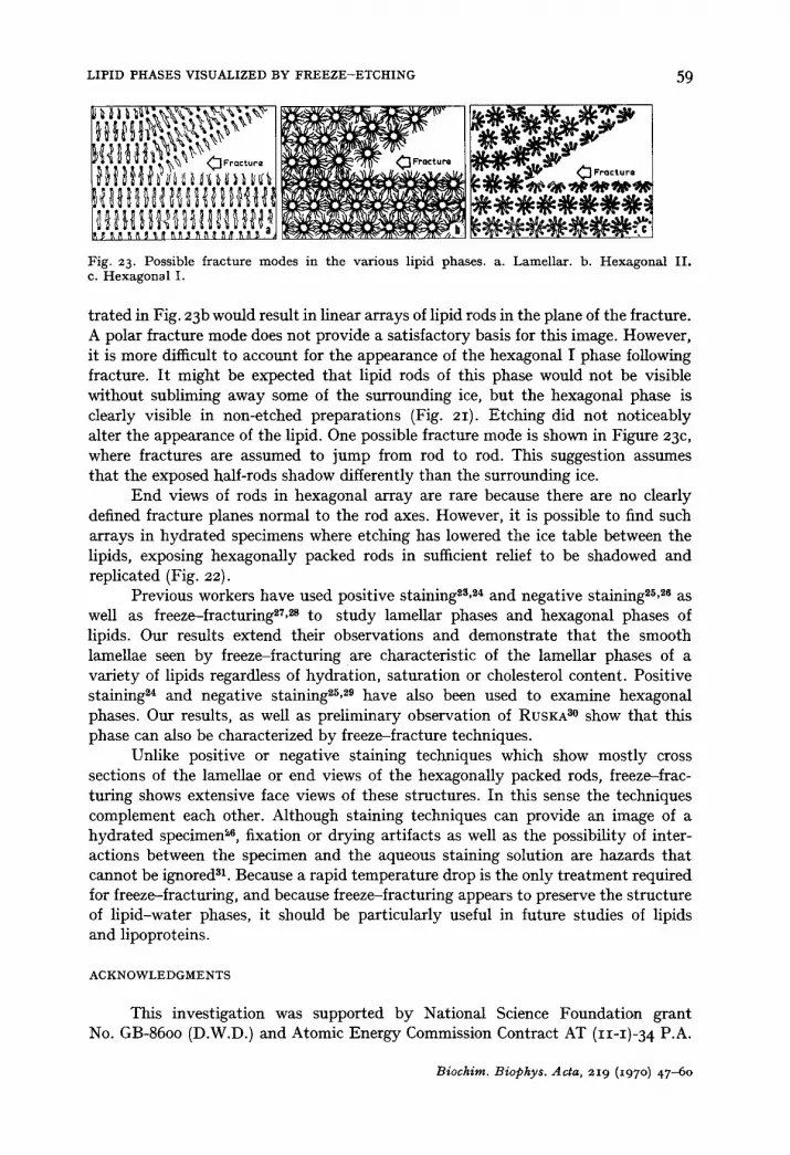
The present observations are consistent with the notion that fractures occur along non-polar regions. Non-polar fractures for lamellar and hexagonal phases are diagrammed in Fig. 23. The lamellar phase fracture is easily understood in terms of non-polar fracture planes (Fig. 23a). Fractures would part lipid bilayers along the plane of hydrocarbon tails where only weak Van der Waal's interactions act to stabilize the bilaye#. Non-polar fracture planes also provide a ready explanation for the appearance of fracture faces in hexagonal n phase. The fracture mode illus-
Biochirn. Biophys. Acta, 219 (I97 o) 47-60
LIPID PHASES VISUALIZED BY FREEZE-ETCHING 59
~ ~ l l ~ ~,~;~ ~.~-~.~,~ i ~ ~ ~ ,
~ t b ~ d'l Fro¢tur*
Fig. 2 3. Possible f rac tu re m o d e s in t he va r ious lipid phases , a. Lamel la r . b. H e x a g o n a l I I . c. H e x a g o n a l I.
trated in Fig. 23b would result in linear arrays of lipid rods in the plane of the fracture. A polar fracture mode does not provide a satisfactory basis for this image. However, it is more difficult to account for the appearance of the hexagonal I phase following fracture. I t might be expected that lipid rods of this phase would not be visible without subliming away some of the surrounding ice, but the hexagonal phase is clearly visible in non-etched preparations (Fig. 21). Etching did not noticeably alter the appearance of the lipid. One possible fracture mode is shown in Figure 23c, where fractures are assumed to jump from rod to rod. This suggestion assumes that the exposed half-rods shadow differently than the surrounding ice.
End views of rods in hexagonal array are rare because there are no clearly defined fracture planes normal to the rod axes. However, it is possible to find such arrays in hydrated specimens where etching has lowered the ice table between the lipids, exposing hexagonally packed rods in sufficient relief to be shadowed and replicated (Fig. 22).
Previous workers have used positive staining~3, 24 and negative stalning2Sm as well as freeze-fracturing~7, ~ to study lameUar phases and hexagonal phases of lipids. Our results extend their observations and demonstrate that the smooth lameUae seen by freeze-fracturing are characteristic of the lamellar phases of a variety of lipids regardless of hydration, saturation or cholesterol content. Positive staining 24 and negative staining25, ~9 have also been used to examine hexagonal phases. Our results, as well as preliminary observation of RUSKA 3° show that this phase can also be characterized by freeze-fracture techniques.
Unlike positive or negative staining techniques which show mostly cross sections of the lamellae or end views of the hexagonally packed rods, freeze-frac- turing shows extensive face views of these structures. In this sense the techniques complement each other. Although staining techniques can provide an image of a hydrated specimen ~s, fixation or drying artifacts as well as the possibility of inter- actions between the specimen and the aqueous staining solution are hazards that cannot be ignored 31. Because a rapid temperature drop is the only treatment required for freeze-fracturing, and because freeze-fracturing appears to preserve the structure of lipid-water phases, it should be particularly useful in future studies of lipids and lipoproteins.
ACKNOWLEDGMENTS
This investigation was supported by National Science Foundation grant No. GB-86oo (D.W.D.) and Atomic Energy Commission Contract AT (11-1)-34 P.A.
Biochim. Biophys. Acta, 219 (z97 o) 47-60
60 D.W. DEAMER et al.
142 (D.B.). The authors wish to thank Dr. T. Gulik-Kryzwicki and Dr. V. Luzzati for lively interest, discussions and many helpful suggestions. Miss S. Whytock and Mrs. E. Crump provided expert technical assistance.
R E F E R E N C E S
i D. BRANTON, Proc. Natl. Acad. Sci. U.S., 55 (1966) l°48. 2 D. BRANTON, Ann. Rev. Plant Physiol., 2o (1969) 209. 3 n . BRANTON AND R. B. PARK, J. Ultrastruct. Res., 19 (1967) 283. 4 D. W. DEAMER AND n . BRANTON, Science, 158 (1967) 655. 5 P. PINTO DA SILVA AND D. BRANTON, J. Cell Biol., 45 (197 °) 598- 6 E. WEHRLI, K. MUHLETHALER AND H. MOOR, Exptl. Cell Res., 59 (197 o) 336. 7 T. TILLACK AND V. MARCHESI, J. Cell Biol., 45 (I97 °) 649. 8 V. LtlZZATI, in D. CHAPMAN, Biological Membranes, Academic Press, New York, 1968, p. 71. 9 T. GULIK-KRZYWlCKI, E. SCHECHTER, V. LUZZATI AND A. FAURE, Nature, 223 (1969) 1116.
io P. A. SPEGT AND A. E. SKOULIOS, Acta Cryst., 17 (1964) 198. I I C. H. LEA AND D. N. RHODES, Biochim. Biophys. Acta, 17 (1955) 416. 12 M. FAURE AND J. J. COULON-MoRELEC, Ann. Inst. Pasteur, 95 (1958) 18o. 13 H. MOOR AND K. MUHLETHALER, J. Cell Biol., 17 (1963) 609. 14 J. GALL, J. Cell Biol., 31 (1966) I3oA. 15 D. FLUCK, A. HENSON AND D. CHAPMAN, J. Ultrastruct. Res., 29 (I969) 416. 16 J. REEVES AND R. DOWBEN, J. Cell Physiol., 73 (1969) 49. 17 M. BOORGES, D. M. SMALL AND D. G. DERVlClAN, Biochim. Biophys. Acta, 137 (1967) 157. 18 H. LECUYER AND D. G. DERVlClAN, J. Mol. Biol., 45 (1969) 39. 19 B. LADBROOKE, R. WILLIAMS AND D. CHAPMAN, Biochim. Biophys. Acta, 15o (1968) 333. 20 P. A. SPEGT AND A. E. SKOULIOS, Acta Cryst., 21 (1966) 892. 21 E. RIVAS AND V. LUZZATI, J. Mol. Biol., 41 (1969) 261. 22 R. REISS-HUSSON AND V. LUZZATI, Advan. Biol. Med. Phys., I I (1967) 87. 23 W. STOECKENIUS, J. Cell Biol., 12 (1962) 221. 24 W. STOECKENIUS, Proc. European Conf. on Electron Microscopy, Delft, 2 (196o) 716. 25 A. D. BANGHAM AND R. W. HORNE, J. Mol. Biol., 8 (1964) 660. 26 J . A. L u c y AND A. M. GLAUERT, J. Mol. Biol., 8 (1964) 727 . 27 L. A. STAEHELIN, J. Ultrastruct. Res., 22 (1968) 326. 28 J. H. BUCKINGHAM AND L. A. STAEHELIN, J. Microscopy, 90 (1969) 83. 29 E. JONGER AND H. REINAUER, Biochim. Biophys. Acta, 183 (1969) 3o4 • 3 ° C. RUSKA, Nat**rwissenschaften, 12 (1969) 637. 31 A. M. GLAUERT AND J. A. LUCY, J. Microscopy, 89 (1968) I.
Biochim. Biophys. Acta, 219 (197 o) 47-60