Embed Size (px)
Citation preview
1
(Submitted to APL – 20 May 2010)
LaCrO3 heteroepitaxy on SrTiO3(001) by molecular beam epitaxy
L. Qiao1, T.C. Droubay1, M.E. Bowden2, V.Shutthanandan2, T.C. Kaspar1,
S. A. Chambers1
1Fundamental and Computational Science Directorate, Pacific Northwest National
Laboratory, Richland, WA 99352
2Environmental and Molecular Sciences Laboratory, Pacific Northwest National
Laboratory, Richland, WA 99352
Abstract
Stoichiometric, epitaxial LaCrO3 films have been grown on TiO2-terminated SrTiO3(001)
substrates by molecular beam epitaxy using O2 as the oxidant. Film growth occurred in a
layer-by-layer fashion, giving rise to structurally excellent films and surfaces which
preserve the step-terrace structure of the substrate. The critical thickness is in excess of
500 Å. Near-surface Cr(III) is highly susceptible to further oxidation to Cr(V), leading to
the formation of a disordered phase upon exposure to atomic oxygen. Recovery of the
original epitaxial LaCrO3 phase is readily achieved by vacuum annealing.

2
Complex oxides exhibit a range of physical properties unparalleled by any other class
of materials. Their electronic, magnetic, and optical characteristics span the field of
possibilities, and a wealth of applications awaits our ability to understand and control
these properties. Materials synthesis is key to realizing the full potential of complex
oxides, particularly in thin-film form. Gaining understanding of and control over
composition and defect density are of major importance. Moreover, different synthetic
methods appear to have different effects on defect creation. For instance, record high
electron mobilities in La-doped SrTiO3 (STO) grown by hybrid molecular beam epitaxy
(MBE) were recently demonstrated, surpassing what had been achieved previously by
bulk synthesis and thin-film growth.1 Perfecting stoichiometric control and minimizing
defect creation appear to have been critical.2
LaCrO3 (LCO) is an antiferromagnetic insulator with a charge transfer gap of 3.3 eV.3
In the bulk, this perovskite exhibits an orthorhombic structure (space group Pbnm) with
lattice parameters a = 5.513 Å, b = 5.476 Å, c = 7.759 Å. 4 However, isolation of a
pseudocubic cell within the orthorhombic structure yields a = b = c = 3.885 Å (see
structural models in Fig. 1). Thus, LCO is a candidate for heteroepitaxy on SrTiO3(001),
for which a = b = c = 3.905 Å.
In this Letter, we describe the epitaxial growth of LCO on SrTiO3(001) by MBE. 5
Atomically flat, TiO2-terminated SrTiO3 (STO) substrates were prepared as described
previously.6 La and Cr were evaporated from an effusion cell and an electron beam
evaporator, respectively, and the O2 partial pressure was kept constant at ~6 × 10⁻⁸ Torr.
The La flux was measured before and after deposition by a quartz crystal oscillator
(QCO), and the Cr flux was monitored and controlled during deposition by atomic
absor
two-c
samp
diffra
grow
were
photo
qualit
meas
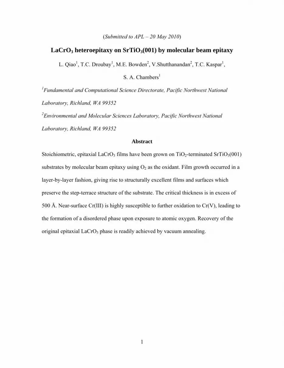
W
grow
persis
persis
FIG. Insetspatterscan
rption. The s
color infrare
ples were coo
action (RHE
wth rate and s
determined
oelectron spe
ty were dete
ured by tapp
We show in
wth time for a
stent oscillat
stent layer-b
1 RHEED ins include strurns for an LCfor a LCO fi
substrate tem
d pyrometer
oled to room
ED) intensit
surface struc
by Rutherfo
ectroscopy (X
ermined by x
ping-mode a
Fig. 1 RHE
a 25 unit cell
tions were o
by-layer grow
ntensity osciuctural modCO film and
film.
mperature du
r. The O2 va
m temperatur
ty oscillation
cture, respect
ord backscatt
XPS), respe
x-ray diffract
atomic force
EED specular
l (u.c., refere
bserved for
wth. A typic
illations as adels of orthord a starting S
3
uring growth
alve was clos
e in vacuo.
ns and patter
tively. Film
tering spectr
ctively. Lat
tion (XRD),
microscopy
r beam inten
enced to the
film thickne
cal pair of RH
a function ofrhombic andTO substrat
was 650° ±
sed immedia
Reflection h
rns were use
m stoichiomet
rometry (RB
ttice paramet
and surface
(AFM).
nsity oscillati
pseudocubic
esses up to 1
RHEED patte
f growth timed pseudocubie, and a typi
50oC, as me
ately after gr
high-energy
d to monitor
try and oxid
BS) and in-si
ters and crys
e morphologi
ions as a fun
c cell) film.
30 u.c., dem
erns for the s
e for a 25 u.ic LCO, typiical AFM im
easured by a
rowth and
electron
r the overall
dation states
itu x-ray
stallographic
ies were
nction of
Similar
monstrating
starting
c. LCO filmical RHEED
mage and line
a
c
m. D
e
4
substrate and the film after cooling to ambient temperature are shown as insets, along
with an AFM image and a line profile. The surface periodicity evolves from (1×1) for the
substrate to (2×2) for the film surface. Moreover, the film surface exhibits a well-defined
terrace-step structure with typical step heights of ~ 4 Å, the c value for pseudocubic LCO.
This result confirms the layer-by-layer growth mode revealed by RHEED oscillations and
the presence of a single surface termination, which we expect is CrO2, based on the fact
that all growths were terminated after an integral number of oscillations were recorded.
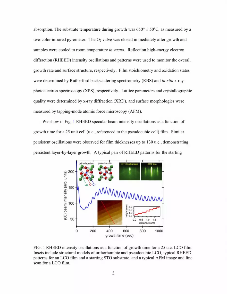
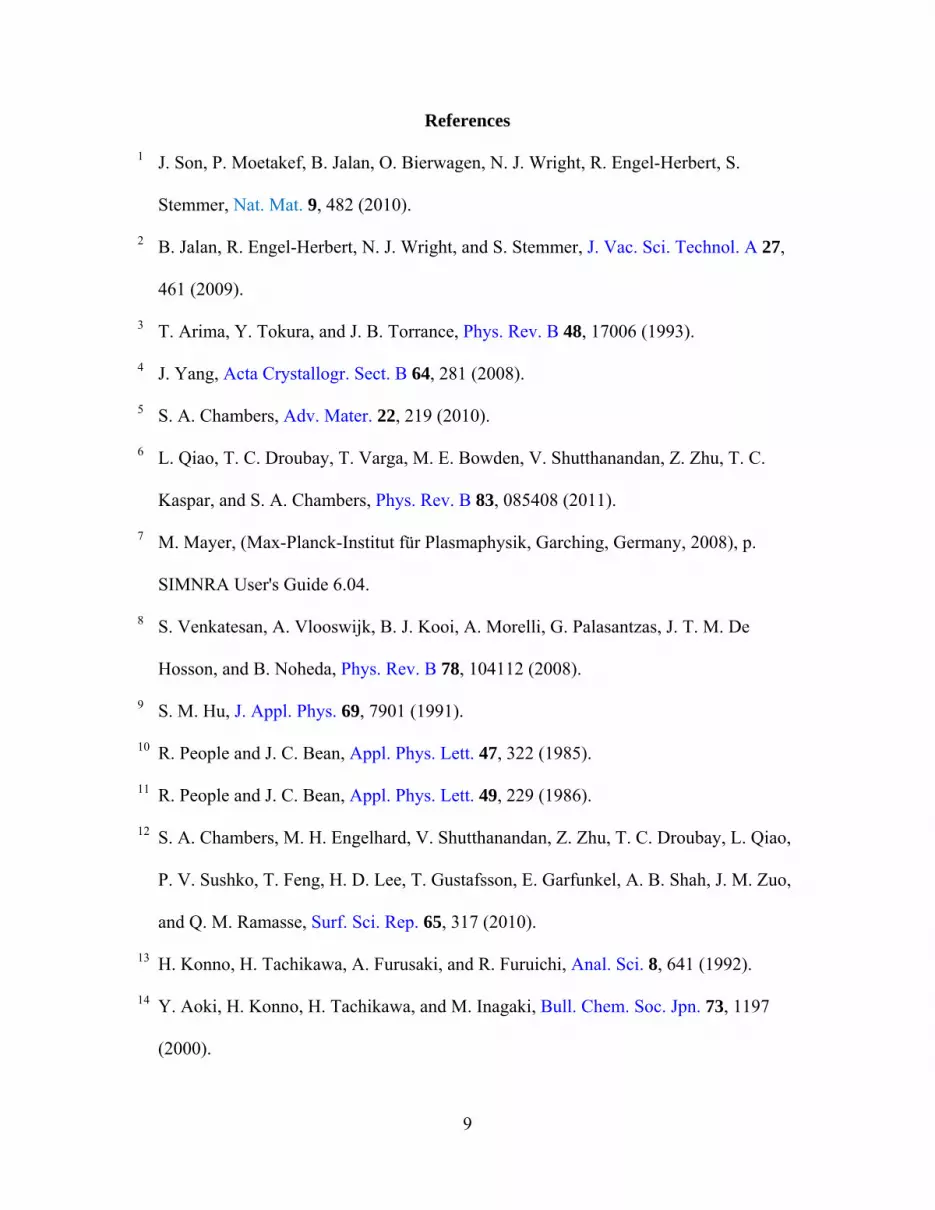
We show in Fig. 2 a typical RBS spectrum obtained for a 25 u.c. film with the
incident beam oriented 7o off normal in a random geometry and a scattering angle of 150o.
Also, shown is a simulated SIMNRA7 spectrum in which the La:Cr atom ratio is 1:1. The
statistical uncertainty for the La peak area (√N/N, where N is the total counts in the peak
above background) is quite low (0.4%) because of the high intensity of this feature and
the low background.
FIG. 2 Non-aligned RBS for a 25 u.c. LCO film using a 2 MeV He ion beam with the incident beam oriented 7o off normal and a scattering angle of 150o. Insets – the Cr peak along (squares) along with a family of simulations for Cr percentages ranging from 17 to 23 at. % (left), and a least-squares fit of these simulations to the Cr feature (right).
Ion energy
Cr
0.6
0.4
0.2
0.0222018
% Cr
2
–2
min
) /
2 min
8000
6000
4000
2000
0
Cou
nts
180017001600150014001300
Ion Energy (keV)
experimentsimulation La:Al = 1.00(4)
LaSr
Cr
Ti
5
In contrast, the uncertainty in the Cr peak area is larger because the Cr peak sits on the
background of the Sr plateau, and is of lower intensity due to the smaller atomic number
of Cr compared to La. After subtraction of the background Sr plateau, the statistical
uncertainty in the Cr peak area is ~5%. Least-squares fits of these two peaks to model
SIMNRA simulations yield absolute La and Cr atomic percentages of 19.3 ± 0.1% and
19.2 ± 0.8%, respectively, leading to a La:Cr ratio of 1.00 ± 0.04. This analysis is shown
for the Cr peak as insets in Fig. 2. The family of simulations in the left inset span an
absolute Cr concentration from 17 to 23 at. %. The 2 values referenced to 2 at the
minimum are plotted in the right inset. The Cr percentage range associated with (2 -
2min) / 2
min = 0.05 below the minimum to (2 - 2min) / 2
min = 0.05 above the minimum
is our estimated uncertainty. RBS data collected on a representative set of thick LCO
films were utilized to calibrate XPS peak area ratios, which were measured in situ on all
films.
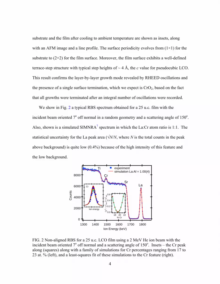
A first indication of the crystallographic quality in these films is revealed by the
persistent Kiessig fringes surrounding the LCO (002) Bragg reflections in the XRD 2θ-ω
scan, as seen in Fig. 3. Matching experiment to simulations yields a film thickness of ~
500 Å, in good agreement with the values obtained from RHEED oscillations and RBS
analysis. The film and substrate reflections are easily distinguished, and the Bragg peak
angle for the film indicates a smaller c dimension than that for the substrate. By
simultaneously fitting profiles to the (202), (113), (114), and (123) in-plane reflections
(not shown), we determine a = b = 3.904 Å and c = 3.881 Å, the latter dimension fully
consistent with the peak position observed in the out-of-plane scan. The film is thus fully
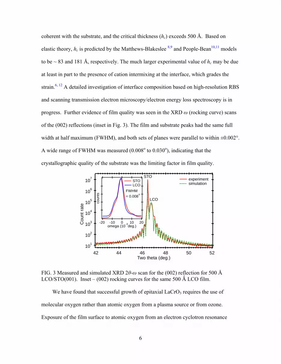
6
coherent with the substrate, and the critical thickness (hc) exceeds 500 Å. Based on
elastic theory, hc is predicted by the Matthews-Blakeslee 8,9 and People-Bean10,11 models
to be ~ 83 and 181 Å, respectively. The much larger experimental value of hc may be due
at least in part to the presence of cation intermixing at the interface, which grades the
strain.6, 12 A detailed investigation of interface composition based on high-resolution RBS
and scanning transmission electron microscopy/electron energy loss spectroscopy is in
progress. Further evidence of film quality was seen in the XRD ω (rocking curve) scans
of the (002) reflections (inset in Fig. 3). The film and substrate peaks had the same full
width at half maximum (FWHM), and both sets of planes were parallel to within ±0.002°.
A wide range of FWHM was measured (0.008o to 0.030o), indicating that the
crystallographic quality of the substrate was the limiting factor in film quality.
FIG. 3 Measured and simulated XRD 2θ-ω scan for the (002) reflection for 500 Å LCO/STO(001). Inset – (002) rocking curves for the same 500 Ǻ LCO film. We have found that successful growth of epitaxial LaCrO3 requires the use of
molecular oxygen rather than atomic oxygen from a plasma source or from ozone.
Exposure of the film surface to atomic oxygen from an electron cyclotron resonance
101
102
103
104
105
106
107
Cou
nt
rate
525048464442Two theta (deg.)
experimentsimulation
LCO
STO
coun
ts
-20 -10 0 10 20omega (10
-3deg.)
STOLCO
FWHM
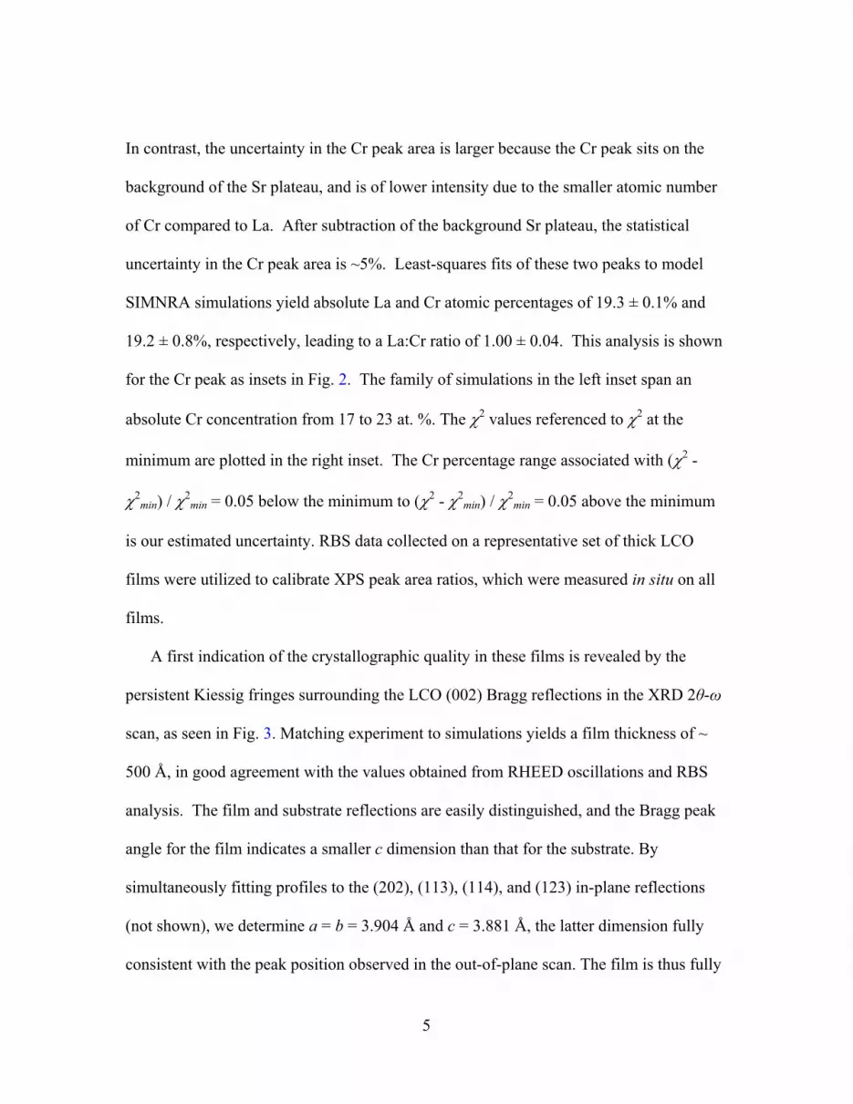
= 0.008o

oxyg
disor
This
occur
regio
polyc
LaCr
comp
More
annea
FIG. vacuuimag The h
LaCr
Intere
en plasma at
dered as Cr(
result is illu
rs as new fea
ns, respectiv
crystalline L
rO4, is ~1.5 n
plete oxidatio
eover, the su
aling in vacu
4 Cr 2p XPSum annealedes for the fil
hypothesis th
rO4 exhibits
estingly, this
t 2.0×10⁻⁵ To
(III) ions in t
strated in Fig
atures appea
vely. These
aCrO4.13,14 T
nm thick bas
on, and is sta
rface can be
uum at the fi
S spectra ford (green) LClm during dif
hat this surfa
a monoclinic
s redox proc
orr for a few
the near-surf
g. 4. A sign
ar at ~576 eV
new peaks a
This Cr(V)-c
ed on angle-
able in high
e reduced bac
ilm growth t
r an as-growCO film. Inse
fferent stage
ace phase is
c structure in
ess at least p
7
w minutes cau
face region a
nificant chan
V and ~586 e
are character
containing la
-resolved XP
vacuum ove
ck to the orig
emperature,
wn (red), oxygets show the es of this red
disordered L
ncommensur
partially pres
uses the surf
are oxidized
nge in the Cr
eV in the 2p3
ristic of Cr(V
ayer, which m
PS and assum
er a period o
ginal epitaxi
as also seen
gen plasma tcorrespondi
dox process.
LaCrO4 is re
rate with ST
serves the te
face to becom
to a higher
2p XPS line
3/2 and 2p1/2
V), as measu
might be dis
ming uniform
of at least two
ial LaCrO3 p
n in Fig 4.
treated (blueing RHEED
easonable be
TO and LCO
errace-step st
me
charge state
e shape
spectral
ured in
sordered
m and
o weeks.
phase by
e), and and AFM
cause
O.15
tructure of
.

8
the original film surface, as seen in the AFM images shown in Fig. 4. A blunting of the
steps and a roughening of the terraces is evident. Additionally, the RHEED pattern
exhibits higher background after recovery of the original phase, indicating a higher point
defect density than was present on the original surface.
In summary, high-quality epitaxial LaCrO3 has been successfully grown on TiO2-
terminated SrTiO3(001) by MBE. Layer-by-layer growth was confirmed by both RHEED
oscillations and AFM images of the final surface. Structural Cr(III) in the near-surface
region is readily oxidized to a disordered phase containing Cr(V) upon exposure to
atomic oxygen, and annealing in vacuo results in conversion back to crystalline LaCrO3.
This interesting result may render this complex oxide useful in fundamental studies of
surface photophysics and photochemistry.
This work was supported by the Office of Science, Division of Materials Sciences
and Engineering and Division of Chemical Sciences, U.S. Department of Energy, and
was performed in the Environmental Molecular Sciences Laboratory, a national scientific
user facility sponsored by the Office of Biological and Environmental Research of the
Department of Energy and located at Pacific Northwest National Laboratory.
9
References
1 J. Son, P. Moetakef, B. Jalan, O. Bierwagen, N. J. Wright, R. Engel-Herbert, S.
Stemmer, Nat. Mat. 9, 482 (2010).
2 B. Jalan, R. Engel-Herbert, N. J. Wright, and S. Stemmer, J. Vac. Sci. Technol. A 27,
461 (2009).
3 T. Arima, Y. Tokura, and J. B. Torrance, Phys. Rev. B 48, 17006 (1993).
4 J. Yang, Acta Crystallogr. Sect. B 64, 281 (2008).
5 S. A. Chambers, Adv. Mater. 22, 219 (2010).
6 L. Qiao, T. C. Droubay, T. Varga, M. E. Bowden, V. Shutthanandan, Z. Zhu, T. C.
Kaspar, and S. A. Chambers, Phys. Rev. B 83, 085408 (2011).
7 M. Mayer, (Max-Planck-Institut für Plasmaphysik, Garching, Germany, 2008), p.
SIMNRA User's Guide 6.04.
8 S. Venkatesan, A. Vlooswijk, B. J. Kooi, A. Morelli, G. Palasantzas, J. T. M. De
Hosson, and B. Noheda, Phys. Rev. B 78, 104112 (2008).
9 S. M. Hu, J. Appl. Phys. 69, 7901 (1991).
10 R. People and J. C. Bean, Appl. Phys. Lett. 47, 322 (1985).
11 R. People and J. C. Bean, Appl. Phys. Lett. 49, 229 (1986).
12 S. A. Chambers, M. H. Engelhard, V. Shutthanandan, Z. Zhu, T. C. Droubay, L. Qiao,
P. V. Sushko, T. Feng, H. D. Lee, T. Gustafsson, E. Garfunkel, A. B. Shah, J. M. Zuo,
and Q. M. Ramasse, Surf. Sci. Rep. 65, 317 (2010).
13 H. Konno, H. Tachikawa, A. Furusaki, and R. Furuichi, Anal. Sci. 8, 641 (1992).
14 Y. Aoki, H. Konno, H. Tachikawa, and M. Inagaki, Bull. Chem. Soc. Jpn. 73, 1197
(2000).

10
15 S. G. Manca and E. J. Baran, J. Appl. Crystallogr. 15, 102 (1982)