Embed Size (px)
Citation preview
.f
ISOLATION AND IDENTIFICATION OF ANTIBIOTIC
PRODUCING MICROORGANISMS FROM NATURAL
HABITATS IN THE KWAZULU-NATAL MIDLANDS
By
Vincent Ifeanyi Okudoh
B.Tech. Hans
Submitted in fulfilment
ofthe academic requirements for the degree of
MASTER OF SCIENCE IN AGRICULTURE
in the
Discipline ofMicrobiology
School ofApplied Environmental Sciences
Faculty ofScience and Agriculture
UNIVERSITY OF NATAL
PIETERMARITZBURG
January, 2001
..
ABSTRACT
The search for new antibiotics continues in a rather overlooked hunting ground. In the course of
screening for new antibiotic-producing microorganisms, seventy-nine isolates showing
antimicrobial activity were isolated from soil samples from various habitats in the KwaZulu
Natal midlands, South Africa. Existing methods of screening for antibiotic producers together
with some novel procedures were reviewed. Both modified agar-streak and agar-plug methods
were used in the primary screens. The use ofselective isolation media, with or without antibiotic
incorporation and/or heat pretreatment, enhanced the development of certain actinomycete
colonies on the isolation plates. Winogradsky's nitrite medium (Winogradsky, 1949), M3 agar
(Rowbotham and Cross, 1977), and Kosmachev's medium (Kosmachev, 1960), were found to
be selective for actinomycetes. Statistical analysis showed highly significant interactions
between isolates, assay media and the test organisms. The diameters of inhibition zones were
found to be larger on Iso-sensitest agar (ISTA)[Oxoid, England] than in nutrient agar plates. Of
the 79 isolates that showed antimicrobial activity, 44 isolates were selected for confirmatory
screening. Of these, 13 were selected for secondary screening. Criteria for selection were based
on significant inhibition of at least two test organisms and/or the inhibition of the specifically
targeted organisms, Pseudomonas and Xanthomonas species. Following secondary screening
eight isolates were considered for further investigation. The isolates were tentatively identified .
on the basis of morphological features, using both light microscopy and scanning electron
microscopy(SEM); their ability to utilize various carbon sources; and selected physiological and
staining tests. Suspected actinomycetes were further characterized on the basis of selected
chemical properties using thin layer chromatography (TLC) and high pressure liquid
chromatography (HPLC) techniques. High pressure liquid chromatography analysis (Beckman
6300 analyzer) detected the presence ofdiaminopimelic acid (DAP) in whole-cell hydrolysates
1
of six of the isolates while TLC analysis confirmed the type ofDAP present. The isolates N2,
N12, N16, N19 and N35 were tentatively identified as Thermomonospora, Saccharopolyspora,
Nocardiodes, Corynebacterium and Promicromonospora, respectively. Isolate N30 was
identified as belonging to the coryneform group ofbacteria, possibly an Arthrobacter species.
Isolate, N8, tentatively identified as Actinosynnema, was unique among the isolates tested as it
showed good antimicrobial activity against all the Gram- positive and Gram-negative bacteria,
and yeasts used as test organisms in the present investigation.
11
DECLARATION
I hereby declare that the work reported in this thesis, except where otherwise indicated, is entirely
the result ofmy own research efforts.
111

ACKNOWLEDGMENTS
My sincere thanks to :
ProfF. M. Wallis, my supervisor for expert criticism and stimulating suggestions. Profwas a dad
to me. His support and advice, even in non-academic matters and his readiness to assist me
despite his tight schedules, gave me strength and courage to complete this project.
Mrs P Nortje, the departmental secretary, for her motherly support, advice and assistance during
my depressing times. Her help in typing out the first draft and valuable information that helped
alleviate my fmancial problems, cannot be quantified in words. I will remain ever grateful.
Miss D Fowlds, Celeste Christianson, Ingrid Schlosser ofMicrobiology Discipline for handling
orders and provision ofneeded materials.
Prof J Hastings, Mr Mervyn Beukes of the Department of Genetics and Prof J Van Staden of
Botany Department for valuable information and material assistance.
Mrs M Hundley of Animal Science Department for help in amino acid analysis.
The staff of the Centre for Electron Microscopy for technical assistance.
The Mlangeni family, Mrs Okeke and family, the director of Camco Farms for help in sample
collection.
Mr and Mrs Gavin and Denyse Webbstock, Mr George Tembo, Dr Uzor Francis, Dr Ufo Uzodike
for support and encouragement.
Dr Esther Ugoji, and my fellow students, Yobo, Sanisha, Pulane, Candice, for their penetrating
criticisms and stimulating suggestions.
IV
To my late father,
ChiefDenis(Ononenyi),
My mother, Josephine,
and my brothers and sisters,
Chudi(Proj), Nkem, Nnanna,
Mbanefo and Obianuju.
v

TABLE OF CONTENTS
Chapter 1: Literature Review
1.1 Introduction 1
1.2 What are Antibiotics? 3
1.3 Classification and Nomenclature of Antibiotics 4
1.4 Antibiotics from Microorganisms - A Review 8
1.5 Biochemistry of Antibiotic Production 14
1.6 Mechanism of Antimicrobial Action. 16
1.7 Spectra of Antimicrobial Activity 20
1.8 Review of Screening Methods. 22
1.8.1 Primary Screening Methods 22
1.8.2 Primary Testing of Antibiotic Production 23
1.8.3 Factors Affecting Antibiotic Production 24
1.8.4 Extraction and Purification Methods 26
1.9 Suggested Newer Methods of Searching for Antibiotic-Producing Microorganisms 26
Chapter 2: Isolation of Antibiotic-Producing Microorganisms
2.1 Introduction
2.2 Materials and Methods
2.2.1 Sample Collection /
2.2.2 Isolation Media
2.2-.3 Scope of the Isolation Programme
30
33
33
34
35
2.2.6 Purification ofIsolates
2.3 Results and Discussion
~~::::::::~~:'-~>--'------'-==-------, ..-"---j-~--------- -------'37 ,-- ---
39
VI
Chapter 3: Screening of Isolates for Antimicrobial Activity
3.1 Introduction
3.2 Materials and Methods
3.2.1 Isolates
3.2.2· Test Organisms
3.2.3 Assay Media
3.2.4 Primary Screening
3.2.5 Secondary Testing of Antibiotic Production
3.2.5.1 Ant~biotic-production Media,
3.2.5.2 Production of the Antibiotic
3.2.5.3 Bioassay for Antibiotic Activity- .
3.2.6 Determination ofMinimum Inhibitory Concentration [MIC]
3.3 Results and Discussion
44
47
47
47
47
47
48
48
48
48
49
51
Chapter 4: Identification of Antibiotic-Producing
Microorganisms I: DESCRIPTION OF ISOLATES
4.1 Introduction 64
4.2 Materials and Methods 67
4.2.1 Organisms 67
4.2.2 Morphological Characterization 67
4.2.2.1 Light Microscopy 67
4.2.2.2 Electron Microscopy 67
4.2.3 Physiological Characterization 68
4.2.3.1 Formation ofMelanin Pigment 68
4.2.3;2 Carbohydrate Utilization 68
4.2.3.3 Other Physiological Tests 69
4.2.4 Biochemical Characterization 69
4.2.4.1 Preparation of Whole-Cell Hydrolysates 69
4.2.4.2 Determination of Sugar Occurring in Whole-cell Hydrolysates 70
4.2.4.2.1 One-way Paper Chromatography for Sugar Determination 70
4.2.4.3 Determination of diaminopimelic acid (DAP) isomers 70
4.2.4.3.1 HPLC Analysis of Whole-cell Hydrolysates for DAP Detection 71
vu
4.2.4.3.2 Two-Dimensional Thin Layer Chromatography for
DAP Determination
4.2.4.3.3 One-Way Paper/Thin-Layer Chromatography for
DAP Determination
4.3 Results and Discussion
Chapter 5: Identification of Antibiotic-Producing
Microorganisms 11: TENTATIVE IDENTITY OF ISOLATES
71
72
73
5.1 Identity of Isolate N2 95
5.2 Identity ofIsolate N8 98
5.3 Identity ofIsolate NI2 98
5.4 Identity ofIsolate NI6 99
5.5 Identity ofIsolate NI9 100
5.6 Identity ofIsolate N30 100
5.7 Identity ofIsolate N33 101
5.8 Identity ofIsolate N35 101
Chapter 6
GENERAL DISCUSSION
APPENDICES
REFERENCES
Vlll
103
107
118
Chapter!
Literature Review
1.1 Introduction
The diversity ofsoil microorganisms was ofgreat significance as a factor promoting the early discovery
ofantibiotics (Woodruff, 1996). Many types ofmicroorganisms such as moulds, bacteria, protozoa and
algae, all competing for limited nutrients in the soil, have to devise strategies to survive. Among these
microbes are autotrophs, free living nitrogen fixers, thermophiles, acidophiles, pathogens and
saprophytes (Woodruff,1996).
Support for the fact that antagonistic interrelationships occur among microorganisms can be traced back
to Pasteur's observation that an injection of a mixed population of soil microbes, which included
anthrax spores, was less infective for animals than injections of the anthrax organism alone
(Woodruff,1996). Waksman and Foster (1937) noted that certain soil actinomycetes exert antagonistic
effects against other soil microbes. Between 1939 and 1940, Howard Florey and his associates at Oxford
University and ReneDubos at the Rockefeller Institute reported that the antagonistic effect is based on
certain chemical entities produced by antagonistic organisms (Dubos, 1939; Chain et al., 1940). These
reports initiated the search for antimicrobial chemicals and hence the initiation ofantibiotic studies by
Waksman and his associates. Thereafter, the study ofinteractions among soil inhabiting microbes ceased
in favour of screening for new antibiotic producers (Woodruff, 1996).
Although the first commercially produced antibiotic, penicillin, was discovered by chance, most present
day antibiotics are discovered by systematic searching. As the soil is a vast repository of
microorganisms, many of which remain undetected, it is a potential source of many species with the
ability to produce new antibiotics. Thus attention is most often turned to the soil whenever new
antibiotic producers are being sought (Okafor, 1987).
The earliest concerted search for antibiotic producing microbes in the soil would appear to be that of
1
Dubos. He used essentially an enrichment method in which polysaccharides of Capsular III
pneumococci were incubated in soil to bait soil organisms able to decompose them (Okafor, 1987). Later
Dubos introduced the actual pathogens into the soil. Although he obtained from Bacillus brevis, an
antibiotic, Tyrothricin (later shown to consist oftwo antibiotics, Gramicidin and Tryocidin), his method
presupposed that the antibiotic producing organism acts by attacking the cell wall material of sensitive
strains. Therefore, it is not surprising that this method was later replaced by a more suitable method
developed by Waksman and associates (Okafor,1987).
It was an appreciation ofthe diversity of soil micro-organisms that led Dubos, Waksman and those that
followed to choose soil as the source for their experiments (Woodruff,1996). Thousands of antibiotics
have already been discovered. However, only a very small proportion ofknown antibiotics are used
clinically because the others are either too toxic, or show other undesirable properties. For example, by
1978, nearly 5000 antibiotics had been described, of which only 95 were being used for therapy
(Okafor, 1987).
In spite of the many antibiotics available, the need still exists for the discovery of new ones to solve
urgent therapeutic problems, including:
• The continuing problem of development of drug resistance among pathogenic species;
• Organisms which were previously commensal are now becoming pathogens because of wide
spread abuse ofantibiotics. Examples are Proteus spp, Staphylococcus spp and several yeasts;
• Currently few satisfactory systemic antifungal antibiotics outside Amphotericin B seem to exist,
and even Amphotericin B is not always effective;
• The growing needs for antibiotics for use in Agriculture, for combating plant diseases;
• Antiviral agents also need to be developed (Berdy,1980).
These problems cannot be solved by chemical synthesis of antibiotics alone. New types of antibiotics,
exhibiting new profiles ofactivity and mechanism ofaction, may be found mainly by further systematic
screening of microorganisms, performed according to fundamentally new principles.
2
With the advent of new techniques of screening, coupled with advances made in the field of genetic
engineering, many microbiologists are taking up this challenge. This challenge is being brought to
Africa because of lack of previous systematic searches on much of the continent. Thus the KwaZulu
Natal midlands region ofSouth Africa, with its abundance ofnature was deemed a good hunting ground
and was selected as the focus area of the present investigation.
Antibiotics and antibiotic-producing microorganisms indigenous to African soils may contribute
significantly to keeping up with the spirit of this crusade.
1.2 What are Antibiotics?
Antibiotics have been defined by Gottlieb and Shaw (1967) as U Organic substances that are produced
by microorganisms and are harmful at low concentrations to growth and metabolic activities of other
organisms". Lancini and Parenti (1982) limited the definition to special inhibitory products oflow
molecular weight and excluded enzymes, lactic acid, ethanol, and other similar substances that prevent
growth ofsome microorganisms. Powerful antibiotics such as penicillin and cephalosporin are ofsuch
tremendous importance in medicine, that an ever increasing search is going on in the hope that agents
superior to these and other antibiotics now in use might be isolated.
According to Berdy (1980), higher forms oflife (algae, lichens, plants and animals) have been shown
to produce anti-microbial substances oflow molecular weight. These are secondary metabolites, just
like conventional antibiotics. Because of this, over the past two decades, there has been a tendency to
extend the term antibiotic to all secondary metabolites, irrespective of their origin, which are able to
inhibit various growth processes at low concentration (Okafor, 1987). It is not altogether an
unreasonable redefining ofterms, since the word antibiotic, derives from two origins, anti (against) and
bios (life). Nothing in the word itself, either in origin or in use, restricts the term antibiotic to substances
of microbial origin. Proponents of this view argue that any products from a living organism which, at
low concentrations, kill any other living organism, be they microorganisms, higher plants or animals,
should be described as antibiotics (Okafor, 1987).
Perhaps most microbiologists, while accepting that the term antibiotic need not be limited to those
3
products with anti-microbial activity, may prefer to restrict the term to only those metabolites produced
by microorganisms. After all, the microbiologist by his training feels more at home with the Petri dish
than with zoological or botanical gardens (Okafor,1987).
1.3 Classification and Nomenclature of Antibiotics
Several methods ofantibiotic classificationhave been used by various authors. Some methods are based
on the mode ofaction ofcompounds, e.g. whether they act on the cell wall or membrane or are inhibitors
ofprotein or nucleic acid synthesis/functions, or interfere with the whole system ofcellular metabolism.
However, several mechanisms of action may operate simultaneously, making such classification
difficult (Berdy, 1974). In some cases, antibiotics have been classified on the basis of the producing
organism. However, some organisms may produce several antibiotics, e.g., the production ofpenicillin
N and cephalosporin C by Cephalosporium acremonium (Price, 1969 ; Berdy,1980) [Tablel.1].
Alternatively, the same antibiotic may be produced by different organisms. For example, penicillin has
now been shown to be produced by a wide range of organisms including species of the fungi
Aspergillus, Cephalosporium, and Trichophyton, and some actinomycetes, such as Streptomyces spp.
(Queener et al., 1978).
Antibiotics have also been classified according to the route ofbiosynthesis. However, several different
biosynthetic routes often have much commonality [sectionl.4]. The spectra of organisms
killed/inhibited have also been used e.g. those affecting specifically bacteria, fungi, protozoa etc.
However, some antibiotics belonging to a particular group may have a different spectrum from others
in the same group. For instance, paromomycin is generally classified as antiprotozoal, when its entire
spectrum ofactivity and chemical structure is almost identical with that ofother amino-glycosides (viz.
neomycin, kanamycin) which are regarded as antibacterial antibiotics (Berdy, 1974).
More recent classification methods are based on the chemical structure of the antibiotics. Queener et
at (1978) divided antibiotics into thirteen groups [Tablel.2], based strictly on their chemical structures.
This enables the accommodation of new compounds as they are discovered. The question of
nomenclature of antibiotics is highly confusing and the same antibiotic may have as many as thirteen
4
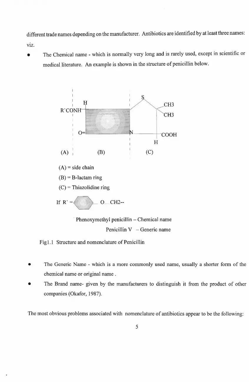
different trade names depending on the manufacturer. Antibiotics are identified by at least three names:
VIZ.
• The Chemical name - which is normally very long and is rarely used, except in scientific or
medical literature. An example is shown in the structure ofpenicillin below.
I
I
II
R'CONHI
II
I
I
I
I
I(A) I (B)
CH3
CH3
jL-1--~~---+-COOH
H
(C)
(A) = side chain
(B) = B-Iactam ring
(C) = Thiazolidine ring
If R' = _ O_CH2--
Phenoxymethyl penicillin - Chemical name
Penicillin V - Generic name
Fig1.1 Structure and nomenclature of Penicillin
• The Generic Name - which is a more commonly used name, usually a shorter form of the
chemical name or original name .
• The Brand name- given by the manufacturers to distinguish it from the product of other
companies (Okafor, 1987).
The most obvious problems associated with nomenclature of antibiotics appear to be the following:
5
• There is a nearly complete lack ofuniform denomination for antibiotics described by different
scientists and found later to be identical e.g. desdanin, netropsin, neomycin etc.
• Designations used in patents constitute a problem in themselves. Frequently only numbers or
letter-number combinations are used, which are not identical with names applied in later
publications. e.g. (a) SF-733 antibiotic -- patent designation; or Ribostamycin -- generic name
(non proprietary name); or Vistamycin -- brand (commercial name)/ trade name: (b) M-141,
U-18409 -- patent designation; or Actinospectacin -- generic name; or Spectinomycin -
brand/trade name.
• Very often several names for one agent are in general use in VarIOUS publications:
(Paromomycin, aminosidin, monomycin); (Pentamycin, lagosin, fungichromin); (Nigericin,
polyetherin A) etc.
• At present there are more than a thousand superfluous antibiotic names in current use (
Berdy,1974; 1980).
6
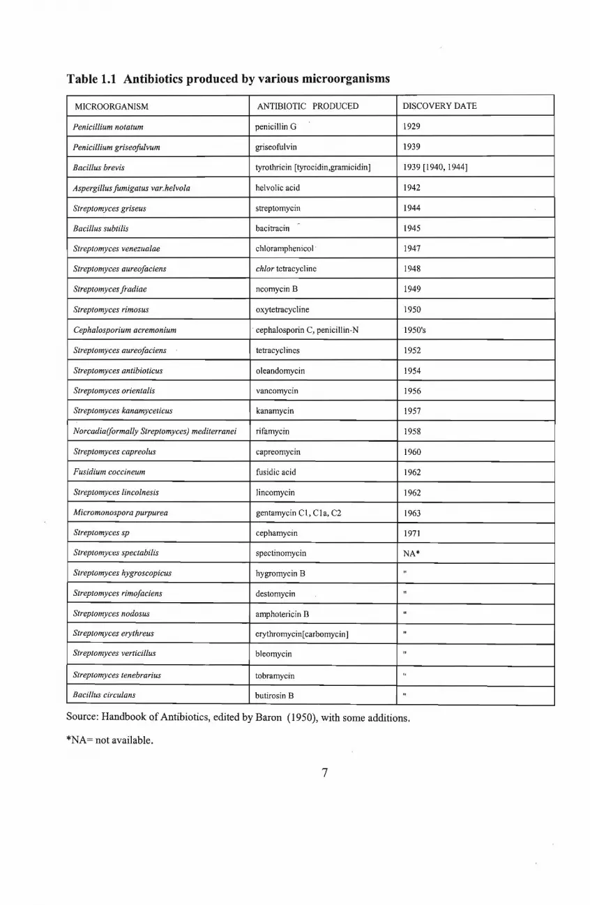
Table 1.1 Antibiotics produced by various microorganisms
MICROORGANISM ANTIBIOTIC PRODUCED DISCOVERY DATE
Penicillium notatum penicillin G 1929
Penicillium griseojitlvum griseofulvin 1939
Bacillus brevis tyrothricin [tyrocidin,gramicidin] 1939 [1940, 1944]
Aspergillus fumigatus var.helvola helvolic acid 1942
Streptomyces griseus streptomycin 1944
Bacillus subtilis bacitracin - 1945
Streptomyces venezualae chloramphenicol' 1947
Streptomyces aureofaciens chlor tetracycline 1948
Streptomyces fradiae neomycin B 1949
Streptomyces rimosus oxytetracycline 1950
Cephalosporium acremonium cephalosporin C, penicillin-N 1950's
Streptomyces aureofaciens tetracyclines 1952
Streptomyces antibioticus oleandomycin 1954
Streptomyces orientalis vancomycin 1956
Streptomyces kanamyceticus kanamycin 1957
Norcadia(formally Streptomyces) mediterranei rifamycin 1958
Streptomyces capreolus capreomycin 1960
Fusidium coccineum fusidic acid 1962
Streptomyces lincolnesis lincomycin 1962
Micromonospora purpurea gentamycin Cl, CIa, C2 1963
Streptomyces sp cephamycin 1971
Streptomyces spectabilis spectinomycin NA*
Streptomyces hygroscopicus hygromycin B "
Streptomyces rimofaciens destomycin "
Streptomyces nodosus amphotericin B "
Streptomyces erythreus erythromycin[carbomycin] "
Streptomyces verticillus bleomycin "
Streptomyces tenebrarius tobramycin "
Bacillus circulans butirosin B "
Source: Handbook of Antibiotics, edited by Baron (1950), with some additions.
*NA= not available.
7
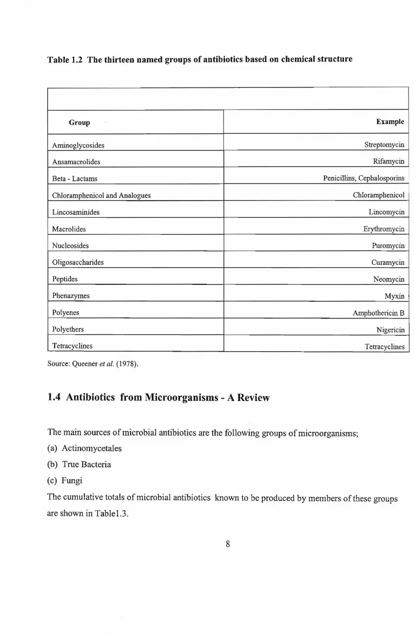
Table 1.2 The thirteen named groups of antibiotics based on chemical structure
Group Example
Aminoglycosides Streptomycin
Ansamacrolides Rifamycin
Beta - Lactams Penicillins, Cephalosporins
Chloramphenicol and Analogues Chloramphenicol
Lincosaminides Lincomycin
Macrolides Erythromycin
Nucleosides Puromycin
oligosaccharides Curamycin
Peptides Neomycin
Phenazymes Myxin
Polyenes Amphothericin B
Polyethers Nigericin
Tetracyclines Tetracyclines
Source: Queener et al. (1978).
1.4 Antibiotics from Microorganisms - A Review
The main sources ofmicrobial antibiotics are the following groups ofmicroorganisms;
(a) Actinomycetales
(b) True Bacteria
(c) Fungi
The cumulative totals ofmicrobial antibiotics known to be produced by members of these groups
are shown in Tablel.3.
8
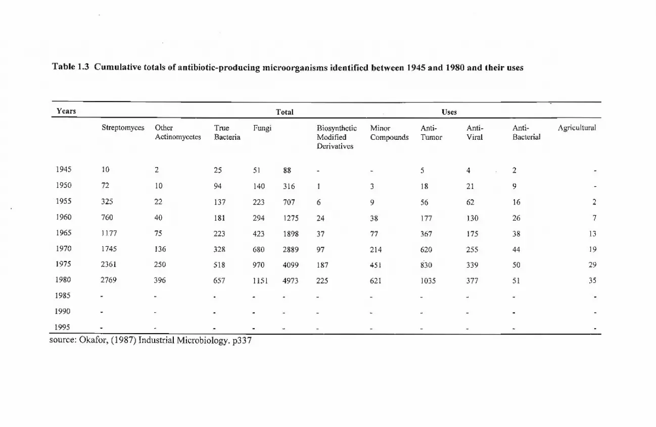
Table 1.3 Cumulative totals of antibiotic-producing microorganisms identified between 1945 and 1980 and their uses
Years Total Uses
Streptomyces Other True Fungi Biosynthetic Minor Anti- Anti- Anti- AgriculturalActinomycetes Bacteria Modified Compounds Tumor Viral Bacterial
Derivatives
1945 10 2 25 51 88 - - 5 4 2
1950 72 10 94 140 316 1 3 18 21 9
1955 325 22 137 223 707 6 9 56 62 16 2
1960 760 40 181 294 1275 24 38 177 130 26 7
1965 1177 75 223 423 1898 37 77 367 175 38 13
1970 1745 136 328 680 2889 97 214 620 255 44 19
1975 2361 250 518 970 4099 187 451 830 339 50 29
1980 2769 396 657 1151 4973 225 621 1035 377 51 35
1985
1990
1995
source: Okafor, (1987) Industrial Microbiology_ p337
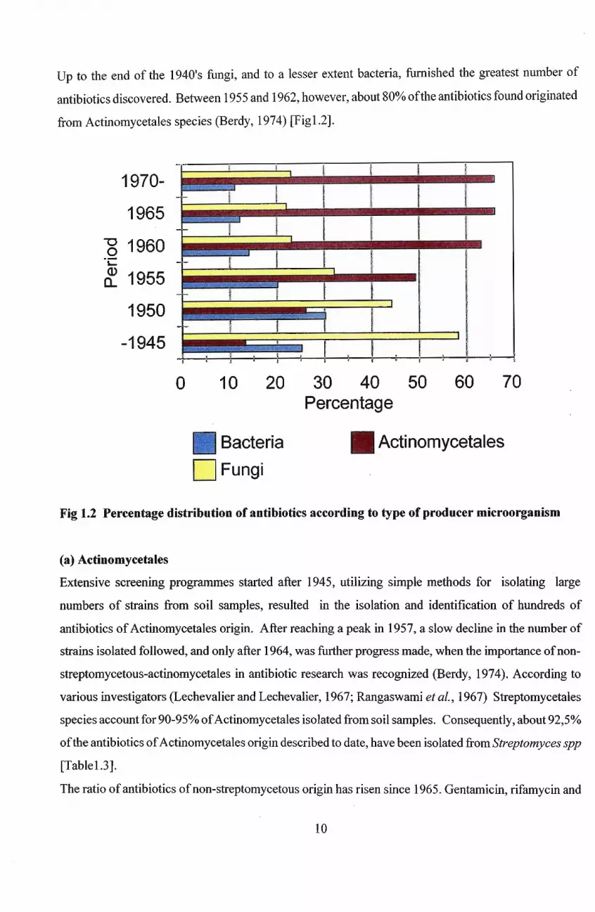
Up to the end of the 1940's fungi, and to a lesser extent bacteria, furnished the greatest number of
antibiotics discovered. Between 1955 and 1962, however, about 80% ofthe antibiotics found originated
from Actinomycetales species (Berdy, 1974) [Figl.2].
1970-
1965
-g 1960"C
rf. 1955
1950
-1945f
: . I I I' ,-!_',~~~, r--~ il ~
o 10 20 30 40 50 60 70Percentage
Bacteria • Actinomycetales
DFungi
Fig 1.2 Percentage distribution of antibiotics according to type of producer microorganism
(a) Actinomycetales
Extensive screening programmes started after 1945, utilizing simple methods for isolating large
numbers of strains from soil samples, resulted in the isolation and identification of hundreds of
antibiotics ofActinomycetales origin. After reaching a peak in 1957, a slow decline in the number of
strains isolated followed, and only after 1964, was further progress made, when the importance ofnon
streptomycetous-actinomycetales in antibiotic research was recognized (Berdy, 1974). According to
various investigators (Lechevalier and Lechevalier, 1967; Rangaswami et al., 1967) Streptomycetales
species account for 90-95% ofActinomycetales isolated from soil samples. Consequently, about 92,5%
ofthe antibiotics ofActinomycetales origin described to date, have been isolated from Streptomyces spp
[Tablel.3].
The ratio ofantibiotics ofnon-streptomycetous origin has risen since 1965. Gentamicin, rifamycin and
10
[Table1.3].
The ratio ofantibiotics ofnon-streptomycetous origin has risen since 1965. Gentamicin, rifamycin and
ristocetin originate from Micromonospora and Nocardia spp although sometimes problems have arisen
in identification ofthe microorganisms. For example, rifamycin-producing Nocardia mediteranei was
for a long time thought to be a Streptomyces sp (Thiemann et al., 1969).
Of the 25 new antibiotics introduced into clinical practice between 1960-1970, all except fusidic acid
were produced by actinomycetales. Actinomycetales have furnished the greatest number ofantibiotics
in commercial use; altogether about 72 of them being utilized for various purposes (Berdy,1974).
Cb)True Bacteria.
Among this group, the organisms that deserve particular mention include Pseudomonas species and
Bacillus species [Table1.4]. Members of these genera produce several useful compounds including:
colistin, polymyxin B and M, gramicidin, tyrothricin, bacitracin, from Bacillus species; pyocyanin and
pyrrolnitrin from Pseudomonas species (Berdy, 1974).
(c) Fungi.
Penicillium, Cephalosporium, Aspergillus and some members of the fungi imperfecti are of greatest
importance here. The number ofantibiotics produced by different fungi is shown in Table 1.5. Ofthese
about ten have been commercialized, viz. penicillin G, V, 0, cephalosporin, griseofulvin, fumagillin,
variotin, fusidic acid, siccanin and xanthocillin (Berdy, 1974). It is interesting to note that most ofthe
commonly used fungal antibiotics e.g. penicillins, cephalosporin C and fusidic acid are found among
metabolites of different fungal species (Berdy, 1974).
Table1.4 Number of antibiotics produced by different schizomycetes
Pseudomonadales 8784
Pseudomonadaceae
Pseudomonas82
Aerobacter2
11

Spirillaceae 3274
EubacterialesAzobacteraceae 1
2Rhizobiaceae
9Achromobacteriaceae
36Enterobacteriaceae
3Brucellaceae
16Micrococcaceae
28Lactobacillaceae
2Propionibacteriaceae <t
1Brevibacteriaceae
5Corynebacteriaceae
171Bacillaceae
167Bacillus
4Clostridium
Actinomycetales 20784
Mycobacteriaceae
18Actinoplanaceae
6Actinoplanes
2Spillospora
7Streptosporangium
3Microcellobosporia
1950Streptomycetaceae
Streptomyces1922
Streptoverticillium 19
Chainia8
Kitasatoa1
Micromonosporaceae41
Micromonospora 41
12
1711lernaoactinomycetaceae
9Thermoactinomyces
4MicrobisporaThermomonospora 4
48Nocardiaceae
45Nocardia
2Micropolyspora
1Thermomonospora
Myxobacteriales(Cytophaga,Myxococcus, etc 9
Mycoplasmatales(Mycoplasma) 2
Source: Berdy (1974). Advances in Applied Microbiology 18: p328
Table1.5 Number of antibiotics produced by different fungi
Myxothallophytes (Myxomycotinia) 4
(Fuligo, Physarum, Lycologa sp)
Eumycotinia (Mycophyta), ' true fungi' 768
Phycomycetes 14
(Phytophthora, Mucor, Rhizopus sp.)
Ascomycetes 299
Protoascomycetes (Ascosporogenous yeasts ) 8
Euascomycetes 291
Plectascales 248
Aspergillaceae 242
Penicillium 123
Aspergillus 115
Other orders of Euascomycetes 43
Basidiomycetes 140
13
Fungi imperfecti 315
Moniliales 269
Moniliaceae 102
Cephalosporium 20
Tichoderma 13
Oospora 10
Acrostalagmus 7
Cylindrocladium 9
Demetaciae 69
Helminthosporium ( Ophiobolus) 23
Alternaria 10
Phoma 8
Tuberculariae 79
Fusarium 46
Myrothecium 26
Stillbaceae (Isaria, Metarrhyzium) 10
Melanconiales 1
Mycelia sterilia 5
Dermatophyta 9
Asporogenous yeasts 19
Unidentified fungi imperfecti 12
Source: Berdy (1974). Advances in applied microbiology 18: p330
1.5 Biochemistry of Antibiotic Production
Antibiotics are considered generally as secondary metabolites, although some, for example, Nisin, have
been considered by some workers to be primary metabolites (Hammond and Lambert, 1978). Secondary
metabolism is characteristic of lower life forms and the metabolites are often species specific . In
contrast with primary metabolites, from which they are created, secondary metabolites often accumulate
14
and are usually excreted into the medium. They have no obvious function in the growth ofthe cell and
are usually produced late in the growth phase (Hammond and Lambert, 1978). The actual functions
ofantibiotics in the producing microorganism are still a matter ofdebate. There are three main schools
of thought on this issue:
• They are simply waste products of metabolism - this view is no longer tenable, as many
secondary metabolites have been found to have biological activity.
• They provide a competitive mechanism between organisms - this seems most likely.
Unfortunately, very little is known about the synthesis ofantibiotics in the natural environment.
It could explain why actinomycetes, which as slow growers have to compete with fast growing
microbes, produce many antibiotics.
• They serve as a metabolic regulation mechanism - this is unlikely to have general validity, as
many antibiotics are not active against the producing organism. Some antibiotics have
mechanisms of action that are difficult to reconcile with a regulatory role (Lancini and
Parenti,1982).
Hammond and Lambert (1978), proposed that antibiotics, from their position as secondary metabolites,
may function as a means of detoxification to protect the microbial cell from adverse conditions in the
environment and may also inhibitmacromolecule synthesis under hostile conditions, thereby preventing
active growth. The enzyme reactions that lead to the synthesis of antibiotics are not fundamentally
different from those that lead to the synthesis of primary metabolites. Only small variations occur in
the principal biosynthetic pathways. Three major categories ofantibiotics may be derived on the basis
of their biosynthetic route.
• Antibiotics derived from a single primary metabolite .e.g. Chloramphenicol and nucleoside
antibiotics.
• Antibiotics derived by condensation of several metabolites .e.g. Lincomycin and Novobiocin.
• Antibiotics derived by oligomer or polymer formation. There are about four sub-classes in this
group.
(a) Polypeptide antibiotics:- from condensation of amino acids, e.g. Gramicidin Sand Penicillins.
15
(b) Acetate/propionate polymerization antibiotics, e.g. Griseofulvin, Tetracyclines, and Erythromycin.
(c) Terpenoid antibiotics:- through isoprenoid synthesis, e.g. Fusidic acid.
(d) Aminoglycoside antibiotics: - made through a condensation reaction, similar to that of
polysaccharides. e.g. Streptomycin and Neomycin.
The production of antibiotics is not rigorously species specific (Lancini and Parenti, 1982). The same
antibiotic can be produced by different species ofthe same genera. And the reverse is also true; that is,
the same species can produce different antibiotics. Most often microbial species or strains produce
several biologically and chemically related antibiotics that constitute a 'family', which is to say that the
same strain makes two or more antibiotics that resemble each other. The capacity to produce" families"
of compounds is not unique to antibiotic biosynthesis but a general characteristic of secondary
metabolites, mainly due to the large size of intermediate molecules (Lancini and Parenti,1982).
The concepts of 'metabolic trees' and 'metabolic nets' were used by Lancini and Parenti (1982) to
explain the reason for these metabolic events. The same metabolite can be a substrate for two or more
reactions generating different products which may be enzymatically transformed, giving rise to a
metabolic tree. Or the same metabolite can be obtained through different pathways that vary only in the
order of enzymatic reactions, giving rise to a metabolic net. They also noted, a combination pathway
of these 'trees' and 'nets' was possible.
1.6 Mechanism of Antimicrobial Action
It is essential that a successful antibiotic must be selective in its mechanism of action, that is, it must
have the ability to penetrate and concentrate in the microbial cell and should not interact with the cells
of the host. They should show suitable absorption and distribution properties within the host body
(Franklin and Snow, 1981).
Antimicrobial agents generally bring about microbial inhibition by interacting with specific cellular
16
components and disordering cell metabolism. They block the growth of sensitive microorganisms by
inhibiting the action of a macromolecule, such as enzymes or a nucleic acid essential to the function
of the cell. This means that the antibiotic molecule is able to bind to a specific site on the target
macromolecule, forming a complex that is useless to the cell (Lancini and Parenti,1982).
Several authors have classified antibiotics based on the site ofaction in the microorganism (Hammond
and Lambert, 1978; Franklin and Snow, 1981; Lancini and Parenti, 1982).
(a) Inhibitors of cell wall synthesis
Examples include Penicillin, Cephalosporin, Cycloserine and Vancomycin (Hammond and Lambert,
1978). About 50 - 70% of the Gram - positive bacterial cell wall mass and to a lesser extent (10
15%) in Gram-negatives is composed of peptidoglycan (sometimes referred to as Murein or
Mucopeptide). Its cross-linked structure provides a tough, fibrous fabric, giving strength and shape to
the cell and enabling it to withstand a high internal osmotic pressure (Franklin and Snow,1981).
Most of the antibiotics in this group affect peptidoglycan synthesis, while others interfere with the
synthesis or assembly of other components of the wall, e.g. teichoic acid. ~-lactam antibiotics
(penicillins, cephalosporins), so-called because of the presence of a cyclic amide forming a four atom
ring (~-lactam ring) in the molecule, all have a similar but not identical mechanism of action. They
prevent peptidoglycan maturation by inhibiting the cross-linking of the linear peptidoglycan strands
(Hammond aild Lambert, 1978; Franklin and Snow,1981; Lancini and Parenti, 1982). Action of these
antibiotics ultimately results in bacterial cell lysis.
Vancomycin will inhibit the transfer ofmuramyl pentapeptide-acetylglucosamide from the lipid carrier
to the peptidoglycan chain being formed. Conversely, cycloserine, which is particularly active against
mycobacteria, acts at the terminal position ofthe pentapeptide ofmuramic acid. All known inhibitors
of cell wall synthesis are:
• Bactericidal in action;
17
• Inactive against resting cells;
• Inactive against bacteria that lack a conventional cell wall, e.g. Mycoplasmas, L- forms and
protoplasts (Lancini and Parenti,1982).
(b) Inhibitors of cell membrane functions.
Examples include PolYmyxin, tyrocidin, valinomycin and amphothericin B (Hammond and Lambert,
1978). Cell membranes have very similar constituents throughout the phylogenetic ladder from bacteria
to mammalian cells. The only important difference is that there are no sterols in bacterial cell
membranes, while zYmosterol and ergosterol are present in the cell membranes offungi and plants, and
cholesterol in those of mammals. Some antibiotics in this group disorganize the super-molecular
structure of the membrane, thus causing loss of cellular substance to the outside, while some act as
carriers of specific ions (Ionophores) and cause an abnormal accumulation of ions inside the cell
(Lancini and Parenti, 1982).
PolYmixins and tryocidin are both cyclic polypeptide antibiotics. Their action disturbs membrane
function by allowing leakage of cytoplasmic components (Ca2+ ions; Mg2+ ions) and uncoupling of
oxidative phosphorylation (prevention of ATP generation during sugar oxidation). Valinomycin
specifically drains the cell of potassium ions (K+) and growth ceases because of the requirement for
potassium in protein biosynthesis (Franklin and Snow, 1981). Amphotericin B, a typical polyene
antifungal antibiotic, creates instability in the membrane, by forming complexes with sterol components
of the membrane that alter trans-membrane cation permeability. The majority of inhibitors of cell
membrane functions are non-selective and consequently are too toxic to be given systemically, and are
therefore used exclusively as antiseptics or topical agents (Franklin and Snow,1981).
(c) Inhibitors of transcription and replication of genetic material (nucleic acids)
Examples include, rifamycin, actinomycin D and acridine dyes (Hammond and Lambert, 1978).
18
The synthesis ofDNA and the various classes ofRNA is an essential function ofdividing and growing
cells. Thus inhibition ofDNA synthesis rapidly results in inhibition ofcell division (Franklin and Snow,
1981). Rifamycins are enzyme inhibitors. They bind to, and inhibit the DNA dependant - RNA
polymerase of sensitive bacteria, but the precise mechanism of inhibition is still uncertain (Harnmond
and Lambert,1978).
ActinomycinD molecules insert themselves into the smaller groove ofthe DNA double helix and form
a reversible complex bound by hydrogen bonds. The complex does not permit RNA po'iymerase to
travel along the DNA template thereby inhibiting synthesis of RNA (Hammond and Lambert, 1978).
---Acridines show antimicrobial activity by distorting the sugar phosphate backbone of the DNA helix
created by the intercalated dye molecules (Franklin and Snow, 1981). Most ofthe inhibitors ofgenetic
material are used in cancer chemotherapy rather than as antimicrobial agents. Unfortunately, many of
these do not show sufficient selectivity against the tumour cells and are too toxic to be used effectively
(Hammond and Lambert, 1978).
(d) Inhibitors of Protein Synthesis.
Examples include streptomycin, tetracycline and chloramphenicol (Hammond and Lambert, 1978).
Some antibiotics in this group affect amino acid activation and transfer reactions, while others interfere
with the functions of the 30S or 50S ribosomal subunit (Lancini and Parenti, 1982). Streptomycin and
tetracycline distort the 30S ribosomal subunit enough to prevent normal bonding between the codon
ofmRNA and the anti-codon oftRNA. This causes mis-coding ofthe proteins, bringing normal protein
synthesis to a halt (Hammond and Lambert, 1978).
Chloramphenicol acts on the 50S subunit by inhibiting peptide bond formation between the amino acids.
Inhibitors of protein synthesis are bacteriostatic if they do not form irreversible bonds with some
essential component of the synthetic system. If they do, they are bactericidal (Franklin and Snow,
1981).
19

1.7 Spectra of antimicrobial activity
Antibiotics may be grouped into five classes according to their spectra of activity; i.e. according to the
classes ofmicroorganisms they inhibit.
[1] Antiviral
[2] Antifungal
[3] Antiprotozoal
[4] Antibacterial
[5] Antitumour agents - justifiably classified as antibiotics because the compounds were originally
isolated on the basis of their antimicrobial activities (Lancini and Parenti, 1982).
Occasionally an antibiotic is active against all microorganisms, but usually different antibiotics are
active against selected organisms. The group of microorganisms whose growth is inhibited by an
antibiotic constitutes its spectrum ofactivity. As an index ofantimicrobial activity, microbiologists and
clinicians use the Minimum Inhibitory Concentration (MIC), defined as the lowest antibiotic
concentration that will inhibit the growth of a specific organism (Hammond and Lambert, 1978).
To determine MIC, small bottles of liquid growth medium, containing graded doses of antibiotic, are
inoculated with the test organism. After suitable incubation, growth will occur in those bottles where
the antibiotic is below the inhibitory level and the culture will become turbid (cloudy) from the large
number ofmicroorganisms present. Growth will not occur above the inhibitory level and the medium
will remain clear. Samples are taken from the bottles and plated out on nutrient agar. The plate
containing the lowest antibiotic concentration which shows no microbial colonies after incubation is the
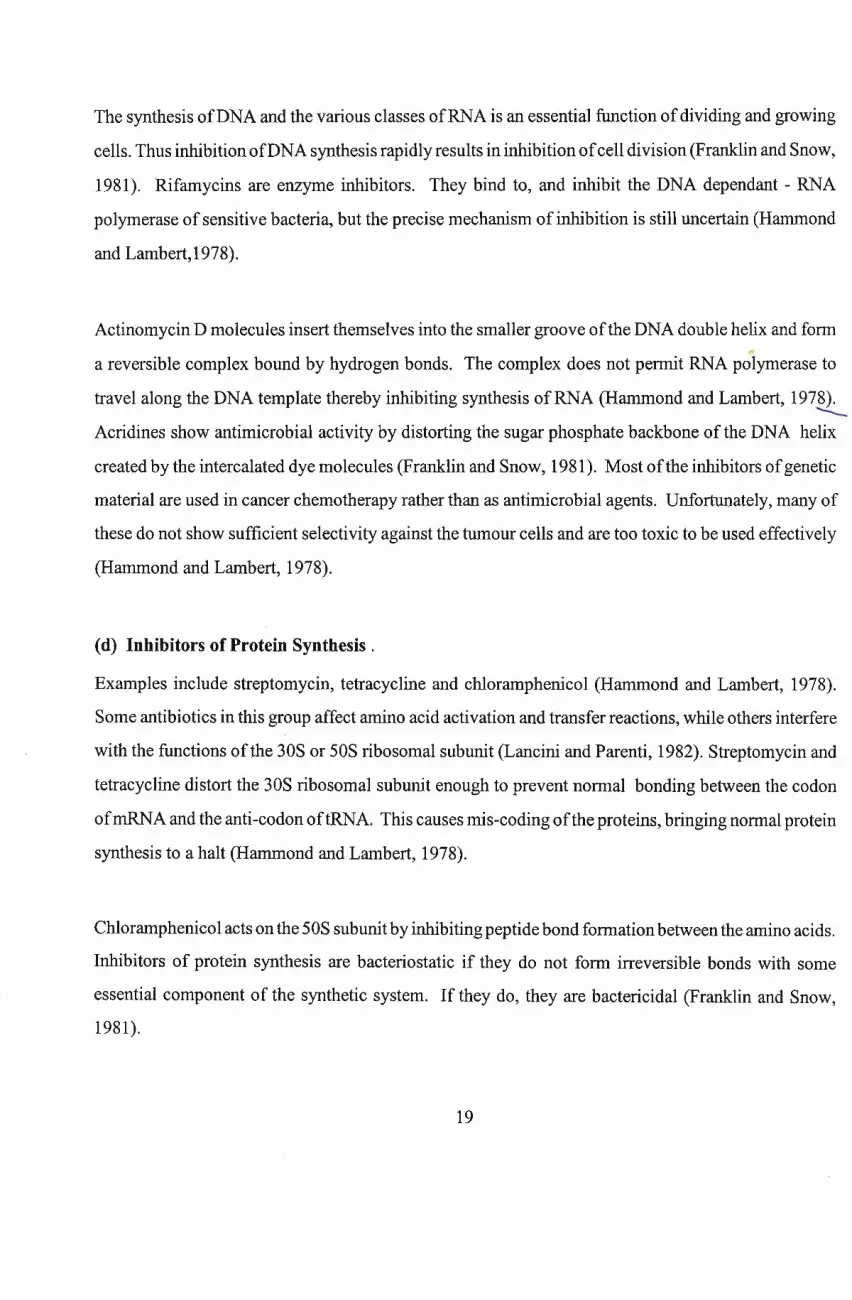
one with the MIC (Hammond and Lambert, 1978). Table1.6 shows the MIC of some important
antibiotics against selected pathogenic microorganisms. Antibacterial antibiotics are said to have a
narrow spectrum of activity if they are active against few species of either Gram - positive or Gram
negative bacteria only, and broad spectrum if they are active against majority ofboth Gram - positive
and Gram -negative bacteria (Lancini and Parenti, 1982).
20
Table 1.6 MICs Illg/ml) of some antibiotics against representative pathogens
Penicillin G Amp Cephalo Erythro Tetra Chloram Strept AmphoB
Gram-positive cocci
Diplococcus pneumoniae 0.010 0.04 0.003 0.01 - 2 0.05 - 1 1 - 3 0.2 -1 uns
Staphylococcus aureus pnp 0.04 0.1 0.05 0.01 - 2 0.1 4-8 1 - 5 unspp 50 Dns 13 0.01 - 2 0.1 4-8 1 - 5
Gram-negative cocci
Neisseria gonorrhoeae 0.05 0.1 - 0.6 0.1 - 4 0.02 - 1 0.1 - 3 1 2 - uns uns
Gram-positive Bacilli
Clostridium tetani 0.01 - 3 0.02 0.5 0.5 - 2 0.03 - 1 0.1 - uns uns uns
Corynebacterium diphtheriae 0.3 - 3 - 1 0.5 - 5 0.5 - 5 0.5 - 5 0.5 - uns unsN...... Gram-negative bacilli
Escherichia coli uns 2 - uns 1 - uns 2 - 10 0.5 - 5 2 - 12 1 - uns uns
Klebsiella spp 6 1.5 6 - uns uns 0.5 - uns 0.3 - 1.5 8 - uns uns
Pseudomonas aeruginosa uns uns uns uns 4 - 200 16 - uns 20 - 500 uns
Mycobacterium tuberculosis uns - 10 uns uns 6 - 30 0.5 - 5 uns
Yeast and Fungi
Candida albicans uns uns uns uns uns uns uns 0.1 - 3
Dermatophilus spp uns uns uns uns uns uns uns 0.2 - 40
uns = unsusceptible; Amp= Ampicillin; Ceph = Cephalosporin; Erythro= Erythromycin; Tetra = Tetracycline; Chloram= Chloramphenicol; Strept = Streptomycin; Ampho B
= Amphotericin B; pnp= penicillase-non-producer; pp= penicillase producer; source: Lancini and Parenti, 1982. Antibiotics, An integrated View. pp24 - 25.
1.8 Review of screening methods
1.8.1 Primary Screening Methods
General screening methods have not changed fundamentally in the past 40 years. Millions ofdifferent
microorganisms, mostly actinomycete species originating from soils, were screened all over the world
by means of the classical methods of Waksman and Dubos.
[1] The crowded plate method
This method is used to isolate organisms able to produce antibiotics active against other soil organisms.
A heavy aqueous suspension (1: 10; 1:100) of soil is plated on agar in such a way as to ensure the
development of confluent growth. Colonies surrounded by clear zones are purified for further study.
By altering the media used, different groups oforganisms can be encouraged to develop (Okafor, 1987).
[2] Dilution plate method
This method is applied when the aim is to isolate antibiotics against a known organism or organisms.
It is based on the principle that'Activity against a single test species/strain is a sufficient criterion for
the selection of antagonistic organisms' (Okafor, 1987). The procedure involves mixing a sample of
soil at a suitable dilution with an appropriate melted agar and the mixture poured into plates. After the
agar has solidified, the plate is inverted and incubated until scattered colonies appear. The plate is then
flooded with a suspension of the test organism and re-incubated. The test species forms a lawn on the
surface ofthe agar interspersed with clear zones ofinhibition. The clear zones indicate the presence of
colonies from the soil sample which are capable of producing antibiotics active against the test
bacterium. Those colonies surrounded by clear zones are purified and transferred to an agar slant to
be held for further studies. By employing a series of soil agar plates each flooded with a different test
species, it is possible to isolate forms which are antagonistic to a large number of organisms.
The essence ofWaksman's method is that it facilitates preliminary screening of antibiotic producers

through development of an easily visualized clear zone in a confluent growth of the test organism
(Waksman and Lechevalier, 1962).
1.8.2 Primary Testing of Antibiotic Production
Once an organism showing evidence of antibiotic action has been detected and isolated, it is then
purified and several other steps are carried out to confirm antibiotic production. Some of the methods
will be reviewed here.
[1] The cross streak method
This method is suitable for testing individual isolates. The purified isolate is streaked across the upper
third of a plate containing a medium which supports its growth as well as that of the test organisms. A
variety ofmedia may be used for streaking the antibiotic producer. It is allowed to grow for up to 7
days, in which time, any antibiotic produced should have diffused a considerable distance from the
streak. Test organisms are streaked at right angles to the producer isolate and the extent of inhibition
of the various test organisms observed/measured (Okafor, 1987).
., [2] The agar plug method
This method is particularly useful for testing fungal isolates for antibiotic production, especially when
the test organism(s) grow(s) poorly in the medium that supports growth ofthe producer fungus. Plugs
about 0.5 cm in diameter are made with a sterile cork borer from the fungus isolate plate. These plugs
are then placed on plates seeded with different test organisms and incubated. Inhibition zones appear
around those isolates producing antimicrobial compounds. This method may be used with actinomycetes
(Okafor, 1987).
[3] The replica plating method
Ifa large number oforganisms are to undergo primary screening, one rapid method is the use ofreplica
plating. This is a well known method used in microbial genetics (Okafor, 1987). The method consists
23
ofplacing a sterile velveteen pad over the colonies growing on a soil agar plate, or on a plate containing
separate, individually distinct, colonies to be tested for antibiotic properties. The pad is thereafter
carefully touched onto the surface of four or five plates seeded with the test organism. Because an
orientation mark is placed on the pad as well as on the plates, it is possible to tell which colonies are
causing the cleared zones on the test plates (Okafor, 1987). There are various other screening techniques
employed, e.g. ditch plates, gradient plates etc., that are not described here because of the remote
possibility ofusing them in the present study.
1.8.3 Factors affecting antibiotic production
In all the procedures described above there are certain factors that will affect antibiotic production:
(1) Nutritional conditions (2) Growth rate of the producing organism (3) The assay conditions.
Positive inhibition oftest organisms by antibiotic-producing isolates, depends upon complex interactions
between the antibiotic producer and its culture medium, and on cultural conditions such as temperature,
pH and availability ofoxygen (Lancini and Parenti, 1982). Several workers have reported that a strong
relationship exists between nutritional factors and antibiotic production, for example, glutamate, as sole
nitrogen source supported good growth of Streptomyces griseus but a very low yield of its antibiotic,
streptomycin. Conversely, L- proline, supplied as sole nitrogen source, promoted both biomass and
streptomycin yield (Dulaney, 1948). Also nitrogen, glucose and phosphate regulation of antibiotic
production have been reported (Gallo and Katz :1972; Aharonowitz and Demain :1979; Martin and
Demain :1976;1980; Hodgson:1982).
The growth rate of the producing organism is yet another factor affecting antibiotic production.
Microorganisms seem to be programmed to produce antibiotics only when the specific growth rate
decreases below a certain level (Martin and Demain, 1980). Expression of the genes coding for
antibiotic biosynthesis usually does not occur at high growth rates, suggesting that during rapid growth,
either antibiotic synthetases are not formed or, ifformed, their activity is inhibited (Martin and Demain,
1980). The enzymes involved in the synthesis of antibiotics have been monitored by various workers
(Liras et aI., 1977; Madry and Pape, 1982; Takeshima et al., 1989).
24
lons are very important in determining the in vitro activity ofan antibiotic. Lancini
suggested that all test conditions must be specified precisely and if possible
'ishes to use inhibition of bacterial growth for quantitative determination of the
G and to have data that are reproducible in different laboratories. The assay
d be standardized include:
organism/s .
.lsition [pH, ions, serum etc.]
lsity [etherogeneticy ofpopulation etc.]
ns [time, temperature, aeration]
It must be noted that antibiotic activity may differ depending on whether the testing was done in a liquid
or on a solid medium and also when the total number ofbacteria in the inoculum is very high, there is
a greater probability that some ofthe cells will be less susceptible to the antibiotic (Lancini and Parenti,
1982).
The effect of pH, temperature and alkali metal ions on antibiotic activity has also been studied by
various investigators (James et al., 1991; Kirpekar and Kirwan, 1991; Eckwall and Schottel, 1997; Paik
et al., 1997). Eckwall and Schottel (1997) observed that an antibiotic produced by the scab disease
suppressive Streptomyces diastatochromogenes strain Pon SS 11 was stable for weeks at acidic pH but
very labile at alkaline pH conditions. The pH instability ofthis compound may have some implications
for the effectiveness of the organism as a bio-control agent in soils at neutral or alkaline pH. Some
antibiotics, such as nitrofurantoin, are more active at acidic pH while aminoglycosides and sulfonamides
are more active in alkaline medium (Jawetz et al., 1984). Several antimicrobial agents lose their activity
at commonly used incubation temperature (37°C). For example, chlortetracycline is inactivated rapidly
and penicillin more slowly, whereas the aminoglycosides, chloramphenicol and polymixin B are quite
stable for longer periods at this temperature (Jawetz et al., 1984).
25
1.8.4 Extraction and purification methods
Over the years, various workers have used different methodologies to extract and/or purify antibiotics
: solvent extraction (Arcamone et al., 1969); a combination ofmolecular exclusion chromatography, ion
exchange chromatography and sodium dodecyl sulphate polyacrilamide gel electrophoresis (Galvez et
al., 1986); adsorption to ion exchange resins (Homer et al., 1989); flash chromatography (Stankovic
et al., 1990); HPLC (high performance liquid chromatography, Franco and Coutinho, 1991); a
combination of thin layer chromatography and HPLC using three reagents Amberlite XAD -2, DEAE
Sephadex A-50 and SP Sephadex C-25 (Eckwall and Schottel, 1997). Most ofthese methods are based
on the affinity of different antibiotics for different eluting solvents and different matrices such as ion
exchangers.
1.9 Some suggested newer methods of searching for antibiotic-producing microorganisms
Between the mid 1960s and mid 1970s, although the search for antibiotics continued, very few new
antibiotics were in fact discovered. It was found that the majority of new isolates produced the well
known antibiotics (Berdy, 1980). This was because the classical method of Waksman and Dubos
described earlier was mainly based on inhibition/clear zones on the agar plate and also on the use of a
few selected test organisms, usually ofmedical importance. Some suggested new methods have led to
the discovery ofnew antibiotics.
[i] The use of super sensitive mutants
By using super sensitive strains oftest organisms in primary screening procedures, organisms producing
only small amounts of an antibiotic may be detected. Antibiotics so produced are useful because they
seem to have a wider spectrum than those of the same class already in existence (Okafor,1987).
Furthermore, they may provide better substrates for semi- or muta-synthesis. This method has led to
the discovery ofnovel p-Iactam antibiotics, thienamycin, olivanic acid, nocardicins, clavulanic acid, etc.
The use of super sensitive mutants has shown that p- lactam antibiotics are produced by a wider
spectrum oforganisms - ascomycetes, fungi imperfecti and actinomycetes - than was previously thought
26

(Okafor, 1987).
[ii] The application of criteria other than death or inhibition
Reactions such as irregular growth of fungal mycelium and inhibition ofsporulation, rather than death,
may be used to follow an antibiotic's effect/so When antifungal antibiotics are sought, the clear zone
principle is employed using yeasts or fungal spores in the test plate. With this method, the e~isting
antifungal antibiotics, namely, polyene as well as cycloheximide and actimycins were found. Ifcriteria
less drastic than death (e.g. abnormal growth ofhyphae or inhibition ofzygospore formation) are applied
instead, a wide range of new antibiotics may be found. Using this method, some new actinomycete
antibiotics have been found including boromycin, venturicidin and mikkomycin (Okafor, 1987).
[Hi] The use of chemical assays
The use of chemical assays based on various chemical reactions (chemical screening) of the culture
broth of an organism, using thin-layer chromatography with reagents specific for certain chemical
groups, high pressure liquid chromatography etc., may lead to the discovery ofnew antibiotics, as was
the case with dienomycins and arglecins (Okafor, 1987).
[iv] Search in novel environments
Systematic searches for antibiotics are usually from soil. Ofcourse, other natural sources exist, although
none can match the soil in the ubiquity and richness offlora. Nevertheless, searches in unusual habitats
may provide novel organisms. One such habitat is the sea. It contains large amounts ofsalts and other
mineral nutrients, has a fairly constant temperature, high hydrostatic pressure and less sunlight in the
deep areas. The coastal area is constantly changing with the tides and such areas should be expected to
have a wide variety oforganisms, peculiar to the littoral environment. The search for antibiotics in the
sea has indeed led to the discovery ofnew and unique compounds. These include: antibiotic SS-228R,
from Chainia sp., effective against gram-positive bacteria and tumours; bromo-pyrrol, from a marine.---
Pseudomonas, and leptosphaenin from marine fungi (Okafor, 1987). The point is that novel
environments, tree barks, leafsurfaces, sewage, all provide distinct environments, whichmay encourage
the growth ofmicroorganisms possessing a unique set of enzymes and, hence, the possible capacity to
27

synthesize new antibiotics (Okafor,1987).
(v) Changed culture conditions
The production of new antibiotics may be encouraged, even in known cultures, by subjecting them to
new culture conditions such as a different carbon source, temperature, level ofaeration etc. This is done
in the hope that certain pathways may be blocked and others enhanced. Such carbon sources include
monosaccharides, glycosides, substituted sugars, polyhydric alcohols, oligosaccharides, terpenes and
hydrocarbons (Bunge et aI., 1978).
Isolation ofmicroorganisms is done by sprinkling soil on plates containing the above carbon sources.
A perfusion technique, in which soil is bathed constantly with a solution containing the chosen carbon
source, may also be used. Isolates are grown in shake flasks using the various carbon sources and a
variety ofenvironmental variables, including pH, mineral composition and concentration, temperature,
nitrogen source and aeration. Tablel.7 illustrates new products formed when new culture conditions
were provided to producers of well established antibiotics (Neijssel and Tempest, 1979).
Table1.7 New microbial products resulting from changed culture conditions
New conditions Orpanisms New Products
Increased POl Pseudomonas sp Pyrrolnitrin
Nocardia uniformis Thiopeptin
Streptomyces tatyamensis Nocardin
S . saptoronensis Bicyc10mycin
Lower Incubation Temp
27°C - 12°C S. griseus Cryomycin
27°C - 15°C S. griseus Holomycin
Increased O2 supply S. lavendulae Chlorocarcin
28

[vi] Strategy based on multiple antibiotic resistance
This strategy is based on the principle that antibiotics are not species specific, but rather strain specific.
Dsually one may regard resistance to own antibiotics as a specific phenotype conferred by a selfresistant
gene (Cundliffe, 1992). However, the resistance to specific antibiotics is not specific to the strains
which produce them, because there are many strains with multiple- antibiotic resistance (Cundliffe,
1992). For example, there are many streptomycin-resistant actinomycetes other than streptomycin
producers (Hotta and Okami, 1996). These methods involve random selection ofmany strains, from soil
isolates with or without antibiotic resistance and examining them for antibiotic production by incubating
them in a starch - soy bean meal medium (Hotta et al., 1983). Strains with known resistance patterns are
selected and screened for their ability to produce antibiotics. Two novel antibiotics, dopsisamine and
bagougeramine (a backyard product) have been isolated using this method (Hotta and Okami, 1996).
29
Chapter 2
Isolation of Antibiotic-Producing Microorganisms
2.1 Introduction
According to a survey done in 1993, over 70% ofthe novel bio- active substances ofmicrobial origin
isolated in the decade 1984 - 1993 were derived from actinomycetes (Miyadoh, 1993). It is believed
that actinomycetes will continue to play a major role in providing novel bio-active substances in the
next decade (Hotta and Okami, 1996).
However, as the number ofknown secondary metabolites increases, the probability offinding new ones
becomes increasingly remote. Innovative methods will be required in order to enhance the probability
of finding new antibiotics. Accordingly, a new basis will need to be established for isolating and
distinguishing strains with 'potential productivity of novel bio-active metabolites (Hotta and Okami,
1996).
Over the past two decades, taxonomists have shown increasing interests in rare actinomycetes as a
potential source ofbio-active secondary metabolites. Methods designed to isolate a wide variety ofrare
actinomycetes have been developed (Hayakawa and Nonomura, 1989; Shomura, 1993).
Five steps were observed in the modem isolation methods. These included the choice of substrate,
composition of the isolation medium, pre-treatment and incubation conditions, colony selection and
purification (Cross, 1982; Nolan and Cross, 1988). Of the five factors, composition of the isolation
medium, pre-treatment and incubation conditions are the most important since they determine which
organism will develop on the isolation plates.
Taxonomic characterization has many limitations since it still remains essentially at the species level,
although the production of antibiotics and other secondary metabolites is strain specific (Hotta and
Okami, 1996). Therefore a new basis will be required to distinguish isolates at the strain level in terms
ofpotential productivity ofnovel antibiotics (Hotta and Okami, 1996).
30
Hotta and Okami (1996) also recommended primary metabolism as a key to establishing a new basis
in the search for new antibiotics. Obviously, the supply of primary metabolites as precursors is
necessary for biosynthesis of secondary metabolites. Therefore, it seems likely that the primary
metabolism of antibiotic producers is appropriately regulated to supply precursors for biosynthesis of
their specific secondary metabolites. For example, in the case of aminoglycoside producers, primary
metabolism should be regulated to supply sugar precursors for aminoglycoside biosynthesis. Similarly,
producers ofamino acid-related antibiotics and polyketides, such as macrolides, will have specifically
regulated primary metabolisms to supply precursors for their characteristic products. Therefore, any
discovery of phenotypes relating to characteristic primary metabolism, will form a good basis for
screening for novel antibiotics (Hotia and Okami, 1996).
In actinomycetes, however, primary metabolism has been studied rather poorly compared to secondary-..:.-
metabolism, and more studies on the genetics and biochemistry ofprimary metabolism will definitely
be necessary. The present study incorporated some of the new ideas by employing changing culture
conditions, such as different carbon sources, temperature and aeration during the growth cycle of the
isolates investigated. This was done in the hope that certain metabolic pathways might be blocked and
others enhanced.
Due to the slow growth rate of actinomycetes, numerous efforts have been made to develop selective~procedures for their isolation from natural habitats. Such procedures include, use ofselective media as
well as mechanical and chemical separation of other unwanted organisms that might interfere with
actinomycete isolation (Cross, 1982; Karwowski, 1986). The incorporation of antibiotics in
actinomycete isolation media was also employed (Athalye and Lacey, 1981 ; Orchard and Goodfellow,
1974). Pretreatment methods used by some investigators (Cross, 1982; Ntuli, 1994) include: nutrient
enrichment and physical and! or chemical treatment applied individually, or in combination, to samples
or sample suspensions, before plating out on suitable media.
Natal soils have been reported as a potential source ofnovel antibiotic producing microorganisms. For
example, the organism, Streptomyces natalensis, an actinomycete, first used in industrial production
ofpimaricin, was originally isolated from Natal soils (Baecker and Ryan, 1987).
31

The main aim of this study was to further the progress made in this area to isolate new antibiotic
producers by applying some new ideas and methodologies. Members of the genus Streptomyces were
excluded from the study, since they have been extensively studied in the past (Okami and Hotta, 1988;
Ntuli, 1994).
32

2.2 Materials and Methods
2.2.1 Sample collection
All samples were collected from KwaZulu-Natal, Republic of South Africa. Samples were collected
from: Charles Johnson Memorial Hospital; Nqutu compost dump, which consisted mainly oflitter from
cut trees and grass left to decompose with occasional burning; several Dundee farms, mainly cow
manure and dung; Camco Farm just outside Pietermaritzburg on the road to Richmond, comprised
mainly ofchicken litter and droppings. About 20 samples were collected from each site at depths of 3
5cm below the surface.
Additional samples were collected from the Mlangeni farm in Bisley, Pietermaritzburg, which consisted
mainly ofdecayed chicken manure and droppings; from the banks ofthe Mzinyathi river at Rorkes Drift
(northern KwaZulu-Natal); from uncultivated farmland in the Escourt area, and from the outskirts of
Pietermaritzburg. See Table 2.1 for more details.
All samples, ofwhich approximately 1500 were collected during the period ofthis investigation, were
kept in a refrigerator(±4QC) until required.
Table 2.1 Nature and location of samples collected and investigated over the period 1998-2000
Sample Type
Cow Manure
Chicken Litter
Chicken Manure
Chicken Manure
Location
Dundee farms, Dundee, Northern
Natal
Camco farm, Pietermaritzburg
Mlangeni's Poultry Farm,
Pietermaritzburg
Family Poultry Farm, Sweet Waters
33
Description
3cm depth of soil covered with dung
and decomposed cow feed.
Poultry farm, litter covered with
chicken droppings.
3cm depth of soil; decomposed feed
and droppings.
5cm depth of soil; decomposed feed
and droppings.
Marshy soil
Compost Soil
Creosote Contaminated Soil
Uncultivated Farm Land
Cane Sugar Field
Forest Soil
Mzinyathi River Bank, Rorke's Drift
Charles Johnson Memorial
Hospital,Nqutu
Project Site,
University of Natal
NI Freeway, Escourt
Natal Midlands
Illovo Farms Ltd,
Ellanspruit, Natal Midlands
Sappi Farms Ltd,
Seven Oaks, Natal Midlands
5cm depth of marshy, muddy soil
covered with decomposing grasses
and algae.
5cm depth of soil;
Cut branches, leaves and refuse left
to decompose with occassional
burning
Phd Student's sample
3cm depth of soil.
3cm depth of soil; freshly cultivated
sugar cane field.
5cm depth of soil;
decomposing leaves.
2.2.2 Isolation Media
Distilled water was used for all medium preparation. Attempts were made to use selective media in
order to avoid the growth of Streptomyces species. Antibiotics were also added to autoclaved and
cooled media (to prevent thermal destruction of the antibiotics) as filter-sterilized solutions, and after
gentle swirling, the media were poured into petri dishes.
For chemical composition and preparation of the media used, refer to Appendix 1.
Czapek's medium [Cross and Attwell,1974] referred to by Cross, 1981]. 25f.1g/ml ofNovobiocin and
50llg/ml of cycloheximide were added when antibiotic supplementation was required.
Kosmachev's medium (Kosmachev, 1960)
M3 Agar (Rowbotham and Cross, 1977).
34
M G A (Nonomura and Ohara, 1971).
The medium was modified by omitting soil extract and vitamin solution but was made more
selective by the addition of 10IJ.g/ml polymixin Band 12IJ.g1ml oxytetracycline.
Modified Czapek's Agar (Higgins and Lechevalier, 1969).
The medium was further modified by addition of50IJ.g1ml cycloheximide and 4IJ.g/ml Thiamine-
HCI.
Modified Nutrient Agar On many occasions half strength nutrient agar was used and made more
selective by addition of 50IJ.g/ml cycloheximide.
Oatmeal Agar [ISP medium 3] (Shirling and Gothlieb, 1966). Gentamicin (50IJ.g1ml) was added when
required.
Bacto Tryptic Soy Agar (Difco). Prepared as per manufacturers instructions. Gentamicin was
sometimes added to the autoclaved and cooled medium to give a fmal concentration of50IJ.g/mI.
Winogradsky's Nitrite medium (Winogradsky, 1949)
Yeast Extract Agar [ISP medium 2] (Pridham et al., 1956-1957).
2.2.3 Scope of the Isolation Programme
It is difficult to estimate the exact number of colonies screened in this programme. However, a rough
idea of the scale of the isolation programme may be obtained by ~stimating the number of isolation
plates used. For each of220 samples investigated, three suspension dilutions (10-2 to 10-4) were divided
into four equal aliquots, and each aliquot was then treated differently before plating on triplicate plates
ofat least 8 appropriate isolation media. The treatments were as follows: (1) heat pretreatment followed
by plating on an antibiotic(s) containing medium; (2) heat treatment followed by plating on an isolation
medium with no antibiotics; (3) no heat pretreatment applied and no antibiotics added to the isolation
35
medium; (4) pretreatment omitted - suspensions plated on an isolation medium containing selective
antibiotics. Thus a total of220 x 3 x 4 x 3 x 8 = 63,360 isolation plates were used in this investigation.
This figure is, however, an underestimation, since in some cases all 10 isolation media listed above
were used.
2.2.4 Primary Isolation·
As far as possible, aseptic procedures were used throughout the investigation. Each sample was
prepared, firstly, by vigorous hand shaking in an autoc1aved 1L beaker. All stones or debris present
were subsequently removed. Samples were then placed in an autoc1aved Waring blender mixing
container fitted with four cutting blades and blended into a powder. About Ig of the sample was
weighed out on a filter paper and transferred to an oven at 100°C for 1 hour after which it was sprinkled
onto the surface of the isolation media (Pisano et aI., 1987).
Alternatively, a 19 portion ofthe sample was suspended in 10m125% (w/v) strength Ringer's solution
containing 0,5g/1 gelatin (Cross, 1981 ) and serially diluted to 10-4• The 10-1 dilution was prepared by
vortexing the suspension at maximum speed for 5 minutes. The 10-2 to 10-4 dilutions were separately
plated. In some instances, samples were heat pre-treated in a water bath at 55°C for 6 minutes
(Rowbotham and Cross, 1977), or at 70°C for 10 minutes (Sandrack, 1977,cited by Cross, 1981), and
selective antibiotics added to the isolation media.
For the crowded-plate method, there was neither pretreatment ofthe samples nor antibiotic incorporation
into the media used. All the isolation media mentioned earlier were employed during the primary
isolation stage.
2.2.5 Primary screening
'7 Primary screening of isolates for antimicrobial activity was carried out using a modified cross- streak
method. Both plant and human pathogenic bacteria and yeasts were used as test organisms. The test
organisms were Staphylocccus aureus (1), Escherichia coli (2), Pseudomonas fluorescens (3), Serratia
marcescens (4), Streptococcus faecalis (5), Candida utilis (6), and Xanthomonas campestris pv
campestris (7). Individual colonies were picked offwith a sterile loop and streaked along the diameter
36
\
ofNutrient Agar (NA), Iso-sensitest agar (ISTA) [Oxoid] or Mueller-Hinton agar (Oxoid) plates. The
plates were incubated for 4- 6 days at 30°C, after which the test organisms were streaked randomly at
right angles to the producer isolate growth. Standardized inoculum ofthe test organisms was achieved
with the aid ofa sterilized 4mm loop. Streaking was done outwards starting approximately Imm from
the edge ofthe producer isolate growth. The plates were then re-incubated for 24hours at 30°C before
diameters of zones of inhibition were observed and measured.
2.2.6 Purification of Isolates
Purification of isolates was carried out. Colonies of isolates showing antimicrobial activity were
transferred separately to 40ml sterilised distilled water. Tween 60 (3-5 drops) were added as a wetting
agent. Mycelial or spore suspenSIOns were then homogenised using an ULTRA- TURRAX
homogenizer fitted with a 18N shaft. A drop of the resultant suspension was placed on a
haemocytometer, covered with a cover slip and mycelial fragments and for spores counted under a
bright-field microscope.
The original suspension was then diluted to give a concentration of 1 colony forming unit (cfu) per
microlitre. A grid comprising ± 36 x lcm2 squares was drawn on the bottom of the agar plates and IIJI
of each cell suspension pipetted onto the centre of each square. Plates were sealed with masking tape
and incubated at 30°C for 7 days to 3 weeks. In some cases the colonies were purified using the three
way streak method before screening was carried out [Plate2.1E].
37
1
,ij
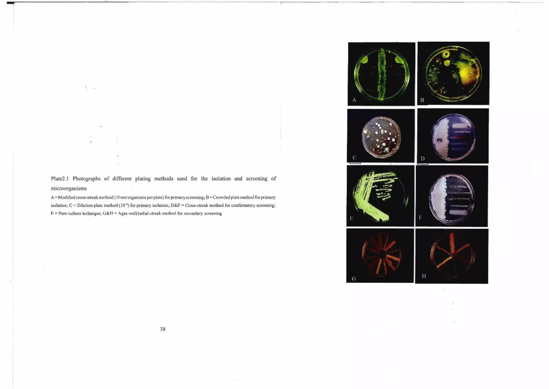
Plate2.1 Photographs of different plating methods used for the isolation and screening of
mIcroorgamsms
A =Modified cross-streak method (10 test organisms perplate) for primary screening; B =Crowded plate method for primary
isolation; C = Dilution plate method (10·4) for primary isolation; D&F = Cross-streak method for confmnatory screening;
E = Pure culture technique; G&H = Agar-welllradial-streak method for secondary screening
38
2.3 Results and DJscussion
.~.,..
Plating Techniques
The crowded plate technique yielded profuse growth of many organisms [Plate2.1B]. However, the
combined treatment ofheat and antibiotic incorporation into the media greatly reduced the number of
organisms present on the plates, The results were similar to those previously reported by several,.
authors (Williams and Davies, 1965; Pisano et al., 1986; Ntuli, 1994). The disadvantage ofantibiotic
addition is that a potent antibiotic producer, which is sensitive to the antibiotic added, may be inhibited
and hence will not be detected. On the other hand, the antibiotic makes the medium more selective by
limiting the number of organisms developing on the plates [Table 2.2].
Table 2.2 Percentage of the total number" of isolates active against all or some of the test
organisms* on primary screening
no. isolates per pre
treatment
H+A 38
H+NA 46
NH+A 94
NH+NA 166
H+A 34
H+NA 61
NH+A 123
NH+NA 205
H+A 43
H+NA 55
NH+A 58
NH +NA 157
NH+A 45
NH+NA 76
39
no. isolates active
12
18
11
2

o
no. isolates active
7
2
23
4
no. isolates per pre-
treatment
NH+A 56
H+A 23
NH+NA 103
NH+A 13
NH+NA 45
H+A 36
NH+A 97
NH+NA 167
H+A 66
H+NA 105
NH+NA 5
NH+A 71
NH+NA 128
H+A 61
NH+A 101
NH+NA 169
H+A 56
H+NA 60
Grand Total 79
*StaphylococcuS aureus, Escherichia coli, Pseudomonas fluorescens, Serratia marcescens,
Streptococcusfaecalis, Candida uti/is andXanthomonas campestris; NH=Non-Heated; H=Heated;A=Antibiotics
added; NA = No Antibiotics added; Atotal number based on the average colony count of the 10-2 to 10-4 dilutions of the sample.
Also the heat pretreatment selected thermophilic organisms, which means that any novel strains which
are heat sensitive may not have been detected. Sprinkling of heat pre-treated soil directly onto the
media led to overcrowding ofthe colonies at particular places on the plates. Antimicrobial activity, if
present, would be difficult to detect in such instances.
In contrast to the above procedure, the dilution plate method was more effective in isolating individual
40
colonies [Plate2.1q. However, very few colonies showed antimicrobial activity, expressed as
surrounding zones ofclearing. Zones ofinhibition were noticed on three plates; chicken manure plated
on Czapek's agar; chicken litter, also on Czapek' s agar, and cow manure on Kosmachev's medium. All
other samples showed no noticeable zones of inhibition.
Although no direct comparisons of the media used in this investigation were made, significant
observations were nonetheless possible. Some of the isolation media, namely, M3, Kosmachev's
medium and Winogradsky nitrite medium were found to be highly selective for actinomycetes without
the addition of any antibiotics to, or heat pretreatment of, the samples. Other authors have reported
similar observations (Williams and Davies, 1965; Ntuli, 1994). A fairly wide variety of organisms
grew on Czapek's agar which also enhanced pigment production in some of the organisms. MGA
medium supported prolific fungal growth and Kosmachev's medium proved to be highly selective for
actinomycetes, as most of the plates appeared to be colonized with almost pure cultures of
actinomycetes, probably, polysporic actinomycetes (Kosmachev, 1960). Unamended YEA plates were
overgrown with yeasts and fungi, but on antibiotic addition, growth of additional minute mycelial
colonies was noted. The antibiotics used in this investigation seemed to inhibit the growth offungi and
yeasts while favouring the growth of small mycelial colonies, probably actinomycetes, or bacteria
which were the main focus of this investigation.
The combination of selective pretreatment, selective media and incubation conditions, were helpful in
eliminating the growth of unwanted Streptomyces species. Dry heat treatment of some samples, in an
oven at 100°C for 1 hour before plating, meant that only actinomycetales having more heat resistant
spores such as Micromonospora, Microbispora and Streptosporangium were selected (Labeda, 1990).
The relatively mild heating regimens in a water bath (55°C for 6 minutes, Rowbotham and Cross, 1977)
and 70°C for 10 minutes (Cross, 1981) eliminated the spreading bacterial colonies and enhanced the
selective isolation of Micromonospora respectively (Labeda, 1990). Most of the media used were
selective for specific genera. M3 agar (Rowbotham and Cross, 1977), modified Czapek's agar (Higgins
and Lechevalier, 1969) and Winogradsky's nitrite medium (Winogradsky, 1949) were selective for
nocardiae and rhodococci. The addition ofantibacterial antibiotics such as gentamicin, oxytetracycline
and novobiocin to the other isolation media used, also meant that non-streptomycetous actinomycetales
41
were selected for. Actinomycetales differ significantly in their sensitivity toward antibacterial
antibiotics. Streptomycetes fortunately belong to the antibiotic sensitive group (Kutzner, 1981). It must
be noted, however, that this author doubts the selectivity of some of the so-called selective media as a
few colonies of other bacteria occasionally developed on the plates.
In the dilution plate method, there were remarkable variations in the numbers of organisms observed
at different dilutions. A 10-4 dilution was found to be most suitable for isolating single colonies as it
contained individual colonies ofa large variety oforganisms [Plate2.1 C]. Although particular attention
was directed in this study to organisms resembling actinomycetes, it was very important that each and
every colony that developed on the isolation plates was treated as a potential antibiotic producer. The
use of selective procedures minimized the laborious work involved in screening thousands of isolates.
Preliminary screening
The problem of screening each individual isolate with an overlay of a particular test organism was
overcome, in part, by employing a modified cross-streak method [Plate2.1A]. Using this method
facilitated testing a particular isolate against all the test organisms simultaneously. Only 79 of the
approximately 2600 isolates screened, showed signs of antimicrobial activity [Table2.2]. All inactive
isolates were discarded. The highest percentage ofactive isolates came from the SAPPI forest soil while
the lowest percentage came from samples from the banks ofthe Mzinyati river. The composition ofthe
medium is a very impor,tant factor affecting the production of active antibiotics (Lancini and Parenti,
1982). It is possible, therefore, that the nature of the media used was not conducive to antibiotic
production by the riparian microbiota. Decolouration of Serratia marcescens was not regarded as a
form of inhibition, as incubation at a lower temperature restored the red coloration.
Comparison of the number of antimicrobially-active isolates to the total number of isolates screened
from the various samples is shown in Fig 2.2. It is clearly evident, that the number of culturable
antibiotic-producing microorganisms constitute a small percentage ofthe total microbial population in
the different samples studied.
42
500 ....-------------------,
400
en2 300ro"0.!Q'0200oc
100
ocm cl cow cs us rb sf ccs sw csf
• total isolates antimicrobially active
Fig 2.2 Comparison of antimicrobially-active isolates to total number of isolates following primary
screemng
Only those isolates that inhibited two or more test organisms were considered worthy of further analysis
except in the case ofPseudomonas or Xanthomonas, which are notoriously difficult to control. Based
on these criteria, only 44 isolates were selected for re-screening on NA and Mueller Hinton Agar to
confirm the preliminary results [Chapter 3].
These preliminary findings indicate that KwaZulu-Natal soils do harbour antibiotic-producing
microorganisms, confirming the results obtained by previous investigators (Baecker and Ryan, 1987;
Ntuli, 1994).
43
CHAPTER 3
Screening of Isolates for Antimicrobial Activity
3.1 Introduction
Thousands of samples and millions ofmicrobial colonies have been screened all over the world in the
past 50 years, but compared to the screening rates, reports of success were minimal. At the beginning
of the 1950's the screening of 10,000 microorganisms resulted in one single useful agent. Later the
screening of 400,000 microorganisms over a ten year period also resulted in three utilizable drugs
(Berdy, 1974).
The rediscovery ofknown substances has become more frequent. In order to maximize the chances of
success, sampling and plating need to be a continuous exercise, so that screening itself becomes a
continuous process. In the present study, about 220 samples and approximately 2600 colonies, from
10 different locations, were screened over a three year period. This involved plating, on average, two
samples per week. Only 44 isolates showed any meaningful antimicrobial activity [Appendix 2].
Screening methods have metamorphosed over the years. From the Waksman and Dubos classical
methods ofagar-zone- diffusion (Linton, 1983), to discs impregnated with antibiotic substances (Pelczar
and Reid, 1972), and to the more complex use of selected mutants (Paik et al., 1997). Basically most
ofthese methods were dependent on one criterion; - the prodllction ofgrowth inhibition zones on agar~'",
media. However, some authors have recommended the use of other <;riteria less drastic than death or
inhibition of the test organism, such as the inhibition of sporulation, or abnormal growth ofhyphae, as
a novel way of screening for new antibiotics (Okafor, 1987).
\\_ Antibiotics diffuse through agar gel at different rates, .e.g. penicillin-G and chloramphenicol diffuse
rapidly, while, polymyxin diffuses slowly, so that the zone sizes produced when testing specifi~ isolates
I\t) with different antibiotics are not directly comparable. In conclusion, it is wrong to assume that an'\J
~ organism is more susceptible to one agent than another on the basis of a larger inhibition:' zone sizeU'
(Hammond and Lambert, 1978).
44
The size of the growth inhibition zone is a consequence of two dynamic systems proceeding
simultaneously:
• the diffusion of the antibiotic from its source,
• the growth rate of the test organism.
The position ofthe growth-inhibition zone edge in an agar gel is fixed at a point in time when specific
criteria in the two systems are fulfilled independently. An equation can be applied to the diffusion of
antibiotics through gels (Linton, 1983).
X2 = 4 DT 2. 3 ( Log Mo - Log M' )
Where
X = The distance between the antibiotic source and the zone edge.
D = The diffusion coefficient of the antibiotic.
T = The time taken for the zone to be fixed.
Mo = The antibiotic concentration at the source.
M' = The critical concentration of the antibiotic which inhibits the test organism.
Under standard conditions X and Mo are directly related. D is affected by temperature and viscosity
ofthe solvent. A high Mo drives M' well out into the agar in T, forming large growth inhibition zones.
Under normal assay conditions, Mo may be considered constant throughout the period T. However, in
disc susceptibility tests, Mo is not known, since although the disc is impregnated with a known amount
ofthe drug, this dissolves in an unknown amount of water in the assay agar. Hence the concentration
cannot be determined. It is not possible to relate initial antibiotic concentration directly to the size of
the inhibition zone produced (Linton, 1983).
However, the above problem may be minimized by determining the Minimum Inhibitory Concentration
[MIC] of the antibiotic. The MIC, defined as the lowest antibiotic concentration that will inhibit the
growth of a specific organism, has the advantage that it is performed in broth, thereby eliminating the
disadvantage of slower diffusion rates in the case of large-molecular mass antibiotics (Okafor, 1987).
A knowledge ofMICs is very important to microbiologists and clinicians, as an index ofantimicrobial
activity (Hammond and Lambert, 1978). An attempt was made in this study to determine the MICs of
45
the antimicrobial compounds produced by the most promising isolates.
46

3.2 Materials and Methods
3.2.1 Isolates
Organisms screened for antimicrobial activity were isolated from samples from various habitats in
KwaZulu-Natal midlands [section 2.2.1]. Stock cultures of pure isolates were kept on agar slants and
on thick NA plates at ± 4°C until required.
3.2.2 Test organisms
The test organisms, Staphylococcus aureus (1), Escherichia coli (2), Pseudomonas fluorescens (3),
Serratia marcescens (4), Streptococcus faecalis (5), Candida utilis (6), and Xanthomonas campestris
pv campestris (7) were provided by D. Fowlds ofthe Department ofMicrobiology & Plant Pathology
Laboratory, University of Natal. They were all grown on thick Nutrient agar (NA) plates except for
Candida utilis, which was grown on Yeast Extract agar (YEA), and kept as stock cultures in a
refrigerator (± 4°C). The test organisms were subcultured every 4 months throughout the period ofthis
investigation.
3.2.3 Assay media
Nutrient Agar and Iso-sensitest agar (ISTA) [Oxoid] were used for antimicrobial-activity assay. Also
f Potato Dextrose agar (PDA) was used for assaying fungal isolates. The responses ofthe isolates to the
different selective media were statistically analyzed using the SAS system.
3.2.4 Primary Screening
Individual purified isolates were streaked along the diameter ofNA and ISTA (Oxoid) plates using a
flamed sterilized loop. Each isolate was cultured for 6 - 7 days at 30°C, in which time any antibiotic
produced would have diffused a considerable distance from the streak. Test organisms were then
streaked at right angles to the original isolates starting from approximately Imm distance from the edge
ofthe producer organism's growth. Plates were then re-incubated for 24 hours at 30°C and the extent
of the inhibition ofthe various test organisms observed. All platings were done in replicates of 3 to 5.
47
3.2.5 Secondary Testing of Antibiotic Production
3.2.5.1 Antibiotic-production media
Media used included Nutrient broth (NB ) and M3 Broth (M3B ) - an M3 medium without agar (
Rowbotham and Cross, 1977). Other media used were Glucose-Yeast-Malt- Extract Broth (GYMB)
and Oatmeal Broth [International Streptomyces Project media 3 and 2 respectively, in which agar had
been omitted] (Shirling and Gottlieb, 1966).
3.2.5.2 Production of the antibiotic
Isolates which produced growth inhibition zones in the primary screens, were grown in nutrient broth
or in one/several ofthe media mentioned above. A loopful ofthe isolate was introduced aseptically into
100 ml NB or in any of the production media contained in a 250 ml Erlenmeyer flask and cultured in
a shaking incubator at 30°C for 5 to 8 days.
Attempts were made to produce large amounts ofthe antibiotics by using increased volumes ofbroth
and culturing under static conditions. Since there were no significant increases in antibiotic production,
static cultures were discontinued. After incubation, the cultures were centrifuged in a Heraeus Labofuge
200 at 9500 x g for 10-15 minutes. Aliquots of the supernatant (3-5ml) were filter sterilized (0.22Ilm)
into sterile screw capped glass vials. In some instances the filtration procedure was repeated to ensure
that cell-free supernatant (culture filtrate) was used for the antibiotic activity bioassay.
3.2.5.3 Bioassay for antibiotic activity
Samples of the culture filtrate were tested for antibiotic activity using a modification ofthe agar well
diffusion method ofPaik and Glatz (1995). After pouring the agar (5 mm deep), plates were incubated
for 24 hours at room temperature before wells were cut (7mm diameter). The plates were further
incubated at 37°C for 2 hours to dry, thereby facilitating sample diffusion into the agar. Culture filtrate
(50Il1) obtained by filter sterilization (0.22Ilm) ofthe supernatant above, was pipetted into the agar well
and the test organisms streaked radially outwards, starting 1mm from the edge ofthe well. The plates
were then incubated at 30°C for 24 hours and observed for growth inhibition zones. All assays were
carried out in triplicate (3 plates per test organism per isolate). Plates in which the wells were filled with
48
uninoculated culture media served as controls.
Also some samples were tested using the spot-on-lawn assay method of Eckwall and Schottel (1997).
A 50111 aliquot of culture filtrate was spotted onto filter paper disks (6.5mm, Whatman No.2). These
disks were allowed to dry before they were placed on Petri plates containing ISTA (Oxoid) medium
which had been freshly seeded with the test organism. The plates were then incubated at 30°C for 3 days
before the diameters of growth inhibition zones were measured.
The above method produced remarkable zones of growth inhibition but was discontinued due to its
laborious and uneconomical nature. It involved testing each isolate with only a single test organism at
a time, compared to the cross-streak method, which involved testing all the test organisms
simultaneously, in a single Petri plate.
3.2.6 Determination of Minimum Inhibitory Concentration (MIC)
Minimum inhibitory concentration (MIC) was determined using the broth micro-dilution method as
recommended by the Swedish Reference Group for antibiotics and its sub committee on methodology
[SRGA] (webite: www.SRGA) and as per standard protocol of Hancock's Doubling dilution method
(website: www.interchg.ubc.ca/bobh/methods.html).
Preparation ofIndicator test organisms
The organisms used as indicators were inhibited by the antimicrobially-active isolates on
primary/secondary screens. The indicator test organisms were prepared by growing them in Nutrient
Broth(NB) to an optical density (OD) ofO.5 -1.0 at 420nm. AfterwhichXlll cell suspension were added
to Iml NB. The value of X was calculated using the equation:
Xlll = 10/0D
Where OD = Optical Density
A 100111 of this cell suspension was re-suspended in 10ml Nutrient broth or 50111 to 5ml of the broth
prior to testing.
49
Broth micro-dilution method
Dilutions ofthe antibiotic-producer culture filtrate (stock solution) were prepared in a micro-titre plate
(IOxIO wells). From the stock solution, nine dilutions were made using the growth medium. To each
ofthe wells in micro-titre plate were added I00111 broth, except the fust well. The first and second wells
of the plate then received IOOlll each, of the stock solution. The contents of the second well were
thoroughly mixed, and 100111 transferred from this second well to the third. This process was continued
until the dilution series was completed. A step-wise dilution factor of2 (1.0, 0.5, 0.25, 0.125, 0.063 )
was maintained throughout the dilution procedure.
Finally, lOOlll ofthe prepared suspension ofthe indicator test organism were added to each ofthe wells
in the micro-titre plate and incubated at 30°C. The plates were examined for growth after 12h, 24h and
48h incubation. The last well showing no visible growth, i.e. a clear medium, was regarded as the MIC.
The results were confirmed by transferring O.lml aliquot, from each well, to nutrient agar plates,
incubating at 30°C for 24 hours and observing for colony formation. Practically, 99.9% reduction in
colony forming units (cfu) was recorded as no growth (Spooner and Sykes, 1972).
50
3.3 Results and Discussion
Primary screening
Of the 79 isolates from various samples that showed antimicrobial activity against the test organisms
on preliminary screening [Chapter2], 44 were selected for re-screening. The results of the primary
screening programme are shown in Appendix 2. It should be noted that only isolates that inhibited two
or more test organisms were considered except in the case of Pseudomonas or Xanthomonas spp.
Pseudomonas infections still require very high doses ofeven the most active penicillins during treatment
(Lancini and Parenti, 1982) and Xanthomonas infections remain a problem to plant pathologists.
The distribution ofthe isolates varied with the sample source and the isolation pre-treatment conditions.
The chicken manure sample yielded isolate N8, that showed antimicrobial activity against the majority
of test organisms used [Plate3.1A], while the SAPPI forest samples yielded isolates N19 [Plate3.1B]
and N33 that showed good activity against Pseudomonas fluorescens, and isolate N35, which showed
good activity against the plant pathogen, Xanthomonas campestris pv campestris, following isolation
from an unheated sample of SAPPI forest soil [Table 3.1]. The forest soil, rich in organic content and
acidic in nature may offer a good hunting ground for antibiotics judging from this result. Waksman et
al. (1942) also reported that certain acid forest soils do harbour antibiotic producing microorganisms.
The majority ofthe isolates from the Sweet Waters sample showed no meaningful antimicrobial activity
against the test organisms, with the exception of one isolate, N4a, which had a fairly good activity
against Pseudomonas fluorescens [Plate3.1 C&D]. Isolates from the banks of the Mzinyathi river at
Rorke's Drift, showed negative results. The highly waterlogged soils devoid ofoxygen may present an
unfavorable environment for aerobic microorganisms, especially, actinomycetes. It may also be
possible that the nature of the media used was not conducive to antibiotic production by the isolates
screened.
51
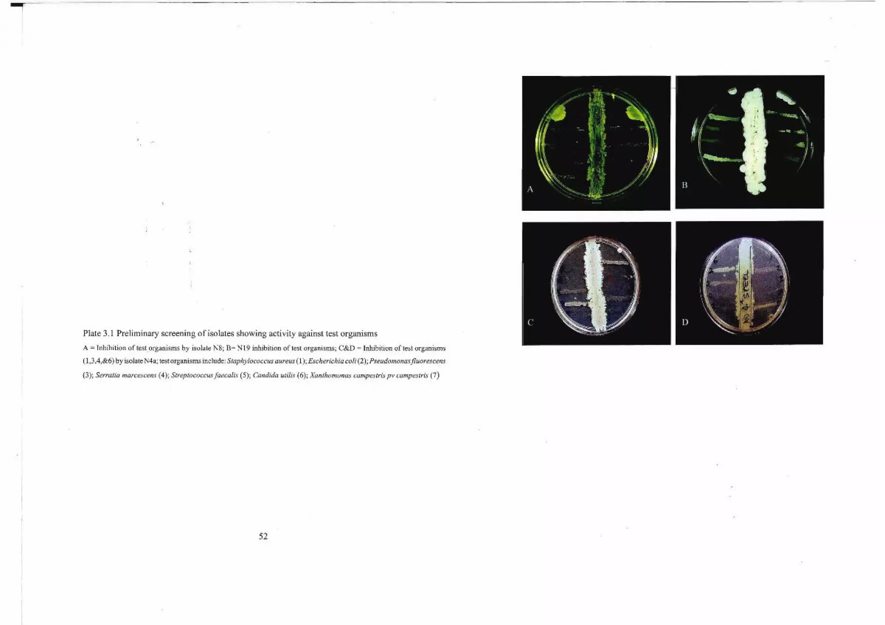
Plate 3.1 Preliminary screening of isolates showing activity against test organisms
A = Inhibition of test organisms by isolate N8; B= Nl9 inhibition of test organisms; C&D = Inhibition of test organisms
(1,3,4,&6) by isolate N4a; test organisms include: Staphylococcus aureus (1); Escherichia coli (2); Pseudomonasfluorescens
(3); Serratia marcescens (4); Streptococcusfaecalis (5); Candida utilis (6); Xanthomonas campestris pv campestris (7)
52
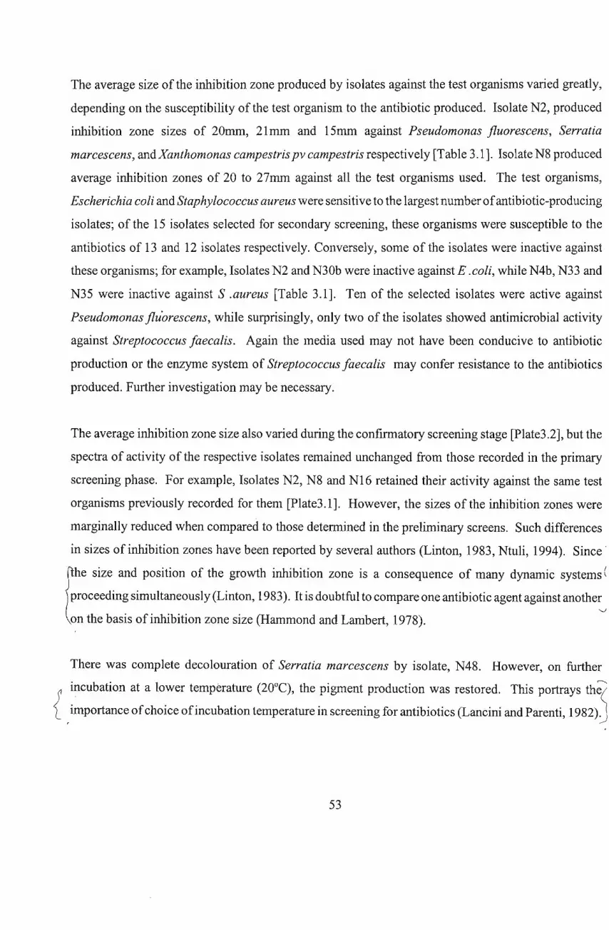
The average size of the inhibition zone produced by isolates against the test organisms varied greatly,
depending on the susceptibility of the test organism to the antibiotic produced. Isolate N2, produced
inhibition zone sizes of 20mm, 21mm and 15mm against Pseudomonas jluorescens, Serratia
marcescens, andXanthomonas campestris pv campestris respectively [Table 3.1]. Isolate N8 produced
average inhibition zones of 20 to 27mm against all the test organisms used. The test organisms,
Escherichia coli and Staphylococcus aureus were sensitive to the largest number ofantibiotic-producing
isolates; of the 15 isolates selected for secondary screening, these organisms were susceptible to the
antibiotics of 13 and 12 isolates respectively. Conversely, some of the isolates were inactive against
these organisms; for example, Isolates N2 and N30b were inactive against E .coli, while N4b, N33 and
N35 were inactive against S .aureus [Table 3.1]. Ten of the selected isolates were active against
Pseudomonas jluorescens, while surprisingly, only two of the isolates showed antimicrobial activity
against Streptococcus faecalis. Again the media used may not have been conducive to antibiotic
production or the enzyme system of Streptococcus faecalis may confer resistance to the antibiotics
produced. Further investigation may be necessary.
The average inhibition zone size also varied during the confirmatory screening stage [Plate3.2], but the
spectra of activity of the respective isolates remained unchanged from those recorded in the primary
screening phase. For example, Isolates N2, N8 and N16 retained their activity against the same test
organisms previously recorded for them [Plate3.1]. However, the sizes of the inhibition zones were
marginally reduced when compared to those determined in the preliminary screens. Such differences
in sizes of inhibition zones have been reported by several authors (Linton, 1983, Ntuli, 1994). Since'
f:he size and position of the growth inhibition zone is a consequence of many dynamic systems (
proceedin~ sim~lt~n~~US1Y (Lint~n, 1983). It is doubtful to compare one antibiotic agent against anothe~
on the basIs of InhIbItIon zone SIze (Hammond and Lambert, 1978). .
There was complete decolouration of Serratia marcescens by isolate, N48. However, on further
incubation at a lower temperature (20°C), the pigment production was restored. This portrays th~
{ •importance ofchoice ofincubation temperature in screening for antibiotics (Lancini and Parenti, 1982Jj
53

1
Plate3.2 Primary screening of,isolates confirming activity against test organisms
Photographs (A&B) == N8 activity against nos. 1,2,3,4&7 test organisms; (C&D) = N16 activity against nos. 1,2,3,&7 test
organisms; (E, F&H) = N2 activity; against nos.3&6; (G) = N19 showing inactivity against Serratia marcescens and
Streptococcusfaecalis. Test organisms include: Staphylococcus aureus (1); Escherichia coli (2); Pseudomonas
fluorescens (3); Serratia marcescens (4); Streptococcus fa ecalis (5); Candida utilis (6); Xanthomonas
campestris pv campestris (7)
54

Table 3.1 Average size of inhibition zones produced by isolates against test organisms [recommended for
secondary screening]*
Test organisms/ Inhibition zone size (mm)
Isolate source 0 @ @} 0 0 <D @
Sa Ec Pf Srn Sf Cu Xc
N2 CL 13 10 20 21 20 15
N3 CM 25 20 25 nt
N4a SW 15 30 20 nt
N4b SW 30 15
N16 CCS 11 25 3 3 3
N8 CM 20 25 20 23 22 27 24
N15 CL 20 21 2 30
N16a CL 25 5 14 Rs
N25a CL 2 30 30
N48 CS 18 30 -dc 21 19
N12b CSF(NH+A) 3 7 8 5
N19b SF(NH+A) 3 4 10 5
N30b CSF(H) 5 20
N33 SF(NH) 3 10
N35 SF(NH) 3 10
* Data generated from the results presented in Appendix 3; Sa = Staphylococcus aureus; Ec = Escherichia coli; Pf =
Pseudomonasfluorescens; Sm = Serratia marcescens; Sf= Streptococcus faecalis; Cu = Candida utilis; Xc = Xanthomonas
campestris pv campestris; Rs = Resistant strain; CL = Chicken litter; CM = Chicken manure; CS= Compost Soil; US =
Uncultivated soil; CCS = Creosate Contaminated Soil; SW = Sweet Waters (Chicken manure); nt = not tested; -dc =
decoloration; H=Heated; NH=Non-heated; A= Antibiotics added; SF=SAPPI forest; CSF=Cane sugar field.
The spectra of activity of isolates N2 and N8 were quite broad, although N2 was inactive against
Streptococcus faecalis [Fig 3.3]. Morphological observations showed that the organisms may likely
belong to the actinomycete group [Chapter4]. Isolate N48 was active against four ofthe test organisms;
viz. S. aureus, E. coli, C. utilis and X campestris pv campestris, with E. coli being the most susceptible.
Although isolate N16 showed broad activity against the majority of the test organisms, their
55
susceptibilities were quite low, except for E. coli, whichwas highly susceptible to the antibiotic produced
by the isolate [Fig3.3]. Only three ofthe test organisms used were susceptible to the antibiotic produced
by isolate N3, however, this was quite remarkable considering the large size of the inhibition zones
produced, especially against P.jluorescens [Fig3.3].
Activity spectra of isolates
N48N8N16Isolates
N3
----"'"-.-0------
N2O-i --La--
30-~.----------------------,.--~.........E525"-.m~ 20.~
~ 15co~ 10o
+=':.0 5 -----.r:.c
Sa 11 Ec D Pf 11 Srn Sf 11 Cu Xc
Fig 3.3 Comparison of antimicrobial spectra of some adive isolates on primary screening.
Some of the isolates lost a substantial part of their activity on extended incubation . This may suggest
that the antibiotic produced may have been inactivated, either by a substance released late into the
medium or some resistant strains developed and grew in the inhibition zone. It is also possible that the
initial inhibitoryantibiotic concentration, have been altered as a result ofantibiotic diffusion through the
agar medium. Isolated colonies which grew within an inhibition zone were recorded as resistant strains.
Similar findings have been reported by other authors (Linton,1983).
56
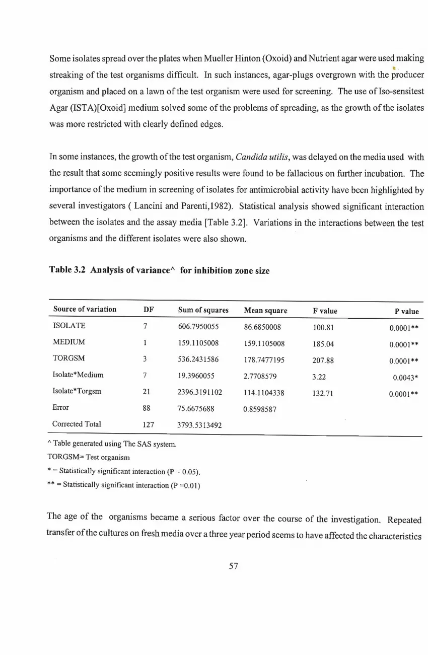
Some isolates spread over the plates when Mueller Hinton (Oxoid) and Nutrient agar were used making~.
streaking of the test organisms difficult. In such instances, agar-plugs overgrown with the producer
organism and placed on a lawn of the test organism were used for screening. The use of Iso-sensitest
Agar (ISTA)[Oxoid] medium solved some of the problems of spreading, as the growth of the isolates
was more restricted with clearly defined edges.
In some instances, the growth ofthe test organism, Candida uti/is, was delayed on the media used with
the result that some seemingly positive results were found to be fallacious on further incubation. The
importance of the medium in screening of isolates for antimicrobial activity have been highlighted by
several investigators ( Lancini and Parenti,1982). Statistical analysis showed significant interaction
between the isolates and the assay media [Table 3.2]. Variations in the interactions between the test
organisms and the different isolates were also shown.
Table 3.2 Analysis of varianceA for inhibition zone size
Source of variation DF Sum of squares Mean square F value P value
ISOLATE 7 606.7950055 86.6850008 100.81 0.0001 **
MEDIUM 1 159.1105008 159.1105008 185.04 0.0001 **
TORGSM 3 536.2431586 178.7477195 207.88 0.0001 **
Isolate*Medium 7 19.3960055 2.7708579 3.22 0.0043*
Isolate*Torgsm 21 2396.3191102 114.1104338 132.71 0.0001 **
Error 88 75.6675688 0.8598587
Corrected Total 127 3793.5313492
/\ Table generated using The SAS system.
TORGSM= Test organism
* = Statistically significant interaction (P = 0.05).
** = Statistically significant interaction (P =0.01)
The age of the organisms became a serious factor over the course of the investigation. Repeated
transfer ofthe cultures on fresh media over a three year period seems to have affected the characteristics
57
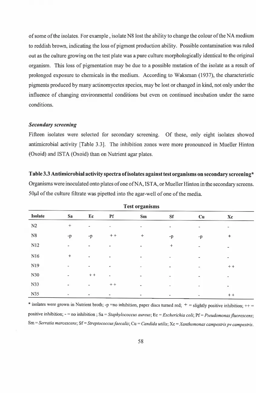
ofsome ofthe isolates. For example, isolate N8 lost the ability to change the colour ofthe NA medium
to reddish brown, indicating the loss ofpigment production ability. Possible contamination was ruled
out as the culture growing on the test plate was a pure culture morphologically identical to the original
organism. This loss of pigmentation may be due to a possible mutation of the isolate as a result of
prolonged exposure to chemicals in the medium. According to Waksman (1937), the characteristic
pigments produced by many actinomycetes species, may be lost or changed in kind, not only under the
influence of changing environmental conditions but even on continued incubation under the same
conditions.
Secondary screening
Fifteen isolates were selected for secondary screemng. Of these, only eight isolates showed
antimicrobial activity [Table 3.3]. The inhibition zones were more pronounced in Mueller Hinton
(Oxoid) and ISTA (Oxoid) than on Nutrient agar plates.
Table 3.3 Antimicrobial activity spectra ofisolates against test organisms on secondary screening*
Organisms were inoculated onto plates ofone ofNA, ISTA, or Mueller Hinton in the secondary screens.
50111 ofthe culture filtrate was pipetted into the agar-well of one ofthe media.
Test organisms
Isolate
N2
N8
N12
N16
N19
N30
N33
N35
Sa
+
-p
+
Ec
-p
++
Pf
++
++
Srn
+
Sf
-p
+
Cu
-p
Xc
+
++
++
* isolates were grown in Nutrient broth; -p =no inhibition, paper discs turned red; + = slightly positive inhibition; ++ =
positive inhibition; - = no inhibition; Sa = Staphylococcus aureus; Ec = Escherichia coli; Pf = Pseudomonas fluorescens;
Srn = Serratia marcescens; Sf= Streptococcus faecalis; Cu = Candida utilis; Xc = Xanthomonas campestris pv campestris.
58
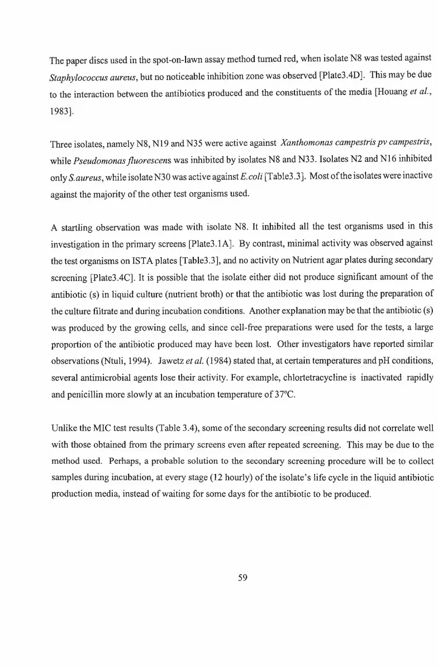
The paper discs used in the spot-on-lawn assay method turned red, when isolate N8 was tested against
Staphylococcus aureus, but no noticeable inhibition zone was observed [Plate3.4D]. This may be due
to the interaction between the antibiotics produced and the constituents of the media [Houang et al.,
1983].
Three isolates, namely N8, N19 and N35 were active against Xanthomonas campestris pv campestris,
while Pseudomonas fluorescens was inhibited by isolates N8 and N33. Isolates N2 and N16 inhibited
only S.aureus, while isolate N30 was active againstE.coli [Table3.3]. Most ofthe isolates were inactive
against the majority ofthe other test organisms used.
A startling observation was made with isolate N8. It inhibited all the test organisms used in this
investigation in the primary screens [Plate3 .1A]. By contrast, minimal activity was observed against
the test organisms on ISTA plates [Table3.3], and no activity on Nutrient agar plates during secondary
screening [Plate3.4C]. It is possible that the isolate either did not produce significant amount of the
antibiotic (s) in liquid culture (nutrient broth) or that the antibiotic was lost during the preparation of
the culture filtrate and during incubation conditions. Another explanation may be that the antibiotic (s)
was produced by the growing cells, and since cell-free preparations were used for the tests, a large
proportion of the antibiotic produced may have been lost. Other investigators have reported similar
observations (Ntuli, 1994). Jawetz et al. (1984) stated that, at certain temperatures and pH conditions,
several antimicrobial agents lose their activity. For example, chlortetracycline is inactivated rapidly
and penicillin more slowly at an incubation temperature of 37°C.
Unlike the MIC test results (Table 3.4), some ofthe secondary screening results did not correlate well
with those obtained from the primary screens even after repeated screening. This may be due to the
method used. Perhaps, a probable solution to the secondary screening procedure will be to collect
samples during incubation, at every stage (12 hourly) of the isolate's life cycle in the liquid antibiotic
production media, instead ofwaiting for some days for the antibiotic to be produced.
59
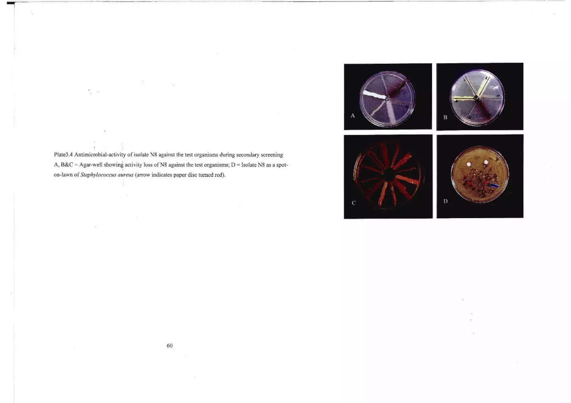
Plate3.4 Antimicrobial-activity of isolate N8 against the test organisms during secondary screening
A, B&C = Agar-well showing activity loss ofN8 against the test organisms; D = Isolate N8 as a spot
on-lawn of Staphylococcus aureus (arrow indicates paper disc turned red).
60
Minimal Inhibitory Concentration( MIC)
The method used for MIC determinations was appropriate but as a crude extract (culture filtrate) was
used in the investigation, the concentration ofthe antibiotic in the crude filtrate could not be ascertained.
Hence, the term, Arbitrary activity Unit, may be more suitable. The arbitrary activity unit (AU) of an
isolate may be regarded as the reciprocal of the dilution of the last clear well in a micro-titre plate. The
value is adjusted to obtain activity units per ml [AU/ml] by multiplying by ten ( Papathanasopoulos,
1997). Isolate N2 showed activity against Staphylococcus aureus, Escherichia coli, Pseudomonas
jluorescens and Xanthomonas campestris pv campestris. Its highest activity was against P.jluorescens
with an MIC ofO,0039flg/ml, while the MIC forXcampestris pv campestris was O,25flg/ml [Table 3.4].
Table 3.4 MIC's lflg/ml] ofthe selected antimicrobially-active isolates against the test organisms
Isolate No Sa Ec Pf Srn Sf Co XC
N2 0,0078 0,0156 0,0039 uns uns nt 0,25
N8 0,0078 0,0039 0,0625 0,125 0,25 0,002 0,0025
N12 nt nt 0,25 uns uns uns nt
N16 0,125 0,0039 0,0039 uns uns uns 0,002
N19 nt nt 0,125 uns uns 0,25 0,25
N30 nt uns 0,0625 uns uns uns uns
N33 uns nt 0,002 uns uns uns uns
N35 uns 0,031 uns uns uns uns 0,0039
*Uns= Unsusceptible; nt= not tested; Sa = Staphylococcus aureus; Bc = Escherichia coli; Pf= Pseudomonas
fluorescens; Srn = Serratia marcescens; Sf = Streptococcus faecalis; Cu = Candida utilis; Xc = Xanthomonas
campestris pv campestris.
The activity against Pseudomonasjluorescens was the main reason N2 was selected for further studies.
N8 appears to be the isolate with the greatest chemotherapeutic value in the present investigation on the
results obtained from screening and MIC determinations. It showed activity against all the test
organisms used [Table 3.4], especially the Pseudomonas and Xanthomonas species, with MICs of
O,0625flg/ml and O,0025flg/ml respectively. However, specificity ofaction ofantibiotics is much valued
61
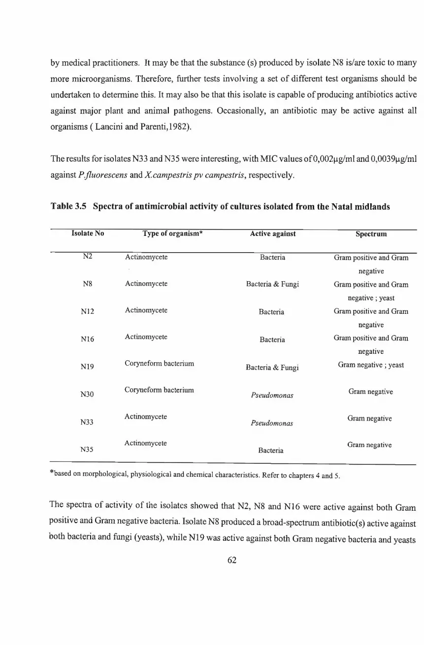
by medical practitioners. It may be that the substance (s) produced by isolate N8 is/are toxic to many
more microorganisms. Therefore, further tests involving a set of different test organisms should be
undertaken to determine this. It may also be that this isolate is capable ofproducing antibiotics active
against major plant and animal pathogens. Occasionally, an antibiotic may be active against all
organisms (Lancini and Parenti,1982).
The results for isolates N33 and N35 were interesting, with MIC values ofO,002~g/ml and O,0039~g/ml
against P.fluorescens and Xcampestris pv campestris, respectively.
Table 3.5 Spectra of antimicrobial activity of cultures isolated from the Natal midlands
Isolate No Type of organism* Active against Spectrum
N2 Actinomycete Bacteria Gram positive and Gram
negative
N8 Actinomycete Bacteria & Fungi Gram positive and Gram
negative; yeast
Nl2 Actinomycete Bacteria Gram positive and Gram
negative
Nl6 Actinomycete Bacteria Gram positive and Gram
negative
Nl9 Coryneform bacterium Bacteria & Fungi Gram negative; yeast
N30Coryneform bacterium
Pseudomonas Gram negative
N33Actinomycete
PseudomonasGram negative
Actinomycete Gram negativeN35 Bacteria
*based on morphological, physiological and chemical characteristics. Refer to chapters 4 and 5.
The spectra of activity of the isolates showed that N2, N8 and N16 were active against both Gram
positive and Gram negative bacteria. Isolate N8 produced a broad-spectrum antibiotic(s) active against
both bacteria and fungi (yeasts), while N19 was active against both Gram negative bacteria and yeasts
62
[Table 3.5]. Isolates N30, N33 and N35 were active against the Gram negative bacteria.
The MIC values shown in Table 3.4, serve as an index of the antimicrobial-activity of the isolates
investigated in this study.
63
Chapter 4
Identification of Antibiotic-Producing Microorganisms
I: DESCRIPTION OF ISOLATES
4.1 Introduction
Identification ofcultures producing antibiotics is a necessary part ofa patent application (Dietz, 1999).
It is imperative for an industrial taxonomist to obtain as many type cultures as possible, study them
under standardized conditions, and develop a practical in house identification scheme (Dietz, 1999).
Since the beginnings ofmicrobiology, a legion ofcharacterization tests has been developed and studied.
Some of these tests have enjoyed popularity because they allowed categorization of strains, and as a
means for communicating identifications. Many ultimately met with disfavor and were abandoned
because study of additional strains led to detection of the inevitable 'different' strain or strains whose
characteristics did not seem to correlate properly with other data or an overwhelming number ofstrains
gave similar results and the hoped for separations could not be made (pridham, 1999).
In early studies of actinomycetes, classification was based solely on morphological observations
(Lechevalier et al., 1961; Thiemann and Beretta, 1968). However, accurate morphological
characterization of actinomycetes is dependent upon the use of a culture medium which supports good
sporulation (Shirling and Gottlieb, 1966).
Morphological characteristics such as the nature of aerial mycelium and mode of spore fonnation are
very important for generic identifications. The fonn of spores and sporophores are constant for every
actinomycete species. For example, Streptomyces produces conidia (spores) that are spherical, oval or
elongated while Nocardia oidiospores are cylindrical. However, morphology in itself is not adequate
to differentiate many actinomycete genera. It has to be complemented with chemical and physiological
investigations (Labeda,1987).
Lechevalier and Lechevalier (1965) pointed out that actinomycetes can be separated into broad groups
on the basis of chemical and morphological characteristics. This approach is still the simplest and the
64
best (Lechevalier, 1989). Cell wall composition, whole-cell sugarpattems, mycolic acid structures, and
more recently phospholipids, have been used as taxonomic markers in the actinomycetes (Lechevalier,
1999). It is possible by using one-way paper chromatography ofwhole-cell hydrolysates to determine
the type ofcell wall or diagnostic sugars present in the cell wall ofan unknown organism. This simple
method will reveal whether or not the cell wall of the unknown isolate contains diaminopimelic acid
(DAP) and, if so, the isomers present, viz. the meso- or the L-form (Lechevalier,1999).
Identification to the species level requires physiological or biochemical characterization. There have
been many attempts to utilise physiological reactions which represent multiple enzymatic activities, for
characterization, classification and identification of actinomycetes. These attempts at applying
conventional systematic criterion have been disappointing (Trejo,1970), Luedemann (1971), and
Pridham (1976).
Characterization ofprogeny ofstrains is less reproducible, less understandable, and less communicable
(Pridham, 1999). Major obstacles are the superfluity of tests and methodologies and their
standardization. Beyond that, the reproducibility of conventional characterizations and their
interpretation leaves much to be desired. From the examination of two volumes of Bergeys Manual it
is apparent that little standardization of test methods prevails. The same is true for most other
systematics treatises.
Gottlieb (1976) has addressed this problem to some extent in a letter to the editor of the Newsletter of
the US Federation for Culture Collections. Even within the same genus of Actinomycetales,
comparative data are lacking. For example, information on production of 20-p-keto reductase was
presented for only one species of Streptomycetes (Pridham, 1999).
In summary, conventional systematics ofthe Actinomycetales is still in an unresolved state because of
reliance on imprecise methodology for determining physiological characteristics. This in turn affects
the nature of the species concept derived therefrom (Pridham, 1999).
An attempt was made to identify some of the antibiotic-producing actinomycetes isolated during this
65
investigation, up to the species level, using methods currently available in the literature.
66
4.2 Materials and Methods
4.2.1 Organisms
The organisms to be identified were isolated from soil samples collected from the KwaZulu-Natal
midlands, South Africa. Eight isolates viz- N2, N8, N16, N12, N19, N30, N33, and N35 were selected
from 13 isolates that showed good antimicrobial activity on both primary and secondary screening. The
criteria for selection were indicated in Chapter 3.
Subcultures were inoculated on the same media used for their isolation and incubated at 30°C for 4 to
6 days. Stock cultures were grown at 30°C on slants and/or thick Nutrient Agar or Yeast Extract Agar
(YEA) - ISP medium 2 (Shirling and Gottlieb,1966) plates and then stored at 4°C. In all the
investigations, the inocula used were 7-14 day old stock cultures.
4.2.2 Morphological Characterization
The morphological features of each isolate grown on each of the ISP media 2,3,4 & 5 (Shirling and
Gottlieb,1966) were studied under brightfield and phase contrast light microscopy. A more detailed
study of the surfaces of spores and the spore chains was carried out with the aid of both the
Environmental Scanning Electron microscope (ESEM) and the conventional Scanning Electron
Microscope (SEM).
4.2.2.1 Light Microscopy
Micro-morphology ofthe actinomycete isolates was determined by either preparing Gram stained slides
from 7 to 21 day-old cultures or by observing the organisms in situ. The presence of spore chains and
aerial and/or substrate mycelium was observed in situ under bright field illumination using either a
Nikkon (1000 X ) or a Zeiss Axiophot light microscope which was used to take photographs at 400x and
1000x magnifications. Observations were made on cultures growing on all the media used [section
4.2.1 ].
4.2.2.2 Electron microscopy
Small agar plugs (2x2rnm) were removed from the plate cultures, transferred to separate plastic -capped
67
glass vials, and fixed overnight in 3% cacodylate buffered gluteraldehyde. The fixed specimens were
then washed (30 mins x 2 changes) in 0.05M cacodylate buffer and dehydrated in an alcohol series: once
(10 mins each) in each of30%, 50%, 70%, 80%, 90%, and thrice (10 mins each) in 100% alcohol. The
dehydrated specimens were transferred to a Hitachi HCP-2 critical point dryer (CPD) under 100%
alcohol and dried. These were then sputter coated with Gold palladium in a Polaron SEM coating unit
E5100 (Polaron Equipment Ltd) and observed in a Hitachi S-570 SEM.
For Environmental Scanning Microscopy (ESEM), small agar plugs (2x2mm) covered with the
actinomycete culture were cut and mounted on brass stubs using double sided cellotape. The specimens
were then viewed in a Philips XL30 ESEM.
4.2.3 Physiological Characterization
Many physiological tests have been found unreliable and of little taxonomic value if a wide species
concept is employed (Kutzner, 1981). Only two seem to have stood the test oftime and have been used
in the International Streptomyces Project (ISP) [Shirling and Gottlieb, 1966]. These include:
• Formation of pigments
• Utilization of nine carbon sources (mainly sugars and sugar alcohols).
4.2.3.1 Formation of melanin pigment
Melanin pigment production was tested on ISP medium 6 and ISP medium 7 as detailed by Shirling and
Gottlieb (1966).
4.2.3.2 Carbohydrate utilization
Utilization of sugars and sugar alcohols by the isolates was determined using the Agar culture method
of Nitsch and Kutzner (1973). The basal medium contained one-third agar (Difco AATCC) strength
(Appendix1) while the carbon sources were filter sterilized and added as 10% solutions. Glucose was
used as a positive control while the basal medium without any carbon source was used as a negative
control. To prepare shake cultures, 0.2 ml carbon sources were added to sterile test tubes ( 1Ox1OOmm)
and inoculated with 0.1 ml of a dense spore suspension ofthe isolate. Basal medium (1,7 ml; warmed
to 70°C) was finally added and the test tubes incubated for 2 to 7 days at 30°C.
68
4.2.3..3 Other physiological tests
All the isolates were Gram stained using Hucker's modification of the Gram stain procedure (Corm et
al., 1960) while acid fastness was determined using the Ziehl-Neelsen staining method (modified by
adding 50:50 volumes of 3% HCI and alcohol for decolorisation).
The catalase and oxidase tests were carried out on 7-day old cultures of the isolates. Kovac's method
was used for the oxidase test. Examination ofthe isolate's spores was by Conklin's modification ofthe
Wirtz spore stain. All tests were performed according to the Manual of Microbiological Methods,
McGraw-Hill, New York (1957) and the examples from Bergey's Manual of Determinative
Bacteriology (8th ed., 1974) and Bergey's Manual of Systematic Bacteriology (VolA., 1989).
Motility was determined using two procedures; the hanging drop method ; and an in situ motility
determination method. The latter method involved placing a drop of sterile distilled water directly on
the bacterial growth(single colonies) on the plates, covering with a coverslip and gentle pressing down
to spread the water evenly. Motility determination were made after 18 hrs, three days and seven days
incubation, under a light microscope (oil lens, 1000x magnification; Zeiss Axiophot).
4.2.4 Biochemical Characterization
For the actinomycete isolates, the first stage in the chemical procedure was the examination ofwhole
cell and/or cell wall hydrolysates for diagnostic sugars and diaminopimelic acid (DAP) isomers (Becker
et al., 1964; Lechevalier, 1968; Berd, 1973; Staneck and Roberts, 1974). Different solvent systems, or
combinations thereof, were used.
4.2.4.1 Preparation of the whole-cell hydrolysates
The isolates were grown in modified Santon's medium as detailed by Mordaska et al. (1972). The
organisms were aseptically inoculated by transferring growth from stock cultures with an inoculating
loop, into 50ml medium in 250ml Erlenmeyer flasks and incubated at 30°C in a rotary shaker [New
Brunswick, Edison, USA] at 300rpm for seven days. The biomass was harvested by centrifugation at
3000 x g for 10 mins in a Heraeus Labofuge 200. The organisms were then washed once with 0,5M
NaCl in distilled water, twice with distilled water and freeze-dried under vacuum in a GAMMA 1freeze
69
\
dryer.
Diagnostic cell wall sugars were extracted from freeze-dried biomass using a combination of two
methods: organisms were hydrolyzed by the method ofMurray and Procter (1965) ; and the hydrolysate
then evaporated using the method ofStaneck and Roberts (1974).
Freeze-dried cells (50mg) ofeach isolate were suspended separately in 3ml of5% H2SO4in 20ml screw
cap bottles and autoclaved at 5lb/sq in. for 20 minutes. After cooling, 19 Ba(OH)2 was added to the
hydrolysates. The mixtures were adjusted to pH5.0 - 5.5 with 5%H2S04, centrifuged at 6000 x g in a
Heraeus Labofuge 200, the supematants removed by Pasteur pipette and the remaining liquid evaporated
over boiling water. The residues were re-suspended in 1ml dH20 and centrifuged to remove insoluble
material before the solutions were used for chromatography.
4.2.4.2 Determination of sugars occurring in whole-cell hydrolysates
4.2.4.2.1 One-way paper chromatography for sugar determination
The sugar standards: Galactose, Arabinose and Xylose (1 st standard) and Rhamnose, Mannose, Glucose
and Ribose (2nd standard) were prepared separately as 1% (w/v) solutions. In all cases 20x20 cm sheets
of Whatman's chromatographic paper were used and the sample and standards (lOlll each) applied
separately, one centimeter from the bottom edge of the chromatogram. The chromatograms were
developed in the solvent system ofBecker et al. (1964). Completed chromatograms were air-dried with
a hair-dryer and visualized by spraying with 0,2% ninhydrin in acetone followed by heating at 105°C
for two to five minutes.
4.2.4.3 Determination of Diaminopimelic acid (DAP) Isomers
The presence and form ofDAP was determined by the method ofBecker et al. (1964) as described by
Ntuli (1994). For each isolate, fifty milligrams of freeze-dried biomass were mixed with 5m16M HCl
in a small screw-cap bottles. The mixture was hydrolyzed at 100°C for two hours in a hot air oven. After
cooling to room temperature the hydrolysate was filtered through Whatman No.2 paper. The filtrate was
collected in a 50ml glass beaker and evaporated to dryness over boiling water. The residue was re
dissolved in 5ml distilled water and re-evaporated and the final residue re-suspended in 1ml distilled
70

water before being used for chromatography.
4.2.4.3.1 HPLC analysis of whole-cell hydrolysates for DAP detection
The freeze-dried biomass (50mg) ofeach isolate was hydrolyzed separately in 6N HCl at 110°C for 24
hours according to the protein hydrolysis procedure ofMoore and Stein (1948). An internal standard
was added to correct for any inaccuracies that may have occurred during sample preparation.
The hydrolysates were transferred separately to a Beckman 6300 analyzer. The analyzer comprised a
single-column ion-exchange chromatography utilizing HPLC techniques involving; sodium elution
buffers, a regenerating reagent (ninhydrin), and a sodium high perfonnance column. The standard
detection system featured the classical Moore and Stein (1948) ninhydrin reagent. The combined
column effluent/reagent mixture was heated at 135°C and ran at a flow rate of 14.5mllmin for a period,
prior to photometric visualisation at wavelengths of 440 and 520 nanometers. These analyses were
carried out in the Animal Science Laboratory of the University ofNatal.
4.2.4.3.2 Two-Dimensional Thin Layer Chromatography for DAP Determination
For the actinomycete isolates, the DAP isomers were detennined by the method described by Harper
and Davis (1979). Commercial cellulose-coated aluminum sheets were used. Isopropanol-acetic acid
water (75:10:15) and methanol-pyridine-10N HCl-water (64:8:2:14) were used as first and second
solvents respectively. To ensure saturation ofthe tank atmosphere with the solvents, two walls of each
tank (Shandon TLC tanks 30x22x13cmX2) were lined internally with absorbent paper and 100ml of
solvent poured into the tank 24hrs before chromatograms were run.
Cellulose-coated aluminum sheets (20x20cm, Merck, cat. No.5552) were marked with origin points, 1,5
cm from two sheet edges and pencil lines made 15,5cm from, and parallel to, the sheet edges. Each two
dimensional analysis was made with 2 to 4111 ofhydrolysate with the sample spot size kept as minimal
as possible. The strips, 4,5cm wide, marked on the sheet were used to make one-dimensional runs of
the sample and control mixtures of standard amino acids in each solvent. The first dimension solvent
mentioned above took approximately 3h to reach the 15,5cm mark. The chromatogram was then dried
overnight in a ventilated hood at room temperature or in a ventilated oven at 10YC for 2 hours.
71
The second solvent mentioned above took approximately 1,25h to reach the 15,5 mark. Completed
chromatograms were then air-dried and visualized with 0,2% ninhydrin in acetone by spraying and
heating at 105°C.
4.2.4.3.3 One-way paper/thin-layer chromatography for DAP determination.
Diaminopimelic acid (DAP) isomers were also determined by one-way paper/thin-layer
chromatography. Whole-cell hydrolysis was carried out by the modified method ofBecker et al. (1964)
as described earlier. The following solvent mixture was used for separation of the amino acids:
methanol-water-lON HCI-pyridine (80: 17.5: 2.5: 10, byvolume).
72
4.3 Results and Discussion
Selective media were used for isolation ofthe organisms to be identified. Lechevalier (1989) practical
guide to generic identification of Actinomycetes was adhered to, and the results compared with
descriptions ofsimilar and/or related organisms in Bergey' s Manual ofSystematic Bacteriology (VolA,
1989).
Isolate N2
Aforphologicalproperties
The colonies were curled and irregularly branched in young cultures, forming a mycelium resembling
a contour map [Plate4.1B]. Colonies were white to pale orange [Plate4.1D]. The young hyphae
penetrate the agar [Plate4.1 C], and after 14 days incubation, fuse together at the surface ofthe colonies
[Plate4.1D]. Environmental Scanning Electron Microscopy (ESEM) revealed the spores silhouette
contained in sheaths of extracellular material [Plate4.1E]. The surface of the oval shaped spores is
smooth and the spores are packed in dense clusters [Plate4.1F] as seen under the Scanning Electron
Microscope (SEM).
Physiological tests
Cells were Gram-positive, non-acid fast [Plate4.1A], catalase positive and formed moderate filiform
growth on nutrient agar slants. A pellicle was formed in test tube cultures which later detached and fell
to the bottom of the tube. The broth remained clear afterwards. A reddish brown soluble pigment was
produced on ISP medium 7 and in nutrient broth shake cultures incubated for seven days. No diffusible
pigment was produced on the other ISP media used. The oxidase test was negative.
Carbohydrate utilization
On ISP medium 9, the organism utilized L-arabinose, fructose, glucose, inositol, rhamnose and sucrose
and to a lesser extent raffinose and D-xylose as carbon sources [Table 4.1]. Pink-brown pigment was
produced on all the carbon sources except raffinose and D-xylose. There was no noticeable growth in
the control which contained only the basal medium without any carbon source.
73
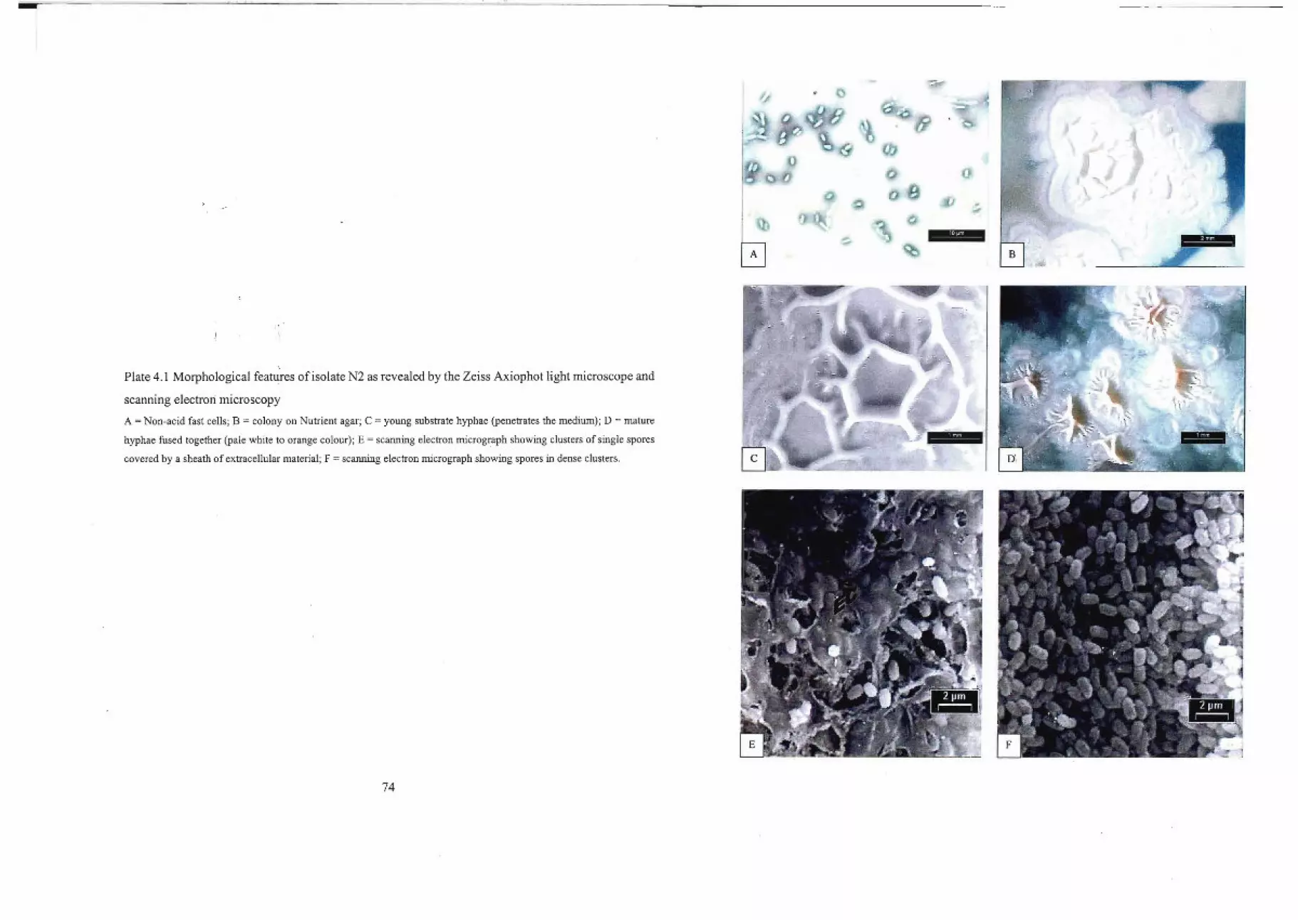
..rl~
o
i"'" ."
~:"
\'.
o
.',
Plate 4.1 Morphological features of isolate N2 as revealed by the Zeiss Axiophot light microscope and
scanning electron microscopy
A = Non-acid fast cells; B = colony on Nutrient agar; C = young substrate hyphae (penetrates the medium); D = mature
hyphae fused together (pale white to orange colour); E = scanning electron micrograph showing clusters of single spores
covered by a sheath of extracellular material; F = scanning electron micrograph showing spores in dense clusters.
74
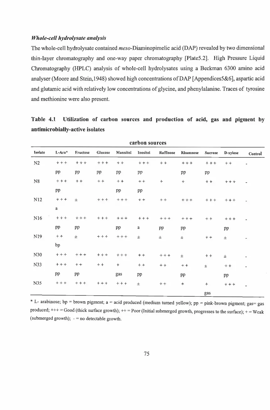
Whole-cell hydrolysate analysis
The whole-cell hydrolysate contained meso-Diaminopimelic acid (DAP) revealed by two dimensional
thin-layer chromatography and one-way paper chromatography [Plate5.2]. High Pressure Liquid
Chromatography (HPLC) analysis of whole-cell hydrolysates using a Beckman 6300 amino acid
analyser (Moore and Stein,1948) showed high concentrations ofDAP [Appendices5&6], aspartic acid
and glutamic acid with relatively low concentrations ofglycine, and phenylalanine. Traces of tyrosine
and methionine were also present.
Table 4.1 Utilization of carbon sources and production of acid, gas and pigment by
antimicrobially-active isolates
carbon sources
Isolate L-Ara* Fructose Glucose Mannitol Inositol Raffinose Rhamnose Sucrose D-xylose Control
N2
N8
+++
pp
+++
pp
+++
pp
++
+++
pp
++
++
pp
++
pp
+++
pp
++
pp
++
+
+++
pp
+
+++
pp
++
++
+++
NI2 +++ ±
a
+++ +++ ++ ++ +++ +++ +++
Nl6
Nl9
N30
N33
N35
+++
pp
++
bp
+++
+++
pp
+++
+++
pp
±
+++
++
pp
+++
+++
+++
+++
++
+++
+++
pp
+++
+++
+
gas
+++
+++
a
±
++
++
pp
±
+++
pp
±
+++
++
++
+++
pp
±
±
++
pp
+
++
++
++
±
+
gas
+++
pp
±
±
++
pp
+++
* L- arabinose; bp = brown pigment; a = acid produced (medium turned yellow); pp = pink-brown pigment; gas= gas
produced; +++ = Good (thick surface growth); ++ = Poor (Initial submerged growth, progresses to the surface); + = Weak
(submerged growth); - = no detectable growth.
75
Diagnostic sugar analysis
Separation ofsugars in whole-cell hydrolysates by one-way paper chromatography showed that isolate
N2 contained none of the characteristic actinomycete sugars [Appendix4]. Thus, the culture can be
considered to have a type III cell wall according to the scheme of Becker et al. (1965) and a type C
whole-cell sugar pattern (WCSP) according to the scheme of Lechevalier (1968).
Isolate N8
The physiological and chemical characteristics were similar to those of isolate N2 described above.
However, there are variations in morphology and utilization ofcarbon sources. Unlike N2, N8 could not
utilize raffinose and rhamnose as carbon sources [Table 4.1]. Isolate N2 produced pink-brown pigment
on fructose, glucose, rhamnose and sucrose carbon sources, whereas no pigment was produced when
grown on any of these sugars by isolate N8 [Table 4.1]. The cells of isolate N8 were non-acid fast
[Plate4.2A].
Morphological Properties
On nutrient agar plates, N8 appeared as pale white to orange colonies that resembles a contour map
[Plate4.2B]. The young hyphae were fleshy, intertwined, and penetrated the medium [Plate 4.2C], while
the mature hyphae fused together [Plate4.2D] to form dark brown to gray coloured dome-like bodies
that contained the spores [Plate 4.2E]. In broth the long branching hyphae arose from a synnemata
(which is a collection ofhyphae fused together) that bears spores [Plate 4.2 F&G].
Environmental scanning electron microscopy (ESEM) revealed that the spores are enclosed in a thick
slimy layer/sheath [Plate4.3A&B], which when broken exposes the spore mass [Plate4.3D]. Scanning
electron microscopy (SEM) showed the spores as smooth surfaced, oval to rod shaped [Plate 4.3C], and
packed in dense clusters. Growth on agar slants was moderate and filiform in pattern. The pellicle
formed by broth cultures in test tubes later detached and fell to the bottom of the test tube leaving the
broth clear.
76
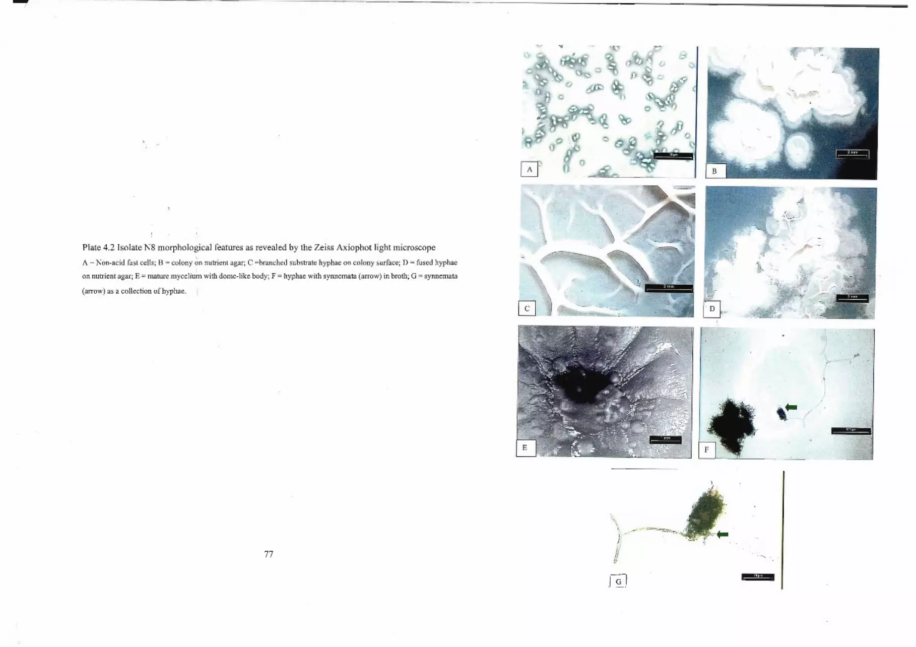
----/
Plate 4.2 Isolate N8 morphological features as revealed by the Zeiss Axiophot light microscope
A = Non-acid fast cells; B = colony onnutrient agar; C =branched substrate hyphae on colony surface; D = fused hyphae
on nutrient agar; E = mature mycelium with dome-like body; F = hyphae with synnemata (arrow) in broth; G = synnemata
(arrow) as a collection of hyphae.
1.'7<f"''<'
0~
'I ~~
,~ ~-
pe
;;.)
(
77
. ,-~ ~-;;:"-:::.s-~·~----..,.
fGl
"--'''",
~..•
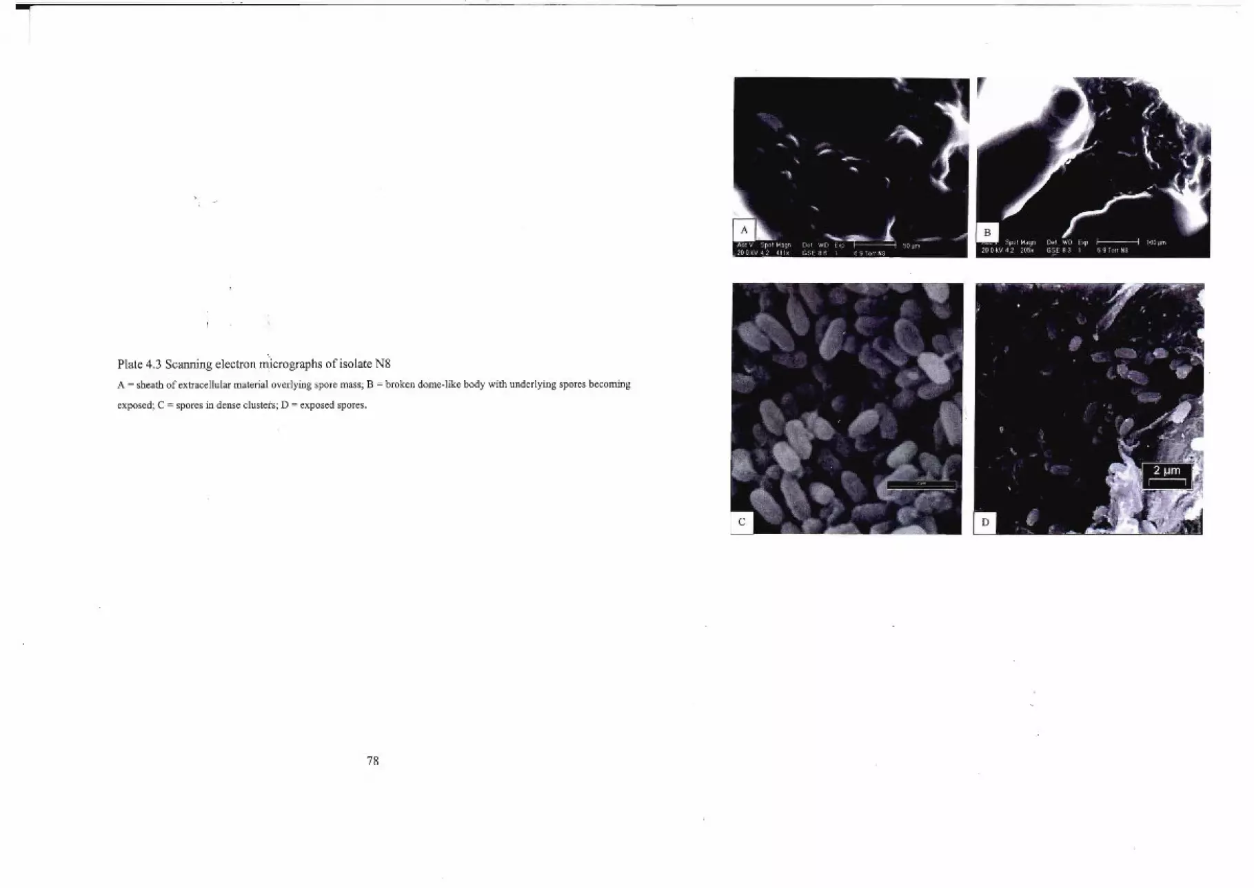
1--_._--
Plate 4.3 Scanning electron mIcrographs of isolate N8
A = sheath of extracellular material overlying spore mass; B = broken dome-like body with underlying spores becoming
exposed; C = spores in dense clusters; D = exposed spores.
78
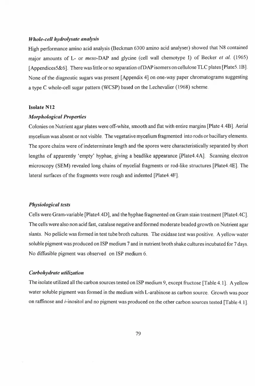
Whole-cell hydrolysate analysis
High performance amino acid analysis (Beckman 6300 amino acid analyser) showed that N8 contained
major amounts of L- or meso-DAP and glycine (cell wall chemotype I) of Becker et at. (1965)
[Appendices5&6]. There was little or no separation ofDAP isomers on cellulose TLC plates [Plate5.1B].
None ofthe diagnostic sugars was present [Appendix 4] on one-way paper chromatograms suggesting
a type C whole-cell sugar pattern (WCSP) based on the Lechevalier (1968) scheme.
Isolate NI2
Morphological Properties
Colonies on Nutrient agar plates were off-white, smooth and flat with entire margins [plate 4AB]. Aerial
mycelium was absent or not visible. The vegetative mycelium fragmented into rods or bacillary elements.
The spore chains were of indeterminate length and the spores were characteristically separated by short
lengths of apparently 'empty' hyphae, giving a beadlike appearance [Plate4.4A]. Scanning electron
microscopy (SEM) revealed long chains of mycelial fragments or rod-like structures [Plate4AE]. The
lateral surfaces of the fragments were rough and indented [Plate4.4F].
Physiological tests
Cells were Gram-variable [Plate4AD], and the hyphae fragmented on Gram stain treatment [Plate4.4C].
The cells were also non acid fast, catalase negative and formed moderate beaded growth on Nutrient agar
slants. No pellicle was formed in test tube broth cultures. The oxidase test was positive. A yellow water
soluble pigment was produced on ISP medium 7 and in nutrient broth shake cultures incubated for 7 days.
No diffusible pigment was observed on ISP medium 6.
Carbohydrate utilization
The isolate utilized all the carbon sources tested on ISP medium 9, except fructose [Table 4.1]. A yellow
water soluble pigment was formed in the medium with L-arabinose as carbon source. Growth was poor
on raffinose and i-inositol and no pigment was produced on the other carbon sources tested [Table 4.1].
79

Whole-cell hydrolysate analysis
The whole-cell hydrolysate contained meso-DAP on one dimensional TLC analysis [Plate5.2].
Arabinose and galactose were the diagnostic sugars present. Thus, N12 can be considered to have a cell
wall of type IV according to the scheme of Becker et al. (1965) and a type A WCSP based on
Lechevalier (1968) scheme.
80
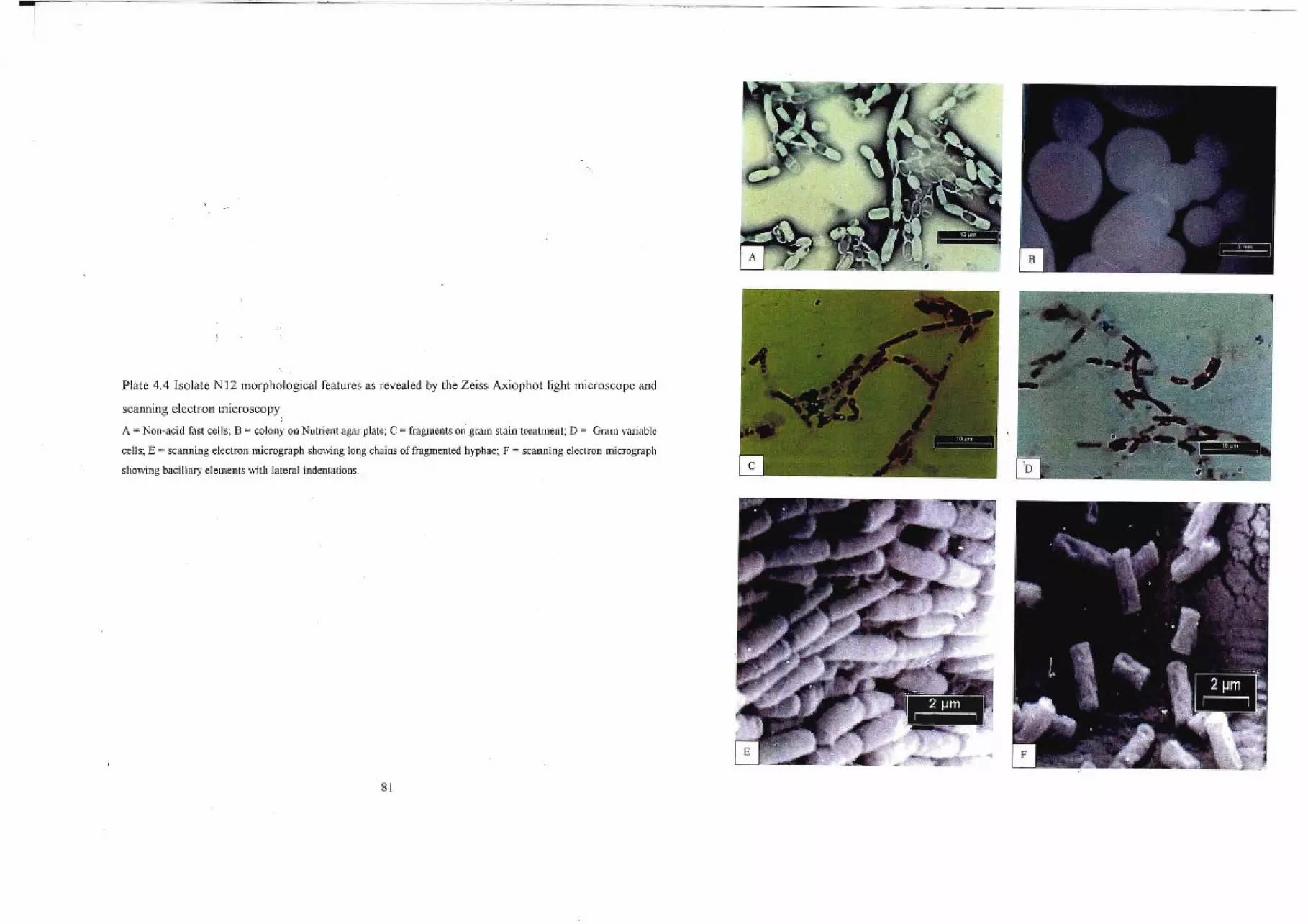
Plate 4.4 Isolate N12 morphological features as revealed by the Zeiss Axiophot light microscope and
scanning electron microscopy
A = Non-acid fast cells; B = colony on Nutrient agar plate; C =fragments on gram stain treatment; D = Gram variable
cells; E = scanning electron micrograph showing long chains of fragmented hyphae; F = scanning electron micrograph
showing bacillary elements with lateral indentations.
81

Isolate N16
Morphological properties
The primary mycelium were branched and irregularly wrinkled [Plate4.5C], cream colored with some
tinges ofpink/orange [Plate 4.5D]. And in the older cultures, the mycelium fragmented into short rods
or coccoid elements. No aerial hyphae or synnemata was observed. Scanning electron micrograph
(SEM) revealed that the surfaces ofspores were covered by a few strands [Plate4.5F] to several sheaths
of slimy extracellular material (Williams et aI., 1976). SEM revealed oval to rod-shaped cells packed
in clusters [Plate 4.5E].
Physiological characteristics
Cells were Gram-positive [Plate4.5B], non acid fast [Plate4.5A] and oxidase negative. Catalase was
produced. A pellicle was developed on the surface oftest tube broth cultures. The broth was clear as the
pellicle remained at the top, indicative of strong aerobic growth. In agar shake cultures incubated for
seven days, a reddish-brown water soluble pigment was produced. Moderate arborescent growth was
observed on agar slants.
Carbohydrate utilization
Isolate N16 utilized all nine carbon sources tested on ISP medium 9. Pink to brown pigment was
produced on all the sugars except glucose and sucrose [Table 4.1]. Acid was produced in i-inositol and
no gas was produced on any of the carbon sources tested.
Chemical properties
The whole-cell hydrolysates contained low amounts ofL-DAP [Plate5.2&Appendix6] with arabinose
and galactose as the diagnostic sugars present. Thus, N16 can be considered to have a type I cell wall
according to the scheme of Becker et al. (1965) and a WCSP type A sensu based on Lechevalier and
Lechevalier (1970) scheme.
82
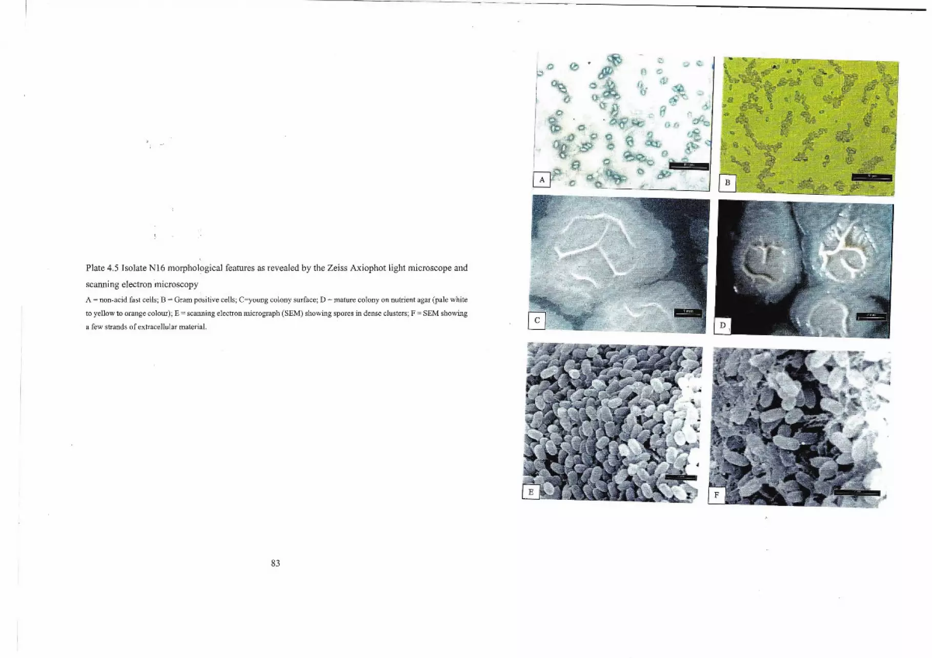
l
Plate 4.5 Isolate N16 morphological features as revealed by the Zeiss Axiophot light microscope and
scanning electron microscopy
A = non-acid fast cells; B = Gram positive cells; C=young colony surface; D = mature colony on nutrient agar (pale white
to yellow to orange colour); E = scanning electron micrograph (SEM) showing spores in dense clusters; F = SEM showing
a few strands of extracellular material.
83
Isolate N19
Aforphologicalproperdes
N19 was isolated on Czapek's agar as tiny punctifonn, flat, translucent colonies. On agar slants, growth
was abundant and spreading. Two distinct growth fonns were observed on nutrient agar. A yellow to
orange, oval to lemon shaped colony, surrounded at the edges or covered entirely, by a milky flat
spreading growth [Plate4.6D], possibly a hyphal outgrowth. In liquid culture, mycelium was lacking
except for sparse club-shaped cell doublets, joined at the tapered ends [Plate 4.6C]. No spores were
fonned. Both acid-fast and Gram-stained preparations revealed, an angular arrangement of cells,
sometimes in V-fonnations [Plate4.6A&B]. Individual cells were rod-shaped [Plate4.6F]and densely
packed [Plate 4.6E] when viewed under the Scanning electron microscope.
Physiological characteristics
Gram reaction, catalase and oxidase tests were positive for isolate N19. Cells were non acid-fast but
tended to retain carbol fuschin [Plate4.6B]. Motility was not observed.
Carbohydrate utilisation
Only glucose and mannitol was well utilized in ISP medium 9 [Table 4.1]. Sucrose and L-arabinose
yielded poor growth, with brown pigment production in the case of the latter sugar. There was little
or no utilization offructose, inositol, raffinose, rhamnose and D-xylose. Gas and/or acid production was
absent in all cases.
Whole-cell hydrolysate analysis
High perfonnance amino acid analysis revealed that N19 contained only a trace amount of DAP
[Appendix 5] and very high glycine concentration [Appendix 6]. Separation of DAP on silica and
cellulose TLC plates was doubtful. However, with paper chromatography, L-DAP was evident from the
Rf value calculated. The presence ofL-DAP and glycine is characteristic oftype I cell wall chemotype
ofBecker et al. (1965). Arabinose and galactose were the diagnostic sugars present. Thus, N19 can be
considered to have a cell wall chemotype I and a type A WCSP based on the scheme of Lechevalier
(1968).
84
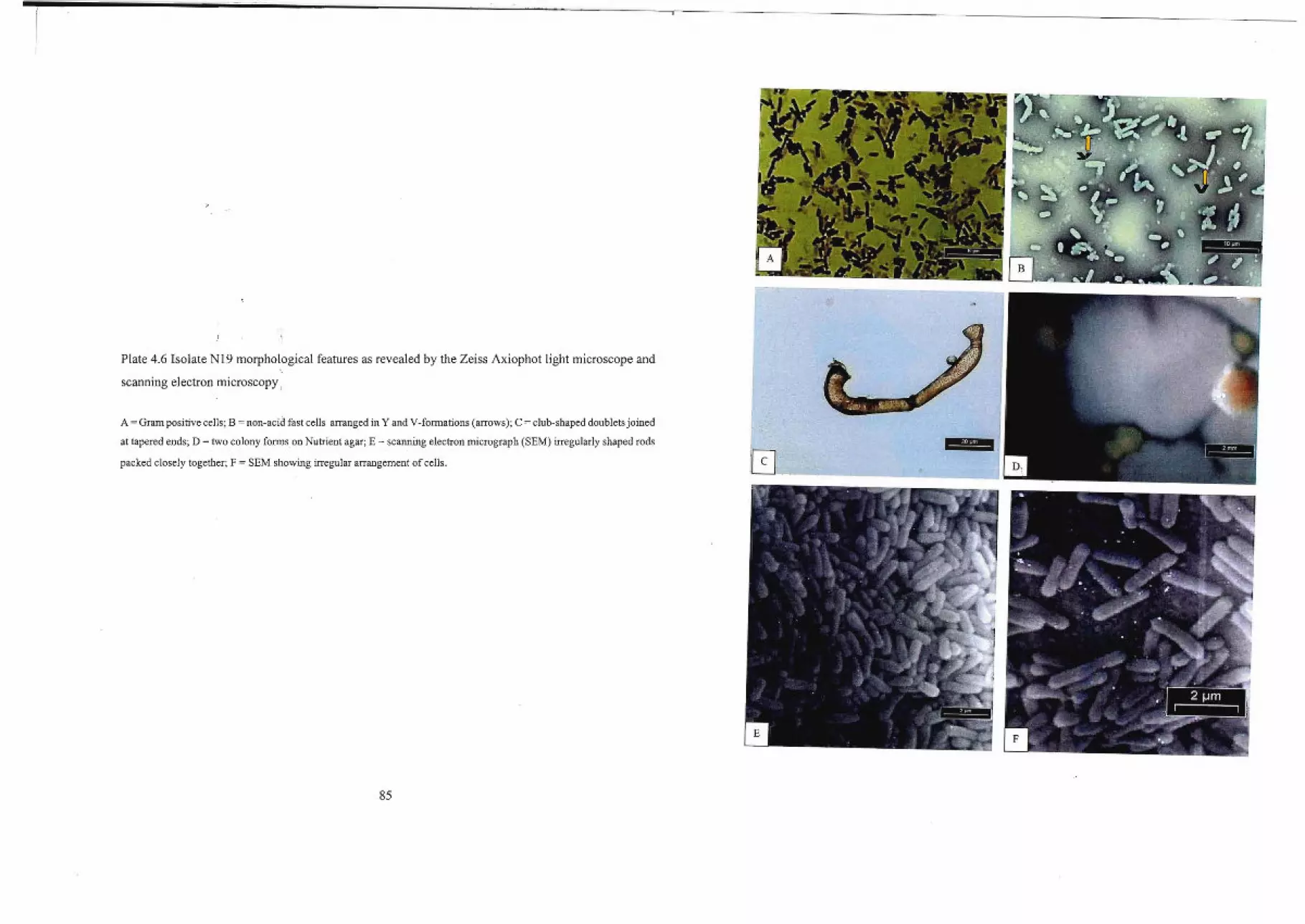
r
,;1
Plate 4.6 Isolate Nl9 morphological features as revealed by the Zeiss Axiophot light microscope and
scanning electron microscopy ,
A =Gram positive cells; B =non-acid fast cells arranged in Y and V-formations (arrows); C = club-shaped doublets joined
at tapered ends; D = two colony forms,on Nutrient agar; E = scanning electron micrograph (SEM) irregularly shaped rods
packed closely together; F = SEM showing irregular arrangement of cells.
85
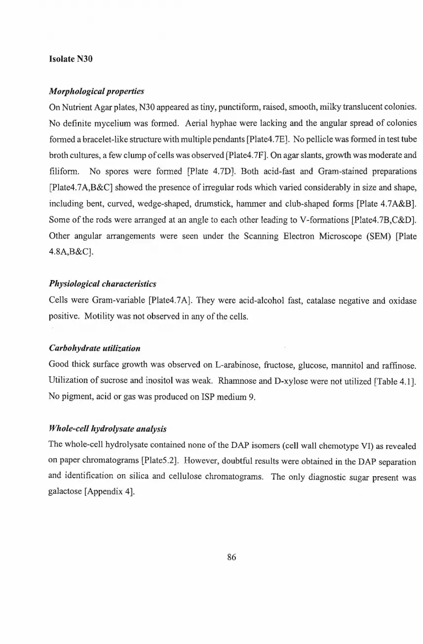
Isolate N30
Morphological properties
On Nutrient Agar plates, N30 appeared as tiny, punctiform, raised, smooth, milky translucent colonies.
No definite mycelium was formed. Aerial hyphae were lacking and the angular spread of colonies
formed a bracelet-like structure with multiple pendants [Plate4.7E]. No pellicle was formed in test tube
broth cultures, a few clump ofcells was observed [Plate4.7F]. On agar slants, growth was moderate and
filiform. No spores were formed [Plate 4.7D]. Both acid-fast and Gram-stained preparations
[Plate4.7A,B&C] showed the presence of irregular rods which varied considerably in size and shape,
including bent, curved, wedge-shaped, drumstick, hammer and club-shaped forms [Plate 4.7A&B].
Some of the rods were arranged at an angle to each other leading to V-formations [Plate4.7B,C&D].
Other angular arrangements were seen under the Scanning Electron Microscope (SEM) [Plate
4.8A,B&C].
Physiological characteristics
Cells were Gram-variable [Plate4.7A]. They were acid-alcohol fast, catalase negative and oxidase
positive. Motility was not observed in any of the cells.
Carbohydrate utilization
Good thick surface growth was observed on L-arabinose, fructose, glucose, mannitol and raffinose.
Utilization of sucrose and inositol was weak. Rhamnose and D-xylose were not utilized [Table 4.1].
No pigment, acid or gas was produced on ISP medium 9.
Whole-cell hydrolysate analysis
The whole-cell hydrolysate contained none of the DAP isomers (cell wall chemotype VI) as revealed
on paper chromatograms [Plate5.2]. However, doubtful results were obtained in the DAP separation
and identification on silica and cellulose chromatograms. The only diagnostic sugar present was
galactose [Appendix 4].
86

1 I
Plate 4.7 Isolate N30 morphological features as revealed by Zeiss Axiophot light microscope
A = Gram variable cells; B = drumsticks (D), hammer (H), club-shaped, bent, curved and V-formations (arrows); C = non
acid fast cells; D = mainly vegetativ~ cells on Ziehl-Neelsen spore stain; E = angular spread of colonies on agar plate; F =
clump of cells in Nutrient broth.
87
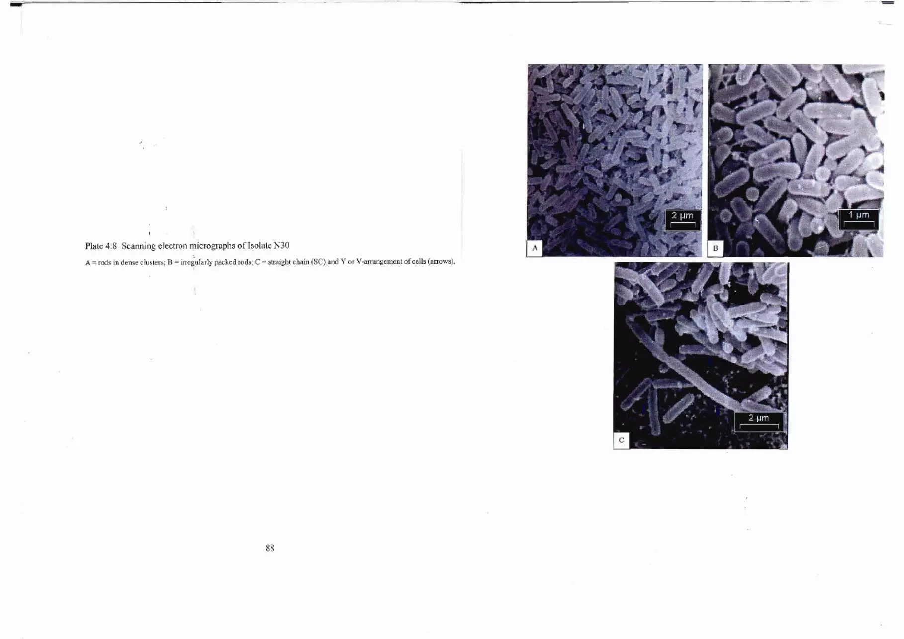
I
Plate 4.8 Scanning electron micrographs ofIsolate N30
A = rods in dense clusters; B = irregularly packed rods; C = straight chain (Se) and Y or V-arrangement of cells (arrows).
88
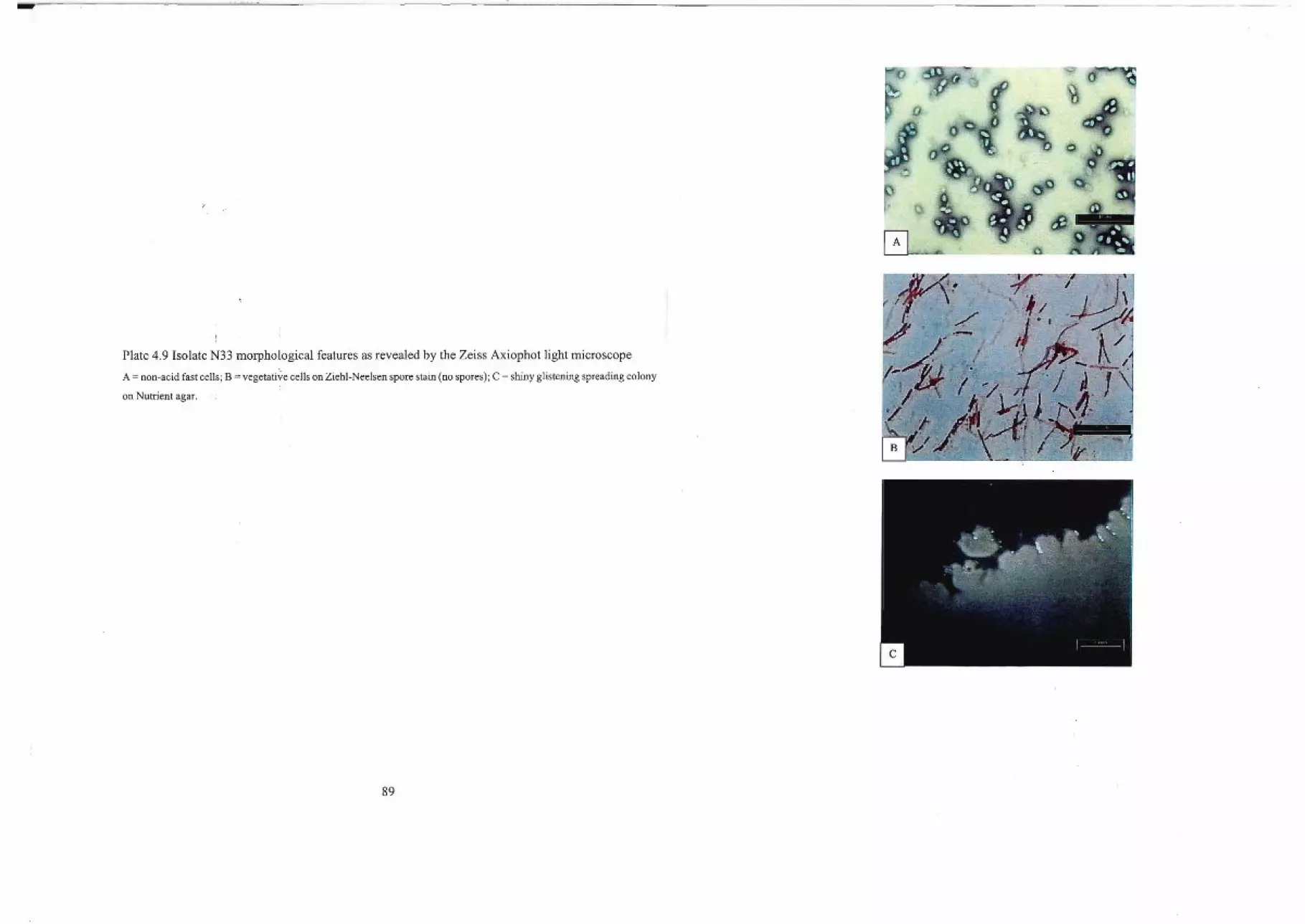
Plate 4.9 Isolate N33 morphological features as revealed by the Zeiss Axiophot light microscope
A = non-acid fast cells; B = vegetative cells on Ziehl-Neelsen spore stain (no spores); C = shiny glistening spreading colony
on Nutrient agar.
I
! .\
89
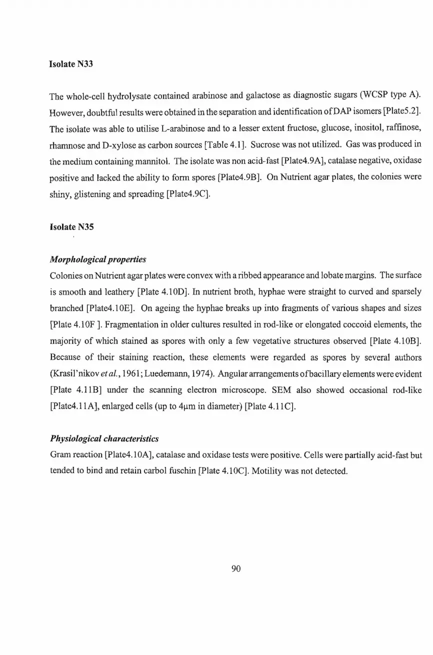
Isolate N33
The whole-cell hydrolysate contained arabinose and galactose as diagnostic sugars (WCSP type A).
However, doubtful results were obtained in the separation and identification ofDAP isom«rs [Plate5.2].
The isolate was able to utilise L-arabinose and to a lesser extent fructose, glucose, inositol, raffinose,
rhamnose and D-xylose as carbon sources [Table 4.1]. Sucrose was not utilized. Gas was produced in
the medium containing mannitol. The isolate was non acid-fast [Plate4.9A], catalase negative, oxidase
positive and lacked the ability to form spores [Plate4.9B]. On Nutrient agar plates, the colonies were
shiny, glistening and spreading [Plate4.9C].
Isolate N35
Morphological properties
Colonies on Nutrient agar plates were convex with a ribbed appearance and lobate margins. The surface
is smooth and leathery [Plate 4.l0D]. In nutrient broth, hyphae were straight to curved and sparsely
branched [Plate4.l0E]. On ageing the hyphae breaks up into fragments of various shapes and sizes
[Plate 4.l0F]. Fragmentation in older cultures resulted in rod-like or elongated coccoid elements, the
majority of which stained as spores with only a few vegetative structures observed [Plate 4. lOB].
Because of their staining reaction, these elements were regarded as spores by several authors
(Krasil 'nikov etaI., 1961; Luedemann, 1974). Angular arrangements ofbacillary elements were evident
[Plate 4.llB] under the scanning electron microscope. SEM also showed occasional rod-like
[Plate4.llA], enlarged cells (up to 4~m in diameter) [Plate 4.llC].
Physiological characteristics
Gram reaction [Plate4.1OA], catalase and oxidase tests were positive. Cells were partially acid-fast but
tended to bind and retain carbol fuschin [Plate 4.1 OC]. Motility was not detected.
90
.,I
tN".....".
1111 11l- --- 1
f;.'.o,
,
,,<
Plate 4.10 Isolate N35 morphological features as revealed by the Zeiss Axiophot light microscope
A = Gram positive cells; B = spores with few vegetative forms on Ziehl-Neelsen stain; C = partially acid fast cells;
D =ribbed, smooth, leathery surface colony on Nutrient agar plate; E =branched hyphae in broth; F =hyphal fragmentation
in broth.
~/
t\\\
~ ~
\ ,;~,/
\ '-\ ~.' .. (-x' /)\? ./I. .~~ .
;rJ 1 !/rc /)\.\ ,r I
> { .>-:t ' ..... f' /.. ,,/ \,,~~/ 1-./)-v ! _/ / \.._'
;' ' ' \
" ~ j 'r I "J} (,\)- / ;
0' ,'. tr --'h" "eiiii I
J ,i
.1./ ." '-.'.' ,-M'V.' \ • .' > I ;• _ \ \" .-t-/ I
'b'"
91

r I
PJ
i]
,,-, ~.
~ '" -,.. ,t. ....11 :;,." "11. !~." '0.....
(' I. if; ~ -\
".. . ,'. f,' I. ..' , '...' ..J1"'• t·' ~.~" ~~ ,1-;
11 ,.' "'~
.' I . ' >~'tY.I, I'~"• ,"', .. :J<:.~l1,,;l!(,
oa '"'' • ~;i ,,' ':.- :: .4~:.,< ~:"""'" "' ....: ~:
.f U' '\. .. ' ti'''.' ",.f ," "oJ;....."ii'-' ., .• .;, ~~:''''.,J.,"~.~ • 'I..:"'\.. . ,,~":, ,'./' • .,' • 't' "," .,~" r' ,.".ilI " ..-ti. ~",'"[25]'';' ,~.. j~
(l" JJrn ,~
~ t' §~ ~~, ~.~ '. -~..,l~. 'I
.. I\!""y ~ 1\i . ,,, •." ~. .
..
III
~
•
.. ~,'"jj '\
"
oPlate 4.11 Scanning electron tpicrographs of Isolate N35
A = rod-shaped cells; B= angular arrangement of bacillary elements; C = enlarged cells (up to 4~m in diameter).
92
Carbohydrate utilization
On ISP medium 9, the isolate utilised L-arabinose, fructose, glucose, mannitol and D-xylose as carbon
sources. Raffinose was poorly utilised, while very weak growth was observed in sucrose, rhamnose and
inositol [Table 4.1]. Gas pockets were produced in the ISP medium containing sucrose as carbon
source. No pigment was observed in any ISP media used.
Whole-cell hydrolysate analysis
Diaminopimelic acid (DAP) was not detected on paper [Plate5.2], cellulose and silica chromatograms.
Galactose was present as the only diagnostic sugar. Thus, the culture can be considered to have cell wall
chemotype VI based on the scheme ofLechevalier and Lechevalier (1970).
The morphological, physiological and chemical characteristics of the antimicrobially-active isolates
investigated, are summarised in Table 4.2.
93
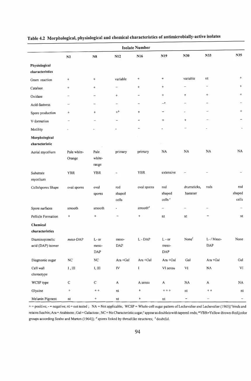
Table 4.2 Morphological, physiological and chemical characteristics of antimicrobially-active isolates
Isolate Number
N2 N8 N12 N16 N19 N30 N33 N35
Physiological
characteristics
Gram reaction + + variable + + variable nt +
Catalase + + + + +
Oxidase + + + + +
Acid-fastness
Spore production + + +b + +
V-formation + +
Motility
Morphological
characteristic
Aerial mycelium Pale white- Pale primary primary NA NA NA NA
Orange white-
range
Substrate YBR YBR YBR extensive
mycelium
Cells/spores Shape oval spores oval rod oval spores rod drumsticks, rods rod
spores shaped shaped hammer shaped
cells cells C cells
Spore surfaces smooth smooth smoothd
Pellicle Formation + + + nt nt nt
Chemical
characteristics
Diaminopimelic meso-DAP L- or meso- L-DAP L - or None f L-/Meso- Noneacid (DAP) isomer meso- DAP meso- DAP
DAP DAP
Diagnostic sugar Ne NC Ara+Gal Ara+Gal Ara+Gal Gal Ara+Gal Gal
Cell wall I, III I, III IV VI sensu VI NA VIchemotype
wesp type e C A A sensu A NA A NA
Glycine + ++ nt + +++ nt ++ nt
Melanin Pigment nt + nt + nt
+ = positive; - = negative; nt = not tested; NA = Not applicable; wesp = Whole-cell sugar pattern of Lechevalier and Lechevalier (1965): binds and
retains fuschin;Ara= Arabinose; Gal = Galactose; NC = No Characteristic sugar;' appear as doublets with tapered ends; *YBR=Yellow-Brown-Red [color
groups according Szabo and Marton (1964)]; d spores linked by threadlike structures; f doubtful.
94
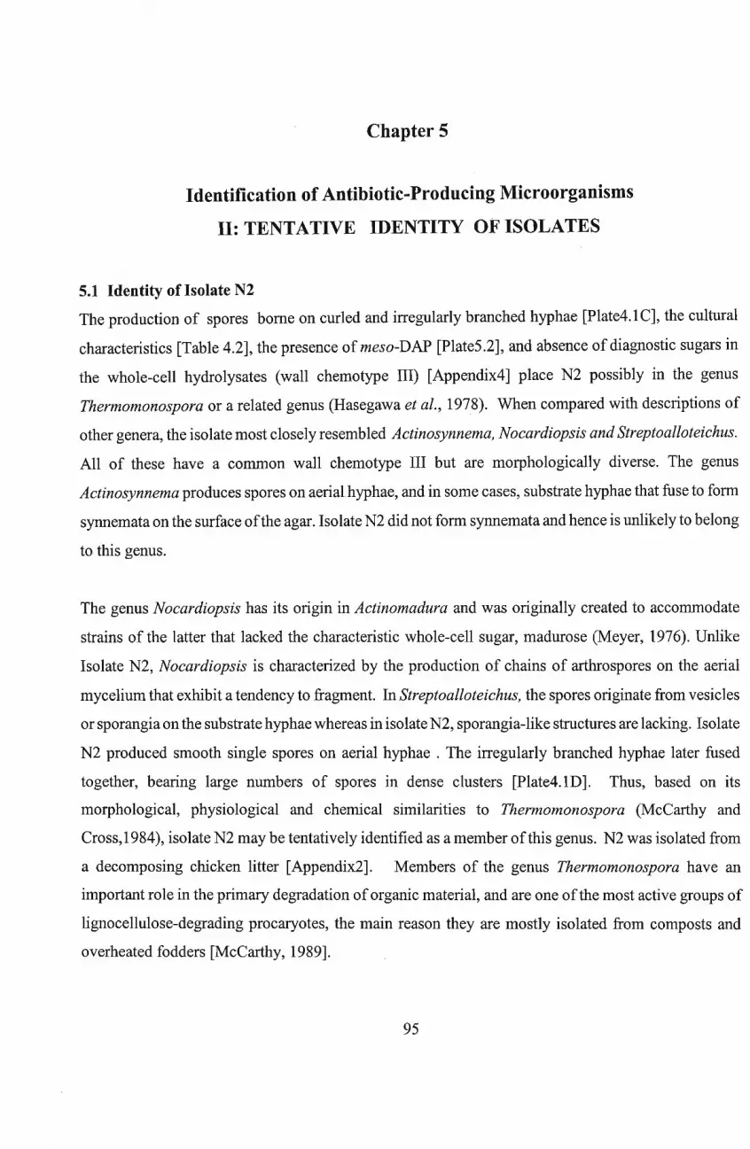
Chapter 5
Identification of Antibiotic-Producing Microorganisms
11: TENTATIVE IDENTITY OF ISOLATES
5.1 Identity of Isolate N2
The production of spores borne on curled and irregularly branched hyphae [Plate4.1C], the cultural
characteristics [Table 4.2], the presence of meso-DAP [Plate5.2], and absence of diagnostic sugars in
the whole-cell hydrolysates (wall chemotype Ill) [Appendix4] place N2 possibly in the genus
Thermomonospora or a related genus (Hasegawa et al., 1978). When compared with descriptions of
other genera, the isolate most closely resembled Actinosynnema, Nocardiopsis and Streptoalloteichus.
All of these have a common wall chemotype III but are morphologically diverse. The genus
Actinosynnema produces spores on aerialhyphae, and in some cases, substrate hyphae that fuse to form
synnemata on the surface ofthe agar. Isolate N2 did not form synnemata and hence is unlikely to belong
to this genus.
The genus Nocardiopsis has its origin in Actinomadura and was originally created to accommodate
strains of the latter that lacked the characteristic whole-cell sugar, madurose (Meyer, 1976). Unlike
Isolate N2, Nocardiopsis is characterized by the production of chains of arthrospores on the aerial
mycelium that exhibit a tendency to fragment. In Streptoalloteichus, the spores originate from vesicles
or sporangia on the substrate hyphae whereas in isolate N2, sporangia-like structures are lacking. Isolate
N2 produced smooth single spores on aerial hyphae . The irregularly branched hyphae later fused
together, bearing large numbers of spores in dense clusters [Plate4.1D]. Thus, based on its
morphological, physiological and chemical similarities to Thermomonospora (McCarthy and
Cross,1984), isolate N2 may be tentatively identified as a member of this genus. N2 was isolated from
a decomposing chicken litter [Appendix2]. Members of the genus Thermomonospora have an
important role in the primary degradation oforganic material, and are one ofthe most active groups of
lignocellulose-degrading procaryotes, the main reason they are mostly isolated from composts and
overheated fodders [McCarthy, 1989].
95
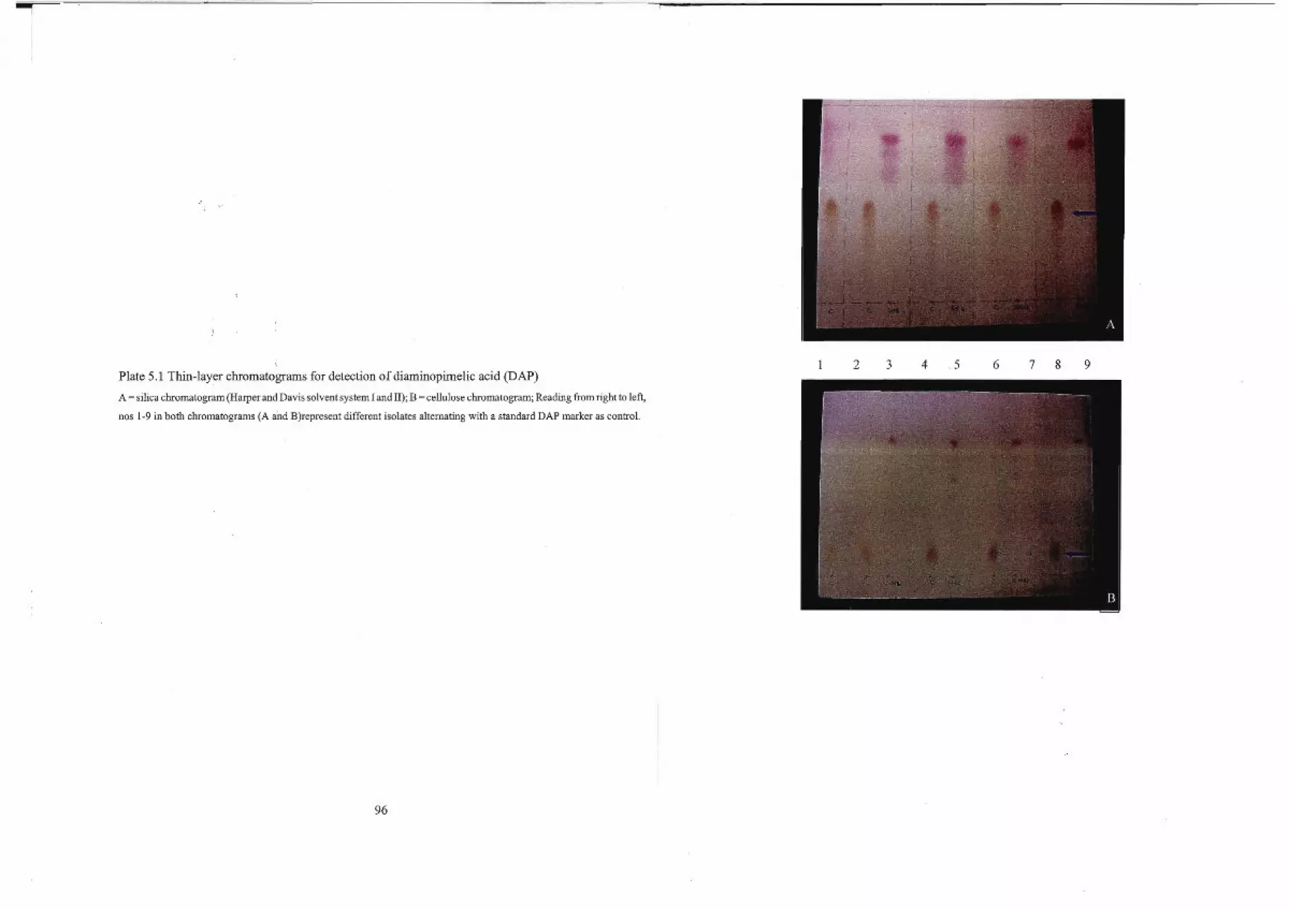
I"!
Plate 5.1 Thin-layer chromatograms for detection of diaminopimelic acid (DAP)
A = silica chromatogram (Harper and Davis solvent system I and 11); B = cellulose chromatogram; Reading from right to left,
nos 1-9 in both chromatograms (A and B)represent different isolates alternating with a standard DAP marker as control.
96
1 2 3 4 5 6 7 8 9
't- .
I
Plate 5.2 Detection of diamin~pimelicacid (DAP) on paper chromatogram
Long arrows indicate meso-DAP ,(dark pink colour); short arrow indicate L-DAP (light pink); C=control (DAP
(SIGMA)standard, mixture ofLL-, DD-, and meso-DAP); No distinction is made between the meso and D forms ofthis acid.
97
L
5.2 Identity of Isolate N8
The absence of diagnostic sugars in whole-cell hydrolysates (wall chemotype Ill) [Appendix4], the
cultural characteristics [Table 4.2] and the branched filaments that penetrate the agar [Plate4.2C] and
later fuse together to form dome-like bodies [Plate4.2E] and synnemata [Plate4.2F&G], place isolate
N8 possibly in the genus Actinosynnema or a related genus.
Comparisons with descriptions of other genera in Bergey's Manual of Systematic Bacteriology
(Vo1.4,1989) showed isolate N8 to closely resemble members of the genera Sporichthya,
Planomonospora, Planobispora, Spirillospora, Dermatophilus, and Geodermatophilus. All members
ofthe above, except Sporichthya, have a type III cell wall, but in contrast to isolate N8, they have a type
B WCSP (Lechevalier and Lechevalier, (1965). Though Geodermatophilus species do have type C
WCSP, similar to N8, the absence of spore bearing aerial mycelium indicates that isolate N8 does not
belong to this genus. The presence of substrate mycelium [Plate4.2B] and synnemata [Plate4.2F&G],
in isolate N8 and their absence in the genus Sporichthya presents evidence for non-inclusion ofisolate
N8 in this genus.
The genus Actinosynnema (Hasegawa et al., 1978; 1983), apart from the formation ofsynnemata, also
called coremia, possesses similar morphological, physiological and chemical properties to isolate N8.
Thus, N8 may be identified possibly as an Actinosynnema sp.
5.3 Identity of Isolate NI2
The production ofa primary mycelium that fragments into bacillary or rod-shaped elements [Plate4.4F],
the presence of major amounts of meso-DAP [plate5.2], and the occurrence of L-arabinose, and
galactose in the whole-cell hydrolysates (wall chemotype IV) [Appendix4] placeisolate N12 possibly
in the genus Nocardia or related genera. The above description is, however, also applicable to
Rhodococcus, Actinopolyspora, Bacterionema, Corynebacterium, Micropolyspora, Mycobacterium,
Saccharomonospora, Saccharopolyspora and Pseudonocardia (Gochnauer et al., 1975; Goodfellow
and Minnikin, 1977) and by the "aurantica" taxon, which contains organisms previously classified as
Gordona aurantiaca (Goodfellow et al., 1978). Unfortunately, lipid analysis, useful in distinguishing
98
representatives ofNocardia and Rhodococcus from actinomycetes that share cell wall chemotype IV,
was not carried out. However, other morphological, physiological and chemical properties [Table 4.2]
correlate closely with those in published descriptions of Nocardia and Saccharopolyspora. The
disintegration of the mycelium into rod-shaped elements [Plate4.4D&F], together with the beadlike
chains of spores [Plate4.4A], suggest Saccharopolyspora (Lacey and Goodfellow, 1975) is the more
likely.
Thus, isolate N12 may be identified tentatively as Saccharopolyspora species. Isolate N12, designated
as N12b [Appendix2], was isolated from cane sugar bagasse, the same natural habitat where the original
isolate described by Lacey and Goodfellow (1975) was isolated.
5.4 Identity of Isolate N16
The presence ofmajor amounts ofL-DAP [Plate5.2], detection ofglycine (cell wall chemotype I sensu
Becker et al., 1965)[Appendix6], the occurrence of arabinose and galactose in the whole-cell
hydrolysates [Appendix4], together with other morphological, chemical and physiological properties
[Table 4.2] place isolate N16 in the genus Nocardiodes or closely related genera (Prauser, 1976). When
compared with descriptions of other genera, the isolate most closely resembled Pimelobacter,
Intrasporangium, Mycobacterium, Nocardia, Spirochthya, Arachnia, Streptomycetaceae, Rhodococcus,
Oerskovia, and Promicromonospora. However, unlike N16, Pimelobacter, Spirochthya and
Mycobacterium, do not produce primary mycelium on agar media. Hence, the isolate is unlikely to
belong to these genera. The primary mycelium ofisolate N16 breaks up completely into smooth surface
spore-like elements [Plate4.5E], not normally observed in Mycobacterium and Streptomycetaceae.
Nocardia are partially acid-alcohol fast, whereas N16 was not and, hence, may be excluded from this
genus. Most species of the genera, Intrasporangium, Arachnia, Rhodococcus, and Oerskovia do not
produce aerial mycelium, which was observed in isolateN16 [Plate4.5D]. Promicromonospora has a
cell wall chemotype VI, whereas, N16 has a chemotype I cell wall, and hence is unlikely to belong to
this genus. The presence of a few to several layers of slimy extracellular material, with a pasty
consistency, overlying the colonies [Plate4.5D], in isolate N16, point strongly to the genus Nocardiodes
(Prauser, 1976).
Thus, based on its morphological, physiological and chemical similarities to Nocardiodes (prauser,
99
1976), isolate N16 may be tentatively identified as a member of this genus.
5.5 Identity of N19
The presence ofmajor amounts ofglycine [Appendix6], the detection ofL- or meso-DAP [Appendix5],
the formation of an extensive mycelium [Plate4.6D], the angular arrangement of cells in Y or V
formations [Plate4.6B], and other morphological and chemical properties [Table 4.2], place isolate N19
possibly in the genus Corynebacterium or related genera (Barksdale, 1970). When compared with
published descriptions of various genera, isolate N19 most closely resembled genera in the aerobic
nocardioform, Mycobacterium and coryneform groups (O'Donnell et a!., 1982; Colliins et al., 1983;
Suzuki and Komagata, 1983). Unfortunately, mycolic acid analysis necessary to separate these genera
effectively was not carried out.
Most of the nocardioforms have a type IV cell wall composition, as do the coryneform bacteria
(Lechevalier, 1981), whereas N19 has a chemotype IV sensu of Lechevalier and Lechevalier (1970).
This, together with other morphological, physiological and chemical properties, points strongly to the
genus Corynebacterium (Barksdale, 1970). Thus, isolate N19 was tentatively identified as a member
of this genus.
5.6 Identity of N30
The presence of galactose and total absence of arabinose [Appendix4], doubt as to the absence ofL- or
meso-DAP [Plate 5.2], and the cultural and morphological characteristics displayed [Table 4.2] suggest
N30 possibly belongs in the genus Arthrobacter or related genera. When compared with published
descriptions ofthe genera within this group, isolate N30 most closely resembled the Cure and Keddie
(1973) description ofthe coryneform group ofbacteria. The organisms originally placed in this group,
included, Corynebacterium, Arthrobacterand Cellulomonas, with Brevibacterium and Microbacterium
as genera incertae sedis and 'tentatively' Kurthia (Keddie, 1978). Barksdale (1970) had earlier
suggested that the word 'coryneform' be dropped because of the large, complex and broad
morphological and chemical differences among members of this group (Keddie, 1978).
100

Although, N30 appeared to have a close phenotypic relationship [Plate4.7A,B&C] with Arthrobacter,
doubt concerning the presence ofmeso- DAP, disallowed confident assignment to this genus. Additional
chemotaxonomic tests are necessary before this can be done.
In Bergey 's Manual ofSystematic Bacteriology CV01.4, 1989), the genus Arthrobacter was described
as catalase positive whereas isolate N30 did not produce catalase. Keddie (1978) noted that
Arthrobacter tumescens was wrongly described in the 7th edition of Bergey's Manual as catalase
negative. To the present author's knowledge, the problem of catalase activity in taxonomic
identification ofArthrobacter species has not yet been adequately addressed. Further investigations are
necessary to clarify the situation.
5.7 Identity of N33
Although fragmentation into bacillary to coccoid elements (a property isolate N33 shares with
Nocardioform actinomycetes) was observed [Plate4.9A&B], insufficient information was obtained to
assign this organism to any specific genus. Additional chemotaxonornic tests are necessary before this
can be done. This isolate was included in the investigation since it showed strong bactericidal activity
againstPseudomonasfluorescens (Chapter 3) and to the author's knowledge, treatment ofPseudomonas
infections still poses a problem in medicine.
5.8 Identity of N35
The absence ofDAP in the whole-cell hydrolysate [Plate5.2], and galactose being the only diagnostic
sugar (cell wall chemotype VI) present [Appendix6], and other physiological and morphological
characteristics [Table 4.2], suggest isolate N35 possibly belongs in the genus Promicromonospora or
related genera (Krasil'nikov et al., 1961). Comparisons with published descriptions showed close
resemblance ofN35 to Cellulomonas, Oerskovia and the so-called non-motile organisms (NMOs) of
Lechevalier and Lechevalier (1981), and also to Nocardia cellulans, Brevibacteriumfermentans, and
'Corynebacterium manihot'. The inability ofN35 to form aerial hyphae, which are characteristic ofthe
abovementioned organisms, warrants its exclusion from these genera. Promicromonospora is
distinguished from all these genera by its inability to form aerial hyphae. Thus, based on its
101
morphological, physiological and chemical similarities to Promicromonospora (Krasil'nikov et aI.,
1961), isolate N35 may be tentatively identified as a member of this genus.
102

Chapter 6
GENERAL DISCUSSION
The results of the screening program carried out in this study (Chapters 2, 3,4 and 5) have shown that
KwaZulu-Natal soils do harbour microorganisms (both coryneform and non-streptomycetous
actinomycetes), capable ofproducing antibiotics against some major plant and human pathogens. The
numbers and types ofactinomycetes present in a particular soil are strongly influenced by geographical
location, soil temperature, soil type, soil pH, organic matter content, cultivation practices, aeration and
moisture content (Williams et al., 1971; 1972). All these factors were taken into consideration during
sample collection.
Some of the isolation media used, were found to be selective for actinomycetes. For example,
Winogradsky's Nitrite medium (Winogradsky, 1949), M3 medium (Rowbotham and Cross, 1977), and
Kosmachev's medium (Kosmachev, 1960) were selective without heat pretreatment and/or antibiotic
incorporation, although a few colonies of other bacteria did occasionally develop on the plates.
Although no direct comparisons of the media used in this investigation were made, some differences
between them were noted. Modified Czapek's agar (Higgins and Lechevalier,1969) appeared to
enhance pigment production, or to select for pigment-producing strains ofactinomycetes, most probably
Nocardioform group. MGA (Nonomura and Ohara, 1971) supported growth of mainly filamentous
organisms, possibly actinomycetes with aerial hyphae, such as Actinomadura as reported by Athalye
and Lacey (1981). Ofall the media used, Kosmachev' s medium (Kosmachev, 1960) appeared to be the
most selective, as most ofthe plates contained virtually pure cultures of actinomycetes. This medium
has been reported to be selective for Excellospora and related genera (Kosmachev, 1960). Some doubt
has been expressed regarding the selectivity of some of the other so-called selective media used in this
study, as portrayed in literature (Goodfellow and Minnikin, 1981; Rowbotham and Cross, 1977).
Heat pretreatment ofthe samples was beneficial as it eliminated most of the unwanted Gram-negative
bacteria that produce mucoid, spreading colonies on soil dilution plates (Williams and Wellington,
1982), thereby facilitating the isolation ofactinomycetous organisms. It is conceded that such treatment
may have concomitantly eliminated other heat sensitive producers ofnovel_antibiotics. A 10-4 dilution
~
103
was found to be optional for isolation as it resulted in reasonably well separated individual colonies of
a variety of organisms. Antibiotic incorporation into the media, together with heat pretreatment,
eliminated most of the unwanted fungal and yeast growth. The present study did not target fungi and
yeasts as part of the screening programme. Non-streptomycetous actinomycetes, coryneform bacteria
and other related organisms were the main targets. Streptomyces species were also disregarded mainly
because the large number ofsuch organisms would have increased the scope ofthe investigation beyond
the capacity ofthe facilities available, and many ofthe isolates would be found to produce already well
documented antibiotics.
The simplest and least time consuming screening methods (modified cross-streak and agar-plug) were
used. The methods employed were suitable for the kind of routine, high-rate screening programme
(approximately 63,000 isolation plates used) embarked on in this study, and produced acceptably
reproducible results. The highest percentage of antimicrobially-active isolates came from the SAPPI
forest soil, while the lowest percentage came from the banks ofthe Mzinyathi river. Forest soils usually
have a relatively low pH and contain a predominance of actinomycetes that are tolerant of acid
conditions (Davies and Williams, 1970; Williams et al., 1971). Actinomycete populations are usually
small in water-logged soils with low oxygen tension (Labeda, 1990), and the nature of the isolation
media used may not have been conducive to antibiotic production by the riparian microbiota.
Many of the other samples screened, such as, compost soil, cow and chicken manure, and soil sugar
cane fields, were rich in organic matter and contained a large variety of actinomycetes. The dry,
uncultivated soil sample contained fewer actinomycete isolates than the other samples examined.
However, arid soils usually have an alkaline pH and have been reported to contain more ofthe so-called
'rare' actinomycetes (Labeda, 1990). The results of a study on the distribution of actinomycetes in
South African soils were recently reported by Kizuka et at. (1997). Apart from this report, current
quantitative data on the geographical and ecological distribution ofactinomycetes appears to be scarce
in the general scientific literature (Goodfellow and Williams, 1983; Williams and Vickers, 1986).
Antibiotic production by organisms is a complex process and assessment thereofconsideration ofmany
interacting factors. The mere observation of an apparent inhibition zone on agar plates may be
deceptive at times. For example, the complete decolouration ofSerratia marcesens observed on plates
104
inoculated with isolate N 48 and incubated at 30°C was thought to indicate an inhibitory effect.
However, on further incubation at a lower temperature (20°C), pigment production was fully restored,
indicating that no antibiotic action had in fact occurred. In some instances the growth of the test
organism was delayed inion the media used, with the result that some of the early, seemingly positive,
results were found to be negative after longer incubation.
The use ofIso-SensiTest Agar (ISTA) [Oxoid] is recommended for primary screening as the zone edges
of the isolate/test organism were better defined. Some isolates tended to spread profusely on Mueller
Hinton medium and, sometimes, on nutrient agar making streaking and reading of the plates very
difficult. This reiterates the importance of the culture medium, in any screening programme. Other
factors that need to considered are the test organisms, the inoculum size, pH and incubation conditions,
oxygen availability, which must be standardized if one wishes to have data that is reproducible and
comparable under standard laboratory conditions.
No attempt was made in this study to isolate or purify the antibiotic substance(s) produced. This is a
complicated task, constituting a project in itself. Time and economic constraints were also considered.
Hopefully, future attempts to isolate and purify the antibiotics produced, especially by isolate N8, will
prove to be worthwhile.
The actinomycetes are a large, heterogenous and complex group of microorganisms. Their
identification requires detailed morphological, physiological and biochemical investigation. An attempt
was made in this study to identify the isolated antibiotic producers to the species level, using as many
as possible of the methods described in literature. Despite this the amount of data obtained (Chapter
4), was not sufficient to allow incontrovertible identification ofthe species. To simultaneously identify
several potential antibiotic producers to species level proved very difficult, mainly due to time, and
financial constraints and lack offacilities. This was not too serious however since detailed identification
of antibiotic-producing organisms is only required when a patent application is sought (Dietz, 1999).
Much time was spent on standardising the numerous tests and methodologies used in the investigation.
This was felt to be necessary since an examination of both the 8th and 9th edition ofBergey's Manual
revealed that little standardization ofthe many methods used in the identification of actinomycetes has
105
been achieved.
Separation of diaminopimelic acid isomers (L- or meso-DAP) during chemical analysis of the whole
cell hydrolysates was successfully carried out in most instances, although dubious results were obtained.
Sometimes different results were obtained depending on whether paper, silica or cellulose thin layer
chromatography plates were used. Other problems were also encountered with this test. Firstly, the
DAP (SIGMA) standards obtained from the manufacturers failed to separate in the prescribed solvent
system. Secondly, migration ofthe sample spots sometimes failed in the recommended solvents. This
led to the author having to conduct extensive trials on many solvent systems and combinations ofplates
and paper before acceptable results were obtained. Two dimensional thin layer chromatography based
on the method ofHarper and Davis (1979) proved helpful in some cases where separation could not be
obtained using the conventional one-dimensional paper chromatography technique. However, it should
be recommended only as a second option, i.e. where one-way paper chromatography does not yield
satisfactory results, since it is time consuming and expensive, especially when large numbers ofsamples
are to be analysed.
The techniques employed in the present investigation yielded reproducible results and were thus
considered reliable. Isolate N8 should be further investigated since it showed strong inhibitory action
against all the test organisms used, including Gram positive and Gram negative bacteria and Eukaryotes.
Further testing ofthe spectrum ofthe antibiotic produced by isolate N8 should be undertaken in clinical
trials, perhaps by a multinational pharmaceutical company, to determine its efficacy against the many
human and animal pathogenic microorganisms that have developed resistance to the currently widely
used antibiotics. The substance might prove to very useful ifit could be shown to have broad antifungal
activity. The activity ofthe compound should also be tested against a range ofphytopathogenic bacteria
and fungi since a major issue at present is the development ofbiocontrol agents to replace chemical
treatments. Whether or not this substance is novel, only the future will tell.
106

APPENDICES
Appendix 1
Media Composition and Preparation:
Distilled water was used for all media preparation. Except where contra-indicated, all media were
autoclaved at 121QC for 15 minutes before use.
1.1 Czapek's medium (Cross and Attwell, 1974., referred to by Cross, 1981). (g/l): Sucrose, 30.0
; NaN02, 2.0; MgS04.7H20, 0.5; KC1, 0.5; FeS04.7H20, 0.01; KH2P04, 1.0; Yeast extract, 2.0;
Vitamin-free casamino acids, 6.0; Agar, 13.0.
1.2 Kosmachev's medium (Kosmachev, 1960) (g/l): KN03, 1.0; (NH4)2S04' 1.0; N~HP04.12H20,
1.0; MgS04.7H20, 0.5; FeS04.7H20, 0.01; CaC03, 4.0; Yeast extract, 3.0; Agar, 15.0.
1.3 M3 Agar (Rowbotham and Cross, 1977). (g/l): KH2P04, 0.466; N~HP04.12H20, 0.732; KN03,
0.10; NaCl, 0.29; MgS04.7H20, 0.10; CaC03, 0.02; Sodium propionate, 0.20; FeS04.H20,
200~g; ZnS04.7H20, 180~g; MnS04.7H20, 20~g; Agar, 18.0; Cycloheximide and Thiamine
HCl, 4.0 mg each; pH 7.0. Cycloheximide and thiamine-HCl were dissolved in 70% (v/v)
alcohol, filter- sterilised and added to the autoclaved and cooled medium.
1.4 MGA (Nonomura and Ohara, 1971). (g/l): Glucose, 2.0; L-asparagine, 1.0; K2HP04, 0.5;
MgS04.7H20, 0.5; Trace salts solution, 1.0ml; Agar, 20.0. Trace salts solution consisted of
mg/ml: FeS04.7H20, 10.0; MnS04.7H20, 1.0; CuS04.5H20, 1.0; ZnS04.7H20, 1.0.
Sufficient polymixin B and oxyteracycline were dissolved in 70% alcohol, filter-sterilised and
added to the medium to give a [mal concentration of lO~g/ml and 12~g/ml respectively.
1.5 Modified Czapek's Agar (Higgins and Lechevalier ,1969). (g/l): NaN03,2.0; K2HP04, 1.0;
MgS04.7H20, 0.5; KC1, 0.5; FeS04, 0.01; Sucrose, 30.0; Yeast extract, 2.0; Agar, 15.0; pH 7.2.
1.6 Ringer's solution (~ strength) (g/l): NaCl, 2.25; KCl, 0.105; CaC12, 0.06; NaHC03, 0.05;
gelatin, 0.5.
107
1.7 Trace salts solution (Shirling and Gottlieb, 1966) (g/l): FeS04.7HzO, 0.1; MnClz.4HzO,O.l;
ZnS04.7HzO, 0.1.
1.8 Bacto Tryptic Soy Agar (Difco). This medium was prepared as per the manufacture's
instructions. Gentamicin sulphate was added to the autoc1aved and cooled medium to give a final
concentration of 50\!g/ml.
1.9 Winogradsky's nitrite medium (Winogradsky, 1949).
NazC03' 1.0; KHZP04, 0.5; Agar, 15.
(g/l): NaN0z, 2.0; Anhydrous
1.10 Yeast extract agar [ISP medium 2] (pridham et a!., 1956-1957). (g/l): Yeast extract, 4.0; Malt
extract, 10.0; Glucose, 4.0; Agar, 15.0; pH 7.0.
International Streptomyces Project (lSP) media used in these investigations (Shirling and Gottlieb,
1966 )
ISP medium 1: Tryptone- yeast extract broth (g/l) Bacto-tryptone (Difco), 5.0; Bacto-yeast
extract (Difco), 3.0; pH 7.0 - 7.2.
ISP medium 2 : Yeast extract agar ( see 1.10 above)
ISP medium 3: Oatmeal agar (Shirling and Gottlieb, 1966). (g/l):
Oatmeal, 20.0; Agar, 18.0; Trace salts solution, 1 ml. 1L of distilled water was poured into a
2 L conical flask, and 20.0g ofpinhead oatmeal (Tiger Oats Co.,S.A) were added and boiled for
20 minutes. The medium was then filtered through cheese cloth. The volume of filtrate was
restored to 1L by addition of de-ionized water. 1.0 ml trace salts solution was then added and
the pH adjusted to 7.2. 18g agar were added and heated until completely dissolved. The
complete medium was then dispensed into 500ml flasks in 250ml quantities and autoclaved at
121°C for 15 minutes. Gentamicin sulphate was added to the cooled medium to give a final
concentration of 50\!g/ml.
108
ISP medium 4: Inorganic salts-starch agar
Solution 1: 1O.Og ofsoluble starch was made to a paste, and then brought to 500ml with distilled
water.
Solution II : (g/500ml): K2HP04 ( anhydrous basis), 1.0; MgS04.7H20, 1.0; NaCl, 1.0;
(NH4)2S04, 2.0; CaC03, 2.0; trace salts solution, 1.0ml.
Solutions I and IT were mixed and 18g agar added and the medium boiled until the agar
dissolved, the medium was then autoclaved at 121°C for 15 minutes.
ISP medium 5: Glycerol-asparagine agar
( g/l): L-asparagine ( anhydrous basis ), 1.0; glycerol, 10.0; K 2HP04 ( anhydrous basis), 1.0;
trace salts solution, 1.0ml; agar, 18.
ISP medium 6: peptone - yeast extract iron agar
(g/l) : Bacto-peptone, 15; protease peptone, 5.0; Ferric ammonium citrate, 0.5; dipotassium
phosphate, 1.0; sodium thiosulphate, 0.08; yeast extract, 1.0; agar, 15.
ISP medium 7: tyrosine agar
(g/l): Glycerol, 15.0; L-tyrosine, 0.5; L-asparagine, 1.0; K 2HP04 ( anhydrous), 0.5;
MgS04.7H20, 0.5; NaCI, 0.5; FeS04.7H20, 0.01; trace salts solution, 1.0ml; agar, 18.
ISP medium 9: carbon utilization medium
Carbon sources were added as filter sterilized 10% solutions (w/v) in basal mineral salts agar
to give a final concentration of 1% (10ml of a 10% solution to 100ml basal medium).
D-glucose was added as a positive control, whereas the basal mineral salts agar without any
carbon source, served as a negative control.
A. Sterile carbon sources
L- arabinose D- fructose
Sucrose Rhamnose
D-xylose Glucose
I - inositol Raffinose
D- mannitol Cellulose
109
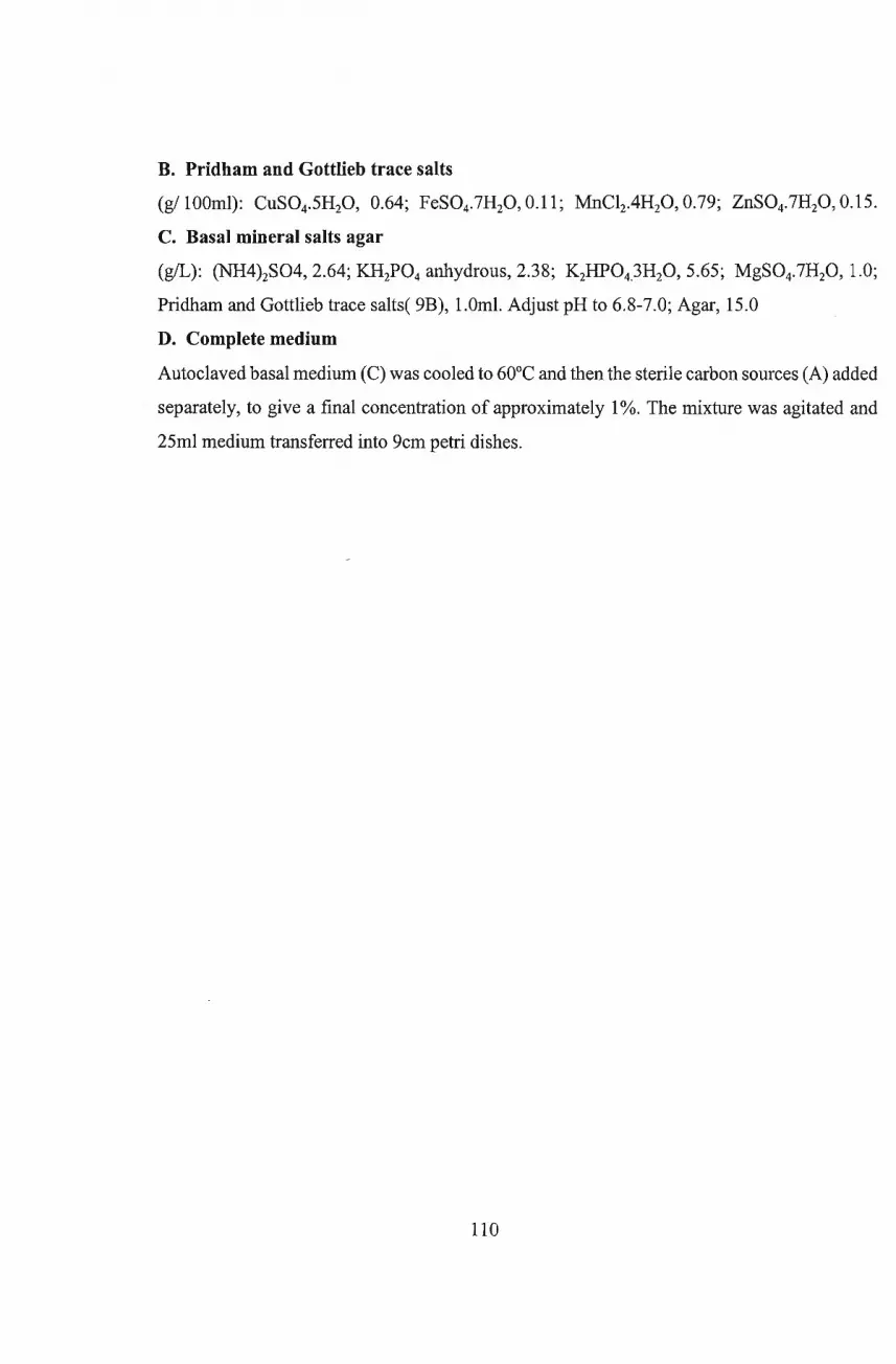
B. Pridham and Gottlieb trace salts
(g/lOOml): CuS04.5H20, 0.64; FeS04.7H20, 0.11; MnCI2.4H20, 0.79; ZnS04.7H20,0.15.
C. Basal mineral salts agar
(g/L): (NH4)2S04, 2.64; KH2P04 anhydrous, 2.38; K2HP043H20, 5.65; MgS04.7H20, 1.0;
Pridham and Gottlieb trace salts( 9B), 1.0m!. Adjust pH to 6.8-7.0; Agar, 15.0
D. Complete medium
Autoclaved basal medium (C) was cooled to 60°C and then the sterile carbon sources (A) added
separately, to give a final concentration of approximately 1%. The mixture was agitated and
25ml medium transferred into 9cm petri dishes.
110
Appendix 2
Antimicrobial-activity of selected isolates against test organisms on primary screening medium, NA
Test organisms
Isolate sample 0 @ @) 0 0 CD fI
number source Sa Ec Pf Srn Sf Cu Xc
NI CM + 0+
N2* CL 0+ ++ ++ ++ ++ ++
N3* CM +++ ++ +++ nt
N4a* SW ++ +++ ++ nt
N4b* SW +++ ++
N5 " + nt 0+
N7 " nt nt + +
N8 " ++ +++ ++ +++ ++++ ++++ +++
N12a CL 0+ +
NI3 " +++ Rs
N14 " + 0+ ++
N15 " ++Rs ++ 0+ ++++
N16a* " +++Rs +Rs ++ 0+ 0+
N17a " + ++++
N20 COW +
N22 CL +++ +++Rs
N23 " + 0+ Rs
N24 " + 0+
N25a* " 6+ + + + +rs ++++
N26 " 6+ +
N27 COW + + 0+ +
N28a " + +
N30a CL + +
N37 CL +Rs +
N41 " ++Rs ++++ +
N45 CS +++ 6+ 0+
111
N48 11 ++ ++++ -dis +++ ++
Nl7b US + ++++
N16b 11 +Rs ++++
N19a 11 +Rs ++
NlO 11 ++
N6* CCS + +++ + + +
N4c SF(NH) +
N12b* CSF(NH+A ++ ++ ++ 6+ + ++
)
N17c SF(NH) + ++
N19b* SF(NH+A +++ ++ +++ + ++ ++
N25b SF(NH) ++ +Rs +
N28b 11 +
N30b* CSF(H) + +++Rs
N31 11 + Rs
N33 SF(NH) + ++ +++ nt
N34 CSF(H) + ++ nt
N35 SF(NH) ++ + +++
N36 11 + ++
N38 SF(H) 6+ ++
+ positive; --negative; nt = not tested; 0+ =partially positive Rs = Resistant strains; SF = sappi forest; CSF = cane sugar field;
CM = chicken manure; CL = chicken litter; CS = compost soil; CCS = creosate contaminated soil; COW = cow manure;
SW = chicken manure from sweet waters; US = uncultivated soil; A = antibiotics added; NA = no antibiotics added; H =
heated; NH = non-heated. Sa = Staphylococcus aureus; Ec = Escherichia coli; Pf= Pseudomonasfluorescens; Srn =Serratia
marcescens; Sf = Streptococcus faecalis; Cu = Candida utilis; Xc = Xanthomonas campestris pv campestris
112

Appendix 3
Average diameters of inhibition zones produced by selected active isolates against test organisms on primary
screening medium, NA
Test organisms/ Inhibition zones (mm)
Isolate sample 0 @ ~ 0 ~ (i) 8
number source Sa Ec Pf Srn Sf Cu Xc
NI CM 5 2
N2* CL 13 10 20 21 20 15
N3* CM 25 20 25 nt
N4a* SW 15 30 20 nt
N4b* SW 30 15
N5 " 5 nt 2
N7 " nt nt 6 nt
N8* " 20 25 20 23 22 27 24
N12a CL 3 5
NB " 30
N14 " 9 5 15
N15* " 20 21 2 30
N16a* " 25 5 14 Rs
N17a " 2 26
N20 COW 8
N22 CL 25 30
N23 " 7 3 4
N24 " 5 3
N25a* " 2 30 30
N26 " 3 5
N27 COW 5 6 3 14
N28a " 7 5
N30a CL 6 5
N37 CL 5 4
N41 " 20 30 2
113
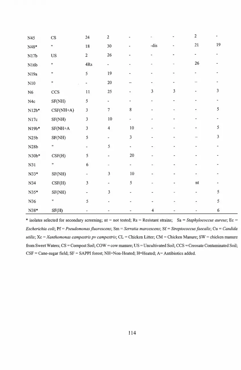
N45 CS 24 2 2
N48* " 18 30 -dis 21 19
N17b US 2 26
N16b " 4Rs 26
N19a " 5 19
N10 " 20
N6 CCS 11 25 3 3 3
N4c SF(NH) 5
N12b* CSF(NH+A) 3 7 8 5
N17c SF(NH) 3 10
N19b* SF(NH+A 3 4 10 5
N25b SF(NH) 5 3 3
N28b " 5
N30b* CSF(H) 5 20
N31 " 6
N33* SF(NH) 3 10
N34 CSF(H) 3 5 nt
N35* SF(NH) 3 5
N36 " 5 5
N38* SF(H) 4 6
* isolates selected for secondary screening; nt = not tested; Rs = Resistant strains; Sa = Staphylococcus aureus; Ec =
Escherichia coli; Pf = Pseudomonas fluorescens; Srn = Serratia marcescens; Sf = Streptococcus faecalis; Cu = Candida
utilis; Xc = Xanthomonas campestris pv campestris; CL = Chicken Litter; CM = Chicken Manure; SW = chicken manure
from Sweet Waters; CS = Compost Soil; COW = cow manure; US = Uncultivated Soil; CCS = Creosate Contaminated Soil;
CSF = Cane-sugar field; SF = SAPPI forest; NH=Non-Heated; H=Heated; A= Antibiotics added.
114
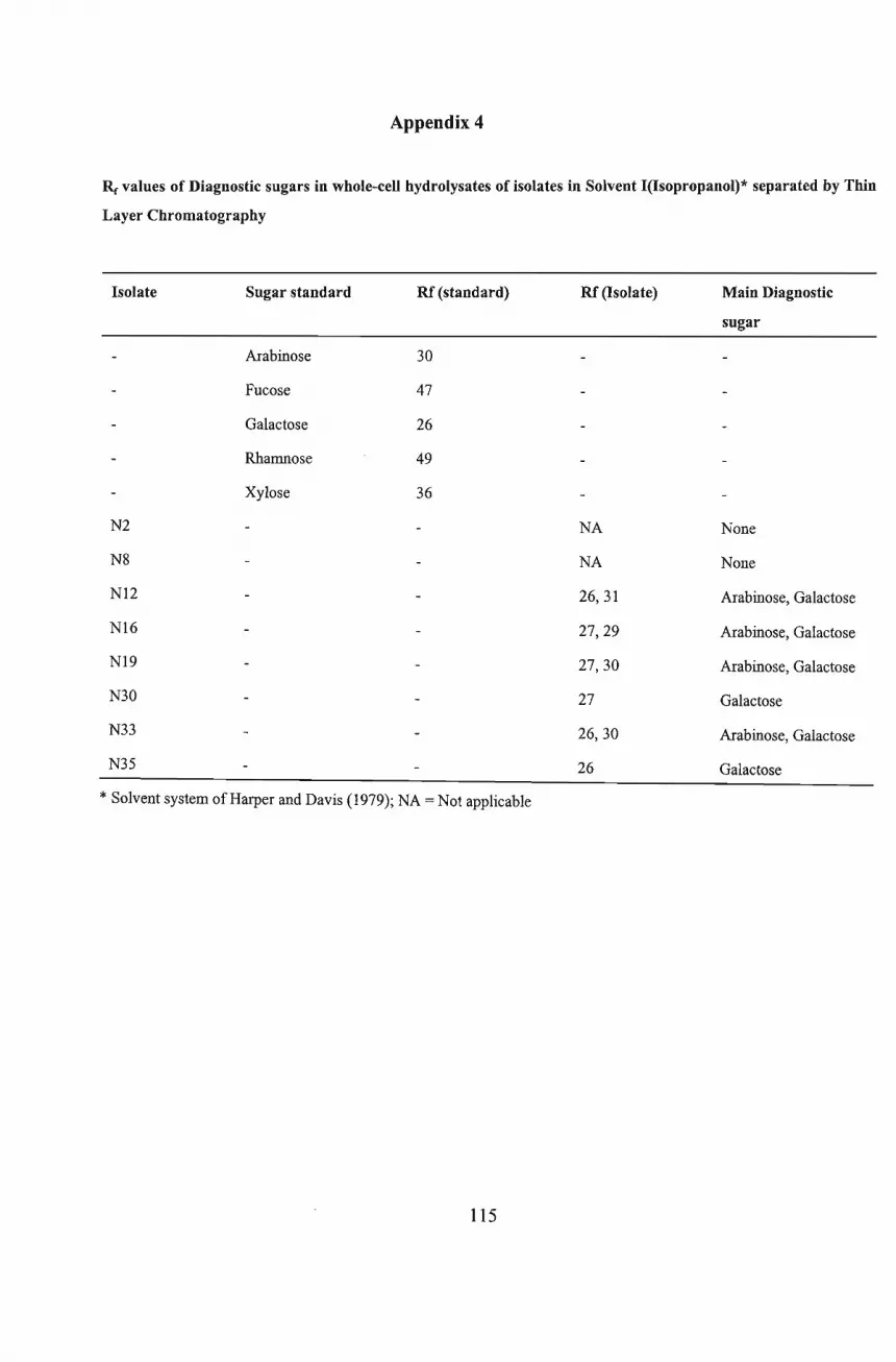
Appendix 4
Rc values of Diagnostic sugars in whole-ceU hydrolysates of isolates in Solvent I(Isopropanol)* separated by Thin
Layer Chromatography
Isolate Sugar standard RC (standard) RC (Isolate) Main Diagnostic
sugar
Arabinose 30
Fucose 47
Galactose 26
Rhamnose 49
Xylose 36
N2 NA None
N8 NA None
NI2 26,31 Arabinose, Galactose
NI6 27,29 Arabinose, Galactose
NI9 27,30 Arabinose, Galactose
N30 27 Galactose
N33 26,30 Arabinose, Galactose
N35 26 Galactose
* Solvent system of Harper and Davis (1979); NA = Not applicable
115
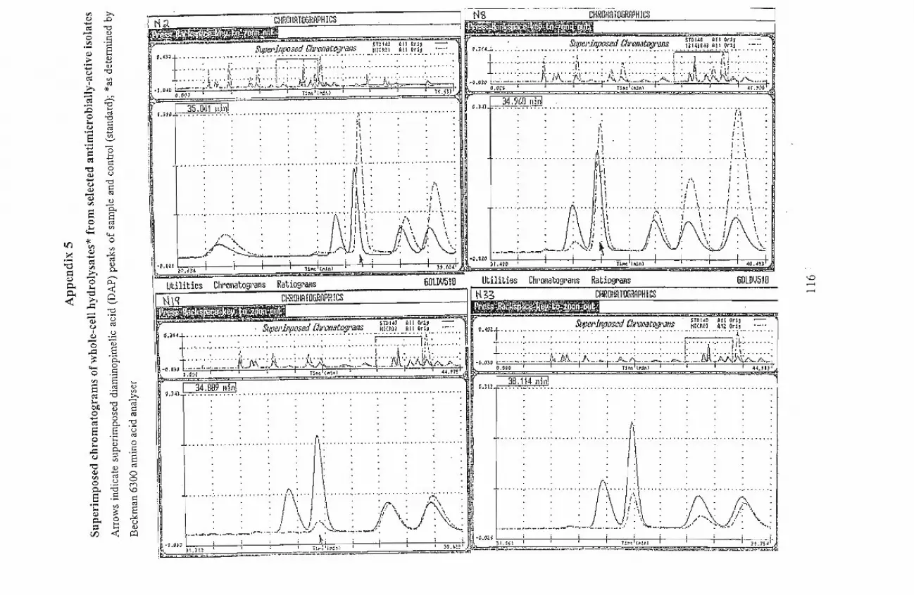
'" >-.Q)..... .0c-= '"Cl-0 a.>
.~ .5Q)
§.:: £..... a.>u '"Clc-= <J')
I
.Q«:l
*'-c-= ;..::
:c '"Cl....0
«:l... '"Clu I::.... 13S <J')
'-':;:: '0I:
c-= .J:j
"0I::0
Q) u.....u '"ClQ) I::- «:lQ)
'" ~
S S-o..::
«:ltI)
i<'+-<
l()
'"0
~Q)
tI)
.... ..... ~
"0 c-= «:l
= '"a.>
>. p.,Q.) "0 ,-..,
0- ... p...
~"0 <t::» Cl
..::: '-'
- '"Cl- .-Q.) uU «:l
I.~Q)- d)0
..:= .8~ p.,
"""'0
0 .5'" eS
....«:l a.>
c-= :.a tI)
... >-.
OJ) '"Cl c;j
0a.> I::<J')..... 0
«:l
c-=S S-
'"Cl'(3
0 'C ro... a.> 0
..:= p., I::u ::l .§
"0tI)
Q)a.> «:l.....,
'" «:l 0
0 u 0
0- :.a ....S .5
\0I::
'C tI)ro
Q) ::: ]0- 0::l t:: u
en <t::a.>~
~HRm.lOHR'PH.~~' _ -"-"' ,.:-=J ~~>~" ,,,. ~",'._PHlCS ._ .-.-~.. . . d 'T'W ~II Or', • s.' 1a 'Tbll~ 011 Od- "'to!. o.mt ~'f:':~~~~~1~~.. F!~'~~~.t~;1:'~~ J~I~~:"} ~!~ .0~~3._ . :-:::.. W4t !~'~'~Jf~;:~f ~~~.t~~:~~ i.II~I.!~~..A~~ .o~~i :-::: ..
t····· .:.) ,i: ;. ·l·· ." ~="TJ:\ : : : : : ~ J : ; } : ~.: :.. ·..1· .: .. .1 : ~! ..: :.Q 04cl~:~:f:t~rj;f' ~·.j~}~ ....:;.i;LJt.).k~L;,,:;,,:,~·.~;,.J;;l~~~ -o.m ..~.~~::- :~:~:A)~\:jiL.::~·.: '/to;,.·~.~;':''':·A.:.Lj\J~j(~JiL;~·~~·.~
;~. O.~N : i 1:",'(:\1•• ) I I i H.>!! • o.CCO. . ;;...-:=r!I., 'Nn} 41.1."0 ~
!1·'C'lI~ll .. ~~:~I!.':l~~l~ , C.l~l .. ?~:?~~.~1~~)\ .I, .,.... I',· . .. /,\
. . . . . . \. . . . . '.'I • •••••• i" . . . it: ! . ~
: : : : : : i\: : : : ir.: : : : ! : ! :. . . . . ., \. . . . Al' . . . I ' \ .
......: :., : : : :, ..!.\ ..: :., ~ : '!:1:i"'" .: : : , ..: i .:..i :pi i !:j . i i
J
\\ .. :t'· J!i ./\ i ~1I I . . .. ". \" . i I
\:::::: i i : : :! i : '.. i, . . :! \ : i: i :
\ ~ ~:~~ ~ ~ ~J""'\~..I r '~'\"f!\:::!:\: r: \ "i~~\""'r '\ '~""j .. ~ \ ..;I • i-...-....... . 1 I I . 1\\ r \ \ : ... I' i j:\', if\. '\. J!\. ".I' f--~(1 ~~. ,;,... f".; \:.._..} '0: \.:.::; ._._-;--_.;..-- ."'-:"',<) ~ .~. .._.j: \) \~~j : '-~:
\
.0 01' • ~ I' '1 I I -0.010 I I f I~ • • 21'.0)& '....._'.-...J.. l1M (I"L1n) !:t.024 '\ )1.0400 I T1~ INn}" 40.~
\
Utilities ChrmlOtogl'atls RatiOBl'al~S GOLDV510 Utiliti~ Chl'QNatogl'ilt-lS RatiogriYls GOLDIJ510
~ Cl. CHHOHATOGRPPHICS ~~HRm!ATllGRAPHICS\ ~'. "., _r=-.:.:..:..::.:.:.::.::...;;;:.:.:..-----------
I ~ o.l:,.j. :. ~~1~:'!J.1J!~1!~!~ (~~'~~J~!?,;J1,~~ )~!:~.!. ..m.~~i}._ ..~ ro.mt ~~!~;;~q~~.t?'~ ~~r1H .~~~t ~7!!, :, : ; ;, ; ,.. ; ~ : ·11\·:·· ·: T"" ..: : ; ; ,; : ,t , •• ,{i.: :
t.. · : ~\ '1\,"; .1-\ •••. ;., {i' .~ : t .1-: ; ii.i: ;. -:J. T" -: i 'b~";""":"'" ,~ '" ':- .. ,··f:-····A -: !;.\:- -:
.u;~ .'_' t._·._._._ .....:_). ,._ ...f,._· ... t ...' 0·,,:=._':-:,..,_,_J.:.,. .Bl1J\· 'J'N./'.·~'''''·_I ·O.OlO -'-'--' " ._._(,. ·(J.·-ll\_....p.~c.....ft-·_·.".,.· .c....;..._.MfN,V\i;.c:-.../,._,I '.m I 'c ""'W..~, """" ".m ..... w. ,.... ~ , '-; ...m'I .,.. H ~:~~l? H HH H H HH HH:H Hon HHH' H"" HHH ''''1,,1!'?!?! HH H H
r,
H'
I [·····:························Y::: I r· H
. ' r H
•••.•..•.•••••
H
I.., .\..(t \. I· \, '.1 .,::!.\ . . \. /,1. \,. ! .\ I .",' . f \. r . \'. . f#i " ) \ I . " • • ""\ J,I' ,
\._,_._.~.~.,_ ..;...J- _..:..~~~.. :~,-.:.,.__./ ,.I: \':,.....: ~ .-.-.-.-.-.-.-.-----~.-.:.-.-,.;: ..~_.:..-..- .. ,.. "" "'~"-'!./' .......... ,
-O.u, i ; ~ i j i i i i.. M:t ~c..aH. ..: I i '" i i i!~\;;;;;! ~I.~I? "".~_.... __..... 1_ ~~.:-~I~;...i:l; I t I !ls·l.SJs~·::'- .u' ._..__ • = ._u__t ~_T~,._=, ..."eee=--~=r_~~~~:;:~~:J
\0..............
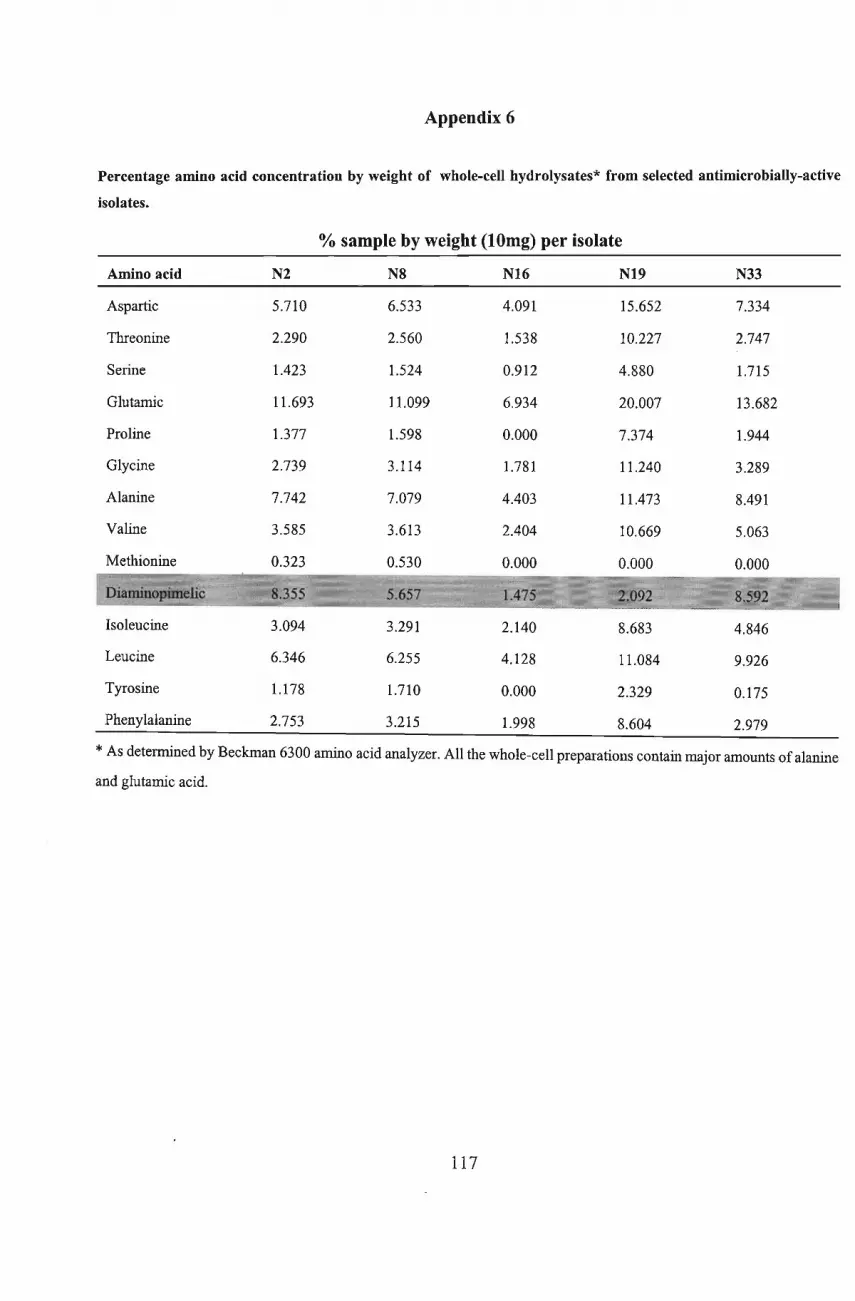
Appendix 6
Percentage amino acid concentration by weight of whole-cell hydrolysates* from selected antimicrobially-active
isolates.
% sample by weight (lOmg) per isolate
Amino acid N2 N8 N16 N19 N33
Aspartic 5.710 6.533 4.091 15.652 7.334
Threonine 2.290 2.560 1.538 10.227 2.747
Serine 1.423 1.524 0.912 4.880 1.715
Glutamic 11.693 11.099 6.934 20.007 13.682
Proline 1.377 1.598 0.000 7.374 1.944
Glycine 2.739 3.114 1.781 11.240 3.289
Alanine 7.742 7.079 4.403 11.473 8.491
Valine 3.585 3.613 2.404 10.669 5.063
Methionine 0.323 0.530 0.000 0.000 0.000
Isoleucine
Leucine
Tyrosine
Phenylalanine
3.094
6.346
1.178
2.753
3.291
6.255
1.710
3.215
2.140
4.128
0.000
1.998
8.683
11.084
2.329
8.604
4.846
9.926
0.175
2.979
*As determined by Beckman 6300 amino acid analyzer. All the whole-cell preparations contain major amounts ofalanine
and glutamic acid.
117
REFERENCES
Aharonowitz,Y and Demain, AL (1979) Nitrogen Nutrition and Regulation of Cephalosporin
Production in Streptomyces clavuligerus. Canadian Journal ofMicrobiology 25, 61 - 67
Arcamone, F., Cassinelli, G., Fantini, G., Grein, A.,Orezzi, P., Pol, C. and Spalla, C. (1969)
Adriamycin, 14 Hydroxydaunomycin, A New Anti-Tumour Antibiotic from S. peucetius var
. caesius. Biotechnology and Bioengineering XI: 1101 - 1110.
As per standard protocol ofHancock's Doubling Dilution Method for determination ofMlC using the
micro titre plate. website: www.interchg.ubc.ca/bobh/methods.htrnl.
As per SRGA ( Swedish Reference Group for Antibiotics and its sub committee of methodology)
Method for MIC determination. website: www.SRGA.
Athalye M and Lacey, J (1981). Selective Isolation and Enumeration of Actinomycetes using
Rifampicin. Journal ofApplied Bacteriology 51: 289 - 297.
Baecker. AA and Ryan, KC (1987) Improving the Isolation ofActinomycetes from soil by high- speed
homogenization. South African Journal ofPlant and Soil 4: 165 -170.
Barksdale, L (1970). Corynebacterium diphtheriae and its relatives. Bacteriological Review. 34: 378
-422.
Baron, AL (1950) Handbook ofAntibiotics, Reinhold Publishing Corp., New York.
Becker, B., Lechevalier, MP., Gordon, RE and Lechevalier, HA.(1964). Rapid Differentiation between
Nocardia and Streptomyces by Paper Chromatography of Whole-Cell Hydrolysates. Applied
Microbiology. 12: 421- 423.
118
Becker, B., Lechevalier, MP and Lechevalier, HA (1965). Chemical Composition of Cell Wall
Preparations from Strains ofVarious Genera ofAerobic Actinomycetes. AppliedMicrobiology.
13: 236 - 243.
Berd, D (1973). Laboratory Identification of Clinically Important Aerobic Actinomycetes. Applied
Microbiology. 25: 665 - 681.
Berdy, J. (1974) Recent Development of Antibiotic Research and Classification of Antibiotics
According to Chemical Structure. Advances in Applied Microbiology 18: 309 - 406.
Berdy, J. (1980) Recent Advances in and Prospects ofAntibiotic Research. Process Biochemistry 15
: 28 - 35.
Bergey's Manual ofDeterminative Bacteriology. 8th Ed. 1974. Buchanan, RE and Gibbons, NE (eds.)
Williams and Wilkins, Baltimore.
Bergey's Manual ofDeterminative Bacteriology. 7th Ed. 1957. Buchanan, RE and Gibbons, NE (eds.)
Williams and Wilkins, Baltimore.
Bergey's Manual of Systematic Bacteriology. 1989. Vo14. Williams, ST., Sharpe, ME and Holt, TG.
(eds) . Williams and Wilkins, Baltimore.
Bunge, RH., McReady, DE., Balta, LA., Graham, BD., French, JC., Dion, HW (1978) In: Carter, SK.,
Umezawa, H., Douros, J and Sakurai,Y (eds.) Anti- tumour Antibiotics. Springer - Verlag ,
New York: 77 - 84.
Chain, E., Florey, HW., Gardiner, AD., Heatly, WG., Jennings, MA., Ewing, 1. and Sanders (1940)
Penicillin as a Chemotherapeutic Agent. Lancet 239: 226 - 228 .
119
Collins, MD., Keddie, RM and Kropenstedt, RM (1983). Lipid Composition ofArthrobacter simplex,
Arthrobacter tumescens and Possibly Related Taxa. Systematic andAppliedMicrobiology 4: 18
26.
Conn, Hl,Darrow, MA., Emmel, VM., Davenport, HA., Gwinner, G., Kauffman, BP., Kornhauser, SI.,
Longley, JB., Lillie, RD., Pratt, C and Smith, FH (1960). Stains for Microorganisms in Smears.
In Staining Procedures 2nd Ed. pp. 223 - 243.
Cross, T (1981). The Monosporic Actinomycetes In: The Prokaryotes vol n. Starr, MP. Stolp, H,
Truper, H.G, Balows, A and Schlegel, H.G., (eds). New York: Springer. pp 2091-2102.
Cross, T (1982) Invitation ONR Lecture - Actinomycetes : A Continuing Source ofNew Metabolites.
Developments in Industrial Microbiology 23,1-18.
Cundliffe, E. (1992) Self Protection Mechanism in Antibiotic Producers. In : Secondary Metabolites
, Their Function and Evolution ( Derek, J C and Whelan, l, eds.) John Wiley and Sons,
Chichester UK : 199 - 214.
Cure, GL and Keddie, RM (1973) Methods for the Morphological Examination ofAerobic Corynefonn
Bacteria. In Sampling - Microbiological Monitoring ofEnvironments. Board, RG and Lovelock,
DW., eds. pp123 - 135. London: Academic Press.
Davies, FL and Williams, ST (1970) Studies on the Ecology of Actinomycetes in Soil. I. The
Occurrence and Distribution of Actinomycetes in Pine Forest Soil. Soil Biology Biochemistry
2: 227-238.
Dietz, A. (1999). Application of Electron Microscopy to Cultures ofIndustrial Significance. Journal
ofIndustrial Microbiology and Biotechnology. 22: 372 - 380.
120
Dubos, RJ (1939) Bactericidal Effect of an Extract of Soil Bacillus on Gram Positive Cocci. Journal
ofExperimental Medicine 70: 1 - 17.
Dulaney, EL (1948) Observations on Streptomyces griseus H. Nitrogen Sources for Growth and
Streptomycin Production. Journal ofBacteriology 56 : 305 - 313.
Eckwall, EC and Schottel, JL (1997) Isolation and Characterization of an Antibiotic Produced by the
Scale-Disease-Suppressive Streptomyces diastatochromogenes strain Pon SSII. Journal of
Industrial Microbiology and Biotechnology 19 : 220 - 225.
Florey, HW., Chain, E., Heatley, NG., Jennings, MA., Sanders, AG., Abraham, EP, and Florey, ME
(1949) A Survey ofPenicillin, Streptomycin, and Other Antimicrobial Substances from Fungi,
Actinomycetes, Bacteria, and Plants. In: Antibiotics, Vol. I & II, Oxford University Press,
London, New York.
Franco, CMM and Coutinho, LEL (1991) Detection of Novel Secondary Metabolites. Critical
Reviews in Biotechnology 11 : 193 - 276.
Franklin, TJ and Snow, GA (1981) Biochemistry ofAntimicrobial Action. - 3rd ed. Chapman and Hall,
New York: 1-56.
Gallo, M and Katz, E (1972) Regulation of Secondary Metabolite Biosynthesis, Catabolite Repression
ofPhenoxazinone Synthase and Actinomycin Formation by Glucose. Journal ofBacteriology
109: 659 - 667.
Galvez, A., Maqueda, M., Valdivia, E., Quesada, A., and Montoya, E (1986) Characterization and
Partial Purification ofBroad Spectrum Antibiotic A5 - A8 Produced by Streptococcus fa ecalis .
Canadian Journal ofMicrobiology 32: 765 -771.
121
Antibiotics and Antimicrobial Action. London: Edward
Gochnauer, MB., Leppard, GG., Komaratat, P., Kates, M., Novitsky, T and Kusher, DJ (1975).
Isolation and Characterization ofActinopolyspora halophila, gen.nov. an Extremely Halophilic
Actinomycete. Canadian Journal ofMicrobiology. 21 : 1500 - 1511.
Goodfellow, M., Orlean, PAB, Collins, MD, Alshamaony, Land Minnikin, DE (1978). Chemical and
Numerical Taxonomy of Some Strains Received as Gordona aurantiaca. Journal ofGeneral
Microbiology. 109: 57 - 68.
Goodfellow, M and Minnikin, DE (1977). Nocardioform Bacteria. Annual Review ofMicrobiology 31:
159 - 180.
Goodfellow, M and Minnikin, (1981). The Genera Nocardia and Rhodococcus . In: Starr, Stolp, Truper,
Balows and Schlegel (eds), The Prokaryotes; A Handbook on Habitats, Isolation and
Identification ofBacteria, Springer, Verlag, B~rlin, pp. 2016 - 2017.
Goodfellow, M and Williams, ST (1983) Ecology ofActinomycetes. Annual Review ofMicrobiology
37: 189-216.
Gottlieb, D (1976). Actinomycete Taxonomy. Letter to the Editor. Newsletter ofthe Us. Federation
of Culture Collection 6 (2): 5 -7.
Gottlieb, D and Shaw, PD (1967) Antibiotics; Vol] and Vo12. Springer Verlag , Berlin and New
York.
~ammond, SM. and Lambert, PA (1978)
Arnold : pp5 - 52.
Harper, JJ and Davis, HG (1979). Two-Dimensional Thin-Layer Chromatography for Amino Acid
122
Analysis ofBacterial Cell Walls. International Journal ofSystematic Bacteriology. 29: 56 - 58.
Hasegawa, T., Lechevalier, MP and Lechevalier, HA (1978). New Genus of the Actinomycetales:
Actinosynnema gen. novo International Journal ofSystematic Bacteriology. 28: 304 - 310.
Hasegawa, T., Tanida, S, Hatano, K, Higashide, E and Yoneda, M (1983). Motile Actinomycetes:
Atinosynnema pretiosum subsp. pretiosum sp. nov and Actinosynnema pretiosum subsp.
auranticum subsp. novo International Journal ofSystematic Bacteriology. 33: 314 - 320.
Hayakawa, M and Nonomura, H (1989) A New Method for the Intensive Isolation of Actinomycetes
from Soil. Actinomycetologica 3 : 95 - 104.
Higgins, ML and Lechevalier, (1969) Poorly Lytic Bacteriophage from Dactylosporangium
thailandensis (Actinomycetales). Journal ofVirology 3: 210-216.
Hodgson, DA (1982) Glucose Repression of Carbon Source Uptake and Metabolism in Streptomyces
coelicolor A3 [2] and its Pertubation in Mutants Resistance to 2- Deoxyglucose. Journal of
General Microbiology 128: 2417 - 2430.
Homer, T., Zahner, H., Kellner, R., and Jung, G (1989) Fermentation and Isolation of Epidermin, a
Lanthonine Containing Polypeptide Antibiotic from Staphylococcus epidermidis. Applied
Microbiology and Biotechnology 30 :219 - 225.
Hotta, K and Okami, Y (1996) Diversity in Aminoglycoside Antibiotic Resistance of Actinomycetes
and its Exploitation in the Search for Novel Antibiotics. Journal ofIndustrial Microbiology 17:
352 - 358.
Hotta, KA., Takahashi, A., Okami, Y., and Umezawa, H (1983) Relationship Between Antibiotic
123
Resistance and Antibiotic Productivity in Actinomycetes which Produce Aminoglycoside
Antibiotics. Journal ofAntibiotics 36: 1789 - 1791 .
Houang, ET. Hince, C and Howard, AJ (1983) The Effect of Composition of Culture Media on MIC
values ofAntibiotics. In Antibiotics: Assessment ofAntimicrobial Activity and Resistance. eds.
Russel AD and Quesnell, LB pp 31 - 48. London Academic Press.
James, PDA., Edwards, C and Dawson, M (1991) The Effect ofTemperature, pH and Growth Rate on
Secondary Metabolism in Streptomyces thermoviolaceus Grown in Chemostat . Journal of
General Microbiology 137 :1715 - 1720.
Jawetz, E., Melnick, JL and Adelberg, EA (1984) Antimicrobial Chemotherapy. In: Review ofMedical
Microbiology 16th ed. Lange Medical Publications. Los Altos, California. ppI28-129.
Kalakoutskii, LV., Kirillova, JP and Krasil'nikov, NA (1967). A new genus ofthe Actinomycetales
Intrasporangium gen.nov. Journal ofGeneral Microbiology. 48: 79-85.
Karwowski, JP (1986) The Selective Isolation of Micromonospora from Soil by Caesium Chloride
Density Gradient Ultracentrifugation. Journal ofIndustrial Microbiology 1: 181-186.
Keddie, RM (1978). What Do We Mean by Coryneform Bacteria. In: Coryneform Bacteria., eds.
Bousfield, IJ and Callely, AG., ppl - 12. London, Academic Press.
Kirpekar, AC and Kirwan, DJ (1991) Effect of Glutamate, Glucose, Phosphate and Alkali Metal Ions
on Cephamycin C Production by Norcadia lactamdurans in Defined Media. Biotechnology and
Bio-Engineering 38: 1100 - 1109.
Kizuka, M., Enokita, R., Takahashi, K and Okazaki, T (1997). Distribution of Actinomycetes in the
Republic ofSouth Africa Using a Newly Developed Isolation Method. Actinomycetologica. 11:
124
pp 54-58.
Kosmachev, AK (1960) Preservation ofViability ofThermophilic Actinomycetes After Long Storage.
Microbiology [English translation ofMikrobiologiya] 29: 210 - 211.
Krasil'nikov, NA., Kalakoutskii, LV and Kirillova, NF (1961). A New Genus of Ray Fungi
-Promicromonospora gen. novo (in Russian). Izv. Akad.Nauk SSSR (Ser. Bioi.) 1: 107-112.
Kutzner, HJ (1981) The Family Streptomycetaceae. In: The Prokaryotes; A Handbook on Habitats,
Isolation and Identification ofBacteria. eds. Staff, Stolp, Balows and Schlegel. Springer, Verlag,
Berlin, pp 2028-2090.
Labeda, DP (1987). Transfer of the Type Strain Streptomyces erythraeus (Waksman 1923) Waksman
and Henrici 1948 to Genus Saccharopolyspora Lacey and Goodfellow 1975 as
Saccharopolyspora eryghraea sp. novo and Designation ofa New Type Strain for Streptomyces
erythraeus. International Journal ofSystematic Bacteriology. 37: 19 - 22.
Labeda, DP (1990). Isolation of Actinomycetes for Biotechnological Organisms from Nature.
Environmental Biotechnology Series. McGraw Hill, ppl- 15.
Lacey, J and Goodfellow, M (1975) A Novel Actinomycete from Sugar-Cane Bagasse,
Saccharopolyspora hirsuta gen. et sp. novo Journal ofGeneral Microbiology 88: 75 - 85.
Lancini, G and Parenti, F (1982) Antibiotics, an Integrated View. Staff, MP. - ed. New York: Springer
-Verlag: ppl - 241.
Lechevalier, HA (1989) Practical Guide to Generic Identification of Actinomycetes. In: Bergey's
Manual ofSystematic Bacteriology. Vo14. 1989. eds. Williams, ST., Sharpe, ME and Holt, TG.
Williams and Wilkins, Baltimore.
125

J
Lechevalier, HA (1999). Thorn Award Address. Actinomycetes in the Family. Journal ofIndustrial
Microbiology and Biotechnology. 22: 518 - 525.
Lechevalier, MP (1968). Identification of Aerobic Actinomycetes of Clinical Importance. Journal of
Laboratory Clinical Medicine. 71: 934 - 944.
Lechevalier, MP (1981) Ecological Associations Involving Actinomycetes. Zentralblatt fuer
Bakteriologie, Mikrobiologie, und Hygiene. I Abteilung, Supplement. 11: 159-166.
Lechevalier, MP and Lechevalier, HA (1965) Classification des Actinomycetes Aerobes Basee sur Leur
Morphologie et Leur Composition Chimique. Annales Institut Pasteur. 108: 662 - 673.
Lechevalier, HA and Lechevalier, MP (1967) Biology of the Actinomycetes. Annual Review of
Microbiology 21: 71-100.
Lechevalier, MP and Lechevalier, HA (1970). Chemical Composition as a Criterion in the
Classification ofAerobic Actinomycetes. International Journal ofSystematic Bacteriology. 20:
435 - 443.
Lechevalier, HA and Lechevalier, MP (1981) Introduction to the Order Actinomycetales In :The
prokaryotes Vol II eds. Starr, MP., Stolp, H., Trupier ,HG., Barlows, A and Schlegel, HG.
New York Springer :pp 1915 - 1922.
Lechevalier, HA., Solotorovsky, M and McDurmont, Cl (1961). A New Genus of Actinomycetales:
Micropolyspora gen. novo Journal o/General Microbiology. 26: 11 - 18.
Linton, AH (1983) Theory of Antibiotic Inhibition Zone Formations, Disc Sensitivity Methods and
MIC. In Antibiotics; Assessment ofAntimicrobial Activity and Resistance. eds. Russel AD and
126

Quesnell, LB. London: Academic Press pp 19-30..
Liras, P., Villanneva, JR and Martin (1977) Sequential Expression ofMacromolecule Biosynthesis and
Candicidin Formation in Streptomyces griseus. Journal ofGeneralMicrobiology 102: 269-277.
Luedemann, GM (1971). Species Concept and Criteria in the Genus Micromonospora. Transactions.
New York Academy ofSciences. 33: 207 - 218.
Luedemann, GM (1974). Addendum to Micromonosporaceae. In Buchanan and Gibbons ( eds).
Bergey's Manual ofDeterminative Bacteriology, 8th Ed. The Williams and Wilkins Company,
Baltimore. pp 848 - 855.
Madry, Nand Pape, H (1982) Formation of Secondary Metabolism Enzymes in the Tylosin
Producer Streptomyces T59 - 235. Archives ofMicrobiology 131 : 170 -173.
Makkar, NS and Cross, T (1982) Actinoplanetes in Soil on Plant Litter from Fresh Water Habitats.
Journal ofApplied Bacteriology 52: 209-218.
Manual of Microbiological Method by the Society of American Bacteriologists, Committee on
Bacteriological Technic, New York. McGraw-Hill, 1957.
Martin, JF and Demain, AL (1976) Control by Phosphate ofCandicidin Production. Biochemical and
Biophysical Research Communications 71: 1103 - 1109.
Martin, JF and Demain, AL (1980) Control ofAntibiotic Biosynthesis. Microbiological Reviews 44
: 230 - 251.
McCarthy, AJ (1989) Thermomonospora and Related Genera. In: Bergey's Manual of Systematic
Bacteriology. 1989. Vol4. Williams, ST., Sharpe, ME and Holt, TG. (eds) . Williams and
127
Wi1kins, Baltimore. pp 2552-2561.
McCarthy, AJ and Cross, T (1984). A Taxonomic Study ofThermomonospora and Other Monosporic
Actinomycetes. Journal ofGeneral Microbiology. 130: 5 - 25.
Meyer, J (1976). Nocardiopsis, A New Genus ofthe Order Actinomycetales. International Journal
ofSystematic Bacteriology. 26: 487 - 493.
Miyadoh, S (1993) Research on Antibiotic Screening in Japan over the Last Decade: A Producing
Microorganisms Approach. Actimomycetologica 7 : 100 - 106 .
Moore, S., and Stein, WH (1948). Journal ofBiological Chemistry. 176: 367 - 388.
Mordaska, H., Mordaski, M and Goodfellow, M (1972). Chemotaxonomic Characters and
Classification ofSome Nocardiofonn Bacteria. Journal ofGeneral Microbiology. 71: 77 - 86.
Murray, IG and Procter, AGJ (1965). Paper Chromatography as an Aid to the Identification of
Nocardia species. Journal ofGeneral Microbiology. 41: 163 - 167.
Neijsse1, OM and Tempest DW (1979) In: Bull, A,T, E1wood, DC and Ratledge, C : Microbial
Technology: Current State Future Prospects. Cambridge Dniv Press, Cambridge: 59-62
Nitsch, B., Kutzner, HJ (1973) Wachstum von Streptomyceten in Schutte1agarkultur: Eine neue
Methode zur Feststellung des C-Quellen-Spektrums. 3. Symposium Techn. Mikrobio10gie,
Berlin, pp. 481-486.
No1an, RD and Cross, T (1988) Isolation and Screening of Actinomycetes. In :Actinomycetes in
Biotechnology. eds. Goodfellow, M., Williams, ST and Mordarski, M. London: Academic
128
Press: pp 1 - 32.
Nonomura, H and Ohara, Y (1971) Distribution of Actinomycetes in Soil. (X) New Genus and
Species of Monosporic Actinomycetes. Journal ofFermentation Technology 49: 895-903.
/ Ntuli, NB (1994) Isolation ofAntibiotic Producing Microorganisms from Natural Habitats in Natal
and Partial Purification of their Active Compounds. M Sc Thesis. University of Natal,
Pietermaritzburg, South Africa.
O'Donnell, AG., Goodfellow, M and Minnikin, DE (1982). Lipids in the Classification of
Nocardioides; Reclassification of Arthrobacter simplex (Jensen) Lochhead in the Genus
Nocardioides (Prauser) emend. 0 'Donnell et al. , as Nocardioides simplex comb.nov. Archives
of Microbiology. 133: 323 - 329.
Okafor, N (1987) Antibiotics and Anti - Tumour Agents. In: Industrial Microbiology. University
ofUK Press Ltd.: pp 336 - 369.
Okami, Y and Hotta, K (1988) Search and Discovery of New Antibiotics. In: Goodfellow, M.,
Williams, ST., and Mordarski, M: Actinomycetes in Biotechnology. Academic Press London
: pp 33 - 67.
Orchard, VA and Goodfellow, M (1974) Short Communications: The Selective Isolation ofNocardia
from Soil Using Antibiotics. Journal ofGeneral Microbiology 85, 160-162.
Paik, HD and Glatz, BA (1995) Purification and Partial Amino Acid Sequence ofPropionicium PLG
1, a Bacteriocin Produced by Propionibacterium thoenii P127. Lait 75: 367-377.
Paik, HD., Bae, SS., Park, SH. and Pan, JG (1997) Identification and Partial Characterization of
Tochicin, a Bacteriocin Produced by Bacillus thuringiensis sub sp tochigiensis. Journal of
129

Industrial Microbiology and Biotechnology. 19 : 294 - 298 .
Papathanasopoulos, MA (1997) Antimicrobial Activity of Lactic Acid Bacteria Associated With
Vacuum Packaged Processed Meats. PhD Thesis. University ofNatal, Pietermaritzburg, South
Africa. pp 5 -25.
Pelczar JR and Reid, RD (1972) Antibiotics and Other Chemotherapeutic Agents in Microbiology.
New York, McGraw-Hill Book Company. pp 463-494.
Pisano, MA., Sommer, MJ and Lopez, MM (1986) Application ofPre-treatments for the Isolation of
Bioactive Actinomycetes from Marine Sediments. Applied Microbiology and Biotechnology
25: 285-288.
Pisano, MA., Sommer, MJ and Brett, BP (1987) Hudson River Sediments as a Source of
Actinomycetes Exhibiting Antifungal Activity. AppliedMicrobiology and Biotechnology 27:
214-217
Prauser, H (1976) Nocardiodes, a New Genus of the Order Actinomycetales. International Journal
o/Systematic Bacteriology 26: 58-65.
Price, KE (1969) Structure-Activity Relationships ofSemisynthetic Penicillins. Advances in Applied
Microbiology 11 :17 - 72 .
Pridham, TG., Anderson, P., Foley, C., Lindenfelser, HA., Hesseltine, CW., Benedict, RG (1956
1957) A Selection of Media for Maintenance and Taxonomic Study of Streptomyces.
Antibiotics Annual 1956-1957: 947-953.
Pridham, TG and Tresner, HD (1974). Family Streptomycetaceae Waksman and Henrici. In
Buchanan and Gibbons( eds), Bergey's Manual of Determinative bacteriology, 8th Ed., The
130
Williams and Wilkins Co. Baltimore, pp 747 - 748.
Pridham, TG (1976). Contemporary Species Concepts in Actinomycetales. In: Arai (ed),
Actinomycetes: The Boundary Microorganisms, Toppan Co. Ltd., Tokyo, pp 163 - 174.
Pridham, TG (1999). Physiological Characteristics and the Species Concept in Actinomycetales.
Journal ofIndustrial Microbiology and Biotechnology. 22: 361 - 371.
Queener, SW., Sebek, OK., Vezina, C (1978) Mutants Blocked in Antibiotic Synthesis. Annual
Review ofMicrobiology 32: 593 - 636.
Rangaswami, G., Oblisami, G., and Swaminatham, R (1967) Antagonistic Actinomycetes in the Soils
of South India. Phoenix Press, Visveswarapuram, Bangalore, India.
Rowbotham, TJ and Cross, T (1977) Ecology of Rhodococcus coprophilus and Associated
Actinomycetes in Fresh Water and Agricultural Habitats. Journal ofGeneral Microbiology
100: 231- 240.
Shirling, EB and Gottlieb, D (1966). Methods for Characterization ofStreptomyces sp. International
Journal ofSystematic Bacteriology 16 : 313 - 340.
Shomura, T. (1993). Screening for new products of new species of Dactylosporangium and other
actinomycetes. Actinomycetologica. 7: 88 - 98.
Spooner, DF and Sykes, G (1972) Laboratory Assessment ofAntimicrobial Activity. In: Methods in
Microbiology, Vol 7. B . New York Academic Press, pp 211-276.
Staneck, JC and Roberts, GD (1974). Simplified Approach to Identification of Aerobic
Actinomycetes by Thin-Layer Chromatography. Applied Microbiology. 28: 226- 231.
131

Stankovic, Cl, Delfino, JM and Schreiber, SL (1990) Purification of Gramicidin A. Analytical
Biochemistry 184 : 100 - 103.
Suzuki, K and Komagata, K (1983). Pimelobacter gen.nov.- A New Genus of Coryneform Bacteria
with LL-Diaminopimelic Acid in the Cell Wall. Journal ofGeneralAppliedMicrobiology. 29:
59 - 71.
Szabo, I and Marton, M (1964) Comments on the First Results ofthe International Cooperative Work
on Criteria Used in Characterization of Streptomycetes. International Bulletin ofBacteria
Nomenclature and Taxonomy. 14: 17-38.
Takeshima, H., Inokishi, l., Takada, Y., Tanaka, Hand Omura, S (1989) A Deacylation Enzyme for
Aculeacin A, a Neutral Lipopeptide Antibiotic from Actinoplanes utahensis: Purification and
Characterization. Journal ofBiochemistry 105: 606-610~
Thiemann, lE and Beretta, G (1968) A New Genus oftheActinoplanaceae: Planobispora, gen. novo
Archives Mikrobiologie. 62: 157 - 166.
Thiemann, IT., Zucco, G., and Pelizza, G (1969) Combined Novels. Archives ofMicrobiology. 67:
147-155.
Trejo, WH (1970) An Evaluation of Some Concepts and Criteria Used in the Speciation of
Streptomycetes. Transactions. New York Academy ofSciences. Series 11. 32: 989 - 997.
Waksman, SA (1937) Associative and Antagonistic Effects ofMicroorganisms I: Historical Review
of Antagonistic Relationships. Soil Science 43: 51-68.
Waksman, SA. and Foster, JW (1937) Associative and Antagonistic Effects of Microorganisms
Grown on Artificial Substrates. Soil Science 43 : 69 - 76.
132
Waksman, SA and Lechevalier, HA (1962) Antibiotics of Actinomycetes. In: The Actinomycetes
In. The Williams and Wilkins Company, Baltimore, Maryland.
Waksman, SA., Homing, ES., Welsch, M and Woodruff, HB (1942) Distribution of Antagonistic
Actinomycetes in Nature. Soil Science 54: 281-296.
Williams, ST and Davies FL (1965) Use of Antibiotics for Selective Isolation and Enumeration
ofActinomycetes in Soil. Journal ofGeneral Microbiology 38: 251-261.
Williams, ST and Vickers, Je (1986) The Ecology ofAntibiotic Production. Microbial Ecology 12:
43-52
Williams, ST and Wellington, EMH (1981) The Genera Actinomadura, Actinopolyspora,
Excellospora, Microbiospora, Microp0lyspora, Microtetraspora, Nocardiopsis,
Saccharopolyspora and pseudonocardia. In: The Prokaryotes Vol n. eds. Starr, M.P, Stolp,
H, Truper, HG, Balows, A and Schlegel, HG. New York: Springer pp 2091 - 2102.
Williams ST and Wellington, EMH (1982) Actinomycetes. In: Methods ofSoil Analysis, Part 2.
Chemical and Biological Properties - Agronomy Monograph no. 9 (2nd Ed.). pp 969 - 987.
Williams, ST., Davies, FL., Mayfield, Cl and Khan, MR (1971) Studies on the Ecology of
Actinomycetes in Soil n. The pH Requirements ofStreptomycetes from Two Acid Soils. Soil
Biology and Biochemistry 3: 187-195.
Williams, ST., Shameemullah, M., Watson, ET and Mayfield, Cl (1972) Studies on the Ecology of
Actinomycetes in Soil. VI. The Influence ofMoisture Tension on Growth and Survival. Soil
Biology and Biochemistry 4: 215 - 225.
Williams, ST., Sharples, GP, Serrano, JA, Serrana, AA and Lacey, J (1976). The Micromorphology
133
and Fine Structure ofNocardiofoffil Organisms. In: Goodfellow, Brownell, Serrano (eds) The
Biology ofNocardiae, Academic Press, London. pp. 102 - 140.
Winogradsky, S. (1949) Microbiologie du sol. Paris: Masson et Cie.
Woodruff, HB (1996) Impact ofMicrobial Diversity on Antibiotic Discovery, A Personal History.
Journal ofIndustrial Microbiology 17 : 323 - 327.
134


