Embed Size (px)
Citation preview
ISOLATION AND EFFECTS OF CITRUS LIMONOIDS ON CYTOCHROME
P450 INHIBITION, APOPTOTIC INDUCTION AND CYTOTOXICITY ON
HUMAN CANCER CELLS
A Dissertation
by
SHIBU M. POULOSE
Submitted to the Office of Graduate Studies of Texas A&M University
in partial fulfillment of the requirements for the degree of
DOCTOR OF PHILOSOPHY
December 2005
Major Subject: Horticulture
ISOLATION AND EFFECTS OF CITRUS LIMONOIDS ON CYTOCHROME
P450 INHIBITION, APOPTOTIC INDUCTION AND CYTOTOXICITY ON
HUMAN CANCER CELLS
A Dissertation
by
SHIBU M. POULOSE
Submitted to the Office of Graduate Studies of Texas A&M University
in partial fulfillment of the requirements for the degree of
DOCTOR OF PHILOSOPHY
Approved by:
December 2005
Major Subject: Horticultural Sciences
Co-Chairs of Committee,
Bhimanagouda S. Patil Leonard M. Pike
Committee Members, Head of Department,
Edward D. Harris Mani Skaria Jorge A. daSilva Kendall Hirschi Richard T. Mayer Tim D. Davis

iii
ABSTRACT
Isolation and Effects of Citrus Limonoids on Cytochrome P450 Inhibition,
Apoptotic Induction and Cytotoxicity on Human Cancer Cells. (December 2005)
Shibu M. Poulose, B.Sc.; M.Sc., University of Agricultural Sciences,
Bangalore, India
Co-Chairs of Advisory Committee: Dr. Bhimanagouda S. Patil Dr. Leonard M. Pike
This dissertation illustrates an efficient purification method for citrus
limonoids and flavonoids, while examining their effects on cytochrome P450
inhibition and apoptotic induction on human neuroblastoma (SH-SY5Y) and
colonic adenocarcinoma (Caco-2) cells. The first study developed a bulk
purification method for limonoids, from seeds and molasses of citrus fruits, using
a combination of chromatographic techniques. This also resulted in an efficient
purification method for naringin and hesperidin from citrus byproducts.
The second study investigated the inhibitory effects of purified limonoids
and flavonoids on the enzymatic activities of different isoforms of human
cytochrome P450. O-Dealkylase and hydroxylase activities of CYP1A2,
CYP1B1, CYP3A4 and CYP19, using specific substrates such as
ethoxyresorufin (ethoxyresorufin O-dealkylase, EROD), methoxyresorufin
(methoxyresorufin O-dealkylase, MROD), and dibenzylfluorescein (DBF), were
iv
found to be significantly (P < 0.001) reduced at micromolar levels. A kinetic
analysis showed competitive and non-competitive modes of inhibition by
limonoids, on CYP19 hydroxylase activity. The results corroborate the active role
of limonoids in the redox cycling mechanisms.
The third study examined the antioxidant and apoptotic inducing ability of
limonoid glucosides on human neuroblastoma cells. Four limonoid glucosides,
LG (17-D glucopyranoside limonin), OG (obacunone 17-D glucopyranoside),
NAG (nomilinic acid 17-D glucopyranoside), and DNAG (deacetylnomilinic acid
17-D glucopyranoside), have shown superoxide scavenging at millimolar
levels. Micromolar amounts of LG and OG induced rapid necrosis of SH-SY5Y
cells. Cytotoxicity was correlated (P = 0.046) to a concentration and time-
dependent increase in caspase 3/7 activity. Analyses of DNA content during the
S phase of the cell cycle indicated reductions of 86.6% for LG and 82.3% for OG
as compared to untreated. The results validate the antineoplastic distinctiveness
of limonoid glucosides.
In the fourth study, cytotoxic effects of limonoid aglycones and glucosides
were assessed on human SH-SY5Y neuroblastoma and colon carcinoma
(CaCo-2) cell lines and compared with the non-cancerous Chinese hamster
ovary (CHO) cells. Significant (P < 0.001) cytotoxic effects were observed only
on cancerous cells, over 24 to 36 h. The study revealed a marked increase in
the DNA content of aneuploidic cells, which results in cell cycle arrest. The
results confirm that glycosides are the most active apoptotic inducing form.
v
This dissertation is dedicated to my beloved wife, parents, siblings, mentors,
friends and to those who are affected by cancer, for their faith and patience.

vi
TABLE OF CONTENTS
Page
ABSTRACT……………………………………………………………………… iii
DEDICATION …………………………………………………..…................... v
TABLE OF CONTENTS...……………………………………………………... vi
LIST OF FIGURES……………………………………………………………... ix
LIST OF TABLES..…………………………………………….……………….. xi
CHAPTER
I INTRODUCTION………………………………………………………. 1
1.1 Background………………………………….…………………. 1.2 Cytochrome P450 isoenzymes………………………….…… 1.3 Antioxidant activity………………………………………….…. 1.4 Apoptotic induction……………..………………..…………… 1.5 Cytotoxicity on colonic adenocarcinoma……………….....…
1 4 6 6 8
II ISOLATION OF LIMONOIDS AND FLAVONOIDS FROM
SEEDS AND MOLASSES OF CITRUS FRUITS ……………………
10
2.1 Synopsis………..……………………………………………… 2.2 Introduction………………………………………………..…… 2.3 Materials and methods………………………………………..
10 11 13
Limonoids and flavonoids source………………………… Resins and other chemicals………………………………. Selection of resins for glycosidic extracts from seeds..… Separation of limonoid glucosides and flavonoids……… Preparative HPLC separation…………………………….. Extraction of limonoid aglycones…………………………. Vacuum liquid chromatography (VLC)…………………… Open column chromatography……………………………. Synthesis of limonilic acid from limonin………………….. Isolation of hesperidin from orange peels………………..
13 13 14 14 17 17 19 19 20 21
vii
CHAPTER 2.4 Results……………………………………………………..……
Page
21 Selection of resins……………………….……………….....
Separation of limonoids, flavonoids and naringin……….. Purification of limonoid aglycones…………………..……. Limonilic acid, hesperidin and narirutin……..……………
21 23 25 26
2.5 Discussion………….………………………………………….. 27
III INHIBITORY EFFECTS OF CITRUS LIMONOIDS AND FLAVONOIDS ON HUMAN CYTOCHROME P450 ISOENZYMES……………………………………………….………...
30
3.1 Synopsis…………..……………………………………………. 3.2 Introduction………..…………………………………………… 3.3 Materials and methods………..……………………………….
30 31 33
Limonoids and flavonoids ……………………………..….. CYP isoforms and substrates……………………..…….… Methoxy-, ethoxy- -O-dealkylase and hydroxylase activities of CYPs…………………………………..………. Statistical analysis…………..……………………………...
33 34
34 35
3.4 Results……………………….………………………………… 3.5 Discussion…………….………………………………………..
35 40
IV LIMONOID GLUCOSIDES INDUCE APOPTOSIS IN SH-SY5Y HUMAN NEUROBLASTOMA CELLS AND HAVE ANTIOXIDANT PROPERTIES…………….………………………..
44
4.1 Synopsis………………………………………………….……. 4.2 Introduction………………………………………………..…… 4.3 Materials and methods……………….………………….……
44 45 46
Limonoids and chemicals………..…………………..……. Cell cultures……………………….………….…………….. Superoxide radical quenching…………..………………... Measurements of cell viability…………..……………….… Assay of caspase 3/7 Activity………….……………..…… Flow cytometry analysis……………….………………..…. Statistical analysis…………………………………………..
46 47 48 48 49 49 50
4.4 Results………………………………….……………………… Superoxide quenching activity…………………………….. Cell viability ……………………………………….……….. Test of apoptosis…………………………………………… Flow cytometry……………………………………….…….
51 51 53 56 59
viii
CHAPTER
DNA fragmentation………………………………………….
Page
60 4.4 Discussion…………………………………………………..…. 63
V EFFECT OF LIMONOIDS ON COLON CARCINOMA (Caco-2), NEUROBLASTOMA AND NON-CANCEROUS OVARY (CHO) CELLS………………………………………………………………..
68
5.1 Synopsis……………………………………..…………….…… 5.2 Introduction……………………………….……………….…… 5.3 Materials and methods…………………..………………….…
68 69 70
Cell cultures ………………………………………………… Cell viability………………………………………………….. Flow cytometry analysis……………………………………. Statistical analysis…………………………………………..
70 71 72 72
5.4 Results………………………………………………………..… Effect of limonoids on the viability of SY5Y, Caco-2 and
CHO cells………………………………………………..….. Limonoids induce aneuploidy……………………..……… 5.5 Discussion………………………………………..……………..
73
73 78 80
VI CONCLUSIONS…………………………………………..……….... 83
LITERATURE CITED………….………………………………………..……… 88
VITA……………….………………………………………..……………..…….. 101
ix
LIST OF FIGURES
FIGURE Page
2.1 Sequence of ion-exchange columns in the purification of limonoids glucosides and flavonoids from molasses of oranges and grapefruits………………………………………
16
2.2 Separation of limonoids from flavonoids using clarified molasses on an anion exchange column Q-sepharose, with increasing gradient of sodium chloride..…………….….
24
2.3 Structures and analytical HPLC chromatograms with retention time for the individual compounds purified by ion-exchange chromatography and preparative HPLC…………
25
2.4 Structures and analytical HPLC chromatograms with retention time for the individual limonoid aglycones.............
27
3.1 Oxidation-reduction catalytic mechanisms of CYPs…......… 33
3.2 Ethoxyresorufin and methoxyresorufin O-dealkylase
(EROD, MROD) activities of CYP1B1 treated with limonoid glucosides and limonoid aglycones………………..…………
36
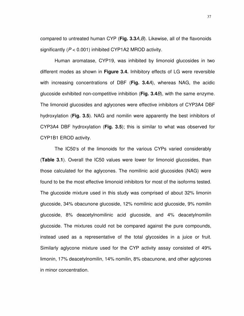
3.3 Effect of limonoid glucosides (A) and limonoid aglycones (B) on MROD activity of CYP1A2. Panels C,D indicate the effect of flavonoids on EROD and MROD activities of CYP1B1 and CYP1A2…………………………………...….…
38
3.4 Lineweaver-Burk plot for the mode of inhibition of NAG and LG on CYP 19 hydroxylase activity………………...…...
39
3.5 Effect of limonoid glucosides and aglycones on the
hydroxylation of DBF, a measure of CYP3A4 activity …..…
39
x
FIGURE Page
4.1 Purified citrus limonoid glucosides used for the study…….. 47
4.2 Quenching of oxygen radicals by limonoid glucosides in vitro………………………………………………………………
52
4.3 Toxicity of limonoid glucosides against SH-SY5Y cells in
culture…………………………………………………………….……
54
4.4 Cell viability analysis based on MTT reduction, measured
by the formation of formazon at 550 nm……………..…..….
55
4.5 Survivability of human neuroblastoma cells treated with LG alone or LG + Ac-DEVD-CHO, a known caspase inhibitor...
57
4.6 Induction of caspase 3/7 activity by limonoid glucosides….. 58
4.7 Flow cytometry analysis of DNA in SH-SY5Y cells as a
function of limonoid glucoside treatment……………..……...
61
4.8 DNA fragmentation analysis of SH-SY5Y cells after limonoid treatment…………………………………………..…
62
5.1 Time-dependent changes in percent viable cells following
treatment with pure aglycones or limonoid mixtures…....…
74
5.2 Confocal images of SH-SY5Y neuroblastoma cells treated with limonoid glucosides and aglycones……………...…..…
77
5.3 Confocal images of Caco-2 and non-cancerous Chinese
hamster ovary (CHO) cells treated with limonoids….……...
78
5.4 Flow cytometric DNA content analysis for cell cycle of SH-SY5Y neuroblastoma cells stained with propidium iodide after 36 h exposure……………………………………….……
79
xi
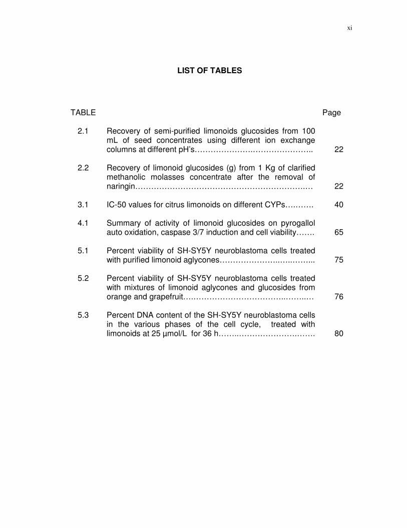
LIST OF TABLES
TABLE Page
2.1 Recovery of semi-purified limonoids glucosides from 100 mL of seed concentrates using different ion exchange columns at different pH’s………………….…………………..
22
2.2 Recovery of limonoid glucosides (g) from 1 Kg of clarified methanolic molasses concentrate after the removal of naringin……………………………………………………….…
22
3.1 IC-50 values for citrus limonoids on different CYPs….……. 40
4.1 Summary of activity of limonoid glucosides on pyrogallol auto oxidation, caspase 3/7 induction and cell viability…….
65
5.1 Percent viability of SH-SY5Y neuroblastoma cells treated
with purified limonoid aglycones…………………..…..……...
75
5.2
Percent viability of SH-SY5Y neuroblastoma cells treated with mixtures of limonoid aglycones and glucosides from orange and grapefruit….……………………………..……..…
76
5.3
Percent DNA content of the SH-SY5Y neuroblastoma cells in the various phases of the cell cycle, treated with limonoids at 25 µmol/L for 36 h……..………………….…….
80
1
CHAPTER I
INTRODUCTION 1.1 Background
Citrus is an important fruit crop of tropical and subtropical areas with over
4000 years history of cultivation (1). The United States contributes nearly one
sixth of the world citrus production, accounting for nearly 70 million metric tons
(2). Consumed both as fresh fruits and processed products, the U.S. export
value of citrus is estimated to be $300 million in 2004-2005 season (3).
Consumption of citrus has long been known to prevent many human diseases,
from scurvy to several types of cancers. The health enhancing effects of citrus
consumption is attributed to the wide assortment of bioactive compounds that
makeup citrus phytochemicals (4). The major class of citrus phytochemicals
includes flavonoids, carotinoids, limonoids, folic acid, pectin, coumarins, vitamin
C, high quality soluble fiber and mineral potassium (5). Unique among these are
the limonoids found predominantly in Rutaceae, Meliaceae, Cneoraceae and
Simaroubaceae families, with citrus being the only edible fruits among these
families. Limonoids are triterpenoids derived from the precursor 4,4,8-trimethyl-
17-furanylsteroid, with furan and lactone moieties on their ring structures.
______________________
This dissertation follows the style and format of the Journal of Nutrition.

2
Limonoids occur both as aglycones, otherwise known as citrus bitter
principles and as tasteless water-soluble glycosides (6,7). Two enzymes,
UDPG-limonoid glycosyl transferase and limonoid D-ring lactone hydrolase
perform the inter-conversion of free to carbohydrate bearing moieties during fruit
maturation (8). More than thirty-six limonoid aglycones and seventeen limonoid
glucosides have thus far been isolated from citrus and its hybrids (9). Limonoid
aglycones contribute to the “delayed bitterness” problem in citrus juices (10).
The most common aglycones are limonin, nomilin, obacunone and
deacetylnomilin which are present relatively large concentrations in citrus seeds
and molasses (6,10).
A glucose moiety attached to C-17 position of aglycones by a -glycosidic
linkage makes the respective limonoid glucosides nonbitter and water-soluble.
Most common among them are limonin 17-D glucopyranoside (LG),
nomilin17-D glucopyranoside (NG), nomilinic acid 17-D glucopyranoside
(NAG), obacunone 17-D glucopyranoside (OG), and deacetylnomilinic acid
17-D glucopyranoside (DNAG). These are found in relatively higher
concentrations in seeds, and molasses (6-8,11,12). Orange, grapefruit, and
lemon juices contain an average of 320, 190 and 82 parts per million (ppm) of
limonoid glucosides (13) and a glass of orange juice contains on average of 66
mg/L of limonoid glucosides (14).
Contemporary studies have shown that numerous health benefits are
derived from limonoid ingestion such as reducing the effects of HIV infections
3
(15,16), serving as antimalarial and anti-inflammatory agents (17), and acting as
cytotoxic agents against cancers of breast (18), colon (19,20), stomach (21),
buccal pouch (22-24) and blood (25). Limonoids also have been found to lower
blood cholesterol (26,27) and induce detoxifying enzymes such as glutathione S-
transferase (GST) (28,29) and cytochrome P450 (30), which are among the
main targets for antineoplastic strategies. Some, if not all of these properties
appear to be associated with the ability of limonoids to affect cell growth (31).
With the plethora of health promoting properties, citrus limonoids appear to be
potential food supplementations and nutrient additives (11).
It is necessary to evaluate the biological activity of pure compounds to
determine the chemopreventive agents in citrus fruits. However, the low
concentration of limonoids in fruits and their byproducts makes their extraction
and purification difficult (12). Several analytical methods using reversed-phase
and normal-phase HPLC methods coupled with UV detection have been used to
separate and detect limonoid aglycones and glucosides in small quantities
(9,32). Additionally, the methods to isolate, identify and quantify limonoids lack
specificity and sensitivity and, therefore, have not been very successful in the
bulk purification of individual compounds required for large scale biological
activity studies. Many of the carcinogenesis studies involving limonoids were
performed only with topical applications in animals because of the dearth of
purified compounds. Recently ion exchange and affinity based synthetic resins
4
have been used for the purification of naturally occurring compounds and could
be applied for the bulk purification of limonoids from seeds and molasses.
Despite remarkable treatment advances, cancer remains the leading
death causing disease among children up to age 14 years in the U.S. and the
second leading cause of death among adults next to coronary heart disease
(33). As many as one-third of all cancer deaths in the U.S. could be prevented
with a healthy, balanced diet emphasizing vegetables, fruits, whole grains and
beans which have a proven effect in maintaining a healthful weight (33).
Generation of reactive oxygen species, activation of cytochrome P450 enzymes,
alteration of cell cycles, and suppression of tumor suppressor genes are among
the leading causes of the onset of several types of cancers (34,35).
Consequently, strategies to counter the malignancies of these cancers are vitally
important.
1.2 Cytochrome P450 isoenzymes
Cytochrome P450 (CYP) enzymes are hemoprotein monooxygenases
that catalyze a wide variety of endogenous and xenobiotic compounds (36,37).
In humans, CYP enzymes have been classified into 18 gene families with over
50 enzymes in each. Individual isoenzymes are thought to be responsible for the
metabolism of specific substrates. Some of the most important CYP isoenzymes
are CYP3A4, CYP1A2, CYPB1 and CYP9, which have a specific role in the
onset of several types of cancers (38-42). CYP3A4 isoenzyme, the most

5
abundant subfamily, has been implicated in the etiology of prostate cancer (38).
A higher CYP1A2 level is known to influence risk of lung and colorectal cancers
(39,40). Recent studies on high levels of E2-4 hydroxylation in estrogen
responsive tissues have shown CYP1B1 to play an important role in estrogen
related tumerogenesis (41). CYP19 aromatase catalyzes the conversion of
androgens to estrogens in the last step of the estrogen biosynthesis pathway. It
is believed that increased exposure to estrogens is a risk factor for breast cancer
and, therefore, the human aromatase (CYP19) gene is a plausible candidate for
low-penetrance breast cancer susceptibility (42). Since CYP enzymes are
involved in both activation and detoxification in the first pass metabolism, their
partial inhibition by naturally occurring compounds may represent a novel
anticarcinogenesis strategy (42). Further, they could reduce the costs on drugs
by increasing the bioavailability of orally administered drugs (43). Flavonoids of
grapefruit have been shown to inhibit the activity of certain other human
cytochrome P450 enzymes (44). Assessing the alkoxyresorufin O-dealkylase
activity by CYP isoforms is reported to be a good assay system to test the
inhibitory effects of naturally occurring compounds from citrus and to determine
the kinetic properties of the inhibition (44). Since bioactive compounds in citrus
juice have been shown to interact with phase I enzymes, it is imperative to
determine the inhibitory effect of purified citrus limonoids on CYP isoforms.
6
1.3 Antioxidant activity
An array of bioactive compounds from fruits and vegetables has been
shown to act against free radicals that have the potential to damage DNA and
lead to cancer, coronary heart disease, and other chronic diseases (45-47).
Activation of GST (28,29) and inhibition of CYP enzymes (30), both enzymes are
known to work against redox cycling, by citrus limonoids has led to speculation
that these compounds could be considered as antioxidants. However, no firm
experimental data are available that support limonoids as having free radical
quenching abilities. Pyrogallol (1,2,3-benzenetriol) undergoes auto-oxidation in
the presence of superoxide anion radicals (O2.-) to form purpurogallin.
Superoxide dismutase (SOD) inhibits this reaction (48) by quenching the
superoxide radicals which can be measured spectrophotometricaly. This in vitro
assay serves as an excellent method to test the superoxide quenching ability of
citrus limonoids.
1.4 Apoptotic induction
One of the molecular mechanisms underlying the chemoprevention
properties of limonoids are thought to be through the induction of apoptosis.
Although there is evidence that suggests limonoids restrain the proliferation of
some cancerous cell lines, the underlying mechanism is unknown. The human
SH SY5Y neuroblastoma cell line is an excellent experimental model for the
study of apoptosis. Members of the Caspase 3/7 family of proteases have been
7
found to be crucial effectors of the downstream apoptosis signaling (49). So
deciphering the cytological and biochemical evidence of limonoid induction of
apoptosis is very revealing.
Neuroblastomas are embryonal tumors of the peripheral nervous system
that arise from the neural crest; they account for about 10 percent of childhood
cancer in U.S. (50). Chemotherapy has not made a significant impact on curing
these pediatric malignancies (51). A few citrus flavonoids have been shown to
protect against neuronal injury presumably through caspase pathway (52).
Human SH-SY5Y neuroblastoma cell lines have been widely used to study
anticancer drug therapy (53) and could be used to study the biochemical effects
of citrus limonoids on the induction of apoptosis. Apoptosis, i.e., programmed
cell death, plays a crucial role in development of several diseases.
Biochemically, apoptosis is characterized by fragmentation of the genome and
cleavage and/or degradation of several cellular proteins. The inhibitory effects
of citrus limonoids on human cancer cell proliferation in certain types of cancer
cell lines are postulated to be through the induction of apoptosis. High
concentrations of limonoids have been shown to induce apoptosis in breast
cancer (MCF-7) cells through the use of flow cytometry (31). Members of
caspase 3/7 family of proteases have been found to be crucial mediators of the
complex biochemical events associated with apoptosis. These enzymes have
substrate specificity for the amino acid sequence Asp-Glu-Val-Asp (DEVD) and
cleave a number of different proteins including poly (ADP-ribose) polymerase
8
(PARP), DNA dependent protein kinase, and actins. Z-DEVD-R110 (N-CBC-
Lasp-Lglu-Lval-Lasp-Rhodamine110) is a profluorescent substrate which is
cleaved at the aspartic acid residue by caspase 3/7, releasing highly fluorescent
rhodamine-110. This assay system may be used with either cell extracts or
primary cell suspensions and enables one to identify the regulatory events in the
apoptotic death process. Overall, studying the effects of limonoids on human
neuroblastoma cells and their regulatory mechanisms could give fundamental
evidence for the molecular mechanism of apoptotic induction. Furthermore,
studying the effects of these compounds at different stages of the cell cycle
provides critical evidences for the cytostatic effects of these bioactive
compounds.
1.5 Cytotoxicity on colonic adenocarcinoma
Colorectal cancer is the second most common cause of deaths related to
cancer in U.S. Based on the American Cancer Society’s Facts and Figures,
about 145,000 new cases and about 57,000 deaths due to colorectal cancer will
occur in 2005 in the U.S. alone (33). Epidemiological studies and animal
experiments consistently proved the protective effects citrus fruits and their
bioactive compounds against a wide range of cancers in humans (5). Grapefruit
juice has been shown to suppress the carcinogen (PhIP) induced colon DNA
damage (54). Furthermore, orange juice, which is rich in limonoids and
flavonoids, significantly inhibited azoxymethane induced colon cancer in male
9
Fisher 344 rats (55). Similarly, limonin and obacunone inhibited aberrant crypt
foci (ACF) formation, thereby, inhibiting the pathogenesis of colon cancer (20).
Considering the advantages of limonoid glucoside over aglycones in water
solubility and absorption, it is imperative to study the cytotoxic effects of limonoid
glucosides and other aglycones on human colon cancer cells. It is also important
to assess the effect of these bioactive compounds on non-cancerous
mammalian cells, to understand the toxic effects. Hence Chinese hamster ovary
cells provide an excellent non-cancerous cell model to assess the toxicity of
these bioactive compounds.

10
CHAPTER II
ISOLATION OF LIMONOIDS AND FLAVONOIDS FROM SEEDS AND
MOLASSES OF CITRUS FRUITS
2.1 Synopsis
Limonoids with putative cancer preventive properties were purified from
seeds and molasses of citrus fruits using a combination of ion exchange and
size exclusion chromatographies. pH-dependent cold precipitation of a large
quantity of naringin, prior to the separation of limonoids from flavonoids on a
styrene/divenylbenzene anionic resin using an increasing salt gradient, was
found to be critical for higher yields of semi-purified limonoid glucosides.
Substantial differences were observed with use of different resins on the total
recovery of limonoid glucosides. High purity limonoid glucosides were obtained
via reverse phase preparative high performance liquid chromatography and high
purity limonoid aglycones through direct crystallization. The purity and structures
of individual compounds were confirmed using LC-MS and NMR. Limonilic acid
was synthesized from limonin for the structure-bioactivity studies. Hesperidin, a
major flavonoid of oranges was isolated from orange peels using a pH
dependent potassium iodide precipitation method. A large scale purification
method of limonoids and flavonoids from citrus byproducts is presented.
11
2.2 Introduction
Plant compounds are sources for the development of new drugs and
model structures for synthetic formulations (56). Citrus fruits contain most of the
important class of health promoting bioactive compounds including flavonoids,
limonoids, carotenoids, vitamin C, coumarins, pectin, folic acid, glucaric acid,
high quality soluble fiber, and minerals such as potassium (5). Although initial
speculations on the biological activities of citrus compounds were associated
with vitamin C, it was later proved that other components impart health
promoting properties. Limonoids are abundant in citrus fruits, and occur both as
water insoluble aglycones and soluble glucosides (6,7). The bitter limonoid
aglycones are converted to nonbitter and tasteless glucosides by UDP-D-
glucose-limonoid glucosyltransferase during fruit maturation (8). Evidence
suggests that intestinal absorption of limonoids and flavonoids is greatly
enhanced when ingested as glucosides (57,58).
Citrus limonoids have numerous health promoting properties in animals
and human cell culture studies (15-31). Major flavonoids of citrus fruits, naringin,
narirutin and hesperidin have also been shown to have numerous health
promoting properties (59-64). Flavonoids are being evaluated as food additives
by the FDA (65). Because of the growing evidence on the health promoting
properties, these bioactive compounds could be developed as food nutrient
additives. Limonilic acid, a modified limonin, has been shown to possess

12
significant antifeedant activity with Colorado potato beetle larvae (66), and thus
could be used for insect control.
However, the activity studies of these bioactive compounds rely on the
availability of pure compounds. Many of the carcinogenesis investigations with
limonoids were performed either with mixtures or using topical applications in
animals due to inadequate supplies of pure compounds. Citrus byproducts from
processing plants are excellent sources for limonoids with more than 500 ppm in
seeds and molasses (6-8,11,67).
Procedures to isolate pure limonoids (both aglycones and glucosides)
have advanced steadily and it is now feasible to prepare and evaluate pure
materials for anticancer effectiveness. Several methods such as normal-phase
HPLC (32, 68, 69) reversed-phase HPLC (70,71), NMR (7,72,73), LC/ESI/MS
(74 -76), have been developed to quantify and/or identify limonoids with different
degrees of specificity and sensitivity. Adsorption and ion exchange resins have
greatly facilitated the isolation and purification of many flavonoid and glucoside
mixtures (11,77). Despite these recent advances, there is still room to improve
the separation efficiency and increase yields. The current study establishes an
efficient method for isolation of limonoids, hesperidin, and synthesis of limonilic
acid from limonin. In addition, a bulk purification method for naringin that
involves a combination of chromatographic techniques is described.

13
2.3 Materials and methods
Limonoids and flavonoids source. Seeds of Mexican oranges
(varieties; Valencia & Navel) were obtained in 100 Kg lots from Monterrey,
Mexico, dried under shade, and finely powdered in a coffee blender, before
solvent extraction. Orange and grapefruit molasses were obtained from the
Texas Citrus Exchange (TCX), a commercial citrus juice plant in Mission, Texas.
The molasses was stored at 4oC with 0.2% sodium azide added until used.
Resins and other chemicals. Commercial macroporous, neutral resins,
such as SP-70, XAD-16 and XAD-2, were purchased from Supelco. Weak anion
exchangers, i.e., WA-30 and high performance Q-sepharose with exchange and
binding capacities of 150-200 µeq/mL and of 120 mg/L human serum albumin
(HSA/mL) respectively, were obtained from Sigma-Aldrich Co. The strong
cationic exchanger Dowex-50 was also purchased from Sigma Chemical Co.
Dowex-50 had an ion exchange capacity of 1.7 meq/mL wet resin, of 100-200 µ
mesh size, and 50-58% water retention capacity. Small columns (2.5 cm X 10
cm) were used for evaluation and selection of appropriate resins, e.g., XAD-16,
XAD-2 and WA30. Bulk purification was accomplished using Dowex-50 (10 cm x
40 cm column), SP-70 (10 cm x 30 cm column), and Q-sepharose (10 cm x 30
cm column). The resins were presoaked in methanol, made into a slurry using
deionized water and added to the column until about two thirds of it was filled.
The top end of the first column was fitted to a low pressure peristaltic pump and
the series of columns were interconnected (Fig. 1). The resins were washed
14
until the conductivity of the eluant decreased below 100 µΩ and backwashed
with three column volumes (CV) of deionized water.
Selection of resins for glycosidic extracts from seeds. Ethanolic
extracts of Mexican orange seeds were concentrated by rotary-vacuum
evaporation. A portion (100 g) of the dried extract was mixed 1:3 with water. The
pH was adjusted from 4.0 to 6.5 to determine the optimum pH for purification
and yield with the various resins. After loading, the columns were washed with
four column volumes of water and then the compounds were eluted with 80%
ethanol. The solvent was removed by rotary-evaporation and the product
weights were determined after freeze-drying.
Separation of limonoid glucosides from flavonoids. Orange and
grapefruit molasses (1 kg; 56 and 53o brix of total soluble sugars respectively
(TSS)) were placed in beakers and the TSS adjusted to 13o Brix with deionized
water. The slurry was centrifuged at 10,000 gmax for 45 min to remove solids
and the supernatant was vacuum filtered through Whatman #5 (2.5 µmol/L) filter
paper. The clarified solution (2400 ml) was loaded onto the Dowex-50 at a flow
rate 46 mL/min using a peristaltic pump. The Dowex-50 column was connected
directly to SP-70 adsorbent column. After loading, the columns were washed
with 5 CV water. The loading capacity and washing efficiency was determined
by sampling the discharge from SP-70 and analyzing by TLC (silica gel). The
compounds adsorbed to SP-70 were eluted with 80% methanol at a flow rate of
50 mL/min. The eluant was concentrated and the solvent was recovered. The

15
method described by Schoch et al.(11) for efficient separation of limonoids from
flavonoids was improved by removing naringin in large quantities. After passing
through Dowex-50 and SP-70 resin columns, 1 Kg of molasses yielded 650 ml
concentrate (from roto-evoparating the methanol eluant) of pH 2.68. Naringin
was precipitated from this concentrate by adjusting the pH to 3.5, stirring
overnight at 4o C. The precipitate was vacuum filtered using Whatman #5 filter
paper. The precipitate was then washed with ice cold water to remove all the
impurities. The precipitated naringin was subsequently dried at 60oC and
analyzed for purity. The pH of the remaining filtered supernatant was adjusted to
6.5 and the supernatant loaded onto a Q-sepharose anion exchange column at
35 mL/min. A linear gradient of NaCl (0-0.6 mol/L) was used to elute the
compounds and 200 fractions (25 mL each) were collected. The fractions were
desalted by passing them through a SP-70 column (2.5 cm x 30 cm) and eluted
with 70% methanol. Limonoid glucosides were detected by silica gel TLC
analysis (mobile phase of 5:3:1:1 ethylacetate: ethylmethylketone: formic acid:
water). Compounds were detected on the TLC plate using Elrich’s reagent (1%
p-dimethylaminobenzaldehyde in ethanol) in an HCl gas chamber. The
flavonoids were detected on a UV spectrophotometer at 254 and 280 nm. The
fractions were freeze-dried and then were weighed. The purification scheme
through ion-exchange chromatography and the points where mass balances
were taken is shown in Figure 2.1.

16
FIGURE 2.1 Sequence of ion-exchange columns in the purification of limonoids
glucosides and flavonoids from molasses of oranges and grapefruits. A, B, and
C represents the points where the mass balances were taken.
17
Preparative HPLC separation. Semi-purified limonoid glucoside mixture
(1 g) was dissolved in 10 ml deionized water and injected to a preparative
HPLC. Previously described methods (75,78) were modified to optimize
resolution of individual peaks by using a mobile phase of 10-25% acetonitrile
gradient, with 0.003% phosphoric acid. A reverse phase C-18 column (10 cm x
40 cm, Novapak, Waters Inc.) at 950/ 1150 psi (radial/ back) pressure was used.
Peaks were monitored using a Waters UV-Photo Diode Array (PDA) detector.
Individual peaks were collected based on the retention time. Acetonitrile was
removed using a rotary-evaporator, and the compounds were lyophilized. The
purity of the compounds and their structures were determined through, HPLC,
LC-MS and NMR.
Extraction of limonoid aglycones. The limonoid aglycones were
extracted by modification of existing methods (79,80). Finely powdered
grapefruit (varieties; Mexican red and Rio red) and orange seeds (varieties;
Navel and Valencia) (10 kg/batch) were extracted with 15 L hexane in the
Soxhlet apparatus for 24 h at 600C. The hexane extract was recovered and
filtered using Whatman # 5 filterpaper. The hexane extract was partitioned with
methanol at 1:1 ratio and allowed to settle for 1 hour. The methanol fraction was
separated and subject to concentrated under reduced pressure (rotary-
evaporation). The resultant concentrate was dissolved in hexane and allowed to
stand overnight at room temperature. The next day the precipitants were filtered
using Whatman # 5 filterpaper. The silica gel TLC analysis of the filtered

18
precipitate indicated high concentrations of obacunone and nomilin. This mixture
was vacuum dried, powdered, and used for open silica column chromatography.
The hexane defatted seed powder (6.5 kg) was further extracted with acetone
(15 L) at 60oC for 12 h in a Soxhlet apparatus to remove the aglycones. The
acetone extract was concentrated under reduced pressure and a rotary-
evaporator. The aglycone mixture dissolved in acetone was spotted on silica gel
TLC plates (Sigma Aldrich Co.) using ethylacetate:hexane (3:2) mobile phase,
sprayed with 10% sulfuric acid in methanol, and developed at 110oC for 1 min.
The Rf values were compared with those of the standards. The compounds
were further quantified on an analytical HPLC (Waters, Novapack RP-18, 8 mm
x 40 mm, UV 6000LP detector at 210 nm) using a 10-50% linear gradient of
water:acetonitrile with 0.003% phosphoric acid. The major aglycone limonin was
precipitated from the concentrate by dissolving it 1:2 in dichloromethane, and
allowing it to stand for 1 hour and then vacuum filtering through a Buckner funnel
using Whatman # 5 filterpaper. Limonin was further purified by washing
repeatedly with isopropanol and recrystallizing in dichloromethane. Most of the
limonin was removed by recrystallization (3–4 times). The remaining filtrate was
rotoevaporated and redissolved in minimum acetone and left at 4oC for 72 h to
crystallize nomilin. The nomilin crystals were washed with cold isopropanol. The
leftover filtrate was concentrated and subjected to vacuum liquid
chromatography to purify minor individual aglycones.

19
Vacuum liquid chromatography (VLC). VLC has been used as an
alternative technique to conventional chromatographic methods (81) and for the
purification of bioactive diterpenoids and other compounds (82). This technique
was used to purify individual aglycones from bulk limonoid aglycone mixtures.
The filtrate obtained after removal of limonin and nomilin contained concentrated
amounts of deacetylnomilin, obacunone and traces of limonin and nomilin. The
filtrate was mixed with TLC grade silica gel (EM Science) with gypsum binder
(mesh size 2-2.5 µm, pore size 0.75 cm2/g, pore diameter 60oA, Bet surface
area 500 m2/g, pH 6.8 at 10% aqueous suspension) and subjected to VLC to
purify individual compounds. The mixture was finely powdered (until free
flowing) and added to dry TLC grade silica gel of same properties already in a
VLC column. A vacuum was applied for about 40 minutes for uniform packing
and then washed with 100% hexane to check the uniform movement of solvent
through the column. The mixture was firmly placed on the top of the silica
packing, covered with a filter paper and vacuum was applied to compact the
silica packing. A step gradient of 20-80% of ethylacetate:hexane at 5%
increments (50 mL/step) was used to elute the aglycones from the column and
individual fractions were analyzed with TLC and pooled based on the individual
compounds content of the fraction.
Open column chromatography. Open silica column chromatography
with a range of mobile phases is widely used for the efficient purification of
individual bioactive compounds. This is a rapid technique for the separation of

20
minute quantities of compounds (83). Aglycone mixtures devoid of limonin and
other crystallized aglycones were dissolved in acetone and mixed with equal
quantities (w/w) of 63-200 mesh size silica gel. The mixture was packed into a
24/40 column 2.5 cm x 30 cm with 54 g of silica and eluted with 40-70%
ethylacetate-hexane. The effluent was collected (20 mL fractions) until complete
elution. The fractions were analyzed by TLC and then pooled depending on their
content. The unresolved limonin-deacetylnomilin was further separated on 1:1
mixture of 230-400 mesh silica, eluted with 55% ethylacetate-hexane.
Synthesis of limonilic acid from limonin. The method for synthesis of
limonilic acid reported by Emerson (84) was modified. Limonin (8 g) was
suspended in 250 ml of 0.2 N KOH in methanol contained in a round bottom
flask equipped with a water cooled condenser. The solution was refluxed for 30
minutes. After cooling, the methanol was removed under vacuum. The residue
was dissolved in 100 mL water and 3 g each of Iodine and potassium iodide
were added. To this mixture 2N NaOH was added until the iodine color was
changed to a light red/strong orange yellow. After 15 minutes, 5 mg/L of sodium
sulfite was added and the solution was acidified with 5N HCl. At pH 1 the
limonilic acid was precipitated. The precipitate was filtered and washed with
isopropanol and then with hexane to remove Iodine. The purity was confirmed
via analytical HPLC.
21
Isolation of hesperidin from orange peels. Large quantities of
hesperidin were isolated via modification of a previously described method (85).
2.5 Kg of freshly peeled mandarin and orange peels were finely ground with a
coffee blender, and mixed with 4 L of deionized water. The pH of the slurry was
raised to 11-12 by addition of calcium hydroxide granules. The elevated pH
slurry was stirred for 90 minutes and filtered using cheese cloth (3 layers). The
filtrate was collected and the solids were discarded. The pH of filtrate was
adjusted to 4.7 with concentrated HCl. The solution heated to 60oC in a water
bath for 4 h, stirred for 2 h and held at room temperature overnight. The
precipitated slurry was vacuum filtered, washed with methanol, and then with
hexane to remove the impurities. The filtrate was dried and analyzed using
analytical HPLC.
2.4 Results
Selection of resins. The total glucoside yield results, using different
resins at different pH are summarized in Table 2.1. The yields were relatively
higher for SP-70 and XAD-16, compared to other resins tested. Total glucoside
yield was higher at pH 4.5, compared to other pH ranges tested (Table 2.1).
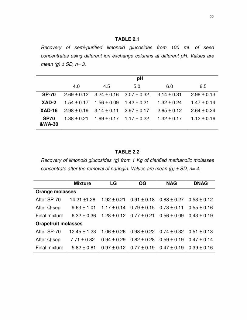
22
TABLE 2.1
Recovery of semi-purified limonoid glucosides from 100 mL of seed
concentrates using different ion exchange columns at different pH. Values are
mean (g) ± SD, n= 3.
pH
4.0 4.5 5.0 6.0 6.5
SP-70 2.69 ± 0.12 3.24 ± 0.16 3.07 ± 0.32 3.14 ± 0.31 2.98 ± 0.13
XAD-2 1.54 ± 0.17 1.56 ± 0.09 1.42 ± 0.21 1.32 ± 0.24 1.47 ± 0.14
XAD-16 2.98 ± 0.19 3.14 ± 0.11 2.97 ± 0.17 2.65 ± 0.12 2.64 ± 0.24
SP70 &WA-30
1.38 ± 0.21 1.69 ± 0.17 1.17 ± 0.22 1.32 ± 0.17 1.12 ± 0.16
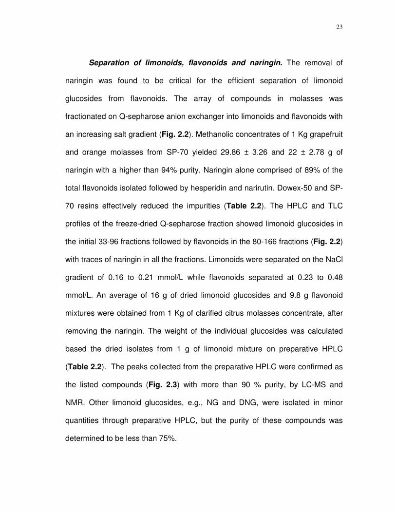
TABLE 2.2
Recovery of limonoid glucosides (g) from 1 Kg of clarified methanolic molasses
concentrate after the removal of naringin. Values are mean (g) ± SD, n= 4.
Mixture LG OG NAG DNAG
Orange molasses
After SP-70 14.21 ±1.28 1.92 ± 0.21 0.91 ± 0.18 0.88 ± 0.27 0.53 ± 0.12
After Q-sep 9.63 ± 1.01 1.17 ± 0.14 0.79 ± 0.15 0.73 ± 0.11 0.55 ± 0.16
Final mixture 6.32 ± 0.36 1.28 ± 0.12 0.77 ± 0.21 0.56 ± 0.09 0.43 ± 0.19
Grapefruit molasses
After SP-70 12.45 ± 1.23 1.06 ± 0.26 0.98 ± 0.22 0.74 ± 0.32 0.51 ± 0.13
After Q-sep 7.71 ± 0.82 0.94 ± 0.29 0.82 ± 0.28 0.59 ± 0.19 0.47 ± 0.14
Final mixture 5.82 ± 0.81 0.97 ± 0.12 0.77 ± 0.19 0.47 ± 0.19 0.39 ± 0.16
23
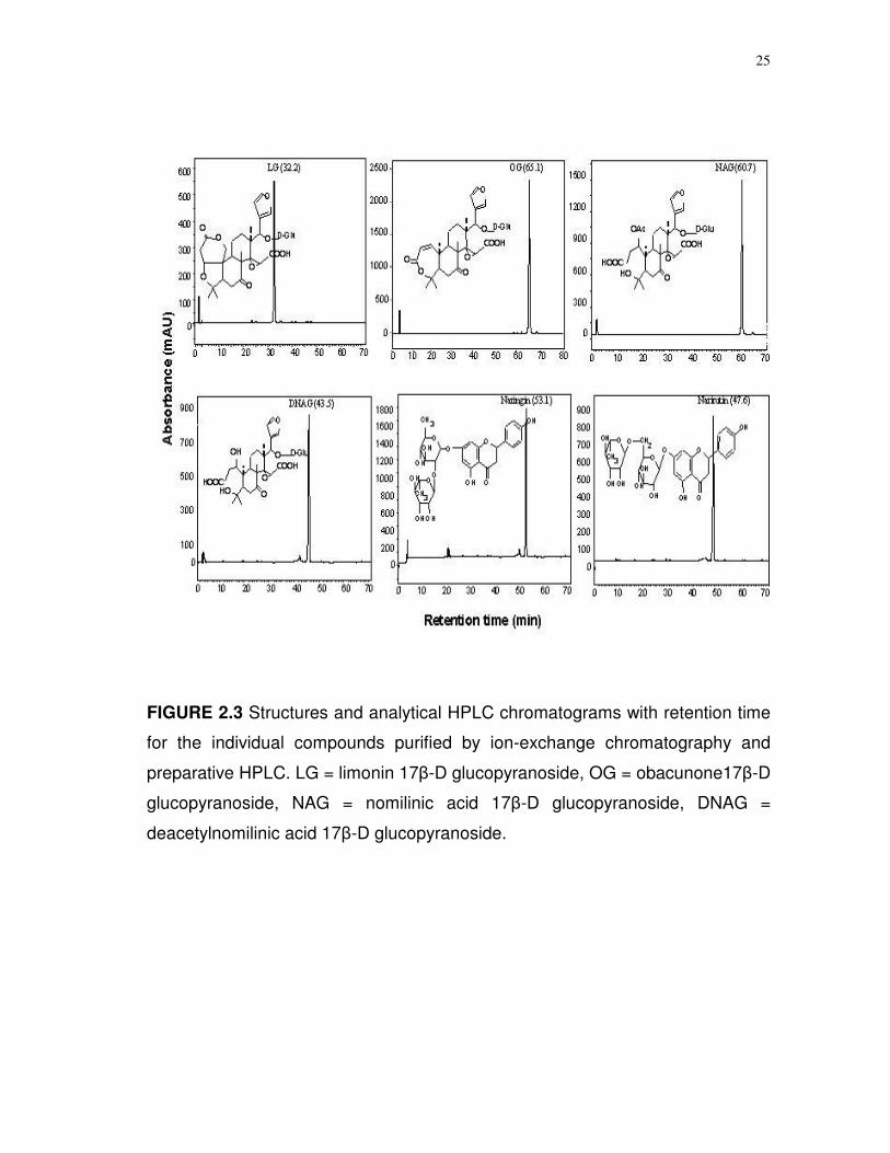
Separation of limonoids, flavonoids and naringin. The removal of
naringin was found to be critical for the efficient separation of limonoid
glucosides from flavonoids. The array of compounds in molasses was
fractionated on Q-sepharose anion exchanger into limonoids and flavonoids with
an increasing salt gradient (Fig. 2.2). Methanolic concentrates of 1 Kg grapefruit
and orange molasses from SP-70 yielded 29.86 ± 3.26 and 22 ± 2.78 g of
naringin with a higher than 94% purity. Naringin alone comprised of 89% of the
total flavonoids isolated followed by hesperidin and narirutin. Dowex-50 and SP-
70 resins effectively reduced the impurities (Table 2.2). The HPLC and TLC
profiles of the freeze-dried Q-sepharose fraction showed limonoid glucosides in
the initial 33-96 fractions followed by flavonoids in the 80-166 fractions (Fig. 2.2)
with traces of naringin in all the fractions. Limonoids were separated on the NaCl
gradient of 0.16 to 0.21 mmol/L while flavonoids separated at 0.23 to 0.48
mmol/L. An average of 16 g of dried limonoid glucosides and 9.8 g flavonoid
mixtures were obtained from 1 Kg of clarified citrus molasses concentrate, after
removing the naringin. The weight of the individual glucosides was calculated
based the dried isolates from 1 g of limonoid mixture on preparative HPLC
(Table 2.2). The peaks collected from the preparative HPLC were confirmed as
the listed compounds (Fig. 2.3) with more than 90 % purity, by LC-MS and
NMR. Other limonoid glucosides, e.g., NG and DNG, were isolated in minor
quantities through preparative HPLC, but the purity of these compounds was
determined to be less than 75%.
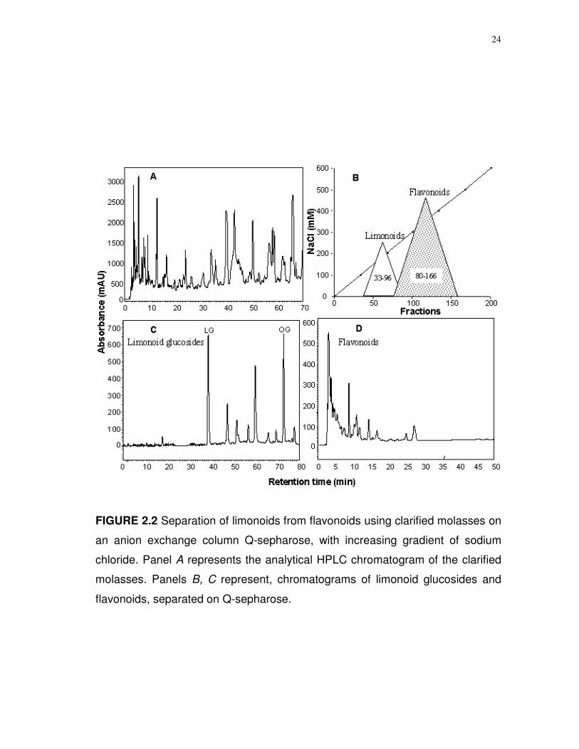
24
FIGURE 2.2 Separation of limonoids from flavonoids using clarified molasses on
an anion exchange column Q-sepharose, with increasing gradient of sodium
chloride. Panel A represents the analytical HPLC chromatogram of the clarified
molasses. Panels B, C represent, chromatograms of limonoid glucosides and
flavonoids, separated on Q-sepharose.
25
FIGURE 2.3 Structures and analytical HPLC chromatograms with retention time
for the individual compounds purified by ion-exchange chromatography and
preparative HPLC. LG = limonin 17-D glucopyranoside, OG = obacunone17-D
glucopyranoside, NAG = nomilinic acid 17-D glucopyranoside, DNAG =
deacetylnomilinic acid 17-D glucopyranoside.
26
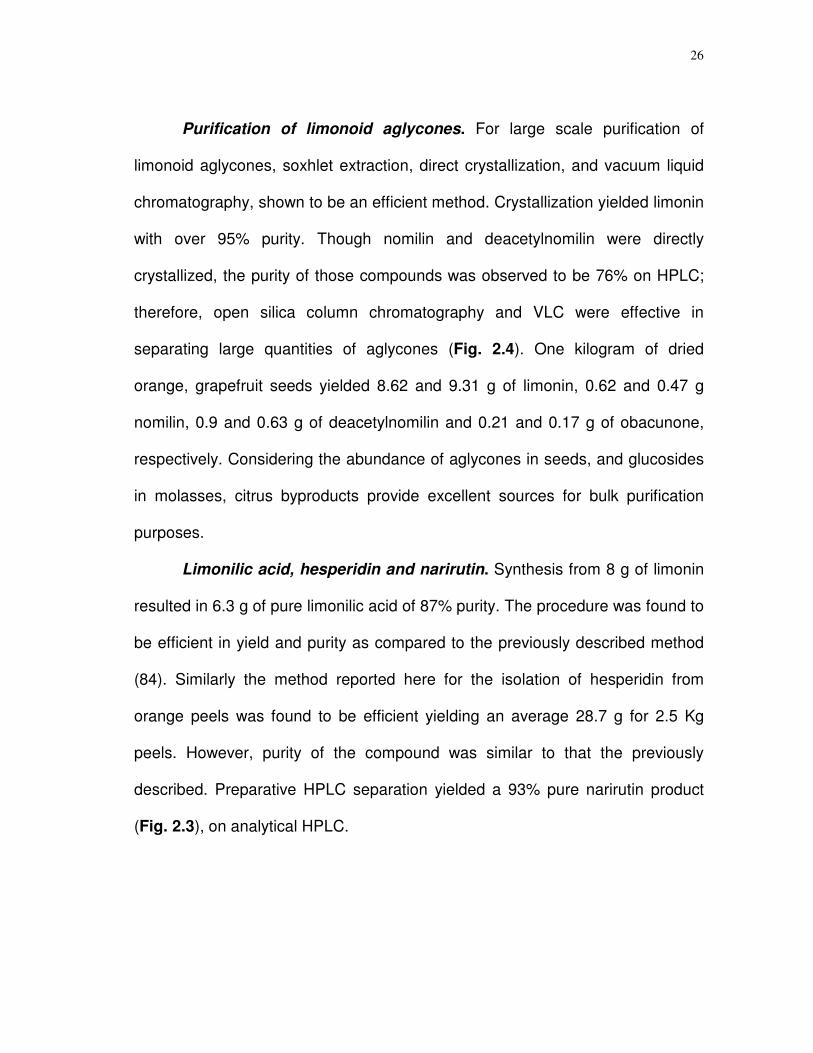
Purification of limonoid aglycones. For large scale purification of
limonoid aglycones, soxhlet extraction, direct crystallization, and vacuum liquid
chromatography, shown to be an efficient method. Crystallization yielded limonin
with over 95% purity. Though nomilin and deacetylnomilin were directly
crystallized, the purity of those compounds was observed to be 76% on HPLC;
therefore, open silica column chromatography and VLC were effective in
separating large quantities of aglycones (Fig. 2.4). One kilogram of dried
orange, grapefruit seeds yielded 8.62 and 9.31 g of limonin, 0.62 and 0.47 g
nomilin, 0.9 and 0.63 g of deacetylnomilin and 0.21 and 0.17 g of obacunone,
respectively. Considering the abundance of aglycones in seeds, and glucosides
in molasses, citrus byproducts provide excellent sources for bulk purification
purposes.
Limonilic acid, hesperidin and narirutin. Synthesis from 8 g of limonin
resulted in 6.3 g of pure limonilic acid of 87% purity. The procedure was found to
be efficient in yield and purity as compared to the previously described method
(84). Similarly the method reported here for the isolation of hesperidin from
orange peels was found to be efficient yielding an average 28.7 g for 2.5 Kg
peels. However, purity of the compound was similar to that the previously
described. Preparative HPLC separation yielded a 93% pure narirutin product
(Fig. 2.3), on analytical HPLC.

27
FIGURE 2.4 Structures and analytical HPLC chromatograms with retention time
for the individual limonoid aglycones. Resolved on a mobile phase of 10-50%
acetonitrile in water with 0.003% phosphoric acid, using a reverse phase C-18
column at 1 mL/min flow rate.
2.5 Discussion
Adsorption and ion-exchange resin load capacity is a key parameter for
limonoid purification. When compared to the other resins, SP-70 had an
increased yield at pH 4.5. The ion-exchange mechanism involved in this
purification process is well characterized for other compounds (86). The
28
adsorption-desorption properties of limonoids on adsorbent resins like SP-70
can be summarized in the following steps: (i) selective movement of limonoids in
solution to the boundary of resin particles, (ii) attachment of limonoid glucosides
to the surface of the resin, (iii) diffusion to the intra-particle active sites from the
surface, (iv) attachment and desorption depending upon the molecular size,
charge of the molecule and polarity of the mobile phase. The differences in
yields at various pH’s could be due to conformational changes that glucosides
undergo as a function of pH.
The ion-exchange process depends on the ionic strength of the mobile
phase and desorption increases with increase in the conductivity (87). A lower
degree of cross-linking in a cationic exchanger like Dowex-50 eliminates
polyelectrolytes and emulsifier anions (88) which aid in the elimination of colored
impurities from molasses. The rate of the adsorption/desorption processes on an
anionic resin is determined by the number of charged groups on the molecule.
Structural properties of limonoid glucosides exhibit a negatively charged open D-
ring with a glucose moiety attached to it. In the case of acidic glucosides like
NAG and DNAG, both the A and D rings are open. From the fractionation pattern
(Fig. 2.2) it is evident that the negative charges on limonoid glucosides and
flavonoids molecules alter the conductivity and separation on anionic resins is
affected by an increasing salt gradient. The increase in yield over the previously
described method (11) can be ascribed to the separation of naringin at the initial
stages of separation. Although more than 17 limonoid glucosides have been
29
identified from citrus (6), only the four most abundant glucosides have been
characterized (Table 2.2) for use in further biological activity studies.
Extraction of aglycones using a Soxhlet apparatus remains to be proven
an efficient method of extraction. Direct crystallization was successful in
providing a large quantity of high purity limonin. The availability of seeds and
molasses from the juice processing plants makes for an inexpensive source of
raw materials. Large scale purification of desired products from seeds and
molasses via the Soxhlet extraction, direct crystallization, VLC, and silica column
chromatography proves to be inexpensive and efficient. These methods are
applicable for industrial purification. Similarly, the synthesis of limonilic acid,
precipitation of hesperidin, and separation of narirutin are also suitable for the
bulk purification to meet commercial needs.
In conclusion, large amounts of pure limonoids and flavonoids are needed
to elucidate their cancer preventive properties. The bulk purification methods
reported in this chapter prove to be economical and efficient. The isolation and
activity studies with pure compounds could provide critical information regarding
the structure-activity relationship, molecular mechanisms, and biochemical
interaction, thereby could aid as lead structures for new drug development.
30
CHAPTER III
INHIBITORY EFFECTS OF CITRUS LIMONOIDS AND FLAVONOIDS ON
HUMAN CYTOCHROME P450 ISOENZYMES
3.1 Synopsis
The effects of limonoids and flavonoids on O-dealkylase and hydroxylase
activities of human cytochrome P450 (CYPs) isozymes such as CYP1A2,
CYP1B1, CYP3A4 and CYP19 were measured using the substrates
ethoxyresorufin (ethoxyresorufin O-dealkylase, EROD), methoxyresorufin
(methoxyresorufin O-dealkylase, MROD), and dibenzylflurescein (DBF). The
kinetic studies resulted in variable inhibition of CYPs in concentration dependent
assays. Significant (P < 0.001) reductions in enzyme activities were observed,
with purified compounds above 2 µmol/L. Kinetic analysis indicated that limonin
glucoside inhibited CYP19 hydroxylase competitively, while nomilinic acid
glucoside in a non-competitive mode. IC50 values were less for glucosides than
for the aglycones. The results support prior suggestions for an active role of
these compounds in the redox cycling. The variation in efficacy by different
limonoids can be ascribed to the structural variation of the molecules. Limonoid
inhibition of key CYPs involved in carcinogenesis supports growing evidences
that citrus bioactive compounds can act as anticancer agents.
31
3.2 Introduction
Exposure to environmental chemicals is considered as a major risk factor
for several types of cancers. These exposures can result in the generation of
reactive oxygen (e.g., singlet oxygen, superoxide) and nitrogen (e.g.,
peroxynitrite, nitrogen dioxide) species (89) which have been implicated as
causative agents for many diseases including inflammatory and degenerative
diseases, arthritis, retinitis pigmentosis, coronary artery diseases, and many
types of cancers (89,90). In humans, many of the chemicals are pre-carcinogens
and are activated to carcinogenic and mutagenic substances by microsomal
enzymes (89,91). The human microsomal enzyme system consists of scores of
cytochrome P450 isoenzymes. In vitro studies with certain phytochemicals
revealed a reduction in activities involved with generation of carcinogens through
partial inhibition of these enzymes (44,92,93).
Cytochrome P450 enzymes are electron-transporting proteins that
contain a heme-prosthetic group in which the iron alternates between reduced
ferrous (Fe2+) and oxidized (ferric, Fe3+) state (Fig. 3.1) (94). The CYPs are
known to be involved in the metabolic activation and detoxification of
environmental carcinogens, orally administered drugs, steroids, bile acids, fatty
acids, eicosanoids, and fat soluble vitamins (91,95). Activation involves the
formation of reactive oxygen species (ROS) that may interact with DNA of target
cells (91,96). In humans, CYP enzymes have been classified into 18 gene
families with over 50 enzymes in each (97) and are mostly found on the
32
endoplasmic reticulum. Individual isoenzymes are thought to be responsible for
the metabolism of specific substrates. Some of the most important cytochrome
P450 isoenzymes are CYP3A4, CYP1A2, CYP1B1, and CYP19, which have
specific roles in the onset of several types of cancers (38-41,91,96). Thus,
inhibition of CYPs by naturally occurring compounds may represent a novel
approach in the anticarcinogenesis strategy (42,95).
Citrus products contain several vitamins, minerals, and an array of
bioactive compounds. The bioactive compounds in citrus fruits have been shown
to possess numerous cancer preventive properties (2,5). Much of current
research is being focused on limonoids and flavonoids. Limonoids are unique to
citrus. Contemporary studies have shown that limonoids have numerous health
benefits such as countering the effects of HIV infections, serving as
antimalarials, anti-inflammatory agents, and as cytotoxic-cytostatic agents
against several types of cancers (15-31). Limonoids are shown to be non-toxic
to the normal cells (31). The effect of limonoids and flavonoids in inducing
glutathione S-transferase (GST), a major phase II detoxifying enzyme (5,24,28)
and specific role on certain CYP activity (30) lead us to postulate that one of the
antineoplastic properties of these compounds could be through inhibition of
cytochrome P450 enzymes, thereby reducing carcinogenesis. Here we report
the effects of citrus limonoid aglycones, glucosides, and flavonoids on the
activity of human cytochrome p450 isoenzymes (CYP1A2, CYP1B1, CYP3A4,
CYP19) using alkoxyresorufins and dibenzylfluorescein (DBF) as substrates.

33
FIGURE 3.1 Oxidation-reduction catalytic mechanisms of CYPs
3.3 Materials and methods
Limonoids and flavonoids. The compounds were isolated from seeds
and molasses of citrus fruits as described in the previous chapter. The pure
compounds and mixtures were used to elucidate the inhibitory effects on CYP
isoforms. The glucoside mixture was comprised of about 32% limonin glucoside,
34% obacunone glucoside, 12% nomilinic acid glucoside, 9% nomilin glucoside,
8% deacetylnomilinic acid glucoside, and 4% deacetylnomilin glucoside. The
34
aglycone mixture used for the CYP activity assay consisted of 49% limonin, 17%
deacetylnomilin, 14% nomilin, 8% obacunone, and 12% other aglycones in
minor concentrations, based on the HPLC profiles. The stock solutions (1 mol/L)
were prepared in DMSO for individual limonoid aglycones and in water for pure
limonoid glucosides. The stock solutions for the mixtures were in ppm. The
DMSO concentration was maintained below 0.1% in the CYP reaction mixtures.
CYP isoforms and substrates. Alkoxyresorufins (e.g., methoxyresorufin,
and ethoxyresorufin) were synthesized as described by Mayer et al.(98). DBF
and ketoconazole were purchased from Sigma-Aldrich Co. (St. Louis, MO,
USA). Human liver expressed CYP isoenzymes CYP1A2, CYP1B1, CYP3A4,
CYP19 and NADPH oxidoreductase were purchased from BD Gentest Inc.
(Woburn, MA, USA). Activity of all these isoforms were expressed from human
CYP1A2 cDNA using baculovirus infected (BTI-TN-5B1-4) expression system.
Methoxy-, ethoxy-O-dealkylase and hydroxylase activities of CYPs.
Methoxyresorufin and ethoxyresorufin were used as substrates for CYP1A2 and
CYP1B1. DBF was the substrate for CYP3A4 and CYP19. Ketoconazole (0.25
µmol/L) was used as positive control. The MROD and EROD activities of
CYP1B1 and CYP1A2 were determined fluorometrically as described by Burke
et al. (99). The substrates (1 mol/L) were prepared in DMSO. The reaction
mixture (500 µL) contained 0.5 to 10 µmol/L citrus limonoids, 0.2 to 40 µmol/L
substrates, 2-10 pmol/L CYP. The volume was made up using proportional
volumes of 1.3 mmol/L NADP+, 3.3 mmol/L glucose 6-phosphate. 0.4 units/mL
35
glucose 6-phosphate dehydrogenase, 3.3 mmol/L MgCl2 in 0.1M phosphate
buffer, pH 7.4. Ice cold NADPH oxidoreductase was added just before the start
of the reaction. A Synergy HT fluorescence plate reader (Bio-Tek Instruments,
Inc., Winooski, VA), equipped with 530 ± 15 nm excitation and 590 ± 15 nm
emission filters was used to determine the kinetic and endpoint fluorescence of
resorufin. Hydroxylation of DBF was measured by following the formation of
fluorescein (485 ± 15 nm excitation, 538 ± 15 nm emission). The DBF reactions
were terminated at 30 minutes duration with 2 mol/L NaOH, using the same
plate reader. Reaction products were quantified using resorufin and fluorescein
standards. Inhibitor concentrations that produce 50% inhibition (IC50) were
calculated as previously described (100).
Statistical analysis. All the experiments were done in triplicate. The data
were analyzed by one way ANOVA using SPSS statistical analysis software
(Version 11.0). Comparisons were made within and between groups using
Fisher’s least significant difference test and Dunnet’s paired comparisons.
3.4 Results
All of the limonoids significantly (P < 0.001) inhibited EROD
(ethoxyresorufin O-dealkylase) activity of CYP1B1 (Fig. 3.2A,B). However, there
were no significant differences between 0.5 and 2.0 µmol/L of LG (P = 0.350),
limonin (P = 0.120), and 2 to 5 µmol/L obacunone (P = 0.011) and nomilin (P =
0.02). Limonoid glucosides were better inhibitors than aglycones (Fig. 3.2A,B).

36
FIGURE 3.2 Ethoxyresorufin and methoxyresorufin O-dealkylase (EROD,
MROD) activities of CYP1B1 treated with limonoid glucosides (panels A,C) and
limonoid aglycones (panels B,D). Values are mean ± SEM, n = 3. C (control),
Lim (limonin), Obc (obacunone), Nom (nomilin) and Dan (deacetylnomilin).
The acidic limonoid glucoside, NAG, had the highest inhibitory effect. All
of the limonoid glucosides significantly (P < 0.001) inhibited MROD activity of
CYP1B1 (Fig. 3.2C,D). Fisher’s least square differences on mean metabolite
production (MROD) by CYP1A2 treated with different levels of limonoids
indicated that all the compounds were effective (P < 0.005) inhibitors when
37
compared to untreated human CYP (Fig. 3.3A,B). Likewise, all of the flavonoids
significantly (P < 0.001) inhibited CYP1A2 MROD activity.
Human aromatase, CYP19, was inhibited by limonoid glucosides in two
different modes as shown in Figure 3.4. Inhibitory effects of LG were reversible
with increasing concentrations of DBF (Fig. 3.4A), whereas NAG, the acidic
glucoside exhibited non-competitive inhibition (Fig. 3.4B), with the same enzyme.
The limonoid glucosides and aglycones were effective inhibitors of CYP3A4 DBF
hydroxylation (Fig. 3.5). NAG and nomilin were apparently the best inhibitors of
CYP3A4 DBF hydroxylation (Fig. 3.5); this is similar to what was observed for
CYP1B1 EROD activity.
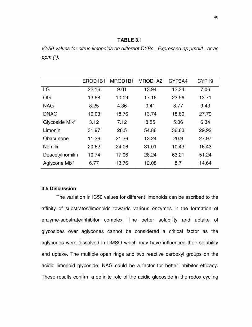
The IC50‘s of the limonoids for the various CYPs varied considerably
(Table 3.1). Overall the IC50 values were lower for limonoid glucosides, than
those calculated for the aglycones. The nomilinic acid glucosides (NAG) were
found to be the most effective limonoid inhibitors for most of the isoforms tested.
The glucoside mixture used in this study was comprised of about 32% limonin
glucoside, 34% obacunone glucoside, 12% nomilinic acid glucoside, 9% nomilin
glucoside, 8% deacetylnomilinic acid glucoside, and 4% deacetylnomilin
glucoside. The mixtures could not be compared against the pure compounds,
instead used as a representative of the total glycosides in a juice or fruit.
Similarly aglycone mixture used for the CYP activity assay consisted of 49%
limonin, 17% deacetylnomilin, 14% nomilin, 8% obacunone, and other aglycones
in minor concentration.
38
FIGURE 3.3. Effects of limonoid glucosides (A) and limonoid aglycones (B) on
MROD activity of CYP1A2. Panels C,D indicate the effects of flavonoids on
EROD and MROD activities of CYP1B1 and CYP1A2 respectively. Values are
mean + SEM, n = 3.

39
FIGURE 3.4 Lineweaver-Burk plot for the mode of inhibition of NAG and LG on
CYP19 hydroxylase activity. Nomilinic acid glucoside (NAG) inhibited CYP19 in
anon competitive mode (A), while limonin glucoside (LG) inhibited competitively
(B). Values are mean ± SEM, n = 3.
FIGURE 3.5 Effect of limonoid glycosides and aglycones on the hydroxylation of
DBF, a measure of CYP3A4 activity. Values are in mean ± SEM, n = 4.
40
TABLE 3.1
IC-50 values for citrus limonoids on different CYPs. Expressed as µmol/L. or as
ppm (*).
3.5 Discussion
The variation in IC50 values for different limonoids can be ascribed to the
affinity of substrates/limonoids towards various enzymes in the formation of
enzyme-substrate/inhibitor complex. The better solubility and uptake of
glycosides over aglycones cannot be considered a critical factor as the
aglycones were dissolved in DMSO which may have influenced their solubility
and uptake. The multiple open rings and two reactive carboxyl groups on the
acidic limonoid glycoside, NAG could be a factor for better inhibitor efficacy.
These results confirm a definite role of the acidic glucoside in the redox cycling
EROD1B1 MROD1B1 MROD1A2 CYP3A4 CYP19
LG 22.16 9.01 13.94 13.34 7.06
OG 13.68 10.09 17.16 23.56 13.71
NAG 8.25 4.36 9.41 8.77 9.43
DNAG 10.03 18.76 13.74 18.89 27.79
Glycoside Mix* 3.12 7.12 8.55 5.06 6.34
Limonin 31.97 26.5 54.86 36.63 29.92
Obacunone 11.36 21.36 13.24 20.9 27.97
Nomilin 20.62 24.06 31.01 10.43 16.43
Deacetylnomilin 10.74 17.06 28.24 63.21 51.24
Aglycone Mix* 6.77 13.76 12.08 8.7 14.64
41
mechanism which involves many enzymes including CYPs. However, compared
to the IC50’s of many citrus flavonoids and furocoumarins (37,42,101), limonoids
are poorer inhibitors for the isoenzymes tested. This study confirms the
previously reported non-significant effects of limonin and nomilin, on various
isoforms of CYPs in liver and intestine of rats at lower doses (30).
In humans, cytochrome P450 monooxygenases catalyze a wide variety of
both endogenous and xenobiotic compounds (37). Recent studies on high levels
of E2-4 hydroxylation in estrogen responsive tissues have shown CYP1B1 to
play an important role in estrogen related tumerogenesis (42). Limonoid
glucosides are better inhibitors of CYP1B1 than the limonoid aglycones.
Moreover, all of the compounds partially inhibited these isoenzymes at
micromolar levels. Epidemiological studies have shown increased risk of colon
cancer in individuals with high CYP1A2 activity based on caffeine metabolism
(102). Higher CYP1A2 activity has also been shown to influence the risk of lung
cancer (39). Tanaka et al. (19,20) had earlier reported that obacunone and
limonin reduced the azoxymethane induced colon carcinogenesis in rats. This
study confirms the effects of other limonoids as inhibitors of CYP1A2 which has
a direct role in the etiology of colon cancer. This also corroborates earlier claims
that bioactive compounds in orange and grapefruit juice can reduce the food
carcinogen PhIP (2-amino-1-methyl-6-phenylimidazo pyridine) and AOM
(azoxymethane) induced colon cancer in rats (54,55). CYP19, the steroid
aromatase, catalyzes the conversion of androgens to estrogens. An increased
42
exposure to estrogens is considered a risk factor for breast cancer. Therefore,
CYP19 is a plausible candidate for low-penetrance breast cancer (41,95). The
decreased expression of CYP19 is propitious for the reduction of estrogen
dependent tumors (103). LG and NAG inhibited CYP19 aromatase activity in
competitive and non-competitive modes, respectively. This supports earlier
claims by Miller et al. (13), So et al. (18), and Tian et al. (31), that some citrus
limonoids and other bioactive compounds in orange and grapefruit juice reduce
mammary tumerogenesis in rats as well as in the human breast cancer cells.
CYP3A4, the most abundant subfamily of CYPs, has been implicated in the
etiology of prostate cancer (38). Therefore, partial inhibition of CYP3A4 by citrus
bioactive compounds, could aid in the reduction of prostate cancer. Increased
plasma concentrations of orally administered drugs can be caused by the
inhibition of CYP3A4 enzymes which play a pivotal role in the oxidative
biotransformation of many drugs (37,93,95,101). Consequently, partial inhibition
of these enzymes would prolong the bioavailability of administered drugs by
lowering their turnover; inhibition could reduce the medical costs by extending
drug half-lives (43,95). Partial inhibition of CYP3A4 by limonoids, also could
contribute to the increased bioavailability of orally administered drugs when co-
administered with grapefruit juice. But the IC50 values observed for limonoids
are much higher compared to other compounds such as furocoumarins and
flavonoids, present in the grapefruit juice. Further research on dose response
relationships of drugs interacting with citrus bioactive compounds could reduce

43
the threat of potential toxicity problems resulting from elevated drug levels. In
mammalian systems, the induction of CYPs is known to be mediated through the
expression of specific receptors like aryl hydrocarbon receptor (AhR) (93). AhR
receptors have been primarily implicated in the over-expression of CYP1A2,
CYP1B1 and other CYP1 family of enzymes (93). Some flavonoids have been
shown to bind the AhR receptors without activating the transcription factor,
thereby acting as antagonists of CYPs (93). Thus, we can speculate that the
limonoids bind to some of this specific receptor, triggering the inhibitory effect.
In conclusion, citrus limonoids and flavonoids have been shown to effect
redox-cycling enzymes in a dose dependent and dose independent manner.
Limonoids partially inhibited the dealkylase and hydroxylase activity of
cytochrome P450 isoforms at the levels tested. Inhibition of CYP enzymes
represents a unique mechanism in the anticarcinogenesis strategy, part of which
includes reducing the generation of reactive oxygen species. The presence of
citrus bioactive compounds increased the bioavailability of orally administered
drugs, by inhibiting CYP3A4 and could reduce medical costs. Dose responses
and interactions need to be determined with prescribed drugs.
44
CHAPTER IV
LIMONOID GLUCOSIDES INDUCE APOPTOSIS IN SH-SY5Y HUMAN
NEUROBLASTOMA CELLS AND HAVE ANTIOXIDANT PROPERTIES* 4.1 Synopsis Limonoid glucosides have been postulated to have free radical-
scavenging and apoptosis-inducing properties against certain types of cancers.
In this study four highly purified limonoid glucosides. i.e, LG, OG, NAG, and
DNAG, were tested for superoxide radical (O2-) quenching activity and
cytotoxicity against cultured undifferentiated human SH-SY5Y neuroblastoma
cells. Four limonoid glucosides have shown superoxide scavenging abilities and
NAG, in particular, emulated the quenching effect to an equivalent concentration
of vitamin C. Micromolar amounts of LG and OG induced rapid necrosis of
cultured SH-SY5Y cells. Viability studies showed significant reduction in live
cells within 24 h of treatment with LG and OG. Cytotoxicity was correlated (P =
0.046) to a concentration and time-dependent increase in caspase 3/7 activity.
DNA content at the S phase of the cell cycle indicated a significant reduction
with limonoid treatment. The results strongly suggest the antineoplastic
properties of limonoid glucosides.
* Reprinted with permission from “Citrus Limonoids Induce Apoptosis in Human Neuroblastoma Cells and Have Radical Scavenging Activity” by Poulose, S. M., Harris, E. D. & Patil, B. S., 2005, Journal of Nutrition. 135: 870-877. Copyright 2005 by Journal of Nutrition.
45
4.2 Introduction
Limonoid glucosides are a class of furan-containing triterpenes that are
highly water soluble, non-bitter in taste and that have been shown to be effective
against several types of cancers (4,5). Thus far, more than 19 limonoid
glucosides have been isolated and characterized from citrus and their related
species. Two enzymes, UDPG-limonoid glycosyl transferase and limonoid D-ring
lactone hydrolase perform the inter-conversion of bitter limonoid aglycones to
carbohydrate-bearing limonoid glucosides (8). The transferase gene has been
isolated and cloned (104). Glucoside content in citrus juice is much higher than
that of aglycones (13) and the glucosides are absorbed better due to higher
water solubility (11). However, bioavailability studies have shown that the once
absorbed, glucosides lose the glucose moiety and appear as aglycones in the
blood serum (105).
Recent studies have shown numerous health benefits from limonoid
ingestion, including the prevention of several types of cancer (15-31). Some, if
not all of these cancer preventive properties appear to be associated with
limonoid glucosides effects on cell growth (31). This study focuses on the
mechanism for blocking cancer cell growth.
Bioactive compounds are known to play pivotal roles in detoxifying
reactive oxygen species (ROS) and limiting their severity or preventing the
occurrence of many types of cancers. While there is one report on the

46
antioxidant capacity of a few limonoids (106), there is no concrete evidence on
the type of reactive species involved in supporting such a role. Studies on
carcinogen detoxification properties of limonoids, in mammalian system, have
focused on the induction of glutathione S-transferase activity (12,16,17). We
have shown the inhibitory effects of limonoids on several cytochrome P450
isoforms. GSTs and CYPs are known to work against redox cycling and assist in
detoxification of many types of compounds. Here we provide biochemical
evidence that specific limonoid glucosides have the capacity to quench
superoxide radicals and intercede with the production of ROS.
Neuroblastoma, a common extra-cranial solid tumor in children, accounts
for 10% of all cancers and up to 50% of malignancies among children (50,107).
In this study reports a second facet of limonoid glucosides in inducing apoptosis,
as measured by cytotoxic action against the neuroblastoma cell line SH-SY5Y.
A surprising finding was an inequality in quenching and apoptotic-inducing
properties of individual limonoid glucosides, suggesting that chemopreventative
properties rely on the arrangement of specific functional groups within the
structure of the molecule.
4.3 Material and methods
Limonoids and chemicals. Four limonoid glucosides (Fig 4.1), purified
via preparative HPLC as described in the previous chapters were used for the
activity studies described below. Pyrogallol, ascorbic acid, camptothecin, MTT
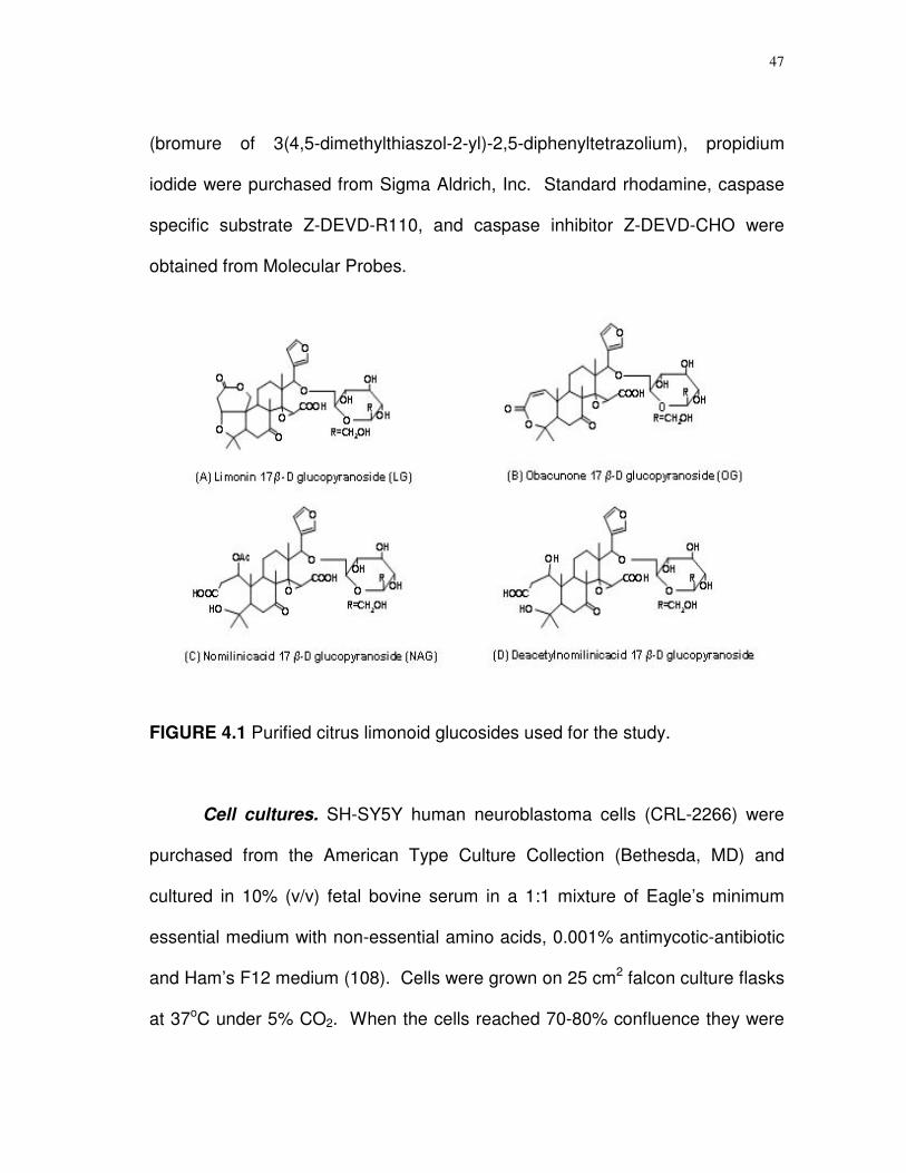
47
(bromure of 3(4,5-dimethylthiaszol-2-yl)-2,5-diphenyltetrazolium), propidium
iodide were purchased from Sigma Aldrich, Inc. Standard rhodamine, caspase
specific substrate Z-DEVD-R110, and caspase inhibitor Z-DEVD-CHO were
obtained from Molecular Probes.
FIGURE 4.1 Purified citrus limonoid glucosides used for the study.
Cell cultures. SH-SY5Y human neuroblastoma cells (CRL-2266) were
purchased from the American Type Culture Collection (Bethesda, MD) and
cultured in 10% (v/v) fetal bovine serum in a 1:1 mixture of Eagle’s minimum
essential medium with non-essential amino acids, 0.001% antimycotic-antibiotic
and Ham’s F12 medium (108). Cells were grown on 25 cm2 falcon culture flasks
at 37oC under 5% CO2. When the cells reached 70-80% confluence they were
48
detached from the flasks with Pucks EDTA solution and quantified in a
hemocytometer. Approximately 15 x 103 cells were subcultured into 12, 24- and
96-well plates for experiments.
Superoxide radical quenching. Assays for O2- radical quenching
followed the procedure described by Marklund and Marklund (48) with the
exception that the assays were performed at pH 7.6, which is closer to biological
pH, and pyrogallol was present at a concentration of 0.2 mmol/L. Stock
solutions of 1 mmol/L pyrogallol were prepared in 0.5 M HCl and diluted to the
desired concentration and pH with 0.2 mol/L Tris-HCl. These adjustments
allowed for a more sensitive measurement of limonoid quenching. Individual
limonoid glucosides were added to a final concentration of 0.1-10 mmol/L and
continuous absorbance readings were taken over 10 min in a Hitachi U2001
recording UV-visible spectrophotometer at wavelengths corresponding to
purpurogallin (325 nm), purpurogalloquinone (600 nm) and the peroxidation
species of purpurogalloquinone (440 nm). Decomposition rates were
determined by the slope of the absorbance curve. Quenching was assessed by
measuring decomposition rate against samples treated with 40 U of superoxide
dismutase (Sigma-Aldrich).
Measurements of cell viability. SH-SY5Y cells in triplicate were
incubated in culture medium supplemented with limonoid glucosides (1-100
µmol/L final concentration) and cell viability was monitored at 12 h intervals over
the next 72 h. Viability determinations were based on trypan blue exclusion as
49
measured in a hemocytometer and the MTT reagent (3[4,5-dimethylthiazol-2-yl]-
2,5-diphenyltetrazolium) as described (109).
Assay of caspase 3/7 activity. Upon reaching confluence, SH-SY5Y
cells were harvested and co-cultured into 24-well culture plates. After 24 h
individual cultures in triplicate were treated with limonoid glucosides. At 12, 24,
36 and 48 h after treatment, the cells were harvested, washed three times with
PBS buffer and collected by centrifugation (5000 x g, 2 min, 4oC) and the cell
pellet was taken up in 200 µL of lysis buffer (0.2 mol/L Tris, pH 7.5, 2 mol/L
NaCl, 0.02 mol/L EDTA, 0.02% (v/v) Triton-X100). Aliquots were analyzed for
protein using the BCA reagent (Pierce) and a lysate volume equivalent to 50 µg
of protein was brought to 100 µL with buffer in a 96-well flat bottom black
microtiter plate. An equal volume of caspase substrate (Z-DEVD-R110) was
added with mixing and the plate was covered with aluminum foil and set aside at
37oC for 30 min. Fluorescence measurements were performed over a 2 h period
in a Perkin-Elmer LS50B luminescence spectrometer, using 499 nm excitation
and 521 nm emission wavelengths. Controls with no limonoid glucosides or with
camptothecin, a strong apoptosis inducer, were run concurrently. Caspase
activity was expressed as pmol/L rhodamine/min/µg protein. Other controls
consisted of limonoid glucosides or camptothecin combined with a known
caspase 3/7 inhibitor AC-DEVD-CHO.
Flow cytometry analysis. The cells were treated with limonoid
glucosides or camptothecin for 12, 24, 36, and 48 h and harvested as before.
50
Between 106 to 107 cells in PBS were centrifuged (200 x g, 6 min, 25oC), and the
cell pellet was taken up in 50 mL of PBS, 450 mL of 70% ethanol, and stored at -
20oC until used. Cells were separated from the ethanol by centrifugation at 200
x g for 5 min, decanting the ethanol, and suspended in 1 mL of 0.1% (v/v) Triton
X-100 in PBS, to which was added 200 µg of DNase-free RNase A and 20 µL of
propidium iodide. After 15 min incubation at 37oC to destroy RNA, the
suspension was filtered through nylon and collected in tubes that fit a Beckton
Dickenson FACS Caliber flow cytometer. DNA fluorescence readings were
taken with an excitation blue light set and 488 nm and a detector emission of red
fluorescence through a 585 nm filter. Pulse width area signals were used to
discriminate between G2 cells and cell doublets. The data were analyzed using
ModFitLT software version 3.1 (PMac).
Statistical analysis. Experimental results represent triplicate
experiments for each treatment group and are expressed as means ± standard
error. A two-way ANOVA was used with a P-value of less than 0.05 (Fisher’s
least significant difference test) considered to indicate significance and Dunnet’s
paired comparison. Analyses were done with SPSS statistical analysis software,
(Version 11.0). The coefficient of variation in the DNA analysis was less than
6%. Reduced chi square goodness of fit was assumed to be 0.8 to 3.0. IC50
values for the inhibition concentration for individual compounds were calculated
after Stahle et al. (100), using a linear regression equation.
51
4.4 Results
Superoxide quenching activity. The potential for the purified limonoid
glucosides to quench free radicals was ascertained using pyrogallol in slightly
alkaline (pH 7.6) solutions. Quenching superoxide anion (O2-) effectively stops
the spontaneous decomposition of the pyrogallol, which can be measured
spectrophotometrically at wavelengths corresponding to the oxidized products of
pyrogallol. Assays were run with limonoid glucoside concentrations varying
between 0.1 and 10 mmol/L. Typically, a short lag phase at the start was used
to measure quenching activity (Fig. 4.2). As shown in Figure 4.2A, at 5 mmol/L,
clearly discernible differences in the quenching efficacies of the different
limonoid glucosides were observed. NAG was superior to LG, OG, and DNAG
over the 5 min assay time (Fig. 4.2A). At 5 mmol/L NAG had nearly the same
quenching efficacy as L-ascorbic acid and superoxide dismutase. Testing NAG
alone showed that strong and sustained inhibition, which was not seen until the
concentration was above 1 mmol/L (Fig. 4.2B). Neither change in the pH nor
pyrogallol concentration affected the profiles for the different limonoid
glucosides. The rate of inhibition of purpurogallin formation by NAG was found
to be diminishing after three minutes of the start of the reaction indicating a rapid
turnover of the compound and unquenchable spontaneous auto-oxidation of
pyrogallol (Fig. 4.2C). The acidic glucosides did not effect any noticeable
change in pH on the buffer medium, at the levels tested.
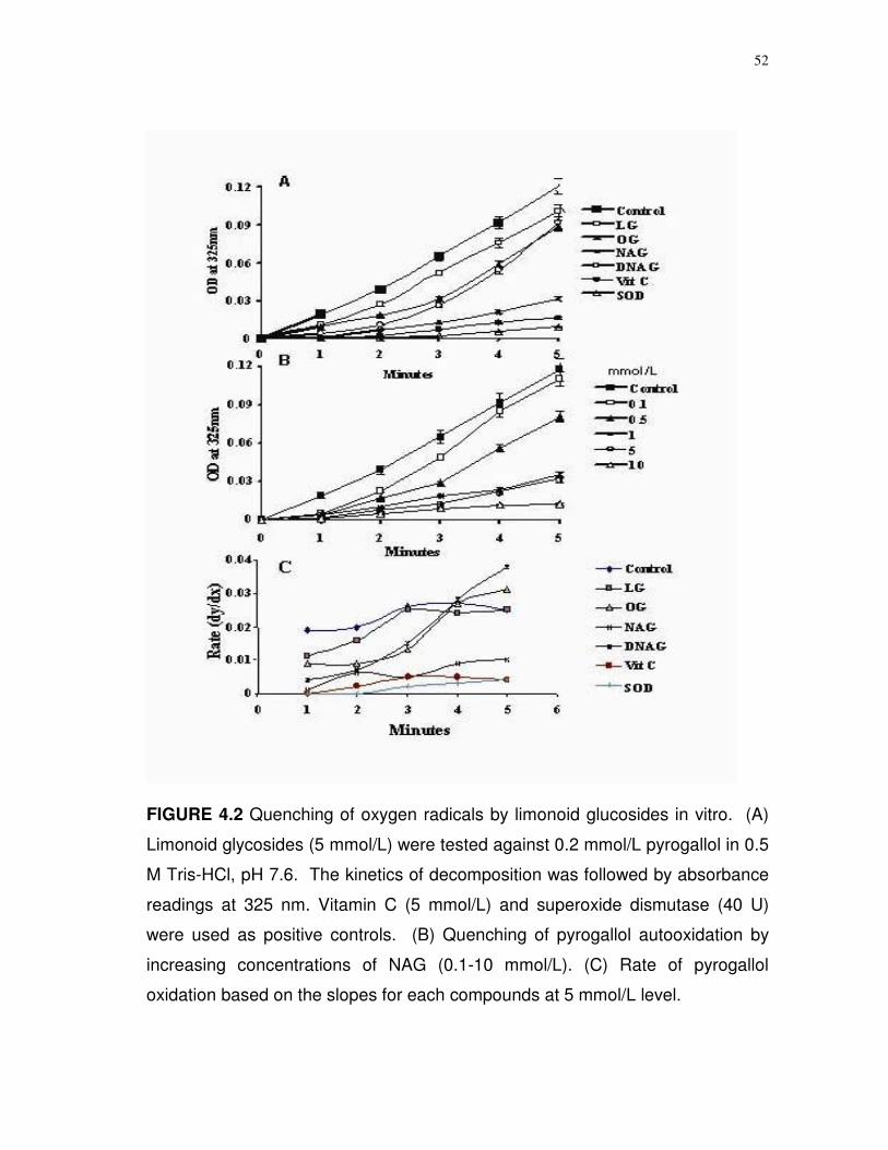
52
FIGURE 4.2 Quenching of oxygen radicals by limonoid glucosides in vitro. (A)
Limonoid glycosides (5 mmol/L) were tested against 0.2 mmol/L pyrogallol in 0.5
M Tris-HCl, pH 7.6. The kinetics of decomposition was followed by absorbance
readings at 325 nm. Vitamin C (5 mmol/L) and superoxide dismutase (40 U)
were used as positive controls. (B) Quenching of pyrogallol autooxidation by
increasing concentrations of NAG (0.1-10 mmol/L). (C) Rate of pyrogallol
oxidation based on the slopes for each compounds at 5 mmol/L level.
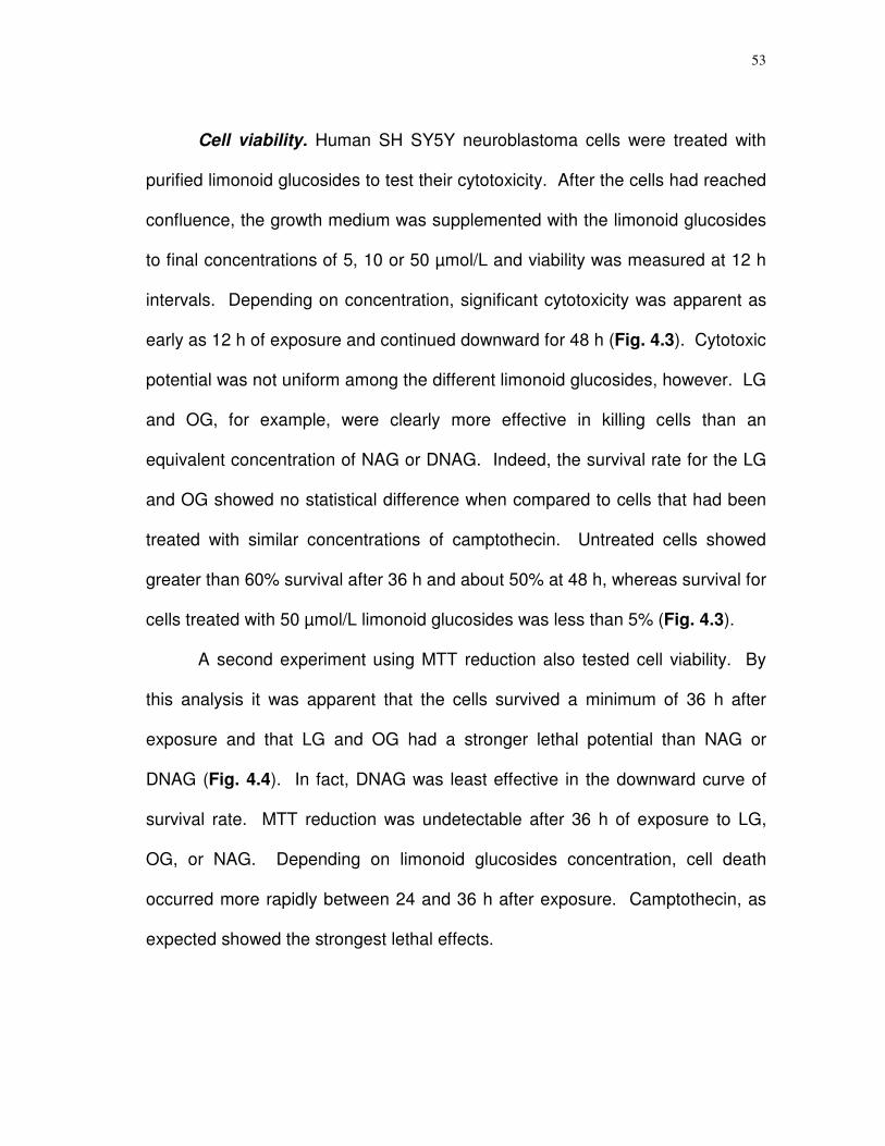
53
Cell viability. Human SH SY5Y neuroblastoma cells were treated with
purified limonoid glucosides to test their cytotoxicity. After the cells had reached
confluence, the growth medium was supplemented with the limonoid glucosides
to final concentrations of 5, 10 or 50 µmol/L and viability was measured at 12 h
intervals. Depending on concentration, significant cytotoxicity was apparent as
early as 12 h of exposure and continued downward for 48 h (Fig. 4.3). Cytotoxic
potential was not uniform among the different limonoid glucosides, however. LG
and OG, for example, were clearly more effective in killing cells than an
equivalent concentration of NAG or DNAG. Indeed, the survival rate for the LG
and OG showed no statistical difference when compared to cells that had been
treated with similar concentrations of camptothecin. Untreated cells showed
greater than 60% survival after 36 h and about 50% at 48 h, whereas survival for
cells treated with 50 µmol/L limonoid glucosides was less than 5% (Fig. 4.3).
A second experiment using MTT reduction also tested cell viability. By
this analysis it was apparent that the cells survived a minimum of 36 h after
exposure and that LG and OG had a stronger lethal potential than NAG or
DNAG (Fig. 4.4). In fact, DNAG was least effective in the downward curve of
survival rate. MTT reduction was undetectable after 36 h of exposure to LG,
OG, or NAG. Depending on limonoid glucosides concentration, cell death
occurred more rapidly between 24 and 36 h after exposure. Camptothecin, as
expected showed the strongest lethal effects.
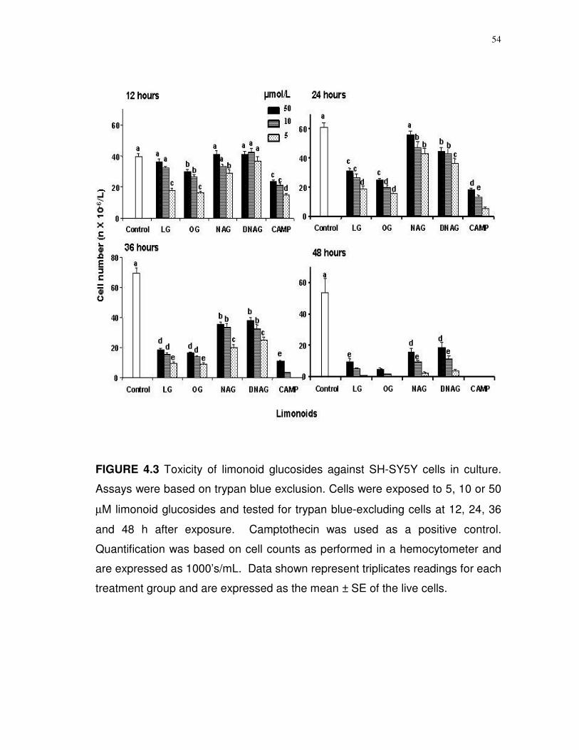
54
FIGURE 4.3 Toxicity of limonoid glucosides against SH-SY5Y cells in culture.
Assays were based on trypan blue exclusion. Cells were exposed to 5, 10 or 50
µM limonoid glucosides and tested for trypan blue-excluding cells at 12, 24, 36
and 48 h after exposure. Camptothecin was used as a positive control.
Quantification was based on cell counts as performed in a hemocytometer and
are expressed as 1000’s/mL. Data shown represent triplicates readings for each
treatment group and are expressed as the mean ± SE of the live cells.
55
FIGURE 4.4 Cell viability analysis based on MTT reduction, measured by the
formation of formazon at 550 nm. The values shown represent mean ± SE for
n=3. Survival percentage was determined using control (untreated) cells as a
norm. Camptothecin was used as a positive control.

56
A striking observation, however, was seen in cells that had been treated
with LG and Ac-DVED-CHO in combination. Cell death was suppressed for at
least 48 h, even with the concentration of LG, the most lethal limonoid glucoside,
as high as 50 µmol/L (Fig. 4.5). Since Ac-DVED-CHO is a potent inhibitor of
caspase activity, there was a clear indication that toxic action of limonoid
glucosides could be exerted through the induction of apoptosis.
Test of apoptosis. It should be possible to observe apoptotic changes in
the cells, if apoptosis induction is responsible for the decline in cell viability by
limonoid glucosides. Therefore, we tested for caspase 3/7 induction and DNA
fragmentation. In the caspase induction assay, the cells were treated with the
limonoid glucosides for 12 h, harvested, washed with PBS and then lysed.
Aliquots matched for their protein content were assayed for caspase 3/7 activity
in the presence and absence of a caspase inhibitor. Camptothecin was used as
a positive control. All of the limonoid glucosides at a set concentration (10
µmol/L) induced caspase 3/7 activity (Fig. 4.6A). Similar to the lethal potential
on cell viability, there was a clear difference among the different limonoid
glucosides. Though the basal caspase activity changed very little over the 24 h
experimental period, the caspase activity appeared to peak at 12 h and declined
thereafter. Although all limonoid glucosides were less effective than
camptothecin, LG and OG were superior to NAG and DNAG in their caspase
inducing efficacy.
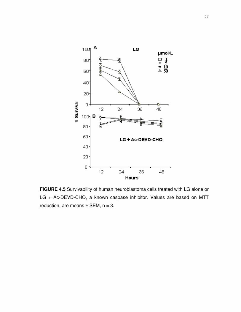
57
FIGURE 4.5 Survivability of human neuroblastoma cells treated with LG alone or
LG + Ac-DEVD-CHO, a known caspase inhibitor. Values are based on MTT
reduction, are means ± SEM, n = 3.

58
FIGURE 4.6 Induction of caspase 3/7 activity by limonoid glucosides. The cells
were exposed to 10 µmol/L limonoid glucosides for 6, 12 or 24 h. (A) Kinetics
of caspase induction for each of the different limonoid glucosides. Controls
received no limonoids. (B) The induction of caspase 3/7 activity at 12 h as a
function of different concentration of limonoids. Camptothecin (CAMP) was
used as a positive control. Values are mean ± SD, n = 3. * Significantly different
from control at P < 0.001. The activity is expressed as picomoles of
rhodamine110 formed per µg of cell lysate protein per minute.
59
Figure 4.6B shows that caspase induction was a function of the
concentration of limonoid glucosides in the medium. As little as 1 µmol/L
brought about a noticeable increment over basal (control) caspase activity; 5
µmol/L brought about a significant difference (P < 0.05). Importantly, those
limonoid glucosides that were the most effective suppressors of cell growth, viz.,
OG and LG, were the strongest inducers of caspase 3/7 activity. The 12 h
caspase activity had a strong correlation (r = -0.998, P = 0.002) to the cell
survival. The data, therefore, confirmed the earlier study, which suggested that
the cytotoxic effect of limonoid glucosides was manifested through apoptotic
induction mechanism.
Flow cytometry. As a further confirmation of apoptosis induction by the
limonoid glucosides, a flow cytometry analysis was performed. Cells were
exposed to 10 µmol/L limonoid glucosides for 24 h, harvested, fixed in 70%
ethanol, and treated with propidium iodide before the analysis. Aggregates were
removed by filtration through nylon. Figure 4.7 shows that treated and control
cells were mostly in the G0 and G2M phases with less numbers of cells in the S
phase subpopulation. The position marked A shows univariate DNA content, a
marker of DNA degradation, was significantly higher for cells treated with LG,
OG and camptothecin. Background aggregates and debris (BAD) between G1
and G2 phase, an indicator of apoptosis, was determined to be 35.4% for
camptothecin, 31.3% for LG and 30.9% for OG, which was significantly higher (P
< 0.05) compared to control (8.6%), NAG (17.5%) and DNAG (11.1%). There
60
was no significant reduction in cell numbers at the G0/G1 phase between all
treatment groups, but OG, LG and camptothecin significantly reduced the
number of S phase cells, which essentially blocks the crucial proliferation phase.
DNA content at S phase of the cell cycle indicated a reduction of 86.67% with
LG, 82.36% with OG, 57.46% with NAG and 38.35% with DNAG, as compared
to the known inhibitor like camptothecin. OG also significantly suppressed the
G2/M phase. Overall, the results provided evidence that limonoid glucosides act
as antiproliferative agents through arresting critical growth stages of the cell
cycle and inducing apoptosis.
DNA fragmentation. The potential for limonoid glucosides to induce DNA
breakage in sequel to apoptosis induction was determined through agarose gel
electrophoresis. In this assay DNA was extracted from control and treated cells,
precipitated with ethanol, and chromatographed on 1% (w/v) agarose. An equal
amount of cellular DNA was applied to each lane of an electrophoresis gel.
Figure 4.8 shows that untreated cells had high molecular weight DNA that was
virtually intact as shown by the heavy staining of slow moving components,
whereas cells treated with LG, OG, or camptothecin showed a spectrum of
fragments that were present in a familiar ladder pattern typical of endonuclease
cleaved DNA. Fragmentation of DNA was less evident in cells treated with NAG
and DNAG. These data support the flow cytometry results and provide direct
evidence for DNA degradation of neuroblastoma cells as a consequence of
limonoid treatment.
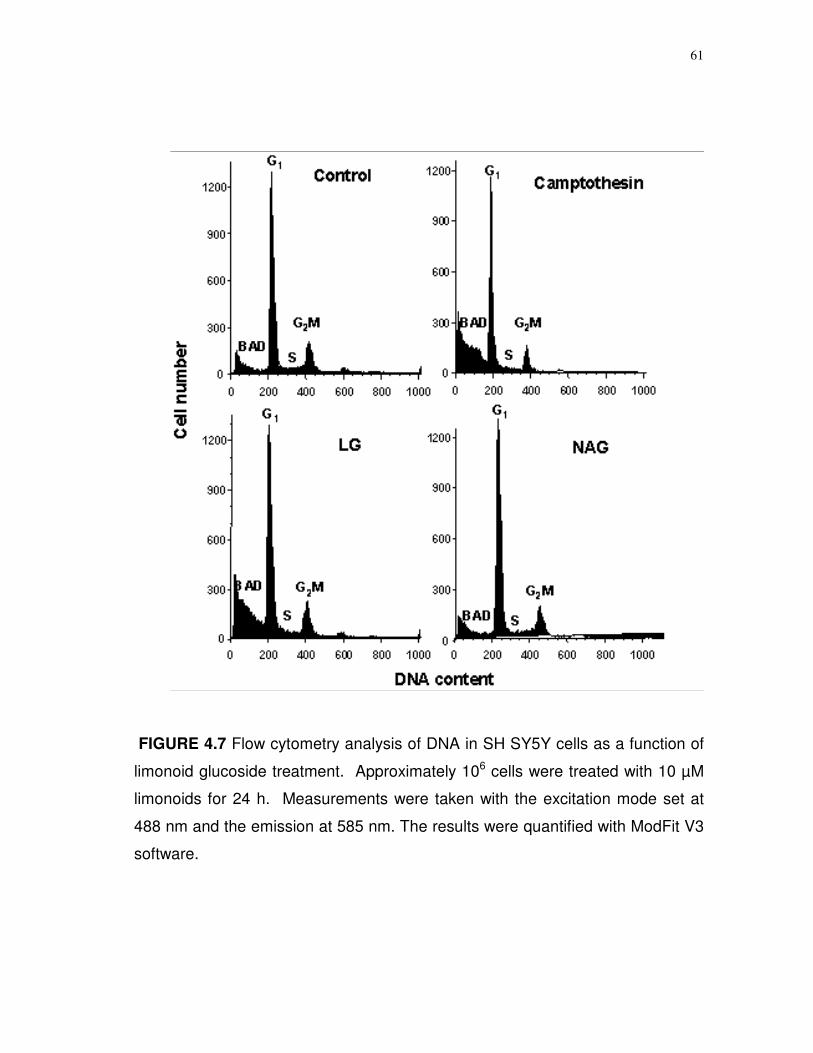
61
FIGURE 4.7 Flow cytometry analysis of DNA in SH SY5Y cells as a function of
limonoid glucoside treatment. Approximately 106 cells were treated with 10 µM
limonoids for 24 h. Measurements were taken with the excitation mode set at
488 nm and the emission at 585 nm. The results were quantified with ModFit V3
software.

62
FIGURE 4.8 DNA fragmentation analysis of SH-SY5Y cells after limonoid
treatment. Approximately 106 cells were treated with 10 or 50 µmol/L limonoid
glucosides for 24 h. An equal amount of DNA from each treatment group was
applied to a 1% agarose gel. Lane marked MR shows 100 bp to 1000 bp
molecular weight ladder.
Cont LG OG NAG DNAG CAMP MR 10 50 10 50 10 50 10 50 10 50
63
4.4 Discussion
Many epidemiological and controlled diet studies have reported that high
intake of fruits and vegetables lower the risk of numerous degenerative diseases
including cancer (110). Although many bioactive compounds from citrus fruits
were implicated, the potential of limonoid glucosides is believed to dwell in
antioxidant and apoptosis inducing properties.
Two important facets of limonoid glucosides action, such as, an ability to
quench O2- radicals and to induce apoptotic changes in neuroblastoma cells
were studied here. Quenching, as detected by an in vitro spectrophotometric
assay, was evident only when relatively high (mmol/L) levels of the limonoid
glucosides were present, which may limit chemopreventative significance.
Furthermore, quenching appeared to occur only in the early stages of the
reaction and, depending on concentration, did not endure beyond the first two or
three minutes. The fact that quenching did not persist suggests the limonoid
glucosides act stoichiometrically with O2- and, once modified, are unable to
regenerate active forms. Because of the high concentrations needed and the
failure to recycle to active forms weakens the suggestion that limonoid
glucosides are free radical scavengers in situ.
The limonoid glucosides tested in this study caused rapid death to
undifferentiated neuroblastoma cells in culture. Lethality occurred at micromolar
concentrations, which suggests neuroblastoma cells are highly sensitive to their
action. Tian et al. (31) showed that the obacunone glucoside was superior to

64
nomilinic acid glucoside in causing the death of MCF-7 breast cancer cells in
culture. Moreover, the aglycones of NG and LG were as effective as the
glucosides in killing cells, suggesting the glucoside appendage on the molecule
is not important for toxic action. The previous study with some limonoid
glucosides, however, ruled out apoptosis as a cause because of the relatively
high amounts (100 µg/L) needed to induce an apoptotic response (31). In the
present study, it has been demonstrated that limonoid glucosides as low as 1-5
µmol/L (0.6-3.0 µg/L) induced caspase activity and fragmented DNA in SH-
SY5Y cells. Why MCF-7 cells fail to show a similar heightened sensitivity
against limonoid glucosides is not clear. Apoptosis can be induced in SH-SY5Y
cells by a number of external factors and conditions such as tumor necrosis
factor- (111), amyloid--peptides (112), UV radiation (113) and ceramide (114).
In contrast, retinoic acid, PGE-2, leptin, guanosine, interleukin (IL)-1 and IL-6
appear to protect SH-SY5Y cells against oxidants and neurotoxins (115-118), as
do flavones such as lutoelin, apigenin, and epigallocatechin (119-122). The
data suggest that limonoid glucosides are more apt to fit the category of toxic
agents as opposed to protective agents, and unlike the flavones offer little or no
protection against oxidants and neurotoxins in situ. This hypothesis, however, is
tempered by the observation that cancer cell lines HL-60, NCI-SNU-1, HeLa,
SCOV-3, and HepG2 are unaffected by limonoid glucosides (31), which implies
lethal action may be selective and dependent on cell type or differentiation state.
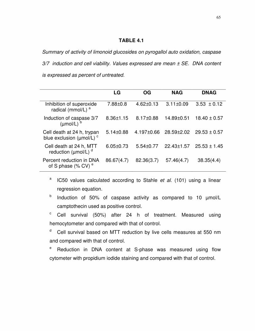
65
TABLE 4.1
Summary of activity of limonoid glucosides on pyrogallol auto oxidation, caspase
3/7 induction and cell viability. Values expressed are mean ± SE. DNA content
is expressed as percent of untreated.
LG OG NAG DNAG
Inhibition of superoxide radical (mmol/L) a
7.88±0.8 4.62±0.13 3.11±0.09 3.53 ± 0.12
Induction of caspase 3/7 (µmol/L) b
8.36±1.15 8.17±0.88 14.89±0.51 18.40 ± 0.57
Cell death at 24 h, trypan blue exclusion (µmol/L) c
5.14±0.88 4.197±0.66 28.59±2.02 29.53 ± 0.57
Cell death at 24 h, MTT reduction (µmol/L) d
6.05±0.73 5.54±0.77 22.43±1.57 25.53 ± 1.45
Percent reduction in DNA of S phase (% CV) e
86.67(4.7) 82.36(3.7) 57.46(4.7) 38.35(4.4)
a IC50 values calculated according to Stahle et al. (101) using a linear
regression equation. b Induction of 50% of caspase activity as compared to 10 µmol/L
camptothecin used as positive control. c Cell survival (50%) after 24 h of treatment. Measured using
hemocytometer and compared with that of control. d Cell survival based on MTT reduction by live cells measures at 550 nm
and compared with that of control. e Reduction in DNA content at S-phase was measured using flow
cytometer with propidium iodide staining and compared with that of control.
66
It is important to note the difference in the efficacy by different limonoid
glucosides. It is clear that LG and OG induced cell death at one-fourth the
concentrations of NAG and DNAG (Table 4.1). Caspase induction and DNA
fragmentation was also achieved at lower concentrations of LG and OG than
NAG and DNAG. Quenching, however, favored NAG. A careful inspection of
the structures (Fig. 4.1) shows that NG and LG share the same structural
features as NAG and DNAG with the exception of a sealed A ring in the OG and
LG molecules. Miller et al. (23) earlier had speculated that the A ring as
opposed to the D, is more decisive in its biological activity of limonoid
glucosides. Our data strongly support that by showing greater apoptosis-
inducing potential, that may also be associated with the A ring of the molecule.
Future research endeavors can now turn to the mechanism of apoptosis
induction to explain the underlying mechanism of the limonoid glucosides on SH-
SY5Y cells. Calcium, acting through the activation of two Ca2+-dependent
proteases, mu-calpain, and caspase 12, has been strongly implicated in
induction of apoptosis and the regulation of the apoptotic signaling pathway
(123). Mitochondria regulate apoptosis in vertebrates by releasing cytochrome
c, an inducer of caspase 3, and Bcl-2 regulates this release (124). Activation of
the JNK signaling pathway, an early event in the induction of apoptosis in
neuronal cells (111), leads to the phosphorylation of c-Jun and Bcl-2, two factors
that control apoptosis. What effect limonoid glucosides may have on these
internal events is ripe for further investigation. One more consideration is the
67
synergistic action by which one limonoid promotes the action of another.
Mertens-Talcott (125), showed that ellagic acid significantly potentiated the
action of quercetin in stimulating caspase activity in cultured MOLT-4 human
leukemia cells. A combination of the two fruit polyphenols gave robust
anticarcinogenic effects than either one alone. This observation suggests
limonoid glucosides could act in concert with flavones to promote an action. In
nutrition synergistic health promoting effects achieved by whole fruit
consumption, rather than individual molecules.
In conclusion, the data in this study strongly support limonoid glucosides
at micromolar concentrations as being toxic to cultured neuroblastoma. Because
limonoid glucosides are not lethal to all cancer cells (31), they may target
specific cells types. Consequently, it is vital to recognize the vulnerable site(s) in
cells that underlie their mechanisms of action.
68
CHAPTER V
EFFECT OF LIMONOIDS ON COLONIC ADENOCARCINOMA (Caco-2),
NEUROBLASTOMA AND NON-CANCEROUS OVARY (CHO) CELLS
5.1 Synopsis
Purified limonin, limonin glucoside, obacunone, obacunone glucoside,
nomilin, and deacetylnomilin along with mixtures of limonoid aglycones and
glucosides were tested for toxic effects on human SH-SY5Y neuroblastoma and
colon carcinoma (Caco-2) cell lines and were compared with the mammalian
non-cancerous Chinese hamster ovary (CHO) cells. Limonoid glucosides have
been shown to have greater cytotoxic effects than aglycones and that the effects
were specific to cancerous cells. The effects were significant (P < 0.001) over 24
to 36 h when compared with the untreated cells. Flow cytometric analyses of
limonoid treated neuroblastoma cells showed a marked increase in DNA content
of aneuploidic cells within 24 h, which are considered to be a prognostic factor in
neuroblastoma. The percentage of aneuploidic cells following treatment were
36% for LG, 26% for limonin and more than 42% for limonoid glucoside
mixtures. The results confirm that both limonoid aglycones and glycosides are
lethal to cancer cell lines; however, glycosides are the most active apoptotic
inducing form, even though both forms arrest cell growth.
69
5.2 Introduction
Secondary metabolites have often been proven to be beneficial to human
health. More than 120 important prescription drugs in the U.S. are derived from
secondary plant metabolites which comprise about 25% of the total drugs in use
(126). With innumerable chemopreventive properties, these phytochemicals
have been divided into either cancer-blocking agents or cancer-suppressing
agents (92). The blocking agents work through scavenging the carcinogens by
CYP/GST mediated metabolism and detoxification, quenching ROS, and
blocking the cellular targets. The suppressing agents restrain the proliferation
and progression of the cancer by activating several transcription factors),
inducing apoptosis, arresting the cell cycle (3-19, 92).
Statistics from American Cancer Society show an estimated 1,372,910
new cases and a total of 570,280 deaths by cancer in U.S. in 2005 (33). Of
these, about 145,000 new cases and about 56,000 deaths will occur due to
colorectal cancer. Neuroblastomas account for about 10% childhood cancer with
an annual incidence of 6-8 per million (50,127). Furthermore, the overall costs of
cancer for the year of 2005 is estimated to be $189.8 billion with $69.4 billion in
direct medical expenditures and $103.5 billion in indirect mortality costs, which
include the costs of lost productivity due to premature death (33). An estimate by
the National Cancer Institute indicates that one in four cancer deaths are diet
related and that eight of ten cancer cases have a nutrition/diet component (33).
70
National Cancer Institute designer programs identified citrus fruits as
especially nutrient rich foods containing more than a dozen classes of beneficial
phytochemicals (4,5). Chemopreventive properties of citrus limonoids have been
discussed in detail in the previous chapters. Epidemiological, cell culture and
animal studies have shown that citrus limonoids can prevent several types of
cancers (15-31). Though the antiproliferative effects of citrus limonoids are found
to be mediated through inhibition of cytochrome P450, antioxidant activity and
apoptotic induction in earlier experiments, their effects on normal cells and other
cancer cell lines have yet to be ascertained. Strategies to offset the
malignancies of colorectal cancer and neuroblastomas are vital. This study
report the cytotoxic and apoptotic effects of the purified limonoids on human SH-
SY5Y neuroblastoma (CRL-2266), colonic adenocarcinoma (Caco-2) and
assessed the effects with a non-cancerous Chinese hamster ovary cells (CHO).
5.3 Materials and methods
Cell cultures. Human SH-SY5Y neuroblastoma cells (CRL-2266), colonic
adenocarcinoma cells (Caco-2), and Chinese hamster ovary cells (CHO) were
purchased from the American Type Culture Collection (Bethesda, MD). SH-
SY5Y cells were cultured in 10% (v/v) fetal bovine serum in a 1:1 mixture of
Eagle’s minimum essential medium with non-essential amino acids, 0.001%
antimycotic-antibiotic and Ham’s F12 medium as previously described (108).
HepG2 and Caco-2 cells were grown in 10% (v/v) and 20% (v/v) fetal bovine
71
serum in Eagle’s minimum essential medium with 2 mmol/L L-glutamine and
Earle’s balanced salt solutions (BSS) adjusted to contain 17.85 mmol/L sodium
bicarbonate, 0.1 mmol/L non-essential amino acids, and 1 mmol/L sodium
pyruvate. CHO cells were cultured in 10% (v/v) fetal bovine serum Ham’s F12K
medium with 2 mmol/L L- glutamine adjusted to contain 17.85 mmol/L sodium
bicarbonate. Cells were grown on 25 cm3 Falcon culture flasks at 37oC under
5% CO2. When the cells reached 70-80% confluence they were detached from
the flasks using either Pucks EDTA (SH-SY5Y, CHO) or trypsin EDTA (CacO-2)
and quantified in a hemocytometer. Approximately 104 cells/mL (1000/well)
were sub-cultured into 96-well culture plates and incubated for 24 h (SH-SY5Y,
CHO) and 48 h (Caco-2), to attain 70% confluence before the treatments. Fresh
medium was added to each well at the beginning of the treatments. Stock
solutions of aglycones were prepared in DMSO and glucosides in water.
Cell viability. The cells in triplicate were incubated in culture medium
supplemented with limonoid glucosides (5-50 µmol/L final concentration) and
cell viability was monitored at 12 h intervals over the next 48 h. Care was taken
to keep the DMSO levels below 0.5% which had no apparent effect on cell
growth. Viability determinations were based on the reduction of MTT reagent
(3[4,5-dimethylthiazol-2-yl]-2,5-diphenyltetrazolium) by active mitochondria as
described (109). The morphological differences between limonoid treated and
untreated cancer cell lines were ascertained using a confocal microscopy using
a green fluorescent filter.
72
Flow cytometry analysis. Cells were treated with limonoids for 36 h and
harvested as before. Between 106 to 107 cells in PBS were centrifuged (200 x g,
6 min, 25oC), washed 3 times with PBS and the cell pellet was resuspended in
500 µL of PBS to which added 0.1% (v/v) Triton X-100 in PBS, 200 µg/mL of
DNase-free RNase A and 50 µL/mL of propidium iodide in 4 mmol/L sodium
citrate buffer. After a 15 min incubation at 37oC to destroy RNA, the suspension
was filtered through nylon and collected in separate tubes that fit a FACS
Caliber flow cytometer (Beckton Dickenson, San Jose, CA USA), equipped with
a 15 mW argon ion laser. DNA fluorescence readings were taken with an
excitation of 488 nm and emission with a 585 nm filter, with a doublet
discrimination module to differentiate G2 cells from cell doublets using pulse
width area. The existence of DNA aneuploidy was described as the existence of
two Go/G1 peaks in the DNA histogram as described by Abad et al. (128).
Statistical analysis. Experimental results for cell viability represent
triplicate for each treatment group and are expressed as means ± standard error
(percent). A one-way ANOVA was used with a P-value of less than 0.001 with
Fisher’s least significant difference test for inter group comparisons. Analyses
were done with SPSS statistical analysis software (Version 11.0). Distribution
analysis of Go/M, S and G2/M phases of the cell cycle were performed on
propidium iodide stained gated cells for the exclusion of doublets using
ModFitLT software (Version 3.1, PMac, Beckton Dickenson).

73
5.4 Results
Effect of limonoids on the viability of SH-SY5Y, Caco-2 and CHO cells.
In accordance with the previous report on the effect of limonoid glucosides on
the induction of apoptosis in human SH-SY5Y neuroblastoma cell lines (127), a
slow but similar effect for the limonoid aglycones was observed. The
morphological differences observed using confocal microscopy clearly indicated
a rapid response for the limonin 17-D glucopyranoside (LG) as opposed to that
of limonin and obacunone aglycones (Fig. 5.1). The effects were consistent with
both time and dose dependent assays (Table 5.1). While limonoid mixtures had
definitive effects on the viability of both Caco-2 and SH-SY5Y cell lines, the
response of both pure as well as mixtures was selective towards cancerous cell
lines (Fig. 5.2). Neuroblastoma cell lines were found to be more sensitive than
the colon carcinoma cells for nomilin (Fig. 5.2). Although all of the pure
compounds seemed to have similar levels of cytotoxicity on SH-SY5Y cells the
relative effects appeared to be strong for obacunone followed by limonin,
nomilin, and deacetylnomilin (Table 5.1). There were no significant differences
(P < 0.001) between limonin and obacunone. Limonin, nomilin, and obacunone
had same cytotoxicity towards Caco-2 and SH-SY5Y cell lines at 48 h.
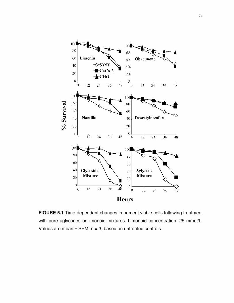
74
FIGURE 5.1 Time-dependent changes in percent viable cells following treatment
with pure aglycones or limonoid mixtures. Limonoid concentration, 25 mmol/L.
Values are mean ± SEM, n = 3, based on untreated controls.
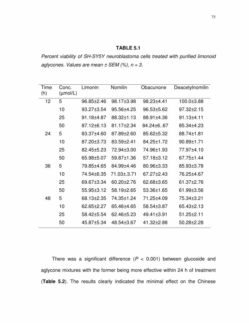
75
TABLE 5.1
Percent viability of SH-SY5Y neuroblastoma cells treated with purified limonoid
aglycones. Values are mean ± SEM (%), n = 3.
There was a significant difference (P < 0.001) between glucoside and
aglycone mixtures with the former being more effective within 24 h of treatment
(Table 5.2). The results clearly indicated the minimal effect on the Chinese
Time (h)
Conc. (µmol/L)
Limonin Nomilin Obacunone Deacetylnomilin
12 5 96.85±2.46 98.17±3.98 98.23±4.41 100.0±3.88
10 93.27±3.54 95.56±4.25 96.53±5.62 97.32±2.15
25 91.18±4.87 88.32±1.13 88.91±4.36 91.13±4.11
50 87.12±6.13 81.17±2.34 84.24±6..67 85.34±4.23
24 5 83.37±4.60 87.89±2.60 85.62±5.32 88.74±1.81
10 87.20±3.73 83.59±2.41 84.25±1.72 90.89±1.71
25 82.45±5.23 72.94±3.00 74.96±1.93 77.97±4.10
50 65.98±5.07 59.87±1.36 57.18±3.12 67.75±1.44
36 5 79.85±4.65 84.99±4.46 80.96±3.33 85.93±3.78
10 74.54±6.35 71.03±.3.71 67.27±2.43 76.25±4.67
25 69.67±3.34 60.20±2.76 62.68±3.65 61.37±2.76
50 55.95±3.12 58.19±2.65 53.36±1.65 61.99±3.56
48 5 68.13±2.35 74.35±1.24 71.25±4.09 75.34±3.21
10 62.65±2.27 65.46±4.65 58.54±3.87 65.43±2.13
25 58.42±5.54 62.46±5.23 49.41±3.91 51.25±2.11
50 45.87±5.34 48.54±3.67 41.32±2.88 50.28±2.28
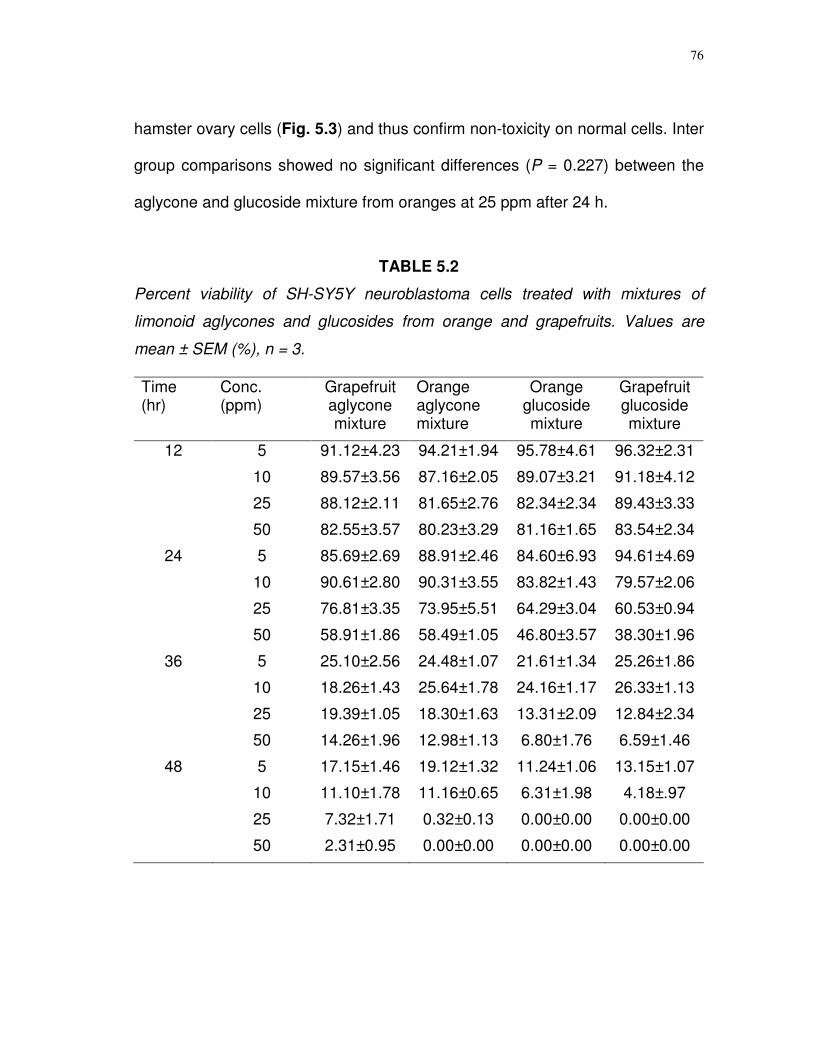
76
hamster ovary cells (Fig. 5.3) and thus confirm non-toxicity on normal cells. Inter
group comparisons showed no significant differences (P = 0.227) between the
aglycone and glucoside mixture from oranges at 25 ppm after 24 h.
TABLE 5.2
Percent viability of SH-SY5Y neuroblastoma cells treated with mixtures of
limonoid aglycones and glucosides from orange and grapefruits. Values are
mean ± SEM (%), n = 3.
Time (hr)
Conc. (ppm)
Grapefruit aglycone mixture
Orange aglycone mixture
Orange glucoside mixture
Grapefruit glucoside mixture
12 5 91.12±4.23 94.21±1.94 95.78±4.61 96.32±2.31
10 89.57±3.56 87.16±2.05 89.07±3.21 91.18±4.12
25 88.12±2.11 81.65±2.76 82.34±2.34 89.43±3.33
50 82.55±3.57 80.23±3.29 81.16±1.65 83.54±2.34
24 5 85.69±2.69 88.91±2.46 84.60±6.93 94.61±4.69
10 90.61±2.80 90.31±3.55 83.82±1.43 79.57±2.06
25 76.81±3.35 73.95±5.51 64.29±3.04 60.53±0.94
50 58.91±1.86 58.49±1.05 46.80±3.57 38.30±1.96
36 5 25.10±2.56 24.48±1.07 21.61±1.34 25.26±1.86
10 18.26±1.43 25.64±1.78 24.16±1.17 26.33±1.13
25 19.39±1.05 18.30±1.63 13.31±2.09 12.84±2.34
50 14.26±1.96 12.98±1.13 6.80±1.76 6.59±1.46
48 5 17.15±1.46 19.12±1.32 11.24±1.06 13.15±1.07
10 11.10±1.78 11.16±0.65 6.31±1.98 4.18±.97
25 7.32±1.71 0.32±0.13 0.00±0.00 0.00±0.00
50 2.31±0.95 0.00±0.00 0.00±0.00 0.00±0.00

77
FIGURE 5.2 Confocal images of SH-SY5Y neuroblastoma cells treated with
limonoid glucosides and aglycones, from Figure 5.1. Panels A and B untreated
cells at 0 and 48 h. C and D, 25 µmol/L LG for 24 and 48 h; E and F, 25 µmol/L
obacunone.
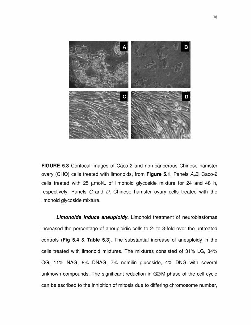
78
FIGURE 5.3 Confocal images of Caco-2 and non-cancerous Chinese hamster
ovary (CHO) cells treated with limonoids, from Figure 5.1. Panels A,B, Caco-2
cells treated with 25 µmol/L of limonoid glycoside mixture for 24 and 48 h,
respectively. Panels C and D, Chinese hamster ovary cells treated with the
limonoid glycoside mixture.
Limonoids induce aneuploidy. Limonoid treatment of neuroblastomas
increased the percentage of aneuploidic cells to 2- to 3-fold over the untreated
controls (Fig 5.4 & Table 5.3). The substantial increase of aneuploidy in the
cells treated with limonoid mixtures. The mixtures consisted of 31% LG, 34%
OG, 11% NAG, 8% DNAG, 7% nomilin glucoside, 4% DNG with several
unknown compounds. The significant reduction in G2/M phase of the cell cycle
can be ascribed to the inhibition of mitosis due to differing chromosome number,
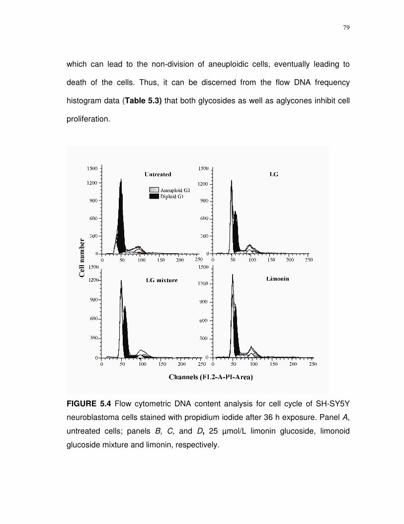
79
which can lead to the non-division of aneuploidic cells, eventually leading to
death of the cells. Thus, it can be discerned from the flow DNA frequency
histogram data (Table 5.3) that both glycosides as well as aglycones inhibit cell
proliferation.
FIGURE 5.4 Flow cytometric DNA content analysis for cell cycle of SH-SY5Y
neuroblastoma cells stained with propidium iodide after 36 h exposure. Panel A,
untreated cells; panels B, C, and D, 25 µmol/L limonin glucoside, limonoid
glucoside mixture and limonin, respectively.
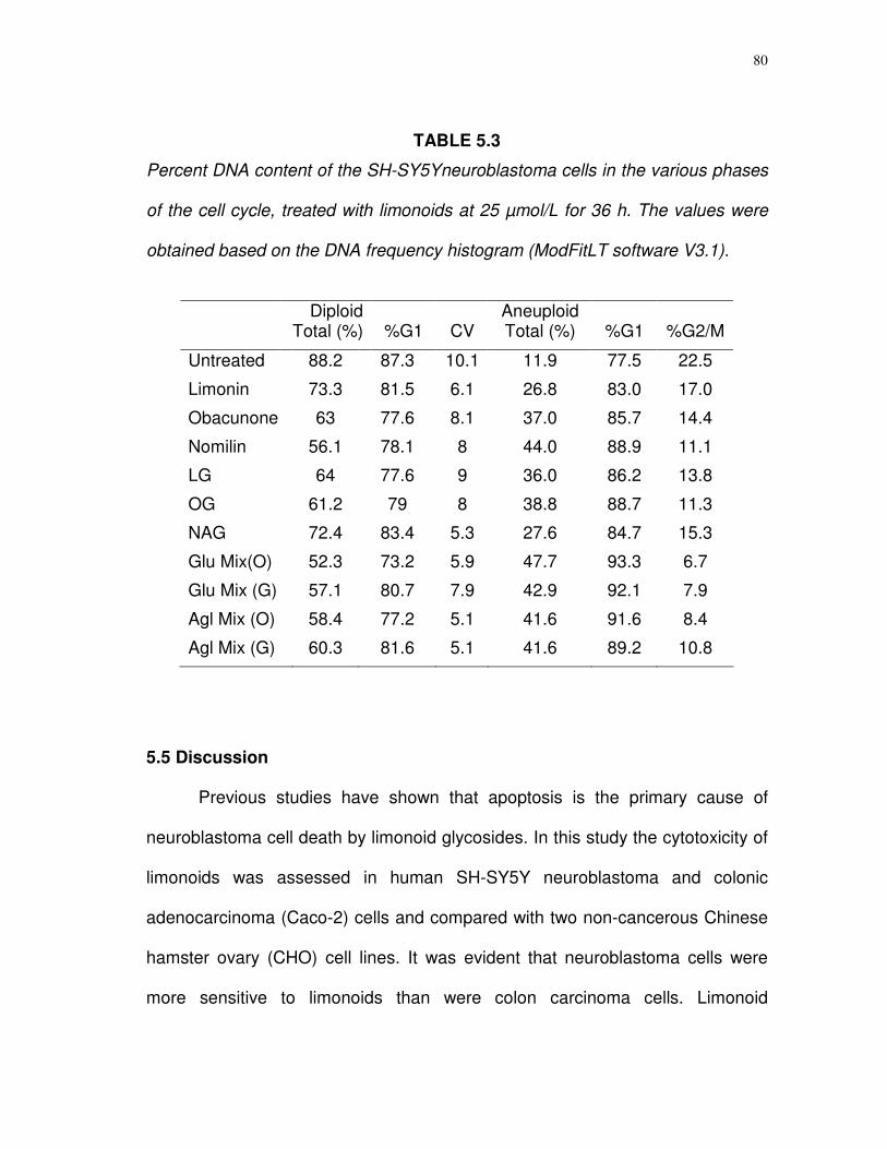
80
TABLE 5.3
Percent DNA content of the SH-SY5Yneuroblastoma cells in the various phases
of the cell cycle, treated with limonoids at 25 µmol/L for 36 h. The values were
obtained based on the DNA frequency histogram (ModFitLT software V3.1).
5.5 Discussion
Previous studies have shown that apoptosis is the primary cause of
neuroblastoma cell death by limonoid glycosides. In this study the cytotoxicity of
limonoids was assessed in human SH-SY5Y neuroblastoma and colonic
adenocarcinoma (Caco-2) cells and compared with two non-cancerous Chinese
hamster ovary (CHO) cell lines. It was evident that neuroblastoma cells were
more sensitive to limonoids than were colon carcinoma cells. Limonoid
Diploid
Total (%) %G1 CV Aneuploid Total (%) %G1 %G2/M
Untreated 88.2 87.3 10.1 11.9 77.5 22.5
Limonin 73.3 81.5 6.1 26.8 83.0 17.0
Obacunone 63 77.6 8.1 37.0 85.7 14.4
Nomilin 56.1 78.1 8 44.0 88.9 11.1
LG 64 77.6 9 36.0 86.2 13.8
OG 61.2 79 8 38.8 88.7 11.3
NAG 72.4 83.4 5.3 27.6 84.7 15.3
Glu Mix(O) 52.3 73.2 5.9 47.7 93.3 6.7
Glu Mix (G) 57.1 80.7 7.9 42.9 92.1 7.9
Agl Mix (O) 58.4 77.2 5.1 41.6 91.6 8.4
Agl Mix (G) 60.3 81.6 5.1 41.6 89.2 10.8
81
cytotoxicity varied depending on the type of cancer cell and the type of limonoid
applied. While both limonoid aglycones and glucosides at micromolar levels
appeared to arrest cell growth, biochemical and morphological data showed that
the glycosides were clearly more effective in inducing rapid cell death. Neither
pure compounds nor mixtures showed toxicity against the viable mammalian
CHO epithelial cells.
Corresponding to our previous report showing the apoptotic inducing
ability of LG and OG, here the cell death on both SY5Y as well as Caco-2 cells
by limonin and obacunone was observed, but at a significantly slower pace and
never to the same extent. This confirms the killing potential of limonoid
aglycones as reported by Miller et al. (22, 23). Considering the solubility factor
that could hinder the killing potential, the aglycones were supplemented through
non-toxic concentrations of DMSO, giving both compounds equal access to the
cell’s interior and sensitive site(s). Obacunone and other purified aglycones were
as effective as glycosides in stopping cell division, but considerably inferior in
rapid killing. The data, therefore, imply that stopping cell division and inducing
cell death by apoptosis may be two separate and independent actions of
limonoids on cancer cells. Aglycones are capable of arresting the cell
proliferation, but weak in inducing the apoptosis. The propensity of aglycone and
glycosidic mixtures speaks strongly for synergistic events beyond absorption,
and from a nutritional perspective, whole fruit or whole juice consumption to
maximize beneficial effects.

82
Chromosomal aberrations in cell DNA content (aneuploidy) measured by
flow cytometry provide objective information and major prognostic factors for
neuroblastomas (127). Aneuploidy is common in neuroblastoma cells and has
been used to predict tumor aggressiveness and successful response to
chemotherapy. In neuroblastoma tumor cells absence of aneuploidy (near
diploid or near tetraploid DNA content) is confirmed to be the more aggressive
pattern of neuroblastoma (127-130). Look et al. (130) reported that tumors with
greater numbers of aneuploidic cells responded well to chemotherapy.
Limonoids increased the number of aneuploidic SH-SY5Y cells. A previous
report correlated cell cycle arrest with an increase in G1 phase cells, thus
inferring a block in the transition to the S phase of the cell cycle. Enhancing
aneuploidy raises the possibility that cell cycle arrest could also be occurring at a
later stage of cell division, particularly the separation into two daughter cells
which is shown as an arrest at the G2/M phase.
In conclusion, with proven anticancer properties, limonoids can now be
considered as lead structures for anticancer drugs. Viability studies indicate that
molecules patterned after limonoid glycosides may have greater commercial
potential. As cellular permeability, water solubility, and metabolic change all play
pivotal roles in the overall efficacy of a natural compound’s performance (131),
the glycosides appear to excel over aglycones. These studies advocate citrus
fruit as a valuable functional food which may thwart cancer in human and could
augment demand for citrus and citrus products.
83
CHAPTER VI
CONCLUSIONS
Combination of ion-exchange and crystallization techniques presented in
this dissertation provides an efficient and economical method for the large scale
purification of limonoids and flavonoids from citrus byproducts. The methods
developed were found to be highly specific and with more than 80-fold increase
in total yields, than present methods in practice for the isolation of limonoid
glucosides. The removal of large amounts of naringin in particular, increased the
purification efficiency. One kilogram of dried orange and grapefruit seeds yielded
8.62 and 9.31 g of limonin, 0.62 and 0.47 g nomilin, 0.9 and 0.63 g of
deacetylnomilin and 0.21 and 0.17 g of obacunone respectively. An average of
16 g of dried limonoid glucosides and 9.8 g flavonoid mixtures were obtained
from 1 Kg of clarified citrus molasses concentrate. Preparative HPLC separation
of these semi-purified mixtures resulted in high yields of limonoid glucosides with
more than 90% purity. Considering the abundance of aglycones in seeds, and
glucosides in molasses, citrus byproducts provide an excellent source for bulk
purification.
In our first biological activity study using the purified compounds, limonoids
and flavonoids have shown to effect redox-cycling enzymes in a dose dependent
and dose independent manner. Limonoids selectively and quantitatively inhibited
the dealkylase and hydroxylase reactions performed by cytochrome P450
84
isoforms. All the limonoids significantly (P < 0.001) inhibited EROD and MROD
activity of CYP1B1 and CYP1A2. Limonoid glucosides were found to be better
inhibitors than aglycones. The acidic limonoid glucoside, NAG, had the highest
inhibitory effect on most of the isoforms tested. Correspondingly, the flavonoids
tested also significantly (P < 0.001) inhibited EROD and MROD activity of
CYP1B1 and CYP1A2. Hydroxylation activity of human CYP aromatase, CYP19,
was inhibited reversibly by LG and non-competitively by NAG. The limonoid
glucosides and aglycones were also found to inhibit CYP 3A4 hydroxylation.
NAG and nomilin were the best inhibitors of CYP3A4, drug metabolizing CYP
isoenzyme. The IC50 for limonoids varied considerably, depending upon the
type of isoforms. The limonoid glucosides were found to be the better inhibitors
when compared to the aglycones. The effects of citrus bioactive compounds on
the partial inhibition of CYP enzymes represent a unique mechanism in the
anticarcinogenesis strategy. One of the mechanisms is through the reduction in
the generation of reactive oxygen species. This study also supports the role of
citrus bioactive compounds on the increased bioavailability of orally
administered drugs. Partial inhibition of CYP3A4 may represent an alternate
strategy for the reduction in medical costs, but needs further studies on the
mechanistic and dose response relations. Structural variations in different
limonoid and flavonoid molecules can be ascribed to the differences in efficacy
in inhibiting different CYP isoenzymes.
85
A complimentary means by which to address the question “Does limonoids
acts as antioxidants?” relies on their ability to quench the reactive oxygen
species. In vitro studies with four pure limonoid glucosides have shown effective
superoxide scavenging and perceptible differences in the quenching efficacies,
at 5 mmol/L. NAG emulated a quenching effect to an equivalent concentration
of vitamin C, and was superior to LG, OG, and DNAG.
The study conducted to evaluate the cytotoxic effects of limonoid
glucosides confirmed that micromolar amounts of LG and OG induce rapid
necrosis of SH-SY5Y cells in culture. Viability studies showed significant (P <
0.001) reduction in survivability by LG and OG within 24 h of treatment. The 12 h
caspase 3/7 activity had a strong negative correlation (r = -0.998, P = 0.002) to
the cell survival. The data confirmed that the cytotoxic effect of limonoid
glucosides was manifested through apoptotic induction mechanism which was
further endorsed through DNA fragmentation profile. Univariate DNA content
was significantly higher for cells treated with LG, OG. Background aggregates
and debris (BAD) between G1 and G2 phase, an indicator of apoptosis, was
significantly higher (P < 0.05) for LG and OG compared to control. Similarly DNA
content at S phase of the cell cycle indicated a significant reduction by LG and
OG as compared to NAG, DNAG (86.67% with LG, 82.36% with OG, 57.46%
with NAG, and 38.35% with DNAG). Overall, these results confirm the
antiproliferative property of limonoid glucosides are manifested through arresting
critical growth stages of the cell cycle and inducing apoptosis.
86
The last study conducted to elucidate the cytotoxic effects of citrus limonoid
aglycones and glucosides on human cancer cell lines such as SH-SY5Y
neuroblastoma and colon carcinoma (Caco-2) cell lines showed a specific
cytotoxic effects on cancerous cells and spared the non cancerous Chinese
hamster ovary (CHO) cells. Limonoid glucosides have shown greater cytotoxic
effects than aglycones and the effects were significant (P < 0.001) over the
period of 24 to 36 h when compared with the untreated cells. The morphological
differences observed on the confocal microscopy clearly designate a rapid
response for the LG as against limonin and obacunone aglycones. The effects
were consistent with both time and dose dependent assay. Neuroblastoma cell
lines were found to be more sensitive than the colon carcinoma cells incase of
mixtures and nomilin during the initial period of exposure. The flow cytometric
analysis of limonoid treated neuroblastoma cells showed a marked increase in
DNA content of aneuploidic cells within 24 h. Percent aneuploidic cells were
found to be 36% for LG, 26% for limonin and more than 42% for limonoid
glucoside mixtures indicating a synergistic effect of different limonoid glucosides.
The significant reduction in G2/M phase of the cell cycle confirms the results in
chapter IV that limonoids inhibit S phase of the cell cycle. The inhibition of
mitosis due to differing chromosome number in the aneuploidic cells, can lead to
the non division of cells, eventually leading to death. These results confirm that
both limonoid aglycones and glycosides are lethal to cancer cell lines; however
87
glycosides are the most active apoptotic inducing form, even though both forms
arrest cell cycle.
In summary, with wide biological applications, anticancer properties and
possible role in cell signaling mechanisms, limonoids can now be considered as
a possible lead structure for a new class of anticancer drugs. The bulk
purification methods could aid further research in the area of clinical trials, food
additives, and nutritional supplements. As cellular permeability, water solubility,
and metabolic change all play pivotal roles in the overall efficacy of a natural
compound’s performance, the glycosides appear to excel over aglycones. With
citrus being a store house for many nutritionally important bioactive compounds,
our studies strongly advocate citrus as a valuable functional food, particularly for
the prevention of cancer in children.
88
LITERATURE CITED
1. Calabrese, F. (2002) Citrus: Origin and history. In Citrus: The Genus Citrus (Dugo, G., & Giacomo, A. D., eds.) pp.1-15, Taylor & Francis Inc. New York.
2. USDA (2005) World Horticultural Trade & US Export Opportunities,
Situation and outlook for citrus, www.fas.usda.gov, pp.1-8, Accessed April, 2005.
3. US Department of Commerce, (2005), US citrus exports, Florida
Department of Citrus: Economic & market research report # EX-05-03, 1-2. 4. Miller, E.G., Taylor, S.E., Berry, C.W., Zimmerman, J.A. &
Hasegawa, S. (2000) Citrus limonoids: Increasing importance as anticancer agents. In: Citrus Limonoids-Functional Chemicals in Agriculture and Foods (Berhow, M.A., Hasegawa, S., & Manners, G.D., eds.), pp.132-144. American Chemical Society, Washington D.C.
5. Montanari, A., Widmer, W. & Nagy, S. (1997) Health promoting
phytochemicals in citrus fruit and juice products: Recent advances in phytochemistry. In: Functionality of Food Phytochemicals (Johns, T. A. & Romeo, J. T. eds.) pp.31 -52. Meeting of the Phytochemical Society of North America on Functionality, Plenum Press, New York.
6. Hasegawa, S. & Miyake, M. (1996) Biochemistry and biological
functions of citrus limonoids. Food Rev. Int. 12: 413-435. 7. Hasegawa, S., Bennet, R. D., Herman, Z., Fong, C. H. & Ou, P.
(1989) Limonoid glucosides in citrus. Phytochemistry 28: 1717-1724. 8. Hasegawa, S., Ou, P., Fong, C. H., Herman, Z., Coggins, C.W., Jr.
& Atkin, D.R. (1991). Changes in the limonoate A-ring lactone and limonin 17-D-glucopyranoside content of navel oranges during fruit growth and maturation. J. Agric. Food Chem. 39: 262-265.
9. Hasegawa, S., Berhow, M. A. & Fong, C. H. (1995) Analysis of bitter principles in citrus. In: Modern Methods of Plant Analysis. (Linskens, H. F., Jackson, J. F. & Allen, M. S., eds.) Vol.18, pp. 59-80. Springer-Verlag, Berlin, Germany.
89
10. Maier, V. P. & Beverly, G. D. (1968) Limonin monolactone, the
nonbitter precursor responsible for delayed bitterness in certain citrus juices. J. Food Sci., 33: 488-492.
11. Schoch, T. K., Manner, G. D. & Hasegawa, S. (2002) Recovery of
limonoid glucosides from citrus molasses. J. Food Sci., 67: 3159-3163. 12. Hasegawa, S., Fong, C. H., Herman, Z., & Miyake M. (1992)
Glucosides of limonoids. Flavour precursors. Am. Chem. Soc. 86-97. 13. Miller, E. G., Porter, J. L., Binnie, W. H., Guo, I. Y. & Hasegawa, S.
(2004) Further studies on the anticancer activity of citrus limonoids. J. Agric. Food Chem., 52: 4908-4912.
14. Herman, Z., Fong, C. H., Ou, P. & Hasegawa, S. (1990) Limonoid
glucosides in orange juices by HPLC. J. Agric. Food Chem. 38: 1860-1861. 15. Battinelli, L., Menqoni, F., Lichtner, M., Maqzzanti, G., Saija, A.,
Mastroianni, C. M. & Vullo, V. (2003) Effect of limonin and nomilin on HIV-1 replication on infected human mononuclear cells. Plant Medica 69: 910-913.
16. Sunthitikawinsakul, A., Kongkathip, N., Kongkathip, B.,
Phonnakhu, S., Daly, J.W., Spande, T.F., et al. (2003) Anti HIV-1 limonoid: first isolation from Clausena excavata. Phytother. Res. 17: 1101-1103.
17. Bray, D. H., Warhurst, C. C., Connolly, J. D., O’Neill, M. J. &
Phillipson, J. D. (1990) Plants as sources of antimalarial drugs. Part 7. Activity of some species of Meliaceae plants and their constituent limonoids. Phytother. Res. 4: 29-35.
18. So, F. V., Guthrie, N., Chambers, A. F., Moussa, M. & Carroll, K.
K. (1996) Inhibition of breast cancer cell proliferation and delay of mammary tumerogenesis by flavonoids and citrus juices. Nutr. Cancer 25: 167-181.
19. Tanaka, T., Kohno, H., Tsukio, Y., Honjo, S., Tanino, M., Miyake,
M. & Wada, K. (2000) Citrus limonoids obacunone and limonin inhibit azoxymethane-induced colon carcinogenesis in rats. Biofactors 13: 213-218.
20. Tanaka, T., Maeda, M., Kohno, H., Murakami, M., Kagami, S.,
Miyake, M. & Wada, K. (2001) Inhibition of azoxymethane-induced colon carcinogenesis in male F344 rats by the citrus limonoids obacunone and limonin. Carcinogenesis 22: 193-198.
90
21. Lam, L. K. T. & Hasegawa, S. (1989) Inhibition of benzo[a]pyrene-induced forestomach neoplasia in mice by citrus limonoids. Nutr. Cancer 12: 43-47.
22. Miller, E. G., Gonzales, S. A. P., Couvillon, A. M., Wright, J. M.
Hasegawa, S. & Lam, L. K. T. (1992) Inhibition of hamster buccal pouch carcinogenesis by limonin 17 -D-glucopyranoside. Nutr. Cancer 17: 1-7.
23. Miller, E. G., Fanous, R., Rivera, H. F., Binnie, W. H., Hasegawa,
S. & Lam, L. K. T. (1989) The effect of citrus limonoids on hamster buccal pouch carcinogenesis. Carcinogenesis 10: 1535-1537.
24. Miller, E. G., Porter, J. L., Binnie, W. H., Guo I. Y. & Hasegawa, S.
(2004) Further studies on the anticancer activity of citrus limonoids. J. Agric. Food Chem. 52: 4908-4912.
25. Pettit, G. R., Barton, D. H. R., Herald, C. L., Polonsky, J., Schmidt,
J. M. & Connolly, J. D. (1983) Evaluation of limonoids against the murine P388 lymphocytic leukemia cell line. J. Natur. Prod. 46: 379-390.
26. Kurowska, E. M. & Manthey, J. A. (2004) Hydrolipidemic effects
and absorption of citrus polymethoxylated flavones in hamsters with diet-induced hypercholesterolemia. J. Agric. Food Chem. 52: 2879-2886.
27. Kurowska, E. M. & Manthey, J. A. Casaschi, A. & Theriault, A. G.
(2004) Modulation of HepG2 cell net apolipoprotein B secretion by the citrus polymethoxyflavone, tangeretin. Lipids 39:143-151.
28. Lam, L. K. T., Li, Y. & Hasegawa, S. (1989) Effects of citrus
limonoids on glutathione S-transferase in mice. J. Agric. Food Chem. 37: 878-880.
29. Lam, L. K. T., Zhang, J., Hasegawa, S. & Schut, H. A. J. (1994)
Inhibition of chemically induced carcinogenesis by citrus limonoids. In: Food Phytochemicals for Cancer Prevention I. (Huang, M. J., Osawa, T., Ho, C. T. & Rosen, R. T. eds.), pp. 209-219. American Chemical Society, Washington D.C.
30. Kelly, C., Jewell, C. & O’Brien, N. M. (2003) The effect of dietary
supplementation with the citrus limonoids, limonin and nomilin on xenobiotic metabolizing enzymes in the liver and small intestine of the rat. Nutr. Res. 23: 681-690.

91
31. Tian, Q., Miller, E. G. Hassan, A., Tang, L. & Patil, B. S. (2001) Differential inhibition of human cancer cell proliferation by citrus limonoids. Nutr. Canc. 40: 180-184.
32. Manners, G. D. & Hasegawa, S. (1999) A new normal phase liquid
chromatographic method for the analysis of limonoids in citrus. Phytochem. Anal. 10: 76-81.
33. American Cancer Society. (2005) Cancer Facts & Figures 2005.
Atlanta, GA. 34. Halliwell, B. (1991) Reactive oxygen species in living systems:
source biochemistry and role in human disease. Am. J. Med. 9: 14-22. 35. Guengerich, F. P, & Shimada, T. (1991) Oxidation of toxic and
carcinogenic chemicals by human cytochrome P-450 enzymes. Chem. Res. Toxicol. 4: 381-407.
36. Kadlubar, F. F, & Hammons G. J. (1987) The role of cytochrome
P-450 in the metabolism of chemical carcinogens. In: Mammalian cytochromes P-450 (Guengerich, F. P. ed.), pp. 81-130, CRC Press, Boca Raton, FL.
37. Guengerich F. P, & Shimada T. (1991) Oxidation of toxic and
carcinogenic chemicals by human cytochrome P-450 enzymes. Chem. Res. Toxicol. 4: 381-407.
38. Zeigler, J. C. (2001) CYP3A4: a potential prostate cancer risk
factor for high-risk groups. Clin. J. Oncol. Nurs. 5: 153-154. 39. Seow, A., Zhao, B., Lee, E. J., Poh, W. T., The, M. et al. (2001)
Cytochrome P4501A2 activity and lung cancer risk: a preliminary study among Chinese women in Singapore. Carcinogenesis. 22: 673-677.
40. Marchand, L. L., Hankin J. H., Wilkens L. R., Pierce L. M., Franke
A. et al. (2001) Combined effects of well-done red meat, smoking, and rapid N-acetyltransferase 2 and CYP1A2 phenotypes in increasing colorectal cancer risk. Cancer Epidem. Biomarkers & Prev. 10: 1259-1266.
41. Yager, J. D, & Liehr, J. G. (1996) Molecular mechanisms of
estrogen carcinogenesis. Ann. Rev. Pharmacol. Toxicol. 36: 203-232. 42. Guengerich, F. P., Chun, Y. J., Kim, D., Gillam, E. M. J. &
Shimada, T. (2003) Cytochrome P450 1B1: a target for inhibition in anticarcinogenesis strategies. Mut. Res. 523: 173-182.
92
43. Lilja, J. J., Kivisto, K. T. & Neuvonen, P. J. (1998) Grapefruit juice-simvastatin interaction: Effect on serum concentrations of simvastatin, simvastatin acid, and HMG/L-CoA reductase inhibitors. Clinic. Pharmac. Therap. 64: 477-483.
44. Doostdar, H., Burke, M. D. & Mayer, R. T, (2000) Bioflavonoids:
Selective substrates and inhibitors of CYP 450 1A1 and CYP 1B1, Toxicology 144: 31-38.
45. Cao, G., & Sofic, E. (1996) Antioxidant capacity of tea and
common vegetables. J. Agric. Food Chem. 44: 3426 -3431. 46. Steinmetz, K. A. & Potter, J. D. (1996) Vegetables, fruits and
cancer prevention: A review. J. Am. Diet. Assoc. 96: 1027–1037. 47. Block, G., Patterson, B. & Subar, A. (1992) Fruit, vegetables and
cancer prevention; a review of the epidemiological evidence. Nutr. Cancer 18: 1–29.
48. Marklund, S. & Marklund, G. (1974) Involvement of the superoxide
anion radical in the autoxidation of pyrogallol and a convenient assay for superoxide dismutase. Eur. J. Biochem. 47: 469-474.
49. Ikonen, M., Liu, B., Hashimoto, Y., Ma, L., Lee, K. W., Niikura, T.,
Nishimoto, I. & Cohen P. (2003) Interaction between the Alzheimer’s survival peptide humanin and insulin-like growth factor binding protein regulates cell survival and apoptosis. Proc. Natl. Acad. Sci. U.S.A. 100: 13042-13047.
50. Jaffe, N. (1976) Neuroblastoma: Review of the literature and an
examination of its enigmatic character. Cancer Treat. Rev. 3: 61–82. 51. Souhami, R. & Tobias, J. (1998) Cancer and its Management (3rd
Ed), Blackwell Science, Oxford, U.K. 52. Wang, C. N., Chi, C. W., Lin, Y. L., Chen C. F., & Shiao, Y. J.
(2001) The neuroprotective effects of phytoestrogens on amyloid protein-induced toxicity are mediated by abrogating the activation of caspase cascade in rat cortical neurons. J. Biol. Chem. 276: 5287-5295.
53. Nicolini, G., Rigolio, R., Scuteri, A., Miloso, M., Saccomanno, D.,
Cavaletti, G. & Tredici, G. (2003) Effect of trans-resveratrol on signal transduction pathways involved in paclitaxel-induced apoptosis in human neuroblastoma SH-SY5Y cells. Neurochem. Internat. 43: 419-429.
93
54. Miyataa, M., Takanoa, H., Takahashia, K., & Sasakib, Y. F. (2002) Suppression of 2-amino-1-methyl-6-phenylimidazo [4,5-] pyridine-induced DNA damage in rat colon after grapefruit juice intake. Cancer Lett. 183: 17-22.
55. Miyagi, Y., Om, A. S., Chee, K. M., & Bennik, M. R. (2000)
Inhibition of azoxymethane-induced colon cancer by orange juice. Nutr. Cancer 32: 224-229.
56. Balandrin, N. F., Kinghorn, A. D., Farnsworth, N. R. (1993) In:
Human Medicinal Agents from Plants (Kinghorn, A. D. & Balandrin, M. F., eds.), pp. 2-12. American Chemical Society (Symposium Series 534), Washington, D.C.
57. Manners, G. D., Jacob, R. A., Breksa, A. P., Schoch, T. K. &
Hasegawa, S. (2003) Bioavailability of citrus limonoids in humans. J. Agric. Food Chem. 51: 4156-4161.
58. Hollmam, P., Hertog, M. & Katan, M. (1996) Role of dietary
flavonoids in protection against cancer and coronary heart disease. Biochem. Soc. Trans. 24: 785-789.
59. Jagetia, G. C., Venkatesha, V. A. & Reddy, T. K. (2003) Naringin,
a citrus flavonone, protects against radiation-induced chromosome damage in mouse bone marrow. Mutagenesis 18: 337-343.
60. Jeon, S. M., Bok, S. H., Jang, M. K., Lee, M. K., Nam, K. T., Park,
Y. B., Rhee, S. J. & Choi, M. S. (2001) Antioxidative activity of naringin and lovastatin in high cholesterol-fed rabbits. Life Sciences 69: 2855-2866.
61. Gorinstein, S., Leontowicz, H., Leontowicz, M., Krzeminski, R.,
Gralak, M. et al. (2005) Changes in plasma lipid and antioxidant activity in rats as a result of naringin and red grapefruit supplementation. J. Agric. Food Chem. 53: 3223-3228.
62. Kawaii, S., Tomono, Y., Katase, E., Ogawa, K. & Yano, M. (1999)
Antiproliferative activity of flavonoids on several cancer cell lines. Biosci. Biotech. Biochem. 63: 896-899.
63. Tanaka, T., Kohno, H., Murakami, M., Shimada, R., Kagami, S.,
Sumida, T., Azuma, Y. & Ogawa, H. (2000) Suppression of azoxymethane-induced colon carcinogenesis in male F344 rats by mandarin juices rich in -cryptoxanthin and hesperidin. Int. J. Cancer 88: 146-150.
94
64. Scambia, G., Ranelletti, F. O., Benedetti P. P., Piantelli, M., Bonanno, G. et al. (1990) Inhibitory effect of quercetin on OVCA 433 cells and presence of type II estrogen binding sites in primary ovarian tumors and cultured cells. Brit. J. Cancer 62: 942-946.
65. Food and Drug Administration (1982) Evaluation of the health
aspects of hesperidin, naringin, and citrus bioflavonoid extracts as food ingredients. SCOGS-II-3, FDA/BF-82/62; Order No. PB82-192931, Gov. Rep. Announce Index (U.S.A.) 82(15), 2962.
66. Bentley, M. D., Rajab, M. S., Alford, A. R., Mendel, M. J. &
Hassanali, A. (1988) Structure-activity studies of modified citrus limonoids as antifeedants for Colorado potato beetle larvae, Leptinotarsa decemlineata. Entomol. Exper. Applic. 49: 189-193.
67. Ozaki, Y., Ayano, S., Inaba, N., Miyake, M., Berhow, M. A. &
Hasegawa, S. (1995) Limonoid glucosides in fruit, juice, and processing by-products of Satsma mandarin (Citrus unshiu Marcov.). J. Food Sci. 60: 186-189.
68. Rouseff, R. L. & Fisher, J. F. (1980) Determination of limonin and
related limonoids in citrus juices by high performance liquid chromatography. Anal. Chem. 52: 1228-1233.
69. Sawabe, A., Hasegawa, S., Bennet, R. D. & Wong, R.Y. (2000)
LC-MS and NMR technique for the analysis and characterization of citrus limonoids. In: Citrus Limonoids; Functional Chemicals in Agriculture and Foods. (Berhow, M. A., Hasegawa, S. & Manners, G. eds) pp. 40-58. American Chemical Society (Symposia series 758), Washington, D.C.
70. Shaw, P. E. & Wilson, C. W. (1984) A rapid method for the
determination of limonin in citrus juice by reverse-phase high-performance liquid chromatography. J. Chromatogr. 464: 375-386.
71. Van Beek, T. A. & Blaakmeer, A. (1989) Determination of limonin
in grapefruit juice and other citrus juices by high-performance liquid chromatography. J. Chromatogr. 161: 375-86.
72. Bennet, R. D., Hasegawa, S. & Herman, Z. (1989) Glucosides of
acidic limonoids in citrus. Phytochemistry 28: 2777-2781. 73. Hasegawa, S. & Bennett, R. D. (1980) Isolimonic acid, a new citrus
limonoid. Phytochemistry 19: 2417-2419.
95
74. Manners, G. D., Breska III, A. P., Schoch, T. K. & Hidalgo, M. B. (2003) Analysis of bitter limonoids in citrus juices by atmospheric pressure chemical ionization and electrospray ionization liquid chromatography-mass chromatography. J. Agric. Food Chem. 51: 3709-3714.
75. Tian, Q. & Ding, X. (2000) Screening for limonoid glucosides in
Citrus tangerina (Tanaka) Tseng by high-performance liquid chromatography-electrospray ionization mass spectrometry. J. Chromatogr. 874: 13-19.
76. Schoch, T. K., Manner, G.D. & Hasegawa, S. (2001) Analysis of
limonoid glucosides from citrus by electrospray ionization liquid chromatography-mass spectrometry. J. Agric. Food Chem. 49: 1102-1108.
77. Manthey, J. A. (2004) Fractionation of orange peel phenols in
ultrafiltered molasses and mass balance studies of their antioxidant levels. J. Agric. Food Chem. 52: 7586-7592.
78. Tian, Q., Liu, Y., Bhat, N. G. & Patil, B. S. (2000) Total limonoids in
citrus. Proc. Intl. Citricult. Cong. IX. 1261-1264. 79. Emerson, O. H. (1948) Bitter principles in citrus fruit. I. Isolation of
nomilin, a new bitter principle from the seeds of oranges and lemons. J. Am. Chem. Soc. 70: 545-549.
80. Dreyer, D. L. (1965) Citrus bitter principles. III. Isolation of
deacetylnomilin and deoxylimonin. J. Org. Chem. 30: 749-51. 81. Targett, N. M., Kilcoyne, J. P. & Green, B. (1979) Vacuum liquid
chromatography: an alternative to common chromatographic methods. J. Org. Chem. 44: 4962-4964.
82. Coll, J. C. & Bowden, B. F. (1986) The application of vacuum liquid
chromatography to the separation of terpene mixtures. J. Nat. Prod. 49: 934-936.
83. Turcant, A., Premel, C. A., Cailleux, A. & Allain, P. (1988)
Screening for neutral and basic drugs in blood by dual fused-silica column chromatography with nitrogen-phosphorus detection. Clin. Chem. 34: 1492-1497.
84. Emerson, O. H. (1952) Bitter principles in citrus. III. Some
reactions of limonin. J. Am. Chem. Soc. 74: 688-693.
96
85. Hendrickson, R. & Kesterson, J. W. (1956) Crude hesperidin purification. Citrus Industry 37: 7-10.
86. Scordino, M., Di, M. A. Passerini, A. & Maccarone, E. (2004)
Adsorption of flavonoids on resins: cyanidin 3-glucoside. J. Agric. Food Chem. 52: 1965-1972.
87. Levison, R. P. (1996) Ion exchange chromatography and
secondary adsorption effects. In: Downstream Processing of Natural Products, a practical handbook (Michael S. V. ed). Wiley J & Sons, Inc. Chichester, U.K.
88. Vandenhul, H. J. & Wanderhoff, J. W. (1972) The characterization
of latex particle surfaces by ion exchange and conductometric titration. J. Electronal. Chem. 37: 161-168.
89. Halliwell, B. (1991) Reactive oxygen species in living systems:
source biochemistry and role in human disease. Am. J. Med. 9: 14-22. 90. Loo, G. (2003) Redox-sensitive mechanisms of phytochemical-
mediated inhibition of cancer cell proliferation. J. Nutr. Biochem. 14: 64-73. 91. Wrighton, S. A. & Stevens, J. C. (1992) The human hepatic
cytochrome P450 involved in drug metabolism. Crit. Rev. Toxicol. 22: 1-21. 92. Russo, M., Tedesco, I., Iacomino, G., Palumbo, R., Galano, G. &
Russo, G. L. (2005) Dietary phytochemicals in chemoprevention of cancer. Curr. Med. Chem. Immunol. Endocr. Metab. Agents 5: 61-72.
93. Hodek, P., Trefil, P. & Stiborova, M. (2002) Flavonoids-potent and
versatile biologically active compounds interacting with cytochromes p450. Chem. Biol. Interact.139: 1-21.
94. Ortiz de Montellano, P. R. (1986) Oxygen activation and transfer.
In: Cytochrome P-450: Structure, Mechanism, and Biochemistry (Ortiz de Montellano, P. R., ed) pp. 217-271. Plenum Press, New York.
95. Guengerich, F. P. (2003) Cytochrome P450, drugs and disease. Mol.
Interv. 3: 194-204. 96. Miller, E. C. & Miller, J. A. (1976) The metabolism of chemical
carcinogens to reactive electrophiles and their possible mechanisms of action in carcinogenesis, In: Chemical Carcinogenesis Monograph, pp. 737–762. American Chemical Society, Washington, D.C.
97
97. Bartsch, H., Nair, U., Risch, A., Rojas, M., Wikman, H. & Alexandrov, K. (2000) Genetic polymorphism of CYP genes, alone or in combination, as a risk modifier of tobacco-related cancers. Cancer Epidemiol. Biomarkers Prev. 9: 3-28.
98. Mayer R. T., Jermyn, J. W. & Burke, M. D. (1977)
Methoxyresorufin as a substrate for the fluorometric assay of insect microsomal O-dealkylase. Pestic. Biochem. Physiol. 7: 349-354.
99. Burke, M. D., Thompson, S., Weaver, R. J., Wolf C. R. & Mayer, R.
T. (1994) Cytochrome P450 specificities of alkoxyresorufin O-dealkylation in human and rat liver. Biochem. Pharmacol. 48: 923-936.
100. Stahle, E. L., Schloss, L., Sundqvist, V. A., Brytting, M.,
Hokeberg, I., Cox, S., Wahren, B. & Linde, A. (1998) Solid phase ELISA for determination of the virus dose dependent sensitivity of human cytomegalovirus to antiviral drugs in vitro. Antiviral Res. 40: 105-112.
101. Girennavar, B., Poulose, S. M., Brodbelt, J. S., Jayaprakasha G.
K., Bhat, N. G. & Patil, B. S. (2005) Whither grapefruit drug interaction?, American Chemical Society, (ACS symposium series) Washington, D.C. (in press).
102. Lang, N. P., Butler, M. A., Massengill, J., Lawson, M., et al. (1994)
Rapid metabolic phenotypes for acetyltransferase and cytochrome P4501A2 and putative exposure to food-borne heterocyclic amines increase the risk for colorectal cancer or polyps. Cancer Epidemiol. Biomarkers Prev. 3: 675–682.
103. Brodie, A. M. H. (1985), Aromatase inhibition and its
pharmacological implications. Biochem. Pharmacol. 34: 3213-3219. 104. Kita, M., Hirata, Y., Moriguchi, T., Endo-Inagaki, T., Matsumoto, R.,
Hasegawa, S. Suhayda, C. G. & Omura, M. (2000) Molecular cloning and characterization of a novel gene encoding limonoid UDP-glucosyltransferase in citrus. FEBS. Lett. 469: 173-178.
105. Manners, G. D., Jacob, R. A., Breksa, A. P., Schoch, T. K. &
Hasegawa, S. (2003) Bioavailability of citrus limonoids in humans. J. Agric. Food Chem. 51: 4156-4161.
106. Yu, J., Wang, L., Walzem, R. L., Miller, E. G., Pike, L. M. & Patil, B.
S. (2005) Antioxidant activity of citrus limonoids, flavonoids, and coumarins. J. Agric. Food Chem. 53: 2009-2014.
98
107. Gale, G., D’Angio, G., Uri, A., Chatten, J. & Coop, C. E. (1982) Cancer in neonates: the experience at the children’s hospital of Philadelphia. Pediatrics 70: 409–413.
108. Reddy, M. C. M., Majumdar, S. & Harris, E. D. (2000) Evidence for
a Menkes-like protein with a nuclear targeting sequence. Biochem. J. 350: 855-863.
109. Hansen, M. B., Nielsen, S. E. & Berg, K. (1989) Re-examination
and further development of a precise and rapid dye method for measuring cell growth/cell kill. J. Immun. Meth. 119: 203-210.
110. Silalahi, J. (2002) Anticancer and health protective properties of
citrus fruit components. Asia Pac. J. Clin. Nutr. 11: 79-84. 111. Deng, M., Zhao, J. Y., Ju, X. D., Tu, P. F. Jiang, Y. & Li, Z. B.
(2004) Protective effect of tubuloside B on TNF-alpha-induced apoptosis in neuronal cells. Acta Pharmacol. Sin. 25: 1276-1284.
112. Ikonen, M., Liu, B, Hashimoto, Y., Ma, L., Lee, K. W., Niikura, T.,
Nishimoto, I. & Cohen, P. (2003) Interaction between the Alzheimer’s survival peptide humanin and insulin-like growth factor binding protein regulates cell survival and apoptosis. Proc. Natl. Acad. Sci. 100: 13042-13047.
113. Berglund, C. M., Radesater, A. C., Persson, M. A., & Budd H. S. L.
(2004) UV-induced apoptosis in SH-SY5Y cells: contribution to apoptosis by JNK signaling and cytochrome c. J. Neurosci. Res. 78: 580-589.
114. Lovat, P. E., Di Sano, F., Corazzzari, M., Fazi, B., Donnorso, R. P.
et al. (2004) Gangliosides link the acidic sphingomyelinase-mediated induction of ceramide to 12-lipoxygenase-dependent apoptosis of neuroblastoma in response to fenretinide. J. Natl Canc Instit. 96: 1288-1299.
115. Kweon, S. M., Lee, Z. W., Yi, S. J., Kim, Y. M., Han, J A., Paik, S.
G. & Ha, S. S. (2004) Protective role of tissue transglutaminase in the cell death induced by TNF-alpha in SH-SY5Y neuroblastoma cells. J. Biochem. Mol. Biol. 37: 185-191.
116. Lee, E. O., Shin, Y. J., & Chong, Y.H. (2004) Mechanisms
involved in prostaglandin E2-mediated neuroprotection against TNF-alpha: possible involvement of multiple signal transduction and beta-catenin/T-cell factor. J. Neuroimmunol. 155: 21-31.

99
117. Russo, V. C., Metaxas, S., Kobayashi, K., Harris, M. & Werther, G. A. (2004) Antiapoptotic effects of leptin in human neuroblastoma cells. Endocrinology 145: 4103-4112.
118. Pettifer, K. M., Kleywegt, S., Bau, C. J., Ramsbottom, J. D. Vertes,
E., Ciccarelli, R., Caciagli, F., Werstiuk, E. S. & Rathbone M. P. (2004) Guanosine protects SH-SY5Y cells against beta-amyloid-induced apoptosis. Neuroreport. 15: 833-836.
119. Bissonnette, C. J., Klegeris, A., McGeer, P. L. & McGeer, E.G.
(2004) Interleukin 1 alpha and interleukin 6 protect human neuronal SH-SY5Y cells from oxidative damage. Neurosci Lett. 361: 40-43.
120. Kang, S. S., Lee, J. Y., Choi, Y. K., Kim, G. S. & Han, B. H. (2004)
Neuroprotective effects of flavones on hydrogen peroxide-induced apoptosis in SH-SY5Y neuroblastoma cells. Bioorg. Med. Chem. Lett. 15: 2261-2264.
121. Jeong, J. H., Kim, H. J., Lee, T. J., Kim, M. K., Park, E. S. & Choi,
B. S. (2004) Epigallocatechin 3-gallate attenuates neuronal damage induced by 3-hydroxykynurenine. Toxicology 195: 53-60.
122. Zhang, Y. & Zhao, B. (2003) Green tea polyphenols enhance
sodium nitroprusside-induced neurotoxicity in human neuroblastoma SH-SY5Y cells. J. Neurochem. 86: 1189-1200.
123. Sergeev, I. N. (2004) Genistein induces Ca2+-mediated,
calpain/caspase-12-dependent apoptosis in breast cancer cells. Biochem. Biophys. Res. Common. 321: 462-467.
124. Green, D. & Kroemer, G. (1998) The central executioners of
apoptosis: caspases or mitochondria? Trends Cell. Biol. 8: 267-271. 125. Mertens-Talcott, S. U., Talcott, S. T. & Percival, S. S. (2003) Low
concentrations of quercetin and ellagic acid synergistically influence proliferation, cytoxicity and apoptosis in MOLT-4 human leukemia cells. J. Nutr. 133: 2669-2674.
126. Setzer, W. N. & Setzer, M. C. (2003) Plant-derived triterpinoids as
potential anticancer agents. Mini Rev. Med. Chem. 3: 540-556. 127. Taylor, C. P. F., Bown, N. P., McGuckin, A. G., Lunec, J., Malcolm,
A. J., Pearson, A. D. J. & Sheer, D. (2000) Fluorescence in situ hybridization techniques for the rapid detection of genetic prognostic factors in neuroblastoma. Br. J. Cancer 83: 40-49.
100
128. Abad, M., Ciudad, J., Rincon, M. R., Silva, I., Paz-Bouza, J. I.,
Lopez, A., Alonso, A. G., Bullon, A. & Orfao, A. (1998) DNA aneuploidy by flow cytometry is an independent prognostic factor in gastric cancer. Anal. Cell. Pathol. 16: 223-231.
129. Look, A. T., Hayes F. A., Shuster J. J., Douglas E. C., Castleberry
R. P., Bowman L. C., Smith E. I. & Brodeur G. M. (1991) Clinical relevance of tumour ploidy and N-myc gene amplification in childhood neuroblastoma: a pediatric oncology group study. J. Clin. Oncol. 9: 581-591.
130. Gansler T., Chatten J., Varello, M., Bunin G. R. & Atkinson B.
(1986) Flow cytometric analysis of neuroblastoma: correlation with histology and clinical outcome. Cancer 58: 2453-2458.
131. Shoji, Y. & Nakashima, H. (2004) Nutraceutics and delivery systems. J
Drug Targeting 12: 385-391
101
VITA
Shibu M. Poulose 1422 Shadowbrook Trl.
Garland TX 75043 -1781 Ph: (972) 240-3499 Email: [email protected]
EDUCATION
PROFESSIONAL EXPERIENCE
PUBLICATIONS
• Poulose, S. M., Harris, E. D. & Patil, B. S. 2005, Citrus limonoids induce apoptosis in human neuroblastoma cells and have radical scavenging activity. J. Nutr. 135: 870-877.
• Poulose, S. M., Harris, E. D. & Patil, B. S. 2005, Proapoptotic and
anticancer activity of citrus limonoids on human neuroblastoma cells. The FASEB Journal, 19 (5): 830.
• Poulose, S. M., Harris, E. D. & Patil, B. S. Cytotoxic effects of citrus
limonoids: glycosides are more lethal than aglycones against human neuroblastoma and colonic adenocarcinoma cells, in preparation.
• Poulose, S. M., Mayer, R. T., Girennavar, B., Pike, L. M., & Patil, B. S.,
Purification and inhibitory effects of citrus limonoids on human CYP 450 isoenzymes, in preparation.
HONORS AND AWARDS
• 2005 Outstanding Co-operative Ph.D. Student, 2004-2005, TAMUK • 2005 Best Student Research Oral Presentation-Life Sciences. TAMU • 2005 & 2004 Best Student Research Poster Award, Ag Program. TAMU • 2004 Outstanding Graduate Student of the Year 2003-2004, TAMUK
Ph.D. Horticulture Texas A&M University 2005 M.Sc. Crop Physiology Univ. Agric. Sci., Bangalore, India 1998 B.Sc. Agriculture Univ. Agric. Sci., Bangalore, India 1995
Postdoctoral Trainee, Vegetable and Fruit Improvement Center 2005 Graduate Research Assistant, Dept. of Hort. Sci. TAMU 2001-2005 Research Coordinator, GREEN Foundation, Bangalore, India 1998-2001


