Embed Size (px)
Citation preview
R
Irm
KMa
b
2c
d
a
ARRA
KMNMSSMMAVDGM
C
h0
Neuroscience and Biobehavioral Reviews 43 (2014) 48–73
Contents lists available at ScienceDirect
Neuroscience and Biobehavioral Reviews
journa l h om epa ge: www.elsev ier .com/ locate /neubiorev
eview
s meditation associated with altered brain structure? A systematiceview and meta-analysis of morphometric neuroimaging ineditation practitioners
ieran C.R. Foxa,∗, Savannah Nijeboera, Matthew L. Dixona, James L. Flomanb,elissa Ellamila, Samuel P. Rumaka, Peter Sedlmeierc, Kalina Christoff a,d
Department of Psychology, University of British Columbia, 2136 West Mall, Vancouver, BC V6T 1Z4, CanadaDepartment of Educational and Counselling Psychology, and Special Education, University of British Columbia,125 Main Mall, Vancouver, BC V6T 1Z4, CanadaInstitut für Psychologie, Technische Universität Chemnitz, 43 Wilhelm-Raabe Street, Chemnitz, GermanyBrain Research Centre, University of British Columbia, 2211 Wesbrook Mall, Vancouver, BC V6T 2B5, Canada
r t i c l e i n f o
rticle history:eceived 23 December 2013eceived in revised form 20 March 2014ccepted 24 March 2014
eywords:editationeuroimagingeta-analysis
ystematic reviewtructural neuroimaging
a b s t r a c t
Numerous studies have begun to address how the brain’s gray and white matter may be shaped bymeditation. This research is yet to be integrated, however, and two fundamental questions remain: Ismeditation associated with altered brain structure? If so, what is the magnitude of these differences?To address these questions, we reviewed and meta-analyzed 123 brain morphology differences from21 neuroimaging studies examining ∼300 meditation practitioners. Anatomical likelihood estimation(ALE) meta-analysis found eight brain regions consistently altered in meditators, including areas key tometa-awareness (frontopolar cortex/BA 10), exteroceptive and interoceptive body awareness (sensorycortices and insula), memory consolidation and reconsolidation (hippocampus), self and emotion regula-tion (anterior and mid cingulate; orbitofrontal cortex), and intra- and interhemispheric communication(superior longitudinal fasciculus; corpus callosum). Effect size meta-analysis (calculating 132 effect sizes
orphometric neuroimagingindfulness
natomical likelihood estimationoxel based morphometryiffusion tensor imagingray matter concentration
from 16 studies) suggests a global ‘medium’ effect size (Cohen’s d̄ = 0.46; r̄ = .19). Publication bias andmethodological limitations are strong concerns, however. Further research using rigorous methods isrequired to definitively link meditation practice to altered brain morphology.
© 2014 Elsevier Ltd. All rights reserved.
ental practice
ontents
1. Introduction . . . . . . . . . . . . . . . . . . . . . . . . . . . . . . . . . . . . . . . . . . . . . . . . . . . . . . . . . . . . . . . . . . . . . . . . . . . . . . . . . . . . . . . . . . . . . . . . . . . . . . . . . . . . . . . . . . . . . . . . . . . . . . . . . . . . . . . . . . 491.1. Overview of the present meta-analysis . . . . . . . . . . . . . . . . . . . . . . . . . . . . . . . . . . . . . . . . . . . . . . . . . . . . . . . . . . . . . . . . . . . . . . . . . . . . . . . . . . . . . . . . . . . . . . . . . . . . . . . 50
1.1.1. Morphometric neuroimaging meta-analysis . . . . . . . . . . . . . . . . . . . . . . . . . . . . . . . . . . . . . . . . . . . . . . . . . . . . . . . . . . . . . . . . . . . . . . . . . . . . . . . . . . . . . . . 501.1.2. Effect size meta-analysis . . . . . . . . . . . . . . . . . . . . . . . . . . . . . . . . . . . . . . . . . . . . . . . . . . . . . . . . . . . . . . . . . . . . . . . . . . . . . . . . . . . . . . . . . . . . . . . . . . . . . . . . . . . . 52
1.2. Morphometric neuroimaging of brain structure in meditation practitioners . . . . . . . . . . . . . . . . . . . . . . . . . . . . . . . . . . . . . . . . . . . . . . . . . . . . . . . . . . . . . . . 521.3. Does increase of structure equal enhancement of function?. . . . . . . . . . . . . . . . . . . . . . . . . . . . . . . . . . . . . . . . . . . . . . . . . . . . . . . . . . . . . . . . . . . . . . . . . . . . . . . . . 521.4. Are disparate morphometric neuroimaging methods comparable? . . . . . . . . . . . . . . . . . . . . . . . . . . . . . . . . . . . . . . . . . . . . . . . . . . . . . . . . . . . . . . . . . . . . . . . . . 521.5. The varieties of meditative experience . . . . . . . . . . . . . . . . . . . . . . . . . . . . . . . . . . . . . . . . . . . . . . . . . . . . . . . . . . . . . . . . . . . . . . . . . . . . . . . . . . . . . . . . . . . . . . . . . . . . . . . 53
1.6. Prior syntheses . . . . . . . . . . . . . . . . . . . . . . . . . . . . . . . . . . . . . . . . . . . . . . . . . . . . . .2. Review methods . . . . . . . . . . . . . . . . . . . . . . . . . . . . . . . . . . . . . . . . . . . . . . . . . . . . . . . . . . . .2.1. Study selection . . . . . . . . . . . . . . . . . . . . . . . . . . . . . . . . . . . . . . . . . . . . . . . . . . . . . .
2.1.1. Search strategy . . . . . . . . . . . . . . . . . . . . . . . . . . . . . . . . . . . . . . . . . . . .
∗ Corresponding author. Tel.: +1 778 968 3334; fax: +1 604 822 6923.E-mail address: [email protected] (K.C.R. Fox).
ttp://dx.doi.org/10.1016/j.neubiorev.2014.03.016149-7634/© 2014 Elsevier Ltd. All rights reserved.
. . . . . . . . . . . . . . . . . . . . . . . . . . . . . . . . . . . . . . . . . . . . . . . . . . . . . . . . . . . . . . . . . . . . . . . . . . 53 . . . . . . . . . . . . . . . . . . . . . . . . . . . . . . . . . . . . . . . . . . . . . . . . . . . . . . . . . . . . . . . . . . . . . . . . . . 53
. . . . . . . . . . . . . . . . . . . . . . . . . . . . . . . . . . . . . . . . . . . . . . . . . . . . . . . . . . . . . . . . . . . . . . . . . . 53 . . . . . . . . . . . . . . . . . . . . . . . . . . . . . . . . . . . . . . . . . . . . . . . . . . . . . . . . . . . . . . . . . . . . . . . . . . 53
K.C.R. Fox et al. / Neuroscience and Biobehavioral Reviews 43 (2014) 48–73 49
2.1.2. Excluded studies . . . . . . . . . . . . . . . . . . . . . . . . . . . . . . . . . . . . . . . . . . . . . . . . . . . . . . . . . . . . . . . . . . . . . . . . . . . . . . . . . . . . . . . . . . . . . . . . . . . . . . . . . . . . . . . . . . . . 532.1.3. Included studies . . . . . . . . . . . . . . . . . . . . . . . . . . . . . . . . . . . . . . . . . . . . . . . . . . . . . . . . . . . . . . . . . . . . . . . . . . . . . . . . . . . . . . . . . . . . . . . . . . . . . . . . . . . . . . . . . . . . . 53
2.2. Short-term vs. long-term meditation training . . . . . . . . . . . . . . . . . . . . . . . . . . . . . . . . . . . . . . . . . . . . . . . . . . . . . . . . . . . . . . . . . . . . . . . . . . . . . . . . . . . . . . . . . . . . . . . 542.3. Review method . . . . . . . . . . . . . . . . . . . . . . . . . . . . . . . . . . . . . . . . . . . . . . . . . . . . . . . . . . . . . . . . . . . . . . . . . . . . . . . . . . . . . . . . . . . . . . . . . . . . . . . . . . . . . . . . . . . . . . . . . . . . . . . 54
2.3.1. Classification of primary data . . . . . . . . . . . . . . . . . . . . . . . . . . . . . . . . . . . . . . . . . . . . . . . . . . . . . . . . . . . . . . . . . . . . . . . . . . . . . . . . . . . . . . . . . . . . . . . . . . . . . . . 542.3.2. Determining consistent brain structure differences . . . . . . . . . . . . . . . . . . . . . . . . . . . . . . . . . . . . . . . . . . . . . . . . . . . . . . . . . . . . . . . . . . . . . . . . . . . . . . . . 542.3.3. Determining the magnitude of differences . . . . . . . . . . . . . . . . . . . . . . . . . . . . . . . . . . . . . . . . . . . . . . . . . . . . . . . . . . . . . . . . . . . . . . . . . . . . . . . . . . . . . . . . . 55
2.4. Anatomical likelihood estimation (ALE) neuroimaging meta-analysis . . . . . . . . . . . . . . . . . . . . . . . . . . . . . . . . . . . . . . . . . . . . . . . . . . . . . . . . . . . . . . . . . . . . . . 552.5. Effect size meta-analysis . . . . . . . . . . . . . . . . . . . . . . . . . . . . . . . . . . . . . . . . . . . . . . . . . . . . . . . . . . . . . . . . . . . . . . . . . . . . . . . . . . . . . . . . . . . . . . . . . . . . . . . . . . . . . . . . . . . . . . 56
2.5.1. General method . . . . . . . . . . . . . . . . . . . . . . . . . . . . . . . . . . . . . . . . . . . . . . . . . . . . . . . . . . . . . . . . . . . . . . . . . . . . . . . . . . . . . . . . . . . . . . . . . . . . . . . . . . . . . . . . . . . . . 562.5.2. Adjusting for potential inflation of effect sizes . . . . . . . . . . . . . . . . . . . . . . . . . . . . . . . . . . . . . . . . . . . . . . . . . . . . . . . . . . . . . . . . . . . . . . . . . . . . . . . . . . . . . 562.5.3. Other caveats regarding effect sizes in neuroimaging . . . . . . . . . . . . . . . . . . . . . . . . . . . . . . . . . . . . . . . . . . . . . . . . . . . . . . . . . . . . . . . . . . . . . . . . . . . . . . 56
2.6. Estimating publication bias in meta-analytic results . . . . . . . . . . . . . . . . . . . . . . . . . . . . . . . . . . . . . . . . . . . . . . . . . . . . . . . . . . . . . . . . . . . . . . . . . . . . . . . . . . . . . . . . . 562.7. Reporting and classification of results . . . . . . . . . . . . . . . . . . . . . . . . . . . . . . . . . . . . . . . . . . . . . . . . . . . . . . . . . . . . . . . . . . . . . . . . . . . . . . . . . . . . . . . . . . . . . . . . . . . . . . . . 57
3. Results . . . . . . . . . . . . . . . . . . . . . . . . . . . . . . . . . . . . . . . . . . . . . . . . . . . . . . . . . . . . . . . . . . . . . . . . . . . . . . . . . . . . . . . . . . . . . . . . . . . . . . . . . . . . . . . . . . . . . . . . . . . . . . . . . . . . . . . . . . . . . . . . 573.1. Qualitative review of group differences in long-term practitioners and novices . . . . . . . . . . . . . . . . . . . . . . . . . . . . . . . . . . . . . . . . . . . . . . . . . . . . . . . . . . . . 573.2. Anatomical likelihood estimation (ALE) meta-analysis . . . . . . . . . . . . . . . . . . . . . . . . . . . . . . . . . . . . . . . . . . . . . . . . . . . . . . . . . . . . . . . . . . . . . . . . . . . . . . . . . . . . . . 573.3. Qualitative review of structural differences after short-term meditation training . . . . . . . . . . . . . . . . . . . . . . . . . . . . . . . . . . . . . . . . . . . . . . . . . . . . . . . . . . 573.4. Hemispheric asymmetries . . . . . . . . . . . . . . . . . . . . . . . . . . . . . . . . . . . . . . . . . . . . . . . . . . . . . . . . . . . . . . . . . . . . . . . . . . . . . . . . . . . . . . . . . . . . . . . . . . . . . . . . . . . . . . . . . . . . 583.5. Correlations between brain structure and meditation experience or behavioral measures. . . . . . . . . . . . . . . . . . . . . . . . . . . . . . . . . . . . . . . . . . . . . . . . . 583.6. Controls > meditators . . . . . . . . . . . . . . . . . . . . . . . . . . . . . . . . . . . . . . . . . . . . . . . . . . . . . . . . . . . . . . . . . . . . . . . . . . . . . . . . . . . . . . . . . . . . . . . . . . . . . . . . . . . . . . . . . . . . . . . . . 583.7. Global mean effect sizes for morphometric studies of meditation. . . . . . . . . . . . . . . . . . . . . . . . . . . . . . . . . . . . . . . . . . . . . . . . . . . . . . . . . . . . . . . . . . . . . . . . . . . 603.8. Mean effect sizes by tissue type (gray vs. white matter) . . . . . . . . . . . . . . . . . . . . . . . . . . . . . . . . . . . . . . . . . . . . . . . . . . . . . . . . . . . . . . . . . . . . . . . . . . . . . . . . . . . . . 603.9. Mean effect size as a function of meditation experience . . . . . . . . . . . . . . . . . . . . . . . . . . . . . . . . . . . . . . . . . . . . . . . . . . . . . . . . . . . . . . . . . . . . . . . . . . . . . . . . . . . . . 603.10. Assessment of publication bias . . . . . . . . . . . . . . . . . . . . . . . . . . . . . . . . . . . . . . . . . . . . . . . . . . . . . . . . . . . . . . . . . . . . . . . . . . . . . . . . . . . . . . . . . . . . . . . . . . . . . . . . . . . . . . 613.11. Stringent meta-analyses of brain regions implicated in meditation. . . . . . . . . . . . . . . . . . . . . . . . . . . . . . . . . . . . . . . . . . . . . . . . . . . . . . . . . . . . . . . . . . . . . . . . 61
4. Convergent findings . . . . . . . . . . . . . . . . . . . . . . . . . . . . . . . . . . . . . . . . . . . . . . . . . . . . . . . . . . . . . . . . . . . . . . . . . . . . . . . . . . . . . . . . . . . . . . . . . . . . . . . . . . . . . . . . . . . . . . . . . . . . . . . . . . 614.1. Gray matter regions . . . . . . . . . . . . . . . . . . . . . . . . . . . . . . . . . . . . . . . . . . . . . . . . . . . . . . . . . . . . . . . . . . . . . . . . . . . . . . . . . . . . . . . . . . . . . . . . . . . . . . . . . . . . . . . . . . . . . . . . . . . 61
4.1.1. Insular cortex . . . . . . . . . . . . . . . . . . . . . . . . . . . . . . . . . . . . . . . . . . . . . . . . . . . . . . . . . . . . . . . . . . . . . . . . . . . . . . . . . . . . . . . . . . . . . . . . . . . . . . . . . . . . . . . . . . . . . . . . 614.1.2. Somatomotor cortices . . . . . . . . . . . . . . . . . . . . . . . . . . . . . . . . . . . . . . . . . . . . . . . . . . . . . . . . . . . . . . . . . . . . . . . . . . . . . . . . . . . . . . . . . . . . . . . . . . . . . . . . . . . . . . . 624.1.3. Anterior precuneus (BA 7) . . . . . . . . . . . . . . . . . . . . . . . . . . . . . . . . . . . . . . . . . . . . . . . . . . . . . . . . . . . . . . . . . . . . . . . . . . . . . . . . . . . . . . . . . . . . . . . . . . . . . . . . . . 624.1.4. Rostrolateral prefrontal cortex (RLPFC)/BA 10. . . . . . . . . . . . . . . . . . . . . . . . . . . . . . . . . . . . . . . . . . . . . . . . . . . . . . . . . . . . . . . . . . . . . . . . . . . . . . . . . . . . . . 624.1.5. Anterior cingulate cortex (ACC) and mid-cingulate cortex (MCC) . . . . . . . . . . . . . . . . . . . . . . . . . . . . . . . . . . . . . . . . . . . . . . . . . . . . . . . . . . . . . . . . . 634.1.6. Orbitofrontal cortex (OFC/BA 11/13/47) . . . . . . . . . . . . . . . . . . . . . . . . . . . . . . . . . . . . . . . . . . . . . . . . . . . . . . . . . . . . . . . . . . . . . . . . . . . . . . . . . . . . . . . . . . . . 634.1.7. Fusiform and inferior temporal gyri (BA 20/21) . . . . . . . . . . . . . . . . . . . . . . . . . . . . . . . . . . . . . . . . . . . . . . . . . . . . . . . . . . . . . . . . . . . . . . . . . . . . . . . . . . . . 634.1.8. Hippocampus . . . . . . . . . . . . . . . . . . . . . . . . . . . . . . . . . . . . . . . . . . . . . . . . . . . . . . . . . . . . . . . . . . . . . . . . . . . . . . . . . . . . . . . . . . . . . . . . . . . . . . . . . . . . . . . . . . . . . . . . 64
4.2. White matter pathways . . . . . . . . . . . . . . . . . . . . . . . . . . . . . . . . . . . . . . . . . . . . . . . . . . . . . . . . . . . . . . . . . . . . . . . . . . . . . . . . . . . . . . . . . . . . . . . . . . . . . . . . . . . . . . . . . . . . . . . 644.2.1. Corpus callosum . . . . . . . . . . . . . . . . . . . . . . . . . . . . . . . . . . . . . . . . . . . . . . . . . . . . . . . . . . . . . . . . . . . . . . . . . . . . . . . . . . . . . . . . . . . . . . . . . . . . . . . . . . . . . . . . . . . . . 644.2.2. Superior longitudinal fasciculus (SLF) . . . . . . . . . . . . . . . . . . . . . . . . . . . . . . . . . . . . . . . . . . . . . . . . . . . . . . . . . . . . . . . . . . . . . . . . . . . . . . . . . . . . . . . . . . . . . . 64
5. Discussion . . . . . . . . . . . . . . . . . . . . . . . . . . . . . . . . . . . . . . . . . . . . . . . . . . . . . . . . . . . . . . . . . . . . . . . . . . . . . . . . . . . . . . . . . . . . . . . . . . . . . . . . . . . . . . . . . . . . . . . . . . . . . . . . . . . . . . . . . . . . 645.1. Is meditation associated with altered brain structure? . . . . . . . . . . . . . . . . . . . . . . . . . . . . . . . . . . . . . . . . . . . . . . . . . . . . . . . . . . . . . . . . . . . . . . . . . . . . . . . . . . . . . . 645.2. Persistence or transience of structural differences . . . . . . . . . . . . . . . . . . . . . . . . . . . . . . . . . . . . . . . . . . . . . . . . . . . . . . . . . . . . . . . . . . . . . . . . . . . . . . . . . . . . . . . . . . . 655.3. Distinctive morphological differences with different meditation practices? . . . . . . . . . . . . . . . . . . . . . . . . . . . . . . . . . . . . . . . . . . . . . . . . . . . . . . . . . . . . . . . . 655.4. Structural decrease in meditation practitioners? . . . . . . . . . . . . . . . . . . . . . . . . . . . . . . . . . . . . . . . . . . . . . . . . . . . . . . . . . . . . . . . . . . . . . . . . . . . . . . . . . . . . . . . . . . . . 665.5. Underlying cellular basis of macroscale differences in brain structure . . . . . . . . . . . . . . . . . . . . . . . . . . . . . . . . . . . . . . . . . . . . . . . . . . . . . . . . . . . . . . . . . . . . . . 665.6. Correlations between structural measures and experience or behavior . . . . . . . . . . . . . . . . . . . . . . . . . . . . . . . . . . . . . . . . . . . . . . . . . . . . . . . . . . . . . . . . . . . . . 665.7. Integration of anatomical investigations with behavioral measures . . . . . . . . . . . . . . . . . . . . . . . . . . . . . . . . . . . . . . . . . . . . . . . . . . . . . . . . . . . . . . . . . . . . . . . . 665.8. Integration of anatomical investigations with functional neuroimaging . . . . . . . . . . . . . . . . . . . . . . . . . . . . . . . . . . . . . . . . . . . . . . . . . . . . . . . . . . . . . . . . . . . . 66
6. Meta-analytic methods, reliability, and limitations. . . . . . . . . . . . . . . . . . . . . . . . . . . . . . . . . . . . . . . . . . . . . . . . . . . . . . . . . . . . . . . . . . . . . . . . . . . . . . . . . . . . . . . . . . . . . . . . . . 676.1. Reliability of meta-analyses . . . . . . . . . . . . . . . . . . . . . . . . . . . . . . . . . . . . . . . . . . . . . . . . . . . . . . . . . . . . . . . . . . . . . . . . . . . . . . . . . . . . . . . . . . . . . . . . . . . . . . . . . . . . . . . . . . 67
6.1.1. Determining consistent brain structure differences . . . . . . . . . . . . . . . . . . . . . . . . . . . . . . . . . . . . . . . . . . . . . . . . . . . . . . . . . . . . . . . . . . . . . . . . . . . . . . . . 676.1.2. Effect sizes in morphometric neuroimaging . . . . . . . . . . . . . . . . . . . . . . . . . . . . . . . . . . . . . . . . . . . . . . . . . . . . . . . . . . . . . . . . . . . . . . . . . . . . . . . . . . . . . . . . 676.1.3. Publication bias . . . . . . . . . . . . . . . . . . . . . . . . . . . . . . . . . . . . . . . . . . . . . . . . . . . . . . . . . . . . . . . . . . . . . . . . . . . . . . . . . . . . . . . . . . . . . . . . . . . . . . . . . . . . . . . . . . . . . 67
6.2. Selection bias and preexisting brain structure differences . . . . . . . . . . . . . . . . . . . . . . . . . . . . . . . . . . . . . . . . . . . . . . . . . . . . . . . . . . . . . . . . . . . . . . . . . . . . . . . . . . . 676.3. Divergent findings . . . . . . . . . . . . . . . . . . . . . . . . . . . . . . . . . . . . . . . . . . . . . . . . . . . . . . . . . . . . . . . . . . . . . . . . . . . . . . . . . . . . . . . . . . . . . . . . . . . . . . . . . . . . . . . . . . . . . . . . . . . . 676.4. Few research groups and overlapping samples . . . . . . . . . . . . . . . . . . . . . . . . . . . . . . . . . . . . . . . . . . . . . . . . . . . . . . . . . . . . . . . . . . . . . . . . . . . . . . . . . . . . . . . . . . . . . . 686.5. Lack of exploratory analyses (controls > meditators) . . . . . . . . . . . . . . . . . . . . . . . . . . . . . . . . . . . . . . . . . . . . . . . . . . . . . . . . . . . . . . . . . . . . . . . . . . . . . . . . . . . . . . . . . 68
7. Conclusions and directions for future research . . . . . . . . . . . . . . . . . . . . . . . . . . . . . . . . . . . . . . . . . . . . . . . . . . . . . . . . . . . . . . . . . . . . . . . . . . . . . . . . . . . . . . . . . . . . . . . . . . . . . . 687.1. Is meditation associated with consistent alterations of brain structure? . . . . . . . . . . . . . . . . . . . . . . . . . . . . . . . . . . . . . . . . . . . . . . . . . . . . . . . . . . . . . . . . . . . . 687.2. What is the magnitude of brain morphology differences in meditators? . . . . . . . . . . . . . . . . . . . . . . . . . . . . . . . . . . . . . . . . . . . . . . . . . . . . . . . . . . . . . . . . . . . . 687.3. Directions for future research . . . . . . . . . . . . . . . . . . . . . . . . . . . . . . . . . . . . . . . . . . . . . . . . . . . . . . . . . . . . . . . . . . . . . . . . . . . . . . . . . . . . . . . . . . . . . . . . . . . . . . . . . . . . . . . . 697.4. Conclusions . . . . . . . . . . . . . . . . . . . . . . . . . . . . . . . . . . . . . . . . . . . . . . . . . . . . . . . . . . . . . . . . . . . . . . . . . . . . . . . . . . . . . . . . . . . . . . . . . . . . . . . . . . . . . . . . . . . . . . . . . . . . . . . . . . . 69Conflicts of interest . . . . . . . . . . . . . . . . . . . . . . . . . . . . . . . . . . . . . . . . . . . . . . . . . . . . . . . . . . . . . . . . . . . . . . . . . . . . . . . . . . . . . . . . . . . . . . . . . . . . . . . . . . . . . . . . . . . . . . . . . . . . . . . . . . . 70Acknowledgments . . . . . . . . . . . . . . . . . . . . . . . . . . . . . . . . . . . . . . . . . . . . . . . . . . . . . . . . . . . . . . . . . . . . . . . . . . . . . . . . . . . . . . . . . . . . . . . . . . . . . . . . . . . . . . . . . . . . . . . . . . . . . . . . . . . . 70
1
t
. Introduction
A range of effects have been associated with long- and short-erm training in the mental practices broadly referred to as
‘meditation.’ A few striking examples include enhancement ofexecutive functions, such as attention (Jha et al., 2007), work-ing memory (Jha et al., 2010), and introspection (Fox et al.,2012; Sze et al., 2010); improved immune function (Davidson
50 K.C.R. Fox et al. / Neuroscience and Biobehavioral Reviews 43 (2014) 48–73
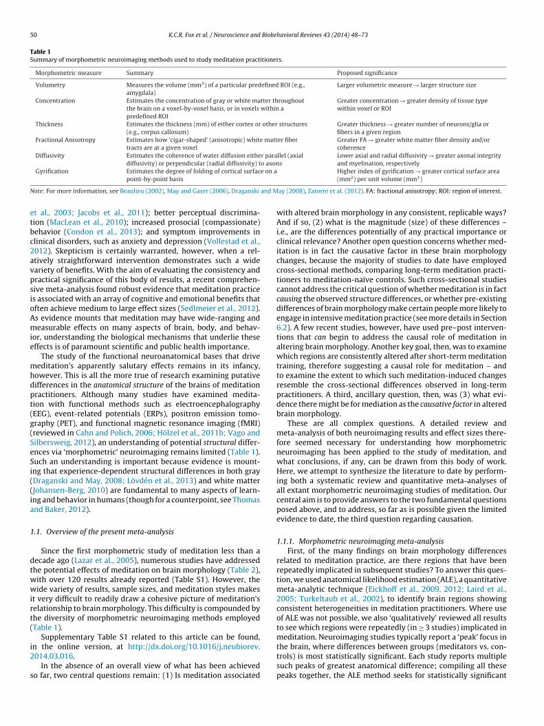
Table 1Summary of morphometric neuroimaging methods used to study meditation practitioners.
Morphometric measure Summary Proposed significance
Volumetry Measures the volume (mm3) of a particular predefined ROI (e.g.,amygdala)
Larger volumetric measure → larger structure size
Concentration Estimates the concentration of gray or white matter throughoutthe brain on a voxel-by-voxel basis, or in voxels within apredefined ROI
Greater concentration → greater density of tissue typewithin voxel or ROI
Thickness Estimates the thickness (mm) of either cortex or other structures(e.g., corpus callosum)
Greater thickness → greater number of neurons/glia orfibers in a given region
Fractional Anisotropy Estimates how ‘cigar-shaped’ (anisotropic) white matter fibertracts are at a given voxel
Greater FA → greater white matter fiber density and/orcoherence
Diffusivity Estimates the coherence of water diffusion either parallel (axialdiffusivity) or perpendicular (radial diffusivity) to axons
Lower axial and radial diffusivity → greater axonal integrityand myelination, respectively
Gyrification Estimates the degree of folding of cortical surface on a Higher index of gyrification → greater cortical surface area
N and M
etbc2avpsioAmie
mhdpt(g(SeSi((ia
1
dtwwirt(
i2
s
point-by-point basis
ote: For more information, see Beaulieu (2002), May and Gaser (2006), Draganski
t al., 2003; Jacobs et al., 2011); better perceptual discrimina-ion (MacLean et al., 2010); increased prosocial (compassionate)ehavior (Condon et al., 2013); and symptom improvements inlinical disorders, such as anxiety and depression (Vollestad et al.,012). Skepticism is certainly warranted, however, when a rel-tively straightforward intervention demonstrates such a wideariety of benefits. With the aim of evaluating the consistency andractical significance of this body of results, a recent comprehen-ive meta-analysis found robust evidence that meditation practices associated with an array of cognitive and emotional benefits thatften achieve medium to large effect sizes (Sedlmeier et al., 2012).s evidence mounts that meditation may have wide-ranging andeasurable effects on many aspects of brain, body, and behav-
or, understanding the biological mechanisms that underlie theseffects is of paramount scientific and public health importance.
The study of the functional neuroanatomical bases that driveeditation’s apparently salutary effects remains in its infancy,
owever. This is all the more true of research examining putativeifferences in the anatomical structure of the brains of meditationractitioners. Although many studies have examined medita-ion with functional methods such as electroencephalographyEEG), event-related potentials (ERPs), positron emission tomo-raphy (PET), and functional magnetic resonance imaging (fMRI)reviewed in Cahn and Polich, 2006; Hölzel et al., 2011b; Vago andilbersweig, 2012), an understanding of potential structural differ-nces via ‘morphometric’ neuroimaging remains limited (Table 1).uch an understanding is important because evidence is mount-ng that experience-dependent structural differences in both grayDraganski and May, 2008; Lövdén et al., 2013) and white matterJohansen-Berg, 2010) are fundamental to many aspects of learn-ng and behavior in humans (though for a counterpoint, see Thomasnd Baker, 2012).
.1. Overview of the present meta-analysis
Since the first morphometric study of meditation less than aecade ago (Lazar et al., 2005), numerous studies have addressedhe potential effects of meditation on brain morphology (Table 2),ith over 120 results already reported (Table S1). However, theide variety of results, sample sizes, and meditation styles makes
t very difficult to readily draw a cohesive picture of meditation’selationship to brain morphology. This difficulty is compounded byhe diversity of morphometric neuroimaging methods employedTable 1).
Supplementary Table S1 related to this article can be found,
n the online version, at http://dx.doi.org/10.1016/j.neubiorev.014.03.016.In the absence of an overall view of what has been achievedo far, two central questions remain: (1) Is meditation associated
(mm2) per unit volume (mm3)
ay (2008), Zatorre et al. (2012). FA: fractional anisotropy; ROI: region of interest.
with altered brain morphology in any consistent, replicable ways?And if so, (2) what is the magnitude (size) of these differences –i.e., are the differences potentially of any practical importance orclinical relevance? Another open question concerns whether med-itation is in fact the causative factor in these brain morphologychanges, because the majority of studies to date have employedcross-sectional methods, comparing long-term meditation practi-tioners to meditation-naïve controls. Such cross-sectional studiescannot address the critical question of whether meditation is in factcausing the observed structure differences, or whether pre-existingdifferences of brain morphology make certain people more likely toengage in intensive meditation practice (see more details in Section6.2). A few recent studies, however, have used pre–post interven-tions that can begin to address the causal role of meditation inaltering brain morphology. Another key goal, then, was to examinewhich regions are consistently altered after short-term meditationtraining, therefore suggesting a causal role for meditation – andto examine the extent to which such meditation-induced changesresemble the cross-sectional differences observed in long-termpractitioners. A third, ancillary question, then, was (3) what evi-dence there might be for mediation as the causative factor in alteredbrain morphology.
These are all complex questions. A detailed review andmeta-analysis of both neuroimaging results and effect sizes there-fore seemed necessary for understanding how morphometricneuroimaging has been applied to the study of meditation, andwhat conclusions, if any, can be drawn from this body of work.Here, we attempt to synthesize the literature to date by perform-ing both a systematic review and quantitative meta-analyses ofall extant morphometric neuroimaging studies of meditation. Ourcentral aim is to provide answers to the two fundamental questionsposed above, and to address, so far as is possible given the limitedevidence to date, the third question regarding causation.
1.1.1. Morphometric neuroimaging meta-analysisFirst, of the many findings on brain morphology differences
related to meditation practice, are there regions that have beenrepeatedly implicated in subsequent studies? To answer this ques-tion, we used anatomical likelihood estimation (ALE), a quantitativemeta-analytic technique (Eickhoff et al., 2009, 2012; Laird et al.,2005; Turkeltaub et al., 2002), to identify brain regions showingconsistent heterogeneities in meditation practitioners. Where useof ALE was not possible, we also ‘qualitatively’ reviewed all resultsto see which regions were repeatedly (in ≥ 3 studies) implicated inmeditation. Neuroimaging studies typically report a ‘peak’ focus in
the brain, where differences between groups (meditators vs. con-trols) is most statistically significant. Each study reports multiplesuch peaks of greatest anatomical difference; compiling all thesepeaks together, the ALE method seeks for statistically significant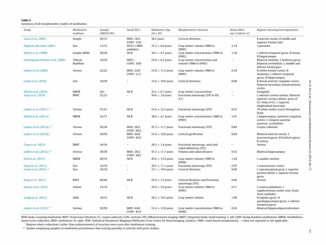
K.C.R
. Fox
et al.
/ N
euroscience and
Biobehavioral R
eviews
43 (2014)
48–73
51Table 2Summary of all morphometric studies of meditation.
Study Meditationtradition
Sample(MED/CON)
Hand (R/L) Meditators Age(M ± SD)
Morphometric measure Mean effectsize (Cohen’s d)
Regions showing heterogeneities
Lazar et al. (2005) Insight 20/15 MED: 18/2CONT: 15/0
38.2 years Cortical thickness – R anterior insula; R middle andsuperior frontal sulci
Pagnoni and Cekic (2007) Zen 13/13 All R (1 MEDambidex)
37.2 ± 6.9 years Gray matter volume (VBM inSPM5)
2.14 L putamen
Hölzel et al. (2008) Insight (BSM) 20/20 All R 34.1 ± 4.7 years Gray matter concentration (VBM inSPM2)
1.18 L inferior temporal gyrus; R insula;R hippocampus
Vestergaard-Poulsen et al. (2009) TibetanBuddhist
10/10 MED:–CONT: 10/0
55.0 ± 6.2 years Gray matter concentration andvolume (VBM in SPM5)
– Bilateral medulla; L fusiform gyrus;bilateral cerebellum; L middle andinferior frontal gyri;
Luders et al. (2009) Various 22/22 MED: 22/0CONT: 21/1
53.0 ± 11.5 years Gray matter volume (VBM inSPM5)
0.34 R orbito-frontal cortex; Rthalamus; L inferior temporalgyrus; R hippocampus
Grant et al. (2010) Zen 19/20 – 37.6 ± 10.9 years Cortical thickness 0.68 R dorsal anterior cingulate cortex;bilateral secondary somatosensorycortex
Hölzel et al. (2010) MBSR 26/– All R 35.2 ± 6.7 years Gray matter concentration – Amygdala*
Tang et al. (2010) IBMT 22/23 – 20.6 ± 1.6 years Fractional anisotropy (DTI in FSL4.1)
– L anterior corona radiata; bilateralsuperior corona radiata; genu ofCC; body of CC; L superiorlongitudinal fasciculus
Luders et al. (2011) � Various 27/27 All R 51.6 ± 12.3 years Fractional anisotropy (DTI) 0.55 18 white matter tracts throughoutbrain
Hölzel et al. (2011a) MBSR 16/17 All R 38.0 ± 4.1 years Gray matter concentration (VBM inSPM5)
1.91 L hippocampus; posterior cingulatecortex; L temporo-parietaljunction; cerebellum
Luders et al. (2012a) � Various 30/30 MED: 28/2CONT: 28/2
47.3 ± 11.7 years Fractional anisotropy (DTI) 0.89 Corpus callosum
Luders et al. (2012b) Various 50/50 MED: 44/6CONT: 45/5
51.4 ± 12.8 years Cortical gyrification 0.64 Bilateral anterior insula; Lprecentral gyrus; R fusiform gyrus;R cuneus
Tang et al. (2012) IBMT 34/34 – 20.5 ± 1.4 years Fractional anisotropy, axial andradial diffusivity (DTI)
– Various
Luders et al. (2012c) � Various 30/30 MED: 28/2CONT: 28/2
47.3 ± 11.7 years Volume and radial distance 0.53 Bilateral hippocampus
Farb et al. (2013) MBSR 20/16 All R 45.6 ± 13.4 years Gray matter volume (VBM inSPM8)
1.24 L caudate nucleus
Fayed et al. (2013) Zen 10/10 – 39.5 ± 11.1 years Fractional anisotropy (DTI) −2.97 L sensorimotor cortex*
Grant et al. (2013) � Zen 18/18 – 37.1 ± 10.9 years Cortical thickness 0.69 L supramarginal gyrus, L superiorparietal lobule, L superior frontalgyrus
Kang et al. (2013) BWV 46/46 All R 25.4 ± 3.3 years Cortical thickness and fractionalanisotropy (DTI)
0.60 Various
Kumar et al. (2013) Soham 14/14 – 29.4 ± 2.0 years Gray matter volume (VBM inSPM5)
0.71 L ventral palladium; Lsupplementary motor area; brainstem (medulla)
Leung et al. (2013) LKM 10/15 All R 50.2 ± 10.5 years Gray matter volume 1.69 R angular gyrus; Rparahippocampal gyrus; L inferiortemporal gyrus
Luders et al. (2013) � Various 50/50 MED: 44/6CONT: 45/5
51.4 ± 12.8 years Gray matter concentration (VBM inSPM8)
0.26 Bilateral hippocampus/subiculum
BSM: body-scanning meditation; BWV: brain wave vibration; CC: corpus callosum; CON: controls; DTI: diffusion tensor imaging; IBMT: integrative body-mind training; L: left; LKM: loving-kindness meditation; MBSR: mindfulness-based stress reduction; MED: meditators; R: right; SPM: Statistical Parametric Mapping (Wellcome Trust Centre for Neuroimaging, London); VBM: voxel-based morphometry. – = data not reported or not applicable.
* Regions where reductions (rather than enhancements) of structure were seen after meditation training.� Studies employing samples of meditation practitioners that overlap partially or entirely with prior studies.
5 Biobe
owai
1
idstCnqwtdgcdssadrwpno
1p
pma(2amsttapnci
soMs2
1
rmfiimI
2 K.C.R. Fox et al. / Neuroscience and
verlaps in the peaks from independent studies. In this way, weere able to compile a list of regions that appear to be consistently
ltered in meditation practitioners, across many independent stud-es and samples (see Section 2 for detailed information).
.1.2. Effect size meta-analysisKnowing that certain regions are consistently different in med-
tation practitioners, however, does not necessarily imply that theifferences are of any practical significance. Even if consistent andtatistically significant, such brain structure differences might beoo small to be considered relevant in a practical, everyday sense.alculation of effect sizes, however, which indicate not the sig-ificance but the magnitude of results, can begin to address theseuestions of practical significance (Cumming, 2013). Simply testinghether a result is significant or not (null-hypothesis significance
esting) is limited by the fact that attaining significance is very muchependent on sample size (Cumming, 2013) – and sample size isenerally quite small in most neuroimaging studies, due to the highosts involved. Effect sizes, however, estimate the magnitude ofifferences between groups, regardless of whether the result wastatistically significant (where non-significance, e.g., might be dueimply to small sample size). Our effect size meta-analysis thereforellowed an overview of the apparent magnitude of brain structureifferences reported in meditators. Although effect sizes are stillarely reported in neuroimaging studies, and their interpretationith respect to brain structure differences remains problematic andoorly developed at the theoretical level (Poldrack et al., 2008), weonetheless aimed to present all quantitative effect size data andffer some preliminary interpretations of their significance.
.2. Morphometric neuroimaging of brain structure in meditationractitioners
Brain ‘morphology’ refers to the structure, shape, and com-osition of the brain; the measurement and analysis of brainorphology via various neuroimaging techniques is gener-
lly known as ‘morphometry’ or ‘morphometric neuroimaging’Draganski and May, 2008; May and Gaser, 2006; Zatorre et al.,012). Broadly speaking, morphometric neuroimaging techniquesim to characterize anatomical differences based on a variety oforphological characteristics. Some relate solely to the brain’s
hape or size (e.g., cortical gyrification), others take into accounthe relative concentration or organization of gray and white mat-er (e.g., gray matter concentration), and yet others combine bothspects (e.g., volumetry of predefined gray matter structures). Mor-hometric neuroimaging stands in contrast, then, to ‘functional’euroimaging techniques such as fMRI, EEG, and PET, which aim toharacterize not brain structure, but brain activity, such as changesn electrical potentials or blood flow.
A brief overview of measures used to date in morphometrictudies of meditation is presented in Table 1 (for in-depth reviewsutside the field of meditation, see Draganski and May, 2008;ay and Gaser, 2006; Zatorre et al., 2012; for specific methods,
ee Ashburner and Friston, 2000; Beaulieu, 2002; Fischl and Dale,000).
.3. Does increase of structure equal enhancement of function?
A sometimes-tacit assumption underlying morphometric neu-oimaging is that greater values (structural ‘increase’) on a givenorphometric measure entail a corresponding enhancement of
unction. The structural increases in question could be, e.g., an
ncreased concentration of gray matter in a given region; anncreased thickness of cerebral cortex; increased integrity of whiteatter fibers; or any number of other measures (see Table 1).n support of this view, there are well-established connections
havioral Reviews 43 (2014) 48–73
between brain maturation and cognitive development, as well as acomplementary link between neurodegenerative disease, or atro-phy, and cognitive decline.
More specifically, there exists fairly robust evidence in favor ofthe brain structure–function connection in both animal models andhuman neuroimaging. Several important studies have establishedrelationships between structural ‘increases’ of both gray and whitematter (for recent reviews, see Taubert et al., 2012; Zatorre et al.,2012) and beneficial outcomes, including achievement in a varietyof fine motor skills, such as juggling (Draganski et al., 2004; Scholzet al., 2009) and musical instrument playing (Hyde et al., 2009).Even gross physical activities, such as aerobic exercise, show an‘enhancing’ effect on brain morphology (Colcombe et al., 2006).
Importantly, such differences are observed not only in responseto physical or motor skill training: some studies have recentlyfound morphometric differences after mental training in reasoning(Mackey et al., 2012) and working memory (Takeuchi et al., 2011).Conversely, structural deterioration or deficiencies measured viamorphometric neuroimaging have been linked to various forms ofcognitive decline, including normal age-related cognitive decline(Good et al., 2001) and Alzheimer’s disease (Frisoni et al., 2007).
The possibility remains, of course, that ‘less is more’ in at leastsome cases: the phenomenon of synaptic pruning provides a force-ful example (Low and Cheng, 2006). Structural increases mightalso indicate functional impairments in at least some cases: severalbrain regions related to stimulus-response learning and habit for-mation show structural increases in obsessive compulsive disorder,for instance (Pujol et al., 2004).
Morphometric neuroimaging in meditation practitioners hasgenerally aimed to explore whether meditation, too, is analogousto a form of (mental) skill learning, and can produce such anatom-ical changes. If so, brain structure increases related to meditativepractice might provide at least a partial neural explanation of thenumerous cognitive and emotional benefits associated with medi-tation (Sedlmeier et al., 2012). It should be acknowledged, however,that both in the field of morphometric neuroimaging as a whole, aswell as within the smaller realm involving meditation practition-ers in particular, the meaning of these brain structure differencesis still very poorly understood. Very few studies have been directlyreplicated, and very few have correlated behavioral changes withbrain structure differences. Enthusiasm about altered brain struc-ture in meditation practitioners should therefore be tempered bythe fact that the significance of these changes remains controver-sial (cf. Thomas and Baker, 2012); indeed, this is one of the mainreasons for the present meta-analysis.
1.4. Are disparate morphometric neuroimaging methodscomparable?
In collating data from multiple morphometric neuroimagingmodalities, our interest is in the regions where differences haveconsistently been reported, irrespective of imaging method. Theassumption is not that morphometric methods are necessarilydirectly comparable, but rather that particular brain regions arereliably involved in particular cognitive and emotional processes.Accordingly, alteration of a region’s structure (regardless of imag-ing method) is presumed to entail a corresponding alteration in itsfunction(s).
Whether a morphological difference in a single region willyield consistent results across morphometric methods is poorlyunderstood. Since very few studies employ multiple methodssimultaneously, direct comparisons are rare. However, there is
preliminary evidence that results from disparate methods are com-parable. For instance, Hutton et al. (2009) found broadly similarresults when comparing two different-aged populations, usingboth gray matter concentration and cortical thickness analysis, and
Biobe
Tcdscfad
1
c2pnVe(sp
armcPoottnlt
deeeipctedphWpt
1
abSotSgieo(l
K.C.R. Fox et al. / Neuroscience and
esta et al. (2004) found that volumetry methods showed resultsonsistent with gray matter concentration analysis. Nevertheless,ifferent methods should not be expected to produce entirely con-istent results, since they likely rely on different underlying cellularhanges for their outcomes (see Section 5.5). Ultimately, the dif-ering sensitivity of various methods may prove to be a source ofdditional information, rather than a shortcoming (for a criticaliscussion, see Lemaitre et al., 2012).
.5. The varieties of meditative experience
Meditation techniques vary enormously in aims, scope, diffi-ulty, and tentatively, recruitment of brain regions (Brewer et al.,011; Lee et al., 2012; Lou et al., 1999; Manna et al., 2010). Zenractice, for instance, tends to involve an open, undirected aware-ess of the present moment (Austin, 1999). Some traditions ofipassana (‘Insight’) meditation, on the other hand, focus veryxplicitly on body sensations in a directed, systematic fashionGoenka, 2000). Yet other practices involve detailed visualizations,imple awareness of the breath, or audible repetition of a particularhrase (a ‘mantra’) (Singh, 1979).
There are several influential attempts to find commonalitiesmong techniques, however. The most well-known scheme catego-izes practices into either ‘focused attention’ or ‘open monitoring’editations (Lutz et al., 2008), alternatively referred to as ‘con-
entrative’ and ‘mindfulness’ techniques, respectively (Cahn andolich, 2006). Focused attention practices involve concentrationf attention on a single object of meditation (e.g., the sensationsf the breath, the recitation of a phrase, or the mental visualiza-ion of an image). Open monitoring practices, sometimes referredo as ‘choiceless awareness,’ instead involve an open, receptive,on-judgmental attitude toward any and all experience, regard-
ess of origin (external/sensory or internal/mental) and affectiveone (positive, negative, or neutral).
With respect to morphometric neuroimaging, however, it isifficult to study the neural basis of each category (much lessach particular technique) independently of the others, for sev-ral reasons. Most practitioners examined to date have substantialxperience with multiple categories, and more specifically, theres a dearth of studies examining only focused attention meditationractitioners (since focused attention meditation is almost alwaysombined with, or followed by, open monitoring and compassionypes of meditation). Moreover, numerous studies mix practition-rs from multiple traditions in their analyses (Table 2). Therefore,espite the potential value of various classification schemes, com-arative analyses based on meditation type were not undertakenere (although, where possible, a tentative discussion is offered).hether distinct patterns of structural differences are related to
articular forms of meditation practice therefore remains a ques-ion for future research.
.6. Prior syntheses
Why the need for a new review and meta-analysis? Although number of major efforts toward theoretical integration haveeen published in recent years (Hölzel et al., 2011b; Vago andilbersweig, 2012; Farb et al., 2012), only a few thorough reviewsf functional and morphometric neuroimaging in meditation prac-itioners have been undertaken (Cahn and Polich, 2006; Chiesa anderretti, 2010; Ivanovski and Mahli, 2007; Rubia, 2009). Thoughenerally comprehensive, several include only ‘mindfulness’ med-tation, and only two recent studies (Sperduti et al., 2012; Tomasino
t al., 2013) have conducted quantitative meta-analyses (ALE)f the burgeoning neuroimaging literature. These meta-analysesSperduti et al., 2012; Tomasino et al., 2013) suffer from certainimitations, such as no calculation or discussion of effect sizes, andhavioral Reviews 43 (2014) 48–73 53
no basic checks to ensure the robustness of the meta-analytic data(e.g., funnel plots or fail-safe N calculations; see Egger et al., 1997).These limitations are common to many earlier meta-analyses ofmeditation’s cognitive and emotional effects as well (see Sedlmeieret al., 2012). Moreover, no synthesis or quantitative meta-analysiswhatsoever of morphometric (i.e., structural) neuroimaging of med-itation practitioners has yet been undertaken, despite the fact thatthe 21 studies examined here have already been cited more than2200 times. Prior reviews and meta-analyses have instead tendedto focus on functional neuroimaging results. In the present work weaim to fill this gap in the literature by providing a systematic reviewand quantitative meta-analysis of all morphometric neuroimagingstudies of meditation.
2. Review methods
2.1. Study selection
2.1.1. Search strategyTwo of us (KCRF and SN) searched MEDLINE (http://www.
pubmed.com), Google Scholar (http://scholar.google.com), andPsycINFO (http://www.apa.org/pub/databases/psycinfo/index.aspx) for all papers containing the word ‘meditation’ since the firstmorphometric study of contemplative practices was published(Lazar et al., 2005). These extensive lists of articles were thenrefined by searching within results for studies that containedany of the words or phrases ‘magnetic resonance imaging’, ‘MRI,’‘neuroimaging,’ ‘diffusion tensor imaging,’ or ‘brain’ within the titleor abstract. Of the remaining results, every abstract was consultedto see if the study indeed employed morphometric methods tostudy meditation. The reference lists of each study found, as wellas those of several major reviews, were also consulted, to ensurethat no studies were missed.
2.1.2. Excluded studiesStudies examining effects of related practices on brain mor-
phology, such as Tai Chi (Wei et al., 2013) and hatha yoga(Froeliger et al., 2012a,b), were excluded due to potential con-founds and non-comparability. Studies examining morphologicalheterogeneities related to ‘dispositional’ (i.e., questionnaire-based)mindfulness measures (Taren et al., 2013; Murakami et al., 2013)were likewise excluded due to the unknown reliability of mind-fulness questionnaires, and their ambiguous relationship to actualmeditation practice and its effects (the widely used ‘Five FacetMindfulness Questionnaire’ [FFMQ], for instance, correlates just aswell with education as with meditation experience; Baer et al.,2008). We also excluded two studies investigating the relation-ship between meditation and brain structure that employed clinicalpopulations with either Parkinson’s disease (Pickut et al., 2013) ormild cognitive impairment (Wells et al., 2013). Although the inter-action between meditation practice, neurodegenerative disease,and brain morphology is of immense interest, these clinical dis-orders are in themselves thought to involve significant alterationsin brain structure (e.g., Jack et al., 1999; Ramirez-Ruiz et al., 2005),presenting an obvious confound if included in our meta-analyses.
2.1.3. Included studiesOnly studies that actually involved either short-term medita-
tion training or long-term meditation practitioners were included(Table 2); that is, studies that used questionnaire-based measurespurporting to measure ‘mindfulness’ or some other construct were
not included (see above, Section 2.1.2). Comparison groups weregenerally age-, sex-, and handedness-matched control subjectswith no meditation experience. For short-term meditation trainingstudies, wait-list controls with an interest in the same meditation
5 Biobe
imwsicc
hucedhptnaas3
2
imrScDmptm
swvtegS
2
ctttt
2
ftotaaew
cc
4 K.C.R. Fox et al. / Neuroscience and
ntervention were usually employed. Evidently, random assign-ent is not possible when comparing long-term practitionersith meditation-naïve controls (for more on the possibility of
elf-selection bias in expert practitioners, see Section 6.2). All stud-es of short-term training, however, used random assignment toontrol and experimental conditions. Overall, 21 studies met ourriteria and were included in the meta-analysis (Table 2).
Three of these 21 included studies used subjects who reportedigh levels of stress on self-report questionnaires and who vol-ntarily enrolled in a mindfulness-based stress reduction (MBSR)ourse for short-term meditation training (Farb et al., 2013; Hölzelt al., 2010, 2011a). High self-reported stress is far from a clinicaliagnosis of a mental health disorder, however, and all subjectsad voluntarily enrolled in such courses (vs. being enrolled by ahysician or other caregiver, for example). We therefore decidedhat these studies warranted inclusion in the meta-analysis, andeed not be excluded (as were the two clinical studies notedbove; Section 2.1.2). In any case, as discussed in Section 3, over-ll meta-analytic results were hardly affected by whether these fewhort-term training studies were included or excluded (see Sections.1 and 3.3).
.2. Short-term vs. long-term meditation training
For the purposes of this meta-analysis, short-term med-tation refers to pre–post intervention-style studies where
editation-naïve novices were given brief meditation training,anging between ∼5 and 60 h of actual meditation practice (Table1). These novices undergoing training were compared to wait-listontrol groups who also had an interest in learning meditation.ifferences in brain structure were examined by comparing brainorphology before and after this meditation intervention (com-
ared to wait-list control groups). Of the 21 studies examined inhe meta-analysis, five were pre–post studies involving short-term
editation training.Studies of ‘long-term’ practitioners were instead cross-sectional
tudies comparing highly advanced meditation experts to controlsith no meditation experience whatsoever. The range of practice
aried enormously for long-term practitioners, but all had at leasthousands of hours, or several years’ worth, of meditation experi-nce (Table S1). Of the 21 studies included in the meta-analysis, thereat majority (16/21) involved long-term practitioners (see Table1).
.3. Review method
As noted in the Introduction, we had two main goals: (1) toompile peak brain foci of anatomical difference between medi-ators and controls and examine whether there were any regionshat have been consistently reported across studies; and if so, (2)o examine effect sizes for these anatomical differences and assessheir magnitude.
.3.1. Classification of primary dataTo begin, we had to decide which results to include. Aside
rom the variety of morphometric measures employed (Table 1),here were differences in statistical significance thresholds; meth-ds of correcting for multiple comparisons; voxel cluster sizehresholds; and combinations of exploratory whole-brain (WB)nd region-of-interest (ROI) analyses. We therefore followed studyuthors’ own criteria for a ‘significant’ result (typically, family wiserror [FWE] rates of <.05 or <.01 across whole brain analyses, or
ithin ROIs).Many studies also reported results trending toward signifi-ance, or that were only significant before correcting for multipleomparisons. With a view toward completeness, we summarize
havioral Reviews 43 (2014) 48–73
and discuss all reported results, indicating statistic (t, Z, F, orr) magnitudes, cluster sizes (k), analysis type (whole-brain orROI), trend results (designated by # symbol), and negative results(controls > meditators; designated by an asterisk [*]) wheneveravailable (all data presented in Table S1). Both trend and stringentresults were included in the main anatomical likelihood estima-tion (ALE) meta-analysis and qualitative review, but to ensurerigorous findings, supplemental ALE meta-analysis and qualitativereview were also undertaken utilizing only stringently significantresults.
Although we summarize every reported result to date (TableS1), we do not systematically discuss every finding, for several rea-sons: many results are (1) reported at lenient statistical thresholds,(2) not yet replicated by other studies or research groups, or (3)based on largely overlapping (non-independent) samples of medi-tation practitioners (the difficulty in recruiting highly experiencedpractitioners has resulted in repeated use of the same subjects inseveral studies; see Table 2 for details). Therefore, even though weinclude all reported results in our ALE and effect size meta-analyses,we focus our discussion on brain regions reported repeatedly, inmultiple studies.
2.3.2. Determining consistent brain structure differencesTo address our first goal of identifying brain regions consis-
tently reported across studies, we conducted both a qualitativereview and a quantitative meta-analysis (the former was neces-sary because not all studies provided quantitative data amenableto an ALE meta-analysis). For the qualitative review, we tabulatedgroup (meditation practitioners vs. controls) and training (pre- vs.post-training) morphology differences reported in all studies (TableS1). In order to discern well-replicated results, we then soughtregions exhibiting differences in three or more independent stud-ies (i.e., regions suggesting substantial consistency across studiesinasmuch as two broad replications have already been obtained inthis relatively small body of work; cf. Kempton et al., 2008). Becausesome studies have used overlapping samples of meditation practi-tioners, however, contributing studies are not necessarily entirelyindependent (see Table 2 for details).
In addition to this ‘qualitative’ review, wherever possible, neu-roimaging data were compiled for a quantitative ALE meta-analysis(detailed methods below). Combining results from both methods,we discuss each brain region either: (1) showing morphologicaldifferences in ≥ 3 separate studies (i.e., two replications of a givenfinding, as in Kempton et al., 2008), or (2) demonstrating signif-icance in the ALE meta-analysis (methods in Section 2.4). Bothmethods result in a largely overlapping list of brain regions (seeSections 3 and 4).
We also performed supplemental qualitative (Table S2) andquantitative ALE (Table S3) meta-analyses in order to determineconsistent brain structure differences in meditators based onlyon results reported at stringent (FWE- or FDR-corrected) statis-tical thresholds. There are many potential pitfalls to assumingthe importance of a result based solely on null hypothesis sig-nificance testing, however (Cumming, 2013), and so we thereforeconducted another ‘stringent’ ALE meta-analysis based not onp-values but instead on effect sizes (Table S4). This final ALEmeta-analysis only included results that met or exceeded a ‘large’effect size (Cohen’s d ≥ ±1.0). As all of these additional analyses(Tables S2–S4) strongly parallel the results of our main neuroimag-ing meta-analyses (Tables 3 and 4), we report them in Supplemental
Materials.Supplementary Tables S2–S4 related to this article can be found,in the online version, at http://dx.doi.org/10.1016/j.neubiorev.2014.03.016.
K.C.R. Fox et al. / Neuroscience and Biobehavioral Reviews 43 (2014) 48–73 55
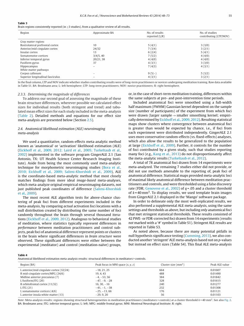
Table 3Brain regions consistently reported (in ≥3 studies), from a qualitative review of all results.
Region Approximate BA No. of resultsreported (L/R)
No. of studiescontributing (LTP/NOV)
Gray matter regionsRostrolateral prefrontal cortex 10 5 (4/1) 3 (3/0)Anterior/mid cingulate cortex 24/32 7 (3/4) 3 (2/1)Insular cortex 13 6 (2/4) 5 (4/1)Somatomotor cortices 3/4/5, 40 7 (5/2) 4 (4/0)Inferior temporal gyrus 20/21, 38 4 (4/0) 4 (4/0)Fusiform gyrus 37 4 (3/1) 3 (3/0)Hippocampus – 7 (4/3) 4 (3/1)White matter pathwaysCorpus callosum – 9 (5/–) 5 (3/2)
I of loni OV: n
2
bsl(m
2m
k(2Att2imwje
tmnrtopjioe
As noted above, because there are many potential pitfalls in
TA
NB
Superior longitudinal fasciculus –
n the final column, LTP and NOV indicate whether studies contributing results weren Table S1. BA: Brodmann area; L: left hemisphere; LTP: long-term practitioners; N
.3.3. Determining the magnitude of differencesTo address our second goal of assessing the magnitude of these
rain structure differences, wherever possible we calculated effectizes for individual results (both stringent and trend) and tabu-ated mean effect sizes for each study included in the meta-analysisTable 2). Detailed methods and equations for our effect size
eta-analysis are presented below (Section 2.5).
.4. Anatomical likelihood estimation (ALE) neuroimagingeta-analysis
We used a quantitative, random-effects meta-analytic methodnown as ‘anatomical’ or ‘activation’ likelihood estimation (ALE)Eickhoff et al., 2009, 2012; Laird et al., 2005; Turkeltaub et al.,002) implemented in the software program GingerALE 2.1 (Sanntonio, TX: UT Health Science Center Research Imaging Insti-
ute). Aside from being the most commonly used meta-analyticechnique for morphometric neuroimaging studies (Bora et al.,010; Eickhoff et al., 2009; Salimi-Khorshidi et al., 2009), ALE
s the coordinate-based meta-analytic method that most closelyatches findings from more ideal image-based meta-analyses,hich meta-analyze original empirical neuroimaging datasets, not
ust published peak coordinates of difference (Salimi-Khorshidit al., 2009).
The most recent ALE algorithm tests for above-chance clus-ering of peak foci from different experiments included in the
eta-analysis, by comparing actual activation foci locations with aull distribution created by distributing the same number of fociandomly throughout the brain through several thousand itera-ions (Eickhoff et al., 2009, 2012). Analogous to behavioral studiesf meditation, where statistics typically represent differences inerformance between meditation practitioners and control sub-ects, peak foci of anatomical difference represent points or clusters
n the brain where significant differences in brain structure werebserved. These significant differences were either between thexperimental (meditator) and control (meditation-naïve) groups,able 4natomical likelihood estimation meta-analysis results: structural differences in meditat
Region (BA) Peak focus in MNI space (x
L anterior/mid cingulate cortex (32/24) −18, 21, 25R mid-cingulate cortex/MFG (24/6) 19, 4, 43
Midline anterior precuneus (7) −4, −53, 56
L fusiform/ITG (20) −47, −9, −28
R orbitofrontal cortex (11/32) 16, 30, −16
L ITG (21) −41, −1, −38
L somatomotor cortices (4/6) −25, −13, 66
L anterior insula white matter (13) −29, 9, 20
ote: Meta-analysis results: regions showing structural heterogeneities in meditation praA: Brodmann area; ITG: inferior temporal gyrus; L: left; MFG: middle frontal gyrus; MN
4 (3/1) 3 (2/1)
g-term practitioners or novices undergoing meditation training. Raw data availableovice practitioners; R: right hemisphere.
or, in the case of short-term meditation training, differences withinthe same subjects at pre- and post-intervention time periods.
Included anatomical foci were smoothed using a full-widthhalf maximum (FWHM) Gaussian kernel dependent on the samplesize (number of participants) of the experiment from which fociwere drawn (larger sample → smaller smoothing kernel; empiri-cally determined by Eickhoff et al., 2009, 2012). Resulting statisticalmaps show clusters where convergence between anatomical fociis greater than would be expected by chance, i.e., if foci fromeach experiment were distributed independently. GingerALE 2.1uses more conservative random-effects (vs. fixed-effects) analyses,which also allow the results to be generalized to the populationat large (Eickhoff et al., 2009). Further, it controls for the numberof foci contributed by a given study, such that studies reportingmany foci (e.g., Kang et al., 2013) do not disproportionately affectthe meta-analytic results (Turkeltaub et al., 2012).
A total of 78 anatomical foci drawn from 14 experiments weremeta-analyzed. The remaining 7 studies either did not provide, ordid not use methods amenable to the reporting of, peak foci ofanatomical difference. Statistical maps provided meta-analytic lociof maximal likely anatomical difference between meditation prac-titioners and controls, and were thresholded using a false discoveryrate (FDR; Genovese et al., 2002) of q = .05 and a cluster thresholdof k = 40 mm3. To display results, we used template brain imagesfrom GingerALE 2.1 displayed in the ‘Mango’ software package.
In order to delineate only the most well-replicated results, wealso performed a supplemental ALE meta-analysis, using the samesettings and thresholds as above, but including only anatomical focithat met stringent statistical thresholds. These results consisted of42 FWE- or FDR-corrected foci drawn from 14 experiments (resultsnot marked with a ‘#’ symbol in Table S1). Stringent ALE results arereported in Table S3.
null hypothesis significance testing (Cumming, 2013), we also con-ducted another ‘stringent’ ALE meta-analysis based not on p-valuesbut instead on effect sizes (Table S4). This final ALE meta-analysis
ors > controls.
, y, z) Cluster size (mm3) Peak ALE value
664 0.01607488 0.01490384 0.01842328 0.01615240 0.01277232 0.01308176 0.01121
56 0.01103
ctitioners (meditators > controls) at a cluster threshold k = 40 mm3. See also Fig. 2.I: Montreal Neurological Institute; R: right.
5 Biobe
o(1
2
2
tt1ospbt(
d
wtri
d
wtfvMAscmd
vSSmi
iasrto
2
pvMresrwfi
ea
6 K.C.R. Fox et al. / Neuroscience and
nly included results that met or exceeded a ‘large’ effect sizeCohen’s d ≥ ±1.0). This ALE meta-analysis included 34 foci from0 studies. Large effect size ALE results are reported in Table S4.
.5. Effect size meta-analysis
.5.1. General methodIn addition to determining which brain regions were consis-
ently altered in meditation practitioners, we sought to evaluatehe magnitude of these differences, i.e., their effect size (Cohen,992; Lipsey and Wilson, 1993; Sedlmeier et al., 2012). In all, 16f 21 studies provided sufficient data to allow calculation of effectizes (see Table 2 for the mean effect size of each study). Whereverossible, we used studies’ t-statistics of group differences betweenrain morphology of meditators vs. controls, or of meditation prac-itioners before and after short-term training, to calculate effect sizeCohen’s d) for each result using Eq. (1) (Ray and Shadish, 1996):
= t
√1ne
+ 1nc
, (1)
here t is the value of the reported peak t-statistic, and ne and nc arehe sample sizes for experimental (meditation) and control groups,espectively. Where means and standard deviations were availablenstead, we used Eq. (2):
= Me − Mc
sp, (2)
here Me and Mc are the means of the experimental and con-rol groups, respectively, and sp is the pooled standard deviationrom both groups. Occasionally, where only F-statistics were pro-ided, effect sizes were calculated using the online Practicaleta-Analysis Effect Size Calculator (Lipsey and Wilson, 2001).
fter calculating effect sizes for 132 unique results, a mean effectize was calculated for each of the 16 studies (Table 2). Further, weompared mean effect sizes by brain tissue type examined (grayatter vs. white matter) to investigate whether meditation has
ifferential effects on given tissue types.To facilitate comparability with prior meta-analyses, we con-
erted Cohen’s d scores to r, following the formula used inedlmeier et al. (2012). In line with the recommendations ofchmidt et al. (2009), we calculated 95% confidence intervals forean effect sizes, to provide an estimate of their precision, follow-
ng Eqs. (8) and (10) in Sedlmeier et al. (2012).Negative effect sizes (results of controls > meditators) were also
ncluded in the above statistical meta-analysis, except where theuthors interpreted decreased morphometric measure values astructural increase. This was the case with, for instance, axial andadial diffusivity (see Table 1). In these cases, the sign of negative-statistics was reversed to correctly count the result as a ‘positive’r increasing effect on brain structure.
.5.2. Adjusting for potential inflation of effect sizesThe standard procedure in neuroimaging literature is to report
eak t- or F-statistics only, which by definition are the extremealues for a given significant cluster of difference between groups.ean t-statistics for an entire cluster of difference are rarely
eported. Our calculated effect sizes thus represent the peak,xtreme effects for each given result. On the other hand, mean t-tatistics for given clusters are guaranteed to be lower. As such, ouresults necessarily overestimate the effect size of the cluster as ahole. This caveat should be kept in mind when interpreting these
ndings.In an effort to address the problem of inflated effect sizes, wexamined studies where both effect sizes based on peak t-statisticsnd those based on mean t-statistics for the entire cluster of
havioral Reviews 43 (2014) 48–73
significant difference were reported. Our aim was to get a sense ofthe inflationary bias caused by reporting of only peak t-statistics(vs. t-statistics for entire clusters of significant difference), andthen adjust (deflate) mean effect sizes from other studies accord-ingly. Unfortunately, only a single study (Kang et al., 2013) providedboth peak and mean cluster t-statistics. This study reported a largenumber of results (n = 44), however, adding some validity to thecomparison. As cluster mean effect sizes were found to be muchsmaller (about 57% as large as effect size from peak foci only), weadjusted other studies’ mean effect sizes accordingly, assuming acomparable difference between cluster and peak effect sizes (seeSection 3.7). Though ideally, of course, such a deflation of effectsizes would be based on more data from multiple studies, no fur-ther data were available to us. This method of adjusting mean effectsizes for this peak vs. cluster bias therefore seemed to us the bestavailable method, given the limited data at our disposal.
2.5.3. Other caveats regarding effect sizes in neuroimagingA further problem is that meta-analysis of mean effect sizes
should also include all null (non-significant) results. Standard pro-cedure in neuroimaging studies is to set a significance threshold andreport only differences that exceed the threshold, i.e., attain signifi-cance. Null results are therefore rarely reported, except where ROIsare investigated. This introduces an unknown amount of inflation-ary bias in the mean effect sizes for the remaining studies, a bias forwhich, to our knowledge, there is no correction as of yet. That said,of the 16 studies contributing effect sizes, a fair number (six) didprovide some null results. This yielded 41 null result-based effectsizes (out of a total of 132 effect sizes calculated) in our effect sizemeta-analysis. Even including these many null results, and afterthe adjustments mentioned above, mean effect sizes here for eachstudy should probably still be considered to be overestimates tosome extent (because many studies here report no null or negative[controls > meditators] results whatsoever).
Another concern is interpretation of effect sizes: the interpretiveguidelines laid down by Cohen (1992) were intended for the behav-ioral sciences, not neuroimaging. Although it seems reasonable tous to use similar guidelines (i.e., the general assumption that a one-half standard deviation difference between groups is meaningfuland of practical significance), rigorous discussion and elaborationof these ideas has, to our knowledge, not yet been undertaken inthe field of neuroimaging. Indeed, even in the behavioral and socialsciences, the interpretation of effect sizes as ‘small,’ ‘medium,’ or‘large’ is ultimately arbitrary (Cohen, 1992).
Despite these limitations, effect sizes have been profitablyemployed in prior meta-analyses of morphometric neuroimagingstudies (e.g., Kempton et al., 2008). The benefits of reporting effectsize information appear to us to outweigh the drawbacks inherentin their calculation and interpretation for neuroimaging studies.We therefore calculate and report effect sizes here for complete-ness and in order to get a general sense of the magnitude of brainstructure differences reported in meditation practitioners, but weemphasize the need for caution in interpreting these results.
2.6. Estimating publication bias in meta-analytic results
The bias toward publication of only positive results (the ‘filedrawer’ problem) is a serious concern (Rosenthal, 1979). We con-structed a funnel plot (scatterplot of effect size against samplesize) to test for potential publication bias in our sample of studies(Egger et al., 1997). Effect sizes were calculated as described above,
and plotted against total sample size (meditators + controls). Fordetailed discussion of funnel plots see Egger et al. (1997); for anexample of their use in a meta-analysis of psychological effects ofmeditation, and further discussion, see Sedlmeier et al. (2012).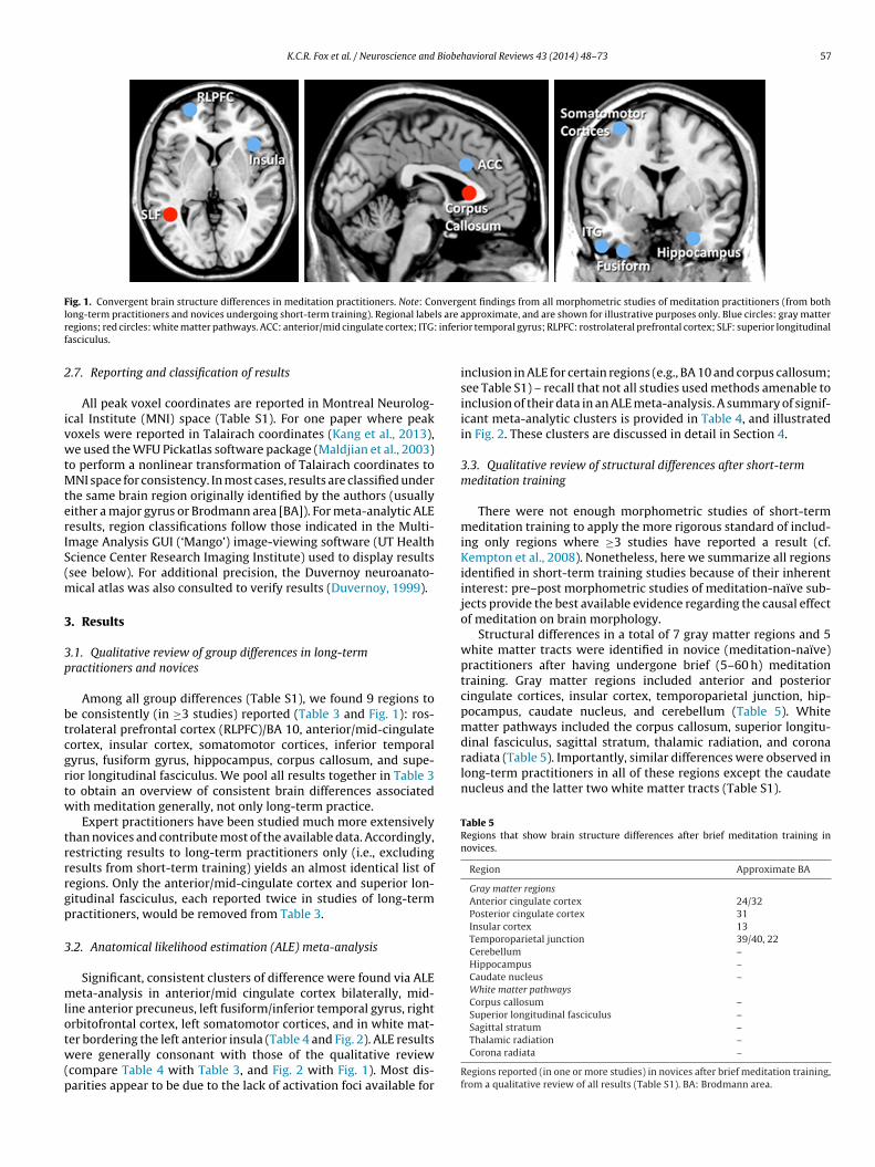
K.C.R. Fox et al. / Neuroscience and Biobehavioral Reviews 43 (2014) 48–73 57
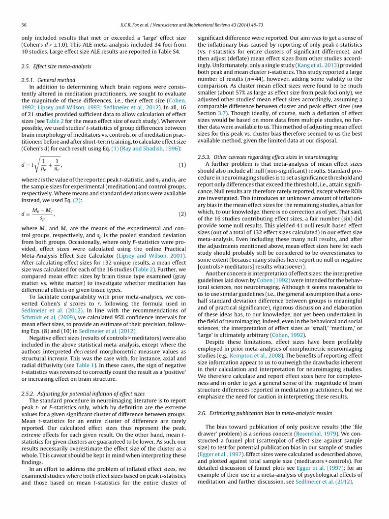
Fig. 1. Convergent brain structure differences in meditation practitioners. Note: Convergent findings from all morphometric studies of meditation practitioners (from bothl ls are ar inferif
2
ivwtMterIS(m
3
3p
btcgrtw
trrrgp
3
mlotw(p
dinal fasciculus, sagittal stratum, thalamic radiation, and coronaradiata (Table 5). Importantly, similar differences were observed inlong-term practitioners in all of these regions except the caudatenucleus and the latter two white matter tracts (Table S1).
Table 5Regions that show brain structure differences after brief meditation training innovices.
Region Approximate BA
Gray matter regionsAnterior cingulate cortex 24/32Posterior cingulate cortex 31Insular cortex 13Temporoparietal junction 39/40, 22Cerebellum –Hippocampus –Caudate nucleus –White matter pathwaysCorpus callosum –Superior longitudinal fasciculus –Sagittal stratum –
ong-term practitioners and novices undergoing short-term training). Regional labeegions; red circles: white matter pathways. ACC: anterior/mid cingulate cortex; ITG:asciculus.
.7. Reporting and classification of results
All peak voxel coordinates are reported in Montreal Neurolog-cal Institute (MNI) space (Table S1). For one paper where peakoxels were reported in Talairach coordinates (Kang et al., 2013),e used the WFU Pickatlas software package (Maldjian et al., 2003)
o perform a nonlinear transformation of Talairach coordinates toNI space for consistency. In most cases, results are classified under
he same brain region originally identified by the authors (usuallyither a major gyrus or Brodmann area [BA]). For meta-analytic ALEesults, region classifications follow those indicated in the Multi-mage Analysis GUI (‘Mango’) image-viewing software (UT Healthcience Center Research Imaging Institute) used to display resultssee below). For additional precision, the Duvernoy neuroanato-
ical atlas was also consulted to verify results (Duvernoy, 1999).
. Results
.1. Qualitative review of group differences in long-termractitioners and novices
Among all group differences (Table S1), we found 9 regions toe consistently (in ≥3 studies) reported (Table 3 and Fig. 1): ros-rolateral prefrontal cortex (RLPFC)/BA 10, anterior/mid-cingulateortex, insular cortex, somatomotor cortices, inferior temporalyrus, fusiform gyrus, hippocampus, corpus callosum, and supe-ior longitudinal fasciculus. We pool all results together in Table 3o obtain an overview of consistent brain differences associatedith meditation generally, not only long-term practice.
Expert practitioners have been studied much more extensivelyhan novices and contribute most of the available data. Accordingly,estricting results to long-term practitioners only (i.e., excludingesults from short-term training) yields an almost identical list ofegions. Only the anterior/mid-cingulate cortex and superior lon-itudinal fasciculus, each reported twice in studies of long-termractitioners, would be removed from Table 3.
.2. Anatomical likelihood estimation (ALE) meta-analysis
Significant, consistent clusters of difference were found via ALEeta-analysis in anterior/mid cingulate cortex bilaterally, mid-
ine anterior precuneus, left fusiform/inferior temporal gyrus, rightrbitofrontal cortex, left somatomotor cortices, and in white mat-
er bordering the left anterior insula (Table 4 and Fig. 2). ALE resultsere generally consonant with those of the qualitative reviewcompare Table 4 with Table 3, and Fig. 2 with Fig. 1). Most dis-arities appear to be due to the lack of activation foci available for
pproximate, and are shown for illustrative purposes only. Blue circles: gray matteror temporal gyrus; RLPFC: rostrolateral prefrontal cortex; SLF: superior longitudinal
inclusion in ALE for certain regions (e.g., BA 10 and corpus callosum;see Table S1) – recall that not all studies used methods amenable toinclusion of their data in an ALE meta-analysis. A summary of signif-icant meta-analytic clusters is provided in Table 4, and illustratedin Fig. 2. These clusters are discussed in detail in Section 4.
3.3. Qualitative review of structural differences after short-termmeditation training
There were not enough morphometric studies of short-termmeditation training to apply the more rigorous standard of includ-ing only regions where ≥3 studies have reported a result (cf.Kempton et al., 2008). Nonetheless, here we summarize all regionsidentified in short-term training studies because of their inherentinterest: pre–post morphometric studies of meditation-naïve sub-jects provide the best available evidence regarding the causal effectof meditation on brain morphology.
Structural differences in a total of 7 gray matter regions and 5white matter tracts were identified in novice (meditation-naïve)practitioners after having undergone brief (5–60 h) meditationtraining. Gray matter regions included anterior and posteriorcingulate cortices, insular cortex, temporoparietal junction, hip-pocampus, caudate nucleus, and cerebellum (Table 5). Whitematter pathways included the corpus callosum, superior longitu-
Thalamic radiation –Corona radiata –
Regions reported (in one or more studies) in novices after brief meditation training,from a qualitative review of all results (Table S1). BA: Brodmann area.
58 K.C.R. Fox et al. / Neuroscience and Biobehavioral Reviews 43 (2014) 48–73
Fig. 2. Anatomical likelihood estimation (ALE) meta-analysis of significant brain morphology differences (meditators > controls). Note: Significant meta-analytic clusters ofmorphological difference were found in 8 regions, and one additional trend-level cluster. (a) Right mid-cingulate cortex/middle frontal gyrus (BA 24/6). (b) White matternear the left anterior/mid-cingulate cortex (BA 32/24). (c) Right orbitofrontal cortex (BA 11/32). (d) Left fusiform gyrus (BA 20). (e) Left inferior temporal gyrus (BA 21).( r preca peaks
T
3
n(�
3e
bao1mw
f) Trend-level cluster in rostrolateral prefrontal cortex (BA 10). (g) Midline anterionterior insula (BA 13). Color bar indicates likelihood that peaks represent actual
able 4 for complete listing. BA: Brodmann area.
.4. Hemispheric asymmetries
Of all lateralized findings (118 results), a somewhat greaterumber were reported in the left (69) vs. right (49) hemisphereTable S1). However, the difference was not statistically significant2(1) = 3.39, p = .066.
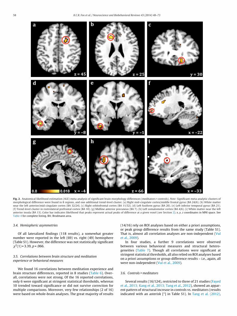
.5. Correlations between brain structure and meditationxperience or behavioral measures
We found 16 correlations between meditation experience andrain structure differences, reported in 8 studies (Table 6). Over-ll, correlations were not strong. Of the 16 reported correlations,
nly 6 were significant at stringent statistical thresholds, whereas0 trended toward significance or did not survive correction forultiple comparisons. Moreover, very few relationships (2 of 16)ere based on whole-brain analyses. The great majority of resultsuneus (BA 7). (h) Left somatomotor cortex (BA 4/6). (i) White matter near the leftof difference at a given voxel (see Section 2). x, y, z coordinates in MNI space. See
(14/16) rely on ROI analyses based on either a priori assumptions,or peak group difference results from the same study (Table S1).That is, almost all correlation analyses are non-independent (Vulet al., 2009).
In four studies, a further 9 correlations were observedbetween various behavioral measures and structural hetero-geneities (Table 7). Though all correlations were significant atstringent statistical thresholds, all also relied on ROI analyses basedon a priori assumptions or group-difference results – i.e., again, allwere non-independent (Vul et al., 2009).
3.6. Controls > meditators
Several results (16/124), restricted to three of 21 studies (Fayedet al., 2013; Kang et al., 2013; Tang et al., 2012), showed an appar-ent pattern of structural increase in controls vs. meditators (resultsindicated with an asterisk [*] in Table S1). In Tang et al. (2012),
K.C.R. Fox et al. / Neuroscience and Biobehavioral Reviews 43 (2014) 48–73 59
Table 6Regions where brain structure differences correlate with meditation experience level.
Region name, peak voxel inMNI space (x, y, z), BA
T at peakvoxel or r
Side Clustersize (k)
Analysistype
Morphometricmeasure
Experience (M ± SD) Age (M ± SD) Study
Visual cortexInferior occipitotemporal* Partial
r = .63R – ROI CT 9.1 ± 7.1 years 38.2 years Lazar et al. (2005)
Middle temporal gyrus–# r = −.59 L – ROI GMV 6456.2 ± 6040.6 h 50.2 ± 10.5
yearsLeung et al. (2013)
Inferior temporal gyrus– r = .40 L – ROI GMC 6254.0 ± 4529.0 h 34.1 ± 4.7 years Hölzel et al. (2008)Inferior temporal lobe– t ∼= 3.5 R – ROI CG 19.8 ± 11.4 years 51.4 ± 12.8
yearsLuders et al.(2012b)
Insula–# r = .36 R – ROI GMC 6254.0 ± 4529.0 h 34.1 ± 4.7 years Hölzel et al. (2008)–# t ∼= 3.5 R – ROI CG 19.8 ± 11.4 years 51.4 ± 12.8
yearsLuders et al.(2012b)
Orbitofrontal cortex1, 45, −16 (BA 11)# t = 4.28 R 185 WB GMC 6254.0 ± 4529.0 h 34.1 ± 4.7 years Hölzel et al. (2008)Somatosensory cortices33, −26, 59 (BA 1–3) t = 5.42 R – WB CT 6404.0 ± 8522.0 h 37.6 ± 7.9 years Grant et al. (2010)−8, −42, 60 (BA 1–3)# t = 3.28 L – ROI CT 6404.0 ± 8522.0 h 37.6 ± 7.9 years Grant et al. (2010)Anterior cingulate7, 22, 31# t = 2.01 R – ROI CT 14.4 ± 8.4 years 37.6 ± 7.9 years Grant et al. (2010)−6, −3, 42# t = 1.82 L – ROI CT 14.4 ± 8.4 years 37.6 ± 7.9 years Grant et al. (2010)Angular gyrus– r = −.76 R – ROI GMV 6456.2 ± 6040.6 h 50.2 ± 10.5
yearsLeung et al. (2013)
Cingulate cortex hippocampus–# r = .40 L – ROI FA 23.3 ± 12.2 years 51.6 ± 12.3
yearsLuders et al. (2011)
Cingulate cortex cingulum–# r = .36 R – ROI FA 23.3 ± 12.2 years 51.6 ± 12.3
yearsLuders et al. (2011)
Premotor cortexBA 6# r = .31 L – ROI CT 3.4 ± 2.3 years 25.4 ± 3.3 years Kang et al. (2012)Hippocampus−18, −37, −11 Partial
r = .33L – ROI GMC 19.8 ± 11.4 years 51.4 ± 12.8
yearsLuders et al. (2013)
BA: Brodmann area; CT: cortical thickness; GMC: gray matter concentration; L: left; MNI: Montreal Neurological Institute; R: right; ROI: region of interest; WB: whole-brain.* Significant after controlling for age and average hemisphere thickness.# Results trending toward significance and/or uncorrected for multiple comparisons.
Table 7Regions where brain structure differences correlate with behavioral measures in meditation practitioners.
Region name, peakvoxel in MNI space (x,y, z), BA
T at peakvoxel or r
Side Cluster size(k)
Analysistype
Morphometricmeasure
Behavioralmeasure
Study
Visual cortexInferioroccipitotemporal
r = .72 R – ROI CT Respiration rate Lazar et al. (2005)
Insula–* Partial
r = .48R – ROI CT Respiration rate Lazar et al. (2005)
51, 2, 6 (BA 13/14) t = 3.57 R – ROI CT Pain sensitivity Grant et al. (2010)Operculum51, −27, 25 (BA 52) t = 3.37 R – ROI CT Pain sensitivity Grant et al. (2010)Anterior cingulate4, 12, 27 (BA 24) t = 3.53 R – ROI CT Pain sensitivity Grant et al. (2010)Parahippocampal gyrus20, −3, −11 (BA 28) t = 4.94 R – ROI CT Pain sensitivity Grant et al. (2010)Amygdala32, 0, −26** t = 3.18 R 10 ROI GMC Perceived stress Hölzel et al. (2010)Posterior corona radiata– r = .41 R – ROI AD Mood disorder Tang et al. (2012)Sagittal stratum– r = .45 L – ROI AD Mood disorder Tang et al. (2012)
AD: axial diffusivity; BA: Brodmann area; CT: cortical thickness; df: degrees of freedom; GMC: gray matter concentration; L: left; MNI: Montreal Neurological Institute; R:right; ROI: region of interest.
* After controlling for age.** Negative correlation between morphometric measure and behavioral measure.
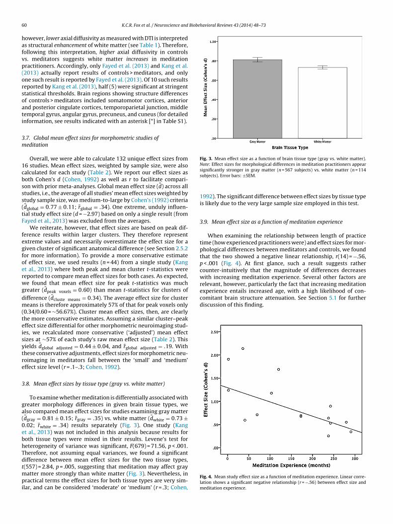
6 Biobehavioral Reviews 43 (2014) 48–73
hafvp(orsoati
3m
1cbsss(tF
fegfoerwgdm(teisytre
3
ga(0ebhTdtmpi
Fig. 3. Mean effect size as a function of brain tissue type (gray vs. white matter).Note: Effect sizes for morphological differences in meditation practitioners appear
comitant brain structure attenuation. See Section 5.1 for furtherdiscussion of this finding.
0 K.C.R. Fox et al. / Neuroscience and
owever, lower axial diffusivity as measured with DTI is interpreteds structural enhancement of white matter (see Table 1). Therefore,ollowing this interpretation, higher axial diffusivity in controlss. meditators suggests white matter increases in meditationractitioners. Accordingly, only Fayed et al. (2013) and Kang et al.2013) actually report results of controls > meditators, and onlyne such result is reported by Fayed et al. (2013). Of 10 such resultseported by Kang et al. (2013), half (5) were significant at stringenttatistical thresholds. Brain regions showing structure differencesf controls > meditators included somatomotor cortices, anteriornd posterior cingulate cortices, temporoparietal junction, middleemporal gyrus, angular gyrus, precuneus, and cuneus (for detailednformation, see results indicated with an asterisk [*] in Table S1).
.7. Global mean effect sizes for morphometric studies ofeditation
Overall, we were able to calculate 132 unique effect sizes from6 studies. Mean effect sizes, weighted by sample size, were alsoalculated for each study (Table 2). We report our effect sizes asoth Cohen’s d (Cohen, 1992) as well as r to facilitate compari-on with prior meta-analyses. Global mean effect size (d̄) across alltudies, i.e., the average of all studies’ mean effect sizes weighted bytudy sample size, was medium-to-large by Cohen’s (1992) criteriad̄global = 0.77 ± 0.11; r̄global = .34). One extreme, unduly influen-ial study effect size (d = −2.97) based on only a single result (fromayed et al., 2013) was excluded from the averages.
We reiterate, however, that effect sizes are based on peak dif-erence results within larger clusters. They therefore representxtreme values and necessarily overestimate the effect size for aiven cluster of significant anatomical difference (see Section 2.5.2or more information). To provide a more conservative estimatef effect size, we used results (n = 44) from a single study (Kangt al., 2013) where both peak and mean cluster t-statistics wereeported to compare mean effect sizes for both cases. As expected,e found that mean effect size for peak t-statistics was much
reater (d̄peak voxels = 0.60) than mean t-statistics for clusters ofifference (d̄cluste means = 0.34). The average effect size for clustereans is therefore approximately 57% of that for peak voxels only
0.34/0.60 = ∼56.67%). Cluster mean effect sizes, then, are clearlyhe more conservative estimates. Assuming a similar cluster–peakffect size differential for other morphometric neuroimaging stud-es, we recalculated more conservative (‘adjusted’) mean effectizes at ∼57% of each study’s raw mean effect size (Table 2). Thisields d̄global adjusted = 0.44 ± 0.04, and r̄global adjusted = .19. Withhese conservative adjustments, effect sizes for morphometric neu-oimaging in meditators fall between the ‘small’ and ‘medium’ffect size level (r = .1–.3; Cohen, 1992).
.8. Mean effect sizes by tissue type (gray vs. white matter)
To examine whether meditation is differentially associated withreater morphology differences in given brain tissue types, welso compared mean effect sizes for studies examining gray matterd̄gray = 0.81 ± 0.15; r̄gray = .35) vs. white matter (d̄white = 0.73 ±.02; r̄white = .34) results separately (Fig. 3). One study (Kangt al., 2013) was not included in this analysis because results foroth tissue types were mixed in their results. Levene’s test foreterogeneity of variance was significant, F(679) = 71.56, p < .001.herefore, not assuming equal variances, we found a significantifference between mean effect sizes for the two tissue types,
(557) = 2.84, p = .005, suggesting that meditation may affect grayatter more strongly than white matter (Fig. 3). Nevertheless, inractical terms the effect sizes for both tissue types are very sim-
lar, and can be considered ‘moderate’ or ‘medium’ (r = .3; Cohen,
significantly stronger in gray matter (n = 567 subjects) vs. white matter (n = 114subjects). Error bars: ±SEM.
1992). The significant difference between effect sizes by tissue typeis likely due to the very large sample size employed in this test.
3.9. Mean effect size as a function of meditation experience
When examining the relationship between length of practicetime (how experienced practitioners were) and effect sizes for mor-phological differences between meditators and controls, we foundthat the two showed a negative linear relationship, r(14) = −.56,p < .001 (Fig. 4). At first glance, such a result suggests rathercounter-intuitively that the magnitude of differences decreaseswith increasing meditation experience. Several other factors arerelevant, however, particularly the fact that increasing meditationexperience entails increased age, with a high likelihood of con-
Fig. 4. Mean study effect size as a function of meditation experience. Linear corre-lation shows a significant negative relationship (r = −.56) between effect size andmeditation experience.
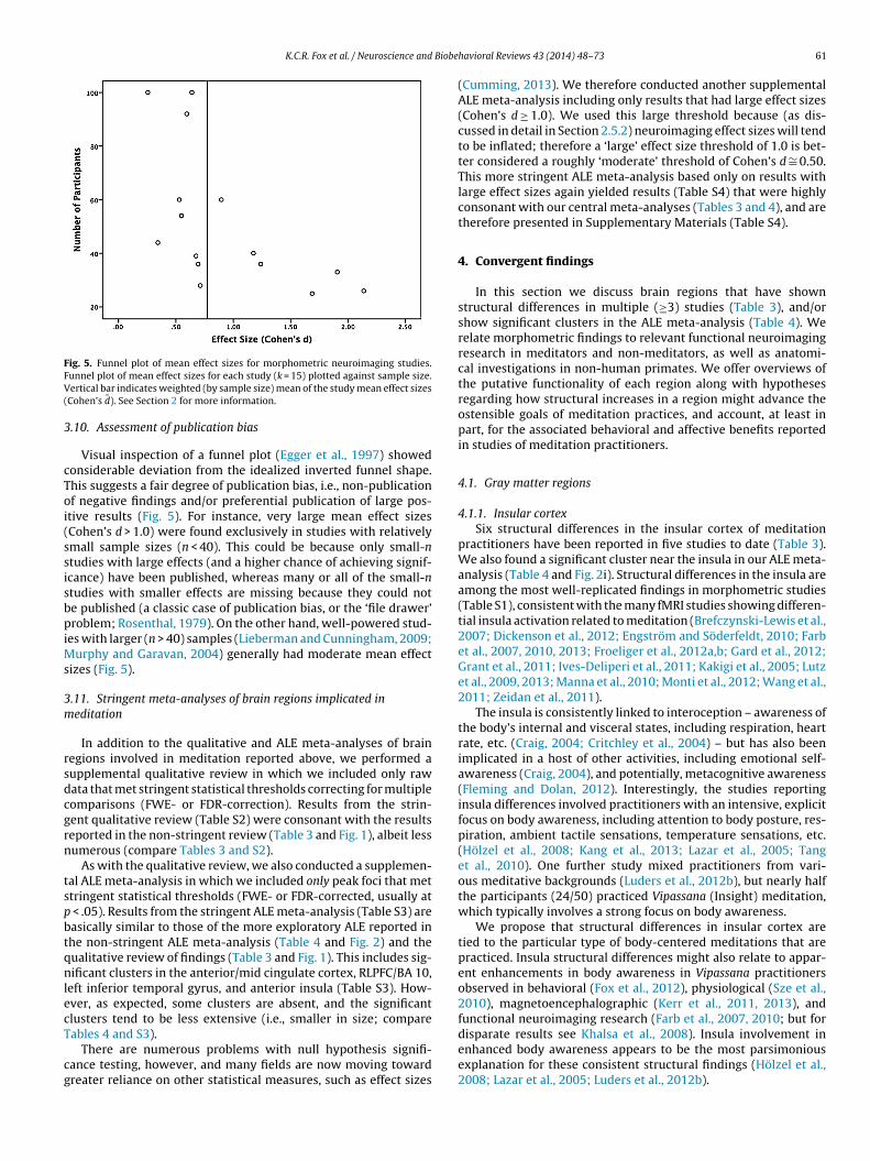
K.C.R. Fox et al. / Neuroscience and Biobe
FFV(
3
cToi(ssisbpiMs
3m
rsdcgrn
tspbtqnlecT
cg
ig. 5. Funnel plot of mean effect sizes for morphometric neuroimaging studies.unnel plot of mean effect sizes for each study (k = 15) plotted against sample size.ertical bar indicates weighted (by sample size) mean of the study mean effect sizes
Cohen’s d̄). See Section 2 for more information.
.10. Assessment of publication bias
Visual inspection of a funnel plot (Egger et al., 1997) showedonsiderable deviation from the idealized inverted funnel shape.his suggests a fair degree of publication bias, i.e., non-publicationf negative findings and/or preferential publication of large pos-tive results (Fig. 5). For instance, very large mean effect sizesCohen’s d > 1.0) were found exclusively in studies with relativelymall sample sizes (n < 40). This could be because only small-ntudies with large effects (and a higher chance of achieving signif-cance) have been published, whereas many or all of the small-ntudies with smaller effects are missing because they could note published (a classic case of publication bias, or the ‘file drawer’roblem; Rosenthal, 1979). On the other hand, well-powered stud-
es with larger (n > 40) samples (Lieberman and Cunningham, 2009;urphy and Garavan, 2004) generally had moderate mean effect
izes (Fig. 5).
.11. Stringent meta-analyses of brain regions implicated ineditation
In addition to the qualitative and ALE meta-analyses of brainegions involved in meditation reported above, we performed aupplemental qualitative review in which we included only rawata that met stringent statistical thresholds correcting for multipleomparisons (FWE- or FDR-correction). Results from the strin-ent qualitative review (Table S2) were consonant with the resultseported in the non-stringent review (Table 3 and Fig. 1), albeit lessumerous (compare Tables 3 and S2).
As with the qualitative review, we also conducted a supplemen-al ALE meta-analysis in which we included only peak foci that mettringent statistical thresholds (FWE- or FDR-corrected, usually at
< .05). Results from the stringent ALE meta-analysis (Table S3) areasically similar to those of the more exploratory ALE reported inhe non-stringent ALE meta-analysis (Table 4 and Fig. 2) and theualitative review of findings (Table 3 and Fig. 1). This includes sig-ificant clusters in the anterior/mid cingulate cortex, RLPFC/BA 10,
eft inferior temporal gyrus, and anterior insula (Table S3). How-ver, as expected, some clusters are absent, and the significantlusters tend to be less extensive (i.e., smaller in size; compare
ables 4 and S3).There are numerous problems with null hypothesis signifi-ance testing, however, and many fields are now moving towardreater reliance on other statistical measures, such as effect sizes
havioral Reviews 43 (2014) 48–73 61
(Cumming, 2013). We therefore conducted another supplementalALE meta-analysis including only results that had large effect sizes(Cohen’s d ≥ 1.0). We used this large threshold because (as dis-cussed in detail in Section 2.5.2) neuroimaging effect sizes will tendto be inflated; therefore a ‘large’ effect size threshold of 1.0 is bet-ter considered a roughly ‘moderate’ threshold of Cohen’s d ∼= 0.50.This more stringent ALE meta-analysis based only on results withlarge effect sizes again yielded results (Table S4) that were highlyconsonant with our central meta-analyses (Tables 3 and 4), and aretherefore presented in Supplementary Materials (Table S4).
4. Convergent findings
In this section we discuss brain regions that have shownstructural differences in multiple (≥3) studies (Table 3), and/orshow significant clusters in the ALE meta-analysis (Table 4). Werelate morphometric findings to relevant functional neuroimagingresearch in meditators and non-meditators, as well as anatomi-cal investigations in non-human primates. We offer overviews ofthe putative functionality of each region along with hypothesesregarding how structural increases in a region might advance theostensible goals of meditation practices, and account, at least inpart, for the associated behavioral and affective benefits reportedin studies of meditation practitioners.
4.1. Gray matter regions
4.1.1. Insular cortexSix structural differences in the insular cortex of meditation
practitioners have been reported in five studies to date (Table 3).We also found a significant cluster near the insula in our ALE meta-analysis (Table 4 and Fig. 2i). Structural differences in the insula areamong the most well-replicated findings in morphometric studies(Table S1), consistent with the many fMRI studies showing differen-tial insula activation related to meditation (Brefczynski-Lewis et al.,2007; Dickenson et al., 2012; Engström and Söderfeldt, 2010; Farbet al., 2007, 2010, 2013; Froeliger et al., 2012a,b; Gard et al., 2012;Grant et al., 2011; Ives-Deliperi et al., 2011; Kakigi et al., 2005; Lutzet al., 2009, 2013; Manna et al., 2010; Monti et al., 2012; Wang et al.,2011; Zeidan et al., 2011).
The insula is consistently linked to interoception – awareness ofthe body’s internal and visceral states, including respiration, heartrate, etc. (Craig, 2004; Critchley et al., 2004) – but has also beenimplicated in a host of other activities, including emotional self-awareness (Craig, 2004), and potentially, metacognitive awareness(Fleming and Dolan, 2012). Interestingly, the studies reportinginsula differences involved practitioners with an intensive, explicitfocus on body awareness, including attention to body posture, res-piration, ambient tactile sensations, temperature sensations, etc.(Hölzel et al., 2008; Kang et al., 2013; Lazar et al., 2005; Tanget al., 2010). One further study mixed practitioners from vari-ous meditative backgrounds (Luders et al., 2012b), but nearly halfthe participants (24/50) practiced Vipassana (Insight) meditation,which typically involves a strong focus on body awareness.
We propose that structural differences in insular cortex aretied to the particular type of body-centered meditations that arepracticed. Insula structural differences might also relate to appar-ent enhancements in body awareness in Vipassana practitionersobserved in behavioral (Fox et al., 2012), physiological (Sze et al.,2010), magnetoencephalographic (Kerr et al., 2011, 2013), andfunctional neuroimaging research (Farb et al., 2007, 2010; but for
disparate results see Khalsa et al., 2008). Insula involvement inenhanced body awareness appears to be the most parsimoniousexplanation for these consistent structural findings (Hölzel et al.,2008; Lazar et al., 2005; Luders et al., 2012b).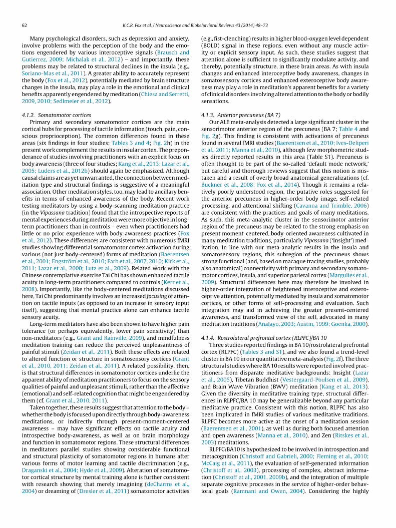
6 Biobe
itGpStcb2
4
csapdb2ciaet(mtlesve2Ca2htis
tnmpteiaq(t
wmaiaiavDtw2
2 K.C.R. Fox et al. / Neuroscience and
Many psychological disorders, such as depression and anxiety,nvolve problems with the perception of the body and the emo-ions engendered by various interoceptive signals (Brausch andutierrez, 2009; Michalak et al., 2012) – and importantly, theseroblems may be related to structural declines in the insula (e.g.,oriano-Mas et al., 2011). A greater ability to accurately representhe body (Fox et al., 2012), potentially mediated by brain structurehanges in the insula, may play a role in the emotional and clinicalenefits apparently engendered by meditation (Chiesa and Serretti,009, 2010; Sedlmeier et al., 2012).
.1.2. Somatomotor corticesPrimary and secondary somatomotor cortices are the main
ortical hubs for processing of tactile information (touch, pain, con-cious proprioception). The common differences found in thesereas (six findings in four studies; Tables 3 and 4; Fig. 2h) in theresent work complement the results in insular cortex. The prepon-erance of studies involving practitioners with an explicit focus onody awareness (three of four studies; Kang et al., 2013; Lazar et al.,005; Luders et al., 2012b) should again be emphasized. Althoughausal claims are as yet unwarranted, the connection between med-tation type and structural findings is suggestive of a meaningfulssociation. Other meditation styles, too, may lead to ancillary ben-fits in terms of enhanced awareness of the body. Recent workesting meditators by using a body-scanning meditation practicein the Vipassana tradition) found that the introspective reports of
ental experiences during meditation were more objective in long-erm practitioners than in controls – even when practitioners hadittle or no prior experience with body-awareness practices (Foxt al., 2012). These differences are consistent with numerous fMRItudies showing differential somatomotor cortex activation duringarious (not just body-centered) forms of meditation (Baerentsent al., 2001; Engström et al., 2010; Farb et al., 2007, 2010; Kirk et al.,011; Lazar et al., 2000; Lutz et al., 2009). Related work with thehinese contemplative exercise Tai Chi has shown enhanced tactilecuity in long-term practitioners compared to controls (Kerr et al.,008). Importantly, like the body-centered meditations discussedere, Tai Chi predominantly involves an increased focusing of atten-ion on tactile inputs (as opposed to an increase in sensory inputtself), suggesting that mental practice alone can enhance tactileensory acuity.
Long-term meditators have also been shown to have higher painolerance (or perhaps equivalently, lower pain sensitivity) thanon-meditators (e.g., Grant and Rainville, 2009), and mindfulnesseditation training can reduce the perceived unpleasantness of
ainful stimuli (Zeidan et al., 2011). Both these effects are relatedo altered function or structure in somatosensory cortices (Grantt al., 2010, 2011; Zeidan et al., 2011). A related possibility, then,s that structural differences in somatomotor cortices underlie thepparent ability of meditation practitioners to focus on the sensoryualities of painful and unpleasant stimuli, rather than the affectiveemotional) and self-related cognition that might be engendered byhem (cf. Grant et al., 2010, 2011).
Taken together, these results suggest that attention to the body –hether the body is focused upon directly through body-awarenesseditations, or indirectly through present-moment-centered
wareness – may have significant effects on tactile acuity andntrospective body-awareness, as well as on brain morphologynd function in somatomotor regions. These structural differencesn meditators parallel studies showing considerable functionalnd structural plasticity of somatomotor regions in humans afterarious forms of motor learning and tactile discrimination (e.g.,
raganski et al., 2004; Hyde et al., 2009). Alteration of somatomo-or cortical structure by mental training alone is further consistentith research showing that merely imagining (deCharms et al.,
004) or dreaming of (Dresler et al., 2011) somatomotor activities
havioral Reviews 43 (2014) 48–73
(e.g., fist-clenching) results in higher blood-oxygen level dependent(BOLD) signal in these regions, even without any muscle activ-ity or explicit sensory input. As such, these studies suggest thatattention alone is sufficient to significantly modulate activity, andthereby, potentially structure, in these brain areas. As with insulachanges and enhanced interoceptive body awareness, changes insomatosensory cortices and enhanced exteroceptive body aware-ness may play a role in meditation’s apparent benefits for a varietyof clinical disorders involving altered attention to the body or bodilysensations.
4.1.3. Anterior precuneus (BA 7)Our ALE meta-analysis detected a large significant cluster in the
sensorimotor anterior region of the precuneus (BA 7; Table 4 andFig. 2g). This finding is consistent with activations of precuneusfound in several fMRI studies (Baerentsen et al., 2010; Ives-Deliperiet al., 2011; Manna et al., 2010), although few morphometric stud-ies directly reported results in this area (Table S1). Precuneus isoften thought to be part of the so-called ‘default mode network,’but careful and thorough reviews suggest that this notion is mis-taken and a result of overly broad anatomical generalizations (cf.Buckner et al., 2008; Fox et al., 2014). Though it remains a rela-tively poorly understood region, the putative roles suggested forthe anterior precuneus in higher-order body image, self-relatedprocessing, and attentional shifting (Cavanna and Trimble, 2006)are consistent with the practices and goals of many meditations.As such, this meta-analytic cluster in the sensorimotor anteriorregion of the precuneus may be related to the strong emphasis onpresent moment-centered, body-oriented awareness cultivated inmany meditation traditions, particularly Vipassana (‘Insight’) med-itation. In line with our meta-analytic results in the insula andsomatosensory regions, this subregion of the precuneus showsstrong functional (and, based on macaque tracing studies, probablyalso anatomical) connectivity with primary and secondary somato-motor cortices, insula, and superior parietal cortex (Margulies et al.,2009). Structural differences here may therefore be involved inhigher-order integration of heightened interoceptive and extero-ceptive attention, potentially mediated by insula and somatomotorcortices, or other forms of self-processing and evaluation. Suchintegration may aid in achieving the greater present-centeredawareness, and transformed view of the self, advocated in manymeditation traditions (Analayo, 2003; Austin, 1999; Goenka, 2000).
4.1.4. Rostrolateral prefrontal cortex (RLPFC)/BA 10Three studies reported findings in BA 10/rostrolateral prefrontal
cortex (RLPFC) (Tables 3 and S1), and we also found a trend-levelcluster in BA 10 in our quantitative meta-analysis (Fig. 2f). The threestructural studies where BA 10 results were reported involved prac-titioners from disparate meditative backgrounds: Insight (Lazaret al., 2005), Tibetan Buddhist (Vestergaard-Poulsen et al., 2009),and Brain Wave Vibration (BWV) meditation (Kang et al., 2013).Given the diversity in meditative training type, structural differ-ences in RLPFC/BA 10 may be generalizable beyond any particularmeditative practice. Consistent with this notion, RLPFC has alsobeen implicated in fMRI studies of various meditative traditions.RLPFC becomes more active at the onset of a meditation session(Baerentsen et al., 2001), as well as during both focused attentionand open awareness (Manna et al., 2010), and Zen (Ritskes et al.,2003) meditations.
RLPFC/BA10 is hypothesized to be involved in introspection andmetacognition (Christoff and Gabrieli, 2000; Fleming et al., 2010;McCaig et al., 2011), the evaluation of self-generated information
(Christoff et al., 2003), processing of complex, abstract informa-tion (Christoff et al., 2001, 2009b), and the integration of multipleseparate cognitive processes in the service of higher-order behav-ioral goals (Ramnani and Owen, 2004). Considering the highly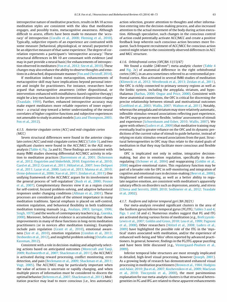
Biobe
imedrTsbrsmtct
meaim(mecnF
4(
lsamteGeOutefrpmemS2ipinDK
i2idVtmoi
K.C.R. Fox et al. / Neuroscience and
ntrospective nature of meditative practices, results in BA 10 acrosseditation styles are consistent with the idea that meditation
ngages, and possibly trains, metacognitive awareness. Althoughifficult to assess, efforts have been made to measure the ‘accu-acy’ of introspection (Corallo et al., 2008; Fleming et al., 2010).ypically, subjective reports of an experience are contrasted withome measure (behavioral, physiological, or neural) purported toe an objective measure of that same experience. The degree of cor-elation represents a participant’s ‘introspective accuracy.’ Thesetructural differences in BA 10 are consonant with evidence (anday in part provide a neural basis) for enhancements of introspec-
ion observed in meditators (Fox et al., 2012; Sze et al., 2010). Thesehanges may also enhance the ability to observe thoughts and emo-ions in a detached, dispassionate manner (Fox and Christoff, 2014).
If meditation indeed trains metacognition, enhancements ofetacognitive skill may have implications beyond personal inter-
st and insight for practitioners. For instance, researchers havergued that metacognitive awareness (either dispositional, orntervention-enhanced with mindfulness-based cognitive therapy)
ight be a key mechanism in the prevention of depression relapseTeasdale, 1999). Further, enhanced introspective accuracy may
ake expert meditators more reliable reporters of inner experi-nce – a crucial step toward a better understanding of the neuralorrelates of higher cognitive functions and subjective experiencesot amenable to study in animal models (Lutz and Thompson, 2003;ox et al., 2012).
.1.5. Anterior cingulate cortex (ACC) and mid-cingulate cortexMCC)
Seven structural differences were found in the anterior cingu-ate cortex (ACC) and mid-cingulate cortex (MCC) (Table 3), and twoignificant clusters were found in the ACC/MCC in the ALE meta-nalysis (Table 4; Fig. 2a and b). These findings are consistent withany fMRI studies showing differential ACC/MCC activity in rela-
ion to meditation practices (Baerentsen et al., 2001; Dickensont al., 2012; Engström and Söderfeldt, 2010; Engström et al., 2010;ard et al., 2012; Grant et al., 2011; Hölzel et al., 2007; Ives-Deliperit al., 2011; Lazar et al., 2000; Lutz et al., 2013; Manna et al., 2010;rme-Johnson et al., 2006; Xue et al., 2011; Zeidan et al., 2011). Onenifying framework of the ACC/MCC argues for its involvement inhe general process of ‘self-regulation’ (Bush et al., 2000; Posnert al., 2007). Complementary theories view it as a region crucialor self-control, focused problem-solving, and adaptive behavioralesponses under changing conditions (Allman et al., 2001). Theserocesses are considered goals of the utmost importance in manyeditation traditions. Special emphasis is placed on self-control,
motion regulation, and behavioral flexibility in both traditionaleditation training manuals (e.g., Analayo, 2003; Iyengar, 1996;
ingh, 1979) and the works of contemporary teachers (e.g., Goenka,000). Moreover, behavioral evidence is accumulating that shows
mprovements in many of these functions in long-term meditationractitioners (or in novices after meditation training). Examples
nclude pain regulation (Grant et al., 2010), emotional aware-ess (Sze et al., 2010), emotion regulation (Condon et al., 2013;esbordes et al., 2012), and insightful problem solving (Ostafin andassman, 2012).
Consistent with a role in decision-making and adaptively select-ng actions based on anticipated outcomes (Morecraft and Tanji,009; Rushworth et al., 2007; Shackman et al., 2011), the ACC/MCC
s activated during reward processing, conflict monitoring, erroretection, and pain (Beckmann et al., 2009; Shackman et al., 2011;ogt, 2005). The ACC/MCC may be particularly important when
he value of actions is uncertain or rapidly changing, and whenultiple pieces of information must be considered to discern the
ptimal behavior (Behrens et al., 2007; Shackman et al., 2011). Med-tation practice may lead to more conscious (i.e., less automatic)
havioral Reviews 43 (2014) 48–73 63
action selection, greater attention to thoughts and other informa-tion entering into the decision-making process, and also increasedattention to the actual movement of the body during action execu-tion. Although speculative, such changes in the conscious controlof action could potentially activate ACC/MCC and create a positivefeedback loop wherein such conscious action becomes more fre-quent. Such frequent recruitment of ACC/MCC for conscious actioncontrol might relate to the consistently observed differences in ACCand MCC structure.
4.1.6. Orbitofrontal cortex (OFC/BA 11/13/47)We found a sizable (240 mm3) meta-analytic cluster (Table 4
and Fig. 2c) of anatomical difference in the right orbitofrontalcortex (OFC), in an area sometimes referred to as ventromedial pre-frontal cortex. Also activated in several fMRI studies of meditation(Klimecki et al., 2012; Westbrook et al., 2013; Zeidan et al., 2011),the OFC is richly connected to primary sensory regions as well asthe limbic system, including the amygdala, striatum, and hypo-thalamus (Barbas, 2000; Ongur and Price, 2000). Consistent withthese anatomical connections, the OFC is critical for discerning theprecise relationship between stimuli and motivational outcomes(Gottfried et al., 2003; Wallis, 2007; Walton et al., 2011). Notably,whereas the amygdala and striatum may store relatively automatic,inflexible stimulus-reward associations reflecting past experience,the OFC may generate more flexible, ‘online’ assessments of stimuliand experience (Schoenbaum and Esber, 2010; Wallis, 2007). Weagree with others (Luders et al., 2009) that meditative training mayeventually lead to greater reliance on the OFC and its dynamic pre-dictions of the current value of stimuli to guide behavior, instead ofrelying on static stimulus-reward associations from the past. Struc-tural heterogeneities in OFC may thus relate to the stated goals ofmeditation in that they may facilitate more conscious and flexiblebehavior.
OFC is implicated not only in online, integrative decision-making, but also in emotion regulation, specifically in down-regulating (Ochsner et al., 2004) and reappraising (Goldin et al.,2008) negative emotional states. This regulatory function may bepart of a broader role for OFC in self-monitoring and integration ofcognitive and emotional cues in decision-making (Beer et al., 2006).Heightened self-monitoring, as well as a better ability to regu-late negative emotion, are consistent with meditation’s apparentlysalutary effects on disorders such as depression, anxiety, and stress(Chiesa and Serretti, 2009, 2010; Sedlmeier et al., 2012; Teasdaleet al., 2002).
4.1.7. Fusiform and inferior temporal gyri (BA 20/21)Our meta-analysis revealed significant clusters in the area of
the fusiform gyrus/inferior temporal gyrus (FG/ITG; Tables 3 and 4;Figs. 1 and 2d and e). Numerous studies suggest that FG and ITGare activated during various forms of meditation (e.g., Brefczynski-Lewis et al., 2007; Goldin and Gross, 2010; Lou et al., 1999; Pagnoniet al., 2008). Other researchers (Hölzel et al., 2008; Luders et al.,2009) have highlighted the possible role of the ITG in the ‘mys-tical’ states associated with meditation, and/or the experience ofenhanced well-being and ‘bliss’ often reported by advanced practi-tioners. In general, however, findings in the FG/ITG appear puzzlingand have been little discussed (e.g., Vestergaard-Poulsen et al.,2009).
Inferior temporal lobe structures are most strongly implicatedin detailed, high-level visual processing, however (Joseph, 2001).As a growing body of research has demonstrated enhanced visualprocessing in meditation practitioners (Brown et al., 1984; Hodgins
and Adair, 2010; Jha et al., 2007; Kozhevnikov et al., 2009; MacLeanet al., 2010; Tloczynski et al., 2000), the most parsimoniousexplanation for our meta-analytic clusters is that structural hetero-geneities in FG and IFG are related to these apparent enhancements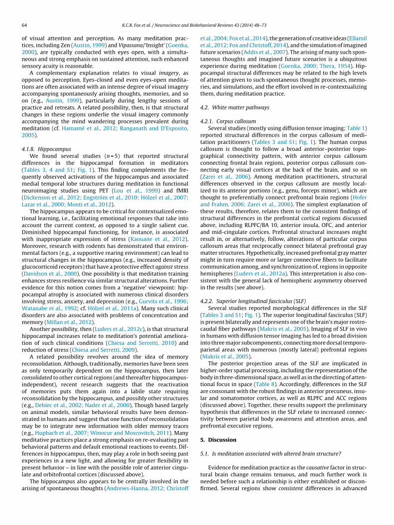
6 Biobe
ot2ns
otaopcam2
4
d(qmn(L
taDwMmsg(eepiWdm
htr
racior(osm(mbfepl
a
4 K.C.R. Fox et al. / Neuroscience and
f visual attention and perception. As many meditation prac-ices, including Zen (Austin, 1999) and Vipassana/‘Insight’ (Goenka,000), are typically conducted with eyes open, with a simulta-eous and strong emphasis on sustained attention, such enhancedensory acuity is reasonable.
A complementary explanation relates to visual imagery, aspposed to perception. Eyes-closed and even eyes-open medita-ions are often associated with an intense degree of visual imageryccompanying spontaneously arising thoughts, memories, and son (e.g., Austin, 1999), particularly during lengthy sessions ofractice and retreats. A related possibility, then, is that structuralhanges in these regions underlie the visual imagery commonlyccompanying the mind wandering processes prevalent duringeditation (cf. Hamamé et al., 2012; Ranganath and D’Esposito,
005).
.1.8. HippocampusWe found several studies (n = 5) that reported structural
ifferences in the hippocampal formation in meditatorsTables 3, 4 and S1; Fig. 1). This finding complements the fre-uently observed activations of the hippocampus and associatededial temporal lobe structures during meditation in functional
euroimaging studies using PET (Lou et al., 1999) and fMRIDickenson et al., 2012; Engström et al., 2010; Hölzel et al., 2007;azar et al., 2000; Monti et al., 2012).
The hippocampus appears to be critical for contextualized emo-ional learning, i.e., facilitating emotional responses that take intoccount the current context, as opposed to a single salient cue.iminished hippocampal functioning, for instance, is associatedith inappropriate expression of stress (Kaouane et al., 2012).oreover, research with rodents has demonstrated that environ-ental factors (e.g., a supportive rearing environment) can lead to
tructural changes in the hippocampus (e.g., increased density oflucocorticoid receptors) that have a protective effect against stressDavidson et al., 2000). One possibility is that meditation trainingnhances stress resilience via similar structural alterations. Furthervidence for this notion comes from a ‘negative’ viewpoint: hip-ocampal atrophy is associated with numerous clinical disorders
nvolving stress, anxiety, and depression (e.g., Gurvits et al., 1996;atanabe et al., 1992; cf. Hölzel et al., 2011a). Many such clinical
isorders are also associated with problems of concentration andemory (Millan et al., 2012).Another possibility, then (Luders et al., 2012c), is that structural
ippocampal increases relate to meditation’s potential ameliora-ion of such clinical conditions (Chiesa and Serretti, 2010) andeduction of stress (Chiesa and Serretti, 2009).
A related possibility revolves around the idea of memoryeconsolidation. Although, traditionally, memories have been seens only temporarily dependent on the hippocampus, then lateronsolidated to other cortical regions (and thereafter hippocampus-ndependent), recent research suggests that the reactivationf memories puts them again into a labile state requiringeconsolidation by the hippocampus, and possibly other structurese.g., Debiec et al., 2002; Nader et al., 2000). Though based largelyn animal models, similar behavioral results have been demon-trated in humans and suggest that one function of reconsolidationay be to integrate new information with older memory traces
e.g., Hupbach et al., 2007; Winocur and Moscovitch, 2011). Manyeditative practices place a strong emphasis on re-evaluating past
ehavioral patterns and default emotional reactions to events. Dif-erences in hippocampus, then, may play a role in both seeing pastxperiences in a new light, and allowing for greater flexibility in
resent behavior – in line with the possible role of anterior cingu-ate and orbitofrontal cortices (discussed above).The hippocampus also appears to be centrally involved in the
rising of spontaneous thoughts (Andrews-Hanna, 2012; Christoff
havioral Reviews 43 (2014) 48–73
et al., 2004; Fox et al., 2014), the generation of creative ideas (Ellamilet al., 2012; Fox and Christoff, 2014), and the simulation of imaginedfuture scenarios (Addis et al., 2007). The arising of many such spon-taneous thoughts and imagined future scenarios is a ubiquitousexperience during meditation (Goenka, 2000; Thera, 1954). Hip-pocampal structural differences may be related to the high levelsof attention given to such spontaneous thought processes, memo-ries, and simulations, and the effort involved in re-contextualizingthem, during meditation practice.
4.2. White matter pathways
4.2.1. Corpus callosumSeveral studies (mostly using diffusion tensor imaging; Table 1)
reported structural differences in the corpus callosum of medi-tation practitioners (Tables 3 and S1; Fig. 1). The human corpuscallosum is thought to follow a broad anterior–posterior topo-graphical connectivity pattern, with anterior corpus callosumconnecting frontal brain regions, posterior corpus callosum con-necting early visual cortices at the back of the brain, and so on(Zarei et al., 2006). Among meditation practitioners, structuraldifferences observed in the corpus callosum are mostly local-ized to its anterior portions (e.g., genu, forceps minor), which arethought to preferentially connect prefrontal brain regions (Hoferand Frahm, 2006; Zarei et al., 2006). The simplest explanation ofthese results, therefore, relates them to the consistent findings ofstructural differences in the prefrontal cortical regions discussedabove, including RLPFC/BA 10, anterior insula, OFC, and anteriorand mid-cingulate cortices. Prefrontal structural increases mightresult in, or alternatively, follow, alterations of particular corpuscallosum areas that reciprocally connect bilateral prefrontal graymatter structures. Hypothetically, increased prefrontal gray mattermight in turn require more or larger connective fibers to facilitatecommunication among, and synchronization of, regions in oppositehemispheres (Luders et al., 2012a). This interpretation is also con-sistent with the general lack of hemispheric asymmetry observedin the results (see above).
4.2.2. Superior longitudinal fasciculus (SLF)Several studies reported morphological differences in the SLF
(Tables 3 and S1; Fig. 1). The superior longitudinal fasciculus (SLF)is present bilaterally and represents one of the brain’s major rostro-caudal fiber pathways (Makris et al., 2005). Imaging of SLF in vivoin humans with diffusion tensor imaging has led to a broad divisioninto three major subcomponents, connecting more dorsal temporo-parietal areas with numerous (mostly lateral) prefrontal regions(Makris et al., 2005).
The posterior projection areas of the SLF are implicated inhigher-order spatial processing, including the representation of thebody in three-dimensional space, as well as in the directing of atten-tional focus in space (Table 8). Accordingly, differences in the SLFare consonant with the robust findings in anterior precuneus, insu-lar and somatomotor cortices, as well as RLPFC and ACC regions(discussed above). Together, these results support the preliminaryhypothesis that differences in the SLF relate to increased connec-tivity between parietal body awareness and attention areas, andprefrontal executive regions.
5. Discussion
5.1. Is meditation associated with altered brain structure?
Evidence for meditation practice as the causative factor in struc-tural brain change remains tenuous, and much further work isneeded before such a relationship is either established or discon-firmed. Several regions show consistent differences in advanced
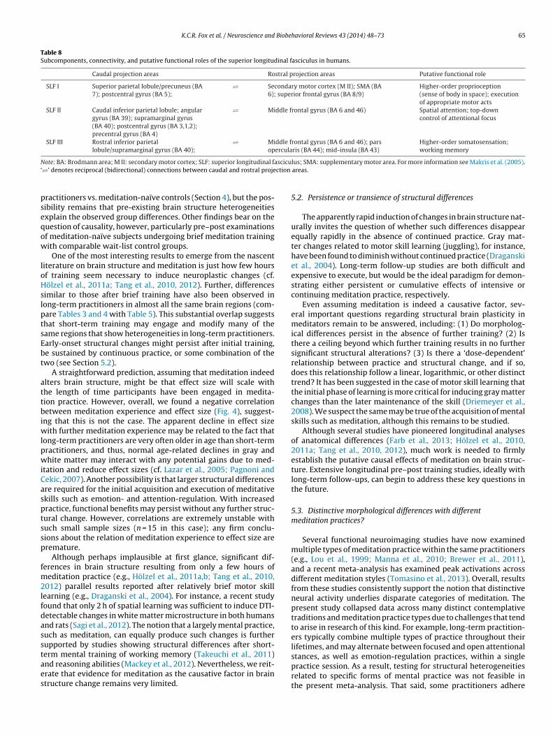
K.C.R. Fox et al. / Neuroscience and Biobehavioral Reviews 43 (2014) 48–73 65
Table 8Subcomponents, connectivity, and putative functional roles of the superior longitudinal fasciculus in humans.
Caudal projection areas Rostral projection areas Putative functional role
SLF I Superior parietal lobule/precuneus (BA7); postcentral gyrus (BA 5);
� Secondary motor cortex (M II); SMA (BA6); superior frontal gyrus (BA 8/9)
Higher-order proprioception(sense of body in space); executionof appropriate motor acts
SLF II Caudal inferior parietal lobule; angulargyrus (BA 39); supramarginal gyrus(BA 40); postcentral gyrus (BA 3,1,2);precentral gyrus (BA 4)
� Middle frontal gyrus (BA 6 and 46) Spatial attention; top-downcontrol of attentional focus
SLF III Rostral inferior parietallobule/supramarginal gyrus (BA 40);
� Middle frontal gyrus (BA 6 and 46); parsopercularis (BA 44); mid-insula (BA 43)
Higher-order somatosensation;working memory
N scicu‘ ction
pseqow
loHslptsEbt
attbiwlpwiCasptssp
fm2lfdasstaes
ote: BA: Brodmann area; M II: secondary motor cortex; SLF: superior longitudinal fa�’ denotes reciprocal (bidirectional) connections between caudal and rostral proje
ractitioners vs. meditation-naïve controls (Section 4), but the pos-ibility remains that pre-existing brain structure heterogeneitiesxplain the observed group differences. Other findings bear on theuestion of causality, however, particularly pre–post examinationsf meditation-naïve subjects undergoing brief meditation trainingith comparable wait-list control groups.
One of the most interesting results to emerge from the nascentiterature on brain structure and meditation is just how few hoursf training seem necessary to induce neuroplastic changes (cf.ölzel et al., 2011a; Tang et al., 2010, 2012). Further, differences
imilar to those after brief training have also been observed inong-term practitioners in almost all the same brain regions (com-are Tables 3 and 4 with Table 5). This substantial overlap suggestshat short-term training may engage and modify many of theame regions that show heterogeneities in long-term practitioners.arly-onset structural changes might persist after initial training,e sustained by continuous practice, or some combination of thewo (see Section 5.2).
A straightforward prediction, assuming that meditation indeedlters brain structure, might be that effect size will scale withhe length of time participants have been engaged in medita-ion practice. However, overall, we found a negative correlationetween meditation experience and effect size (Fig. 4), suggest-
ng that this is not the case. The apparent decline in effect sizeith further meditation experience may be related to the fact that
ong-term practitioners are very often older in age than short-termractitioners, and thus, normal age-related declines in gray andhite matter may interact with any potential gains due to med-
tation and reduce effect sizes (cf. Lazar et al., 2005; Pagnoni andekic, 2007). Another possibility is that larger structural differencesre required for the initial acquisition and execution of meditativekills such as emotion- and attention-regulation. With increasedractice, functional benefits may persist without any further struc-ural change. However, correlations are extremely unstable withuch small sample sizes (n = 15 in this case); any firm conclu-ions about the relation of meditation experience to effect size areremature.
Although perhaps implausible at first glance, significant dif-erences in brain structure resulting from only a few hours of
editation practice (e.g., Hölzel et al., 2011a,b; Tang et al., 2010,012) parallel results reported after relatively brief motor skill
earning (e.g., Draganski et al., 2004). For instance, a recent studyound that only 2 h of spatial learning was sufficient to induce DTI-etectable changes in white matter microstructure in both humansnd rats (Sagi et al., 2012). The notion that a largely mental practice,uch as meditation, can equally produce such changes is furtherupported by studies showing structural differences after short-
erm mental training of working memory (Takeuchi et al., 2011)nd reasoning abilities (Mackey et al., 2012). Nevertheless, we reit-rate that evidence for meditation as the causative factor in braintructure change remains very limited.lus; SMA: supplementary motor area. For more information see Makris et al. (2005).areas.
5.2. Persistence or transience of structural differences
The apparently rapid induction of changes in brain structure nat-urally invites the question of whether such differences disappearequally rapidly in the absence of continued practice. Gray mat-ter changes related to motor skill learning (juggling), for instance,have been found to diminish without continued practice (Draganskiet al., 2004). Long-term follow-up studies are both difficult andexpensive to execute, but would be the ideal paradigm for demon-strating either persistent or cumulative effects of intensive orcontinuing meditation practice, respectively.
Even assuming meditation is indeed a causative factor, sev-eral important questions regarding structural brain plasticity inmeditators remain to be answered, including: (1) Do morpholog-ical differences persist in the absence of further training? (2) Isthere a ceiling beyond which further training results in no furthersignificant structural alterations? (3) Is there a ‘dose-dependent’relationship between practice and structural change, and if so,does this relationship follow a linear, logarithmic, or other distincttrend? It has been suggested in the case of motor skill learning thatthe initial phase of learning is more critical for inducing gray matterchanges than the later maintenance of the skill (Driemeyer et al.,2008). We suspect the same may be true of the acquisition of mentalskills such as meditation, although this remains to be studied.
Although several studies have pioneered longitudinal analysesof anatomical differences (Farb et al., 2013; Hölzel et al., 2010,2011a; Tang et al., 2010, 2012), much work is needed to firmlyestablish the putative causal effects of meditation on brain struc-ture. Extensive longitudinal pre–post training studies, ideally withlong-term follow-ups, can begin to address these key questions inthe future.
5.3. Distinctive morphological differences with differentmeditation practices?
Several functional neuroimaging studies have now examinedmultiple types of meditation practice within the same practitioners(e.g., Lou et al., 1999; Manna et al., 2010; Brewer et al., 2011),and a recent meta-analysis has examined peak activations acrossdifferent meditation styles (Tomasino et al., 2013). Overall, resultsfrom these studies consistently support the notion that distinctiveneural activity underlies disparate categories of meditation. Thepresent study collapsed data across many distinct contemplativetraditions and meditation practice types due to challenges that tendto arise in research of this kind. For example, long-term practition-ers typically combine multiple types of practice throughout theirlifetimes, and may alternate between focused and open attentional
stances, as well as emotion-regulation practices, within a singlepractice session. As a result, testing for structural heterogeneitiesrelated to specific forms of mental practice was not feasible inthe present meta-analysis. That said, some practitioners adhere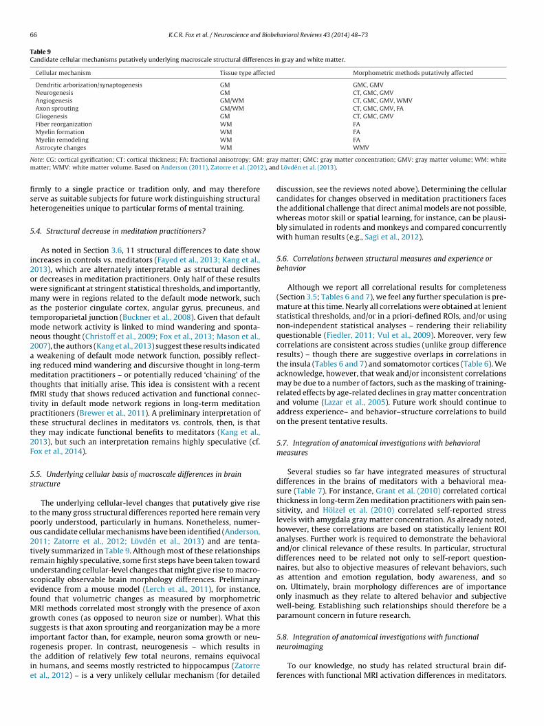
66 K.C.R. Fox et al. / Neuroscience and Biobehavioral Reviews 43 (2014) 48–73
Table 9Candidate cellular mechanisms putatively underlying macroscale structural differences in gray and white matter.
Cellular mechanism Tissue type affected Morphometric methods putatively affected
Dendritic arborization/synaptogenesis GM GMC, GMVNeurogenesis GM CT, GMC, GMVAngiogenesis GM/WM CT, GMC, GMV, WMVAxon sprouting GM/WM CT, GMC, GMV, FAGliogenesis GM CT, GMC, GMVFiber reorganization WM FAMyelin formation WM FAMyelin remodeling WM FAAstrocyte changes WM WMV
N : graym 2), and
fish
5
i2owmatmn2aimtftptt2F
5s
tpo2trusefMgsirtie
ote: CG: cortical gyrification; CT: cortical thickness; FA: fractional anisotropy; GMatter; WMV: white matter volume. Based on Anderson (2011), Zatorre et al. (201
rmly to a single practice or tradition only, and may thereforeerve as suitable subjects for future work distinguishing structuraleterogeneities unique to particular forms of mental training.
.4. Structural decrease in meditation practitioners?
As noted in Section 3.6, 11 structural differences to date showncreases in controls vs. meditators (Fayed et al., 2013; Kang et al.,013), which are alternately interpretable as structural declinesr decreases in meditation practitioners. Only half of these resultsere significant at stringent statistical thresholds, and importantly,any were in regions related to the default mode network, such
s the posterior cingulate cortex, angular gyrus, precuneus, andemporoparietal junction (Buckner et al., 2008). Given that default
ode network activity is linked to mind wandering and sponta-eous thought (Christoff et al., 2009; Fox et al., 2013; Mason et al.,007), the authors (Kang et al., 2013) suggest these results indicated
weakening of default mode network function, possibly reflect-ng reduced mind wandering and discursive thought in long-term
editation practitioners – or potentially reduced ‘chaining’ of thehoughts that initially arise. This idea is consistent with a recentMRI study that shows reduced activation and functional connec-ivity in default mode network regions in long-term meditationractitioners (Brewer et al., 2011). A preliminary interpretation ofhese structural declines in meditators vs. controls, then, is thathey may indicate functional benefits to meditators (Kang et al.,013), but such an interpretation remains highly speculative (cf.ox et al., 2014).
.5. Underlying cellular basis of macroscale differences in braintructure
The underlying cellular-level changes that putatively give riseo the many gross structural differences reported here remain veryoorly understood, particularly in humans. Nonetheless, numer-us candidate cellular mechanisms have been identified (Anderson,011; Zatorre et al., 2012; Lövdén et al., 2013) and are tenta-ively summarized in Table 9. Although most of these relationshipsemain highly speculative, some first steps have been taken towardnderstanding cellular-level changes that might give rise to macro-copically observable brain morphology differences. Preliminaryvidence from a mouse model (Lerch et al., 2011), for instance,ound that volumetric changes as measured by morphometric
RI methods correlated most strongly with the presence of axonrowth cones (as opposed to neuron size or number). What thisuggests is that axon sprouting and reorganization may be a moremportant factor than, for example, neuron soma growth or neu-
ogenesis proper. In contrast, neurogenesis – which results inhe addition of relatively few total neurons, remains equivocaln humans, and seems mostly restricted to hippocampus (Zatorret al., 2012) – is a very unlikely cellular mechanism (for detailedmatter; GMC: gray matter concentration; GMV: gray matter volume; WM: white Lövdén et al. (2013).
discussion, see the reviews noted above). Determining the cellularcandidates for changes observed in meditation practitioners facesthe additional challenge that direct animal models are not possible,whereas motor skill or spatial learning, for instance, can be plausi-bly simulated in rodents and monkeys and compared concurrentlywith human results (e.g., Sagi et al., 2012).
5.6. Correlations between structural measures and experience orbehavior
Although we report all correlational results for completeness(Section 3.5; Tables 6 and 7), we feel any further speculation is pre-mature at this time. Nearly all correlations were obtained at lenientstatistical thresholds, and/or in a priori-defined ROIs, and/or usingnon-independent statistical analyses – rendering their reliabilityquestionable (Fiedler, 2011; Vul et al., 2009). Moreover, very fewcorrelations are consistent across studies (unlike group differenceresults) – though there are suggestive overlaps in correlations inthe insula (Tables 6 and 7) and somatomotor cortices (Table 6). Weacknowledge, however, that weak and/or inconsistent correlationsmay be due to a number of factors, such as the masking of training-related effects by age-related declines in gray matter concentrationand volume (Lazar et al., 2005). Future work should continue toaddress experience– and behavior–structure correlations to buildon the present tentative results.
5.7. Integration of anatomical investigations with behavioralmeasures
Several studies so far have integrated measures of structuraldifferences in the brains of meditators with a behavioral mea-sure (Table 7). For instance, Grant et al. (2010) correlated corticalthickness in long-term Zen meditation practitioners with pain sen-sitivity, and Hölzel et al. (2010) correlated self-reported stresslevels with amygdala gray matter concentration. As already noted,however, these correlations are based on statistically lenient ROIanalyses. Further work is required to demonstrate the behavioraland/or clinical relevance of these results. In particular, structuraldifferences need to be related not only to self-report question-naires, but also to objective measures of relevant behaviors, suchas attention and emotion regulation, body awareness, and soon. Ultimately, brain morphology differences are of importanceonly inasmuch as they relate to altered behavior and subjectivewell-being. Establishing such relationships should therefore be aparamount concern in future research.
5.8. Integration of anatomical investigations with functional
neuroimagingTo our knowledge, no study has related structural brain dif-ferences with functional MRI activation differences in meditators.
Biobe
Fwerfln
6
tlm
6
ioi1Tms
6
afht2sgptc(a
fbrtolsria
ntludcttfiemia
K.C.R. Fox et al. / Neuroscience and
unction and structure differences can corroborate one anotherhen correlated with the same behavioral task, as has been
legantly demonstrated with insula structure and function aselated to interoceptive awareness (Critchley et al., 2004). Suchunctional–structural integration of results represents a major chal-enge – and opportunity – for future research in the cognitiveeuroscience of meditation.
. Meta-analytic methods, reliability, and limitations
In the present section we discuss in more detail the validity ofhe present meta-analysis, the checks of its robustness, and theimitations imposed both by our meta-analytic techniques and the
ethodology of the primary research reviewed.
.1. Reliability of meta-analyses
Although we included all available morphometric neuroimag-ng studies of meditation to date, nonetheless, the sample size forur meta-analysis was fairly small (21 studies total). Only 14 stud-es could be included in quantitative ALE meta-analysis, and only6 studies provided data that enabled calculation of effect sizes.here were also too few studies to test separately among differenteditation traditions, and too few peak foci reported to conduct a
eparate ALE for short-term meditation training results only.
.1.1. Determining consistent brain structure differencesAside from seven studies not contributing to the ALE meta-
nalysis, some studies (especially Kang et al., 2013) contributedar more foci than others (Table S1). The latest ALE algorithms,owever, correct for this bias, reducing the influence of studieshat contribute disproportionate numbers of foci (Turkelbaub et al.,012). ALE also uses random-effects analyses, which are more con-ervative than fixed-effects analyses. In addition, these analyses areeneralizable to the population at large, which is the theoreticalopulation of ‘all meditation practitioners,’ or at least all practi-ioners within a given school of practice. As a further check, weonducted an ALE meta-analysis of stringent statistical results onlyTable S3) and of large effect size results only (Table S4); both bynd large paralleled our primary results (Table 4).
There are also other drawbacks to our meta-analytic techniquesor determining consistent brain regions across studies that shoulde kept in mind when interpreting our results. In our ‘qualitative’eview, the criterion of three reports for a given brain region (i.e.,wo replications of a given result), though used to advantage bythers (Kempton et al., 2008), is somewhat arbitrary. A more oress stringent criterion could just as well be imposed, resulting inhorter or longer lists of brain regions, respectively. Further, theegional labels reported by the source studies are always approx-mate; three findings reported in the ‘same’ region or Brodmannrea may in fact differ considerably in location across studies.
Though ALE avoids the latter problem by using precise coordi-ates (rather than general regional labels) as its input, nonethelesshese peak coordinate foci are highly imperfect indicators of theocation of a significant anatomical difference. The peak coordinatessually represent the weighted center of a cluster of significantifference, but they provide no information about the size of theluster (number of voxels) or its shape. Even when this informa-ion is provided, however, ALE as yet has no means of integratinghis information into its meta-analysis. Finally, ALE attempts tond overlapping clusters of difference by averaging across differ-
nt peak coordinates. There is therefore the risk that, with two orore relatively nearby peak foci, ALE will find an average, ‘signif-cant’ cluster somewhere between these foci, in a brain region notctually reported in any of the source studies.
havioral Reviews 43 (2014) 48–73 67
6.1.2. Effect sizes in morphometric neuroimagingDespite the finding that the overall mean effect size across
15 studies was ‘medium’ (d̄global = 0.77; r̄global = .34), these effectsizes are based on peak t-statistics, as commonly reported in theneuroimaging literature (see Sections 2 and 3). After attemptingto adjust for this bias and estimate effect sizes based on meant-statistics for entire clusters of significant anatomical difference(instead of only peak voxels of difference), our results suggest thatthe effect sizes can be more conservatively estimated at approxi-mately 57% of their reported value (see Section 3). This adjustmentplaces our mean effect size in the realm of ‘moderate’ or ‘medium’(d̄global adjusted = 0.44 and r̄global adjusted = .19). Such effects arecomparable to the mean effect sizes for studies of meditation’sinfluence on attentional, cognitive, and emotional factors (over-all r̄ = .27; Sedlmeier et al., 2012). Nonetheless, this resemblanceshould not be over-interpreted: as already noted, the interpretationof effect sizes in neuroimaging studies remains poorly developed(e.g., Poldrack et al., 2008).
6.1.3. Publication biasFunnel plots (Egger et al., 1997) can indicate the probable degree
of publication bias (Sedlmeier et al., 2012). Although a quantita-tive Egger test (Egger et al., 1997) of funnel plot asymmetry wasprecluded by the lack of data on standard error provided in theoriginal studies meta-analyzed, visual inspection suggested thata fairly large degree of bias was present in averaged effect sizesby study (Fig. 5). Almost all large effect sizes have been reportedin studies with the smallest sample sizes (n < 40), whereas stud-ies with larger (n > 40) samples tend to yield mean effect sizesaround or below the field-wide mean of d̄global = 0.77. The trendtoward more reasonable effect sizes in well-powered, large sam-ple size studies (Fig. 5) could be interpreted as evidence that trueeffects are being converged upon. However, the lack of publicationof almost any negative results in morphometric neuroimaging ofmeditation to date suggests a fair degree of publication bias, andindicates a strong chance of there being a ‘file-drawer problem’ inthis literature (Rosenthal, 1979).
6.2. Selection bias and preexisting brain structure differences
Even short-term meditation training can be very demanding,and consistent, long-term practice involving thousands of hoursof commitment is obviously so. The possibility of a selection biasin favor of participants predisposed to such regimens, and/oralready possessing higher trait levels of body awareness, sus-tained attention, metacognitive awareness, and so on, is clearly amajor potential confound. Such initial differences in personalityand cognitive ability would likely be reflected in brain structureand function. A recent study, for instance, found that differentialbaseline activation in certain brain regions predicted subsequentpractice time of both mindfulness and compassion forms of medi-tation (Mascaro et al., 2013). Longitudinal morphometric studiesin meditation-naïve novices (Hölzel et al., 2011a,b; Farb et al.,2013; Tang et al., 2010, 2012) have begun to mitigate this prob-lem, but extensive research will be necessary to address whetherpreexisting differences explain, or perhaps interact with, appar-ently experience-dependent changes in brain structure. The use ofactive control groups requiring substantial time and commitment(vs. simple wait-list controls) can also help to limit this potentialconfound.
6.3. Divergent findings
Convergent findings aside (Section 4), there are many brainstructure differences observed in only a single study included inthis meta-analysis. There are many reasons why a result might
6 Biobe
hmomsfpe
arAdpivtcportcc
pimltTmsgfi
6
oewfitsspsmpdpgsu
6
iamTeel
8 K.C.R. Fox et al. / Neuroscience and
ave been found in only a single study. First, a great variety ofeditation practices were examined. Although the small number
f studies precluded practice-specific analyses, techniques differedarkedly (e.g., loving-kindness meditation focused on interper-
onal emotional training vs. body-scanning Vipassana meditationocused on tactile sensations). Practitioners focusing on highly dis-arate techniques should not be expected to exhibit identical orven necessarily similar structural differences.
Second, different practices are related to differing lifestyle vari-bles that may in turn affect brain structure. Some meditationegimens, for instance, are particularly strict and austere (e.g., Zen;ustin, 1999), whereas others take a more relaxed approach. Theseiffering styles influence numerous lifestyle variables ostensiblyeripheral (but arguably critical) to the mental meditation practice
tself, including fasting, sleep deprivation, dietary restrictions (e.g.,egetarianism), overall mood and levels of stress, etc. Any ofhese lifestyle factors may interact synergistically or antagonisti-ally with the putative influence on brain structure of the mentalractices themselves. Studies to date have typically controlled fornly age, sex, and handedness. Ideally, future questionnaire-basedesearch would establish demographic models of meditation prac-itioners, and subsequent neuroimaging work could attempt toontrol for the relevant individual difference factors when selectingontrol groups.
Third, error and inconsistencies related to the variety of mor-hometric measures employed (Table 1) is a likely contributor to
nconsistent results across studies. Fourth, and finally, some resultsay simply be the product of chance or error. Considering the
enient statistical thresholds often used in the reviewed studies,he rate of Type I (false positive) error is evidently heightened.hat said, the relative paucity of studies of brain structure andeditation makes any conclusions about not-yet-replicated results
omewhat premature. Future research can more definitively distin-uish results that replicate consistently from anomalous or chancendings.
.4. Few research groups and overlapping samples
The majority of the studies conducted so far have been fromnly a few research groups, and several studies have used largely orntirely overlapping samples of meditation practitioners (markedith a � symbol in Table 2). This is partly because it is often dif-cult to recruit subjects with high levels of expertise. Althoughhese facts in no way invalidate the results obtained, we empha-ize them because it is possible that the samples of practitionerstudied by these groups are atypical in some way (e.g., in havingarticularly effective teachers and practices, or abnormally largetructural brain differences). Further, overlapping samples used inultiple studies (published in multiple papers) may suggest com-
letely independent results whereas findings are, to an unknownegree, dependent. Until larger and more independent samples ofractitioners are studied by a wider variety of research groups,eneralizing from the present results to conclusions about braintructure of meditation practitioners as a population should bendertaken with considerable caution.
.5. Lack of exploratory analyses (controls > meditators)
Beyond stringent statistical tests showing differences for med-tators > controls, many studies reported results of exploratorynalyses examining structural increases in meditators at muchore lenient statistical thresholds (marked with a # symbol in
able S1). Structural increases for controls > meditators are almostntirely absent at stringent thresholds (for exceptions, see Fayedt al., 2013; Kang et al., 2013), but are rarely tested for at moreenient thresholds. This presents a potential problem because many
havioral Reviews 43 (2014) 48–73
meditators > controls differences arereported at lenient thresholds.Although exploratory analyses for controls > meditators may be ofless interest to researchers, we suggest that in future exploratorycontrasts of meditators > controls, the reverse contrast should beconducted as well. Such an approach would allow researchers totest for potential trend differences in controls > meditators thatare typically overlooked, providing a more balanced and com-prehensive understanding of differences between meditators andnon-meditators.
7. Conclusions and directions for future research
At the outset of this review, we asked two fundamentalquestions about the morphometric neuroimaging of meditationpractitioners: (1) Is meditation associated with altered brain mor-phology in any consistent, replicable way? And if so, (2) what isthe magnitude of these differences? In this final section we presentwhat we consider the best available answer to each question, andalso provide some suggestions for future work along these lines.
7.1. Is meditation associated with consistent alterations of brainstructure?
Morphometric studies of the brains of meditation practitionersshow promising preliminary results, but should be interpreted withcaution given many methodological limitations in both the originalsource literature and the meta-analyses employed here. Any firmclaims about whether meditation truly causes differences in brainstructure are still premature. That said, we do find that medita-tion is consistently associated with changes in brain morphology,though the limitations of a qualitative review, and the ALE method(see above), should be kept in mind. Findings converge on sev-eral brain regions hypothesized to be involved in meditation basedon results from functional neuroimaging, behavioral and clinicalresearch, and phenomenological reports of meditative experience.These include regions key to meta-awareness and introspection(RLPFC/BA 10), exteroceptive and interoceptive body awareness(sensory cortices and insular cortex, respectively), memory con-solidation and reconsolidation (hippocampus), self and emotionregulation (anterior and mid-cingulate, and orbitofrontal cortex),and finally intra- and interhemispheric communication (supe-rior longitudinal fasciculus and corpus callosum, respectively).Notably, with the exception of primary and secondary somatomo-tor regions, consistent differences were found almost exclusivelyin higher-order (‘downstream’) executive and association cortices.This suggests that meditation preferentially recruits such general,higher-order brain regions.
In contrast to differences between groups, correlations betweenstructural differences and either total meditation practice time,or other behavioral measures, are mostly statistically non-independent or marginal. No strong conclusions about thesecorrelational analyses can yet be drawn. However, results aftershort-term training of novices closely parallel those observed whencomparing long-term practitioners to meditation-naïve controls.Importantly, this supports the notion that mental training viameditation is the causal source of differences observed in expertpractitioners, but we acknowledge that a great deal of furtherresearch is needed using longitudinal, experimental designs to con-firm this still tenuous relationship.
7.2. What is the magnitude of brain morphology differences inmeditators?
The overall effect sizes from morphometric neuroimaging stud-ies, after correcting for inflation, are ‘moderate’ (d̄ = 0.44; r̄ = .19).However, a funnel plot (Fig. 5) suggests that a fair degree of
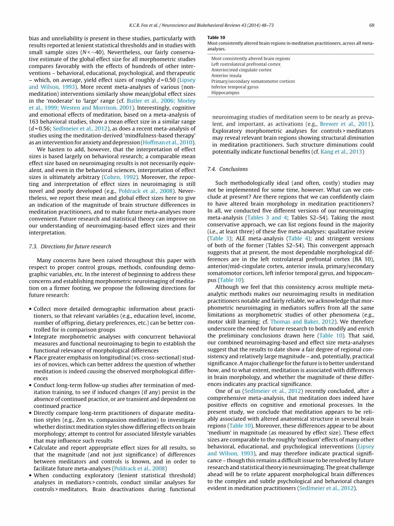
Biobehavioral Reviews 43 (2014) 48–73 69
brstcv–amiea1(sa
seastntamcoi
7
rgctf
•
•
•
•
•
•
•
Table 10Most consistently altered brain regions in meditation practitioners, across all meta-analyses.
Most consistently altered brain regionsLeft rostrolateral prefrontal cortexAnterior/mid cingulate cortexAnterior insulaPrimary/secondary somatomotor corticesInferior temporal gyrus
K.C.R. Fox et al. / Neuroscience and
ias and unreliability is present in these studies, particularly withesults reported at lenient statistical thresholds and in studies withmall sample sizes (N < ∼40). Nevertheless, our fairly conserva-ive estimate of the global effect size for all morphometric studiesompares favorably with the effects of hundreds of other inter-entions – behavioral, educational, psychological, and therapeutic
which, on average, yield effect sizes of roughly d = 0.50 (Lipseynd Wilson, 1993). More recent meta-analyses of various (non-editation) interventions similarly show mean/global effect sizes
n the ‘moderate’ to ‘large’ range (cf. Butler et al., 2006; Morleyt al., 1999; Westen and Morrison, 2001). Interestingly, cognitivend emotional effects of meditation, based on a meta-analysis of63 behavioral studies, show a mean effect size in a similar ranged = 0.56; Sedlmeier et al., 2012), as does a recent meta-analysis oftudies using the meditation-derived ‘mindfulness-based therapy’s an intervention for anxiety and depression (Hoffman et al., 2010).
We hasten to add, however, that the interpretation of effectizes is based largely on behavioral research; a comparable meanffect size based on neuroimaging results is not necessarily equiv-lent, and even in the behavioral sciences, interpretation of effectizes is ultimately arbitrary (Cohen, 1992). Moreover, the repor-ing and interpretation of effect sizes in neuroimaging is stillovel and poorly developed (e.g., Poldrack et al., 2008). Never-heless, we report these mean and global effect sizes here to given indication of the magnitude of brain structure differences ineditation practitioners, and to make future meta-analyses more
onvenient. Future research and statistical theory can improve onur understanding of neuroimaging-based effect sizes and theirnterpretation.
.3. Directions for future research
Many concerns have been raised throughout this paper withespect to proper control groups, methods, confounding demo-raphic variables, etc. In the interest of beginning to address theseoncerns and establishing morphometric neuroimaging of medita-ion on a firmer footing, we propose the following directions foruture research:
Collect more detailed demographic information about practi-tioners, so that relevant variables (e.g., education level, income,number of offspring, dietary preferences, etc.) can be better con-trolled for in comparison groupsIntegrate morphometric analyses with concurrent behavioralmeasures and functional neuroimaging to begin to establish thefunctional relevance of morphological differencesPlace greater emphasis on longitudinal (vs. cross-sectional) stud-ies of novices, which can better address the question of whethermeditation is indeed causing the observed morphological differ-encesConduct long-term follow-up studies after termination of med-itation training, to see if induced changes (if any) persist in theabsence of continued practice, or are transient and dependent oncontinued practiceDirectly compare long-term practitioners of disparate medita-tion styles (e.g., Zen vs. compassion meditation) to investigatewhether distinct meditation styles show differing effects on brainmorphology; attempt to control for associated lifestyle variablesthat may influence such resultsCalculate and report appropriate effect sizes for all results, sothat the magnitude (and not just significance) of differencesbetween meditators and controls is known, and in order to
facilitate future meta-analyses (Poldrack et al., 2008)When conducting exploratory (lenient statistical threshold)analyses in mediators > controls, conduct similar analyses forcontrols > meditators. Brain deactivations during functionalHippocampus
neuroimaging studies of meditation seem to be nearly as preva-lent, and important, as activations (e.g., Brewer et al., 2011).Exploratory morphometric analyses for controls > meditatorsmay reveal relevant brain regions showing structural diminutionin meditation practitioners. Such structure diminutions couldpotentially indicate functional benefits (cf. Kang et al., 2013)
7.4. Conclusions
Such methodologically ideal (and often, costly) studies maynot be implemented for some time, however. What can we con-clude at present? Are there regions that we can confidently claimto have altered brain morphology in meditation practitioners?In all, we conducted five different versions of our neuroimagingmeta-analysis (Tables 3 and 4; Tables S2–S4). Taking the mostconservative approach, we can list regions found in the majority(i.e., at least three) of these five meta-analyses: qualitative review(Table 3); ALE meta-analysis (Table 4); and stringent versionsof both of the former (Tables S2–S4). This convergent approachsuggests that at present, the most dependable morphological dif-ferences are in the left rostrolateral prefrontal cortex (BA 10),anterior/mid-cingulate cortex, anterior insula, primary/secondarysomatomotor cortices, left inferior temporal gyrus, and hippocam-pus (Table 10).
Although we feel that this consistency across multiple meta-analytic methods makes our neuroimaging results in meditationpractitioners notable and fairly reliable, we acknowledge that mor-phometric neuroimaging in mediators suffers from all the samelimitations as morphometric studies of other phenomena (e.g.,motor skill learning; cf. Thomas and Baker, 2012). We thereforeunderscore the need for future research to both modify and enrichthe preliminary conclusions drawn here (Table 10). That said,our combined neuroimaging-based and effect size meta-analysessuggest that the results to date show a fair degree of regional con-sistency and relatively large magnitude – and, potentially, practicalsignificance. A major challenge for the future is to better understandhow, and to what extent, meditation is associated with differencesin brain morphology, and whether the magnitude of these differ-ences indicates any practical significance.
One of us (Sedlmeier et al., 2012) recently concluded, after acomprehensive meta-analysis, that meditation does indeed havepositive effects on cognitive and emotional processes. In thepresent study, we conclude that meditation appears to be reli-ably associated with altered anatomical structure in several brainregions (Table 10). Moreover, these differences appear to be about‘medium’ in magnitude (as measured by effect size). These effectsizes are comparable to the roughly ‘medium’ effects of many otherbehavioral, educational, and psychological interventions (Lipseyand Wilson, 1993), and may therefore indicate practical signifi-cance – though this remains a difficult issue to be resolved by futureresearch and statistical theory in neuroimaging. The great challenge
ahead will be to relate apparent morphological brain differencesto the complex and subtle psychological and behavioral changesevident in meditation practitioners (Sedlmeier et al., 2012).
7 Biobe
C
A
fmSfacfiuC(GaSt(
R
A
A
A
A
A
A
AB
B
B
B
B
B
B
B
B
B
B
a
0 K.C.R. Fox et al. / Neuroscience and
onflicts of interest
The authors declare no conflicts of interest.
cknowledgments
The authors thank Dr. Tal Yarkoni, Dr. David Fresco, and Dr. Clif-ord D. Saron for extremely helpful discussions of meta-analytic
ethods and findings. We also thank Jelena Markovic, Zacharytansfield, Dr. Lawrence M. Ward, and Dr. Todd C. Handy for help-ul comments on earlier drafts of the manuscript, as well as fournonymous reviewers for detailed, considerate, and constructiveomments. Finally, we thank Dr. Norman A.S. Farb and Dr. Sara Lazaror graciously providing previously unpublished statistical data fornclusion in our meta-analyses. This work was supported by a Nat-ral Sciences and Engineering Research Council (NSERC) Vanieranada Graduate Scholarship and University of British ColumbiaUBC) Four-Year Fellowship awarded to K.C.R.F.; NSERC Canadaraduate Scholarships awarded to M.L.D. and M.E.; an Educationalnd Counselling Psychology and Special Education Departmentalcholarship awarded to J.L.F.; a UBC Faculty of Arts Graduate Awardo S.P.R; and grants from the Canadian Institutes of Health ResearchCIHR) and NSERC awarded to K.C.
eferences1
ddis, D.R., Wong, A.T., Schacter, D.L., 2007. Remembering the past and imaginingthe future: common and distinct neural substrates during event constructionand elaboration. Neuropsychologia 45 (7), 1363–1377.
llman, J.M., Hakeem, A., Erwin, J.M., Nimchinsky, E., Hof, P., 2001. The anteriorcingulate cortex. Ann. N. Y. Acad. Sci. 935, 107–117.
shburner, J., Friston, K.J., 2000. Voxel-based morphometry – the methods. Neu-roimage 11 (6), 805–821, http://dx.doi.org/10.1006/nimg.2000.0582.
nalayo, 2003. Satipatthana: The Direct Path to Realization. Windhorse PublicationsLtd, Cambridge, pp. 336 p.
nderson, B.J., 2011. Plasticity of grey matter volume: the cellular and synap-tic plasticity that underlies volumetric change. Dev. Psychobiol. 53 (5),456–465.
ndrews-Hanna, J.R., 2012. The brain’s default network and its adaptive role ininternal mentation. Neuroscientist 18 (3), 251–270.
ustin, J.H., 1999. Zen and the Brain. MIT Press, Boston.aer, R.A., Smith, G.T., Lykins, E., Button, D., Krietemeyer, J., Sauer, S., Walsh, E.,
Duggan, D., Williams, J.M.G., 2008. Construct validity of the five facet mindful-ness questionnaire in meditating and nonmeditating samples. Assessment 15,329–342.
aerentsen, K.B., Hartvig, N.V., Stokilde-Jorgensen, H., Mammen, J., 2001. Onset ofmeditation explored with fMRI. Neuroimage 13 (6), S297.
aerentsen, K.B., Stodkilde-Jorgensen, H., Sommerlund, B., Hartmann, T.,Damsgaard-Madsen, J., Fosnaes, M., Green, A.D., 2010. An investigation ofbrain processes supporting meditation. Cogn. Process. 11, 57–84.
arbas, H., 2000. Connections underlying the synthesis of cognition, memory, andemotion in primate prefrontal cortices. Brain Res. Bull. 52, 319–330.
eaulieu, C., 2002. The basis of anisotropic water diffusion in the nervous system –a technical review. NMR Biomed. 15, 435–455.
eckmann, M., Johansen-Berg, H., Rushworth, M.F., 2009. Connectivity-based par-cellation of human cingulate cortex and its relation to functional specialization.J. Neurosci. 29, 1175–1190.
eer, J.S., John, O.P., Scabini, D., Knight, R.T., 2006. Orbitofrontal cortex and socialbehavior: integrating self-monitoring and emotion-cognition interactions. J.Cogn. Neurosci. 18 (6), 871–879.
ehrens, T.E., Woolrich, M.W., Walton, M.E., Rushworth, M.F., 2007. Learn-ing the value of information in an uncertain world. Nat. Neurosci. 10,1214–1221.
ora, E., Fornito, A., Yücel, M., Pantelis, C., 2010. Voxelwise meta-analysisof grey matter abnormalities in bipolar disorder. Biol. Psychiatry 67,1097–1105.
rausch, A.M., Gutierrez, P.M., 2009. The role of body image and disordered eating
as risk factors for depression and suicidal ideation in adolescents. Suicide Life-Threat. Behav. 39 (1), 58–71.refczynski-Lewis, J.A., Lutz, A., Schaefer, H.S., Levinson, D.B., Davidson, R.J., 2007.Neural correlates of attentional expertise in long-term meditation practitioners.Proc. Natl. Acad. Sci. U. S. A. 104 (27), 11483–11488.
1 References marked with an asterisk indicate studies included in the meta-nalysis.
havioral Reviews 43 (2014) 48–73
Brewer, J.A., Worhunsky, P.D., Grey, J.R., Tang, Y.-Y., Weber, J., Kober, H., 2011. Medi-tation experience is associated with differences in default mode network activityand connectivity. Proc. Natl. Acad. Sci. U. S. A. 108 (50), 20254–20259.
Brown, D., Forte, M., Dysart, M., 1984. Differences in visual sensitivity among mind-fulness meditators and non-meditators. Percept. Mot. Skills 58, 727–733.
Buckner, R.L., Andrews-Hanna, J.R., Schacter, D.L., 2008. The brain’s default network:anatomy, function, and relevance to disease. Ann. N. Y. Acad. Sci. 1124, 1–38.
Bush, G., Luu, P., Posner, M.I., 2000. Cognitive and emotional influences in anteriorcingulate cortex. Trends Cogn. Sci. 4 (6), 215–222.
Butler, A.C., Chapman, J.E., Forman, E.M., Beck, A.T., 2006. The empirical status ofcognitive-behavioral therapy: a review of meta-analyses. Clin. Psychol. Rev. 26,17–31.
Cahn, B.R., Polich, J., 2006. Meditation states and traits: EEG, ERP, and neu-roimaging studies. Psychol. Bull. 132 (2), 180–211, http://dx.doi.org/10.1037/0033-2909.132.2.180.
Cavanna, A.E., Trimble, M.R., 2006. The precuneus: a review of its functional anatomyand behavioural correlates. Brain 129, 564–583.
Chiesa, A., Serretti, A., 2009. Mindfulness-based stress reduction for stress man-agement in healthy people: a review and meta-analysis. J. Altern. Complement.Med. 15 (5), 593–600.
Chiesa, A., Serretti, A., 2010. A systematic review of neurobiological and clinicalfeatures of mindfulness meditations. Psychol. Med. 40 (8), 1239–1252.
Christoff, K., Gabrieli, J.D.E., 2000. The frontopolar cortex and human cognition: evi-dence for a rostrocaudal hierarchical organization within the human prefrontalcortex. Psychobiology 28 (2), 168–186.
Christoff, K., Prabhakaran, V., Dorfman, J., Zhao, Z., Kroger, J.K., Holyoak, K.J., Gabrieli,J.D.E., 2001. Rostrolateral prefrontal cortex involvement in relational integrationduring reasoning. Neuroimage 14 (5), 1136–1149.
Christoff, K., Ream, J.M., Gabrieli, J.D.E., 2004. Cognitive and neural basis of sponta-neous thought processes. Cortex 40, 623–630.
Christoff, K., Ream, J.M., Geddes, L.P.T., Gabrieli, J.D.E., 2003. Evaluating self-generated information: anterior prefrontal contributions to human cognition.Behav. Neurosci. 117 (6), 1161–1168.
Christoff, K., Gordon, A., Smallwood, J., Smith, R., Schooler, J.W., 2009. Experiencesampling during fMRI reveals default network and executive system contrib-utions to mind wandering. Proc. Natl. Acad. Sci. U. S. A. 106 (21), 8719–8724.
Christoff, K., Keramatian, K., Gordon, A.M., Smith, R., Maedler, B., 2009b. Prefrontalorganization of cognitive control according to levels of abstraction. Brain Res.1286, 94–105.
Cohen, J., 1992. A power primer. Psychol. Bull. 112, 155–159.Colcombe, S.J., Erickson, K.I., Scalf, P.E., Kim, J.S., Prakash, R., McAuley, E., Elavsky,
S., Marquez, D.X., Hu, L., Kramer, A.F., 2006. Aerobic exercise training increasesbrain volume in aging humans. J. Gerontol. A 61 (11), 1166–1170.
Condon, P., Desbordes, G., Miller, W., DeSteno, D., Hospital, M.G., DeSteno, D., 2013.Meditation increases compassionate responses to suffering. Psychol. Sci. 24 (10),2125–2127.
Corallo, G., Sackur, J., Dehaene, S., Sigman, M., 2008. Limits on introspection: dis-torted subjective time during the dual-task bottleneck. Psychol. Sci. 19 (11),1110–1117, http://dx.doi.org/10.1111/j.1467-9280.2008.02211.x.
Craig, A.D., 2004. Human feelings: why are some more aware than others? TrendsCogn. Sci. 8 (6), 239–241.
Critchley, H.D., Wiens, S., Rotshtein, P., Öhman, A., Dolan, R.J., 2004. Neuralsystems supporting interoceptive awareness. Nat. Neurosci. 7 (2), 189–195,http://dx.doi.org/10.1038/nn1176.
Cumming, G., 2013. The new statistics: why and how. Psychol. Sci.,http://dx.doi.org/10.1177/0956797613504966, published online beforeprint November 12, 2013.
Davidson, R.J., Jackson, D.C., Kalin, N.H., 2000. Emotion, plasticity, context, and reg-ulation: perspectives from affective neuroscience. Psychol. Bull. 126, 890–909.
Davidson, R.J., Kabat-Zinn, J., Schumacher, J., Rosenkranz, M., Muller, D., Santorelli,S.F., Urbanowski, F., Harrington, A., Bonus, K., Sheridan, J.F., 2003. Alterations inbrain and immune function produced by mindfulness meditation. Psychosom.Med. 65 (4), 564–570.
Debiec, J., LeDoux, J.E., Nader, K., 2002. Cellular and systems reconsolidation in thehippocampus. Neuron 36, 527–538.
deCharms, R.C., Christoff, K., Glover, G.H., Pauly, J.M., Whitfield, S., Gabrieli,J.D.E., 2004. Learned regulation of spatially localized brain activation usingreal-time fMRI. Neuroimage 21 (1), 436–443, http://dx.doi.org/10.1016/j.neuroimage.2003.08.041.
Desbordes, G., Negi, L.T., Pace, T.W., Wallace, B.A., Raison, C.L., Schwartz, E.L., 2012.Effects of mindful-attention and compassion meditation training on amygdalaresponse to emotional stimuli in an ordinary, non-meditative state. Front. Hum.Neurosci. 6, 292.
Dickenson, J., Berkman, E.T., Arch, J., Lieberman, M.D., 2012. Neural correlates offocused attention during a brief mindfulness induction. Soc. Cogn. Affect. Neu-rosci. 8 (1), 40–47.
Draganski, B., Gaser, C., Busch, V., Schuierer, G., Bogdahn, U., May, A., 2004.Neuroplasticity: changes in grey matter induced by training. Nature 427,311–312.
Draganski, B., May, A., 2008. Training-induced structural changes in the adult humanbrain. Behav. Brain Res. 192, 137–142.
Dresler, M., Koch, S.P., Wehrle, R., Spoormaker, V.I., Holsboer, F., Steiger, A., Sämann,P.G., et al., 2011. Dreamed movement elicits activation in the sensorimotor cor-tex. Curr. Biol. 21 (21), 1833–1837, http://dx.doi.org/10.1016/j.cub.2011.09.029.
Driemeyer, J., Boyke, J., Gaser, C., Büchel, C., May, Arne, 2008. Changes in grey matterinduced by learning – revisited. PLoS ONE 3 (7), e2669.
Biobe
D
E
E
E
E
E
E
F
F
F
F
F
F
F
F
F
F
F
F
F
F
F
F
G
G
G
G
G
G
G
G
K.C.R. Fox et al. / Neuroscience and
uvernoy, H.M., 1999. The Human Brain Surface, Blood Supply and Three-Dimensional Sectional Anatomy. Springer Verlag, New York.
gger, M., Smith, G.D., Schneider, M., Minder, C., 1997. Bias in meta-analysis detectedby a simple, graphical test. BMJ 315, 629–634.
ickhoff, S.B., Bzdok, D., Laird, A.R., Kurth, F., Fox, P.T., 2012. Activation likelihoodestimation meta-analysis revisited. Neuroimage 59, 2349–2361.
ickhoff, S.B., Laird, A.R., Grefkes, C., Wang, L.E., Zilles, K., Fox, P.T., 2009. Coordinate-based activation likelihood estimation meta-analysis of neuroimaging data: arandom-effects approach based on empirical estimates of spatial uncertainty.Hum. Brain Mapp. 30, 2907–2926.
llamil, M., Dobson, C., Beeman, M., Christoff, K., 2012. Evaluative and generativemodes of thought during the creative process. Neuroimage 59 (2), 1783–1794.
ngström, M., Pihlsgard, J., Lundberg, P., Soderfelt, B., 2010. Functional magneticresonance imaging of hippocampal activation during silent mantra meditation.J. Altern. Complement. Med. 16, 1253–1258.
ngström, M., Söderfeldt, B., 2010. Brain activation during compassion meditation:a case study. J. Altern. Complement. Med. 16 (5), 597–599.
arb, N.A.S., Segal, Z.V., Mayberg, H., Bean, J., McKeon, D., Fatima, Z., Anderson, A.K.,2007. Attending to the present: mindfulness meditation reveals distinct neuralmodes of self-reference. Soc. Cogn. Affect. Neurosci. 2, 313–322.
arb, N.A.S., Anderson, A.K., Mayberg, H., Bean, J., McKeon, D., Segal, Z.V., 2010. Mind-ing one’s emotions: mindfulness training alters the neural expression of sadness.Emotion 10 (1), 25–33, http://dx.doi.org/10.1037/a0017151.
arb, N.A.S., Anderson, A.K., Segal, Z.V., 2012. The mindful brain and emotion regu-lation in mood disorders. Can. J. Psychiatry 57 (2), 70–77.
arb*, N.A.S., Segal, Z.V., Anderson, A., 2013. Mindfulness meditation training alterscortical representations of interoceptive attention. Soc. Cogn. Affect. Neurosci.8 (1), 15–26.
ayed*, N., Lopez del Hoyo, Y., Andres, E., Serrano-Blanco, A., Bellón, J., Aguilar, K.,Cebolla, A., Garcia-Campayo, J., 2013. Brain changes in long-term Zen meditatorsusing proton magnetic resonance spectroscopy and diffusion tensor imaging: acontrolled study. PLoS ONE 8 (3), e58476.
iedler, K., 2011. Voodoo correlations are everywhere – not only in neuroscience.Perspect. Psychol. Sci. 6 (2), 163–171.
ischl, B., Dale, A.M., 2000. Measuring the thickness of the human cerebral cor-tex from magnetic resonance images. Proc. Natl. Acad. Sci. U.S.A. 97 (20),11050–11055.
leming, S.M., Dolan, R.J., 2012. The neural basis of metacognitive ability. Philos.Trans. R. Soc. B: Biol. Sci. 367 (1594), 1338–1349, http://dx.doi.org/10.1098/rstb.2011.0417.
leming, S.M., Weil, R.S., Nagy, Z., Dolan, R.J., Rees, G., 2010. Relating introspec-tive accuracy to individual differences in brain structure. Science 329 (5998),1541–1543.
ox, K.C.R., Andrews-Hanna, J.R., Spreng, R.N., Christoff, K., 2014. Mind wanderingbeyond the default mode network. Trends Cogn. Sci. (submitted for publication).
ox, K.C.R., Zakarauskas, P., Dixon, M., Ellamil, M., Thompson, E., Christoff, K., 2012.Meditation experience predicts introspective accuracy. PLoS ONE 7 (9), e45370.
ox, K.C.R., Nijeboer, S., Solomonova, E., Domhoff, G.W., Christoff, K., 2013. Dreamingas mind wandering: evidence from functional neuroimaging and first-personcontent reports. Front. Hum. Neurosci. 7 (412), 1–18.
ox, K.C.R., Christoff, K., 2014. Metacognitive facilitation of spontaneous thoughtprocesses: when metacognition helps the wandering mind find its way. In:Fleming, S.M., Frith, C.D. (Eds.), The Cognitive Neuroscience of Metacognition.Springer, pp. 293–319, Ch. 13.
risoni, G.B., Pievani, M., Testa, C., Sabattoli, F., Bresciani, L., Boneetti, M., Beltramello,A., Hayashi, K.M., Toga, A.W., Thompson, P.M., 2007. The topography of greymatter involvement in early and late onset Alzheimer’s disease. Brain 130 (3),720–730.
roeliger, B., Garland, E.L., McClernon, F.J., 2012a. Yoga meditation practitionersexhibit greater grey matter volume and fewer reported cognitive failures: resultsof a preliminary voxel-based morphometry analysis. Evid. Based Complement.Alternat. Med. 821307, 1–8.
roeliger, B., Garland, E.L., Kozink, R.V., Modlin, L.A., Chen, N.-K., et al., 2012b.Meditation-state functional connectivity (msFC): strengthening the DorsalAttention Network and Beyond. Evid. Based Complement. Alternat. Med.680407, 1–9.
ard, T., Hölzel, B.K., Sack, A.T., Hempel, H., Lazar, S.W., Vaitl, D., Ott, U., 2012. Painattenuation through mindfulness is associated with decreased cognitive controland increased sensory processing in the brain. Cereb. Cortex 22 (11), 2692–2702.
enovese, C.R., Lazar, N.A., Nichols, T., 2002. Thresholding of statistical maps in func-tional neuroimaging using the false discovery rate. Neuroimage 15 (4), 870–878.
oenka, S.N., 2000. The Discourse Summaries. Vipassana Research Publications,Onalaska, WA, USA.
oldin, P.R., McRae, K., Ramel, W., Gross, J.J., 2008. The neural bases of emotionregulation: reappraisal and suppression of negative emotion. Biol. Psychiatry63 (6), 577–586.
oldin, P.R., Gross, J.J., 2010. Effects of mindfulness-based stress reduction (MBSR)on emotion regulation in social anxiety disorder. Emotion 10 (1), 83–91.
ood, C.D., Johnsrude, I.S., Asburner, J., Henson, R.N.A., Friston, K.J., Frackowiak, S.J.,2001. A voxel-based morphometric study of ageing in 465 normal adult humanbrains. Neuroimage 14, 21–36.
ottfried, J.A., O’Doherty, J., Dolan, R.J., 2003. Encoding predictive reward value inhuman amygdala and orbitofrontal cortex. Science 301, 1104–1107.
rant*, J.A., Courtemanche, J., Duerden, E.G., Duncan, G.H., Rainville, P., 2010.Cortical thickness and pain sensitivity in Zen meditators. Emotion 10 (1),43–53.
havioral Reviews 43 (2014) 48–73 71
Grant, J.A., Courtemanche, J., Rainville, P., 2011. A non-elaborative mental stance anddecoupling of executive and pain-related cortices predicts low pain sensitivityin Zen meditators. Pain 152, 150–156.
Grant*, J.A., Duerden, E.G., Courtemanche, J., Cherkasova, M., Duncan, G.H., Rainville,P., 2013. Cortical thickness, mental absorption and meditative practice: possibleimplications for disorders of attention. Biol. Psychol. 92, 275–281.
Grant, J.A., Rainville, P., 2009. Pain sensitivity and analgesic effects of mindful statesin Zen meditators: a cross-sectional study. Psychosom. Med. 71, 106–114.
Gurvits, T.V., Shenton, M.E., Hokama, H., Ohta, H., Lasko, N.B., Gilbertson, M.W.,Orr, S.P., Kikinis, R., Jolesz, F.A., McCarley, R.W., Pitman, R.K., 1996. Magneticresonance imaging study of hippocampal volume in chronic, combat-relatedposttraumatic stress disorder. Biol. Psychiatry 40, 1091–1099.
Hamamé, C.M., Vidal, J.R., Ossandón, T., Jerbi, K., Dalal, S.S., Minotti, L., et al., 2012.Reading the mind’s eye: online detection of visuo-spatial working memory andvisual imagery in the inferior temporal lobe. Neuroimage 59 (1), 872–879.
Hodgins, H.S., Adair, K.C., 2010. Attentional processes and meditation. Conscious.Cogn. 19, 872–878.
Hoffman, S.G., Sawyer, A.T., Witt, A.A., Oh, D., 2010. The effect of mindfulness-basedtherapy on anxiety and depression: a meta-analytic review. J. Consult. Clin.Psychol. 78 (2), 169–183.
Hofer, S., Frahm, J., 2006. Topography of the human corpus callosum revisited –comprehensive fiber tractography using diffusion tensor magnetic resonanceimaging. Neuroimage 32, 989–994.
Hölzel, B.K., Ott, U., Hempel, H., Hackl, A., Wolf, K., Stark, R., Vaitl, D., 2007. Differentialengagement of anterior cingulate and adjacent medial frontal cortex in adeptmeditators and non-meditators. Neurosci. Lett. 421, 16–21.
Hölzel*, B.K., Ott, U., Gard, T., Hempel, H., Weygandt, M., Morgen, K., Vaitl, D., 2008.Investigation of mindfulness meditation practitioners with voxel-based mor-phometry. Soc. Cogn. Affect. Neurosci. 3, 55–61.
Hölzel*, B.K., Carmody, J., Evans, K.C., Hoge, E.A., Dusek, J.A., Morgan, L., Pitman,R.K., Lazar, S.W., 2010. Stress reduction correlates with structural changes inthe amygdala. Soc. Cogn. Affect. Neurosci. 5, 11–17.
Hölzel*, B.K., Carmody, J., Vangel, M., Congleton, C., Terramsetti, S.M., Gard, T., Lazar,S.W., 2011a. Mindfulness practice leads to increase in regional brain grey matterdensity. Psych. Res. Neuroimag. 191, 36–43.
Hölzel, B.K., Lazar, S.W., Gard, T., Schuman-Olivier, Z., Vago, D.R., Ott, U., 2011b. Howdoes mindfulness meditation work? proposing mechanisms of action from aconceptual and neural perspective. Perspect. Psycholog. Sci. 6 (6), 537–559.
Hutton, C., Draganski, B., Ashburner, J., Weiskopf, N., 2009. A compari-son between voxel-based cortical thickness and voxel-based morphometryin normal aging. Neuroimage 48 (2), 371–380, http://dx.doi.org/10.1016/j.neuroimage.2009.06.043.
Hupbach, A., Gomez, R., Hardt, O., Nadel, L., 2007. Reconsolidation of episodic mem-ories: a subtle reminder triggers integration of new information. Learn. Mem.14, 47–53.
Hyde, K.L., Lerch, J., Norton, A., Forgeard, M., Winner, E., Evans, A.C., Schlaug, G.,2009. Musical training shapes structural brain development. J. Neurosci. 29 (10),3019–3025.
Ivanovski, B., Mahli, G.S., 2007. The psychological and neurophysiological concomi-tants of mindfulness forms of meditation. Acta Neuropsychiatr. 19 (2), 76–91.
Ives-Deliperi, V.L., Solms, M., Meintjes, E.M., 2011. The neural substrates of mind-fulness: an fMRI investigation. Soc. Neurosci. 6 (3), 231–242.
Iyengar, B.K.S., 1996. Light on the Yoga Sutras of Patanjali. HaperCollinsPublishers,San Francisco.
Jack, C.R., Kokmen, E., Petersen, R.C., Xu, Y.C., O’Brien, P.C., Smith, G.E., et al., 1999.Prediction of AD with MRI-based hippocampal volume in mild cognitive impair-ment. Neurology 52 (7), 1397.
Jacobs, T.L., Epel, E.S., Lin, J.L., Blackburn, E.H., Wolkowitz, O.M., Bridwell, D.A.,Zanesco, A.P., Aichele, S.R., Sahdra, B.K., MacLean, K.A., King, B.G., Shaver, P.R.,Rosenberg, E.L., Ferrer, E., Wallace, B.A., Saron, C.D., 2011. Intensive medita-tion training, immune cell telomerase activity, and psychological mediators.Psychoneuroendocrinology 36 (5), 664–681.
Jha, A., Krompinger, J., Baime, M.J., 2007. Mindfulness training modifies subsystemsof attention. Cogn. Affect. Behav. Neurosci. 7 (2), 109–119.
Jha, A., Stanley, E.A., Kiyonaga, A., Wong, L., Gelfand, L., 2010. Examining the protec-tive effects of mindfulness training on working memory capacity and affectiveexperience. Emotion 10 (1), 54–64.
Johansen-Berg, H., 2010. Behavioural relevance of variation in white mattermicrostructure. Curr. Opin. Neurol. 23, 351–358.
Joseph, J.E., 2001. Functional neuroimaging studies of category specificity in objectrecognition: a critical review and meta-analysis. Cogn. Affect. Behav. Neurosci.1 (2), 119–136.
Kakigi, R., Nakata, H., Inui, K., Hiroe, N., Nagata, O., et al., 2005. Intracerebral painprocessing in a Yoga Master who claims not to feel pain during meditation. Eur.J. Pain 9, 581–589.
Kang*, D., Jo, H.J., Jung, W.H., Kim, S.H., Jung, Y., Choi, C., Lee, U.S., An, S.C., Hang, J.H.,Kwon, J.S., 2013. The effect of meditation on brain structure: cortical thicknessmapping and diffusion tensor imaging. Soc. Cogn. Affect. Neurosci. 8 (1), 27–33.
Kaouane, N., Porte, Y., Vallee, M., Brayda-Bruno, L., Mons, N., Calandreau, L., et al.,2012. Glucocorticoids can induce PTSD-like memory impairments in mice. Sci-ence 335, 1510–1513.
Kempton, M.J., Geddes, J.R., Ettinger, U., Williams, S.C.R., Grasby, P.M., 2008. Meta-analysis, database, and meta-regression of 98 structural imaging studies inbipolar disorder. Arch. Gen. Psychiatry 65 (9), 1017–1032.
Kerr, C.E., Shaw, J.R., Wasserman, R.H., Chen, V.W., Kanojia, A., Bayer, T., Kelley,J.M., 2008. Tactile acuity in experienced Tai Chi practitioners: evidence for
7 Biobe
K
K
K
K
K
K
K
L
L
L
L
L
L
L
L
L
L
L
L
L
L
L
L
L
L
L
L
2 K.C.R. Fox et al. / Neuroscience and
use-dependent plasticity as an effect of sensory-attentional training. Exp. BrainRes. 188 (2), 317–322.
err, C.E., Jonse, S.R., Wan, Q., Pritchett, D.L., Wasserman, R.H., Wexler, A., Villanueva,J.J., Shaw, J.R., Lazar, S.W., Kaptchuk, T.J., Littenberg, R., Hämäläinen, M.S., Moore,C.I., 2011. Effects of mindfulness meditation training on anticipatory alpha mod-ulation in primary somatosensory cortex. Brain Res. Bull. 85, 96–103.
err, C.E., Sacchet, M.D., Lazar, S.W., Moore, C.I., Jones, S.R., 2013. Mindfulness startswith the body: somatosensory attention and top-down modulation of corticalalpha rhythms in mindfulness meditation. Front. Hum. Neurosci. 7, 12.
halsa, S.S., Rudrauf, D., Damasio, A.R., Davidson, R.J., Lutz, A., Tranel, D., 2008.Interoceptive awareness in experienced meditators. Psychophysiology 45 (4),671–677, http://dx.doi.org/10.1111/j.1469-8986.2008.00666.x.
irk, U., Downar, J., Montague, P.R., 2011. Interoception drives increased rationaldecision-making in meditators playing the ultimatum game. Front. Neurosci. 5(49), 1–11.
limecki, O.M., Leiberg, S., Lamm, C., Singer, T., 2012. Functional neural plasticity andassociated changes in positive affect after compassion training. Cereb. Cortex 23(7), 1552–1561.
ozhevnikov, M., Louchakova, O., Josipovic, Z., Motes, M.A., 2009. The enhance-ment of visuospatial processing efficiency through Buddhist Deity meditation.Psychol. Sci. 20, 645–653.
umar*, U., Guleria, A., Kishan, S.S.K., Khetrapal, C.L., 2013. Effect of SOHAM med-itation on human brain: a voxel-based morphometry study. J. Neuroimag.,http://dx.doi.org/10.1111/jon.12040 (Epub ahead of print).
aird, A.R., Fox, M., Price, C.J., Glahn, D.C., Uecker, A.M., Lancaster, J.L., Turkeltaub, P.E.,Kochunov, P., Fox, P.T., 2005. ALE meta-analysis: controlling the false discoveryrate and performing statistical contrasts. Hum. Brain Mapp. 25, 155–164.
azar, S.W., Bush, G., Gollub, R.L., Fricchione, G.L., Khalsa, G., Benson, H., 2000. Func-tional brain mapping of the relaxation response and meditation. Neuroreport11, 1581–1585.
azar*, S.W., Kerr, C.E., Wasserman, R.H., Grey, J.R., Greve, D.N., Treadway, M.T.,McGarvey, M., Quinn, B.T., Dusek, J.A., Benson, H., Rauch, S.L., Moore, C.I., Fischl,B., 2005. Meditation experience is associated with increased cortical thickness.Neuroreport 16, 1893–1897.
ee, T.M.C., Leung, M.-K., Hou, W.-K., Tang, J.C.Y., Yin, J., So, K.-F., Lee, C.-F., Chan,C.C.H., 2012. Distinct neural activity associated with focused-attention medita-tion and loving-kindness meditation. PLoS ONE 7 (8), e40054.
emaitre, H., Goldman, A.L., Sambataro, F., Verchinski, B.A., Meyer-Lindenberg, A.,Weinberger, D., Mattay, V.S., 2012. Normal age-related brain morphometricchanges: nonuniformity across cortical thickness, surface area and grey mattervolume? Neurobiol. Aging 33 (3), 617.e1–617.e9.
erch, J.P., Yiu, A.P., Martinez-Canabal, A., Pekar, T., Bohbot, V.D., et al., 2011. Mazetraining in mice induces MRI-detectable brain shape changes specific to the typeof learning. Neuroimage 54 (3), 2086–2095.
eung*, M., Chan, C.C.H., Yin, J., Lee, C., So, K., Lee, T.M.C., 2013. Increased grey mattervolume in the right angular and posterior parahippocampal gyri in loving-kindness meditators. Soc. Cogn. Affect. Neurosci. 8 (1), 34–39.
ieberman, M.D., Cunningham, W.A., 2009. Type I and Type II error concernsin fMRI research: re-balancing the scale. Soc. Cogn. Affect. Neurosci. 4,423–428.
ipsey, M.W., Wilson, D.B., 1993. The efficacy of psychological, educational, andbehavioral treatment: confirmation from meta-analysis. Am. Psychol. 48 (12),1181–1209.
ipsey, M.W., Wilson, D.B., 2001. Practical Meta-analysis. Sage, Thousand Oaks, CA,Online calculator available at: http://www.campbellcollaboration.org/escalc/html/EffectSizeCalculator-Home.php (accessed August 4, 2013).
ou, H.C., Kjaer, T.W., Friberg, L., Wildschiodtz, G., Holm, S., Nowak, M., 1999. A 15O-H2O PET study of meditation and the resting state of normal consciousness.Hum. Brain Mapp. 7, 98–105.
övdén, M., Wenger, E., Martensson, J., Lindenberger, U., Bäckman, L., 2013. Struc-tural brain plasticity in adult learning and development. Neurosci. Biobehav.Rev., http://dx.doi.org/10.1016/j.neubiorev.2013.02.014.
ow, L.K., Cheng, H.J., 2006. Axon pruning: an essential step underlying the devel-opmental plasticity of neuronal connections. Philos. Trans. R. Soc. Lond. B 361,1531–1544.
uders*, E., Toga, A.W., Lepore, N., Gaser, C., 2009. The underlying anatomical corre-lates of long-term meditation: larger hippocampal and frontal volumes of greymatter. Neuroimage 45, 672–678.
uders*, E., Clark, K., Narr, K.L., Toga, A.W., 2011. Enhanced brain connectivity inlong-term meditation practitioners. Neuroimage 57, 1308–1316.
uders*, E., Phillips, O.R., Clark, K., Kurth, F., Toga, A.W., Narr, K.L., 2012a. Bridgingthe hemispheres in meditation: thicker callosal regions and enhanced fractionalanisotropy (FA) in long-term practitioners. Neuroimage 61, 181–187.
uders*, E., Kurth, F., Mayer, E.A., Toga, A.W., Narr, K.L., Gaser, C., 2012b. The uniquebrain anatomy of meditation practitioners: alterations in cortical gyrification.Front. Hum. Neurosci. 6 (34), 1–9.
uders*, E., Thompson, P.M., Kurth, F., Hong, J.-Y., Phillips, O.R., Wang, Y., Gutman,B.A., Chou, Y.-Y., Narr, K.L., Toga, A.W., 2012c. Global and regional alterations ofhippocampal anatomy in long-term meditation practitioners. Hum. Brain Mapp.,http://dx.doi.org/10.1002/hbm.22153.
uders*, E., Kurth, F., Toga, A.W., Narr, K.L., Gaser, C., 2013. Meditation effects within
the hippocampal complex revealed by voxel-based morphometry and cytoar-chitectonic probabilistic mapping. Front. Psychol. 4 (398), 1–7.utz, A., Greischar, L.L., Perlman, D., Davidson, R.J., 2009. BOLD signal in insula is dif-ferentially related to cardiac function during compassion meditation in expertsvs. novices. Neuroimage 47 (3), 1038–1046.
havioral Reviews 43 (2014) 48–73
Lutz, A., McFarlin, D.R., Perlman, D.M., Salomons, T.V., Davidson, R.J., 2013. Alteredanterior insula activation during anticipation and experience of painful stimuliin expert meditators. Neuroimage 64, 538–546.
Lutz, A., Slagter, H.A., Dunne, J.D., Davidson, R.J., 2008. Attention regula-tion and monitoring in meditation. Trends Cogn. Sci. 12 (4), 163–169,http://dx.doi.org/10.1016/j.tics.2008.01.005.
Lutz, A., Thompson, E., 2003. Neurophenomenology: integrating subjective expe-rience and brain dynamics in the neuroscience of consciousness. J. Conscious.Stud. 10, 31–52.
Mackey, A.P., Whitaker, K.J., Bunge, S.A., 2012. Experience-dependent plasticity inwhite matter microstructure: reasoning training alters structural connectivity.Front. Neuroanat. 6 (32), 1–9.
MacLean, K.A., Ferrer, E., Aichele, S.R., Bridwell, D.A., Zanesco, A.P., King, B.G., Rosen-berg, E.L., Sahdra, B.K., Shaver, P.R., Wallace, B.A., Mangun, G.A., Saron, C.D., 2010.Intensive meditation training improves perceptual discrimination and sustainedattention. Psychol. Sci. 21 (6), 829–839.
Makris, N., Kennedy, D.N., McInerney, S., Sorensen, A.G., Wang, R., Caviness, V.S.,Pandya, D.N., 2005. Segmentation of subcomponents within the superior longi-tudinal fascicle in humans: a quantitative, in vivo. DT-MRI study. Cereb. Cortex15, 854–869.
Maldjian, J.A., Laurienti, P.J., Burdette, J.B., Kraft, R.A., 2003. An automated methodfor neuroanatomic and cytoarchitectonic atlas-based interrogation of fMRI datasets. Neuroimage 19, 1233–1239.
Manna, A., Raffone, A., Perrucci, M.G., Nardo, D., Ferretti, A., Tartaro, A., Londei, A.,Del Gratta, C., Belardinelli, M.O., Romani, G.L., 2010. Neural correlates of focusedattention and cognitive monitoring in meditation. Brain Res. Bull. 82, 46–56.
Margulies, D.S., Vincent, J.L., Kelly, C., Lohmann, G., Uddin, L.Q., Biswal, B.B., Villringer,A., Castellanos, F.X., Milham, M.P., Petrides, M., 2009. Precuneus shares intrinsicfunctional architecture in humans and monkeys. Proc. Natl. Acad. Sci. U.S.A. 106(47), 20069–20074.
Mascaro, J.S., Rilling, J.K., Negi, L.T., Raison, C.L., 2013. Pre-existing brain functionpredicts subsequent practice of mindfulness and compassion meditation. Neu-roimage 69, 35–42.
Mason, M.F., Norton, M.I., Van Horn, J.D., Wegner, D.M., Grafton, S.T., Macrae,C.N., 2007. Wandering minds: the default network and stimulus-independentthought. Science 19, 393–395.
May, A., Gaser, C., 2006. Magnetic resonance-based morphometry: a window intostructural plasticity of the brain. Curr. Opin. Neurol. 19, 407–411.
McCaig, R.G., Dixon, M., Keramantian, K., Liu, I., Christoff, K., 2011. Improved mod-ulation of rostrolateral prefrontal cortex using real-time fMRI training andmeta-cognitive awareness. Neuroimage 55 (3), 1298–1305.
Michalak, J., Burg, J., Heidenrich, T., 2012. Don’t forget your body: mindful-ness, embodiment, and the treatment of depression. Mindfulness 3 (3),190–199.
Millan, M.J., Agid, Y., Brüne, M., Bullmore, E.T., Carter, C.S., et al., 2012. Cognitivedysfunction in psychiatric disorders: characteristics, causes and the quest forimproved therapy. Nat. Rev. Drug Discov. 11, 141–168.
Monti, D.A., Kash, K.M., Kunkel, E.J.S., Brainard, G., Wintering, N., et al., 2012. Changesin cerebral blood flow and anxiety associated with an 8-week mindfulness pro-gramme in women with breast cancer. Stress Health 28, 397–407.
Morecraft, R.J., Tanji, J., 2009. Cingulofrontal interactions and the cingulate motorareas. In: Vogt, B.A. (Ed.), Cingulate Neurobiology and Disease. Oxford UniversityPress, Oxford/New York, pp. 113–144.
Morley, S., Eccleston, C., Williams, A., 1999. Systematic review and meta-analysisof randomized controlled trials of cognitive behaviour therapy and behaviouirtherapy for chronic pain in adults, excluding headache. Pain 80, 1–13.
Murakami, H., Nakao, T., Matsunaga, M., Kasuya, Y., Shinoda, J., Yamada, J., Ohira, H.,2013. The structure of mindful brain. PLoS ONE 7 (9), e46377.
Murphy, K., Garavan, H., 2004. An empirical investigation into the number of subjectsrequired for an event-related fMRI study. Neuroimage 22, 879–885.
Nader, K., Schafe, G.E., LeDoux, J.E., 2000. Reply – reconsolidation: the labile natureof consolidation theory. Nat. Rev. Neurosci. 1, 216–219.
Ochsner, K.N., Ray, R.D., Cooper, J.C., Robertson, E.R., Chopra, S., Gabrieli, J.D.E., Gross,J.J., 2004. For better or for worse: neural systems supporting the cognitive down-and up-regulation of negative emotion. Neuroimage 23, 483–499.
Ongur, D., Price, J.L., 2000. The organization of networks within the orbital andmedial prefrontal cortex of rats, monkeys and humans. Cereb. Cortex 10,206–219.
Orme-Johnson, D.W., Schneider, R.H., Son, Y.D., Nidich, S., Cho, Z., 2006. Neuroimag-ing of meditaiton’s effect on brain reactivity to pain. Neuroreport 17 (12),1359–1363.
Ostafin, B.D., Kassman, K.T., 2012. Stepping out of history: mindfulness improvesinsight problem solving. Conscious. Cogn. 21, 1031–1036.
Pagnoni*, G., Cekic, M., 2007. Age effects of grey matter volume and attentionalperformance in Zen meditation. Neurobiol. Aging 28, 1623–1627.
Pagnoni, G., Cekic, M., Guo, Y., 2008. “Thinking about not-thinking”: neural correlatesof conceptual processing during Zen meditation. PLoS ONE 3 (9), e3083.
Pickut, B.A., Van Hecke, W., Kerckhofs, E., Mariën, P., Vanneste, S., Cras, P., Parizel,P.M., 2013. Mindfulness based intervention in Parkinson’s disease leads to struc-tural brain changes on MRI: a randomized controlled longitudinal trial. Clin.Neurol. Neurosurg. 115 (12), 2419–2425.
Poldrack, R.A., Fletcher, P.C., Henson, R.N., Worsley, K.J., Brett, M., Nichols, T.E., 2008.Guidelines for reporting an fMRI study. Neuroimage 40, 409–414.
Posner, M.I., Rothbart, M.K., Sheese, B.E., Tang, Y., 2007. The anterior cingulategyrus and the mechanism of self-regulation. Cogn. Affect. Behav. Neurosci. 7(4), 391–395.
Biobe
P
R
R
R
R
R
R
R
R
S
S
S
S
S
S
S
S
S
S
S
T
T
T
T
T
T
K.C.R. Fox et al. / Neuroscience and
ujol, J., Soriano-Mas, C., Alonso, P., Cardoner, N., Menchón, J.M., Deus, J., Vallejo,J., 2004. Mapping structural brain alterations in obsessive-compulsive disorder.Arch. Gen. Psychiatry 61 (7), 720–730.
amirez-Ruiz, B., Marti, M.J., Tolosa, E., Bartrés-Faz, D., Summerfield, C., Salgado-Pineda, P., et al., 2005. Longitudinal evaluation of cerebral morphologicalchanges in Parkinson’s disease with and without dementia. J. Neurol. 252 (11),1345–1352.
amnani, N., Owen, A.M., 2004. Anterior prefrontal cortex: insights into functionfrom anatomy and neuroimaging. Nat. Rev. Neurosci. 5, 184–194.
anganath, C., D’Esposito, M., 2005. Directing the mind’s eye: prefrontal, inferior andmedial temporal mechanisms for visual working memory. Curr. Opin. Neurobiol.15, 175–182.
ay, J.W., Shadish, W.R., 1996. How interchangeable are different estimators of effectsize? J. Consult. Clin. Psychol. 64 (6), 1316–1325.
itskes, R., Ritskes-Hoitinga, M., Stodkilde-Jorgensen, H., Baerentsen, K., Hartman,T., 2003. MRI scanning during Zen meditation. Construct. Hum. Sci. 8 (1),85–89.
osenthal, R., 1979. The “file drawer problem” and tolerance for null results. Psychol.Bull. 86 (3), 638–641.
ubia, K., 2009. The neurobiology of meditation and its clinical effectiveness inpsychiatric disorders. Biol. Psychol. 82 (1), 1–11.
ushworth, M.F., Behrens, T.E., Rudebeck, P.H., Walton, M.E., 2007. Contrasting rolesfor cingulate and orbitofrontal cortex in decisions and social behaviour. TrendsCogn. Sci. 11, 168–176.
agi, Y., Tavor, I., Hofstetter, S., Tzur-Moryosef, S., Blumenfeld-Katzir, T., Assaf, Y.,2012. Learning in the fast lane: new insights into neuroplasticity. Neuron 73,1195–1203.
alimi-Khorshidi, G., Smith, S.M., Keltner, J.R., Wager, T.D., Nichols, T.E., 2009. Meta-analysis of neuroimaging data: a comparison of image-based and coordinate-based pooling of studies. Neuroimage 45, 810–823.
chmidt, F.L., Oh, I.-S., Hayes, T.L., 2009. Fixed- versus random-effects modelsin meta-analysis: Model properties and an empirical comparison of dif-ferences in results. Br. J. Math. Stat. Psych. 62, 97–128, http://dx.doi.org/10.1348/000711007X255327.
choenbaum, G., Esber, G.R., 2010. How do you (estimate you will) like them apples?Integration as a defining trait of orbitofrontal function. Curr. Opin. Neurobiol. 20,205–211.
cholz, J., Klein, M.C., Behrens, T.E.J., Johansen-Berg, H., 2009. Training induceschanges in white-matter architecture. Nat. Neurosci. 12, 1370–1371.
edlmeier, P., Eberth, J., Schwarz, M., Zimmermann, D., Haarig, F., Jaeger, S., Kunze,S., 2012. The psychological effects of meditation: a meta-analysis. Psychol. Bull.138 (6), 1139–1171.
hackman, A.J., Salomons, T.V., Slagter, H.A., Fox, A.S., Winter, J.J., Davidson, R.J., 2011.The integration of negative affect, pain and cognitive control in the cingulatecortex. Nat. Rev. Neurosci. 12, 154–167.
ingh, J., 1979. Vijnanabhairvava. Motilal Banarsidass Publishers, Delhi, India, pp.173 p.
oriano-Mas, C., Hernández-Ribas, R., Pujol, J., Urretavizcaya, M., Deus, J., Harrison,B.J., et al., 2011. Cross-sectional and longitudinal assessment of structural brainalterations in melancholic depression. Biol. Psychiatry 69 (4), 318–325.
perduti, M., Mrtinelli, P., Piolino, P., 2012. A neurocognitive model of meditationbased on activation likelihood estimation (ALE) meta-analysis. Conscious. Cogn.21, 269–276.
ze, J.A., Gyurak, A., Yuan, J.W., Levenson, R.W., 2010. Coherence between emotionalexperience and physiology: does body awareness training have an impact? Emo-tion 10 (6), 803–814, http://dx.doi.org/10.1037/a0020146.
akeuchi, H., Taki, Y., Sassa, Y., Hashizume, H., Sekiguchi, A., Fukushima, A.,Kawashima, R., 2011. Working memory training using mental calculationimpacts regional grey matter of the frontal and parietal regions. PLoS ONE 6(8), e23175.
ang*, Y.Y., Lu, Q., Geng, X., Stein, E.A., Yang, Y., Posner, M.I., 2010. Short-term med-itation induces white matter changes in the anterior cingulate. Proc. Natl. Acad.Sci. U. S. A. 107 (35), 15649–15652, http://dx.doi.org/10.1073/pnas.1011043107.
ang*, Y.Y., Lu, Q., Fan, M., Yang, Y., Posner, M.I., 2012. Mechanisms of white mat-ter changes induced by meditation. Proc. Natl. Acad. Sci. U. S. A. 109 (26),10570–10574, http://dx.doi.org/10.1073/pnas.1207817109.
aren, A.A., Creswell, J.D., Gianaro, P.J., 2013. Dispositional mindfulness co-varieswith smaller amygdala and caudate volumes in community adults. PLoS ONE 8
(5), e64574.aubert, M., Villringer, A., Ragert, P., 2012. Learning-related grey and white matterchanges in humans: an update. Neuroscientist 18 (4), 320–325.
easdale, J.D., 1999. Metacognition, mindfulness and the modification of mood dis-orders. Clin. Psychol. Psychother. 6 (2), 146–155.
havioral Reviews 43 (2014) 48–73 73
Teasdale, J.D., Moore, R.G., Hayhurst, H., Pope, M., Williams, S., Segal, Z.V., 2002.Metacognitive awareness and prevention of relapse in depression: empiricalevidence. J. Consult. Clin. Psychol. 70, 275–287.
Testa, C., Laakso, M.P., Sabattoli, F., Rossi, R., Beltramello, A., Soininen, H., Frisoni,G.B., 2004. A comparison between the accuracy of voxel-based morphometryand hippocampal volumetry in Alzheimer’s disease. J. Magn. Reson. Imaging 19(3), 274–282.
Thera, N., 1954. The Heart of Buddhist Meditation. Buddhist Publication Society,Kandy, Sri Lanka.
Thomas, C., Baker, C.I., 2012. Teaching an adult brain new tricks: a critical review ofevidence for training-dependent structural plasticity in humans. Neuroimage,http://dx.doi.org/10.1016/j.neuroimage.2012.03.069.
Tloczynski, J., Santucci, A., Astor-Stetson, E., 2000. Perception of visual illusions bynovice and longer-term meditators. Percept. Mot. Skills 91, 1021–1026.
Tomasino, B., Fregona, S., Skrap, M., Fabbro, F., 2013. Meditation-related activationsare modulated by the practices needed to obtain it and by the expertise: an ALEmeta-analysis study. Front. Hum. Neurosci. 6 (346), 1–14.
Turkeltaub, P.E., Eden, G.F., Jones, K.M., Zeffiro, T.A., 2002. Meta-analysis of thefunctional neuroanatomy of single-word reading: method and validation. Neu-roimage 16, 765–780.
Turkelbaub, P.E., Eickhoff, S.B., Laird, A.R., Fox, M., Wiener, M., Fox, P., 2012. Min-imizing within-experiment and within-group effects in activation likelihoodestimation meta-analyses. Hum. Brain Mapp. 33, 1–13.
Vago, D.A., Silbersweig, D.A., 2012. Self-awareness, self-regulation, and self-transcendence (S-ART): a framework for understanding the neurobiologicalmechanisms of mindfulness. Front. Hum. Neurosci. 6 (296), 1–30.
Vestergaard-Poulsen*, P., van Beek, M., Skewes, J., Bjarkam, C.R., Stubberup, M., Ber-telsen, J., Roepstorff, A., 2009. Long-term meditation is associated with increasedgrey matter density in the brain stem. Neuroreport 20, 170–174.
Vogt, B.A., 2005. Pain and emotion interactions in subregions of the cingulate gyrus.Nat. Rev. Neurosci. 6, 533–544.
Vollestad, J., Nielsen, M.B., Nielsen, G.H., 2012. Mindfulness- and acceptance-basedinterventions for anxiety disorders: a systematic review and meta-analysis. Br.J. Clin. Psychol. 51 (3), 239–260.
Vul, E., Harris, C., Winkielman, P., Pashler, H., 2009. Puzzlingly high correlations infMRI studies of emotion, personality, and social cognition. Perspect. Psychol. Sci.4 (3), 274–290.
Wallis, J.D., 2007. Orbitofrontal cortex and its contribution to decision-making.Annu. Rev. Neurosci. 30, 31–56.
Walton, M.E., Behrens, T.E., Noonan, M.P., Rushworth, M.F., 2011. Giving credit wherecredit is due: orbitofrontal cortex and valuation in an uncertain world. Ann. N.Y. Acad. Sci. 1239, 14–24.
Wang, D.J.J., Rao, H., Korczykowski, M., Wintering, N., Pluta, J., Khalsa, D.S., Newberg,A.B., 2011. Cerebral blood flow changes associated with different meditationpractices and perceived depth of meditation. Psychiatry Res. Neuroimag. 191,60–67.
Watanabe, Y., Gould, E., McEwen, B.S., 1992. Stress induces atropy of apical dendritesof hippocampal CA3 pyramidal neurons. Brain Res. 588, 341–345.
Wei, G., Xu, T., Fan, F., Dong, H., Jiang, L., Li, H., Yang, Z., Luo, J., Zuo, X., 2013. CanTaichi reshape the brain? A brain morphometry study. PLoS ONE 8 (4), e61038.
Wells, R.E., Yeh, G.Y., Kerr, C.E., Wolkin, J., Davis, R.B., et al., 2013. Meditations’ impacton default mode network and hippocampus in mild cognitive impairment: apilot study. Neurosci. Lett. 556, 15–19.
Westbrook, C., Creswell, J.D., Tabibnia, G., Julson, E., Kober, H., Tindle, H.A., 2013.Mindful attention reduces neural and self-reported cue-induced craving insmokers. Soc. Cogn. Affect. Neurosci. 8 (1), 73–84.
Westen, D., Morrison, K., 2001. A multidimensional meta-analysis of treatments fordepression, panic, and generalized anxiety disorder: an empirical examinationof the statuts of empirically supported therapies. J. Consult. Clin. Psychol. 69 (6),875–899.
Winocur, G., Moscovitch, M., 2011. Memory transformation and systems consolida-tion. J. Int. Neuropsychol. Soc. 17, 766–780.
Xue, S., Tang, Y.-Y., Posner, M.I., 2011. Short-term meditation increases networkefficiency of the anterior cingulate cortex. Neuroreport 22, 570–574.
Zarei, M., Johansen-Berg, H., Smith, S., Ciccarelli, O., Thompson, A.J., Matthews,P.M., 2006. Functional anatomy of interhemispheric cortical connections in thehuman brain. J. Anat. 209 (3), 311–320.
Zatorre, R.J., Fields, R.D., Johansen-Berg, H., 2012. Plasticity in grey and white: neu-
roimaging changes in brain structure during learning. Nat. Neurosci. 15 (4),528–536.Zeidan, F., Martucci, K.T., Kraft, R.A., Gordon, N.S., McHaffie, J.G., Coghill, R.C., 2011.Brain mechanisms supporting modulation of pain by mindfulness meditation. J.Neurosci. 31 (14), 5540–5548.


