Embed Size (px)
Citation preview
Is Chytridiomycosis Driving Darwin’s Frogs to Extinction?Claudio Soto-Azat1,2*, Andres Valenzuela-Sanchez1, Barry T. Clarke3, Klaus Busse4, Juan Carlos Ortiz5,
Carlos Barrientos5, Andrew A. Cunningham2
1 Laboratorio de Salud de Ecosistemas, Facultad de Ecologıa y Recursos Naturales, Universidad Andres Bello, Santiago, Chile, 2 Institute of Zoology, Zoological Society of
London, Regent’s Park, London, United Kingdom, 3 Natural History Museum, Department of Life Sciences, Cromwell Rd., London, United Kingdom, 4 Zoologisches
Forschungsmuseum Alexander Koenig, Bonn, Germany, 5 Departamento de Zoologıa, Facultad de Ciencias Naturales y Oceanograficas, Universidad de Concepcion,
Concepcion, Chile
Abstract
Darwin’s frogs (Rhinoderma darwinii and R. rufum) are two species of mouth brooding frogs from Chile and Argentina thathave experienced marked population declines. Rhinoderma rufum has not been found in the wild since 1980. Weinvestigated historical and current evidence of Batrachochytrium dendrobatidis (Bd) infection in Rhinoderma spp. todetermine whether chytridiomycosis is implicated in the population declines of these species. Archived and live specimensof Rhinoderma spp., sympatric amphibians and amphibians at sites where Rhinoderma sp. had recently gone extinct wereexamined for Bd infection using quantitative real-time PCR. Six (0.9%) of 662 archived anurans tested positive for Bd (4/289R. darwinii; 1/266 R. rufum and 1/107 other anurans), all of which had been collected between 1970 and 1978. An overall Bd-infection prevalence of 12.5% was obtained from 797 swabs taken from 369 extant individuals of R. darwinii and 428individuals representing 18 other species of anurans found at sites with current and recent presence of the two Rhinodermaspecies. In extant R. darwinii, Bd-infection prevalence (1.9%) was significantly lower than that found in other anurans (7.3%).The prevalence of infection (30%) in other amphibian species was significantly higher in sites where either Rhinoderma spp.had become extinct or was experiencing severe population declines than in sites where there had been no apparent decline(3.0%; x2 = 106.407, P,0.001). This is the first report of widespread Bd presence in Chile and our results are consistent withRhinoderma spp. declines being due to Bd infection, although additional field and laboratory investigations are required toinvestigate this further.
Citation: Soto-Azat C, Valenzuela-Sanchez A, Clarke BT, Busse K, Ortiz JC, et al. (2013) Is Chytridiomycosis Driving Darwin’s Frogs to Extinction? PLoS ONE 8(11):e79862. doi:10.1371/journal.pone.0079862
Editor: Michael Sears, Clemson University, United States of America
Received July 11, 2013; Accepted September 25, 2013; Published November 20, 2013
Copyright: � 2013 Soto-Azat et al. This is an open-access article distributed under the terms of the Creative Commons Attribution License, which permitsunrestricted use, distribution, and reproduction in any medium, provided the original author and source are credited.
Funding: This research was funded by the Institute of Zoology, Zoological Society of London (ZSL) and the ZSL EDGE Fellowship Programme; the DireccionGeneral de Investigacion y Doctorados, Universidad Andres Bello; the Field Veterinary Programme, Wildlife Health Fund, Wildlife Conservation Society; and aFundacion Futuro Scholarship. AAC is supported by a Royal Society Wolfson Research Merit Award. The funders had no role in study design, data collection andanalysis, decision to publish, or preparation of the manuscript.
Competing Interests: The authors have declared that no competing interests exist.
* E-mail: [email protected]
Introduction
There are two species of Darwin’s frogs, both of which inhabit
the temperate forests of South America: the northern Darwin’s
frog (Rhinoderma rufum), which is endemic to central Chile, and the
southern Darwin’s frog (R. darwinii), which is found in south and
southern Chile and also in adjacent areas of Argentina [1,2]. The
behaviour that sets these frogs apart from all other amphibians is
that the males care for their young by incubating them in their
vocal sacs for at least part of their development, a process known
as neomelia [3,4]. In recent decades, both species have undergone
marked population declines and R. rufum has not been recorded
since 1980 [5]. The reasons for these apparent disappearances
remain poorly understood [6,7]. Throughout the historical
distribution of R. rufum, and within the northern range of R.
darwinii, there has been extensive habitat degradation, due to the
large-scale replacement of native forest with pine (Pinus radiata) and
eucalyptus (Eucalyptus globulus) plantations, and land use change to
agriculture [1,2]. Habitat loss, however, does not fully explain the
enigmatic disappearances of R. rufum from its entire historical
range or of the declines of R. darwinii from undisturbed ecosystems,
including National Parks [8]. In this context, it has been
hypothesised that amphibian chytridiomycosis, an infectious
disease caused by the nonhyphal zoosporic chytrid fungus,
Batrachochytrium dendrobatidis (Bd), might be implicated in the
disappearances of Darwin’s frogs [1,2,8].
Amphibian chytridiomycosis, a recently-described emerging
disease of amphibians [9,10], has been associated with amphibian
epizootic mass mortalities, population declines and global extinc-
tions in different regions of the world [11,12,13,14,15,16,17].
Different genotypes of the fungus have been described, with the
most virulent being a recombinant lineage, termed the global
panzootic lineage (BdGPL) [18]. Recently, Bd whole-genome
sequencing has demonstrated a higher genetic differentiation than
previously recognised (including within BdGPL) [18,19] and a
complex evolutionary history that predates contemporary am-
phibian declines [20]. This highly-pathogenic and readily-trans-
missible pathogen appears to be capable of infecting an entire class
of organism (the Amphibia), with devastating effects [21]. It has
been described as: ‘‘the worst infectious disease ever recorded
among vertebrates in terms of the number of species impacted and
its propensity to drive them to extinction’’ [22]. In 2007,
chytridiomycosis was identified as the cause of death of a group
of 30 wild-caught R. darwinii exported to Germany for captive
PLOS ONE | www.plosone.org 1 November 2013 | Volume 8 | Issue 11 | e79862
breeding [23]. Infection with Bd has been reported in populations
of the invasive African clawed frog, Xenopus laevis [24] in central
Chile. Additionally, Bourke et al. [25,26] recently described Bd
infection in R. darwinii and two other native frog species in the
south of the country. The impacts of this emerging disease on
amphibian populations in Chile, including Darwin’s frogs,
however, have not been studied.
Here, we investigate whether amphibian chytridiomycosis is
implicated in the population declines of Darwin’s frogs. We looked
for evidence of historical Bd infection in Rhinoderma spp. and
amphibians at current and former Rhinoderma sp. sites prior to and
post the onset of declines. Also, we determined how widespread Bd
infection is both in contemporary populations of R. darwinii across
its current range and in other anuran species at sites of Rhinoderma
spp. population decline or recent extinction.
Materials and Methods
Ethics statementThis study was carried out in strict accordance with the
recommendations in the guidelines for use of live amphibians and
reptiles in field research compiled by the American Society of
Ichthyologists and Herpetologists (ASIH). Research was approved
by the ZSL Ethics Committee and was conducted following
Chilean and Argentinian wildlife regulations and according to
permits 1241/08, 7377/09, 7993/10 and 300/12 of the Livestock
and Agriculture Service (SAG) and 20/09, XI-01/09, 28/11 and
X-03/11 of the National Forestry Corporation (CONAF) both in
Chile, and permit 1119/11 of the National Parks Administration
(APN) in Argentina. Archived amphibians were examined in their
museum of origin, by the authors with specific permission given by
all 5 zoological institutions.
Study areaArchived amphibian specimens from museum collections in
Europe and Chile were examined for evidence of Bd infection.
Also, extensive surveys for Bd infection throughout the historical
ranges of R. rufum and R. darwinii were conducted from October
2008 to March 2012. These ranges extended from Zapallar (32u33’ 03’’S, 71u 26’ 37’’W) to Aysen (45u 24’ 24’’S, 72u 41’ 52’’W) in
Chile, and included adjacent areas in the Andes in the Neuquen
and Rıo Negro Provinces in Argentina (Figure 1).
Archived anuransA retrospective study was carried out by examining 555
postmetamorphic Rhinoderma spp. and 107 sympatric anuran
specimens, from the collections of the Zoologisches Museum
Hamburg (ZMH, n = 321); Natural History Museum, London
(BMNH, n = 142); Museo de Zoologıa, Universidad de Concep-
cion, Chile (MZUC, n = 121); Zoologisches Forschungsmuseum
Alexander Koenig, Bonn (ZFMK, n = 46); and Centro de
Investigaciones Zoologicas, Universidad de Chile (CIZ, n = 32).
Specimens were preserved in 70% ethanol (or 70% industrial
methylated spirits for BMNH amphibians) and had been collected
in central and south Chile between 1835 and 1989 (Table 1) for
purposes other than disease investigation.
Living anuransCross-sectional studies were carried out at sites where R. darwinii
was extant and at sites where Rhinoderma spp. had recently (since
1966) become extinct. Sites were delimited and a search effort of
one hour by two researchers was conducted during daylight hours
using a standardised methodology, as previously described [8].
SamplingArchived anurans. The skin of the ventral pelvis and ventral
hind limbs of each amphibian museum specimen was sampled by
brushing with a tapered inter-dental brush (3.2 to 6.0 mm; Oral B
Laboratories), following Soto-Azat et al. [27]. Where multiple
specimens were held in a single jar, they were rinsed with running
tap water prior to sampling to remove possible surface contam-
ination with Bd. Each specimen was handled using a new pair of
disposable nitrile or latex gloves.
Live anurans. Only post-metamorphic and adult anurans
were sampled. Frogs were captured by hand, safely contained in
individual sealed plastic bags and put back immediately after the
capture session in the exact place of capture. Each individual was
handled with the use of clean disposable nitrile gloves. A sterile
dry, rayon-tipped swab (MW100, Medical & Wire Equipment
Co.) was firmly run five times each over the ventral abdomen and
pelvis, each ventral hind limb (femur and tibia) and the plantar
surface of each hind foot, to complete a total of 35 strokes. Dorsal
and ventral pattern photographs were taken of each Darwin’s frog
sampled for identification purposes. In order to minimize any Bd
contamination of samples or the spread of pathogens within or
between study sites by researchers, equipment or materials, a strict
field sampling and disinfection protocol was followed according to
that recommended by the Amphibian and Reptile Groups, UK:
ARG Advice Note 4 (http://www.arguk.org/advice-and-
guidance/view-category). All samples were stored at 280 uC until
processed.
Diagnostic analysisPost sampling, whole interdental brushes and swab tips were
deposited separately in 1.5 ml Eppendorf tubes containing 50 and
60 ml, respectively, of PrepMan Ultra (Applied Biosystems) and
between 30 to 40 mg of Zirconium/silica beads of 0.5 mm
diameter (Biospec Products). For each sample, DNA was extracted
following the protocol of Boyle et al. [28]. Extracted DNA was
diluted (1:10) in double-distilled water and analysed using a
quantitative real-time polymerase chain reaction Taqman assay
(qPCR) with primers specific for the ITS-1/5.8S ribosomal DNA
region of Bd. In addition, bovine serum albumin (BSA) was
included in the Taqman mastermix to minimise inhibition of the
PCR [29]. For each sample, diagnostic assays were performed in
duplicate, and standards of known zoospore concentration were
included within each PCR plate, as were negative controls. A
result was considered positive when: (1) amplification (i.e. a clearly
sigmoid curve) occurred in both replicated PCR assays, (2) values
higher than 0.1 genomic equivalents (GE) were obtained from
both replicated reactions, and (3) average GE from both replicates
were higher than its standard deviation. Extracted DNA from any
positive sample was re-tested in duplicate and only determined to
be positive for the purposes of this study if Bd DNA was clearly
amplified in duplicate wells for a second time.
Data analysisAreas with historical and current presence of Rhinoderma spp. .
2 km from each other were determined to be separate sites or
populations [30]. Statistical analyses were performed using SPSS
(v. 20.0) to detect any significant difference between: 1) Bd
prevalence and time in archived R. darwinii (using Fisher’s exact
test for small sample sizes), 2) Bd prevalence in extant R. darwinii
and sympatric amphibians (using the chi-squared test), 3) Bd
intensity in extant R. darwinii and all other amphibian species
tested (using the Mann-Whitney U-test), and 4) Bd prevalence at
sites with and without evidence of recent Rhinoderma spp.
population decline in extant R. darwinii (using the chi-squared
Chytridiomycosis in Darwin’s Frogs
PLOS ONE | www.plosone.org 2 November 2013 | Volume 8 | Issue 11 | e79862
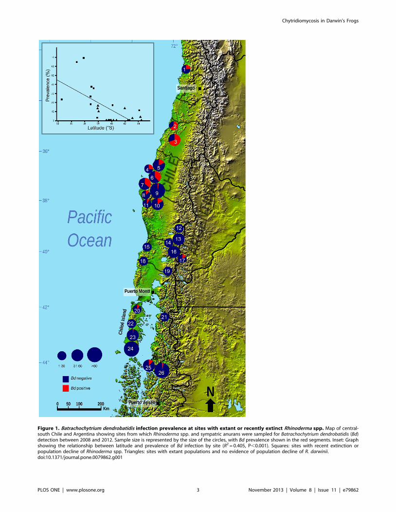
Figure 1. Batrachochytrium dendrobatidis infection prevalence at sites with extant or recently extinct Rhinoderma spp. Map of central-south Chile and Argentina showing sites from which Rhinoderma spp. and sympatric anurans were sampled for Batrachochytrium dendrobatidis (Bd)detection between 2008 and 2012. Sample size is represented by the size of the circles, with Bd prevalence shown in the red segments. Inset: Graphshowing the relationship between latitude and prevalence of Bd infection by site (R2 = 0.405, P,0.001). Squares: sites with recent extinction orpopulation decline of Rhinoderma spp. Triangles: sites with extant populations and no evidence of population decline of R. darwinii.doi:10.1371/journal.pone.0079862.g001
Chytridiomycosis in Darwin’s Frogs
PLOS ONE | www.plosone.org 3 November 2013 | Volume 8 | Issue 11 | e79862
test). Data on Rhinoderma spp. abundance is scarce. To consider a
population having evidence of recent decline, we used data from a
previous study [8], which investigated population sizes and the
extent of declines in Darwin’s frogs. Briefly, populations catego-
rised as having declined comprised those known to have
disappeared since 1966, or (in one case) known to have undergone
a recent marked population decline. A relationship between Bd
prevalence at sites with historical and current Rhinoderma spp.
populations and latitude was also tested using a simple linear
regression model.
Results
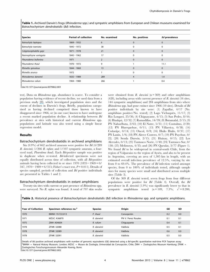
Batrachochytrium dendrobatidis in archived amphibiansSix (0.9%) of 662 archived anurans were positive for Bd (4/289
R. darwinii; 1/266 R. rufum; and 1/107 sympatric anurans, a four-
eyed toad, Pleurodema thaul). Each Bd-positive sample was positive
in duplicate when re-tested. Bd-infected specimens were not
equally distributed across time of collection, with all Bd-positive
animals having been collected in or since 1970 (183521969 = 0/
347, 197021989 = 6/315; Fisher’s exact test, P = 0.011). Details of
species sampled, periods of collection and Bd positive individuals
are presented in Tables 1 and 2.
Batrachochytrium dendrobatidis in extant amphibiansTwenty-six sites with current or past presence of Rhinoderma spp.
were surveyed. No R. rufum was found. A total of 797 skin swabs
were obtained from R. darwinii (n = 369) and other amphibians
(428), including areas with current presence of R. darwinii (16 sites,
144 sympatric amphibians) and 284 amphibians from sites where
Rhinoderma spp. had gone extinct since 1966 (10 sites). Details of Bd
positive individuals by site were: (1) Zapallar, 4/17 (No.
amphibian positive/No. tested); (2) Lago Vichuquen, 11/17; (3)
Rıo Longavı, 25/36; (4) Chiguayante, 4/15; (5) San Pedro, 8/44;
(6) Hualqui, 12/32; (7) Ramadillas, 14/39; (8) Butamalal, 2/15; (9)
PN Nahuelbuta, 2/63; (10) El Natre, 5/31; (11) Contulmo, 2/20;
(12) PN Huerquehue, 0/11; (13) PN Villarrica, 0/38; (14)
Conaripe, 0/14; (15) Oncol, 0/8; (16) Huilo Huilo, 0/37; (17)
PN Lanın, 1/6; (18) PN Alerce Costero, 0/7; (19) PN Puyehue, 0/
22; (20) Senda Darwin, 2/15; (21) Huinay, 0/15; (22) Los
Alerzales, 0/13; (23) Tantauco Norte, 1/40; (24) Tantauco Sur, 0/
130; (25) Melimoyu, 4/35; and (26) PN Queulat, 3/77 (Figure 1).
We found Bd to be widespread in central-south Chile, from the
region of Valparaiso to the region of Aysen, and also to be present
in Argentina, covering an area of 1,305 km in length, with an
estimated overall infection prevalence of 12.5%, varying by site
from 0 to 69.4%. The prevalence of Bd infection varied amongst
species, from 0 to 100% of individuals tested, although sample
sizes for many species were small and distributed across multiple
sites (Table 3).
Of the 369 R. darwinii tested, seven frogs from four different
populations were positive for Bd (Table 4). Overall, the Bd
prevalence in R. darwinii (1.9%) was significantly lower to that in
sympatric amphibians tested (n = 109, 7.3%; x2 = 8.200,
Table 1. Archived Darwin’s frogs (Rhinoderma spp.) and sympatric amphibians from European and Chilean museums examined forBatrachochytrium dendrobatidis (Bd) infection.
Species Period of collection No. examined No. positives Bd prevalence
Batrachyla leptopus 186921932 6 0 0
Batrachyla taeniata 184521972 38 0 0
Calyptocephalella gayi 187121978 21 0 0
Nannophryne variegata 184521962 17 0 0
Pleurodema bufonina 1971 2 0 0
Pleurodema thaul 197021972 9 1 11.1
Rhinella spinulosa 184421860 13 0 0
Rhinella arunco 1972 1 0 0
Rhinoderma darwinii 183521989 289 4 1.4
Rhinoderma rufum 190421980 266 1 0.4
doi:10.1371/journal.pone.0079862.t001
Table 2. Historical presence of Batrachochytrium dendrobatidis (Bd) infection in Rhinoderma spp. and sympatric amphibians.
Year of Collection Specimen reference no.a Species Origin GE SD
1970 BMNH 19.722.013 P. thaul Concepcion 0.2 0.0
1971 MZUC A36870 R. darwinii PN V. Perez Rosales 0.1 0.1
1975 ZMH A04604 R. rufum Chiguayante 0.4 0.1
1978 ZFMK 32088 R. darwinii Valdivia 0.5 0.1
1978 ZFMK 32089 R. darwinii Valdivia 0.6 0.0
1978 ZFMK 32091 R. darwinii Valdivia 0.5 0.0
Details of Bd positive archived amphibians with number of genomic equivalents (GE) detected using a Bd-specific quantitative real-time PCR Taqman assay.aBMNH = Natural History Museum, London; MZUC = Museo de Zoologıa, Universidad de Concepcion, Chile; ZMH = Zoologisches Museum Hamburg; ZFMK =Zoologisches Forschungsmuseum Alexander Koenig, Bonn.doi:10.1371/journal.pone.0079862.t002
Chytridiomycosis in Darwin’s Frogs
PLOS ONE | www.plosone.org 4 November 2013 | Volume 8 | Issue 11 | e79862
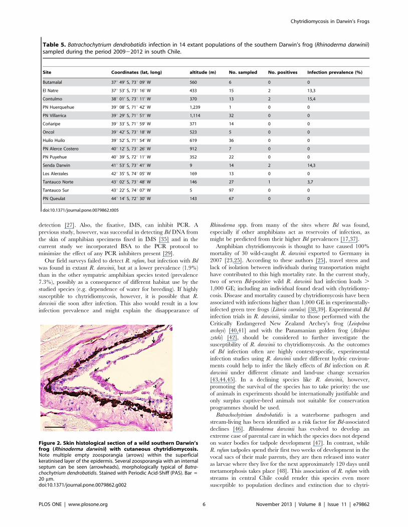
P = 0.004). Contemporary R. darwinii populations sampled in south
and southern Chile and their Bd prevalences are shown in Table 5.
Although R. darwinii had the highest infection intensities (median:
127.1; range: 6.727,059.1 GE) when compared with all other
infected species (13.9; 0.124,481.0 GE) they were not significantly
different (Mann-Whitney U-test; U = 188.0, P = 0.063). Of partic-
ular interest were two R. darwinii from which GE counts over 1,000
were detected. Both frogs belonged to the northernmost known
populations. Of these, one individual (NATRE74/12; 1,020 GE)
was found dead at the capture site and subsequent histopatholog-
ical examination revealed chytridiomycosis as the cause of death
(Figure 2). The prevalence of Bd infection was significantly higher
at sites with either Rhinoderma spp. extinction or severe population
decline (30.0%) than at sites with no apparent Rhinoderma spp.
declines (3.0%; x2 = 106.407, P,0.001). Additionally, when Bd
prevalence by site and geographical location were analysed, a
linear regression revealed an inverse relationship between Bd
prevalence and latitude (R2 = 0.405, P,0.001; Figure 1).
Discussion
Museum amphibian specimens have been increasingly recog-
nised as a valuable source of information for retrospective
epidemiological studies [31,32,33,34,35]. Using such specimens,
we demonstrated historical evidence of Bd infection in three
species of native frogs from south Chile (R. darwinii, R. rufum and P.
thaul). Although we examined similar numbers of frogs that had
been collected prior to 1970 and post-1970, all six Bd-positive
archived amphibians were collected from 1970 to 1978 inclusive: a
time coincident with the onset of the global amphibian population
decline phenomenon, including the disappearance of R. rufum, and
the occurrence of the first amphibian global extinctions subse-
quently associated with Bd [16,35,36]. The only R. rufum Bd-
positive animal was an individual kept in a jar with 179 other R.
rufum specimens, all of which had been collected from Chiguayante
(Biobıo Region, near Concepcion) during a two-day collection
session in December 1975.
As the fixation history of the examined archived amphibians is
not known, the overall infection prevalence (0.9%) and intensity of
infection (GE values 0.120.6) obtained are likely an underesti-
mation of the true situation. For example, although all of the
archived specimens examined were preserved in alcohol, it is
highly possible that many had been initially fixed in formalin, a
chemical known to degrade DNA, reducing the likelihood of Bd
Table 3. Batrachochytrium dendrobatidis (Bd) infection in 19 amphibian species at 26 sites with historical and current presence ofDarwin’s frogs (Rhinoderma spp.), sampled during the period 200822012 in central and south Chile and south-western Argentina.
Species No. Sampled No. Positives Infection prevalence (%) Mean GE GE Range
Alsodes australis 2 0 0 - -
Alsodes barrioi 12 0 0 - -
Alsodes nodosus 12 4 33.3 17.7 1.7253.1
Alsodes verrucosus 2 0 0 - -
Batrachyla antartandica 34 6 17.6 177.6 1.42656.3
Batrachyla leptopus 16 0 0 - -
Batrachyla taeniata 68 3 4.4 180.5 26.52408.9
Calyptocephalella gayi 18 18 100 997.0 79.323,355.0
Eupsophus altor 1 0 0 - -
Eupsophus calcaratus 32 1 3.1 5.4 -
Eupsophus contulmoensis 15 4 26.7 5.0 0.1214.0
Eupophus emiliopugini 4 0 0 - -
Eupsophus nahuelbutensis 59 3 5.1 15.1 0.4240.1
Eupsophus roseus 10 2 20.0 6.4 2,6211,7
Eupsophus vertebralis 1 0 0 - -
Hylorina sylvatica 3 1 33.3 593.3 -
Pleurodema thaul 137 51 37.2 149.5 0.224,481.0
Rhinoderma darwinii 369 7 1.9 1,221.4 6.727,059.1
Telmatobufo bullocki 2 0 0 - -
Genomic equivalents (GE) detected using a Bd-specific quantitative real-time PCR Taqman assay are expressed in means and ranges.doi:10.1371/journal.pone.0079862.t003
Table 4. Details of Batrachochytrium dendrobatidis positiveSouthern Darwin’s frogs (Rhinoderma darwinii) sampledduring the period 200822012 in south Chile with number ofgenomic equivalents (GE) detected using a Bd-specificquantitative real-time PCR Taqman assay.
Reference Site Animal GE SDa
NATRE74/12 El Natre subadult 1,019.5 90.3
NATRE151/12 El Natre brooding male 249.0 40.4
CON123/10 Contulmo adult female 7,059.1 777.4
CON224/11 Contulmo Juvenile 21.0 0.5
SD08/11 Senda Darwin adult male 127.1 9.5
SD03/11 Senda Darwin adult male 67.9 3.7
YAL45/12 Yaldad adult female 6.7 0.3
aStandard deviation.doi:10.1371/journal.pone.0079862.t004
Chytridiomycosis in Darwin’s Frogs
PLOS ONE | www.plosone.org 5 November 2013 | Volume 8 | Issue 11 | e79862
detection [27]. Also, the fixative, IMS, can inhibit PCR. A
previous study, however, was successful in detecting Bd DNA from
the skin of amphibian specimens fixed in IMS [35] and in the
current study we incorporated BSA to the PCR protocol to
minimize the effect of any PCR inhibiters present [29].
Our field surveys failed to detect R. rufum, but infection with Bd
was found in extant R. darwinii, but at a lower prevalence (1.9%)
than in the other sympatric amphibian species tested (prevalence
7.3%), possibly as a consequence of different habitat use by the
studied species (e.g. dependence of water for breeding). If highly
susceptible to chytridiomycosis, however, it is possible that R.
darwinii die soon after infection. This also would result in a low
infection prevalence and might explain the disappearance of
Rhinoderma spp. from many of the sites where Bd was found,
especially if other amphibians act as reservoirs of infection, as
might be predicted from their higher Bd prevalences [17,37].
Amphibian chytridiomycosis is thought to have caused 100%
mortality of 30 wild-caught R. darwinii exported to Germany in
2007 [23,25]. According to these authors [25], travel stress and
lack of isolation between individuals during transportation might
have contributed to this high mortality rate. In the current study,
two of seven Bd-positive wild R. darwinii had infection loads .
1,000 GE; including an individual found dead with chytridiomy-
cosis. Disease and mortality caused by chytridiomycosis have been
associated with infections higher than 1,000 GE in experimentally-
infected green tree frogs (Litoria caerulea) [38,39]. Experimental Bd
infection trials in R. darwinii, similar to those performed with the
Critically Endangered New Zealand Archey’s frog (Leiopelma
archeyi) [40,41] and with the Panamanian golden frog (Atelopus
zeteki) [42], should be considered to further investigate the
susceptibility of R. darwinii to chytridiomycosis. As the outcomes
of Bd infection often are highly context-specific, experimental
infection studies using R. darwinii under different hydric environ-
ments could help to infer the likely effects of Bd infection on R.
darwinii under different climate and land-use change scenarios
[43,44,45]. In a declining species like R. darwinii, however,
promoting the survival of the species has to take priority: the use
of animals in experiments should be internationally justifiable and
only surplus captive-bred animals not suitable for conservation
programmes should be used.
Batrachochytrium dendrobatidis is a waterborne pathogen and
stream-living has been identified as a risk factor for Bd-associated
declines [46]. Rhinoderma darwinii has evolved to develop an
extreme case of parental care in which the species does not depend
on water bodies for tadpole development [47]. In contrast, while
R. rufum tadpoles spend their first two weeks of development in the
vocal sacs of their male parents, they are then released into water
as larvae where they live for the next approximately 120 days until
metamorphosis takes place [48]. This association of R. rufum with
streams in central Chile could render this species even more
susceptible to population declines and extinction due to chytri-
Table 5. Batrachochytrium dendrobatidis infection in 14 extant populations of the southern Darwin’s frog (Rhinoderma darwinii)sampled during the period 200922012 in south Chile.
Site Coordinates (lat, long) altitude (m) No. sampled No. positives Infection prevalence (%)
Butamalal 37u 49’ S, 73u 09’ W 560 6 0 0
El Natre 37u 53’ S, 73u 16’ W 433 15 2 13,3
Contulmo 38u 01’ S, 73u 11’ W 370 13 2 15,4
PN Huerquehue 39u 08’ S, 71u 42’ W 1,239 1 0 0
PN Villarrica 39u 29’ S, 71u 51’ W 1,114 32 0 0
Conaripe 39u 33’ S, 71u 59’ W 371 14 0 0
Oncol 39u 42’ S, 73u 18’ W 523 5 0 0
Huilo Huilo 39u 52’ S, 71u 54’ W 619 36 0 0
PN Alerce Costero 40u 12’ S, 73u 26’ W 912 7 0 0
PN Puyehue 40u 39’ S, 72u 11’ W 352 22 0 0
Senda Darwin 41u 53’ S, 73u 41’ W 9 14 2 14,3
Los Alerzales 42u 35’ S, 74u 05’ W 169 13 0 0
Tantauco Norte 43u 02’ S, 73u 48’ W 146 27 1 3,7
Tantauco Sur 43u 22’ S, 74u 07’ W 5 97 0 0
PN Queulat 44u 14’ S, 72u 30’ W 143 67 0 0
doi:10.1371/journal.pone.0079862.t005
Figure 2. Skin histological section of a wild southern Darwin’sfrog (Rhinoderma darwinii) with cutaneous chytridiomycosis.Note multiple empty zoosporangia (arrows) within the superficialkeratinised layer of the epidermis. Several zoosporangia with an internalseptum can be seen (arrowheads), morphologically typical of Batra-chochytrium dendrobatidis. Stained with Periodic Acid-Shiff (PAS). Bar =20 mm.doi:10.1371/journal.pone.0079862.g002
Chytridiomycosis in Darwin’s Frogs
PLOS ONE | www.plosone.org 6 November 2013 | Volume 8 | Issue 11 | e79862
diomycosis. Although found in only a single archived specimen,
evidence of Bd infection was found in possibly the largest known R.
rufum population [8] five years before the species was last recorded
[5]. This, along with a positive association between Bd prevalence
and Rhinoderma spp. population extinction/decline, suggests a
possible association between chytridiomycosis and the disappear-
ance of R. rufum.
We detected an inverse relationship between Bd prevalence and
latitude, similar to that found by Kriger et al. [49] in the stony
creek frog (Litoria lesueuri) in eastern Australia. Whether this is a
reflection of the historical introduction and spread of Bd in Chile,
with the organism not yet having reached the south of the country,
or if it is due to environmental factors (e.g. temperature) is not yet
clear. Longitudinal sampling of sites across the gradient would
help to answer this question. That such a gradient exists, however,
indicates that northern populations of R. darwinii are likely to be
under a greater threat from chytridiomycosis than those in the
south. It also suggests that the instigation of biosecurity measures
might decrease the rate of spread of the disease to the southern
populations of R. darwinii (assuming that Bd has not already
reached this region and is less readily detected due to the low
temperatures there limiting its growth).
It is not known if the Bd detected in the archived or extant
specimens in the current study is the hypervirulent BdGPL, a
BdGPL-hybrid, or perhaps an endemic lineage (or lineages) of the
fungus. If BdGPL is present in Chile, its spread to the country
might have occurred via the introduction of X. laevis [32]. Feral
populations of this invasive species, which have been established in
central Chile since the 1970s, are known to be Bd-positive,
although other mechanisms of pathogen introduction cannot be
excluded [24].
Conclusions
This is the first report of widespread Bd presence in Chile and
our results provide evidence of an association between the
presence of Bd and mortality in wild R. darwinii. Although,
assessing the role of pathogens in extinctions remains problematic
and infectious diseases are probably an underestimated cause of
biodiversity loss [16], retrospective and prospective epidemiolog-
ical data provide evidence that Bd infection is probably implicated
in the enigmatic disappearance of R. rufum and the declines of R.
darwinii, particularly from the northern part of their historical
range. Nevertheless, further studies, such as the isolation and DNA
sequencing of Bd in Chile, are required to further investigate the
possible role of Bd in Rhinoderma spp. declines.
Acknowledgments
We thank J. Reardon, H. Meredith, S. Wren, R. Monsalve, A. Toro, C.
Espinoza, R. Sanchez, G. Harding and E. Flores for their important
fieldwork support. We also thank S. Sarmiento for laboratory assistance.
We are very grateful to Parque Tantauco, Fundacion Huilo Huilo, Parque
Oncol and Parque Pumalın. This study was carried out as part fulfilment of
the Conservation Medicine Ph. D. degree (by CSA) at the Faculty of
Ecology and Natural Resources, Universidad Andres Bello, Chile.
Author Contributions
Conceived and designed the experiments: CSA AVS AAC. Performed the
experiments: CSA AVS AAC. Analyzed the data: CSA AVS AAC.
Contributed reagents/materials/analysis tools: CSA BTC KB JCO CB
AAC. Wrote the paper: CSA AAC.
References
1. Veloso A, Nunez H, Diaz-Paez H, Formas R (2010) Rhinoderma rufum. IUCN
Red List of Threatened Species. Version 2012.1. Available: http://www.
iucnredlist.org. Accessed 2013 Jan 25.
2. Ubeda C, Veloso A, Nunez H, Lavilla E (2010) Rhinoderma darwinii. IUCN Red
List of Threatened Species. Version 2012.1. Available: http://www.iucnredlist.
org. Accessed 2013 Jan 25.
3. Jimenez de la Espada DM (1872) Sobre la reproduccion de Rhinoderma darwinii.
Anales de la Sociedad de Historia Natural de Madrid 1: 1392151.
4. Burger O (1905) La neomelia de la Rhindoerma darwinii D & B. Santiago:
Imprenta Cervantes. 23 p.
5. Penna M, Veloso A (1990) Vocal diversity in frogs of the South American
temperate forest. J Herpetol 24: 23233.
6. Bourke J, Busse K, Bohme W (2012) Searching for a lost frog (Rhinoderma rufum):
identification of the most promising areas for future surveys and possible reasons
of its enigmatic decline. North-West J Zool 8: 992106.
7. Crump ML, Veloso A (2005) El aporte de observaciones de terreno y del analisis
genetico para la conservacion de Rhinoderma darwinii en Chile. In: Smith-Ramirez
C, Armesto JJ, Valdovinos C, editors. Historia, biodiversidad y ecologıa de los
bosques costeros de Chile. Santiago: Editorial Universitaria. pp. 4522455.
8. Soto-Azat C, Valenzuela-Sanchez A, Collen B, Rowcliffe JM, Veloso A, et al.
(2013) The population decline and extinction of Darwin’s frogs. Plos One 8:
e66957.
9. Berger L, Speare R, Daszak P, Green DE, Cunningham AA, et al. (1998)
Chytridiomycosis causes amphibian mortality associated with population
declines in the rain forests of Australia and Central America. P Natl Acad Sci
U S A 95: 903129036.
10. Longcore JE, Pessier AP, Nichols DK (1999) Batrachochytrium dendrobatidis gen. et
sp. nov., a chytrid pathogenic to amphibians. Mycologia 91: 2192227.
11. Bosch J, Martınez-Solano I, Garcıa-Parıs M (2001) Evidence of a chytrid fungus
infection involved in the decline of the common midwife toad (Alytes obstetricans)
in protected areas of central Spain. Biol Conserv 97: 3312337.
12. Lips KR (1999) Mass mortality and population declines of anurans at an upland
site in Western Panama. Conserv Biol 13: 1172125.
13. Bradley GA, Rosen PC, Sredl MJ, Jones TR, Longcore JE (2002)
Chytridiomycosis in native Arizona frogs. J Wildlife Dis 38: 2062212.
14. Green DE, Converse KA, Schrader AK (2002) Epizootiology of sixty-four
amphibian morbidity and mortality events in the USA, 199622001. Ann N Y
Acad Sci 969: 3232339.
15. Skerratt LF, Berger L, Speare R, Cashins S, McDonald KR, et al. (2007) Spread
of chytridiomycosis has caused the rapid global decline and extinction of frogs.EcoHealth 4: 1252134.
16. Schloegel LM, Hero JM, Berger L, Speare R, McDonald K, et al. (2006) The
decline of the sharp-snouted day frog (Taudactylus acutirostris): the firstdocumented case of extinction by infection in a free-ranging wildlife species?
EcoHealth 3: 35240.
17. Daszak P, Berger L, Cunningham AA, Hyatt AD, Green DE, et al. (1999)Emerging infectious diseases and amphibian population declines. Emerg Infect
Dis 5: 7352748.
18. Farrer RA, Weinert LA, Bielby J, Garner TWJ, Balloux F, et al. (2011) Multipleemergences of genetically diverse amphibian-infecting chytrids include a
globalized hypervirulent recombinant lineage. P Natl Acad Sci U S A 108:18732218736.
19. Schloegel LM, Toledo LF, Longcore JE, Greenspan SE, Vieira CA, et al. (2012)
Novel, panzootic and hybrid genotypes of amphibian chytridiomycosisassociated with the bullfrog trade. Mol Ecol 21: 516225177.
20. Rosenblum EB, James TY, Zamudio KR, Poorten TJ, Ilut D, et al. (2013)
Complex history of the amphibian-killing chytrid fungus revealed with genomeresequencing data. P Natl Acad Sci U S A 110: 938529390.
21. Gower DJ, Doherty-Bone T, Loader SP, Wilkinson M, Kouete MT, et al. (2013)
Batrachochytrium dendrobatidis infection and lethal chytridiomycosis in caecilianamphibians (Gymnophiona). Ecohealth 10: 1732183.
22. Gascon C, Collins JP, Moore RD, Church DR, McKay JE, et al. (2007)
Amphibian conservation action plan. Proceedings: IUCN/SSC AmphibianConservation Summit 2005. GlandSwitzerland and Cambridge, UK: IUCN. 68
p.
23. Werning H (2009) From Darwin’s treasure chest: Rhinoderma. IRCF Reptiles andAmphibians 16: 2472255.
24. Solıs R, Lobos G, Walker SF, Fisher M, Bosch J (2010) Presence of
Batrachochytrium dendrobatidis in feral populations of Xenopus laevis in Chile. BiolInvasions 12: 164121646.
25. Bourke J, Mutschmann F, Ohst T, Ulmer P, Gutsche A, et al. (2010)
Batrachochytrium dendrobatidis in Darwin’s frog Rhinoderma spp. in Chile. Dis AquatOrgan 92: 2172221.
26. Bourke J, Ohst T, Graser Y, Bohme W, Plotner J (2011) New records of
Batrachochytrium dendrobatidis in Chilean frogs. Dis Aquat Organ 95: 2592261.
27. Soto-Azat C, Clarke BT, Fisher MC, Walker SF, Cunningham AA (2009) Non-
invasive sampling methods for the detection of Batrachochytrium dendrobatidis inarchived amphibians. Dis Aquat Organ 84: 1632166.
Chytridiomycosis in Darwin’s Frogs
PLOS ONE | www.plosone.org 7 November 2013 | Volume 8 | Issue 11 | e79862
28. Boyle DG, Boyle DB, Olsen V, Morgan JAT, Hyatt AD (2004) Rapid
quantitative detection of chytridiomycosis (Batrachochytrium dendrobatidis) inamphibian samples using real-time Taqman PCR assay. Dis Aquat Organ 60:
1412148.
29. Garland S, Baker A, Phillott AD, Skerratt LF (2010) BSA reduces inhibition in aTaqMan (R) assay for the detection of Batrachochytrium dendrobatidis. Dis Aquat
Organ 92: 1132116.30. Scribner KT, Arntzen JW, Cruddace N, Oldham RS, Burke T (2001)
Environmental correlates of toad abundance and population genetic diversity.
Biol Conserv 98: 2012210.31. Weldon C, du Preez LH, Hyatt AD, Muller R, Speare R (2004) Origin of the
amphibian chytrid fungus. Emerg Infect Dis 10: 210022105.32. Soto-Azat C, Clarke BT, Poynton JC, Cunningham AA (2010) Widespread
historical presence of Batrachochytrium dendrobatidis in African pipid frogs. DiversDistrib 16: 1262131.
33. Ouellet M, Mikaelian I, Pauli BD, Rodrigue J, Green DM (2005) Historical
evidence of widespread chytrid infection in North American amphibianpopulations. Conserv Biol 19: 143121440.
34. Suarez AV, Tsutsui ND (2004) The value of museum collections for researchand society. Bioscience 54: 66274.
35. Cheng TL, Rovito SM, Wake DB, Vredenburg VT (2011) Coincident mass
extirpation of neotropical amphibians with the emergence of the infectiousfungal pathogen Batrachochytrium dendrobatidis. P Natl Acad Sci U S A 108:
950229507.36. Stuart SN, Chanson JS, Cox NA, Young BE, Rodrigues ASL, et al. (2004) Status
and trends of amphibian declines and extinctions worldwide. Science 306:178321786.
37. Daszak P, Cunningham AA, Hyatt AD (2003) Infectious disease and amphibian
population declines. Divers Distrib 9: 1412150.38. Voyles J, Berger L, Young S, Speare R, Webb R, et al. (2007) Electrolyte
depletion and osmotic imbalance in amphibians with chytridiomycosis. DisAquat Organ 77: 1132118.
39. Voyles J, Young S, Berger L, Campbell C, Voyles WF, et al. (2009) Pathogenesis
of chytridiomycosis, a cause of catastrophic amphibian declines. Science 326:
5822585.
40. Bishop PJ, Speare R, Poulter R, Butler M, Speare BJ, et al. (2009) Elimination of
the amphibian chytrid fungus Batrachochytrium dendrobatidis by Archey’s frog
Leiopelma archeyi. Dis Aquat Organ 84: 9215.
41. Shaw SD, Bishop PJ, Berger L, Skerratt LF, Garland S, et al. (2010)
Experimental infection of self-cured Leiopelma archeyi with the amphibian chytrid
Batrachochytrium dendrobatidis. Dis Aquat Organ 92: 1592163.
42. Bustamante HM, Livo LJ, Carey C (2010) Effects of temperature and hydric
environment on survival of the Panamanian Golden Frog infected with a
pathogenic chytrid fungus. Integr Zool 5: 1432153.
43. Murphy PJ, St-Hilaire S, Corn PS (2011) Temperature, hydric environment,
and prior pathogen exposure alter the experimental severity of chytridiomycosis
in boreal toads. Dis Aquat Organ 95: 31242.
44. Rowley JJL, Alford RA (2013) Hot bodies protect amphibians against chytrid
infection in nature. Sci Rep 3: 1515.
45. Raffel TR, Romansic JM, Halstead NT, McMahon TA, Venesky MD, et al.
(2013) Disease and thermal acclimation in a more variable and unpredictable
climate. Nat Clim Change 3: 1462151.
46. Bielby J, Cooper N, Cunningham AA, Garner TWJ, Purvis A (2008) Predicting
susceptibility to future declines in the world’s frogs. Conserv Lett 1: 82290.
47. Goicoechea O, Garrido O, Jorquera B (1986) Evidence for a trophic paternal-
larval relationship in the frog Rhinoderma darwinii. J Herpetol 20: 1682178.
48. Jorquera B, Pugin E, Garrido O, Goicoechea O, Formas R (1981)
Procedimiento de desarrollo en dos especies del genero Rhinoderma. Medio
Ambiente 5: 58271.
49. Kriger KM, Pereoglou F, Hero JM (2007) Latitudinal variation in the
prevalence and intensity of chytrid (Batrachochytrium dendrobatidis) infection in
eastern Australia. Conserv Biol 21: 128021290.
Chytridiomycosis in Darwin’s Frogs
PLOS ONE | www.plosone.org 8 November 2013 | Volume 8 | Issue 11 | e79862