Embed Size (px)
Citation preview
Iron Traffics in Circulation Bound to a Siderocalin (Ngal)-Catechol Complex
Guanhu Bao*,1, Matthew Clifton*,2, Trisha M. Hoette3, Kiyoshi Mori4, Shi-Xian Deng1,Andong Qiu1, Melanie Viltard1, David Williams1, Neal Paragas1, Thomas Leete1, RitwijKulkarni1, Xiangpo Li1, Belinda Lee1, Avtandil Kalandadze1, Adam J. Ratner1, Juan CarlosPizarro2, Kai M. Schmidt-Ott5, Donald W. Landry1, Kenneth N. Raymond3, Roland K.Strong#,2, and Jonathan Barasch#,1
1College of Physicians and Surgeons of Columbia University, New York2Fred Hutchinson Cancer Research Center, Seattle3University of California and the Lawrence Berkeley National Laboratory, Berkeley, California4Kyoto University Graduate School of Medicine, Kyoto, Japan5Max Delbruck Center for Molecular Medicine, Berlin, Germany
AbstractThe lipocalins are secreted proteins that bind small organic molecules. Scn-Ngal [known asNeutrophil Gelatinase Associated Lipocalin, Siderocalin, Lipocalin 2] sequesters bacterial ironchelators, called siderophores, and consequently blocks bacterial growth. However, Scn-Ngal isalso prominently expressed in aseptic diseases, implying that it binds additional ligands and servesadditional functions. Using chemical screens, crystallography, and fluorescence methods, wereport that Scn-Ngal binds iron together with a small metabolic product called catechol. Theformation of the complex blocked the reactivity of iron and permitted its transport once introducedinto circulation in vivo. Scn-Ngal then recycled its iron in endosomes by a pH sensitivemechanism. Since catechols derive from bacterial and mammalian metabolism of dietarycompounds, the Scn-Ngal:catechol:iron complex represents an unforeseen microbial-hostinteraction, which mimics Scn-Ngal:siderophore interactions, but instead traffics iron in aseptictissues. These results identify an endogenous siderophore, which may link the disparate roles ofScn-Ngal in different diseases.
Users may view, print, copy, download and text and data- mine the content in such documents, for the purposes of academic research,subject always to the full Conditions of use: http://www.nature.com/authors/editorial_policies/license.html#terms
#Corresponding Authors: Roland K. Strong PhD, Division of Basic Sciences, Fred Hutchinson Cancer Center, 1100 FairviewAvenue North, Mailstop A3-025, Seattle, Washington 98109-1024, T: 206-667-5587, [email protected], Jonathan Barasch MD PhD,Department of Medicine, P&S 10-501, Columbia University, 630 West 168th St, New York, N.Y. 10032, T:212-305-1890, F:212-305-3475, [email protected].*Equal Contribution
AUTHORS CONTRIBUTIONS:G.B. identified siderophores, studied the complex in different models, and performed catechol chemistry. K.M., A.K.. B.L. initiatedthese studies. M.C. and R.K.S. identified the structure of Scn-Ngal and the critical sites of molecular recognition; T.M.H., X.L., S.D.,D.W.L., A.J.R., J.C.P. and K.N.R. studied catechol and catechol:iron chemistry, binding affinity and pH sensitivity; N.P., A.Q., T.L.,K.M.S and M.V. designed and performed cell biology experiments; D.W. performed radioautography, J.B. designed and analyzedexperiments and, with contributions from all authors, wrote the paper. There are no conflicts of interests
Additional informationSupplementary information and chemical compound information is available online at http://www.nature.com/naturechemicalbiology/.
NIH Public AccessAuthor ManuscriptNat Chem Biol. Author manuscript; available in PMC 2011 February 1.
Published in final edited form as:Nat Chem Biol. 2010 August ; 6(8): 602–609. doi:10.1038/nchembio.402.
NIH
-PA Author Manuscript
NIH
-PA Author Manuscript
NIH
-PA Author Manuscript
The transport of iron among cells poses a significant problem because free ferric iron isinsoluble (< 10−18 M) in aerobic solutions at physiologic pH1. The solubilization of iron isalso problematic because, even when it is bound to some chelators, iron remains capable ofcatalyzing reactions that produce toxic oxygen species2. Specialized mechanisms areconsequently required to solubilize iron while inhibiting its chemical reactivity. Thesespecialized mechanisms are found in proteins which utilize conserved motifs to directly bindiron (transferrin and ferritin) or in proteins with embedded cofactors such as sulfides3 orheme groups which chelate iron (IRP1, hemoglobin). While extracellular iron transport islargely mediated by transferrin operating in series with a few major-facilitator conductances,mice carrying deletions of these genes displayed surprisingly limited phenotypes4,5,6.Consequently additional, unknown pathways of iron transport are likely to function inparallel with the known transport proteins. We recently found that a member of the lipocalinsuperfamily acted as an iron carrier when binding a novel cofactor.
The lipocalins transport small organic ligands within a cavity (“calyx”) formed by a beta-barrel motif7. In the case of Scn-Ngal, the ligands include the bacterial siderophoreenterochelin (Ent) from Gram-negative bacteria, bacillibactin from Gram-positive bacteriaand carboxymycobactins from mycobacteria8,9. When these siderophores are bound to Scn-Ngal, iron transfer to bacteria is prevented and growth arrest is achieved8,9. Conversely,Scn-Ngal genetically deleted mice displayed excess growth of selected bacterialstrains10,11. Consequently, Scn-Ngal demonstrates a novel mechanism of iron sequestrationby a mammalian protein, reflecting mechanisms that are specific to particular types ofinfections.
Scn-Ngal is expressed at low levels in vivo, but a number of “damage” stimuli raise itsconcentration by orders of magnitude. These include bacterial ligands, but additionally, non-bacterial stimuli such as ischemia, antibiotics and cytotoxic agents which induce a 1000 foldincrease in Scn-Ngal message and a 10–500 fold increase in Scn-Ngal in serum andurine10,12,13. Thereafter, Scn-Ngal traffics in the circulatory and urinary systems and issubsequently captured by one or more plasma membrane receptors including 24p3R14 andmegalin12, suggesting that it delivers a ligand, both in the presence (bacterial siderophores)and in the nominal absence of bacterial infections (unknown ligands). While bacterialsiderophores are not synthesized by mammalian cells, they are composites of well knownfunctional groups such as hydroxybenzoates and hydroxybenzenes which are found in avariety of compounds in mammalian serum and urine15, suggesting that Scn-Ngal may bindthese ligands. However, no additional Scn-Ngal ligands have been identified in mice orhumans and most prior studies have relied on bacterially-expressed, recombinant Scn-Ngal,which already contains enterochelin.
Given that Scn-Ngal is abundant throughout the urinary system12,13, we chose mouse andhuman aseptic urine as a source of novel Scn-Ngal ligands. We developed assays to identifyligands which bind iron and Scn-Ngal. We found that a subset of catechols chelate iron andbind the Scn-Ngal calyx with subnanomolar affinity. The complexes could form in vivo anddeliver iron to cells through endosomes. We elucidated the mechanisms of iron associationand dissociation from Scn-Ngal by structural analyses using x-ray crystallography andfluorescence quenching measurements, which revealed novel mechanisms of iron captureand iron release.
ResultsIdentification of the Scn-Ngal:Catechol:Iron Complex
Using paper chromatography, we found that protein-free filtrates (< 3 KDa) of urine wereable to chelate iron (Supplementary Fig. 1). We then found that a component of the low
Bao et al. Page 2
Nat Chem Biol. Author manuscript; available in PMC 2011 February 1.
NIH
-PA Author Manuscript
NIH
-PA Author Manuscript
NIH
-PA Author Manuscript
molecular weight urine could associate with both Scn-Ngal and iron12 (Supplementary Fig.2) even after this mixture underwent extensive buffer exchanges by different methods(Supplementary Figs. 3 and 4). In contrast, without the addition of urine, Scn-Ngal did notretain iron.
The activity of the urine filtrates (<3 KDa) was partially extractable with ethylacetate,demonstrating that it included organic molecules (Supplementary Fig. 2). Subsequently, ascreen of urinary organic compounds15 identified 18 that mobilized iron on a paperchromatogram which we developed in water (Supplementary Fig. 5a and SupplementaryDataset 1). To determine whether these same compounds interacted with both Scn-Ngal andiron, we incubated Scn-Ngal (10 µM), urinary compounds (0.5–100 µM) and iron (1µM 55Fe +cold FeCl3 9 µM) and then washed these mixtures repetitively on a 10KDa cutofffilter (n = 4–7 independent experiments; Supplementary Fig. 5b). Among these activecompounds, catechol, 3-methylcatechol, 4-methylcatechol and pyrogallol demonstratedsaturable iron retention, but even compounds with more limited activity were structurallyrelated. Hence, a screen of urinary compounds revealed a group of active moleculescontaining the catechol functional group. The interaction of these compounds with iron wasspecific in that iron binding activity was lost upon O-methylation or O-sulfonation of thehydroxyl groups (Supplementary Dataset 1). These findings were reminiscent of theinteraction of Scn-Ngal with enterobacterial siderophores which, while structurally diverse,also contain catechol groups which chelate iron8,9. Indeed, Ent blocked the association ofurinary catechols with Scn-Ngal (Supplementary Fig. 5c; n = 4–5 independent assays;P<0.05–P<10−5) suggesting a critical role of catechol in binding to Scn-Ngal.
Cationic amino acids within the Scn-Ngal calyx recognize the catechol groups of Ent byinteracting with their aromatic electron density and forming cation-π bonds16. To determinewhether Scn-Ngal recognizes endogenous catechols by a similar type of interaction, weevaluated a mutant form of Scn-Ngal. Lysines K125 and K134 interact with the catecholgroups of Ent8,9, and when both of these residues were mutated to alanine, Scn-Ngalbecame ineffective in the capture of both Ent:Fe and catechol:Fe (Supplementary Fig. 5d, n= 3–4, independent experiments; P<0.005–10−5). To further analyze the interaction ofcationic residues and catechol, we calculated the component of binding attributable tocation-π bonds8,9. The quadrupole moment of each catechol, as well as its correspondingquinone oxidation product was established (Supplementary Table 1). Then the cation (Na+)-binding ability of the aromatic unit was calculated. These data indicated that, similar to Ent,simple catechols (catechol > 3-methylcatechol > pyrogallol) should have high affinity forScn-Ngal based on optimized cation- π interactions when bound in the calyx, but thequinone forms would be inactive. Together these data suggest that catechols and Ent sharethe same docking site within the Scn-Ngal calyx.
The association of catechol:iron complexes and Scn-Ngal was examined next. Remarkably,while catechol itself bound with poor affinity (Kd = 0.20±0.06 µM), we detected nanomolarinteractions of catechol and Scn-Ngal in the presence of iron III (a 100 fold increase inbinding affinity). Binding was best described by two dissociation constants (Kd1 = 2.1±0.5nM; Kd2 = 0.4±0.2 nM; Fig. 1), suggesting a stepwise addition of ligands. Given thepredicted pH sensitivity of the catechol:iron complexes, a di-catechol:iron (FeIIIL2) complexwas probably recruited first to the calyx at pH7.4 followed by a single additional catechol togenerate an optimized hexadentate coordination of iron (FeIIIL3) (see Fig. 2).17 In fact, avisible spectral from blue (FeIIIL2: λmax = 575 nm) to red (FeIIIL3: λmax = 498nm) resultedwhen catechol and ironIII were incubated with Scn-Ngal (Fig. 2), producing a spectrum thatwas identical to the tris-catecholate Ent:FeIII. The spectral shift occurred because strong-field catechol ligands (FeIIILn + L=>FeIIILn+1) destabilized iron t2g orbitals, increasing theenergy-gap between the ligand and metal orbitals that are involved in charge transfer and
Bao et al. Page 3
Nat Chem Biol. Author manuscript; available in PMC 2011 February 1.
NIH
-PA Author Manuscript
NIH
-PA Author Manuscript
NIH
-PA Author Manuscript
accounting for the spectral shift to higher energy18,19. The red shift was stable for at least48hrs at room temperature. Hence the formation of a tris-catecholate coordination structure(FeIIIL3), which maximizes both cationic-π interactions (FeIIIL3 contains three catecholgroups) as well as Coulombic interactions (FeIIIL3 is trianionic) within the Scn-Ngal calyxwas likely the mechanism by which the addition of ironIII increased the affinity forcatechols. In contrast, little, if any FeIIL2 or FeIIL3 formed in solution or within the Scn-Ngal calyx, respectively, since appropriate spectral changes were limited, and quenchedwith the addition of ascorbate which limited FeII oxidation (Supplementary Fig. 6). Hence,in the presence of FeIII, Scn-Ngal generates a high affinity catechol:iron complex, by usingcationic-π and electrostatic interactions to recruit its components.
Structural StudiesTo define the specific binding site for the catechols, Scn-Ngal and catechol:iron were co-crystallized using conditions established for the Scn-Ngal:Ent:iron complex (pH 4.5).Structures (dmin = 2.3Å) were determined by direct phasing from a prior structure (PDBaccession code 1L6M) and refined to acceptable statistics (Supplementary Table 2). Whilediffraction data were collected from a number of complexes, difference Fourier synthesesshowed clear, unambiguous ligand density only for catechol:iron and 4-methylcatechol:iron.The binding of 4-methylcatechol was observed in spite of its low affinity for Scn-Ngal (Fig.1a, b) reflecting the high concentration of ligand in the crystallization conditions. None ofthese ligands significantly affected the overall structure of Scn-Ngal when compared withprevious structures (PDB accession codes 1DFV, 1QQS, 1L6M, 1X71, 1X89, 1X8U)8,9,20.For example, pairwise superposition RMSDs between the molecules of the Scn-Ngal:Ent:Fecomplex (1L6M) are X1, Y1, and Z1 for catechol:Fe; and X2, Y2, and Z2 for the 4-methylcatechol:Fe complex (calculated on all common C-alphas in the asymmetric unit).Molecule B showed higher disorder, reflected in poorer quality electron densities and higherB-factors (Supplementary Table 2), accounting for the greater disparity among thesemolecules. Structural conservation extended to residues making direct contact with ligandsin the calyx except for residues W79 and R81 which adopted alternate rotamers from thoseseen in the Ent:iron complex.
A single catechol or 4-methylcatechol occupied pocket #18,20 between the side-chains oflysines K125 and K134 (Fig. 3). 4-methylcatechol was rotated so that the hydroxyl groupsfaced down into the calyx and the ligand was shifted upwards (~1Å) out of the calyx; thisshift accommodated the methyl substitution and relieved steric clashes in pocket #1 (Fig. 3,Supplementary Fig. 7). With the exception of the rotation of the catechol around an axisperpendicular to the rings, they superimposed with the phenyl groups of Ent andCarboxymycobactin in corresponding Scn-Ngal complexes (Supplementary Fig. 8).
Both catechol and 4-methylcatechol were found to coordinate iron, but as a result of thecrystallization at pH 4.5 (see below), which limits binding (Supplementary Fig. 6) oralternatively as a result of the oxidation of catechol to semiquinone, iron was coordinated byone catechol hydroxyl group (Fig. 3), the second facing out of the calyx, resulting in partialoccupancy of iron binding sites. The lack of full hexacoordination of iron also created a netpositive charge, which was compensated by the variable binding of chloride atoms in thecalyx (Supplementary Fig. 9). These data show that pocket #1 determines ligand specificityby contributing the highest affinity for polarized aryl groups.
Traffic of Scn-Ngal:Catechol:IronTo test whether Scn-Ngal binds and transports catechol in vivo, we introduced Scn-Ngal andcatechol:FeIII separately into C57BL/6 mice and harvested the serum 5 minutes later. Scn-Ngal and catechol formed a complex in circulation that was detectable when we fractionated
Bao et al. Page 4
Nat Chem Biol. Author manuscript; available in PMC 2011 February 1.
NIH
-PA Author Manuscript
NIH
-PA Author Manuscript
NIH
-PA Author Manuscript
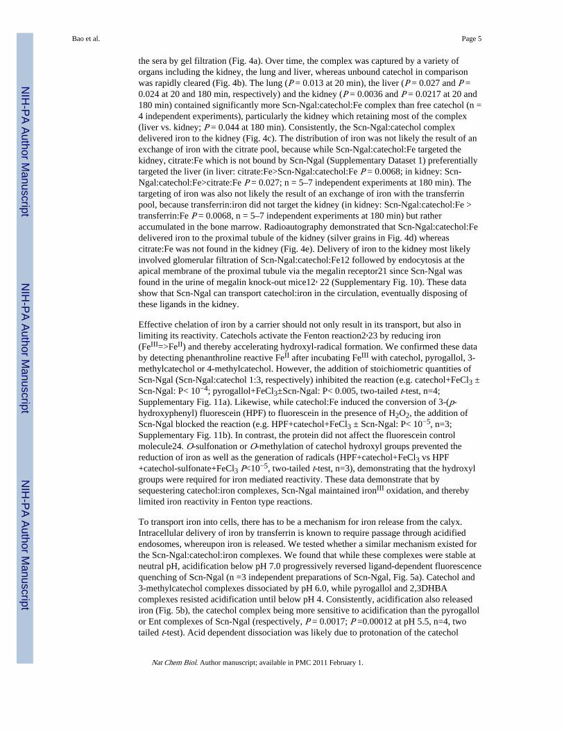
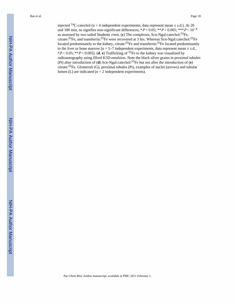
the sera by gel filtration (Fig. 4a). Over time, the complex was captured by a variety oforgans including the kidney, the lung and liver, whereas unbound catechol in comparisonwas rapidly cleared (Fig. 4b). The lung (P = 0.013 at 20 min), the liver (P = 0.027 and P =0.024 at 20 and 180 min, respectively) and the kidney (P = 0.0036 and P = 0.0217 at 20 and180 min) contained significantly more Scn-Ngal:catechol:Fe complex than free catechol (n =4 independent experiments), particularly the kidney which retaining most of the complex(liver vs. kidney; P = 0.044 at 180 min). Consistently, the Scn-Ngal:catechol complexdelivered iron to the kidney (Fig. 4c). The distribution of iron was not likely the result of anexchange of iron with the citrate pool, because while Scn-Ngal:catechol:Fe targeted thekidney, citrate:Fe which is not bound by Scn-Ngal (Supplementary Dataset 1) preferentiallytargeted the liver (in liver: citrate:Fe>Scn-Ngal:catechol:Fe P = 0.0068; in kidney: Scn-Ngal:catechol:Fe>citrate:Fe P = 0.027; n = 5–7 independent experiments at 180 min). Thetargeting of iron was also not likely the result of an exchange of iron with the transferrinpool, because transferrin:iron did not target the kidney (in kidney: Scn-Ngal:catechol:Fe >transferrin:Fe P = 0.0068, n = 5–7 independent experiments at 180 min) but ratheraccumulated in the bone marrow. Radioautography demonstrated that Scn-Ngal:catechol:Fedelivered iron to the proximal tubule of the kidney (silver grains in Fig. 4d) whereascitrate:Fe was not found in the kidney (Fig. 4e). Delivery of iron to the kidney most likelyinvolved glomerular filtration of Scn-Ngal:catechol:Fe12 followed by endocytosis at theapical membrane of the proximal tubule via the megalin receptor21 since Scn-Ngal wasfound in the urine of megalin knock-out mice12, 22 (Supplementary Fig. 10). These datashow that Scn-Ngal can transport catechol:iron in the circulation, eventually disposing ofthese ligands in the kidney.
Effective chelation of iron by a carrier should not only result in its transport, but also inlimiting its reactivity. Catechols activate the Fenton reaction2,23 by reducing iron(FeIII=>FeII) and thereby accelerating hydroxyl-radical formation. We confirmed these databy detecting phenanthroline reactive FeII after incubating FeIII with catechol, pyrogallol, 3-methylcatechol or 4-methylcatechol. However, the addition of stoichiometric quantities ofScn-Ngal (Scn-Ngal:catechol 1:3, respectively) inhibited the reaction (e.g. catechol+FeCl3 ±Scn-Ngal: P< 10−4; pyrogallol+FeCl3±Scn-Ngal: P< 0.005, two-tailed t-test, n=4;Supplementary Fig. 11a). Likewise, while catechol:Fe induced the conversion of 3-(p-hydroxyphenyl) fluorescein (HPF) to fluorescein in the presence of H2O2, the addition ofScn-Ngal blocked the reaction (e.g. HPF+catechol+FeCl3 ± Scn-Ngal: P< 10−5, n=3;Supplementary Fig. 11b). In contrast, the protein did not affect the fluorescein controlmolecule24. O-sulfonation or O-methylation of catechol hydroxyl groups prevented thereduction of iron as well as the generation of radicals (HPF+catechol+FeCl3 vs HPF+catechol-sulfonate+FeCl3 P<10−5, two-tailed t-test, n=3), demonstrating that the hydroxylgroups were required for iron mediated reactivity. These data demonstrate that bysequestering catechol:iron complexes, Scn-Ngal maintained ironIII oxidation, and therebylimited iron reactivity in Fenton type reactions.
To transport iron into cells, there has to be a mechanism for iron release from the calyx.Intracellular delivery of iron by transferrin is known to require passage through acidifiedendosomes, whereupon iron is released. We tested whether a similar mechanism existed forthe Scn-Ngal:catechol:iron complexes. We found that while these complexes were stable atneutral pH, acidification below pH 7.0 progressively reversed ligand-dependent fluorescencequenching of Scn-Ngal (n =3 independent preparations of Scn-Ngal, Fig. 5a). Catechol and3-methylcatechol complexes dissociated by pH 6.0, while pyrogallol and 2,3DHBAcomplexes resisted acidification until below pH 4. Consistently, acidification also releasediron (Fig. 5b), the catechol complex being more sensitive to acidification than the pyrogallolor Ent complexes of Scn-Ngal (respectively, P = 0.0017; P =0.00012 at pH 5.5, n=4, twotailed t-test). Acid dependent dissociation was likely due to protonation of the catechol
Bao et al. Page 5
Nat Chem Biol. Author manuscript; available in PMC 2011 February 1.
NIH
-PA Author Manuscript
NIH
-PA Author Manuscript
NIH
-PA Author Manuscript
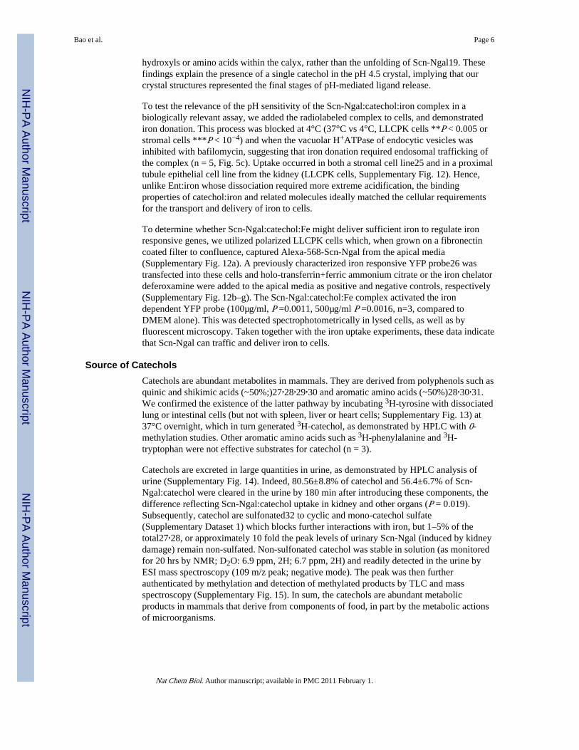
hydroxyls or amino acids within the calyx, rather than the unfolding of Scn-Ngal19. Thesefindings explain the presence of a single catechol in the pH 4.5 crystal, implying that ourcrystal structures represented the final stages of pH-mediated ligand release.
To test the relevance of the pH sensitivity of the Scn-Ngal:catechol:iron complex in abiologically relevant assay, we added the radiolabeled complex to cells, and demonstratediron donation. This process was blocked at 4°C (37°C vs 4°C, LLCPK cells **P < 0.005 orstromal cells ***P < 10−4) and when the vacuolar H+ATPase of endocytic vesicles wasinhibited with bafilomycin, suggesting that iron donation required endosomal trafficking ofthe complex (n = 5, Fig. 5c). Uptake occurred in both a stromal cell line25 and in a proximaltubule epithelial cell line from the kidney (LLCPK cells, Supplementary Fig. 12). Hence,unlike Ent:iron whose dissociation required more extreme acidification, the bindingproperties of catechol:iron and related molecules ideally matched the cellular requirementsfor the transport and delivery of iron to cells.
To determine whether Scn-Ngal:catechol:Fe might deliver sufficient iron to regulate ironresponsive genes, we utilized polarized LLCPK cells which, when grown on a fibronectincoated filter to confluence, captured Alexa-568-Scn-Ngal from the apical media(Supplementary Fig. 12a). A previously characterized iron responsive YFP probe26 wastransfected into these cells and holo-transferrin+ferric ammonium citrate or the iron chelatordeferoxamine were added to the apical media as positive and negative controls, respectively(Supplementary Fig. 12b–g). The Scn-Ngal:catechol:Fe complex activated the irondependent YFP probe (100µg/ml, P =0.0011, 500µg/ml P =0.0016, n=3, compared toDMEM alone). This was detected spectrophotometrically in lysed cells, as well as byfluorescent microscopy. Taken together with the iron uptake experiments, these data indicatethat Scn-Ngal can traffic and deliver iron to cells.
Source of CatecholsCatechols are abundant metabolites in mammals. They are derived from polyphenols such asquinic and shikimic acids (~50%;)27,28,29,30 and aromatic amino acids (~50%)28,30,31.We confirmed the existence of the latter pathway by incubating 3H-tyrosine with dissociatedlung or intestinal cells (but not with spleen, liver or heart cells; Supplementary Fig. 13) at37°C overnight, which in turn generated 3H-catechol, as demonstrated by HPLC with 0-methylation studies. Other aromatic amino acids such as 3H-phenylalanine and 3H-tryptophan were not effective substrates for catechol (n = 3).
Catechols are excreted in large quantities in urine, as demonstrated by HPLC analysis ofurine (Supplementary Fig. 14). Indeed, 80.56±8.8% of catechol and 56.4±6.7% of Scn-Ngal:catechol were cleared in the urine by 180 min after introducing these components, thedifference reflecting Scn-Ngal:catechol uptake in kidney and other organs (P = 0.019).Subsequently, catechol are sulfonated32 to cyclic and mono-catechol sulfate(Supplementary Dataset 1) which blocks further interactions with iron, but 1–5% of thetotal27,28, or approximately 10 fold the peak levels of urinary Scn-Ngal (induced by kidneydamage) remain non-sulfated. Non-sulfonated catechol was stable in solution (as monitoredfor 20 hrs by NMR; D2O: 6.9 ppm, 2H; 6.7 ppm, 2H) and readily detected in the urine byESI mass spectroscopy (109 m/z peak; negative mode). The peak was then furtherauthenticated by methylation and detection of methylated products by TLC and massspectroscopy (Supplementary Fig. 15). In sum, the catechols are abundant metabolicproducts in mammals that derive from components of food, in part by the metabolic actionsof microorganisms.
Bao et al. Page 6
Nat Chem Biol. Author manuscript; available in PMC 2011 February 1.
NIH
-PA Author Manuscript
NIH
-PA Author Manuscript
NIH
-PA Author Manuscript
DiscussionStructural analyses8,9 first suggested that Scn-Ngal may bind ligands other than bacterialsiderophores. This is because the major Scn-Ngal ligand, Ent is composed of functionalgroups that are synthesized not only by bacteria, but also by mammalian cells, implying that‘endogenous’ ligands may bind Scn-Ngal. Here we show that a family of commonmetabolites, called the catechols permitted high affinity binding, effective sequestration andthe transport of iron to cells of the kidney by Scn-Ngal. The data are reminiscent of classicalreports which showed that low molecular weight iron chelators are synthesized bymammalian cells33.
The origin of mammalian catechols has not been fully explained. Dietary intake of plantquinic, shikimic27, and 3,4-dihydroxybenzoic acids28 may contribute since rats fed quinicacid, for example, excrete both free and conjugated catechol in the urine27. Likewise, planthydroxybenzenes such as caffeic acid, chlorgenic acid, catechin, and epicatechin canundergo conversion into catechol29. The restriction of dietary plants consistently reducedthe urinary catechols in half to 40 µM30 while increasing the 4-methylcatechol componentimplied that additional pathways contribute to the catechols such as the conversion ofdietary protein to phenol and then to catechol by a NADPH dependent liver microsomalcytochrome P450 34 , 35 . Microorganisms28 are involved in these pathways since oralneomycin, which sterilizes the gut, reduces catechol excretion in half36 and we found thaturine from mice fed antibiotics or raised in a gnotobiotic facility contained one-half of thesiderophore activity as did conventional urine. Hence the catechols may join the list of ‘co-metabolites’ which derive from intestinal microbes but have function in metabolic reactionsin the mammalian host37. In this light, the Ngal-Scn:catechol complex is an example of abacterial-host interaction, and a second example, after Ent chelation, by which Ngal-Scninteracts with bacterial products, albeit in this case the products of endogenous microflora inthe “non-infected” state.
Urinary catechols are highly abundant: catechol is present at 150 µM34,38 ,39 , 4-methylcatechol at 30 µM, and pyrogallol at 500 µM29,30 (free catechols are 1–5% of thesetotals). This means that when Scn-Ngal plasma levels rise 10–50010,12,13 fold (0.1µM) andiron is released from damaged tissues (such as the kidney affected by acute injury andglomerular diseases),40,41,42, Scn-Ngal (~0.1 µM) may be constitutively saturated byexcess free catechol (~1 µM), especially given that the presence of iron enhances the affinityfor the complex 100 fold. Hence, perhaps even as much as 400 mg (17 µmole) of Scn-Ngal12, ~1mg of iron and ~2mg catechol may undergo filtration by the glomerulus andrecycling in the proximal tubule per day by a megalin (Supplementary Fig. 10) or 24p3Rdependent endosomal pathway. These data emphasize the potential role of Scn-Ngal as ascavenger of catechol, which is known to mobilize FeIII even from proteins17, and ascavenger of the catechol:iron complex, which is known to participate in Fentonreactions2,22. Ligation by the Ngal-Scn calyx conversely quenched iron reduction andhydroxyradical activation by catechol:iron. Additionally, since the donation of iron tomicrobes may be facilitated by different types of catechols such as 2,3-dihydroxybenzoicacid (Salmonella43 and Brucella) and catecholamines (E. coli 44 and Bordatella 45), theclearance of the Scn-Ngal:catechol:iron complex in the kidney might also serve a broad rolein the defense against microorganisms, sharing this property with the clearance of Scn-Ngal:Ent:iron by the kidney12.
Since Scn-Ngal is induced in several human cancers and correlates with a poor prognosis46,a model has been proposed where i) receptor-mediated endocytosis of extracellular Scn-Ngal:iron complexes delivered iron to cells and prevented apoptosis or where ii)internalization of ligand free Scn-Ngal by cells lead to iron efflux resulting in apoptosis and
Bao et al. Page 7
Nat Chem Biol. Author manuscript; available in PMC 2011 February 1.
NIH
-PA Author Manuscript
NIH
-PA Author Manuscript
NIH
-PA Author Manuscript
cell death, mediated by the pro-apoptotic protein Bim14. The results presented here describethe candidate endogenous siderophores necessary for supporting these proposedmechanisms.
In conclusion, we have identified a new family of iron binding cofactors. While cofactorssuch as heme and sulfide groups are well known in intracellular proteins, we describe anovel iron binding cofactor in an extracellular carrier protein that mimics in structure andmolecular recognition a classical bacterial siderophore. The metabolic fate of iron releasedin tissue injury has been unknown, but here we show that a constitutively producedsiderophore, catechol, complexed with a stress activated protein, Scn-Ngal, traffics andclears iron. This pathway may be quantitatively significant in renal proximal tubuleepithelia, where iron is recycled, and perhaps in the urine as well where the Scn-Ngal:catechol:iron complexes are discarded.
MethodsChemicals and Animal Samples
Compounds were obtained commercially (Supplementary Dataset 1). All solvents wereHPLC grade from Fisher. Catechol-14C (100 mCi/mmol) was from Sigma and L-[5-3H]Tryptophan (32.0 Ci/mmol) , L-[4-3H]Phenylalanine (27.0 Ci/mmol), and L-[3,5-3H]Tyrosine (54.0 Ci/mmol) were from Amersham-GE Healthcare. 55FeCl3 (45mCi/mg) was from PerkinElmer. Desferri-Enterochelin (Ent) and ferric Ent (Ent:Fe) were fromEMC Microcollections, Germany. Ngal-Scn was expressed in BL-21 bacteria8,9. Blood,urine and tissues of mice and procedures were performed with approval of the InstitutionalAnimal Care and Use Committee (IACUC) at Columbia University. CD1 Mouse urine wasalso obtained from the Bioreclamation Company, New York, before and after treatment withoral Vancomycin and Neomycin for 1 week. Human urine was pooled from healthy medicalschool students as well as from patients of Columbia University Medical Center, withapproval of the Institutional Review Board.
Computational MethodsComputational studies were conducted at the Molecular Graphics and Computation Facility,College of Chemistry, University of California, Berkeley. To determine the quadrupolemoments, Θzz, the aromatic structures were geometry optimized and characterized viafrequency calculations at the RHF/6-311G** level of theory in the Gaussian 03 package47.To determine the aromatic-cation interaction energies the components were characterizedvia a frequency calculation at the MP2/6-311++G** level of theory and the aromatic-cationinteraction energies corrected for basis set superposition error (BSSE) with the counterpoisemethod in the Gaussian 03 package. In the aromatic-cation calculations the sodium ion wasfixed at a distance of 2.47 Åabove the centroid of the aromatic unit.
CrystallographyRecombinant C87S human Scn-Ngal was expressed and purified as previously described8,9.Protein (10 mg/ml) was mixed with 10 mM catechol or 4-methylcatechol and then with 5mM FeCl3, using extensive washes (YM-10, Millipore) with PNE (25 mM PIPES, 150 mMNaCl, and 1 mM EDTA). Co-crystals of ligand bound human Scn-Ngal were grown byvapor diffusion at 18 °C over reservoirs of 1.0–1.4 M NH4SO4, 100 mM NaCl, 50 mMLiSO4, 100 mM Na acetate (pH 4.5). Crystals typically grew in 5–10 days and were cryo-protected using the mother liquor plus 15% glycerol prior to flash cooling in LiqN2.Diffraction data were collected using synchrotron radiation at the Advanced Light Source(Berkeley, CA) beamline 5.0.1 (wavelength 1.0λ) (Supplementary Methods). Relevant
Bao et al. Page 8
Nat Chem Biol. Author manuscript; available in PMC 2011 February 1.
NIH
-PA Author Manuscript
NIH
-PA Author Manuscript
NIH
-PA Author Manuscript
statistics are shown in Supplementary Table 1 and coordinates deposited in the Protein DataBank: www.rcsb.org (Accession Codes: Catechol = 3FW4; 4-methylcatechol = 3FW5).
Binding Assays(Supplementary Methods) We utilized fluorescence quenching to measure the affinity of theScn-Ngal:catechol interaction. Excitation λexc = 281 nm (5 nm slit band pass) and emissionλem = 340 nm (10 nm slit band pass) data were collected from 100 nM protein solutions(with 32µg/mL ubiquitin and 5% DMSO) exposed to ligands. To prepare FeL3, catechol (12mM, 25µL in DMSO) and ferric chloride (0.33eq.) were combined and then diluted to formthe metal complex 18 µM FeL2 (in iron) in aqueous buffer (pH 7.4; TBS) and 5% DMSO.Apo-catechol solutions were prepared analogously. The pH was adjusted with 0.1 M HClincrementally until the fluorescence signal stopped changing, while fluorescence valueswere corrected for dilution. To monitor the stability of the complex over time, the samplewas stored at 4 °C for 12 hrs. Data were analyzed by a nonlinear regression analysis using aone-site binding model (DYNAFIT)48. Control experiments were performed to ensure thestability of the protein at experimental conditions, including the dilution and the addition ofDMSO and ubiquitin.
Scn-Ngal:Siderophore:Iron Traffic(Supplementary Methods) Mouse embryonic kidney FoxD1+ stromal cells25 (105 cells) andadult kidney proximal tubule LLCPK1 cells (105 cells, ATCC, CL101) were grown inDMEM (low glucose) with 10% FCS for 24 hrs, the FCS removed and complexes Scn-Ngal:14C-catechol or Scn-Ngal:catechol:55Fe, prepared as above, were added to the cells at37 °C or 4 °C for 0–6 hrs. Bafilomycin (0.15 nmole) was tolerated by stromal cells for 3 hrs.Cells were then washed 3 times, briefly trypsinized, and then collected and measured byscintillation counting. To examine the capture of Scn-Ngal in different tissues, the Scn-Ngalcomplexes were introduced by an intraperitoneal route and tissues harvested 1–6 hrs laterfrom C57BL/6 wild type, 8 week old male mice (Charles River) housed with food and waterad libitum. Tissues were diced and dissolved in 2% SDS, 0.1 N NaOH at 60 °C and counted.Each experiment was repeated 3–7 times using independent preparations of recombinantmouse Scn-Ngal and results expressed as means ± s.d. with independent two-tailed Studentt-tests. For radioautography, the kidneys were fixed in 4% glutaraldehyde in phosphatebuffer, post-fixed in 1% OsO4 and embedded in Epon812. Sections (1 µm) were coated withIlford K5D emulsion and developed with Microdol (Kodak) after a 2 month exposure.
Supplementary MaterialRefer to Web version on PubMed Central for supplementary material.
AcknowledgmentsWe thank Drs. Rebecca Abergel, Anna Zawadzka, Q. Al-Awqati for helpful discussion. We are grateful to theGordon lab for gnotobiotic urines and EI Christensen and TE Willnow for megalin knockout urines. We salute theMedical School Classes of 2010 and 2013, College of Physicians and Surgeons for donating urine for this study.Supported by NIH grants AI117448 (KNR), AI59432 (RKS) and the Emerald Foundation, the March of Dimes andby NIH grants DK-55388 and DK-58872 (JB).
References1. Theil EC, Goss DJ. Living with iron (and oxygen): questions and answers about iron homeostasis.
Chem. Rev. 2009; 109:4568–4579. [PubMed: 19824701]
Bao et al. Page 9
Nat Chem Biol. Author manuscript; available in PMC 2011 February 1.
NIH
-PA Author Manuscript
NIH
-PA Author Manuscript
NIH
-PA Author Manuscript
2. Iwahashi H, Morishita H, Ishii T, Sugata R, Kido R. Enhancement by catechols of hydroxyl-radicalformation in the presence of ferric ions and hydrogen peroxide. J. Biochem. 1989; 105:429–434.[PubMed: 2543661]
3. Rouault TA, Tong WH. Iron-sulfur cluster biogenesis and human disease. Trends Genet. 2008;24:398–407. [PubMed: 18606475]
4. Li JY, et al. Scara5 is a ferritin receptor mediating non-transferrin iron delivery. Dev. Cell. 2009;16:35–46. [PubMed: 19154717]
5. Levy JE, Jin O, Fujiwara Y, Kuo F, Andrews NC. Transferrin receptor is necessary for developmentof erythrocytes and the nervous system. Nat. Genet. 1999; 21:396–399. [PubMed: 10192390]
6. Gunshin H, et al. Slc11a2 is required for intestinal iron absorption and erythropoiesis butdispensable in placenta and liver. J Clin. Invest. 2005; 115:1258–1266. [PubMed: 15849611]
7. Akerstrom B, Flower DR, Salier JP. Lipocalins: unity in diversity. Biochim. Biophys. Acta. 2000;1482:1–8. [PubMed: 11058742]
8. Goetz DH, et al. The neutrophil lipocalin NGAL is a bacteriostatic agent that interferes withsiderophore-mediated iron acquisition. Mol. Cell. 2002; 10:1033–1043. [PubMed: 12453412]
9. Holmes MA, Paulsene W, Jide X, Ratledge C, Strong RK. Siderocalin (Lcn 2) also bindscarboxymycobactins, potentially defending against mycobacterial infections through ironsequestration. Structure. 2005; 13:29–41. [PubMed: 15642259]
10. Flo TH, et al. Lipocalin 2 mediates an innate immune response to bacterial infection bysequestrating iron. Nature. 2004; 432:917–921. [PubMed: 15531878]
11. Berger T, et al. Lipocalin 2-deficient mice exhibit increased sensitivity to Escherichia coli infectionbut not to ischemia-reperfusion injury. Proc. Natl. Acad. Sci. USA. 2006; 103:1834–1839.[PubMed: 16446425]
12. Mori K, et al. Endocytic delivery of lipocalin-siderophore-iron complex rescues the kidney fromischemia-reperfusion injury. J. Clin. Invest. 2005; 115:610–621. [PubMed: 15711640]
13. Mishra J, et al. Neutrophil gelatinase-associated lipocalin (NGAL) as a biomarker for acute renalinjury after cardiac surgery. The Lancet. 2005; 365:1231–1238.
14. Devireddy LR, Gazin C, Zhu X, Green MR. A cell-surface receptor for lipocalin 24p3 selectivelymediates apoptosis and iron uptake. Cell. 2005; 123:1293–1305. [PubMed: 16377569]
15. Wishart DS, et al. HMDB: the Human Metabolome Database. Nucleic Acids Res. 2007; 35:D521–D526. [PubMed: 17202168]
16. Hoette TM, et al. The role of electrostatics in siderophore recognition by the immunoproteinSiderocalin. J. Am. Chem. Soc. 2008; 130:17584–17592. [PubMed: 19053425]
17. Sánchez P, et al. Catechol releases iron (III) from ferritin by direct chelation without iron (II)production. Dalton Trans. 2005; 4:811–813. [PubMed: 15702194]
18. Karpishin RB, Gebhard MS, Solomon EI, Raymond KN. Spectroscopic studies of the electronicstructure of iron(III) tris(catecholates). J. Am. Chem. Soc. 1991; 113:2977.
19. Abergel RJ, et al. The Siderocalin/Enterobactin interaction: A link between mammalian immunityand bacterial iron transport. J. Am. Chem. Soc. 2008; 130:11524–11534. [PubMed: 18680288]
20. Goetz DH, et al. Ligand preference inferred from the structure of neutrophil gelatinase associatedlipocalin. Biochemistry. 2000; 39:1935–1941. [PubMed: 10684642]
21. Hvidberg V, et al. The endocytic receptor megalin binds the iron transporting neutrophil-gelatinase-associated lipocalin with high affinity and mediates its cellular uptake. FEBS Lett.2005; 579:773–777. [PubMed: 15670845]
22. Leheste JR, et al. Megalin knockout mice as an animal model of low molecular weight proteinuria.Am. J. Pathol. 1999; 155:1361–1370. [PubMed: 10514418]
23. Rodríguez J, Parra C, Contreras FJ, Baeza J. Dihydroxybenzenes: driven Fenton reactions. WaterSci. Technol. 2001; 44:251–256.
24. Setsukinai K, et al. Development of novel fluorescence probes that can reliably detect reactiveoxygen species and distinguish specific species. J. Biol. Chem. 2003; 278:170–3175.
25. Levinson RS, et al. Foxd1-dependent signals control cellularity in the renal capsule, a structurerequired for normal renal development. Development. 2005; 132:529–539. [PubMed: 15634693]
Bao et al. Page 10
Nat Chem Biol. Author manuscript; available in PMC 2011 February 1.
NIH
-PA Author Manuscript
NIH
-PA Author Manuscript
NIH
-PA Author Manuscript
26. Li JY, et al. Detection of intracellular iron by its regulatory effect. Am. J. Physiol. Cell Physiol.2004; 287:C1547–C1559. [PubMed: 15282194]
27. Booth AN, Robbins DJ, Masri MS, DeEds F. Excretion of catechol after ingestion of quinic andshikimic acids. Nature. 1960; 187:691. [PubMed: 13802680]
28. Martin AK. The origin of urinary aromatic compounds excreted by ruminants. 3. The metabolismof phenolic compounds to simple phenols. Br. J. Nutr. 1982; 48:497–507. [PubMed: 7171537]
29. Lang R, Mueller C, Hofmann T. Development of a stable isotope dilution analysis with liquidchromatography-tandem mass spectrometry detection for the quantitative analysis of di- andtrihydroxybenzenes in foods and model systems. J. Agric. Food Chem. 2006; 54:5755–5762.[PubMed: 16881674]
30. Carmella SG, La VEJ, Hecht SS. Quantitative analysis of catechol and 4-methylcatechol in humanurine. Food Chem. Toxicol. 1982; 20:587–590. [PubMed: 6890513]
31. Bakke OM. Urinary simple phenols in rats fed purified and nonpurified diets. J. Nutr. 1969;98:209–216. [PubMed: 5783302]
32. Rennick B, Quebbemann A. Site of excretion of catechol and catecholamines: renal metabolism ofcatechol. Am. J. Physiol. 1970; 218:1307–1312. [PubMed: 5438255]
33. Jones RL, Peterson CM, Grady RW, Cerami A. Low molecular weight iron-binding factor frommammalian tissue that potentiates bacterial growth. J. Exp. Med. 1980; 151:418–428. [PubMed:6985950]
34. Sawahata T, Neal RA. Biotransformation of phenol to hydroquinone and catechol by rat livermicrosomes. Mol. Pharmacol. 1983; 23:453–460. [PubMed: 6835203]
35. Parke DV, Williams RT. Studies in detoxication. 54. The metabolism of benzene. (a) Theformation of phenylglucuronide and phenylsulphuric acid from [14C]benzene. (b) The metabolismof [14C]phenol. Biochem. J. 1953; 55:337–340. [PubMed: 13093687]
36. Smith AA. Origin of urinary pyrocatechol. Nature. 1961; 190:167. [PubMed: 13690557]
37. Bäckhed F, Ley RE, Sonnenburg JL, Peterson DA, Gordon JI. Host-bacterial mutualism in thehuman intestine. Science. 2005; 307:1915–1920. [PubMed: 15790844]
38. Marrubini G, et al. Direct Analysis of phenol, catechol and hydroquinone in uuman urine bycoupled-column HPLC with fluorimetric detection. Chromatographia. 2005; 62:25–31.
39. Qu Q, et al. Validation of biomarkers in humans exposed to benzene: urine metabolites. Am. J. Ind.Med. 2000; 37:522–531. [PubMed: 10723046]
40. Zager RA. Combined mannitol and deferoxamine therapy for myohemoglobinuric renal injury andoxidant tubular stress. Mechanistic and therapeutic implications. J. Clin. Invest. 1992; 90:711–719.[PubMed: 1325995]
41. Paller MS, Hedlund BE. Role of iron in postischemic renal injury in the rat. Kidney Int. 1988;34:474–480. [PubMed: 3143849]
42. Baliga R, Ueda N, Shah SV. Increase in bleomycin-detectable iron in ischaemia/reperfusion injuryto rat kidneys. Biochem. J. 1993; 291:901–905. [PubMed: 7683877]
43. Jones RL, et al. Effects of iron chelators and iron overload on Salmonella infection. Nature. 1977;267:63–65. [PubMed: 323727]
44. Freestone PP, Walton NJ, Haigh RD, Lyte M. Influence of dietary catechols on the growth ofenteropathogenic bacteria. Int J Food Microbiol. 2007; 119:159–169. [PubMed: 17850907]
45. Anderson MT, Armstrong SK. Norepinephrine mediates acquisition of transferrin-iron inBordetella bronchiseptica. J Bacteriol. 2008; 190:3940–3947. [PubMed: 18390651]
46. Richardson DR. 24p3 and its receptor: dawn of a new iron age? Cell. 2005; 123:1175–1177.[PubMed: 16377555]
47. Frisch, MJT., et al. Gaussian 03, Revision C.02. Wallingford CT: Gaussian, Inc.; 2004.
48. Kuzmic P. Program DYNAFIT for the analysis of enzyme kinetic data: application to HIVproteinase. Anal. Biochem. 1996; 237:260–273. [PubMed: 8660575]
Bao et al. Page 11
Nat Chem Biol. Author manuscript; available in PMC 2011 February 1.
NIH
-PA Author Manuscript
NIH
-PA Author Manuscript
NIH
-PA Author Manuscript
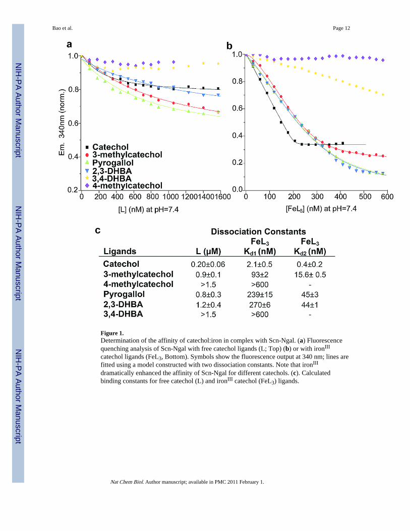
Figure 1.Determination of the affinity of catechol:iron in complex with Scn-Ngal. (a) Fluorescencequenching analysis of Scn-Ngal with free catechol ligands (L; Top) (b) or with ironIII
catechol ligands (FeL3, Bottom). Symbols show the fluorescence output at 340 nm; lines arefitted using a model constructed with two dissociation constants. Note that ironIII
dramatically enhanced the affinity of Scn-Ngal for different catechols. (c). Calculatedbinding constants for free catechol (L) and ironIII catechol (FeL3) ligands.
Bao et al. Page 12
Nat Chem Biol. Author manuscript; available in PMC 2011 February 1.
NIH
-PA Author Manuscript
NIH
-PA Author Manuscript
NIH
-PA Author Manuscript
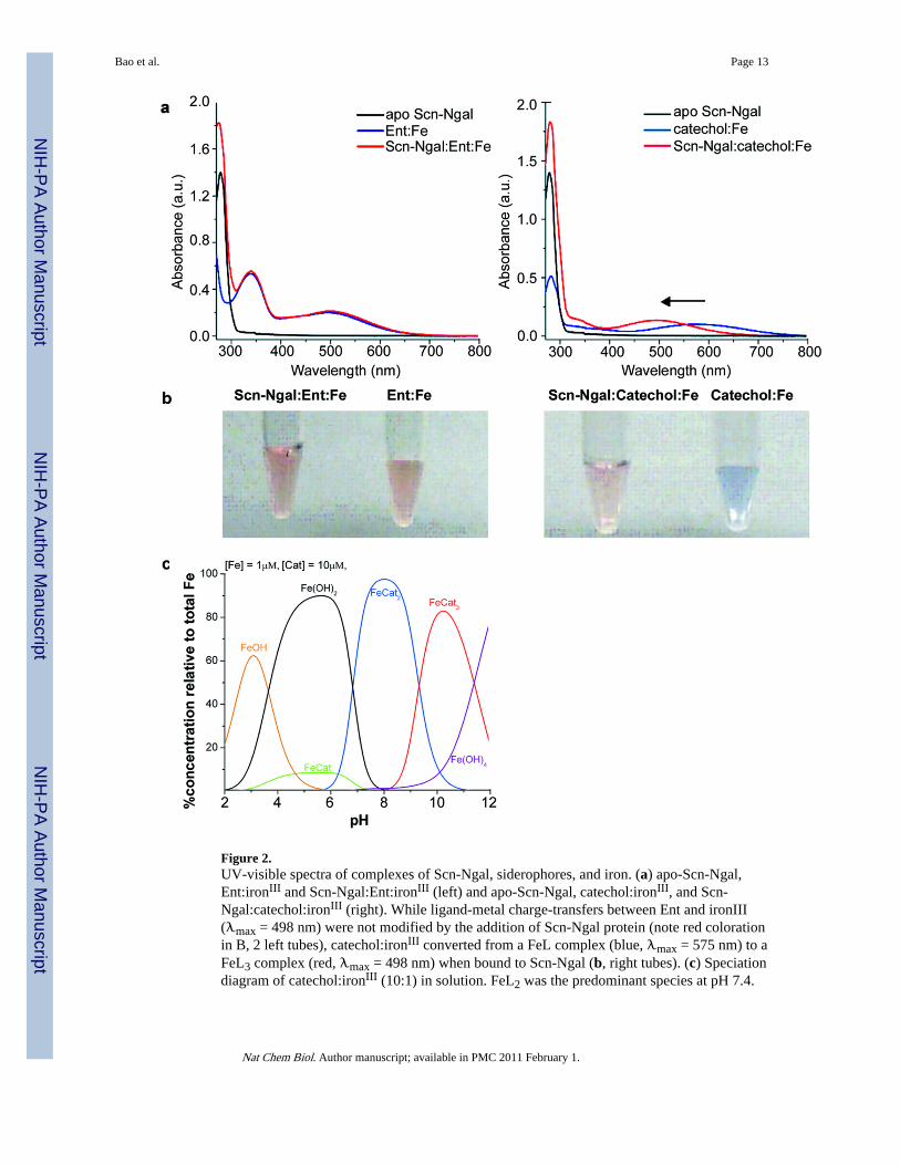
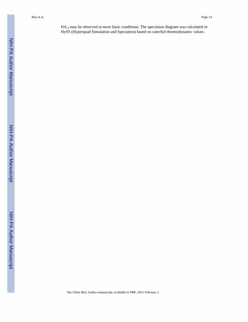
Figure 2.UV-visible spectra of complexes of Scn-Ngal, siderophores, and iron. (a) apo-Scn-Ngal,Ent:ironIII and Scn-Ngal:Ent:ironIII (left) and apo-Scn-Ngal, catechol:ironIII, and Scn-Ngal:catechol:ironIII (right). While ligand-metal charge-transfers between Ent and ironIII(λmax = 498 nm) were not modified by the addition of Scn-Ngal protein (note red colorationin B, 2 left tubes), catechol:ironIII converted from a FeL complex (blue, λmax = 575 nm) to aFeL3 complex (red, λmax = 498 nm) when bound to Scn-Ngal (b, right tubes). (c) Speciationdiagram of catechol:ironIII (10:1) in solution. FeL2 was the predominant species at pH 7.4.
Bao et al. Page 13
Nat Chem Biol. Author manuscript; available in PMC 2011 February 1.
NIH
-PA Author Manuscript
NIH
-PA Author Manuscript
NIH
-PA Author Manuscript
FeL3 may be observed in more basic conditions. The speciation diagram was calculated inHySS (Hyperquad Simulation and Speciation) based on catechol thermodynamic values.
Bao et al. Page 14
Nat Chem Biol. Author manuscript; available in PMC 2011 February 1.
NIH
-PA Author Manuscript
NIH
-PA Author Manuscript
NIH
-PA Author Manuscript
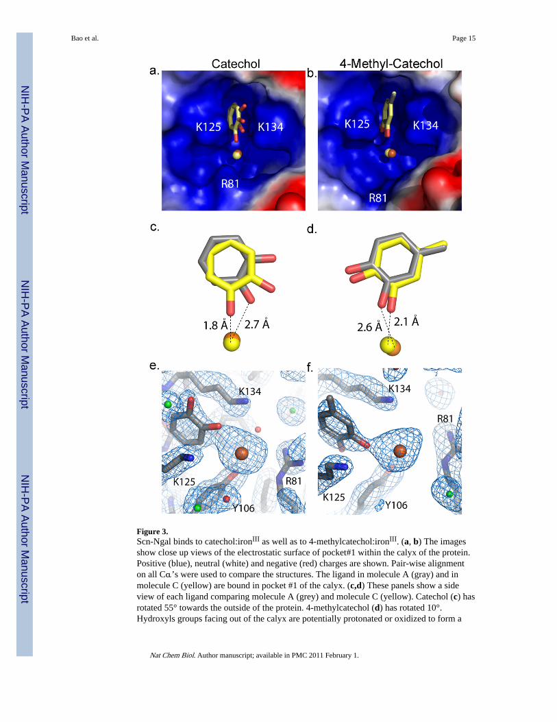
Figure 3.Scn-Ngal binds to catechol:ironIII as well as to 4-methylcatechol:ironIII. (a, b) The imagesshow close up views of the electrostatic surface of pocket#1 within the calyx of the protein.Positive (blue), neutral (white) and negative (red) charges are shown. Pair-wise alignmenton all Cα’s were used to compare the structures. The ligand in molecule A (gray) and inmolecule C (yellow) are bound in pocket #1 of the calyx. (c,d) These panels show a sideview of each ligand comparing molecule A (grey) and molecule C (yellow). Catechol (c) hasrotated 55° towards the outside of the protein. 4-methylcatechol (d) has rotated 10°.Hydroxyls groups facing out of the calyx are potentially protonated or oxidized to form a
Bao et al. Page 15
Nat Chem Biol. Author manuscript; available in PMC 2011 February 1.
NIH
-PA Author Manuscript
NIH
-PA Author Manuscript
NIH
-PA Author Manuscript
semi-quinone species. Iron is shown in orange for molecule A and yellow for molecule C inpanels a–d. (e, f) 2Fo-Fc electron density map of molecule A for catechol (e) and 4-methylcatechol (f) contoured at 1 sigma. Waters are shown in red, chloride in green, iron inorange, and the molecule in gray.
Bao et al. Page 16
Nat Chem Biol. Author manuscript; available in PMC 2011 February 1.
NIH
-PA Author Manuscript
NIH
-PA Author Manuscript
NIH
-PA Author Manuscript
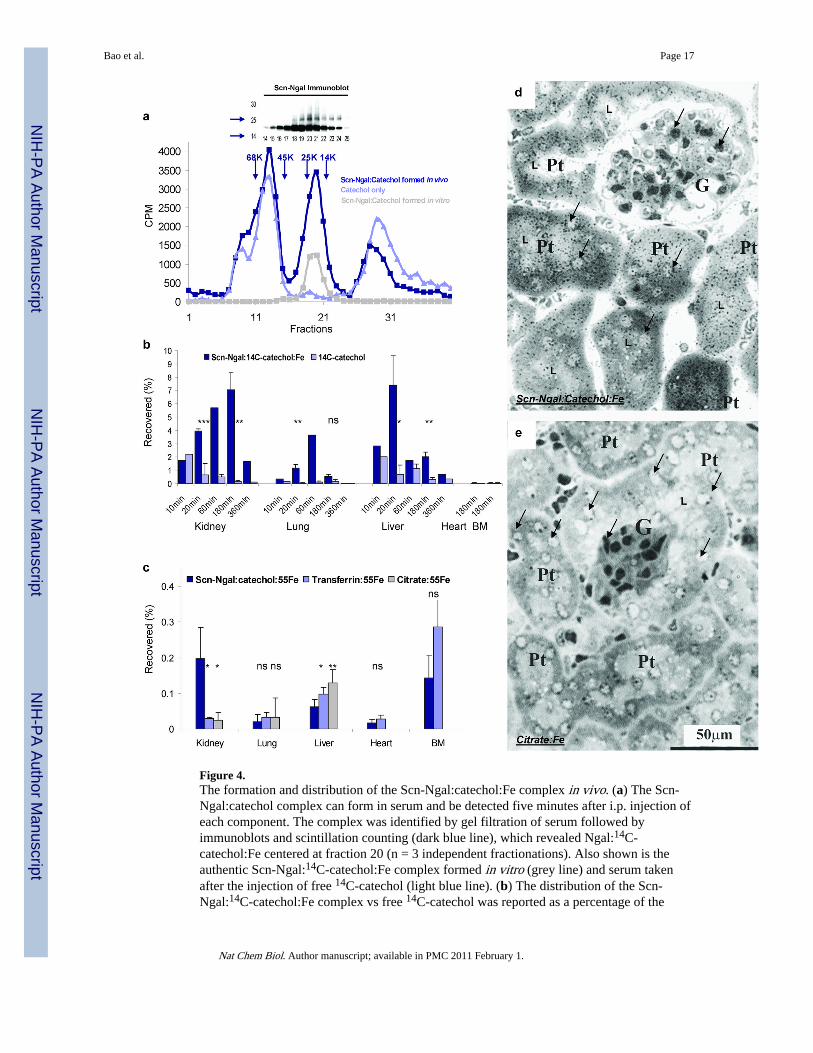
Figure 4.The formation and distribution of the Scn-Ngal:catechol:Fe complex in vivo. (a) The Scn-Ngal:catechol complex can form in serum and be detected five minutes after i.p. injection ofeach component. The complex was identified by gel filtration of serum followed byimmunoblots and scintillation counting (dark blue line), which revealed Ngal:14C-catechol:Fe centered at fraction 20 (n = 3 independent fractionations). Also shown is theauthentic Scn-Ngal:14C-catechol:Fe complex formed in vitro (grey line) and serum takenafter the injection of free 14C-catechol (light blue line). (b) The distribution of the Scn-Ngal:14C-catechol:Fe complex vs free 14C-catechol was reported as a percentage of the
Bao et al. Page 17
Nat Chem Biol. Author manuscript; available in PMC 2011 February 1.
NIH
-PA Author Manuscript
NIH
-PA Author Manuscript
NIH
-PA Author Manuscript
injected 14C-catechol (n = 4 independent experiments; data represent mean ± s.d.). At 20and 180 min, ns signifies non-significant differences, *P < 0.05; **P < 0.005; ***P < 10−4
as assessed by two tailed Students t-test. (c) The complexes, Scn-Ngal:catechol:55Fe,citrate:55Fe, and transferrin:55Fe were recovered at 3 hrs. Whereas Scn-Ngal:catechol:55Felocated predominately to the kidney, citrate:55Fe and transferrin:55Fe located predominantlyto the liver or bone marrow (n = 5–7 independent experiments, data represent mean ± s.d.,*P < 0.05; **P < 0.005). (d, e) Trafficking of 55Fe to the kidney was visualized byradioautography using Ilford K5D emulsion. Note the black silver grains in proximal tubules(Pt) after introduction of (d) Scn-Ngal:catechol:55Fe but not after the introduction of (e)citrate:55Fe. Glomeruli (G), proximal tubules (Pt), examples of nuclei (arrows) and tubularlumen (L) are indicated (n = 2 independent experiments).
Bao et al. Page 18
Nat Chem Biol. Author manuscript; available in PMC 2011 February 1.
NIH
-PA Author Manuscript
NIH
-PA Author Manuscript
NIH
-PA Author Manuscript
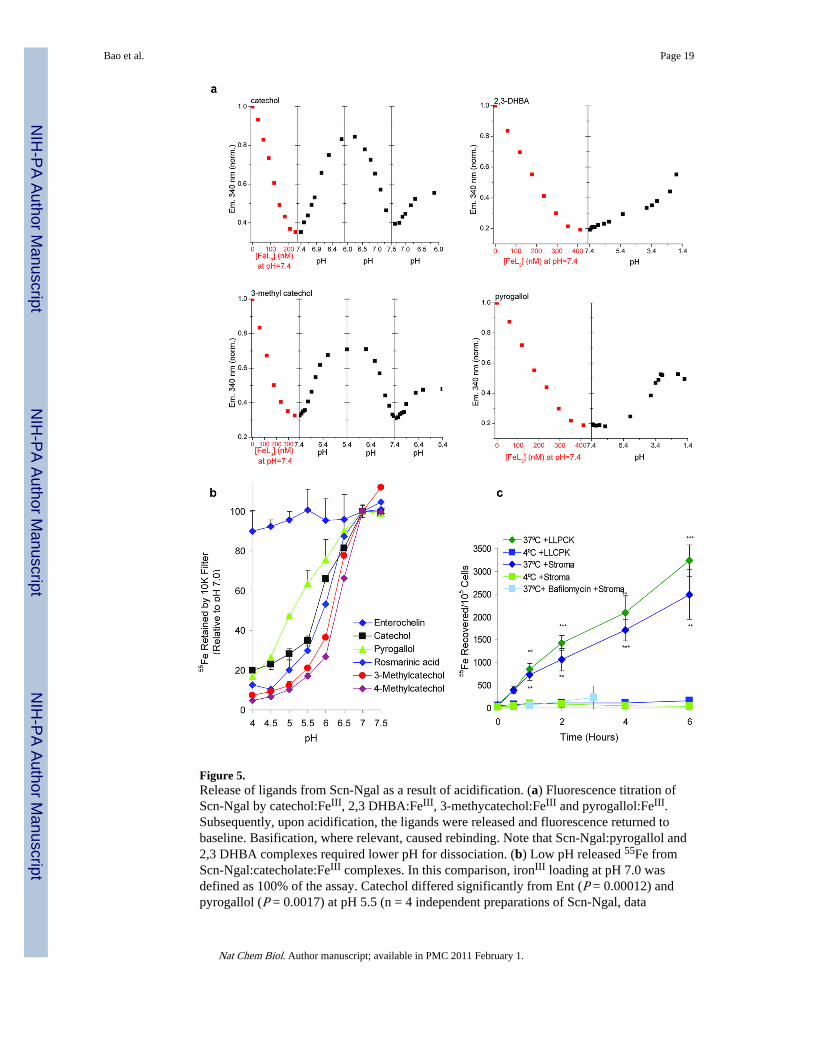
Figure 5.Release of ligands from Scn-Ngal as a result of acidification. (a) Fluorescence titration ofScn-Ngal by catechol:FeIII, 2,3 DHBA:FeIII, 3-methycatechol:FeIII and pyrogallol:FeIII.Subsequently, upon acidification, the ligands were released and fluorescence returned tobaseline. Basification, where relevant, caused rebinding. Note that Scn-Ngal:pyrogallol and2,3 DHBA complexes required lower pH for dissociation. (b) Low pH released 55Fe fromScn-Ngal:catecholate:FeIII complexes. In this comparison, ironIII loading at pH 7.0 wasdefined as 100% of the assay. Catechol differed significantly from Ent (P = 0.00012) andpyrogallol (P = 0.0017) at pH 5.5 (n = 4 independent preparations of Scn-Ngal, data
Bao et al. Page 19
Nat Chem Biol. Author manuscript; available in PMC 2011 February 1.
NIH
-PA Author Manuscript
NIH
-PA Author Manuscript
NIH
-PA Author Manuscript
represents mean ± s.d., two tailed t-test). (c) The capture of 55Fe from the Scn-Ngal:catechol:55Fe complex by kidney stromal cells and kidney proximal tubule LLCPKcells in vitro. 55Fe uptake was inhibited at 4°C or by bafilomycin, an inhibitor of thevacuolar H+ATPase. Data represent mean ± s.d., n =5 independent preparations of Scn-Ngal, and statistical significance (**P < 0.005; ***P < 10−4) was assessed by two tailedStudents t-test comparing uptake at 37°C and 4°C.
Bao et al. Page 20
Nat Chem Biol. Author manuscript; available in PMC 2011 February 1.
NIH
-PA Author Manuscript
NIH
-PA Author Manuscript
NIH
-PA Author Manuscript


