Embed Size (px)
Citation preview
Intestinal Obstruction: Pathophysiological Basis of Clinical Features.
Dr Imran Javed.MBBS, FCPS Surgery.
Associate Professor Surgery.Fiji National University.
Definitions• Intestinal Obstruction: A mechanical blockage arising from a structural abnormality that presents a physical barrier to the progression of gut contents.
• Partial or complete• Simple or strangulated
• Ileus: is a paralytic or functional variety of obstruction.
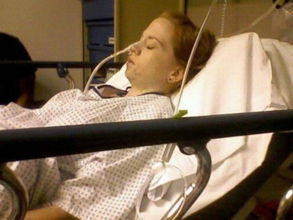
Patient Presentation• A 50 year old gentleman presents with abdominal pain, distension and absolute constipation with repeated episodes of vomiting.
• His vital sign were stable, abdomen distended with diffuse tenderness but minimal peritonism. Bowel Sounds are hyperactive.
Common Questions• Is this bowel obstruction or ileus?• Is this a small or large bowel obstruction?• Is this proximal or distal obstruction?• What is the cause of this obstruction?• Is this a complex or simple obstruction?• How should I start investigating my patient?• What is the role of other supportive investigations?
• What is my immediate/ intermediate treatment plan?
• What are the indications for surgery?
Intestinal Physiology• 8L of isotonic fluid received by the small intestines (saliva, stomach, duodenum, pancreas and hepatobiliary )
• 6L re-absorbed• 2L enter the large intestine and 200 ml excreted in the faeces
• Air in the bowel results from swallowed air ( O2 & N2) and bacterial fermentation in the colon ( H2, Methane & CO2),
600 ml of flatus is released• Enteric bacteria consist of coliforms, anaerobes and strep.faecalis.
• Normal intestinal mucosa has a significant immune role
Pathological events• Distension results from gas and/ or fluid and can exert hydrostatic pressure.(Laplace Law)
• In case of Bowel Obstruction, Bacterial overgrowth can be rapid
• If mucosal barrier is breached it may result in translocation of bacteria and toxins resulting in bactaeremia, septaecemia and toxaemia.
Pathological Basis of clinical events.
• Initial overcoming of the obstruction(CONSTIPATION) by increased peristalsis(COLICKY ABDOMINAL PAIN)
• (ABDOMINAL DISTENTION) & (VOMITING)Increased intraluminal pressure by fluid and gas, sequestration of fluid into the lumen from the surrounding circulation
• Lymphatic and venous congestion resulting in edematous tissues (TISSUE EDEMA).
• Vomiting result in hypovolemia and electrolyte imbalance (ILEUS).
• Further: anoxia, mucosal necrosis and perforation and peritonitis.(COMPLICATIONS)
• Bacterial over growth with translocation of bacteria and it’s toxins causing bacteremia and septicemia.(SYSTEMIC SIGNS)
Principles of Management
SUCK & DRIP.• Decompress with Naso-gatric Tube or Flatus Tube.
• Replace lost fluid in vomiting or 3rd space.• Correct electrolyte abnormalities (Hypcholremic, Hyopnatremic, Hypokalemic, metabolic Alkalosis)
• Recognize strangulation (Hernia) and perforation (Peritonitis)
• Systemic antibiotics (Broad Spectrum).
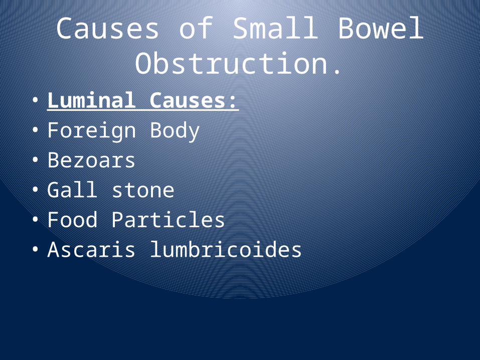
Causes of Small Bowel Obstruction.
• Luminal Causes:• Foreign Body• Bezoars• Gall stone• Food Particles• Ascaris lumbricoides
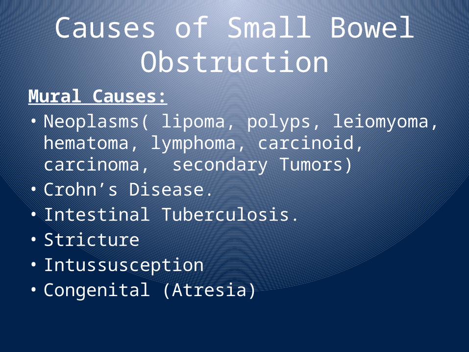
Causes of Small Bowel Obstruction
Mural Causes:• Neoplasms( lipoma, polyps, leiomyoma, hematoma, lymphoma, carcinoid, carcinoma, secondary Tumors)
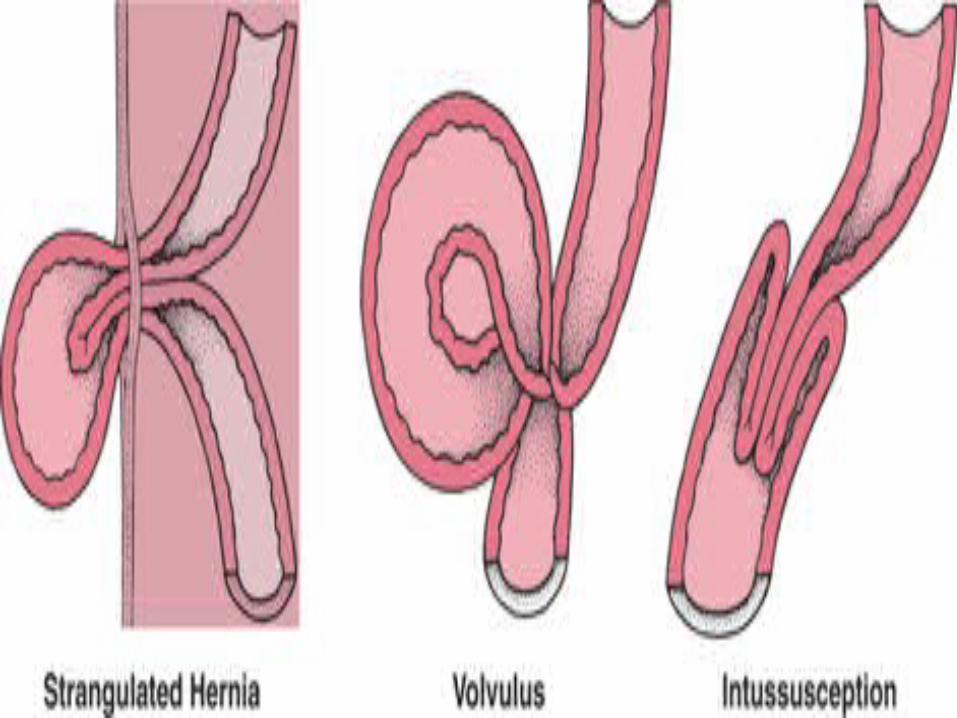
• Crohn’s Disease.• Intestinal Tuberculosis.• Stricture• Intussusception• Congenital (Atresia)
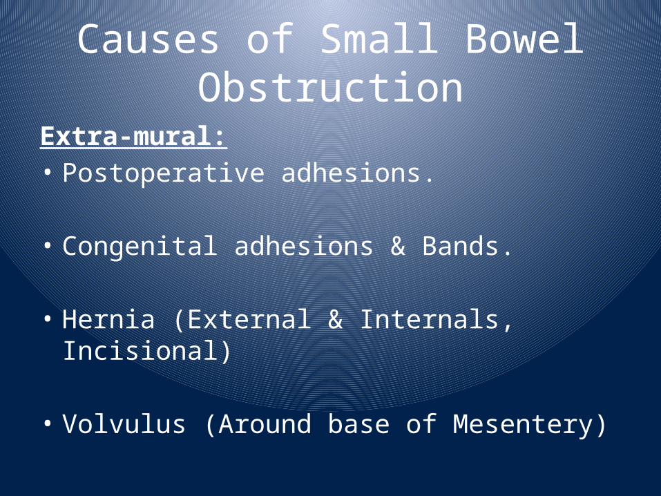
Causes of Small Bowel Obstruction
Extra-mural:• Postoperative adhesions.
• Congenital adhesions & Bands.
• Hernia (External & Internals, Incisional)
• Volvulus (Around base of Mesentery)
Small Bowel Adhesions• Accounts for 60-70% of All Small Bowel Obstructions.• Results from peritoneal injury, platelet activation and fibrin formation.
• Associated with starch covered gloves, intraperitoneal sepsis, haemorrhage and wash with irritant solutions iodine and other foreign bodies.
• As early as 4 weeks post laparotomy. The majority of patients present between 1-5 years
• Colorectal Surgery 25%• Gynaecological 20%• Appendectomy 14%• 70% of patients had a single band• Patients with complex bands are more likely to be readmitted
• Readmission in surgically treated patients is 35%
Hernias• Accounts for 20% of SBO• Commonest 1. Femoral hernia 2. ID inguinal 3. Umbilical 4. Others: incisional and internal H.
• The site of obstruction is the neck of hernia• The compromised viscus is with in the sac. Ischemia occurs initially by venous occlusion, followed by edema and arterial compromise.
• Attempt to distinguish the difference between: • Incarceration, Sliding, Obstruction.• Strangulation is noted by: Persistent pain, Discoloration, Tenderness, Constitutional symptoms
Other Common causes of Small Bowel Obstructions.
• Intussusception: part of the intestine has invaginated into another section of intestine.
• Gall stone Ileus: caused by an impaction of a gallstone within the lumen of the small intestine. Which enters the gut lumen via cholecysto-enteric fistula.
• Crohn’s Disease:is a type of inflammatory bowel disease (IBD)
Large Bowel Obstruction• Distinguishing ileus from mechanical obstruction is challenging
• According to Laplace's law: maximum pressure is at the it’s maximum diameter. Cecum is at the greatest risk of perforation
• Perforation results in the release of formed feaces with heavy bacterial contamination
Causes of Large Bowel Obstructions.
• 1. Carcinoma: The commonest cause, 18% of colonic carcinomas present with obstruction
• 2. Benign stricture: Due to Diverticular disease, Ischemia, Inflammatory bowel disease.
• 3. Volvulus: A. Sigmoid Volvulus: Results from long
redundant, faecaly loaded colon with a narrow pedicleB. Caecal Volvulus
• 4. Hernia.• 5.Congenital:Hirschusbrung, anal stenosis and agenesis
Diagnosis of Intestinal Obstruction
• Clinical Features: Colicky Abdominal Pain, Absolute Constipation, Vomiting & Abdominal Distension.• Radiology: Plain X-ray Films (Erect & Supine). Contrast studies (Single & Double) Diagnostic as well as therapeutic.Ultrasound Scan & Doppler Studies.CT Scan (Plain & Contrast).MRI.
Other Investigations• Hematological:CBC, ESR, Electrolytes, Urea & Creatinine Levels,LFTs, RFTs, Blood Glucose level, Serum Amylase level, cultures etc.• Pathological: Urine Analysis & Cultures, FNAC or Biopsy for enlarged Lymph Nodes.
• Laparoscopic: Diagnostic as well as therapeutic.
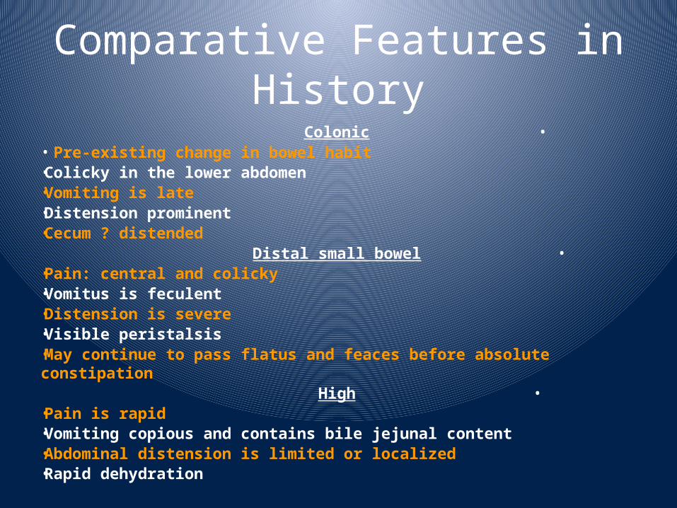
Comparative Features in History
•Colonic• Pre-existing change in bowel habit•Colicky in the lower abdomen•Vomiting is late•Distension prominent•Cecum ? distended
•Distal small bowel•Pain: central and colicky•Vomitus is feculent•Distension is severe•Visible peristalsis•May continue to pass flatus and feaces before absolute constipation
•High•Pain is rapid•Vomiting copious and contains bile jejunal content•Abdominal distension is limited or localized•Rapid dehydration
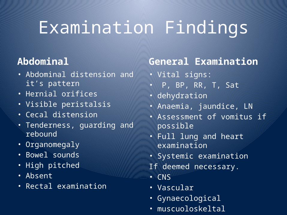
Examination FindingsAbdominal• Abdominal distension and it’s pattern
• Hernial orifices• Visible peristalsis• Cecal distension• Tenderness, guarding and rebound
• Organomegaly• Bowel sounds• High pitched• Absent• Rectal examination
General Examination• Vital signs: • P, BP, RR, T, Sat• dehydration• Anaemia, jaundice, LN• Assessment of vomitus if possible
• Full lung and heart examination
• Systemic examination If deemed necessary.• CNS• Vascular• Gynaecological• muscuoloskeltal
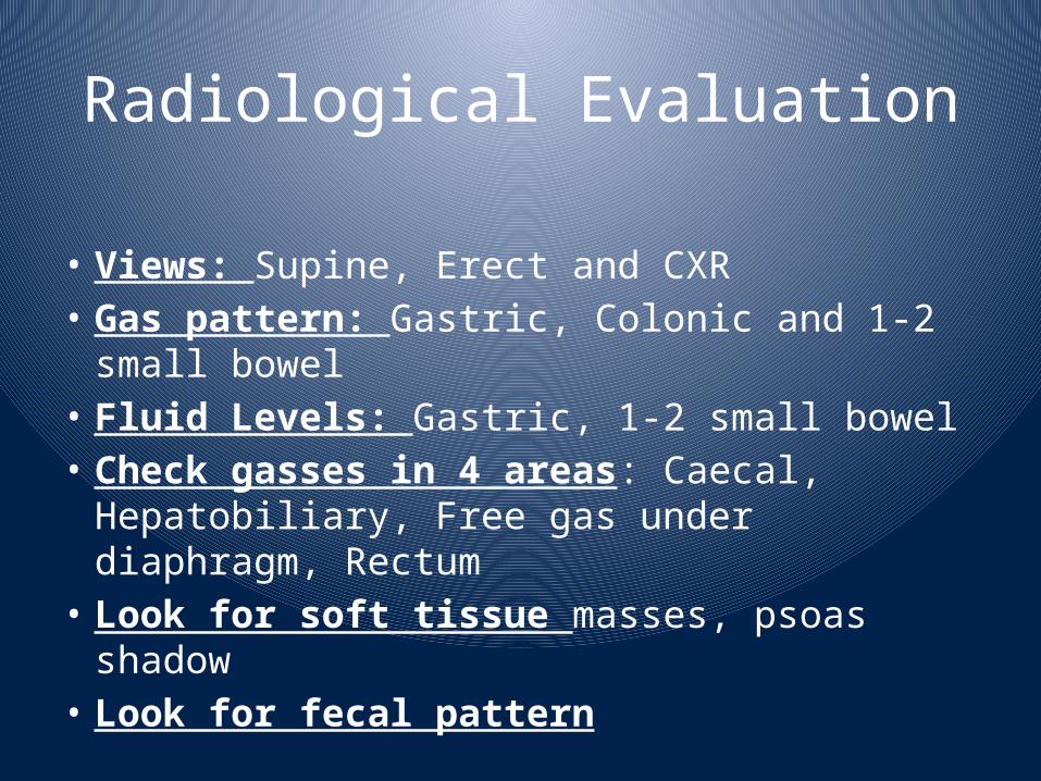
Radiological Evaluation
• Views: Supine, Erect and CXR• Gas pattern: Gastric, Colonic and 1-2 small bowel
• Fluid Levels: Gastric, 1-2 small bowel• Check gasses in 4 areas: Caecal, Hepatobiliary, Free gas under diaphragm, Rectum
• Look for soft tissue masses, psoas shadow
• Look for fecal pattern
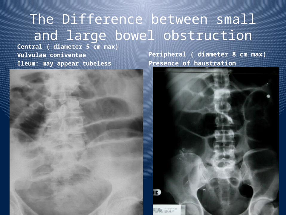
The Difference between small and large bowel obstruction
Central ( diameter 5 cm max)Vulvulae coniventaeIleum: may appear tubeless
Peripheral ( diameter 8 cm max)Presence of haustration
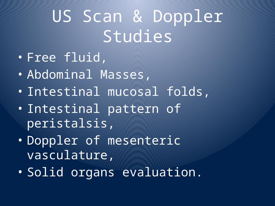
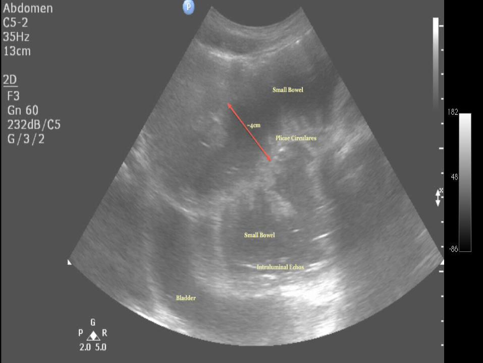
US Scan & Doppler Studies
• Free fluid, • Abdominal Masses, • Intestinal mucosal folds, • Intestinal pattern of peristalsis,
• Doppler of mesenteric vasculature,
• Solid organs evaluation.
Role of CT in Diagnosis• Used with iv contrast, oral and rectal contrast (triple contrast).
• Able to demonstrate abnormality in the bowel wall, mesentery, mesenteric vessels and peritoneum.
• Ensure: patient vitally stable with no renal failure and no previous allergy to iodine.
Findings on CT Scan Abdomen
It can define • the level of obstruction• The degree of obstruction• The cause: volvulus, hernia, luminal and mural causes
• The degree of ischemia• Free fluid and gas
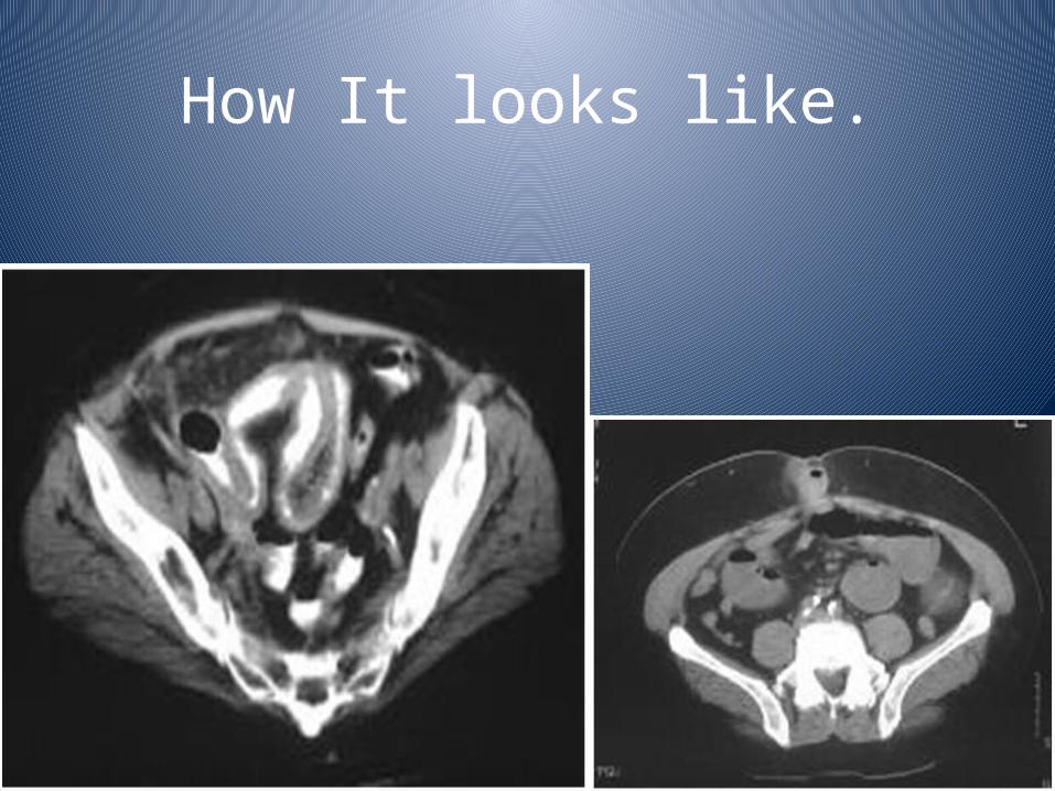
How It looks like.
Barium & Gastrografin studies
• Barium should not be used in a patient with suspected peritonitis.
• As: follow through, enema: Limited use in the acute setting
• Gastrografin is used in acute abdomen but is diluted
• Useful in recurrent and chronic obstruction• May able to define the level and mural causes.
• Can be used to distinguish adynamic and mechanical obstruction
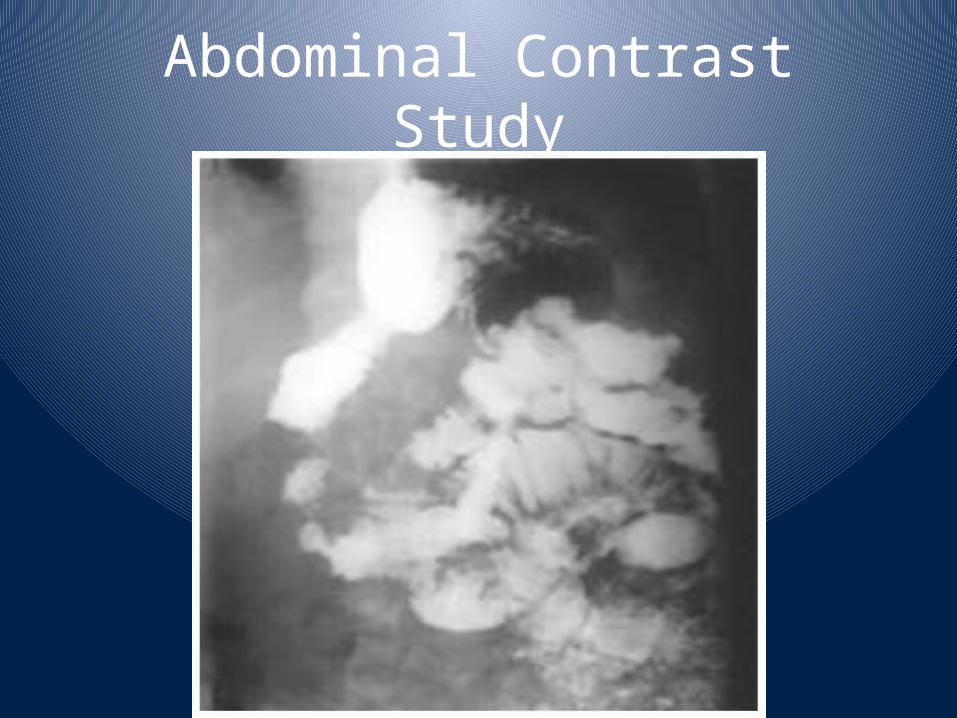
Abdominal Contrast Study
Indications for SurgeryImmediate intervention:• Evidence of strangulation (hernia….etc)• Signs of peritonitis resulting from perforation or ischemia
In the next 24-48 hours:• Clear indication of no resolution of obstruction ( Clinical, radiological).
• Diagnosis is unclear in a virgin abdomen
Intermediate stage:• The cause has been diagnosed and the patient is stabilized
Causes of Intestinal Ileus
• Postoperative and bowel resection• Intraperitoneal infection or inflammation• Ischemia• Extra-abdominal: Chest infection, Myocardia infarction• Endocrine: hypothyroidism, diabetes• Spinal and pelvic fractures• Retro-peritoneal haematoma• Metabolic abnormalities:• Hypokalaemia• Hyponatremia• Uraemia• Hypomagnesemia• Bed ridden• Drug induced: morphine, tricyclic antidepressants
Is this an ileus or obstruction
Clinical features:• Is there an under lying cause?• Is the abdomen distended but tenderness is not marked.
• Is the bowel sounds diffusely hypoactive.
Radiological features:• Is the bowel diffusely distended• Is there gas in the rectum• Are further investigations (CT or Gastrografin studies) helpful in showing an obstruction.
• Does the patient improve on conservative measures
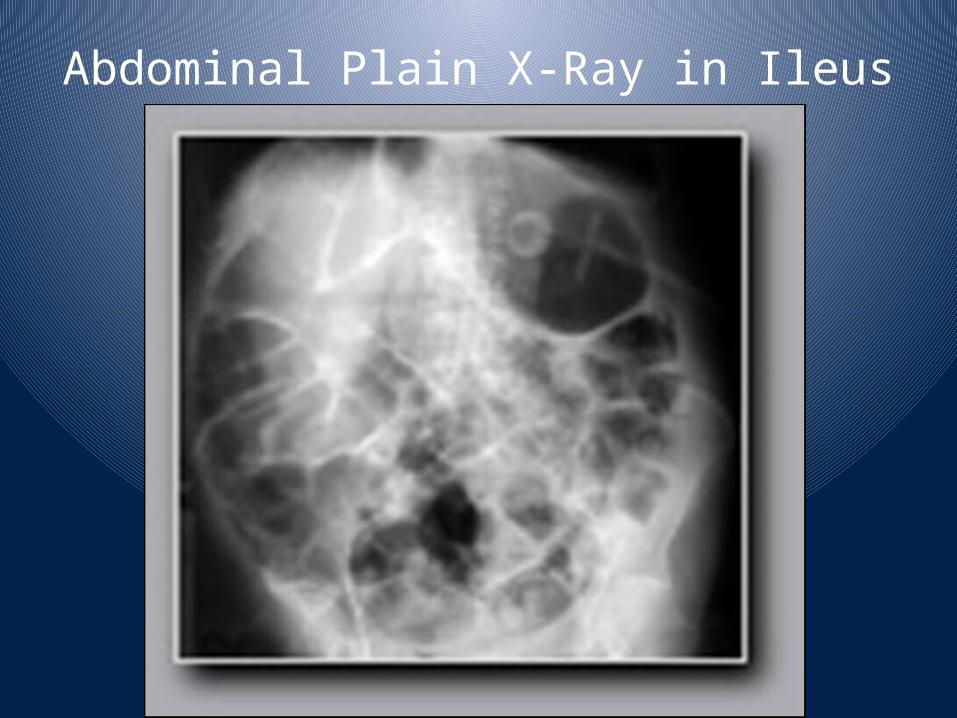
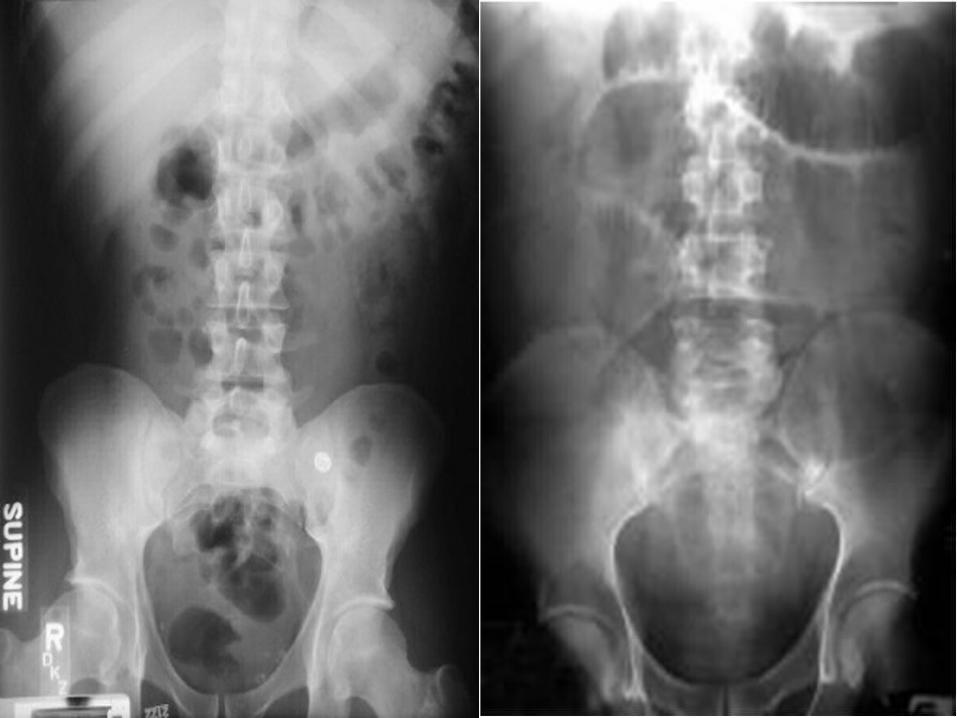
Abdominal Plain X-Ray in Ileus
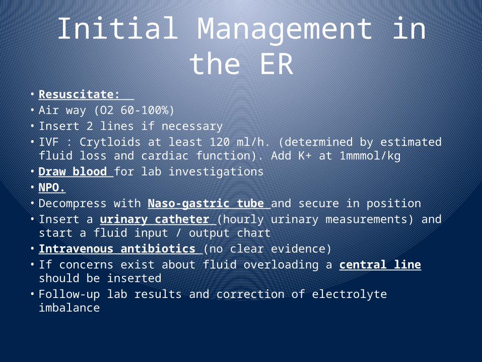
Initial Management in the ER
• Resuscitate: • Air way (O2 60-100%)• Insert 2 lines if necessary• IVF : Crytloids at least 120 ml/h. (determined by estimated fluid loss and cardiac function). Add K+ at 1mmmol/kg
• Draw blood for lab investigations• NPO.• Decompress with Naso-gastric tube and secure in position• Insert a urinary catheter (hourly urinary measurements) and start a fluid input / output chart
• Intravenous antibiotics (no clear evidence)• If concerns exist about fluid overloading a central line should be inserted
• Follow-up lab results and correction of electrolyte imbalance
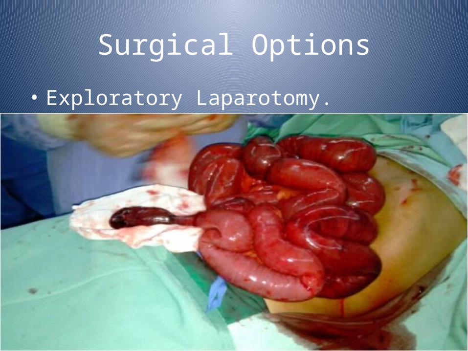
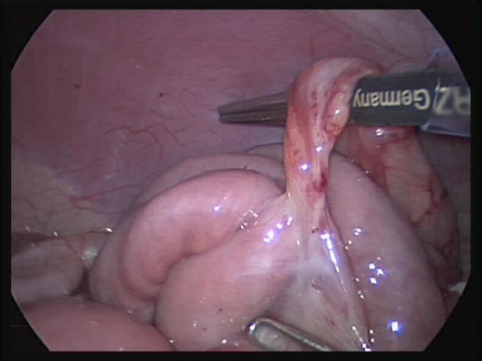
Surgical Options• Exploratory Laparotomy.

Keep the dam flowing !!!!!!!!!


