Embed Size (px)
Citation preview
© 2013 Thekkae Padil and Černík, publisher and licensee Dove Medical Press Ltd. This is an Open Access article which permits unrestricted noncommercial use, provided the original work is properly cited.
International Journal of Nanomedicine 2013:8 889–898
International Journal of Nanomedicine
Green synthesis of copper oxide nanoparticles using gum karaya as a biotemplate and their antibacterial application
Vinod Vellora Thekkae PadilMiroslav ČerníkLaboratory of Chemical Remediation Processes, Institute for Nanomaterials, Advanced Technology and Innovation, Technical University of Liberec, Studentská 1402/2, Liberec, Czech Republic
Correspondence: Miroslav Černík Laboratory Chemical Remediation Processes, Institute for Nanomaterials, Advanced Technology and Innovation (CXI), Technical University of Liberec, Studentská 1402/2, Liberec 1, 461 17, Czech Republic Tel +42 048 535 3017 Fax +42 048 535 3445 Email [email protected]
Background: Copper oxide (CuO) nanoparticles have attracted huge attention due to catalytic,
electric, optical, photonic, textile, nanofluid, and antibacterial activity depending on the size,
shape, and neighboring medium. In the present paper, we synthesized CuO nanoparticles using
gum karaya, a natural nontoxic hydrocolloid, by green technology and explored its potential
antibacterial application.
Methods: The CuO nanoparticles were synthesized by a colloid-thermal synthesis process.
The mixture contained various concentrations of CuCl2 ⋅ 2H
2O (1 mM, 2 mM, and 3 mM) and
gum karaya (10 mg/mL) and was kept at 75°C at 250 rpm for 1 hour in an orbital shaker. The
synthesized CuO was purified and dried to obtain different sizes of the CuO nanoparticles.
The well diffusion method was used to study the antibacterial activity of the synthesized CuO
nanoparticles. The zone of inhibition, minimum inhibitory concentration, and minimum bacte-
ricidal concentration were determined by the broth microdilution method recommended by the
Clinical and Laboratory Standards Institute.
Results: Scanning electron microscopy analysis showed CuO nanoparticles evenly distributed
on the surface of the gum matrix. X-ray diffraction of the synthesized nanoparticles indicates
the formation of single-phase CuO with a monoclinic structure. The Fourier transform infrared
spectroscopy peak at 525 cm−1 should be a stretching of CuO, which matches up to the B2u
mode.
The peaks at 525 cm−1 and 580 cm−1 indicated the formation of CuO nanostructure. Transmission
electron microscope analyses revealed CuO nanoparticles of 4.8 ± 1.6 nm, 5.5 ± 2.5 nm, and
7.8 ± 2.3 nm sizes were synthesized with various concentrations of CuCl2 ⋅ 2H
2O (1 mM, 2 mM,
and 3 mM). X-ray photoelectron spectroscopy profiles indicated that the O 1s and Cu 2p peak
corresponding to the CuO nanoparticles were observed. The antibacterial activity of the syn-
thesized nanoparticles was tested against Gram-negative and positive cultures.
Conclusion: The formed CuO nanoparticles are small in size (4.8 ± 1.6 nm), highly stable, and
have significant antibacterial action on both the Gram classes of bacteria compared to larger sizes
of synthesized CuO (7.8 ± 2.3 nm) nanoparticles. The smaller size of the CuO nanoparticles
(4.8 ± 1.6 nm) was found to be yielding a maximum zone of inhibition compared to the larger
size of synthesized CuO nanoparticles (7.8 ± 2.3 nm). The results also indicate that increase
in precursor concentration enhances an increase in particle size, as well as the morphology of
synthesized CuO nanoparticles.
Keywords: gum karaya, CuO nanoparticles, XRD, FTIR, XPS, antibacterial activity
IntroductionCopper oxide (CuO) nanoparticles are important due to their applications as antimi-
crobials and in gas sensors, batteries, high temperature superconductors, solar energy
conversion tools, and so on.1–4 Human beings have been using copper (Cu) and Cu
Dovepress
submit your manuscript | www.dovepress.com
Dovepress 889
O R I G I N A L R E S E A R C h
open access to scientific and medical research
Open Access Full Text Article
http://dx.doi.org/10.2147/IJN.S40599
International Journal of Nanomedicine 2013:8
complexes for various purposes for centuries, such as water
purifiers, algaecides, fungicides, and as antibacterial and anti-
fouling agents.5 Natural plant materials such as magnolia leaf
extract and stem latex of Euphorbia nivulia have been used
for the synthesis of Cu nanoparticles. The application of these
nanoparticles expresses superior antibacterial activity against
Escherichia coli cells. The nanoparticles have also been used
as nontoxic aqueous formulations for administra tion of cancer
therapy.6–7 Cu and CuO nanoparticles have been studied as
potential antimicrobial agents against infectious organisms
such as E. coli, Bacillus subtilis, Vibria cholera, Pseudomonas
aeruginosa, Syphillis typhus, and Staphylococcus aureus.8–10
Kattumuri et al11 explored the possibility of utilizing gum
arabic as a nontoxic, natural hydrocolloid used in the produc-
tion of readily administrable biocompatible gold nanoparticles
for diagnostic and therapeutic applications. They have also
demonstrated that complex polysaccharides and protein
structures within the gum arabic backbone can effectively
lock gold nanoparticles to produce nontoxic, nanoparticulate
constructs that are stable under in vivo conditions for poten-
tial applications in nanomedicine. Thus, this investigation
elucidates that natural gums could act as nontoxic vehicles
to stabilize and deliver nanoparticles for in vivo applications.
The preparation of CuO nanoparticles uses a variety of
methods including sol-gel, quick precipitation, sonochemi-
cal, electrochemical, solid state reaction, alcohothermal
synthesis, microwave irradiation, and liquid–liquid interface
techniques involving organic solvents and harsh reducing
agents.12–20 Therefore, it was a challenge to find a conve-
nient, mild, nontoxic, natural product to produce metal
nanoparticles in an aqueous environment. Amongst various
natural materials used for nanoparticle construction, plants
seem to be the best candidates, and nanoparticles produced
by plants are more stable, are of various sizes and shapes,
and the rate of production is faster than in the case of
microorganisms.21 Biosynthesis of metal/metal oxide nano-
particles using environmentally friendly methods without the
use of harsh, toxic reducing agents (eg, hydrazine hydrate,
sodium borohydride, dimethylformamide, ethylene glycol,
and so on), and expensive chemicals are the main principle
of green chemistry. Many of these reducing agents have
been associated with environmental toxicity or biological
hazards. With an increasing interest in the minimization or
total elimination of waste and the execution of sustainable
processes through the implementation of the fundamental
principles of green chemistry, the development of natural and
biomimetic approaches for the preparation of nanomaterials
is a desirable aspect.22,23 Hence, there is a need to develop
newer ecofriendly processes for the synthesis of metal/metal
oxide nanoparticles of controlled shapes and sizes, and such
methods could permit the nanoparticles to be extensively
used in biological systems.
Polysaccharide hydrocolloids such as carrageenan,
alginate, agar-agar, starches, pectin, guar and gums
(arabic, tragacanth, and karaya) are high molecular weight
macromolecules. Gums are naturally occurring polysaccha-
ride components in plants, which are fundamentally economi-
cal and easily available. They have assorted applications as
thickeners, food emulsifiers, viscosifiers, sweeteners, and so
on in confectionery, and as binders and drug release modifiers
in pharmaceutical dosage forms.24 Traditionally, India is the
largest producer and exporter of gum karaya (GK), while
Europe is the largest importer of GK.25 Physicochemical
properties, as well as the structural, occurrence, production,
food, and nonfood applications of GK have been widely
studied by different research groups.26–28 GK is a partially
acetylated polysaccharide and has a branched structure and
high molecular mass of ∼16.0 × 10⁹ Da, and is grouped
under substituted rhamnogalacturonoglycan (pectic) type
tree gums. This gum contains about 60% neutral sugars
(rhamnose and galactose) and 40% acidic sugars (glucuronic
acid and galacturonic acids).29 The toxicological evaluation
of GK had established that this gum was nontoxic and has
potential application as a food additive.30
Biosynthesis of metal nanoparticles by plants is currently
under development. The synthesis of metal nanoparticles
using inactivated plant tissue, plant extracts, exudates,
and other parts of living plants is a modern alternative for
their production.31 It is a very cost effective method and
therefore a prospective commercial alternative for large-scale
production.
Natural hydrocolloids isolated from trees are a new class
of potentially economical and environmentally compassionate
biomaterial that exhibit a high specificity for the production
of nanomaterials. Gums from plants may act both as reducing
and capping agents in nanoparticle synthesis. The bioreduc-
tion of metal nanoparticles by a combination of biomolecules
found in plant extracts (eg, enzymes, proteins, amino acids,
vitamins, polysaccharides, and organic acids such as citrates)
is environmentally benign, yet chemically complex. Because of
the important and critical roles of plants in bio-based protocols
for metal nanoparticle production, the green synthesis of metal
nanoparticles using GK is an important subject in this paper.
Further, this study aims to explore the proficiency of these
nanoparticles as antibacterial agents against human patho-
genic bacteria, mainly E. coli and S. aureus.
As part of our ongoing research work on the design and
development of nanoparticulate constructs using natural tree
submit your manuscript | www.dovepress.com
Dovepress
Dovepress
890
Thekkae Padil and Černík
International Journal of Nanomedicine 2013:8
gums, herein we report the development of a new class of
composite material derived from a natural nontoxic GK bio-
polymer with CuO nanoparticles that are stable in vivo. The
present work details a green chemistry approach to the synthesis
of metal oxide nanoparticles using a well-characterized plant
derived natural product GK, which acts as a reducing and
templating agent. In this current research paper CuO nano-
particles have been synthesized and characterized by scanning
electron microscope (SEM), transmission electron microscope
(TEM), Fourier transform infrared spectroscopy (FTIR), X-ray
diffraction (XRD), and X-ray photoelectron spectroscopy
(XPS) analysis. Further, CuO nanoparticles were explored
with respect to their prospective antibacterial application.
Materials and methodsMaterialsGK was obtained from the Sigma-Aldrich Company (Sigma-
Aldrich, St Louis, MO, USA). The chemicals CuCl2 ⋅ 2H
2O,
NaOH, and HCl were analytical grade and used as received
without further purification.
GK preparationGK powder (1 g) was exactly weighed and dispensed into
a clean glass beaker containing 1 L of deionized water. The
procedures for the preparation of GK lyophilized powder was
based on the earlier reported method used for other natural
tree gums.32 The gum solution was gently stirred overnight at
room temperature. Later, undissolved matter was separated
by keeping the gum solution at room temperature (25°C) for
an additional 18 hours and filtered through a sintered glass
funnel (#G-2 followed by #G-4). The obtained clear solution
was freeze-dried and stored for further use.
Preparation of CuO nanoparticlesThe CuO nanoparticles were synthesized by a colloid-
thermal synthesis process.15 In separate 50 mL conical
flasks, 100 µL aliquot of 10 mM solutions of CuCl2 ⋅ 2H
2O
was mixed with 10 mL of the GK solution prepared above
(100 mg gum dispersed in 10 mL of deionized water) and
NaOH ⋅ CuCl
2 ⋅ 2H
2O and NaOH in a molar ratio of 2:5 were
maintained in the mixture. All the solution components were
purged with nitrogen prior to use and all chemical processes
proceeded in the presence of nitrogen. The mixture contain-
ing CuCl2 ⋅ 2H
2O and GK was kept at 75°C at 250 rpm for
1 hour in an orbital shaker. The color of the mixtures gradu-
ally changed from bluish to black, indicating the formation
of CuO nanoparticles. The resulting precipitate was obtained
by centrifugation and washed with ethanol and distilled water,
respectively, and finally freeze-dried.
Characterization of CuO nanoparticlesSEM analysisMorphology of the nanoparticles was studied using SEM
analysis (SEM-LEO S1430 VP from M/S LEO Electron
Microscopy Ltd, Cambridge, UK and UHR FE-SEM Carl
Zeiss ULTRA Plus, Carl Zeiss Meditec AG, Jena, Germany).
X-ray diffraction analysisXRD patterns were obtained with Cu-Kα radiation using
a diffractometer (Philips PW 3710/3020, Netherlands)
equipped with a graphite monochromator. The measure-
ments were made using a step-scanning program with 0.02°
per step and an acquisition time of 5 seconds per step. The
XRD data were analyzed using X’Pert High Score software
(PANanlytical BV Almelo, Netherlands) for the identifica-
tion of the crystalline phases.
Fourier transform infrared spectroscopy analysisFTIR spectra were obtained using a spectrophotometer
(Nicolet IZ10, Thermo Fisher Scientific, Waltham, MA,
USA) in the spectral region of 4000–400 cm−1 using a
resolution of 4 cm−1 and 64 coadded scans. All the colloidal
nanoparticles were freeze-dried in the form of nanopowders
and palletized with KBr for FTIR studies.
Transmission electron microscopyTEM images for these nanoparticles were recorded using a
TEM operating at an acceleration voltage of 15 kV (Tecnai
F12; North American NanoPort, Hillsboro, OR, USA).
For TEM measurements, samples were prepared by dropping
10–20 µL of CuO nanoparticle dispersions on a Cu grid and
subsequently dried at room temperature.
X-ray photoelectron spectroscopy analysisXPS analysis of CuO and GK powder samples was performed
(Kratos XPS system, AXIS HIS-165 Ultra; Shimadzu
Corporation, Kyoto, Japan) to confirm the CuO and GK
composition. Survey scans were recorded in the binding
energy range of 0 to 1100 electron volts (eV) with pass energy
of 80 eV. When comparing the results with the reference,
peaks were identified as being the same if the difference of
the peak position was within 0.5 eV.
Antimicrobial testsBacterial strainsE. coli MTCC 443 and S. aureus MTCC 737 were used
as reference strains for Gram-negative and Gram-positive
bacteria, respectively, following guidelines of the Clinical
and Laboratory Standards Institute.33 Strains were grown on
nutrient agar.
submit your manuscript | www.dovepress.com
Dovepress
Dovepress
891
Antibacterial property of CuO nanoparticles
International Journal of Nanomedicine 2013:8
Agar well diffusion methodThe well diffusion method was used to study the antibacterial
activity of the synthesized CuO nanoparticles. All the glass-
ware, media, and reagents used were sterilized in an autoclave
at 121°C for 20 minutes. The bacterial suspension was pre-
pared by growing a single colony overnight in nutrient broth
and by adjusting the turbidity to 0.5 McFarland standards.
Mueller Hinton agar plates were inoculated with this bacte-
rial suspension, and 50 µg (100 µL) of CuO nanoparticles
were added to the center well with a diameter of 8 mm. The
nanoparticles used here were prepared with 10 mg/mL of GK
solution containing 1 mM, 2 mM, and 3 mM of CuCl2 ⋅ 2H
2O
autoclaved for 60 minutes. Control plates were maintained
with autoclaved GK loaded wells. Antibiotic such as tetra-
cycline was used as a positive control. These plates were
incubated at 37°C for 24 hours in a bacteriological incubator,
and the zone of inhibition (ZOI) was measured by subtracting
the well diameter from the total inhibition zone diameter. All
tests were performed three times.
Determination of minimal inhibitory/bactericidal concentrationsMinimum inhibitory concentrations (MIC) and minimum
bactericidal concentrations (MBC) of the CuO nanoparticles
were determined by the broth microdilution method recom-
mended by the Clinical and Laboratory Standards Institute
in 2000.33 Tetracycline was used as a positive control. The
suspension of GK (10 mg/mL) was used as negative control
in all experiments. A dilution series with 5 mL of nutrient
broth medium containing 10–100 µg/mL of CuO nanopar-
ticles was prepared. For the MIC determination, the suspen-
sions were prepared by suspending one isolate colony from
nutrient plates in 50 µL of nutrient broth. After 24 hours of
growth, suspensions were diluted in distilled water to obtain
final inoculums of 5 × 10⁵–5 × 10⁶ colony-forming units/
mL. The purity of isolates was checked throughout the study
by examining the colony morphology and Gram staining.
Two-fold serial dilutions of CuO nanoparticle solutions were
prepared in nutrient broth in 98-well plates starting from
a stock solution of 10−2 M. An equal volume of bacterial
inoculums was added to each well on the microtiter plate
containing 0.05 mL of the serial compound dilutions. After
incubation for 18–24 hours at 35°C, MIC was determined
with an enzyme-linked immunosorbent assay reader (Infi-
nite 200; Tecan Systems, Inc, San Jose, CA, USA) as the
lowest concentration of compound whose absorbance was
comparable with the negative control wells (broth only or
broth with CuO nanoparticles, without inoculums). Results
are expressed as the mean values of three independent
determinations.
Statistical analysisEach experiment was repeated three times, and the resulting
bacterial growths on three plates corresponding to a particu-
lar sample were reported as the mean ± standard deviation
(n = 3). The MIC and MBC experimental analysis was based
on three independent sample analyses.
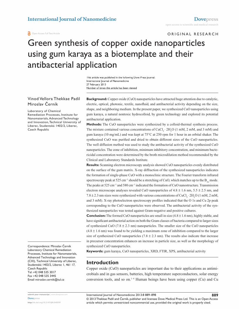
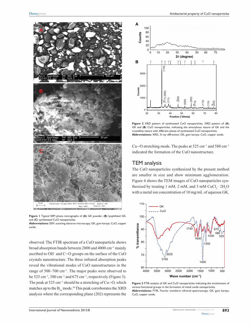
Results and discussionMorphology of GK and CuO nanoparticles by SEM analysisThe morphology of GK before and after the deposition of
CuO nanoparticles studied by SEM is presented in Figure 1.
The morphology of GK indicates irregular particles of
various sizes (Figure 1A), whereas the lyophilized sample
is appearing as crumbled-like structure (Figure 1B). The
CuO nanoparticles appear as small needle-like structures
on the surface of the gum matrix (Figure 1C). The SEM
micrographs were obtained after washing the dried GK-
CuO thrice with deionized water. One can notice that
no metal oxide nanoparticles are seen outside the gum
matrix, which can be attributed to the strong locking of
nanoparticles within the gum matrix. This clearly suggests
that the CuO nanoparticles are bound to the surface of the
gum matrix.
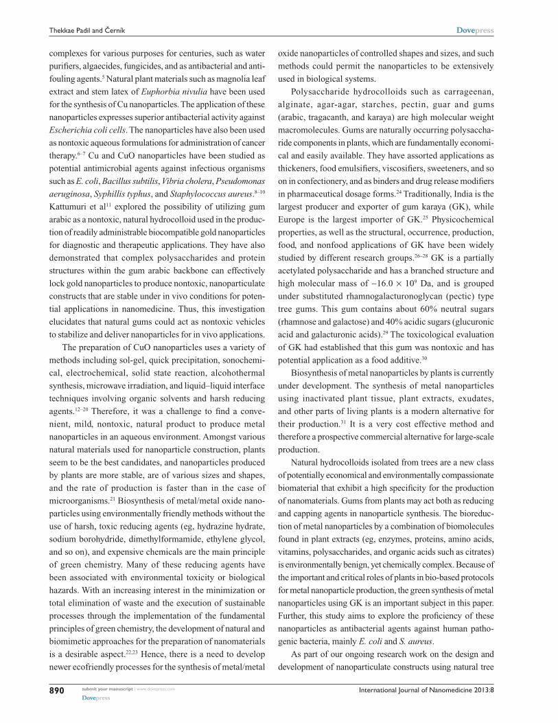
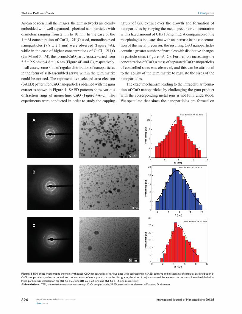
XRD analysisThe XRD pattern of GK and CuO nanoparticles is depicted
in Figure 2. As shown in Figure 2A, the XRD pattern of
GK is amorphous in nature whereas the CuO nanoparticles
(Figure 2B) demonstrate that the CuO is crystalline in nature.
The spectrum is identical to that of pure CuO, indicating the
formation of single-phase CuO with monoclinic structure
(JCPDS-05-0661).34 In the present work, the diffraction
patterns were observed to be at 2θ = 32.47, 35.49, 38.68, 48.65,
53.36, 58.25 and 61.45 were assigned to the reflection lines
of monoclinic CuO nanoparticles. The present experimental
results were found to be in agreement with the reported diffrac-
tion patterns of CuO nanoparticles prepared by Das et al.35
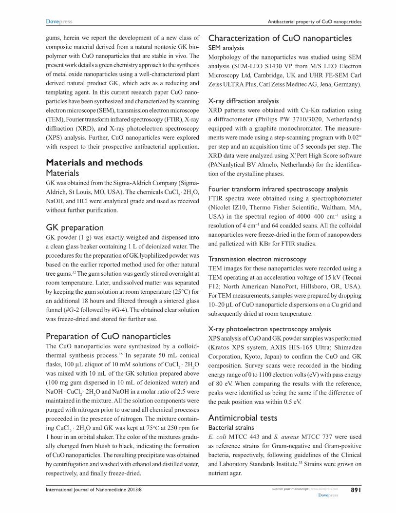
FTIR analysisThe FTIR spectra of GK and CuO nanoparticles are shown in
Figure 3. The main characteristic peaks of GK at 1047 cm−1
and 1422 cm−1 (C−O stretch), 1630 cm−1 (C=O stretch
and N−H bending), 1734 cm−1 (CH3CO group), 2990 cm−1
(C−H stretch), and 3000–3600 cm−1 (O−H stretch) were
submit your manuscript | www.dovepress.com
Dovepress
Dovepress
892
Thekkae Padil and Černík
International Journal of Nanomedicine 2013:8
observed. The FTIR spectrum of a CuO nanoparticle shows
broad absorption bands between 2800 and 4000 cm−1 mainly
ascribed to OH– and C−O groups on the surface of the CuO
crystals nanostructure. The three infrared absorption peaks
reveal the vibrational modes of CuO nanostructures in the
range of 500–700 cm−1. The major peaks were observed to
be 525 cm−1, 580 cm−1 and 675 cm−1, respectively (Figure 3).
The peak at 525 cm−1 should be a stretching of Cu−O, which
matches up to the B2u
mode.36 This peak corroborates the XRD
analysis where the corresponding plane (202) represents the
Cu−O stretching mode. The peaks at 525 cm−1 and 580 cm−1
indicated the formation of the CuO nanostructure.
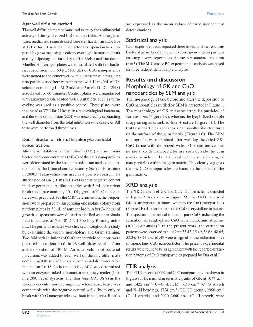
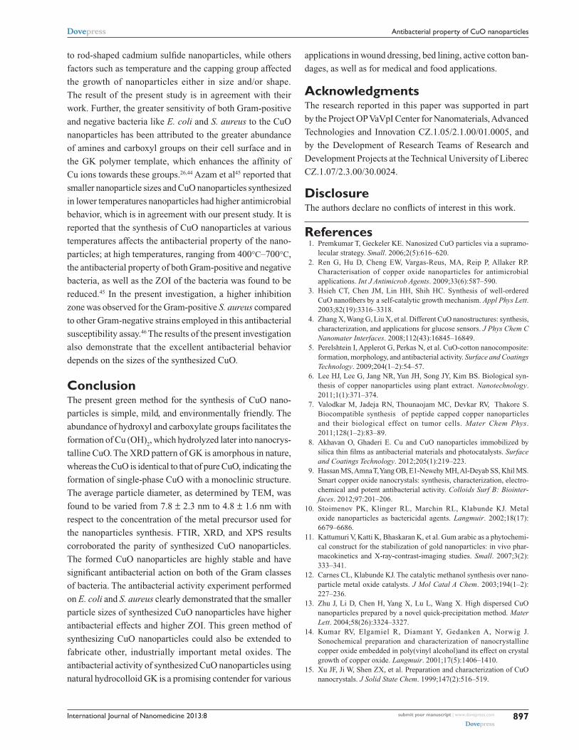
TEM analysisThe CuO nanoparticles synthesized by the present method
are smaller in size and show minimum agglomeration.
Figure 4 shows the TEM images of CuO nanoparticles syn-
thesized by treating 1 mM, 2 mM, and 3 mM CuCl2 ⋅ 2H
2O
with a metal ion concentration of 10 mg/mL of aqueous GK.
A
B
Acc.V Spot Magn Det WD Exp 500 µm25.0 kV 5.0 62× SF 10.6 0
Acc.V Spot Magn Det WD Exp 50 µm25.0 kV 5.0 500× SF 10.6 0
Filament type = W (agar A054) EHT = 20.00 kV WD = 15 mm Signal A = SE1Photo no = 900Mag = 356×
30 µm
C
Figure 1 Typical SEM photo micrographs of (A) GK powder, (B) lyophilized GK, and (C) synthesized CuO nanoparticles.Abbreviations: SEM, scanning electron microscopy; GK, gum karaya; CuO, copper oxide.
100
0 10 20 30 40 50 60 70
20
3000
2000
1000
030
(111
)
(110
)
(202
)
(020
)
(202
)
(113
)
(113
)
(113
)
(222
)
(111
) (0
02)
40 50Position [°2theta]
Co
un
tsC
ou
nts
2θ (degree)
60 70 80
80
60
4020
0
A
B
Figure 2 XRD pattern of synthesized CuO nanoparticles. XRD pattern of (A) GK and (B) CuO nanoparticles, indicating the amorphous nature of GK and the crystalline nature with different planes of synthesized CuO nanoparticles.Abbreviations: XRD, X-ray diffraction; GK, gum karaya; CuO, copper oxide.
110
100
90
80
70
604000 3500 3000 2500
Wave number (cm−1)
% t
ran
smit
tan
ce
3025
31501400
1250
1600
1740
GK
CuO
1030580
525
675
2000 1500 1000 500
Figure 3 FTIR analysis of GK and CuO nanoparticles indicating the involvement of various functional groups in the formation of metal oxide nanoparticles.Abbreviations: FTIR, Fourier transform infrared spectroscopy; GK, gum karaya; CuO, copper oxide.
submit your manuscript | www.dovepress.com
Dovepress
Dovepress
893
Antibacterial property of CuO nanoparticles
International Journal of Nanomedicine 2013:8
As can be seen in all the images, the gum networks are clearly
embedded with well separated, spherical nanoparticles with
diameters ranging from 2 nm to 10 nm. In the case of the
1 mM concentration of CuCl2 ⋅ 2H
2O used, monodispersed
nanoparticles (7.8 ± 2.3 nm) were observed (Figure 4A),
while in the case of higher concentrations of CuCl2 ⋅ 2H
2O
(2 mM and 3 mM), the formed CuO particles size varied from
5.5 ± 2.5 nm to 4.8 ± 1.6 nm (Figure 4B and C), respectively.
In all cases, some kind of regular distribution of nanoparticles
in the form of self-assembled arrays within the gum matrix
could be noticed. The representative selected area electron
(SAED) pattern for CuO nanoparticles obtained with the gum
extract is shown in Figure 4. SAED patterns show various
diffraction rings of monoclinic CuO (Figure 4A–C). The
experiments were conducted in order to study the capping
nature of GK extract over the growth and formation of
nanoparticles by varying the metal precursor concentration
with a fixed amount of GK (10 mg/mL). A comparison of the
morphologies indicates that with an increase in the concentra-
tion of the metal precursor, the resulting CuO nanoparticles
contain a greater number of particles with distinctive changes
in particle sizes (Figure 4A–C). Further, on increasing the
concentration of CuO, a mass of separated CuO nanoparticles
of controlled sizes was observed, and this can be attributed
to the ability of the gum matrix to regulate the sizes of the
nanoparticles.
The exact mechanism leading to the intracellular forma-
tion of CuO nanoparticles by challenging the gum product
with the corresponding metal ions is not fully understood.
We speculate that since the nanoparticles are formed on
20A
B
C
15
10
25
20
15
10
5
0
25
30
20
15
10
5
0
2 4 6
D (nm)
Fre
qu
ency
(%
)
D (nm)
Fre
qu
ency
(%
)
D (nm)
Fre
qu
ency
(%
)
8 10
20 4 6 8 10
5
04 6 8 10
Mean diameter: 7.8 ± 2.3 nm
Mean diameter: 5.5 ± 2.5 nm
Mean diameter: 4.8 ± 1.6 nm
12
Figure 4 TEM photo micrographs showing synthesized CuO nanoparticles of various sizes with corresponding SAED patterns and histograms of particle size distribution of CuO nanoparticles synthesized at various concentrations of metal precursor. In the histograms, the sizes of major nanoparticles are reported as mean ± standard deviation. Mean particle size distribution for (A) 7.8 ± 2.3 nm; (B) 5.5 ± 2.5 nm; and (C) 4.8 ± 1.6 nm, respectively.Abbreviations: TEM, transmission electron microscopy; CuO, copper oxide; SAED, selected area electron diffraction; D, diameter.
submit your manuscript | www.dovepress.com
Dovepress
Dovepress
894
Thekkae Padil and Černík

International Journal of Nanomedicine 2013:8
the surface of the gum and not in the solution, the first step
involves a trapping of metal ions on the surface of the gum
network possibly via an electrostatic interaction between
the metal ions and the negatively-charged carboxylate and
hydroxyl groups present in the biopolymer.25 The presence of
various sugars, amino acids and fatty acids present in the GK
could act as a reducing and capping agent for the formation
of metal oxide nanoparticles.28 Upon hydrolysis, metal ions
may lead to the formation of metal nuclei, and they subse-
quently grow and accumulate in the form of nanoparticles
within the gum matrix. These assumptions can be validated
by FTIR and XRD results.
XPS analysisThe XPS spectra of GK and CuO nanoparticles are presented
in Figure 5A and B, respectively. As can be seen from the
spectra, the XPS survey scans of pure GK show binding
energy peaks characteristic of carbon and oxygen only. The
observed C 1s binding energy peaks assigned to C−C, C−O,
and C=O functional groups present in GK are in agreement
with the observations made by FTIR. The C−O and C=O
peaks can be associated with hydroxyl and carboxylate groups
present in the biopolymer. The O 1s high resolution narrow
scans could be deconvoluted into binding energy peaks at
531.8 eV and 532.7 eV, which can be dedicated to the O in
the C=O and alcoholic C−O groups. The C=O group may
be seen due to the carboxylate groups present in the GK. The
Cu 2p core level spectrum (Figure 5B) represents two peaks
located at 933.4 and 953.8 eV, which corresponds to the Cu
2p3/2 and Cu 2p1/2, respectively. These values match well
with the data reported for the Cu(2p) in CuO.16 The Cu 2P3/2
spectrum (inset Figure 5B) shows that the Cu2+ peak lies at
933.4 eV, with two shake-up satellites 7.2 and 9.8 eV higher
in binding energy than that of the main peak. As shown in
Figure 5B, the O 1s core-level spectrum is broad, and two
O 1s peaks, at a lower energy of 529.5 eV, is in agreement
with O2− in CuO, while the other peak, at a higher energy
of 531.6 eV, is attributed to O adsorbed on the surface of
the CuO particles.37 Thus, the XPS results indicate that the
nanoparticles are composed of CuO. The strong shake-up
satellites recorded in the CuO sample confirm the Cu2+
oxidation state, and rule out the possibility of the existence
of a Cu2O phase.15,16
Antibactericidal testsThe antibacterial activity of the CuO nanoparticle was tested
against the Gram-negative bacteria E. coli and the Gram-
positive S. aureus. The results of the antibacterial activity
30
1000 800 600Binding energy (eV)
Inte
nsi
ty (
CP
S)
× 10
3In
ten
sity
(C
PS
)
Binding energy (eV)
400
O 1s
C 1s
Cu 2p1/2
Cu 2p3/2
200
160
960 952 944
Binding energy (ev)
Inte
nsi
ty (
cps)
936 928
120
80
40
0
0
1000
10
15
20
25
30
5
800 600 400 200 0
A
B
25
20
15
10
5
Figure 5 XPS survey scanning spectra of the (A) GK (B) GK-CuO nanoparticles formation. Note: Inset figure B shows the spectra of the CuO nanoparticles formation.Abbreviations: XPS, X-ray photoelectron spectroscopy; GK, gum karaya; CuO, copper oxide; CPS, counts per second; eV, electron volts.
of CuO nanoparticles against these bacterium (ZOI, MIC,
and MBC) are presented in Table 1. The suspension of GK
(10 mg/mL) as a negative control did not show any antibac-
terial activity. From the above-mentioned experiment it can
be found that the CuO nanoparticles are effective in killing/
inhibiting a range of bacterial growth. The antibacterial
activity of CuO nanoparticles against E. coli and S. aureus
with ZOI are depicted in Figure 6. The ZOI of around
14.5 ± 0.8 mm was observed for the Gram-positive bacteria
strain S. aureus (American Type Culture Collection 25923),
and in the case of the Gram-negative bacteria strain E. coli
(American Type Culture Collection 25922), the detected
ZOI was 16.2 ± 0.8 mm. Synthesized CuO with a size of
4.8 ± 1.6 nm was used in both cases.
The other observation was that CuO nanoparticles
synthesized at the range of 7.8 ± 2.3 nm demonstrated a
smaller ZOI than the smaller particles. The MIC of CuO
nanoparticles was found to be 103.5 ± 4.71 µg/mL for
E. coli and 120.4 ± 8.16 µg/mL for S. aureus, respectively
(Table 1). Like MIC values, particle size dependent CuO
(4.8 ± 1.6 nm) synthesis had MBC values of 125 ± 5.5 µg/mL
for E. coli and 135 ± 8.8 µg/mL for S. aureus (Table 1).
The present experimental values are found to be slightly
higher than the reported MIC for CuO nanoparticles syn-
thesized by continuous gas phase production.38 Based on
these results, it can be concluded that the synthesized CuO
nanoparticles had significant antibacterial action on both
submit your manuscript | www.dovepress.com
Dovepress
Dovepress
895
Antibacterial property of CuO nanoparticles
International Journal of Nanomedicine 2013:8
Table 1 Antibacterial activity of CuO nanoparticles (ZOI, MIC, and MBC) as synthesized using GK extract against Escherichia coli and Staphylococcus aureus laboratory bacterial strains
Bacterial strains used
ZOI (mm) MIC (μg/mL)
MBC (μg/mL)
Control negative (10 mg/mL)
Control positive TC (mm)
Escherichia coli 16.2 ± 0.8 (A) 15.8 ± 0.5 (B) 103 ± 4.7 125 ± 5.5 NA 18 ± 0.8Staphylococcus aureus 14.5 ± 0.6 (C) 13.8 ± 0.4 (D) 120 ± 8.1 135 ± 8.8 NA 16 ± 0.5
Notes: Values are expressed as mean ± standard deviation (n = 3). CuO nanoparticles concentration used are 10–100 µg/mL. Particle size of nanoparticles used, A and C (4.8 ± 1.6 nm); B and D (7.8 ± 2.3 nm). CuO nanoparticles size 4.8 ± 1.6 nm are used for determining MIC and MBC of Escherichia coli and Staphylococcus aureus.Abbreviations: CuO, copper oxide; ZOI, zone of inhibition; MIC, minimum inhibitory concentration; MBC, minimum bactericidal concentration; GK, gum karaya; TC, tetracycline.
Figure 6 Bacterial cultures showing the inhibition zones around wells loaded with 50 µg of CuO nanoparticles and (A) Escherichia Coli (MTCC 443) representing inhibition zone formed with 4.8 ± 1.6 nm size of CuO nanoparticles, (B) Escherichia Coli (MTCC 443) representing inhibition zone formed with 7.8 ± 2.3 nm size of CuO nanoparticles, (C) Staphylococcus aurous (MTCC 737) representing inhibition zone formed with 4.8 ± 1.6 nm size of CuO nanoparticles, and (D) Staphylococcus aurous (MTCC 737) representing inhibition zone formed with 7.8 ± 2.3 nm size of CuO nanoparticles.Abbreviations: CuO, copper oxide; MTCC, Microbial Type Culture Collection.
of the Gram classes of bacteria. The antibacterial activity
of CuO towards Gram-negative bacteria was higher when
compared to Gram-positive bacteria. The difference in activ-
ity against these two types of bacteria could be attributed
to the structural and compositional differences of the cell
membrane.39 Gram-positive bacteria have thicker peptidogly-
can cell membranes compared to the Gram-negative bacteria
and it is harder for CuO to penetrate it, resulting in a low
antibacterial response.40 Perelshtein et al5 reported that in the
case of the CuO-coated fabric, the antibacterial effect was
detected due to the CuO nanoparticles, which can generate
reactive oxygen species that are responsible for damaging
the bacteria’s cells. In this context, a number of mechanisms
have been proposed to interpret the antibacterial behavior
of metal oxides.41
The differential sensitivity of bacteria towards CuO
nanoparticles depends upon particle size, temperature of
synthesis, bacterial cell wall structure, and the degree of
contact with organisms with nanoparticles.42 In our present
investigation, the temperature of synthesis of CuO was main-
tained at 75°C, and the particle size of the nanoparticle was
found to be in the range of 2–10 nm sizes when using vari-
ous concentrations of metal of precursor (1 mM, 2 mM, and
3 mM). Recently, Moloto et al43 have demonstrated that with
an increase in precursor concentration, there was an increase
in particle size, and the morphology evolved from spherical
submit your manuscript | www.dovepress.com
Dovepress
Dovepress
896
Thekkae Padil and Černík
International Journal of Nanomedicine 2013:8
to rod-shaped cadmium sulfide nanoparticles, while others
factors such as temperature and the capping group affected
the growth of nanoparticles either in size and/or shape.
The result of the present study is in agreement with their
work. Further, the greater sensitivity of both Gram-positive
and negative bacteria like E. coli and S. aureus to the CuO
nanoparticles has been attributed to the greater abundance
of amines and carboxyl groups on their cell surface and in
the GK polymer template, which enhances the affinity of
Cu ions towards these groups.26,44 Azam et al45 reported that
smaller nanoparticle sizes and CuO nanoparticles synthesized
in lower temperatures nanoparticles had higher antimicrobial
behavior, which is in agreement with our present study. It is
reported that the synthesis of CuO nanoparticles at various
temperatures affects the antibacterial property of the nano-
particles; at high temperatures, ranging from 400°C–700°C,
the antibacterial property of both Gram-positive and negative
bacteria, as well as the ZOI of the bacteria was found to be
reduced.45 In the present investigation, a higher inhibition
zone was observed for the Gram-positive S. aureus compared
to other Gram-negative strains employed in this antibacterial
susceptibility assay.46 The results of the present investigation
also demonstrate that the excellent antibacterial behavior
depends on the sizes of the synthesized CuO.
ConclusionThe present green method for the synthesis of CuO nano-
particles is simple, mild, and environmentally friendly. The
abundance of hydroxyl and carboxylate groups facilitates the
formation of Cu (OH)2, which hydrolyzed later into nanocrys-
talline CuO. The XRD pattern of GK is amorphous in nature,
whereas the CuO is identical to that of pure CuO, indicating the
formation of single-phase CuO with a monoclinic structure.
The average particle diameter, as determined by TEM, was
found to be varied from 7.8 ± 2.3 nm to 4.8 ± 1.6 nm with
respect to the concentration of the metal precursor used for
the nanoparticles synthesis. FTIR, XRD, and XPS results
corroborated the parity of synthesized CuO nanoparticles.
The formed CuO nanoparticles are highly stable and have
significant antibacterial action on both of the Gram classes
of bacteria. The antibacterial activity experiment performed
on E. coli and S. aureus clearly demonstrated that the smaller
particle sizes of synthesized CuO nanoparticles have higher
antibacterial effects and higher ZOI. This green method of
synthesizing CuO nanoparticles could also be extended to
fabricate other, industrially important metal oxides. The
antibacterial activity of synthesized CuO nanoparticles using
natural hydrocolloid GK is a promising contender for various
applications in wound dressing, bed lining, active cotton ban-
dages, as well as for medical and food applications.
AcknowledgmentsThe research reported in this paper was supported in part
by the Project OP VaVpI Center for Nanomaterials, Advanced
Technologies and Innovation CZ.1.05/2.1.00/01.0005, and
by the Development of Research Teams of Research and
Development Projects at the Technical University of Liberec
CZ.1.07/2.3.00/30.0024.
DisclosureThe authors declare no conflicts of interest in this work.
References 1. Premkumar T, Geckeler KE. Nanosized CuO particles via a supramo-
lecular strategy. Small. 2006;2(5):616–620. 2. Ren G, Hu D, Cheng EW, Vargas-Reus, MA, Reip P, Allaker RP.
Characterisation of copper oxide nanoparticles for antimicrobial applications. Int J Antimicrob Agents. 2009;33(6):587–590.
3. Hsieh CT, Chen JM, Lin HH, Shih HC. Synthesis of well-ordered CuO nanofibers by a self-catalytic growth mechanism. Appl Phys Lett. 2003;82(19):3316–3318.
4. Zhang X, Wang G, Liu X, et al. Different CuO nanostructures: synthesis, characterization, and applications for glucose sensors. J Phys Chem C Nanomater Interfaces. 2008;112(43):16845–16849.
5. Perelshtein I, Applerot G, Perkas N, et al. CuO-cotton nanocomposite: formation, morphology, and antibacterial activity. Surface and Coatings Technology. 2009;204(1–2):54–57.
6. Lee HJ, Lee G, Jang NR, Yun JH, Song JY, Kim BS. Biological syn-thesis of copper nanoparticles using plant extract. Nanotechnology. 2011;1(1):371–374.
7. Valodkar M, Jadeja RN, Thounaojam MC, Devkar RV, Thakore S. Biocompatible synthesis of peptide capped copper nanoparticles and their biological effect on tumor cells. Mater Chem Phys. 2011;128(1–2):83–89.
8. Akhavan O, Ghaderi E. Cu and CuO nanoparticles immobilized by silica thin films as antibacterial materials and photocatalysts. Surface and Coatings Technology. 2012;205(1):219–223.
9. Hassan MS, Amna T, Yang OB, E1-Newehy MH, Al-Deyab SS, Khil MS. Smart copper oxide nanocrystals: synthesis, characterization, electro-chemical and potent antibacterial activity. Colloids Surf B: Biointer-faces. 2012;97:201–206.
10. Stoimenov PK, Klinger RL, Marchin RL, Klabunde KJ. Metal oxide nanoparticles as bactericidal agents. Langmuir. 2002;18(17): 6679–6686.
11. Kattumuri V, Katti K, Bhaskaran K, et al. Gum arabic as a phytochemi-cal construct for the stabilization of gold nanoparticles: in vivo phar-macokinetics and X-ray-contrast-imaging studies. Small. 2007;3(2): 333–341.
12. Carnes CL, Klabunde KJ. The catalytic methanol synthesis over nano-particle metal oxide catalysts. J Mol Catal A Chem. 2003;194(1–2): 227–236.
13. Zhu J, Li D, Chen H, Yang X, Lu L, Wang X. High dispersed CuO nanoparticles prepared by a novel quick-precipitation method. Mater Lett. 2004;58(26):3324–3327.
14. Kumar RV, Elgamiel R, Diamant Y, Gedanken A, Norwig J. Sonochemical preparation and characterization of nanocrystalline copper oxide embedded in poly(vinyl alcohol)and its effect on crystal growth of copper oxide. Langmuir. 2001;17(5):1406–1410.
15. Xu JF, Ji W, Shen ZX, et al. Preparation and characterization of CuO nanocrystals. J Solid State Chem. 1999;147(2):516–519.
submit your manuscript | www.dovepress.com
Dovepress
Dovepress
897
Antibacterial property of CuO nanoparticles
International Journal of Nanomedicine
Publish your work in this journal
Submit your manuscript here: http://www.dovepress.com/international-journal-of-nanomedicine-journal
The International Journal of Nanomedicine is an international, peer-reviewed journal focusing on the application of nanotechnology in diagnostics, therapeutics, and drug delivery systems throughout the biomedical field. This journal is indexed on PubMed Central, MedLine, CAS, SciSearch®, Current Contents®/Clinical Medicine,
Journal Citation Reports/Science Edition, EMBase, Scopus and the Elsevier Bibliographic databases. The manuscript management system is completely online and includes a very quick and fair peer-review system, which is all easy to use. Visit http://www.dovepress.com/ testimonials.php to read real quotes from published authors.
International Journal of Nanomedicine 2013:8
16. Hong ZS, Cao Y, Deng J. A convenient alcohothermal approach for low temperature synthesis of CuO nanoparticles. Mater Lett. 2002; 52(1–2):34–38.
17. Ahmad T, Chopra R, Ramanujachary KV, Lofland SE, Ganguli AK. Canted antiferromagnetism in copper oxide nanoparticles synthe-sized by the reverse-micellar route. Solid State Sciences. 2005;7(7) 891–895.
18. Wang H, Xu JZ, Zhu JJ, Chen HY. Preparation of CuO nanoparticles by microwave irradiation. J Cryst Growth. 2002;244(1):88–94.
19. Sun L, Zhang Z, Wang Z, Wu Z, Dang H. Synthesis and characterization of CuO nanoparticles from liquid ammonia. Mater Res Bull. 2005;40(6): 1024–1027.
20. Saravanan P, Alam S, Mathur GN. A liquid-liquid interface technique to form films of CuO nanowhiskers. Thin Solid Films. 2005;491(1–2):168–172.
21. Iravani S. Green synthesis of metal nanoparticles using plants. Green Chemistry. 2011;13(10):2638–2650.
22. Raveendran P, Fu J, Wallen SL. Completely “green” synthesis and stabilization of metal nanoparticles. J Am Chem Soc. 2003;125(46): 13940–13941.
23. Rao CN, Kulkarni GU, Thomas PJ, Edwards PP. Size-dependent chemistry: properties of nanocrystals. Chemistry. 2002;8(1):28–35.
24. Rana V, Rai P, Tiwary AK, Singh RS, Kennedy JF, Knill CJ. Modified gums: approaches and applications in drug delivery. Carbohydr Polym. 2011;83(3):1031–1047.
25. Verbeken D, Dierckx S, Dewettinck K. Exudate gums: occurrence, production, and applications. Appl Microbiol Biotechnol. 2003;63(1): 10–21.
26. Le Cerf D, Irinei F, Muller G. Solution properties of gum exudates from Sterculia urens (karaya gum). Carbohydr Polym. 1990;13(4): 375–386.
27. Brito ACF, Sierakowski MA, Reicher F, Feitosa JPA, De Paula RCM. Dynamic rheological study of Sterculia Striata and Karaya polysac-charide in aqueous solution. Food Hydrocoll. 2005;19(5):861–867.
28. Silva DA, Brito ACF, de Paula RCM, Feitosa JPA, Paula HCB. Effect of mono and divalent salts on gelation of native, Na and deacetylated Sterculia striata and Stericulia urens polysaccharide gels. Carbohydr Polym. 2003;54(2):229–236.
29. Aspinall GO, Khondo L, Williams BA. The hex-5-enose degradation: cleavage of glycosiduronic acid linkages in modified methylated Sterculia gums. Can J Chem. 1986;65(9):2069–2076.
30. FAO. Karaya Gum. Rome: Food and Agricultural Organization; 1992. Available from: http://www.fao.org/ag/agn/jecfa-additives/specs/Mono-graph1/Additive-244.pdf. Accessed January 1, 2013.
31. Huang J, Li Q, Sun D, et al. Biosynthesis of silver and gold nanopar-ticles by novel sundried Cinnamomum camphora leaf. Nanotechnology. 2007;18(10):105104.
32. Vinod VTP, Sashidhar RB. Bioremediation of industrial toxic metals with gum kondagogu (Cochlospermum gossypium): A natural carbohydrate biopolymer. Indian J biotechnol. 2011;10(1):113–120.
33. Wiegand I, Hilpert K, Hancock RE. Agar and broth dilution methods to determine the minimal inhibitory concentration (MIC) of antimicrobial substances. Nat Protoc. 2008;3(2):163–175.
34. Vaseem M, Umar A, Kim SH, Hahn YB. Low-temperature synthesis of flower-shaped CuO nanostructures by solution process: formation mechanism and structural properties. J Phys Chem C Nanomater Interfaces. 2008;112(15):5729–5735.
35. Das D, Nath BC, Phukon P, Dolui SK. Synthesis and evaluation of antioxidant and antibacterial behavior of CuO nanoparticles. Colloids Surf B Biointerfaces. 2013;101:430–433.
36. Karthik K, Victor Jaya N, Kanagaraj M, Arumugam S. Temperature-dependent magnetic anomalies of CuO nanoparticles. Solid State Commun. 2011;151(7):564–568.
37. Durando M, Morrish R, Musca AJ. Kinetics and mechanism for the reaction of hexafluoroacetylacetone with CuO in supercritical carbon dioxide. J Am Chem Soc. 2008;130(49):16659–16668.
38. Ren G, Hu D, Cheng EW, Vargas-Reus MA, Reip P, Allaker RP. Characterization of copper oxide nanoparticles for antibacterial applications. Int J Antimicrob Agents. 2009;33(6):587–590.
39. Heinlaan M, Ivask A, Blinova I, Dubourguier HC, Kakru A. Toxicity of nanosized and bulk ZnO, CuO and TiO2 to bacteria Vibrio fischeri and crustaceans Daphnia magna and Thamnocephalus platyurus. Chemosphere. 2008;71(7):1308–1316.
40. Tawale JS, Dey K, Pasricha R, Sood KN, Srivastava AK. Synthesis and characterization of ZnO tetrapods for optical and antibacterial applications. Thin Solid Films. 2010;519(3):1244–1247.
41. Gajjar P, Pettee B, Britt DW, Huang W, Johnson WP, Anderson AJ. Antimicrobial activities of commercial nanoparticles against an environmental soil microbe, Pseudomonas putida KT2440. J Biol Eng. 2009;3:9.
42. Liang X, Sun M, Li L, Qiao R, Chen K, Xiao Q, Xu F. Preparation and antibacterial activities of polyaniline/Cu
0.05Zn
0.95O nanocomposites,
Dalton Trans. 2012;41(9):2804–2811. 43. Moloto N, Revaprasadu N, Musetha, PL, Moloto MJ. The effect
of precursor concentration, temperature and capping group on the morphology of CdS nanoparticles. J Nanosci Nanotechnol. 2009;9(8): 4760–4766.
44. Beveridge TJ, Murray RG. Sites of metal deposition in the cell wall of Bacillus subtilis. J Bacteriol. 1980;141(2):876–887.
45. Azam A, Ahmed AS, Oves M, Khan MS, Memic A. Size-dependent antimicrobial properties of CuO nanoparticles against Gram-positive and -negative bacterial strains. Int J Nanomedicine. 2012;7(9): 3527–3535.
46. Son DI, You CH, Kim TW. Structural, optical, and electronic properties of colloidal CuO nanoparticles formed by using a colloid-thermal synthesis process. Appl Surf Sci. 2009;255(21):8794–8797.
submit your manuscript | www.dovepress.com
Dovepress
Dovepress
Dovepress
898
Thekkae Padil and Černík


