Embed Size (px)
Citation preview
doi:10.1182/blood-2006-05-023093Prepublished online August 29, 2006;
Alan A List, Linda S Higgins and Amit VermaEdwin Haghnazari, Li Zhou, Robert Collins, Irene Kerr, Aaron N Nguyen, Yin Xu, Leonidas C Platanias, Tony A Navas, Mani Mohindru, Myka Estes, Jing Ying Ma, Lubomir Sokol, Perry Pahanish, Simrit Parmar, myelodysplastic syndrome progenitorsInhibition of overactivated p38 MAPK can restore hematopoiesis in
(1930 articles)Signal Transduction � (795 articles)Oncogenes and Tumor Suppressors �
(4217 articles)Neoplasia � (3131 articles)Hematopoiesis and Stem Cells �
Articles on similar topics can be found in the following Blood collections
http://bloodjournal.hematologylibrary.org/site/misc/rights.xhtml#repub_requestsInformation about reproducing this article in parts or in its entirety may be found online at:
http://bloodjournal.hematologylibrary.org/site/misc/rights.xhtml#reprintsInformation about ordering reprints may be found online at:
http://bloodjournal.hematologylibrary.org/site/subscriptions/index.xhtmlInformation about subscriptions and ASH membership may be found online at:
digital object identifier (DOIs) and date of initial publication. theindexed by PubMed from initial publication. Citations to Advance online articles must include
final publication). Advance online articles are citable and establish publication priority; they areappeared in the paper journal (edited, typeset versions may be posted when available prior to Advance online articles have been peer reviewed and accepted for publication but have not yet
Copyright 2011 by The American Society of Hematology; all rights reserved.20036.the American Society of Hematology, 2021 L St, NW, Suite 900, Washington DC Blood (print ISSN 0006-4971, online ISSN 1528-0020), is published weekly by
For personal use only. by guest on June 4, 2013. bloodjournal.hematologylibrary.orgFrom
1
Inhibition of overactivated p38 MAPK can restore hematopoiesis in myelodysplastic
syndrome progenitors
Tony A. Navas*3, Mani Mohindru*1,2, Myka Estes*5, Jing Ying Ma3, Lubomir Sokol5, Perry
Pahanish1,2, Simrit Parmar2, Edwin Haghnazari3, Li Zhou1, Robert Collins2, Irene Kerr3, Aaron N.
Nguyen3, Yin Xu2, Leonidas C. Platanias4, Alan A. List5, Linda S. Higgins3, Amit Verma1,2
*Contributed equally
1 Albert Einstein College of Medicine, Bronx, NY 2 University of Texas Southwestern Medical
School and Dallas Veterans Affairs Medical Center, Dallas, TX 3 Scios Inc, Fremont, CA 4
Northwestern University Robert H Lurie Cancer Center, Chicago, IL 5 Moffit Cancer Center,
Tampa, FL
Correspondence to:
Amit Verma, MD
Chanin 601, Albert Einstein Cancer Center
1300 Morris Park Avenue
Bronx, NY 10461
Keywords: Myelodysplasia, p38, MAP kinases, SCIO-469
Supported by NIH 1R01HL082946-01, Harris Methodist foundation grant, VISN-17 award, and
Community foundation for Southwestern Michigan JP McCarthy grant to AV.
Author contributions: Tony Navas, Mani Mohindru, Li Zhou - Designed experiments and performed research Jing Ying Ma, Edwin Haghnazari, Aaron N. Nguyen, Irene Kerr, Linda S. Higgins - Performed research and contributed p38 inhibitors Myka Estes, Lubomir Sokol, Alan A. List - Performed research, contributed patient samples Leonidas C Platanias - Contributed to experimental design Ying Xu, Robert Collins, Simrit Parmar - Contributed patient samples Amit Verma - Designed and performed experiments and wrote the manuscript Abstract 153 words Text 3683 words TN, JYM, EH, IK, ANN and LSH are employed by a SCIOS, Inc whose potential product was studied in the present work.
Blood First Edition Paper, prepublished online August 29, 2006; DOI 10.1182/blood-2006-05-023093
Copyright © 2006 American Society of Hematology
For personal use only. by guest on June 4, 2013. bloodjournal.hematologylibrary.orgFrom
2
ABSTRACT
The myelodysplastic syndromes (MDS) are collections of heterogeneous hematologic diseases
characterized by refractory cytopenias due to ineffective hematopoiesis. Development of
effective treatments has been impeded by limited insights into any unifying pathogenic
pathways. We provide evidence that the p38 MAP kinase is constitutively activated /
phosphorylated in MDS bone marrows. Such activation is uniformly observed in varied
morphologic subtypes of low risk MDS and correlates with enhanced apoptosis observed in
MDS hematopoietic progenitors. Most importantly, pharmacological inhibition of p38α by a novel
small molecule inhibitor, SCIO-469, decreases apoptosis in MDS CD34+ progenitors and leads
to dose-dependant increases in erythroid and myeloid colony formation. Downregulation of the
dominant p38α isoform by siRNA also leads to enhancement of hematopoiesis in MDS bone
marrow progenitors in vitro. These data implicate p38 MAPK in the pathobiology of ineffective
hematopoiesis in low risk MDS and provide a strong rationale for clinical investigation of SCIO-
469 in MDS.
For personal use only. by guest on June 4, 2013. bloodjournal.hematologylibrary.orgFrom
3
INTRODUCTION
The myelodysplastic syndromes (MDS) comprise a spectrum of stem cell malignancies
characterized by cytologic dysplasia and ineffective hematopoiesis1-3. Although approximately
one third of patients may experience progression to acute leukemia, refractory cytopenias are
the principal cause of morbidity and mortality. MDS can be divided into low and high risk
subtypes using the International Prognostic Scoring System (IPSS) based upon features such
as the number of hematopoietic deficits, the percentage of marrow blasts and cytogenetic
pattern4. Approximately two-thirds of patients present with lower risk disease (Low and Int-1
IPSS scores) characterized by increased rates of apoptosis in the progenitor and differentiated
cell compartments in the marrow 5-8. High intramedullary apoptosis leads to ineffective
hematopoiesis and peripheral cytopenias. Higher grade or more advanced disease categories
(Int-2 and High IPSS score) are associated with a significant risk of leukemia transformation with
a corresponding lower apoptotic index and higher percentage of marrow blasts.
Cytokines play important roles in the regulation of normal hematopoiesis and a balance between
the actions of hematopoietic growth factors and myelosuppressive factors is required for optimal
production of different hematopoietic cell lineages. Excess production of inhibitory cytokines
contributes in part to ineffective hematopoiesis in MDS. Tumor Necrosis Factor α (TNFα) has
been implicated in the increased stem cell apoptosis seen in MDS 9,10 and high expression of
TNF receptors and TNF mRNA have been reported in MDS bone marrows 11-14. Transforming
Growth Factor β (TGFβ), Interleukin-6 (IL-6), Vascular Endothelial Growth Factor (VEGF), and
Interferons (IFN γ and α) are also myelosuppressive and these proinflammatory cytokines have
been found to be elevated in serum of MDS patients in various studies and are hypothesized to
play a role in suppressing hematopoiesis in this disease 9,11,15-17. Since multiple cytokines are
involved in promoting abnormal hematopoietic development in MDS, targeting one single
For personal use only. by guest on June 4, 2013. bloodjournal.hematologylibrary.orgFrom
4
cytokine may not yield appreciable clinical benefit. In fact, anti-TNF therapeutic strategies
(monoclonal antibodies and TNFR blockers) have only shown minimal efficacy 18-21. Thus it is
imperative to identify common targetable pathways that regulate many different cytokines. Our
previous studies have shown that myelosuppressive cytokines such as Interferons (IFN α, β and
γ), TGFβ and TNFα can all activate the p38 Mitogen Activated Protein Kinase (MAPK) in
primary human hematopoietic progenitors. MAP kinases are evolutionary conserved family of
enzymes that include Erk 1/2, p38, Jnk and Erk 5 kinases 22,23. p38 MAPK is a serine-threonine
kinase, originally discovered as a stress activated kinase that has now been shown to be
involved in controlling cell cycle or regulating apoptosis, with its effects being cell and context
specific 24-28.
We have previously shown that IFNs α and β, TGFβ and TNFα treatments lead to dose
dependent inhibition of both myeloid and erythroid colonies in methylcellulose colony forming
assays performed with normal human hematopoietic progenitors29,30. Furthermore, we have
shown that activation of p38 is required for effective biologic activities of these cytokines on
hematopoiesis.29,30. Concomitant treatment of hematopoietic cells with pharmacological
inhibitors of p38 MAPK (SB203580 and SB202190) lead to a reversal of the growth inhibitory
effects of these cytokines 30. On the other hand the inactive structure analogue SB202474
(control) or inhibitors of the Mek/Erk pathway (PD98059) do not reverse the growth inhibition by
these three cytokines 1,2. Studies to define the molecular basis of these observations have
shown that p38 activation is required for transcriptional activation of IFN-sensitive genes but this
appears to be unrelated to the effects on DNA binding of Stat complexes or serine
phosphorylation of STATs apparently involving a Stat-independent nuclear mechanism 22,31.
Our previous studies also demonstrate that p38 MAPK inhibition may have a therapeutic role in
acquired aplastic anemia (AA) 30. Overproduction of TNFα and IFNγ have been implicated in the
For personal use only. by guest on June 4, 2013. bloodjournal.hematologylibrary.orgFrom
5
generation of the myelosuppressive state in this disease 32-34. p38 inhibition can stimulate
hematopoietic colony formation in AA by interrupting myelosuppressive cytokine signaling1 and
thus abnormal activation of the p38 pathway may be playing a role in the pathogenesis of AA.
There is evidence that a subset of MDS patients responds to immunosuppressive therapy and
have some similar characteristics with aplastic patients. Moreover, in a recent study it was
demonstrated that pryimidyl imidazol compounds (SB203580, SB202190) or a pyrazole aryl
urea compound (BIX-01208) enhance hematopoietic colony formation from the bone marrows of
a small number of patients with the anemia of chronic disease or myelodysplastic syndromes 35,
suggesting that p38 may play a role in the pathogenesis of these syndromes as well. In the
present study we directly examined whether p38 is phosphorylated/activated in MDS bone
marrows. Our data demonstrate that p38 is constitutively activated in the bone marrows of MDS
patients, which is not seen in bone marrows derived from patients with other causes of
cytopenias. Our data also demonstrate that SCIO-469, a novel clinically relevant p38 inhibitor,
can decrease stem cell apoptosis and stimulate hematopoiesis in primary MDS progenitors.
For personal use only. by guest on June 4, 2013. bloodjournal.hematologylibrary.orgFrom
6
EXPERIMENTAL METHODS
Cells Lines and Reagents Human CD34+ cells were isolated from bone marrows of normal
subjects and patients, after obtaining informed consent approved by the institutional review
boards (IRB) of UT Southwestern Medical School, the Dallas VA Medical Center, the University
of Arizona College of Medicine and the University of South Florida. A portion of human CD34+
cells were also purchased from Cambrex, MA. Erythroid progenitors at the CFU-E level of
differentiation were grown in Iscove’s Modified Dulbecco’s Medium (IMDM) enriched with Insulin
Growth Factor (IGF), Stem Cell Factor (SCF), Interleukin 3 (IL-3) and Erythropoitein (Epo), all
of which were obtained from R&D Systems (Minneapolis, MN) as described in our previous
studies (18, 19, 64,36). MDS1 cell line was derived from a MDS patient with RAEB subtype and
was provided by Dr. Alan List. Human recombinant TNFα was obtained from R&D Systems.
p38 inhibitors SCIO-469 and SD-282 were provided by Scios Inc. SCIO-469 has an in vitro IC50
of 9 nM for inhibition of p38α, about 10-fold selectivity for p38α over p38β, and at least 2000-
fold selectivity for p38α over an in vitro panel of 20 other kinases, including other MAP kinases.
No significant affinity was detected in a panel of 70 enzymes and receptors. SCIO-469 was
diluted in DMSO (20 mM stock solution) and kept at -20°C until use. SB203580, SB202190,
SB202474, and PD98059 were purchased from Calbiochem (La Jolla, CA). Antibodies against
MapKapK-2 and the phosphorylated forms of p38 and MapKapK-2 were obtained from Cell
Signaling Technology Inc. (Beverly, MA). Antibodies against p38α were purchased from Santa
Cruz Biotechnology (Santa Cruz, CA).
Cell Lysis and immunoblotting Cells were lysed in phosphorylation lysis buffer as previously
described 30. In the experiments in which the effects of SCIO-469 were studied, DMSO (diluent)-
treated cells were used as control. Immunoblotting was performed as previously described 30.
For personal use only. by guest on June 4, 2013. bloodjournal.hematologylibrary.orgFrom
7
Immunohistochemistry Paraffin mounted bone marrow core biopsy sections from MDS
patients and controls were obtained after informed consent. Controls had anemia from non-MDS
related causes. Slides were deparaffinized and hydrated. Mercury pigments from B5 fixative
were removed by iodine-sodium thiosulfate sequences. After rinsing 3 times in PBS, all sections
were immersed in 3% hydrogen peroxide for 20 minutes at room temperature to completely
block endogenous peroxidases. Antigen retrieval (Citrate Buffer pH 6.0) was used for all these
antibodies. To prevent non-specific binding with primary antibodies, sections were pretreated
with 15% normal goat serum. After 3 washes with PBS, the sections were incubated with
primary antibodies overnight at 4 oC. The primary antibodies used in this study were rabbit
phospho-p38 monoclonal antibody diluted at 1:50 (Cell Signaling), mouse monoclonal CD34 Ab-
1 (Lab Vision Corp., Fremont, CA) and affinity-purified rabbit activated caspase 3 antibody (R&D
Systems) diluted at 1:400. After 3 washes with PBS, the sections for caspase 3 and CD34
staining were then incubated with biotinylated goat anti-rabbit (Chemicon International, Inc.
Temecula, CA) and goat anti-mouse (Chemicon International) secondary antibodies,
respectively, at 1:2000 dilution at room temperature for 30 min. Normal rabbit or mouse IgG
(Santa Cruz Biotechnology) were used as negative controls. All sections were then treated with
ABC reagents (Vector, Burlingame, CA) and finally stained with diaminobenzidine (Research
Genetic, Calrsbad, CA). Following several more rinses, the sections were counterstained with
hematoxylin and subsequently mounted with Permount mounting medium. The quantification of
phospho-p38 and cleaved-caspase-3 staining was analyzed by counting the total number of
positively stained cells and by measuring the intensity of the positively stained cells in 5 hot
fields (hot field is defined as area of high density of phospho-p38 or caspase 3 staining) for each
patient sample under 400x magnification aided by Image Pro Plus software. The results were
For personal use only. by guest on June 4, 2013. bloodjournal.hematologylibrary.orgFrom
8
expressed as mean number of positively stained cells per field and mean intensity per field for
each individual patient sample.
Flow cytometry:
Apoptosis: Primary human bone marrow mononuclear cells were obtained from healthy
volunteers after IRB approved informed consent. CD34+ cells were obtained after
immunomagnetic selection and were suspended in IMDM media in the presence and absence
of 20ng/ml TNFα and 100nM SCIO-469. Apoptotic cells were evaluated after 24 hours by
staining with Annexin V – Alex Fluor 488 dye (BD Bioscience). Necrotic cells were visualized in
the same assay by staining with nucleic acid dye, Sytox green (Vybrant Apoptosis Kit, Molecular
Probes Inc., Carlsbad, CA).
MDS bone marrow mononuclear cells were obtained after IRB informed consent. They were
cultured in IMDM media with 20% FBS in the presence and absence of 500nM SCIO-469 for 48
hrs. Three MDS samples were evaluated by 3 color flow cytometry after staining them with
CD34-APC, Annexin V-FITC and Propidium Iodide (PI). Four color flow cytometry was
performed in the next 2 samples using CD34-APC, CD71-PE, Annexin V-FITC and 7AAD.
Apoptosis was evaluated in all samples by determining Annexin V positivity in a gated
population of CD34+ cells.
Cell Proliferation: Purified primary BM CD34+ progenitors (5 x 10⁵; Stem Cell Technologies,
Vancouver, BC) were cultured for 6 days in IMDM media with 20% FBS and enriched with TPO,
Flt3L and SCF (all from R&D Systems) with or without 20 ng/mL TNFα and in the presence and
absence of 500nM SCIO-469. Cell cultures were labeled with 10 µM bromodeoxyuridine
(BrDU) for the last 16 hours of incubation. Cells were collected, washed with staining buffer and
For personal use only. by guest on June 4, 2013. bloodjournal.hematologylibrary.orgFrom
9
labeled with anti-CD34-FITC (BD Bioscience). Cells were then fixed, permeabilized, DNAse-
treated and stained with anti-BrDU-APC and 7-AAD using the APC BrDU Flow Kit (BD
Bioscience) and analyzed by flow cytometry using LSR 2. BrDU incorporation was evaluated
against the amount of 7-AAD staining in a gated population of CD34+ cells.
Immunofluorescence : Bone marrow core biopsy sections from MDS patients were obtained
after IRB informed consent. The biopsies were decalcified by prolonged exposure to EDTA. This
was done instead of standard acid decalcification in order to obtain better signals on
immunofluorescence. Paraffin blocks were used to prepare sections. Slides were deparaffinized
and hydrated. After rinsing 3 times in PBS, all sections were immersed in 3% hydrogen peroxide
for 20 minutes at room temperature to completely block endogenous peroxidases. Antigen
retrieval (Citrate Buffer pH 6.0) was used for all these antibodies. To prevent non-specific
binding with primary antibodies, sections were pretreated with 15% normal goat serum. Cytonin
treatment was used for permeablization. After 3 washes with PBS, the sections were incubated
with rabbit phospho-p38 antibody (Cell Signaling) diluted at 1:50 at 4 oC overnight. TACS in situ
Apoptosis Detection Kit (R&D systems, Cat# TA4627) was used to identify apoptotic cells by
detecting DNA fragmentation in bone marrow biopsy. Biotinylated nucleotides are incorporated
into the 3-OH ends of the DNA fragments by Terminal deoxynucleotidyl Transferase (TdT) as
per the directions of the kit. The biotinylated nucleotides were detected using a streptavidin-
fluorescein conjugate. After 3 washes with PBS, the sections were then incubated with goat
anti-rabbit IgG Alexa Fluor 568 (Cat#A11011, Molecular Probes) secondary antibodies at 1:200
dilution at room temperature for 30 min. Both secondary antibodies alone and primary
antibodies alone were used as negative controls. Following several more rinses, the sections
were counterstained with DAPI and subsequently mounted with aqueous mounting medium.
Fluorescence was analyzed by Olympus Fluorescent microscope under 60 X magnification.
For personal use only. by guest on June 4, 2013. bloodjournal.hematologylibrary.orgFrom
10
siRNA Transfections Small interfering RNA duplexes (siRNAs) against p38α were synthesized
and purified by Dharmacon (Lafayette, CO). 100 nM siRNA consisting of mixture of 4 different
RNA duplexes in equimolar concentrations was used for higher knockdown of p38α gene . The
target sequences for siRNAs were GAACUGCGGUUACUUAAAC,
GCACACAGAUGAUGAAAUG, GGAAUUCAAUGAUGUGUAU, GAAGCUCUCCAGACCAUUU.
The siRNA duplexes were labeled with FITC to demonstrate successful transfection in primary
CD34+ cells ( Label IT siRNA Tracker, Mirus Inc, Madison, WI). CD34+ cells were transfected
with either p38α-specific siRNA duplexes or the control scrambled siRNA using the Mirus TKO
transfection system (Mirus Bio Inc.) Fluorescent microscopy demonstrated a high transfection
efficiency of FITC-labeled siRNAs in both Normal and MDS derived CD34 cells (range 60-80%).
Downregulation of p38α in primary CD34 cells was assessed 48 hours after transfection by
immunoblotting against antibodies for p38α (Santa Cruz Biotechnology. The remainder of the
cells were grown in methylcellulose to evaluate for myeloid and erythroid colony formation. The
efficiency of anti-p38α siRNAs in inhibiting p38 MAPK RNA (by RTPCR) and protein (by
immunoblotting) was also confirmed in a variety of hematopoeitic cell lines (data not shown).
Hematopoietic progenitor cell assays Hematopoietic progenitor colony formation was
determined by clonogenic assays in methylcellulose, as in our previous studies 29,30. All
participants in the study signed informed consent, approved by IRB of UT Southwestern and
Dallas VA Medical Center. Granulocyte/macrophage colony-forming (CFU-GM) units and
erythroid burst forming units (BFU-E) from bone marrow samples were scored on day 14 of
culture.
For personal use only. by guest on June 4, 2013. bloodjournal.hematologylibrary.orgFrom
11
RESULTS
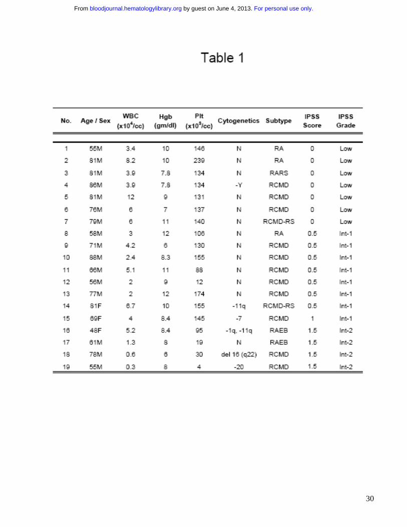
p38 MAPK is constitutively activated in low grade Myelodysplastic Syndromes
Bone marrows of patients with MDS were assessed for the activation / phosphorylation state of
p38 MAPK by immunohistochemistry. Patients were divided into lower (Low and Int-1 IPSS) and
higher (Int-2 and High IPSS scores) grade subtypes of MDS (Table 1). MDS bone marrow
samples were compared to age matched controls with non-MDS causes of cytopenias (1 with
iron deficiency anemia, 1 with B12 deficiency, 2 with chemotherapy related anemia, 2 with
chronic renal insufficiency and 1 with anemia associated with multiple chronic medical problems
with high ferritin in the absence of any dysplasia). Notable activation of p38 was seen in bone
marrow cells of all patients with low grade MDS, (Fig 1A) with a greater number of phospho-
p38-positive staining cells (Fig 1B) and significantly higher intensity of staining (Fig 1C) when
compared to controls. Activation of p38 was seen in all subtypes of low grade MDS examined [1
with Refractory anemia (RA), 1 with Refractory anemia with ringed sideroblasts (RARS) and 7
with Refractory cytopenias with multilineage dysplasia (RCMD)]. The level of activation was
significantly decreased in high grade cases and was comparable to controls.
Since p38 MAPK is ubiquitously expressed, we also investigated the phenotypes of bone
marrow cells that are expressing the activated kinase. Histological examination revealed that
p38 was activated in hematopoietic progenitors of all lineages including erythroid and myeloid
progenitors and even megakaryocytes. Staining with anti-CD3 antibody revealed very few
lymphocytes, most of which appeared to be phospho-p38 negative (data not shown).
Immunohistochemical staining with an antibody against total p38 MAPK was also performed to
determine any changes in p38 MAPK expression in MDS bone marrows when compared to
controls. No significant differences in either staining intensity or in the number of p38-positive
For personal use only. by guest on June 4, 2013. bloodjournal.hematologylibrary.orgFrom
12
cells were seen (Fig 1D,E) suggesting that p38 MAPK is overactivated but not overexpressed in
low grade MDS.
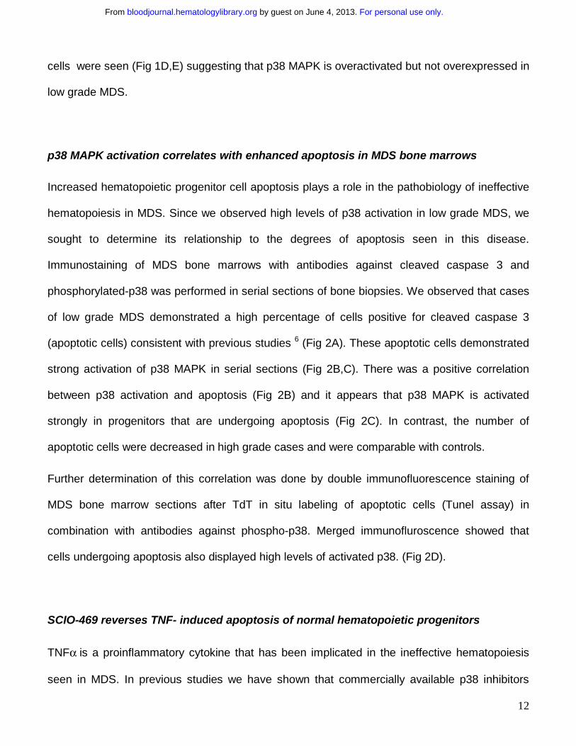
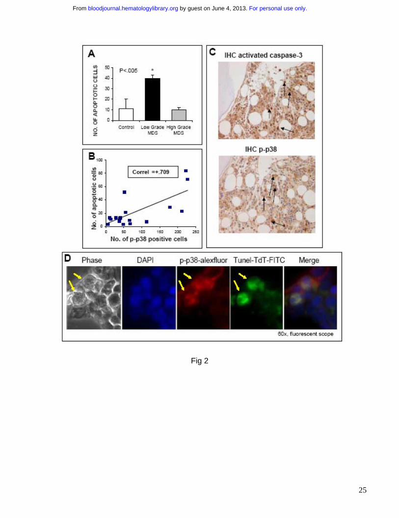
p38 MAPK activation correlates with enhanced apoptosis in MDS bone marrows
Increased hematopoietic progenitor cell apoptosis plays a role in the pathobiology of ineffective
hematopoiesis in MDS. Since we observed high levels of p38 activation in low grade MDS, we
sought to determine its relationship to the degrees of apoptosis seen in this disease.
Immunostaining of MDS bone marrows with antibodies against cleaved caspase 3 and
phosphorylated-p38 was performed in serial sections of bone biopsies. We observed that cases
of low grade MDS demonstrated a high percentage of cells positive for cleaved caspase 3
(apoptotic cells) consistent with previous studies 6 (Fig 2A). These apoptotic cells demonstrated
strong activation of p38 MAPK in serial sections (Fig 2B,C). There was a positive correlation
between p38 activation and apoptosis (Fig 2B) and it appears that p38 MAPK is activated
strongly in progenitors that are undergoing apoptosis (Fig 2C). In contrast, the number of
apoptotic cells were decreased in high grade cases and were comparable with controls.
Further determination of this correlation was done by double immunofluorescence staining of
MDS bone marrow sections after TdT in situ labeling of apoptotic cells (Tunel assay) in
combination with antibodies against phospho-p38. Merged immunofluroscence showed that
cells undergoing apoptosis also displayed high levels of activated p38. (Fig 2D).
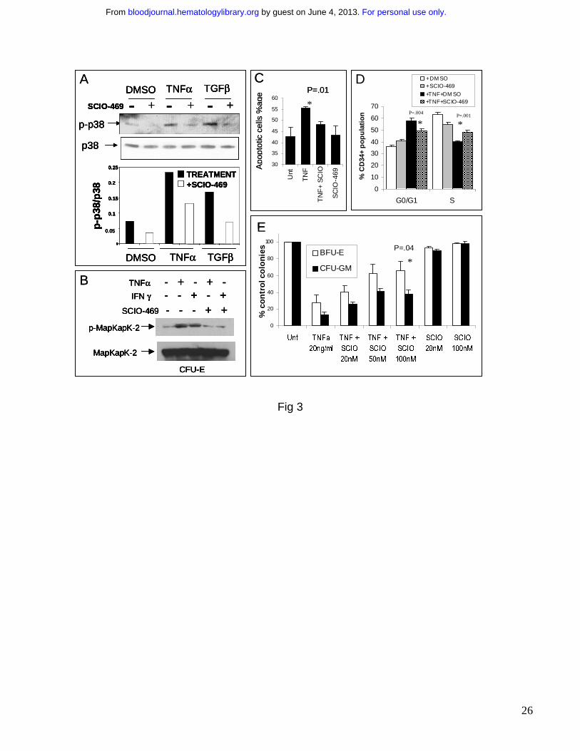
SCIO-469 reverses TNF- induced apoptosis of normal hematopoietic progenitors
TNFα is a proinflammatory cytokine that has been implicated in the ineffective hematopoiesis
seen in MDS. In previous studies we have shown that commercially available p38 inhibitors
For personal use only. by guest on June 4, 2013. bloodjournal.hematologylibrary.orgFrom
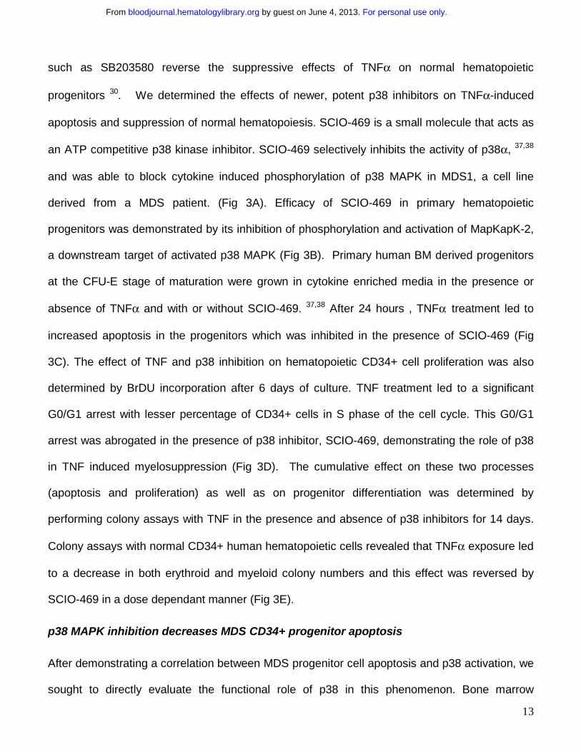
13
such as SB203580 reverse the suppressive effects of TNFα on normal hematopoietic
progenitors 30. We determined the effects of newer, potent p38 inhibitors on TNFα-induced
apoptosis and suppression of normal hematopoiesis. SCIO-469 is a small molecule that acts as
an ATP competitive p38 kinase inhibitor. SCIO-469 selectively inhibits the activity of p38α, 37,38
and was able to block cytokine induced phosphorylation of p38 MAPK in MDS1, a cell line
derived from a MDS patient. (Fig 3A). Efficacy of SCIO-469 in primary hematopoietic
progenitors was demonstrated by its inhibition of phosphorylation and activation of MapKapK-2,
a downstream target of activated p38 MAPK (Fig 3B). Primary human BM derived progenitors
at the CFU-E stage of maturation were grown in cytokine enriched media in the presence or
absence of TNFα and with or without SCIO-469. 37,38 After 24 hours , TNFα treatment led to
increased apoptosis in the progenitors which was inhibited in the presence of SCIO-469 (Fig
3C). The effect of TNF and p38 inhibition on hematopoietic CD34+ cell proliferation was also
determined by BrDU incorporation after 6 days of culture. TNF treatment led to a significant
G0/G1 arrest with lesser percentage of CD34+ cells in S phase of the cell cycle. This G0/G1
arrest was abrogated in the presence of p38 inhibitor, SCIO-469, demonstrating the role of p38
in TNF induced myelosuppression (Fig 3D). The cumulative effect on these two processes
(apoptosis and proliferation) as well as on progenitor differentiation was determined by
performing colony assays with TNF in the presence and absence of p38 inhibitors for 14 days.
Colony assays with normal CD34+ human hematopoietic cells revealed that TNFα exposure led
to a decrease in both erythroid and myeloid colony numbers and this effect was reversed by
SCIO-469 in a dose dependant manner (Fig 3E).
p38 MAPK inhibition decreases MDS CD34+ progenitor apoptosis
After demonstrating a correlation between MDS progenitor cell apoptosis and p38 activation, we
sought to directly evaluate the functional role of p38 in this phenomenon. Bone marrow
For personal use only. by guest on June 4, 2013. bloodjournal.hematologylibrary.orgFrom
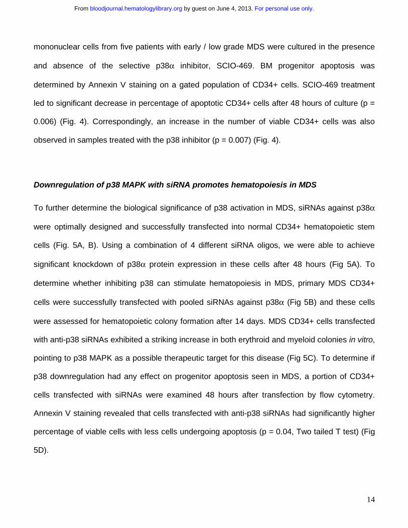
14
mononuclear cells from five patients with early / low grade MDS were cultured in the presence
and absence of the selective p38α inhibitor, SCIO-469. BM progenitor apoptosis was
determined by Annexin V staining on a gated population of CD34+ cells. SCIO-469 treatment
led to significant decrease in percentage of apoptotic CD34+ cells after 48 hours of culture (p =
0.006) (Fig. 4). Correspondingly, an increase in the number of viable CD34+ cells was also
observed in samples treated with the p38 inhibitor (p = 0.007) (Fig. 4).
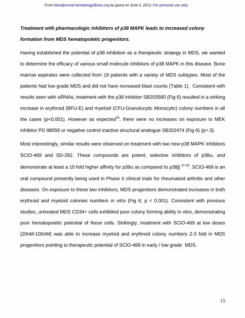
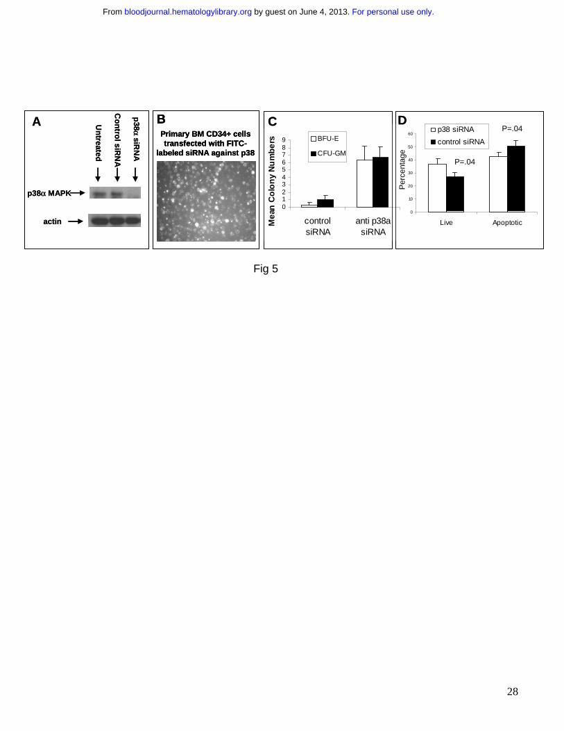
Downregulation of p38 MAPK with siRNA promotes hematopoiesis in MDS
To further determine the biological significance of p38 activation in MDS, siRNAs against p38α
were optimally designed and successfully transfected into normal CD34+ hematopoietic stem
cells (Fig. 5A, B). Using a combination of 4 different siRNA oligos, we were able to achieve
significant knockdown of p38α protein expression in these cells after 48 hours (Fig 5A). To
determine whether inhibiting p38 can stimulate hematopoiesis in MDS, primary MDS CD34+
cells were successfully transfected with pooled siRNAs against p38α (Fig 5B) and these cells
were assessed for hematopoietic colony formation after 14 days. MDS CD34+ cells transfected
with anti-p38 siRNAs exhibited a striking increase in both erythroid and myeloid colonies in vitro,
pointing to p38 MAPK as a possible therapeutic target for this disease (Fig 5C). To determine if
p38 downregulation had any effect on progenitor apoptosis seen in MDS, a portion of CD34+
cells transfected with siRNAs were examined 48 hours after transfection by flow cytometry.
Annexin V staining revealed that cells transfected with anti-p38 siRNAs had significantly higher
percentage of viable cells with less cells undergoing apoptosis (p = 0.04, Two tailed T test) (Fig
5D).
For personal use only. by guest on June 4, 2013. bloodjournal.hematologylibrary.orgFrom
15
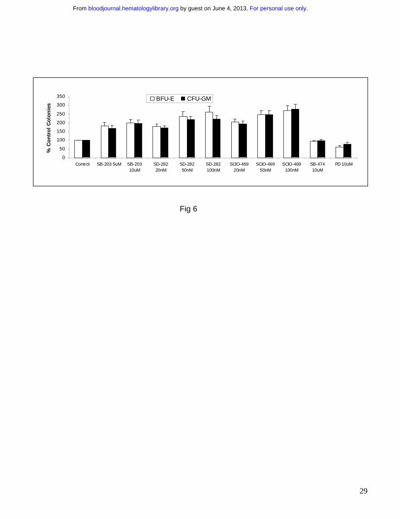
Treatment with pharmacologic inhibitors of p38 MAPK leads to increased colony
formation from MDS hematopoietic progenitors.
Having established the potential of p38 inhibition as a therapeutic strategy in MDS, we wanted
to determine the efficacy of various small molecule inhibitors of p38 MAPK in this disease. Bone
marrow aspirates were collected from 19 patients with a variety of MDS subtypes. Most of the
patients had low grade MDS and did not have increased blast counts (Table 1). Consistent with
results seen with siRNAs, treatment with the p38 inhibitor SB203580 (Fig 6) resulted in a striking
increase in erythroid (BFU-E) and myeloid (CFU-Granulocytic Monocytic) colony numbers in all
the cases (p<0.001). However as expected35, there were no increases on exposure to MEK
inhibitor PD 98059 or negative control inactive structural analogue SB202474 (Fig 6) (p=.3).
Most interestingly, similar results were observed on treatment with two new p38 MAPK inhibitors
SCIO-469 and SD-282. These compounds are potent, selective inhibitors of p38α, and
demonstrate at least a 10 fold higher affinity for p38α as compared to p38β 37-40. SCIO-469 is an
oral compound presently being used in Phase II clinical trials for rheumatoid arthritis and other
diseases. On exposure to these two inhibitors, MDS progenitors demonstrated increases in both
erythroid and myeloid colonies numbers in vitro (Fig 6; p < 0.001). Consistent with previous
studies, untreated MDS CD34+ cells exhibited poor colony forming ability in vitro, demonstrating
poor hematopoietic potential of these cells. Strikingly, treatment with SCIO-469 at low doses
(20nM-100nM) was able to increase myeloid and erythroid colony numbers 2-3 fold in MDS
progenitors pointing to therapeutic potential of SCIO-469 in early / low grade MDS.
For personal use only. by guest on June 4, 2013. bloodjournal.hematologylibrary.orgFrom
16
DISCUSSION
Myelodysplastic syndromes are groups of clonal hematopoietic disorders characterized by
refractory cytopenias with limited treatment options1. These disorders are common in the elderly
and impose a significant burden on healthcare resources. A stumbling block in the discovery of
treatments of this disease has been the heterogeneity observed in subsets of this disease and
the lack of a unifying pathophysiologic target. Our work has identified a kinase, p38 MAPK, that
is activated in bone marrow cells of a large number of MDS patients, even in those with different
chromosomal alterations. Inhibition of this pathway leads to in vitro stimulation of hematopoiesis
suggesting that p38 MAPK pathway is a functionally important inhibitory pathway in MDS. p38
MAPK is not found to be activated in normal bone marrows and consequently p38 inhibition
does not have significant stimulatory effects on hematopoietic progenitors derived from normal
bone marrows.
p38 MAPK was originally discovered as a stress signaling kinase, and recent work has
implicated it as an important mediator of apoptosis in neuronal, cardiac, immune and other cells
41-44. Studies have shown that activation of the p38 MAPK pathway can oppose the proliferative
effects of the Ras-Erk MAPK pathway and can lead to growth arrest and dormancy in tumors 45.
Consistent with its cytostatic properties in non hematopoietic cells, our work has demonstrated
an important role for p38 in cytokine mediated inhibition of human hematopoiesis 29,30. In fact, a
recent report implicates p38 in hematopoietic stem death induced by reactive oxygen species
and suggests that p38 inhibition may be beneficial in prolonging stem cell survival in ATM
knockout mice 46. This report validates our findings of the important role of this MAP kinase in
hematopoiesis and it appears p38 activation is downstream effector of many different cell death
pathways in hematopoietic stem cells.
For personal use only. by guest on June 4, 2013. bloodjournal.hematologylibrary.orgFrom
17
Our data establish a correlation between marrow p38 activation and apoptosis that is
characteristic of early stage MDS. The lack of activation of p38 in nutritional deficiency
anemias, chemotherapy induced anemias and other non MDS cytopenias points to the relative
specificity of p38 activation in low risk/Int-1 MDS. At this stage of the disease, both normal and
cytogenetically abnormal hematopoietic clones are found to exist in the marrow 47. It has been
shown that abnormal MDS progenitor clones are resistant to apoptosis and have higher levels of
anti apoptotic proteins Bcl-2 and Bax 6. Thus it is possible that p38 inhibition may prevent cell
death in the susceptible normal progenitors thereby rescuing normal hematopoiesis in the
early/low grade stage of this disease. Since most MDS cases are low risk and the morbidity
experienced is due to low blood counts, hematopoietic recovery is a major therapeutic goal in
treating these patients. The high numbers of apoptotic progenitors seen by us are consistent
with similar high percentages seen in other reports in low grade MDS5. With disease
progression towards high risk stages, normal progenitors gradually undergo apoptosis resulting
in a bone marrow comprised mainly of the resistant abnormal clones. Thus examination of the
marrow at late stages reveals a low apoptotic index with higher percentages of myeloblasts.
The low level of p38 activation correlates with the reduced apoptosis seen at this stage of the
disease. Instead, constitutive activation of the anti-apoptotic NFκB particularly in MDS
progenitors was observed in a majority of high risk MDS cases 48
MDS is highlighted by a stromal pathology of still unknown causes, which contributes to the
pervasive presence of pro-inflammatory cytokines in the bone marrow. Dysregulation of various
cytokines has been implicated in the pathogenesis of MDS 9,11,15-17. TNFα, TGFβ and IFN γ, α
are myelosuppressive cytokines that have been found to be elevated in serum as well as in the
bone marrow of MDS patients. Our earlier work has shown that p38 MAPK is activated by all of
these cytokines in primary hematopoietic progenitors. We have also shown that p38 MAPK
For personal use only. by guest on June 4, 2013. bloodjournal.hematologylibrary.orgFrom
18
functions at a critical signaling juncture that links upstream signaling pathways induced by
different immunosuppressive cytokines to a common effector pathway that leads to the inhibition
of normal hematopoietic progenitor growth 4,6. Various strategies have targeted inhibitory
cytokines in MDS with varying degrees of success. Clinical trials with monoclonal antibodies
against TNF (Infliximab) 19 and soluble TNF receptors (Etanercept) 18 have yielded
hematological responses in small series of patients. The immunomodulatory (IMID) compounds,
Thalidomide 49 and the newer more efficacious Lenalodimide 50 also target TNFα and have been
shown to reduce TNF mRNA 51 and alter the cytokine milieu. Lenalodimide has been recently
shown to be particularly efficacious of subsets of low grade MDS with 5q deletion 50. Small
molecule inhibitors of the VEGF receptor are presently in clinical trials for MDS. Even though
these approaches are encouraging, they have not been useful for a majority of patients with this
disease. Since p38 MAPK pathway is utilized by many cytokines, it is possible that targeting this
pathway may have wider clinical benefit than treatments targeting individual cytokines alone
(anti-TNF 18 and anti-VEGF strategies) in MDS. We observed that the efficiency of p38 inhibitors
in reversing TNF mediated inhibition was slightly greater in the erythroid lineage in normal CD34
progenitors. This may be due to the different degrees of involvement of p38 signaling in distinct
hematopoeitic growth and differentiation pathways. Nevertheless, our results demonstrate that
p38 inhibition affects both myeloid and erythroid progenitor growth suggesting the role of this
pathway in MDS disease subtypes involving different hematopoietic lineages.
SCIO-469 and SD-282 are novel small molecule inhibitors that occupy the ATP binding site on
the p38 kinase 37-39. Both of these are highly selective for p38α. Even though both the alpha and
beta isoforms are present in hematopoietic cells 52, our results with these inhibitors and siRNA
suggest that p38α is the dominant p38 isoform involved in hematopoietic suppression in MDS.
These results are consistent with our results previously observed with different classes of p38
For personal use only. by guest on June 4, 2013. bloodjournal.hematologylibrary.orgFrom

19
inhibitors in a small series of cases 35. The efficacy of SCIO-469 in inhibiting cytokine signaling
and production have provided preclinical rationale for its use in other cytokine modulated
diseases. It is presently being used in Phase II clinical trials in rheumatoid arthritis and multiple
myeloma. Our data supports the efforts to bring this agent in clinical trials for patients with MDS.
For personal use only. by guest on June 4, 2013. bloodjournal.hematologylibrary.orgFrom
20
FIGURE LEGENDS:
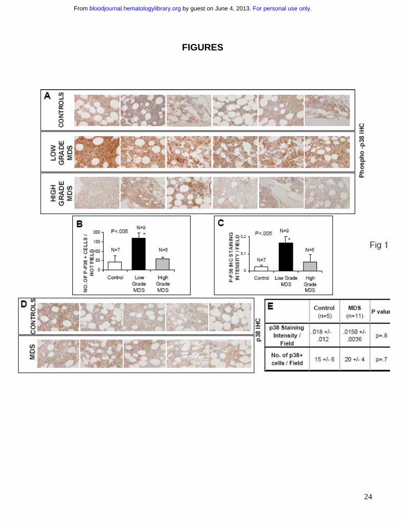
FIGURE 1: p38 MAPK is activated in Low Grade MDS Bone marrow (BM) biopsies from
patients with MDS and controls with non-MDS causes of cytopenias were fixed and
immunostained with antibody against phospho – p38 MAPK (A). Histological examination of 6
representative samples of Low and High grade MDS and controls revealed more intense
staining in low grade MDS samples. The quantification of p-p38 staining was analyzed by
counting the total number of positively stained cells (B) and by measuring intensity of the
positively stained cells (C) in 5- hot fields (defined as area of high density of p-p38 staining) and
aided by Image Pro Plus software. Two tailed T test shows significantly higher p38 activation
per hot field in Low Grade MDS samples. Differences in total p38 MAPK protein expression in 5
representative samples from each group were also evaluated in MDS and control bone marrows
by immunostaining with an antibody against p38 MAPK (D). Statistical analysis shows no
significant differences in number of positive cells and intensity of staining (E).
FIGURE 2: p38 activation correlates with apoptosis in Low Grade MDS. Bone marrow
biopsies from patients with MDS and non-MDS controls were fixed and immunostained with
antibody against cleaved / activated caspase-3. Number of apoptotic cells (cleaved caspase–3-
positive) were determined in cases of Low and High Risk MDS and compared to controls. (A)
Cases of Low Risk MDS had significantly higher number of apoptotic cells ( p< 0.05, Two tailed
T test). The number of apoptotic cells were correlated with numbers of cells positive for
phospho-p38. Pearson’s correlation coefficient was calculated using Microsoft Excel (B). Serial
sections of a representative MDS bone marrow sample show that cells undergoing apoptosis
exhibit greater activation of p38 MAPK (C). Bone marrow biopsies from a representative MDS
patient were stained with rabbit anti human phospho-p38 (red) and fluorescein-TdT (green) after
For personal use only. by guest on June 4, 2013. bloodjournal.hematologylibrary.orgFrom
21
In situ nucleotide labeling for apoptosis detection (Tunel assay) followed by goat anti-rabbit IgG
Alexa Fluor 568 secondary antibodies. Merged immunofluorescence shows apoptotic cells
exhibit activated p38 MAPK (positive for phospho-p38) (D).
Figure 3: p38 inhibitor SCIO-469 can reverse TNF mediated myelosuppression. MDS1
cells were pre-treated for 1 hr with vehicle (-) or 1.0 uM SCIO-469 (+) and then induced with
either 1 ng/mL TNFα or 5 ng/ml TGFβ for 30 min. p-p38 and total p38 levels were analyzed by
Western blotting. Bar graph represents p-p38 levels relative to total p38 in each sample (A).
Immunomagnetically selected bone marrow derived CD34+ cells were differentiated into
hematopoietic progenitors at the CFU-Erythroid stage of maturation as described before 30 .
These cells were treated with 20ng/ml TNFα or 10,000U/mL IFN γ in the presence and absence
of 100nM SCIO-469. Cell lysates were resolved by SDS-PAGE and immunoblotted with an
antibody against the phosphorylated form of MapKapK-2 (threonine 334). The same blot was
stripped and re-probed with an antibody against total MapKapK-2, to control for protein loading
(B). Primary bone marrow derived CD34+ cells were grown in cytokine enriched liquid media in
the presence and absence of 20 ng/mL TNFα and SCIO-469 (100 nM) for 24 hours. The
percentage of apoptotic and dead cells were determined by staining with mixture of Annexin V –
Alexa Fluor 488 and nucleic acid dye, Sytox green respectively (Vybrant Apoptosis Kit,
Molecular Probes) (C). Mean of three independent experiments showed significant decrease in
TNFα-mediated apoptosis in the presence of SCIO-469 (p = 0.01, Paired T Test). BM CD34+
progenitors were cultured with TPO, Flt3L and SCF with or without 20 ng/mL TNFα and in the
presence and absence of 500nM SCIO-469 for 6 days. BrDU incorporation was evaluated
against the amount of 7-AAD by flow cytometry to determine the percent subpopulation at each
cell cycle stage in a gated population of CD34+ cells. Results from 3 experiments were used to
For personal use only. by guest on June 4, 2013. bloodjournal.hematologylibrary.orgFrom
22
compare the proportion of cells in G0/G1 and S phase of cell cycle by using 2 tailed T test. (D).
Primary bone marrow derived CD34+ cells were cultured in methylcellulose in the presence and
absence of 20 ng/ml TNFα and SCIO-469. Colonies were scored on Day 14. Results are
expressed as Mean +/- S.E.M. of three independent experiments.(E) Treatment with SCIO-469
led to a significant reversal of TNF mediated myelosuppression (p = 0.04, T Test).
Figure 4: p38 inhibitor SCIO-469 can decrease apoptosis in MDS CD34+ progenitors. BM
mononuclear cells from patients with MDS were cultured in the presence and absence of 500nM
SCIO-469 for 48 hours. Apoptosis in gated population of CD34+ cells was determined by
Annexin V staining. Comparison of dot plots from 5 independent experiments demonstrate a
decrease in the percentage of Annexin V-positive CD34+ cells in samples treated with SCIO-
469 (A). MDS CD34+ progenitors from 5 patients demonstrate significantly greater viability and
decreased apoptosis after 48 hours of treatment with SCIO-469 (Paired two tailed T test).
Results are presented as Means +/- S.E.M. (B)
Figure 5: Downregulation of p38α by siRNA can stimulate hematopoiesis in MDS CD34+
progenitors. A mixture of 4 siRNAs against p38α were transfected in primary CD34+
hematopoietic progenitors using Mirus TKO transfection reagent. Western blotting showed
specific and significant decrease in total p38 protein levels (A). High transfection efficiency was
demonstrated by using fluorescent labeled siRNAs (B) MDS CD34+ cells were transfected with
either anti-p38α or control scrambled siRNAs and grown in vitro in methylcellulose with
cytokines. Colonies were scored on Day 14 and results were expressed as Means +/- S.E.M. of
3 independent experiments. Significantly higher number of both Myeloid (CFU-GM) and
Erythroid (BFU-E) colonies were observed in cells transfected with anti-p38 siRNAs (C). MDS
For personal use only. by guest on June 4, 2013. bloodjournal.hematologylibrary.orgFrom
23
CD34+ cells transfected with anti-p38α and scrambled control siRNAs were evaluated after 48
hours by Annexin V staining. Flow cytometry revealed significantly higher percentage of viable
cells (p = 0.045, T Test) and fewer number of apoptotic cells (p = 0.04, T Test) when transfected
with anti-p38α siRNA (D). Results are presented as Means +/- S.E.M. of 5 independent
experiments.
Figure 6: Pharmacological p38 inhibitors stimulate hematopoiesis in MDS CD34+
progenitors. MDS Bone marrow derived CD34+ cells from 19 patients were plated in
methylcellulose in the presence and absence of p38 inhibitors SB203580 (5 µM and 10 µM),
SD-282 and SCIO-469; inactive structural analogue SB202474 (10 µM) and Mek-1 inhibitor
PD98059 (10 µM). Colonies were scored at Day 14 and results were expressed as Means +/-
S.E.M of 19 independent experiments
For personal use only. by guest on June 4, 2013. bloodjournal.hematologylibrary.orgFrom
24
FIGURES
For personal use only. by guest on June 4, 2013. bloodjournal.hematologylibrary.orgFrom
25
Fig 2
For personal use only. by guest on June 4, 2013. bloodjournal.hematologylibrary.orgFrom
26
Fig 3
0
20
40
60
80
100
Unt TNFa
20ng/ml
TNF +
SCIO
20nM
TNF +
SCIO
50nM
TNF +
SCIO
100nM
SCIO
20nM
SCIO
100nM
% c
on
tro
l co
lon
ies
BFU-E
CFU-GM
A
B
p-p38
p38
TNFα TGFβ
SCIO-469 ++ + ---
0
0.05
0.1
0.15
0.2
0.25
p-p
38/p
38
TNFα TGFβ
DMSO
DMSO
TREATMENT+SCIO-469
p-p38
p38
TNFα TGFβ
SCIO-469 ++ + ---
0
0.05
0.1
0.15
0.2
0.25
p-p
38/p
38
TNFα TGFβ
DMSO
DMSO
TREATMENT+SCIO-469
TNFα - + - + -IFN γ - - + - +
SCIO-469 - - - + +
p-MapKapK-2
MapKapK-2
CFU-E
TNFα - + - + -IFN γ - - + - +
SCIO-469 - - - + +
p-MapKapK-2
MapKapK-2
CFU-E
*
P=.04
C
Apo
ptot
ic c
ells
%ag
e
30
35
40
45
50
55
60
Unt
TN
F
TN
F+
SC
IO
SC
IO-4
69
P=.01
*
E
0
10
20
30
40
50
60
70
G0/G1 S
% C
D34
+ p
op
ula
tio
n
+ DM SO+ SCIO-469
+TNF+DM SO+TNF+SCIO-469
D
* *P=.004
P=.001
0
20
40
60
80
100
Unt TNFa
20ng/ml
TNF +
SCIO
20nM
TNF +
SCIO
50nM
TNF +
SCIO
100nM
SCIO
20nM
SCIO
100nM
% c
on
tro
l co
lon
ies
BFU-E
CFU-GM
A
B
p-p38
p38
TNFα TGFβ
SCIO-469 ++ + ---
0
0.05
0.1
0.15
0.2
0.25
p-p
38/p
38
TNFα TGFβ
DMSO
DMSO
TREATMENT+SCIO-469
p-p38
p38
TNFα TGFβ
SCIO-469 ++ + ---
0
0.05
0.1
0.15
0.2
0.25
p-p
38/p
38
TNFα TGFβ
DMSO
DMSO
TREATMENT+SCIO-469
TNFα - + - + -IFN γ - - + - +
SCIO-469 - - - + +
p-MapKapK-2
MapKapK-2
CFU-E
TNFα - + - + -IFN γ - - + - +
SCIO-469 - - - + +
p-MapKapK-2
MapKapK-2
CFU-E
*
P=.04
C
Apo
ptot
ic c
ells
%ag
e
30
35
40
45
50
55
60
Unt
TN
F
TN
F+
SC
IO
SC
IO-4
69
P=.01
*
E
0
10
20
30
40
50
60
70
G0/G1 S
% C
D34
+ p
op
ula
tio
n
+ DM SO+ SCIO-469
+TNF+DM SO+TNF+SCIO-469
D
* *P=.004
P=.001
0
10
20
30
40
50
60
70
G0/G1 S
% C
D34
+ p
op
ula
tio
n
+ DM SO+ SCIO-469
+TNF+DM SO+TNF+SCIO-469
D
* *P=.004
P=.001
For personal use only. by guest on June 4, 2013. bloodjournal.hematologylibrary.orgFrom

27
Fig 4
100 101 102 103 104100
101
102
103
104
5.92
22.55
19.452.1
100 101 102 103 104100
101
102
103
104
15.1
0.22
4.580.2
100 101 102 103 104100
101
102
103
104
17.1
28.5 13.7
40.7
100 101 102 103 104100
101
102
103
104
11.2 30.6
11.846.4
100 101 102 103 104100
101
102
103
104
13.1 6.37
7.6272.9
Un
trea
ted
SC
IO-4
69P
rop.
Iodi
deMDS 1
100 101 102 103 104100
101
102
103
104
13.8 3.48
11.371.471.4
MDS 5MDS 3
100 101 102 103 104100
101
102
103
104
4.29
8.52
22.764.5
100 101 102 103 104100
101
102
103
104
.88
9.2
7.6182.3
Annexin-V
7.36 10.96
576.7
100 101 102 103 104100
101
102
103
104
N=6P=.015.51 30
10.354.1
100 101 102 103 104100
101
102
103
104
MDS 4MDS 2
0
20
40
60
80
100
Live Apoptotic Apoptotic+ Necrotic
Necrotic
%ag
e M
DS
CD
34+
Cel
ls
Untreated
SCIO-469p=.007
p=.7
p=.01
p=.006
BA
100 101 102 103 104100
101
102
103
104
5.92
22.55
19.452.1
100 101 102 103 104100
101
102
103
104
100 101 102 103 104100 101 102 103 104100 101 102 103 104100
101
102
103
104
100
101
102
103
104
100
101
102
103
104
5.92
22.55
19.452.1
100 101 102 103 104100
101
102
103
104
100 101 102 103 104100 101 102 103 104100 101 102 103 104100
101
102
103
104
100
101
102
103
104
100
101
102
103
104
15.1
0.22
4.580.2
100 101 102 103 104100
101
102
103
104
17.1
28.5 13.7
40.7
100 101 102 103 104100
101
102
103
104
100 101 102 103 104100 101 102 103 104100 101 102 103 104100
101
102
103
104
100
101
102
103
104
100
101
102
103
104
17.1
28.5 13.7
40.7
100 101 102 103 104100
101
102
103
104
11.2 30.6
11.846.4
100 101 102 103 104100
101
102
103
104
100 101 102 103 104100 101 102 103 104100 101 102 103 104100
101
102
103
104
100
101
102
103
104
100
101
102
103
104
11.2 30.6
11.846.4
100 101 102 103 104100
101
102
103
104
13.1 6.37
7.6272.9
100 101 102 103 104100
101
102
103
104
100 101 102 103 104100 101 102 103 104100 101 102 103 104100
101
102
103
104
100
101
102
103
104
100
101
102
103
104
13.1 6.37
7.6272.9
Un
trea
ted
SC
IO-4
69P
rop.
Iodi
deMDS 1
100 101 102 103 104100
101
102
103
104
100 101 102 103 104100 101 102 103 104100 101 102 103 104100
101
102
103
104
100
101
102
103
104
100
101
102
103
104
13.8 3.48
11.371.471.471.471.4
MDS 5MDS 3
100 101 102 103 104100
101
102
103
104
100 101 102 103 104100 101 102 103 104100 101 102 103 104100
101
102
103
104
100
101
102
103
104
100
101
102
103
104
4.29
8.52
22.764.5
100 101 102 103 104100
101
102
103
104
100 101 102 103 104100 101 102 103 104100 101 102 103 104100
101
102
103
104
100
101
102
103
104
100
101
102
103
104
.88
9.2
7.6182.3
Annexin-V
7.367.36 10.9610.96
5576.776.7
100 101 102 103 104100
101
102
103
104
100 101 102 103 104100 101 102 103 104100 101 102 103 104100
101
102
103
104
100
101
102
103
104
100
101
102
103
104
N=6P=.015.515.51 3030
10.310.354.154.1
100 101 102 103 104100
101
102
103
104
100 101 102 103 104100 101 102 103 104100 101 102 103 104100
101
102
103
104
100
101
102
103
104
100
101
102
103
104
MDS 4MDS 2
0
20
40
60
80
100
Live Apoptotic Apoptotic+ Necrotic
Necrotic
%ag
e M
DS
CD
34+
Cel
ls
Untreated
SCIO-469p=.007
p=.7
p=.01
p=.006
0
20
40
60
80
100
Live Apoptotic Apoptotic+ Necrotic
Necrotic
%ag
e M
DS
CD
34+
Cel
ls
Untreated
SCIO-469p=.007
p=.7
p=.01
p=.006
BA
F
or personal use only. by guest on June 4, 2013.
bloodjournal.hematologylibrary.org
From
28
Fig 5
0
10
20
30
40
50
60
Live Apoptotic
p38 siRNA
control siRNA
0123456789
controlsiRNA
anti p38asiRNA
Mea
n C
olo
ny
Nu
mb
ers BFU-E
CFU-GM
D
p38α MAPK
Co
ntro
l siRN
A
p38α
siRN
A
actin
A Un
treated
Primary BM CD34+ cells transfected with FITC-
labeled siRNA against p38
B
Per
cent
age
CP=.04
P=.04
0
10
20
30
40
50
60
Live Apoptotic
p38 siRNA
control siRNA
0123456789
controlsiRNA
anti p38asiRNA
Mea
n C
olo
ny
Nu
mb
ers BFU-E
CFU-GM
D
p38α MAPK
Co
ntro
l siRN
A
p38α
siRN
A
actin
A Un
treated
Primary BM CD34+ cells transfected with FITC-
labeled siRNA against p38
BPrimary BM CD34+ cells transfected with FITC-
labeled siRNA against p38
B
Per
cent
age
CP=.04
P=.04
For personal use only. by guest on June 4, 2013. bloodjournal.hematologylibrary.orgFrom
29
Fig 6
0
50
100
150
200
250
300
350
Control SB-203 5uM SB-20310uM
SD-28220nM
SD-28250nM
SD-282100nM
SCIO-46920nM
SCIO-46950nM
SCIO-469100nM
SB-47410uM
PD 10uM
% C
on
tro
l C
olo
nie
s
BFU-E CFU-GM
0
50
100
150
200
250
300
350
Control SB-203 5uM SB-20310uM
SD-28220nM
SD-28250nM
SD-282100nM
SCIO-46920nM
SCIO-46950nM
SCIO-469100nM
SB-47410uM
PD 10uM
% C
on
tro
l C
olo
nie
s
BFU-E CFU-GM
For personal use only. by guest on June 4, 2013. bloodjournal.hematologylibrary.orgFrom
30
For personal use only. by guest on June 4, 2013. bloodjournal.hematologylibrary.orgFrom
31
References:
1. Heaney ML, Golde DW. Myelodysplasia. N Engl J Med. 1999;340:1649-1660 2. Sanz GF, Sanz MA, Greenberg PL. Prognostic factors and scoring systems in myelodysplastic syndromes. Haematologica. 1998;83:358-368 3. Greenberg PL. Biologic nature of the myelodysplastic syndromes. Acta Haematol. 1987;78 Suppl 1:94-99 4. Greenberg P, Cox C, LeBeau MM, Fenaux P, Morel P, Sanz G, Sanz M, Vallespi T, Hamblin T, Oscier D, Ohyashiki K, Toyama K, Aul C, Mufti G, Bennett J. International scoring system for evaluating prognosis in myelodysplastic syndromes. Blood. 1997;89:2079-2088 5. Raza A, Gezer S, Mundle S, Gao XZ, Alvi S, Borok R, Rifkin S, Iftikhar A, Shetty V, Parcharidou A, et al. Apoptosis in bone marrow biopsy samples involving stromal and hematopoietic cells in 50 patients with myelodysplastic syndromes. Blood. 1995;86:268-276 6. Greenberg PL. Apoptosis and its role in the myelodysplastic syndromes: implications for disease natural history and treatment. Leuk Res. 1998;22:1123-1136 7. Westwood NB, Mufti GJ. Apoptosis in the myelodysplastic syndromes. Curr Hematol Rep. 2003;2:186-192 8. Ohshima K, Karube K, Shimazaki K, Kamma H, Suzumiya J, Hamasaki M, Kikuchi M. Imbalance between apoptosis and telomerase activity in myelodysplastic syndromes: possible role in ineffective hemopoiesis. Leuk Lymphoma. 2003;44:1339-1346 9. Allampallam K, Shetty V, Mundle S, Dutt D, Kravitz H, Reddy PL, Alvi S, Galili N, Saberwal GS, Anthwal S, Shaikh MW, York A, Raza A. Biological significance of proliferation, apoptosis, cytokines, and monocyte/macrophage cells in bone marrow biopsies of 145 patients with myelodysplastic syndrome. Int J Hematol. 2002;75:289-297 10. Claessens YE, Park S, Dubart-Kupperschmitt A, Mariot V, Garrido C, Chretien S, Dreyfus F, Lacombe C, Mayeux P, Fontenay M. Rescue of early stage myelodysplastic syndrome-deriving erythroid precursors by the ectopic expression of a dominant negative form of FADD. Blood. 2005 11. Kitagawa M, Saito I, Kuwata T, Yoshida S, Yamaguchi S, Takahashi M, Tanizawa T, Kamiyama R, Hirokawa K. Overexpression of tumor necrosis factor (TNF)-alpha and interferon (IFN)-gamma by bone marrow cells from patients with myelodysplastic syndromes. Leukemia. 1997;11:2049-2054 12. Peddie CM, Wolf CR, McLellan LI, Collins AR, Bowen DT. Oxidative DNA damage in CD34+ myelodysplastic cells is associated with intracellular redox changes and elevated plasma tumour necrosis factor-alpha concentration. Br J Haematol. 1997;99:625-631 13. Allampallam K, Shetty V, Hussaini S, Mazzoran L, Zorat F, Huang R, Raza A. Measurement of mRNA expression for a variety of cytokines and its receptors in bone marrows of patients with myelodysplastic syndromes. Anticancer Res. 1999;19:5323-5328 14. Mundle SD, Reza S, Ali A, Mativi Y, Shetty V, Venugopal P, Gregory SA, Raza A. Correlation of tumor necrosis factor alpha (TNF alpha) with high Caspase 3-like activity in myelodysplastic syndromes. Cancer Lett. 1999;140:201-207
For personal use only. by guest on June 4, 2013. bloodjournal.hematologylibrary.orgFrom
32
15. Aguayo A, Kantarjian H, Manshouri T, Gidel C, Estey E, Thomas D, Koller C, Estrov Z, O'Brien S, Keating M, Freireich E, Albitar M. Angiogenesis in acute and chronic leukemias and myelodysplastic syndromes. Blood. 2000;96:2240-2245 16. Zorat F, Shetty V, Dutt D, Lisak L, Nascimben F, Allampallam K, Dar S, York A, Gezer S, Venugopal P, Raza A. The clinical and biological effects of thalidomide in patients with myelodysplastic syndromes. Br J Haematol. 2001;115:881-894 17. Selleri C, Maciejewski JP, Catalano L, Ricci P, Andretta C, Luciano L, Rotoli B. Effects of cyclosporine on hematopoietic and immune functions in patients with hypoplastic myelodysplasia: in vitro and in vivo studies. Cancer. 2002;95:1911-1922 18. Deeg HJ, Gotlib J, Beckham C, Dugan K, Holmberg L, Schubert M, Appelbaum F, Greenberg P. Soluble TNF receptor fusion protein (etanercept) for the treatment of myelodysplastic syndrome: a pilot study. Leukemia. 2002;16:162-164 19. Raza A, Candoni A, Khan U, Lisak L, Tahir S, Silvestri F, Billmeier J, Alvi MI, Mumtaz M, Gezer S, Venugopal P, Reddy P, Galili N. Remicade as TNF suppressor in patients with myelodysplastic syndromes. Leuk Lymphoma. 2004;45:2099-2104 20. Maciejewski JP, Risitano AM, Sloand EM, Wisch L, Geller N, Barrett JA, Young NS. A pilot study of the recombinant soluble human tumour necrosis factor receptor (p75)-Fc fusion protein in patients with myelodysplastic syndrome. Br J Haematol. 2002;117:119-126 21. Greenberg P. Treatment of myelodysplastic syndrome with agents interfering with inhibitory cytokines. Ann Rheum Dis. 2001;60 Suppl 3:iii41-42 22. Uddin S, Lekmine F, Sharma N, Majchrzak B, Mayer I, Young PR, Bokoch GM, Fish EN, Platanias LC. The Rac1/p38 mitogen-activated protein kinase pathway is required for interferon alpha-dependent transcriptional activation but not serine phosphorylation of Stat proteins. J Biol Chem. 2000;275:27634-27640 23. Romerio F, Zella D. MEK and ERK inhibitors enhance the anti-proliferative effect of interferon-alpha2b. Faseb J. 2002;16:1680-1682 24. Johnson GL, Lapadat R. Mitogen-activated protein kinase pathways mediated by ERK, JNK, and p38 protein kinases. Science. 2002;298:1911-1912 25. Kumar S, Boehm J, Lee JC. p38 MAP kinases: key signalling molecules as therapeutic targets for inflammatory diseases. Nat Rev Drug Discov. 2003;2:717-726 26. Parmar S, Katsoulidis E, Verma A, Li Y, Sassano A, Lal L, Majchrzak B, Ravandi F, Tallman MS, Fish EN, Platanias LC. Role of the p38 mitogen-activated protein kinase pathway in the generation of the effects of imatinib mesylate (STI571) in BCR-ABL-expressing cells. J Biol Chem. 2004;279:25345-25352 27. Mayer IA, Verma A, Grumbach IM, Uddin S, Lekmine F, Ravandi F, Majchrzak B, Fujita S, Fish EN, Platanias LC. The p38 MAPK pathway mediates the growth inhibitory effects of interferon-alpha in BCR-ABL-expressing cells. J Biol Chem. 2001;276:28570-28577 28. Platanias LC. Map kinase signaling pathways and hematologic malignancies. Blood. 2003;101:4667-4679 29. Verma A, Deb DK, Sassano A, Uddin S, Varga J, Wickrema A, Platanias LC. Activation of the p38 mitogen-activated protein kinase mediates the suppressive effects of type I interferons and transforming growth factor-beta on normal hematopoiesis. J Biol Chem. 2002;277:7726-7735 30. Verma A, Deb DK, Sassano A, Kambhampati S, Wickrema A, Uddin S, Mohindru M, Van Besien K, Platanias LC. Cutting edge: activation of the p38 mitogen-activated protein kinase signaling pathway mediates cytokine-induced hemopoietic suppression in aplastic anemia. J Immunol. 2002;168:5984-5988 31. Uddin S, Majchrzak B, Woodson J, Arunkumar P, Alsayed Y, Pine R, Young PR, Fish EN, Platanias LC. Activation of the p38 mitogen-activated protein kinase by type I interferons. J Biol Chem. 1999;274:30127-30131
For personal use only. by guest on June 4, 2013. bloodjournal.hematologylibrary.orgFrom
33
32. Welsh JP, Rutherford TR, Flynn J, Foukaneli T, Gordon-Smith EC, Gibson FM. In vitro effects of interferon-gamma and tumor necrosis factor-alpha on CD34+ bone marrow progenitor cells from aplastic anemia patients and normal donors. Hematol J. 2004;5:39-46 33. Dufour C, Corcione A, Svahn J, Haupt R, Poggi V, Beka'ssy AN, Scime R, Pistorio A, Pistoia V. TNF-alpha and IFN-gamma are overexpressed in the bone marrow of Fanconi anemia patients and TNF-alpha suppresses erythropoiesis in vitro. Blood. 2003;102:2053-2059 34. Dufour C, Corcione A, Svahn J, Haupt R, Battilana N, Pistoia V. Interferon gamma and tumour necrosis factor alpha are overexpressed in bone marrow T lymphocytes from paediatric patients with aplastic anaemia. Br J Haematol. 2001;115:1023-1031 35. Katsoulidis E, Li Y, Yoon P, Sassano A, Altman J, Kannan-Thulasiraman P, Balasubramanian L, Parmar S, Varga J, Tallman MS, Verma A, Platanias LC. Role of the p38 MAP Kinase in cytokine mediated hematopoietic suppression in myelodysplastic syndrome. Cancer Res. 2005; 65(19):9029-37. 36. Wickrema A, Uddin S, Sharma A, Chen F, Alsayed Y, Ahmad S, Sawyer ST, Krystal G, Yi T, Nishada K, Hibi M, Hirano T, Platanias LC. Engagement of Gab1 and Gab2 in erythropoietin signaling. J Biol Chem. 1999;274:24469-24474 37. Nikas SN, Drosos AA. SCIO-469 Scios Inc. Curr Opin Investig Drugs. 2004;5:1205-1212 38. Hideshima T, Podar K, Chauhan D, Ishitsuka K, Mitsiades C, Tai YT, Hamasaki M, Raje N, Hideshima H, Schreiner G, Nguyen AN, Navas T, Munshi NC, Richardson PG, Higgins LS, Anderson KC. p38 MAPK inhibition enhances PS-341 (bortezomib)-induced cytotoxicity against multiple myeloma cells. Oncogene. 2004;23:8766-8776 39. Svensson CI, Marsala M, Westerlund A, Calcutt NA, Campana WM, Freshwater JD, Catalano R, Feng Y, Protter AA, Scott B, Yaksh TL. Activation of p38 mitogen-activated protein kinase in spinal microglia is a critical link in inflammation-induced spinal pain processing. J Neurochem. 2003;86:1534-1544 40. Lim MY, Wang H, Kapoun AM, O'Connell M, O'Young G, Brauer HA, Luedtke GR, Chakravarty S, Dugar S, Schreiner GS, Protter AA, Higgins LS. p38 Inhibition attenuates the pro-inflammatory response to C-reactive protein by human peripheral blood mononuclear cells. J Mol Cell Cardiol. 2004;37:1111-1114 41. New L, Han J. The p38 MAP Kinase Pathway and Its Biological Function. Trends Cardiovasc Med. 1998;8:220-228 42. Wang Y, Huang S, Sah VP, Ross J, Jr., Brown JH, Han J, Chien KR. Cardiac muscle cell hypertrophy and apoptosis induced by distinct members of the p38 mitogen-activated protein kinase family. J Biol Chem. 1998;273:2161-2168 43. Xia Z, Dickens M, Raingeaud J, Davis RJ, Greenberg ME. Opposing effects of ERK and JNK-p38 MAP kinases on apoptosis. Science. 1995;270:1326-1331 44. Ichijo H, Nishida E, Irie K, ten Dijke P, Saitoh M, Moriguchi T, Takagi M, Matsumoto K, Miyazono K, Gotoh Y. Induction of apoptosis by ASK1, a mammalian MAPKKK that activates SAPK/JNK and p38 signaling pathways. Science. 1997;275:90-94 45. Aguirre-Ghiso JA, Ossowski L, Rosenbaum SK. Green fluorescent protein tagging of extracellular signal-regulated kinase and p38 pathways reveals novel dynamics of pathway activation during primary and metastatic growth. Cancer Res. 2004;64:7336-7345 46. Ito K, Hirao A, Arai F, Takubo K, Matsuoka S, Miyamoto K, Ohmura M, Naka K, Hosokawa K, Ikeda Y, Suda T. Reactive oxygen species act through p38 MAPK to limit the lifespan of hematopoietic stem cells. Nat Med. 2006;12:446-451 47. Legare RD, Gilliland DG. Myelodysplastic syndrome. Curr Opin Hematol. 1995;2:283-292 48. Braun T, Carvalho G, Coquelle A, Vozenin MC, Lepelley P, Hirsch F, Kiladjian JJ, Ribrag V, Fenaux P, Kroemer G. NF-kappaB constitutes a potential therapeutic target in high-risk myelodysplastic syndrome. Blood. 2006;107:1156-1165
For personal use only. by guest on June 4, 2013. bloodjournal.hematologylibrary.orgFrom
34
49. Raza A, Meyer P, Dutt D, Zorat F, Lisak L, Nascimben F, du Randt M, Kaspar C, Goldberg C, Loew J, Dar S, Gezer S, Venugopal P, Zeldis J. Thalidomide produces transfusion independence in long-standing refractory anemias of patients with myelodysplastic syndromes. Blood. 2001;98:958-965 50. List A, Kurtin S, Roe DJ, Buresh A, Mahadevan D, Fuchs D, Rimsza L, Heaton R, Knight R, Zeldis JB. Efficacy of lenalidomide in myelodysplastic syndromes. N Engl J Med. 2005;352:549-557 51. Verma A, List A. Cytokine targets in Myelodysplastic syndromes. Curr Hematol Rep. 2005;In Print 52. Uddin S, Ah-Kang J, Ulaszek J, Mahmud D, Wickrema A. Differentiation stage-specific activation of p38 mitogen-activated protein kinase isoforms in primary human erythroid cells. Proc Natl Acad Sci U S A. 2004;101:147-152
For personal use only. by guest on June 4, 2013. bloodjournal.hematologylibrary.orgFrom


