Embed Size (px)
Citation preview
Journal of
Clinical Medicine
Review
Indirect Calorimetry in Clinical Practice
Marta Delsoglio 1 , Najate Achamrah 2, Mette M. Berger 3 and Claude Pichard 1,*1 Clinical Nutrition, Geneva University Hospital (HUG), 1205 Geneva, Switzerland2 Nutrition Department, Rouen University Hospital Center, 76000 Rouen, France3 Service of Intensive Care Medicine & Burns, University of Lausanne Hospitals (CHUV),
1005 Lausanne, Switzerland* Correspondence: [email protected]; Tel.: +41-(0)22-372-9345; Fax: +41-(0)22-372-9363
Received: 9 August 2019; Accepted: 30 August 2019; Published: 5 September 2019�����������������
Abstract: Indirect calorimetry (IC) is considered as the gold standard to determine energy expenditure,by measuring pulmonary gas exchanges. It is a non-invasive technique that allows clinicians topersonalize the prescription of nutrition support to the metabolic needs and promote a better clinicaloutcome. Recent technical developments allow accurate and easy IC measurements in spontaneouslybreathing patients as well as in those on mechanical ventilation. The implementation of IC inclinical routine should be promoted in order to optimize the cost–benefit balance of nutrition therapy.This review aims at summarizing the latest innovations of IC as well as the clinical indications,benefits, and limitations.
Keywords: indirect calorimetry; indirect calorimeter; resting energy expenditure; nutrition therapy
1. Introduction
The accurate determination of patients’ energy needs is required to optimize nutritional supportand to reduce the deleterious effects of under- and over-feeding [1]. However nutritional requirementsmay be difficult to predict, especially in patients with acute or chronic conditions where many factors(e.g., stress, brain activity, endocrine profile, inflammation status, feeding state, drugs, etc.) affect theirresting energy expenditure (REE). REE represents the energy expended by the body during a 24 hnon-active period to maintain involuntary functions such as substrate turnover, respiration, cardiacoutput, and body temperature regulation [2]. In healthy sedentary adult subjects REE constitutestwo-thirds of the total energy expenditure (TEE) which includes energy needed for nonobligatoryexpenses such as physical activity and diet-induced thermogenesis. This assumption is probably notentirely correct in sick patients where continuous nutrition, treatments and diseased-related stressincrease, or decrease REE: extreme variations among individuals can be observed within the sameday [3].
Indirect calorimetry (IC) is considered as the gold standard to measure REE, by measuring oxygenconsumption (VO2) and carbon dioxide production (VCO2). Apart from REE, other parameters canbe derived from IC, such as substrate (carbohydrates, fat, and protein) utilization. Indeed, the ratiobetween VCO2 and VO2 ( VCO2
VO2) defines the respiratory quotient (RQ) that corresponds to the substrate
use. The complete oxidation of glucose generates a RQ value of 1.0, while a RQ of 0.7 is indicative of amixed substrate oxidation [2].
Recent technical developments of indirect calorimeters allow accurate, non-invasive, and easyIC measurements in spontaneously breathing patients as well as in those on mechanical ventilation.Recent trials have shown IC allows clinicians to personalize the prescription of nutrition support tothe metabolic needs and to monitor the metabolic response to nutrition therapy promoting a betterclinical outcome in acutely ill patients [4–8]. These results have increased the interest for IC as a toolfor improving the routine nutritional evaluation and prescription.
J. Clin. Med. 2019, 8, 1387; doi:10.3390/jcm8091387 www.mdpi.com/journal/jcm
J. Clin. Med. 2019, 8, 1387 2 of 19
This review aims to summarize the latest innovations of IC as well as the clinical indications,benefits, and limitations of the technique.
2. Indications
Equations predicting REE are relatively reliable in healthy subjects. Contrariwise, in case of diseaseor trauma, REE is influenced by many factors with synergic or antagonist impact (Table 1) [1,2,9].In these conditions, predictive equations are largely inaccurate [10]. As a consequence, IC is mainlyindicated in three different scenarios: (a) clinical conditions significantly modifying REE; (b) failure of anutrition support based on predicted energy needs to maintain or restore body weight; (c) acute criticalillness associated with large and dynamic changes of metabolic stress level [11]. The latter indicationrequires special caution, as the IC results will reflect the patient’s instability: repeated IC studiesare required to observe the evolution of the metabolic response to hemodynamic instability, fever,surgery, weaning from mechanical ventilation, etc. A single IC is rarely sufficient, thereby repeatedmeasurements every second or third day are needed in patients with rapid changes of their clinicalstatus [12–14].
Table 1. Factors influencing resting energy expenditure. Adapted from [1,2,9].
Effects on REE Factors
↑
• Burns• Hyperventilation• Hyperthermia• Hyperthyroidism, pheochromocytoma• Inflammation (interleukins,
interferons, tumor necrosis factors etc.)• Metabolic acidosis
• Morbid obesity• Overfeeding• Physical agitation• Sepsis• Stress (epinephrine, cortisol,
glucagon etc.)
↓
• Coma/deep sleep• General anesthesia• Heavy sedation• Hypothermia• Hypothyroidism• Hypoventilation
• Gluconeogenesis• Metabolic alkalosis• Paralysis• Sarcopenia, cachexia• Starvation/underfeeding/ketosis
Performing IC in clinical routine is recommended not only to set an optimal nutrition therapy,but also to monitor the results of the nutritional interventions, in order to avoid complications ofinappropriate nutrition, i.e., of under- or over-feeding.
3. Practicalities
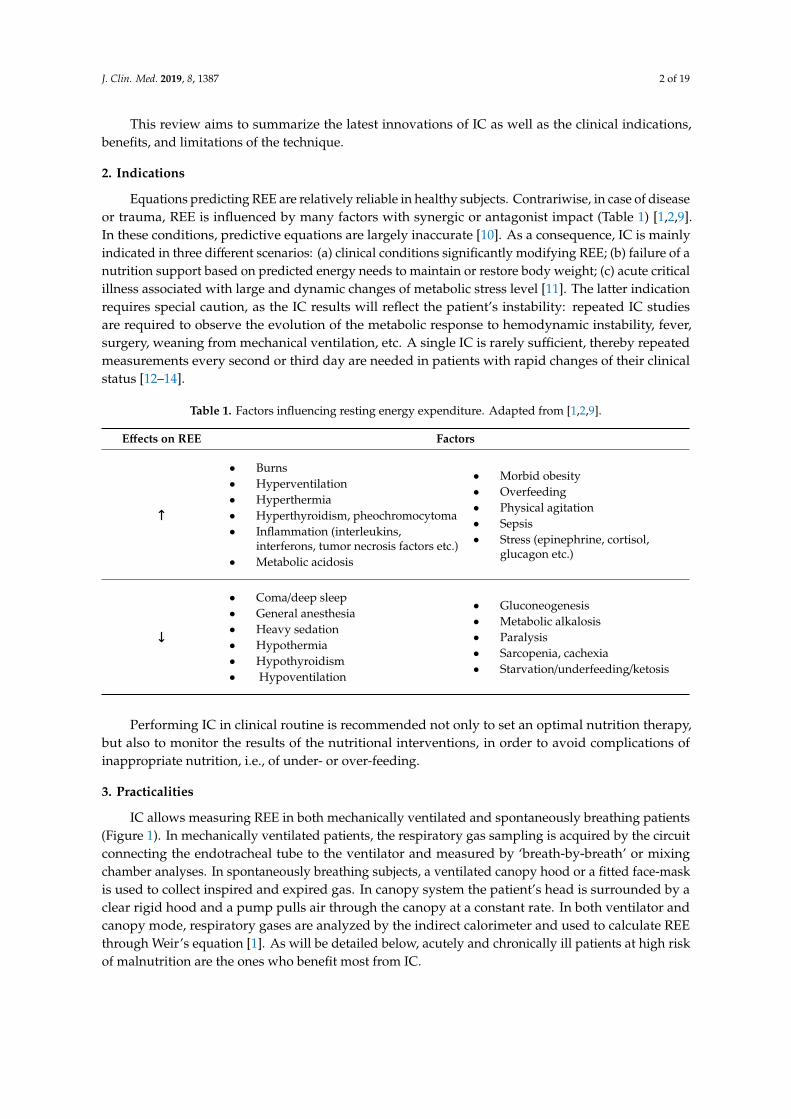
IC allows measuring REE in both mechanically ventilated and spontaneously breathing patients(Figure 1). In mechanically ventilated patients, the respiratory gas sampling is acquired by the circuitconnecting the endotracheal tube to the ventilator and measured by ‘breath-by-breath’ or mixingchamber analyses. In spontaneously breathing subjects, a ventilated canopy hood or a fitted face-maskis used to collect inspired and expired gas. In canopy system the patient’s head is surrounded by aclear rigid hood and a pump pulls air through the canopy at a constant rate. In both ventilator andcanopy mode, respiratory gases are analyzed by the indirect calorimeter and used to calculate REEthrough Weir’s equation [1]. As will be detailed below, acutely and chronically ill patients at high riskof malnutrition are the ones who benefit most from IC.
J. Clin. Med. 2019, 8, 1387 3 of 19
Figure 1. Indirect calorimetry on mechanically ventilated patient (A) and on spontaneous breathingpatient in canopy mode (B). In mechanical ventilation the gas sampling is acquired by the circuitconnecting the endotracheal tube to the ventilator and measured by ‘breath-by-breath’ or mixingchamber analyses. In spontaneous breathing mode, the subject is placed under a clear canopy with aplastic drape to avoid air leakage. Breath exchanges are collected by the calorimeter for gas analysisand enable calculation of REE using Weir’s equation (REE (kcal/day) = [(VO2 × 3.941) + (VCO2 × 1.11)]× 1440).
3.1. Acute Diseases
Metabolic responses to shock and injury were first described by Sir Cuthbertson [15]. According tohis investigations, there is a short ebb phase starting immediately after a traumatic shock and followedby a flow phase of longer duration after 3 to 10 days (Figure 2) [16]. The ebb phase is characterized bya decrease in metabolic rate, oxygen consumption, body temperature, and enzymatic activity. The flowphase, on the contrary, is marked by an increased catabolism, with a high oxygen consumption and anelevated REE rate.

J. Clin. Med. 2019, 8, 1387 4 of 19
Figure 2. Metabolic response to injury proposed by Cuthbertson et al. A short ebb phase characterizedby hypometabolism occurs immediately after the injury and is characterized by a decrease in metabolicrate, oxygen consumption, body temperature, and enzymatic activity. The ebb phase is followed by alonger hypermetabolic flow phase marked by an increased catabolism, with a high oxygen consumptionand an elevated REE rate. Reused with permission from [16].
Recent studies have supported this pattern of longitudinal changes in REE in acutely illpatients [17–19]. The degree of increase from normal REE may reflect the severity of the metabolicresponse to the injury. However, the interaction between natural course of disease, individualinflammatory and immune system response, and medical treatments make it difficult for the clinicianto evaluate caloric needs. For these reasons, IC is the only available tool enabling the individualassessment of the patients’ energy needs, and to ensure precise nutritional interventions in acuteconditions such as post-surgery period, pancreatitis, kidney injury, and sepsis.
The post-operative period of uncomplicated surgery has been associated with a modest 7%increase in energy metabolism as the effect of the surgical operation itself, that cannot be predictedby static equations [20]. Non-septic patients with acute pancreatitis presented a hypermetabolic statewith a REE increase of 120 ± 11% compared to predicted [21]. Energy metabolism in patients withrenal failure has been studied as well. Acute kidney injury (AKI) has not been shown to affect REE,but concomitant conditions, such as sepsis, mostly play role in the hypermetabolism found in thesepatients [22,23]. The presence of hypermetabolism has also been associated with lower age and highervasoactive drug dose [24] and no predictive formula was able to correctly calculate energy needs inAKI patients compared to IC [25]. Sepsis is characterized by a hyperdynamic cardiovascular responseagainst infection and has been reported to increase REE differently among patients with uncomplicatedsepsis, sepsis syndrome, and septic shock (mean REE +55 ± 14%, +24 ± 12%, and +2 ± 24%) [26]. Ahigher REE in severe sepsis adult patients has also been associated with higher mortality [27], howeverfurther studies should investigate the effect of specific individual factors on metabolic evolution duringsepsis and patient outcome.
Critical Illness
Critical illness is frequently associated with a hypermetabolic state related to the activation ofcatabolic hormones and resulting in elevated REE compared to healthy subjects. However, iatrogenicfactors such as beta-blockers, analgesics, and sedatives may attenuate the response and even induce
J. Clin. Med. 2019, 8, 1387 5 of 19
a hypometabolic state. Prolonged bed rest, atrophy of the metabolically active lean body mass, andmechanical ventilation have also been reported to decrease REE [1]. In patients with multiple organfailure, the loss of lean body mass is very rapid and resulted in 22% loss in 10 days [28] and nopredictive equation showed good agreement compared to REE measured by IC [29].
Trauma patients showed hypermetabolism even when heavily sedated or medically paralyzed,however those with head injury on neuromuscular blockade or in a barbiturate coma showed reducedREE compared to similar patients without these agents [30]. Brain trauma was found to increase REEwith high variability among different studies, ranging from 87% to 200% compared to predicted duringthe first 30 days post-injury; surprisingly, in patients admitted with a brain death diagnosis, the valueranged from 75% to 200% compared to predicted during the first 7 days [31].
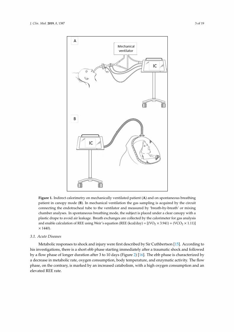
Similarly, post-burn hypermetabolism was shown to increase REE as high as 100% above normal asconsequence of a strong catabolic response mediated by endogenous catecholamines and inflammatorycytokines. Nevertheless, this hypermetabolic state is a direct effect of burn trauma, its persistence leadsto severe septic complications, multiple organ failure, and higher mortality [16]. Nutritional follow upin burn patients shows a highly dynamic and variable REE up to 160 days after injury (Figure 3) [32].
Figure 3. Evolution of measured REE by IC (blue), Toronto predictive equation (dashed blue), deliveredenergy (black), VO2 (red 4), and VCO2 (purple ) in a young man weighing 99 kg upon admissionwith major burns covering 85% body surface over 160 days. The REE variations were important overtime particularly during the early phase (weight gain due to fluid resuscitation was 36 kg by day 3), andparalleled the loss of body weight, i.e., of lean body mass (−31 kg after 3 months, with slow recovery).The REE value on day 1 corresponds to the Harris & Benedict prediction of basal EE. The figure alsoshows the reasonable precision of the Toronto equation, and how difficult it is to feed to measured ICvalue during the first 14 days. Adapted from [32].
The accurate determination of energy needs and the prevention of energy imbalance are essentialin critically ill patients to avoid the harmful consequences of inadequate feeding. Underfeedinghas been shown to increase hospital length of stay, infections, organ failure, to prolong mechanicalventilation, and to increase mortality, while overfeeding has been associated with hyperglycemia,hypertriglyceridemia, hepatic steatosis, azotemia, hypercapnia, and increased mortality [33]. In longstaying patients with dynamic clinical conditions, IC should be repeated to monitor their nutritionalrequirements and avoid energy imbalance [34,35]. Optimal energy delivery targeting REE measured

J. Clin. Med. 2019, 8, 1387 6 of 19
by IC seems to be significantly associated with reduced mortality, stressing the importance of thistechnique to assess caloric needs in ICU patients (Figure 4) [17].
Figure 4. Association of delivered calories/resting energy expenditure (REE) percent by indirectcalorimetry (IC) with 60-day mortality in different models: the authors recalculated their original2016 data to integrate the fact that energy delivery increased progressively during the initial 2–3 days,reducing the mean value in stays <5 days. The lowest ICU mortality was observed when percentof delivered calories by REE obtained by IC was 80% (excluding first two feeding days) and 75%(with >10 evaluable nutrition days) (p < 0.05). On the contrary, increments of the ratio above thatpoint—specifically >110%—were associated with increasing mortality (p < 0.05). Reproduced withpermission (http://creativecommons.org/licenses/by/4.0/) [17].
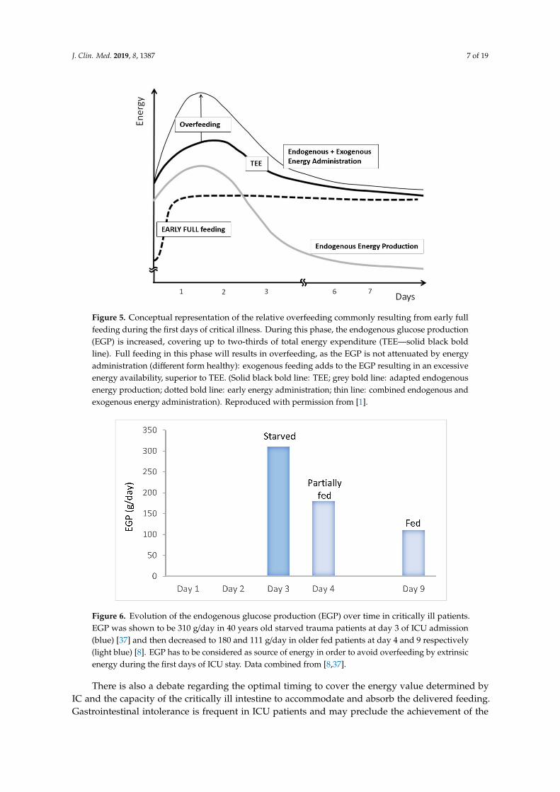
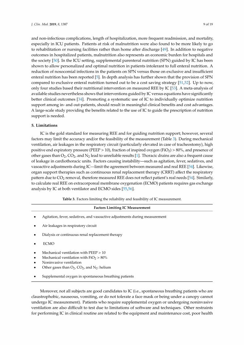
The question which remains debated is the timing from which the measured REE should beprescribed as energy goal. Considering the importance of the early endogenous energy production,which is not suppressed by nutrition in the critically ill [36], feeding to measured value may resultin overfeeding (Figure 5) [1]. Moreover, during the early phase of disease, catecholamines and themultiple treatments needed in these patients generate high instability and directly impact on measuredREE. Tappy et al. showed that, in young starved trauma patients, the endogenous glucose production(EGP) was about 310 g/day (Figure 6), while the measured REE in these patients was 1830 kcal: feedingto the mean measured REE would clearly result in early overfeeding [37]. The figure also showsthat endogenous glucose production continues in the sickest patients for many days even duringfeeding [8] (Figure 6). Therefore, the crude REE value provided by IC requires a careful interpretationto adequately prescribe exogenous energy supply.
J. Clin. Med. 2019, 8, 1387 7 of 19
Figure 5. Conceptual representation of the relative overfeeding commonly resulting from early fullfeeding during the first days of critical illness. During this phase, the endogenous glucose production(EGP) is increased, covering up to two-thirds of total energy expenditure (TEE—solid black boldline). Full feeding in this phase will results in overfeeding, as the EGP is not attenuated by energyadministration (different form healthy): exogenous feeding adds to the EGP resulting in an excessiveenergy availability, superior to TEE. (Solid black bold line: TEE; grey bold line: adapted endogenousenergy production; dotted bold line: early energy administration; thin line: combined endogenous andexogenous energy administration). Reproduced with permission from [1].
Figure 6. Evolution of the endogenous glucose production (EGP) over time in critically ill patients.EGP was shown to be 310 g/day in 40 years old starved trauma patients at day 3 of ICU admission(blue) [37] and then decreased to 180 and 111 g/day in older fed patients at day 4 and 9 respectively(light blue) [8]. EGP has to be considered as source of energy in order to avoid overfeeding by extrinsicenergy during the first days of ICU stay. Data combined from [8,37].
There is also a debate regarding the optimal timing to cover the energy value determined byIC and the capacity of the critically ill intestine to accommodate and absorb the delivered feeding.Gastrointestinal intolerance is frequent in ICU patients and may preclude the achievement of the

J. Clin. Med. 2019, 8, 1387 8 of 19
predefined calorie target due to the incapability of absorbing supplied nutrition [38]. The best feedingroute for each patient has to be assessed in order to limit the risk of stressing the intestine andcumulative caloric deficit [39].
3.2. Chronic Diseases
Energy requirements may be even more difficult to predict in patients with chronic conditionsdue to the large individual variations in REE. Both hyper- and hypo-metabolism have been shown inchronic pathologies due to alterations in metabolism, lean body mass, organ function, and presence ofinflammation. The most common pathologies with important REE alterations are described below andsummarized in Table 2.
Table 2. Common chronic pathologies with effects on resting energy expenditure
Condition Effect on REE
Anorexia nervosa ↓ Low energy intake and reduced lean body mass
Cancer ↑
↓
Cancer growth and inflammationProgressive reduction of lean body mass
Chronic kidney diseases ↑
↓
Metabolic acidosis and inflammationAcute and chronic renal failure
Chronic obstructive pulmonary disease ↑ Increased respiratory efforts
Diabetes ↑ Increased metabolism
Obesity ↑
↓
Increased lean body massSarcopenia
Neuromuscular degenerative diseases ↑
↓
Inflammation and endocrine disordersDysfunction of muscle tissue
Both hyper- and hypo-metabolism, with respectively increased or decreased REE, have beenobserved in cancer patients. Several factors such as type, location, and size of tumor and presence ofliver metastasis may contribute to this variability [16]. Similarly some studies showed increased REEin patients with chronic kidney disease, while others reported equal or even lower REE than thoseof matched healthy controls [40,41]. Renal replacement therapy such as hemodialysis or peritonealdialysis also seem to affect REE, but results are contrasted [42,43]. Neurological diseases, such asAlzheimer’s, Parkinson’s, Huntington’s, and amyotrophic lateral sclerosis significantly affect patients’REE as consequence of motor, endocrine, and metabolic abnormalities [44]. Diabetes is known toalter macronutrient metabolism, and increase the sympathetic activity leading to 5–10% augmentationof REE. On the contrary, the administration of anti-diabetic treatments is associated with a REEdecrease [45]. Patients with chronic obstructive pulmonary disease feature an increased REE associatedtogether with the progression of the disease severity, but a decreased TEE mainly due to a reductionof physical activity [46]. Both obesity and anorexia nervosa have been associated with altered REEmainly due to the different pattern of body composition compared to normal weight subjects. Obesepatients showed significantly higher REE compared to non-obese patients; however, in most casesthis difference disappeared after adjusting for fat-free mass [47]. Underweight and anorectic patientsshowed hypometabolic status with lower REE compared to predicted as a consequence of adaptationto starvation, loss of fat, and fat-free mass [48]. Conducting IC together with body compositionmeasurement is useful to further optimize the nutrition prescription in these patients.
4. Benefits
The major benefit of performing IC in clinical practice is the prevention of both under- andover-feeding among patients with different conditions, thanks to the precise assessment and controlof their energy needs. Inappropriate feeding has been associated with increased risk of infectious
J. Clin. Med. 2019, 8, 1387 9 of 19
and non-infectious complications, length of hospitalization, more frequent readmission, and mortality,especially in ICU patients. Patients at risk of malnutrition were also found to be more likely to goto rehabilitation or nursing facilities rather than home after discharge [49]. In addition to negativeoutcomes in hospitalized patients, malnutrition also represents an economic burden for hospitals andthe society [50]. In the ICU setting, supplemental parenteral nutrition (SPN) guided by IC has beenshown to allow personalized and optimal nutrition in patients intolerant to full enteral nutrition. Areduction of nosocomial infections in the patients on SPN versus those on exclusive and insufficiententeral nutrition has been reported [5]. In depth analysis has further shown that the provision of SPNcompared to exclusive enteral nutrition turned out to be a cost saving strategy [51,52]. Up to now,only four studies based their nutritional intervention on measured REE by IC [53]. A meta-analysis ofavailable studies nevertheless shows that interventions guided by IC versus equations have significantlybetter clinical outcomes [34]. Promoting a systematic use of IC to individually optimize nutritionsupport among in- and out-patients, should result in meaningful clinical benefits and cost advantages.A large-scale study providing the benefits related to the use of IC to guide the prescription of nutritionsupport is needed.
5. Limitations
IC is the gold standard for measuring REE and for guiding nutrition support; however, severalfactors may limit the accuracy and/or the feasibility of the measurement (Table 3). During mechanicalventilation, air leakages in the respiratory circuit (particularly elevated in case of tracheostomy), highpositive end expiratory pressure (PEEP > 10), fraction of inspired oxygen (FiO2) > 80%, and presence ofother gases than O2, CO2, and N2 lead to unreliable results [1]. Thoracic drains are also a frequent causeof leakage in cardiothoracic units. Factors causing instability—such as agitation, fever, sedatives, andvasoactive adjustments during IC—limit the agreement between measured and real REE [54]. Likewise,organ support therapies such as continuous renal replacement therapy (CRRT) affect the respiratorypattern due to CO2 removal, therefore measured REE does not reflect patient’s real needs [54]. Similarly,to calculate real REE on extracorporeal membrane oxygenation (ECMO) patients requires gas exchangeanalysis by IC at both ventilator and ECMO sides [55,56].
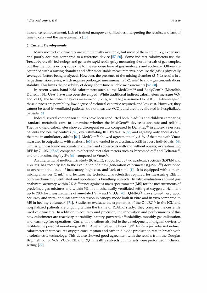
Table 3. Factors limiting the reliability and feasibility of IC measurement.
Factors Limiting IC Measurement
• Agitation, fever, sedatives, and vasoactive adjustments during measurement
• Air leakages in respiratory circuit
• Dialysis or continuous renal replacement therapy
• ECMO
• Mechanical ventilation with PEEP > 10• Mechanical ventilation with FiO2 > 80%• Noninvasive ventilation• Other gases than O2, CO2, and N2: helium
• Supplemental oxygen in spontaneous breathing patients
Moreover, not all subjects are good candidates to IC (i.e., spontaneous breathing patients who areclaustrophobic, nauseous, vomiting, or do not tolerate a face mask or being under a canopy cannotundergo IC measurement). Patients who require supplemental oxygen or undergoing noninvasiveventilation are also difficult to test due to limitations of software and techniques. Other restraintsfor performing IC in clinical routine are related to the equipment and maintenance cost, poor health
J. Clin. Med. 2019, 8, 1387 10 of 19
insurance reimbursement, lack of trained manpower, difficulties interpreting the results, and lack oftime to carry out the measurements [13].
6. Current Developments
Many indirect calorimeters are commercially available, but most of them are bulky, expensiveand poorly accurate compared to a reference device [57–60]. Some indirect calorimeters use the‘breath-by-breath’ technology and generate rapid readings by measuring short intervals of gas samples,but this method is error-prone due to the response time of gas analyzers and software. Others areequipped with a mixing chamber and offer more stable measurements, because the gas is physically‘averaged’ before being analyzed. However, the presence of the mixing chamber (3–5 L) results in alarge dimension device, which requires prolonged measurements (>20 min) to allow gas concentrationsstability. This limits the possibility of doing short-time reliable measurements [57–60].
In recent years, hand-held calorimeters such as the MedGem™ and BodyGem™ (Microlife,Dunedin, FL, USA) have also been developed. While traditional indirect calorimeters measure VO2
and VCO2, the hand-held devices measure only VO2, while RQ is assumed to be 0.85. Advantages ofthese devices are portability, low degree of technical expertise required, and low cost. However, theycannot be used in ventilated patients, do not measure VCO2, and are not validated in hospitalizedpatients [61].
Indeed, several comparison studies have been conducted both in adults and children comparingstandard metabolic carts to determine whether the MedGem™ device is accurate and reliable.The hand-held calorimeter showed discrepant results compared to Deltatrac® in anorexia nervosapatients and healthy controls [62], overestimating REE by 8–11% [63] and agreeing only about 45% ofthe time in ambulatory adults [64]. MedGem® showed agreement only 21% of the time with Vmaxmeasures in outpatients with cirrhosis [65] and tended to overestimate REE in obese individuals [66].Similarly, it was found inaccurate in children and adolescents with and without obesity, overestimatingREE by 7–10% [67,68] compared to other indirect calorimeters such as Parvomedics® and Deltatrac®,and underestimating by 8% [69] compared to Vmax®.
An international multicentric study (ICALIC), supported by two academic societies (ESPEN andESICM), has recently led to the evaluation of a new generation calorimeter (Q-NRG®) developedto overcome the issue of inaccuracy, high cost, and lack of time [1]. It is equipped with a micromixing chamber (2 mL) and features the technical characteristics required for measuring REE inboth mechanically ventilated and spontaneous breathing subjects. In vitro evaluation showed gasanalyzers’ accuracy within 2% difference against a mass spectrometer (MS) for the measurements ofpredefined gas mixtures and within 5% in a mechanically ventilated setting at oxygen enrichmentup to 70% for measurements of simulated VO2 and VCO2 [70]. Q-NRG® also showed very goodaccuracy and intra- and inter-unit precision in canopy mode both in vitro and in vivo compared toMS in healthy volunteers [71]. Studies to evaluate the ergonomics of the Q-NRG® in the ICU andhospitalized patients are ongoing within the frame of ICALIC study: they compare the currentlyused calorimeters. In addition to accuracy and precision, the innovation and performances of thisnew calorimeter are reactivity, portability, battery-powered, affordability, monthly gas calibration,and warm-up free operations. Current innovations also led to the development of original devices tofacilitate the personal monitoring of REE. An example is the Breezing® device, a pocket-sized indirectcalorimeter that measures oxygen consumption and carbon dioxide production rate in breath witha colorimetric technology. This device showed good agreement with the results from the DouglasBag method for VO2, VCO2, EE, and RQ in healthy subjects but no tests were performed in clinicalsetting [72].
J. Clin. Med. 2019, 8, 1387 11 of 19
7. Alternative Methods to IC
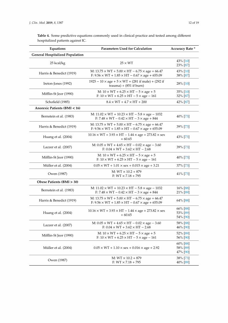
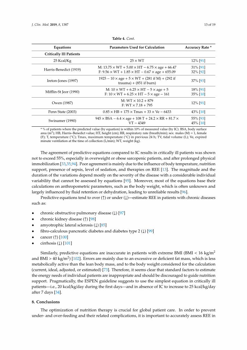
So far, predictive equations remain the most common REE estimation method. They allow arapid calculation of REE using anthropometric data (height, weight, sex, etc.) and have been validatedamong different group of hospitalized patients, but none is optimal (Table 4). Most of these equationshave been developed in healthy subjects, resulting in large errors in case of critical illness and chronicdiseases, despite the use of the correction factors [73]. Other methods for assessing REE have beenexplored and compared to IC in order to find a valid alternative.
Fick’s principle uses cardiac output data, hemoglobin concentration, and arterial and mixedvenous oxygen concentrations obtained from a pulmonary artery catheter to calculate REE. Comparisonwith IC in critically ill patients showed poor correlation with high variability between absolute valuesand variations of VO2 measured, unacceptable for clinical use [74,75]. The error found may be mostlyrelated to five factors: the accuracy of the blood gas analyzer used to calculate arterial and venousoxygen content, the mixed of venous and bronchial arterial blood sample, the hemoglobin levels,the cardiac output variation over the respiratory cycle and the assumption of a fixed RQ to calculateREE [74,76].
The calculation of EE based on CO2 measurement collected from mechanical ventilators (EEVCO2)has also been proposed. This technique considers a fixed value of RQ to calculate the oxygenconsumption (VO2) and REE through the Weir formula. Mainly due to the variability of RQ in criticallyill patients, the accuracy of EEVCO2 compared to the Deltatrac® metabolic monitor is poor [77–80].
The doubly labeled water method implies the oral administration of water with both hydrogen andoxygen atoms labeled with non-radioactive isotopes (2H/1H and 18O/16O) and uses their eliminationrates in body liquids to calculate CO2 production. By assuming a fixed RQ this method allowscalculating TEE in any subjects and any environment, and can been used in complementation to IC tocalculate Physical Activity Level [81,82]. However, this technique is expensive and requires long timeto get the results, limiting its use in daily clinical practice [3].
Motion sensors devices initially developed for fitness settings may be useful in clinical practice tomonitor activity induced energy expenditure (AEE), in particular for patients undergoing physiotherapyduring rehabilitation programs, as well as for the management of daily activity and the dietaryprograms in patients with obesity and/or diabetes. These devices are wearable on the arm, wrist,or waist; user-friendly; relatively low-cost; and non-invasive. Energy expenditure is derived fromacceleration data and individual parameters (sex, age, weight, heart rate) using manufacturer’sconfidential algorithms. However, the energy expenditure derived from these devices generally over-or underestimates the energy expenditure measured by IC by at least 10% [83]. Further studies areneeded to validate their accuracy in clinical practice.
Measurements of body composition by bioelectrical impedance analysis (BIA) or dual energyX-ray absorptiometry (DXA) can be used to estimate REE. To this purpose predictive formula includingFFM and FM values have been developed. This approach has been shown quite inaccurate inclinical populations compared to IC [73,84,85] and cannot be adopted in critically ill patients due totheir abnormalities in hydration state and serum electrolyte concentrations that cause errors in theBIA-derived estimates of FFM and FM [86].
J. Clin. Med. 2019, 8, 1387 12 of 19
Table 4. Some predictive equations commonly used in clinical practice and tested among differenthospitalized patients against IC.
Equations Parameters Used for Calculation Accuracy Rate *
General Hospitalized Population
25 kcal/kg 25 ×WT 43% [10]23% [87]
Harris & Benedict (1919) M: 13.75 ×WT + 5.00 × HT − 6.75 × age + 66.47F: 9.56 ×WT + 1.85 × HT − 0.67 × age + 655.09
43% [10]38% [87]
Ireton-Jones (1992) 1925 − 10 × age + 5 ×WT + (281 if male) + (292 iftrauma) + (851 if burn) 28% [10]
Mifflin-St Jeor (1990) M: 10 ×WT + 6.25 × HT − 5 × age + 5F: 10 ×WT + 6.25 × HT − 5 × age − 161
35% [10]32% [87]
Schofield (1985) 8.4 ×WT + 4.7 × HT + 200 42% [87]
Anorexic Patients (BMI < 16)
Bernstein et al. (1983) M: 11.02 ×WT + 10.23 × HT − 5.8 × age − 1032F: 7.48 ×WT − 0.42 × HT − 3 × age + 844 40% [73]
Harris & Benedict (1919) M: 13.75 ×WT + 5.00 × HT − 6.75 × age + 66.47F: 9.56 ×WT + 1.85 × HT − 0.67 × age + 655.09 39% [73]
Huang et al. (2004) 10.16 ×WT + 3.93 × HT − 1.44 × age + 273.82 × sex+ 60.65 43% [73]
Lazzer et al. (2007) M: 0.05 ×WT + 4.65 × HT − 0.02 × age − 3.60F: 0.04 ×WT + 3.62 × HT − 2.68 39% [73]
Mifflin-St Jeor (1990) M: 10 ×WT + 6.25 × HT − 5 × age + 5F: 10 ×WT + 6.25 × HT − 5 × age − 161 40% [73]
Müller et al. (2004) 0.05 ×WT + 1.01 × sex + 0.015 × age + 3.21 37% [73]
Owen (1987) M: WT × 10.2 + 879F: WT × 7.18 + 795 41% [73]
Obese Patients (BMI > 30)
Bernstein et al. (1983) M: 11.02 ×WT + 10.23 × HT − 5.8 × age − 1032F: 7.48 ×WT − 0.42 × HT − 3 × age + 844
16% [88]21% [89]
Harris & Benedict (1919) M: 13.75 ×WT + 5.00 × HT − 6.75 × age + 66.47F: 9.56 ×WT + 1.85 × HT − 0.67 × age + 655.09 64% [88]
Huang et al. (2004) 10.16 ×WT + 3.93 × HT − 1.44 × age + 273.82 × sex+ 60.65
66% [88]53% [89]54% [90]
Lazzer et al. (2007) M: 0.05 ×WT + 4.65 × HT − 0.02 × age − 3.60F: 0.04 ×WT + 3.62 × HT − 2.68
58% [88]46% [90]
Mifflin-St Jeor (1990) M: 10 ×WT + 6.25 × HT − 5 × age + 5F: 10 ×WT + 6.25 × HT − 5 × age − 161
52% [89]56% [90]
Müller et al. (2004) 0.05 ×WT + 1.10 × sex + 0.016 × age + 2.9260% [88]58% [89]47% [90]
Owen (1987) M: WT × 10.2 + 879F: WT × 7.18 + 795
38% [73]40% [89]
J. Clin. Med. 2019, 8, 1387 13 of 19
Table 4. Cont.
Equations Parameters Used for Calculation Accuracy Rate *
Critically Ill Patients
25 Kcal/Kg 25 ×WT 12% [91]
Harris-Benedict (1919) M: 13.75 ×WT + 5.00 × HT − 6.75 × age + 66.47F: 9.56 ×WT + 1.85 × HT − 0.67 × age + 655.09
31% [91]32% [92]
Ireton-Jones (1997) 1925 − 10 × age + 5 ×WT + (281 if M) + (292 iftrauma) + (851 if burn) 37% [93]
Mifflin-St Jeor (1990) M: 10 ×WT + 6.25 × HT − 5 × age + 5F: 10 ×WT + 6.25 × HT − 5 × age − 161
18% [91]35% [10]
Owen (1987) M: WT × 10.2 + 879F: WT × 7.18 + 795 12% [91]
Penn State (2003) 0.85 × HB + 175 × Tmax + 33 × Ve − 6433 43% [10]
Swinamer (1990) 945 × BSA − 6.4 × age + 108 T + 24.2 × RR + 81.7 ×VT − 4349
55% [93]45% [10]
* % of patients where the predicted value (by equation) is within 10% of measured value (by IC). BSA, body surfacearea (m2); HB, Harris–Benedict value; HT, height (cm); RR, respiratory rate (breath/min); sex: males (M) = 1, female(F); T, temperature (◦C); Tmax, maximum temperature (◦C) in previous 24 h; TV, tidal volume (L); Ve, expiredminute ventilation at the time of collection (L/min); WT, weight (kg).
The agreement of predictive equations compared to IC results in critically ill patients was shownnot to exceed 55%, especially in overweight or obese sarcopenic patients, and after prolonged physicalimmobilization [33,35,94]. Poor agreement is mainly due to the influence of body temperature, nutritionsupport, presence of sepsis, level of sedation, and therapies on REE [13]. The magnitude and theduration of the variations depend mostly on the severity of the disease with a considerable individualvariability that cannot be assessed by equations [95]. Moreover, most of the equations base theircalculations on anthropometric parameters, such as the body weight, which is often unknown andlargely influenced by fluid retention or dehydration, leading to unreliable results [96].
Predictive equations tend to over (↑) or under (↓)—estimate REE in patients with chronic diseasessuch as:
• chronic obstructive pulmonary disease (↓) [97]• chronic kidney disease (↑) [98]• amyotrophic lateral sclerosis (↓) [85]• fibro-calculous pancreatic diabetes and diabetes type 2 (↓) [99]• cancer (↑) [100]• cirrhosis (↓) [101]
Similarly, predictive equations are inaccurate in patients with extreme BMI (BMI < 16 kg/m2
and BMI > 40 kg/m2) [102]. Errors are mainly due to an excessive or deficient fat mass, which is lessmetabolically active than the lean body mass, and to the body weight considered for the calculation(current, ideal, adjusted, or estimated) [73]. Therefore, it seems clear that standard factors to estimatethe energy needs of individual patients are inappropriate and should be discouraged to guide nutritionsupport. Pragmatically, the ESPEN guideline suggests to use the simplest equation in critically illpatients—i.e., 20 kcal/kg/day during the first days—and in absence of IC to increase to 25 kcal/kg/dayafter 7 days [34].
8. Conclusions
The optimization of nutrition therapy is crucial for global patient care. In order to preventunder- and over-feeding and their related complications, it is important to accurately assess REE in
J. Clin. Med. 2019, 8, 1387 14 of 19
individual patients and to ensure adapted nutrition support. IC is considered as the gold standard tothis purpose and ideally any patients in whom energy needs are uncertain should be measured. Recentdevelopments should facilitate the widespread use of IC in medical routine and promote better clinicaloutcomes. Considering the ongoing debate, the widespread use of IC might finally enable the designof prospective studies which will be able to determine the optimal dose of energy to deliver during thedifferent stages of disease, i.e., the ratio of the energy delivered to measured REE and timing of feeding.
Author Contributions: M.D.: conceptualization, writing—original draft preparation, visualization; N.A.:writing—review and editing; M.M.B.: writing—review and editing, supervision; C.P.: writing—review andediting, supervision.
Funding: Financial support came from the Public Foundation Nutrition 2000Plus.
Conflicts of Interest: CP received financial support as an unrestricted academic research grant from publicinstitutions (Geneva University Hospital) and the Foundation Nutrition 2000 Plus. CP received financial supportas research grants and an unrestricted academic research grant, as well as an unrestricted research grant andconsulting fees, from Abbott, Baxter, B. Braun, Cosmed, Fresenius-Kabi, Nestle Medical Nutrition, Novartis,Nutricia-Numico, Pfizer, and Solvay, outside the submitted work. M.M.B. received research grants and publicacademic research grants, unrestricted research grand from Fresenius Kabi International, consulting fees fromFresenius Kabi International, and honoraria for lectures for Fresenius Kabi, Nestle. The other authors declare thatthey have no conflict of interest related to the current work. The funders had no role in the design of the study; inthe collection, analyses, or interpretation of data; in the writing of the manuscript, or in the decision to publishthe results.
References
1. Oshima, T.; Berger, M.M.; De Waele, E.; Guttormsen, A.B.; Heidegger, C.P.; Hiesmayr, M.; Singer, P.;Wernerman, J.; Pichard, C. Indirect calorimetry in nutritional therapy. A position paper by the ICALIC studygroup. Clin. Nutr. 2017, 36, 651–662. [CrossRef]
2. Gupta, R.D.; Ramachandran, R.; Venkatesan, P.; Anoop, S.; Joseph, M.; Thomas, N. Indirect Calorimetry:From Bench to Bedside. Indian J. Endocrinol. Metab. 2017, 21, 594–599. [CrossRef]
3. Fraipont, V.; Preiser, J.C. Energy Estimation and Measurement in Critically Ill Patients. J. Parenter. Enter.Nutr. 2013, 37, 705–713. [CrossRef]
4. Singer, P.; Anbar, R.; Cohen, J.; Shapiro, H.; Shalita-Chesner, M.; Lev, S.; Grozovski, E.; Theilla, M.; Frishman, S.;Madar, Z. The tight calorie control study (TICACOS), a prospective, randomized, controlled pilot study ofnutritional support in critically ill patients. Intensiv. Care Med. 2011, 37, 601–609. [CrossRef]
5. Heidegger, C.P.; Berger, M.M.; Graf, S.; Zingg, W.; Darmon, P.; Costanza, M.C.; Thibault, R.; Pichard, C.Optimisation of energy provision with supplemental parenteral nutrition in critically ill patients: Arandomised controlled clinical trial. Lancet 2013, 381, 385–393. [CrossRef]
6. Petros, S.; Horbach, M.; Seidel, F.; Weidhase, L. Hypocaloric vs Normocaloric Nutrition in Critically IllPatients: A Prospective Randomized Pilot Trial. J. Parenter. Enter. Nutr. 2016, 40, 242–249. [CrossRef]
7. Allingstrup, M.J.; Kondrup, J.; Wiis, J.; Claudius, C.; Pedersen, U.G.; Hein-Rasmussen, R.; Bjerregaard, M.R.;Steensen, M.; Jensen, T.H.; Lange, T.; et al. Early goal-directed nutrition versus standard of care in adultintensive care patients: The single-centre, randomised, outcome assessor-blinded EAT-ICU trial. Intensiv.Care Med. 2017, 43, 1637–1647. [CrossRef]
8. Berger, M.M.; Pantet, O.; Jacquelin-Ravel, N.; Charriere, M.; Schmidt, S.; Becce, F.; Audran, R.; Spertini, F.;Tappy, L.; Pichard, C. Supplemental parenteral nutrition improves immunity with unchanged carbohydrateand protein metabolism in critically ill patients: The SPN2 randomized tracer study. Clin. Nutr. 2018.[CrossRef]
9. Merritt, R. Use of indirect calorimetry in critically ill patients. In The ASPEN Nutrition Support Practice Manual,2nd ed.; American Society for Parenteral and Enteral Nutrit: Silver Spring, MD, USA, 2006; pp. 277–280.
10. Boullata, J.; Williams, J.; Cottrell, F.; Hudson, L.; Compher, C. Accurate determination of energy needs inhospitalized patients. J. Am. Diet. Assoc. 2007, 107, 393–401. [CrossRef]
11. Wooley, J.A.; Sax, H.C. Indirect calorimetry: Applications to practice. Nutr. Clin. Pract. 2003, 18, 434–439.[CrossRef]
J. Clin. Med. 2019, 8, 1387 15 of 19
12. Berger, M.M.; Reintam-Blaser, A.; Calder, P.C.; Casaer, M.; Hiesmayr, M.J.; Mayer, K.; Montejo, J.C.; Pichard, C.;Preiser, J.C.; van Zanten, A.R.H.; et al. Monitoring nutrition in the ICU. Clin. Nutr. 2019, 38, 584–593.[CrossRef]
13. Singer, P.; Singer, J. Clinical Guide for the Use of Metabolic Carts: Indirect Calorimetry-No Longer theOrphan of Energy Estimation. Nutr. Clin. Pract. 2016, 31, 30–38. [CrossRef]
14. Weissman, C.; Kemper, M.; Hyman, A.I. Variation in the resting metabolic rate of mechanically ventilatedcritically ill patients. Anesth. Analg. 1989, 68, 457–461. [CrossRef]
15. Cuthbertson, D.P.; Angeles Valero Zanuy, M.A.; Leon Sanz, M.L. Post-shock metabolic response. 1942. Nutr.Hosp. 2001, 16, 176–182.
16. Rattanachaiwong, S.; Singer, P. Indirect Calorimetry as Point of Care Testing. Clin. Nutr. 2019. [CrossRef]17. Zusman, O.; Theilla, M.; Cohen, J.; Kagan, I.; Bendavid, I.; Singer, P. Resting energy expenditure, calorie and
protein consumption in critically ill patients: A retrospective cohort study. Crit. Care 2016, 20, 367. [CrossRef]18. Plank, L.D.; Hill, G.L. Sequential metabolic changes following induction of systemic inflammatory response
in patients with severe sepsis or major blunt trauma. World J. Surg. 2000, 24, 630–638. [CrossRef]19. Plank, L.D.; Connolly, A.B.; Hill, G.L. Sequential changes in the metabolic response in severely septic patients
during the first 23 days after the onset of peritonitis. Ann. Surg. 1998, 228, 146–158. [CrossRef]20. Brandi, L.S.; Oleggini, M.; Lachi, S.; Frediani, M.; Bevilacqua, S.; Mosca, F.; Ferrannini, E. Energy metabolism
of surgical patients in the early postoperative period: A reappraisal. Crit. Care Med. 1988, 16, 18–22.[CrossRef]
21. Dickerson, R.N.; Vehe, K.L.; Mullen, J.L.; Feurer, I.D. Resting energy expenditure in patients with pancreatitis.Crit. Care Med. 1991, 19, 484–490. [CrossRef]
22. Schneeweiss, B.; Graninger, W.; Stockenhuber, F.; Druml, W.; Ferenci, P.; Eichinger, S.; Grimm, G.;Laggner, A.N.; Lenz, K. Energy metabolism in acute and chronic renal failure. Am. J. Clin. Nutr.1990, 52, 596–601. [CrossRef]
23. Soncini Sanches, A.C.; de Góes, C.R.; Nogueira Berbel Bufarah, M.; Balbi, A.L.; Ponce, D. Does Acute KidneyInjury Alter Energy Metabolism of Septic Patients? Arch. Ren. Dis. Manag. 2016, 2, 19–23. [CrossRef]
24. Goes, C.R.; Balbi, A.L.; Ponce, D. Evaluation of Factors Associated with Hypermetabolism andHypometabolism in Critically Ill AKI Patients. Nutrients 2018, 10, 505. [CrossRef]
25. Sabatino, A.; Theilla, M.; Hellerman, M.; Singer, P.; Maggiore, U.; Barbagallo, M.; Regolisti, G.; Fiaccadori, E.Energy and Protein in Critically Ill Patients with AKI: A Prospective, Multicenter Observational Study UsingIndirect Calorimetry and Protein Catabolic Rate. Nutrients 2017, 9, 802. [CrossRef]
26. Kreymann, G.; Grosser, S.; Buggisch, P.; Gottschall, C.; Matthaei, S.; Greten, H. Oxygen consumption andresting metabolic rate in sepsis, sepsis syndrome, and septic shock. Crit. Care Med. 1993, 21, 1012–1019.[CrossRef]
27. Wu, C.; Wang, X.Y.; Yu, W.K.; Tian, F.; Liu, S.T.; Li, P.; Li, J.; Li, N. Hypermetabolism in the Initial Phase ofIntensive Care Is Related to a Poor Outcome in Severe Sepsis Patients. Ann. Nutr. Metab. 2015, 66, 188–195.[CrossRef]
28. Puthucheary, Z.A.; Rawal, J.; McPhail, M.; Connolly, B.; Ratnayake, G.; Chan, P.; Hopkinson, N.S.; Phadke, R.;Dew, T.; Sidhu, P.S.; et al. Acute skeletal muscle wasting in critical illness. JAMA 2013, 310, 1591–1600.[CrossRef]
29. Jeon, J.; Kym, D.; Cho, Y.S.; Kim, Y.; Yoon, J.; Yim, H.; Hur, J.; Chun, W. Reliability of resting energyexpenditure in major burns: Comparison between measured and predictive equations. Clin. Nutr. 2018.[CrossRef]
30. McClave, S.A.; Martindale, R.G.; Kiraly, L. The use of indirect calorimetry in the intensive care unit. Curr.Opin. Clin. Nutr. Metab. Care 2013, 16, 202–208. [CrossRef]
31. Foley, N.; Marshall, S.; Pikul, J.; Salter, K.; Teasell, R. Hypermetabolism following moderate to severetraumatic acute brain injury: A systematic review. J. Neurotrauma 2008, 25, 1415–1431. [CrossRef]
32. Berger, M.M.; Pichard, C. Feeding should be individualized in the critically ill patients. Curr. Opin. Crit. Care2019, 25, 307–313. [CrossRef]
33. Ndahimana, D.; Kim, E.K. Energy Requirements in Critically Ill Patients. Clin. Nutr. Res. 2018, 7, 81–90.[CrossRef]
J. Clin. Med. 2019, 8, 1387 16 of 19
34. Singer, P.; Blaser, A.R.; Berger, M.M.; Alhazzani, W.; Calder, P.C.; Casaer, M.P.; Hiesmayrh, M.; Mayeri, K.;Montejoj, J.C.; Pichard, C.; et al. ESPEN guideline on clinical nutrition in the intensive care unit. Clin. Nutr.2019, 38, 48–79. [CrossRef]
35. Zusman, O.; Kagan, I.; Bendavid, I.; Theilla, M.; Cohen, J.; Singer, P. Predictive equations versus measuredenergy expenditure by indirect calorimetry: A retrospective validation. Clin. Nutr. 2018. [CrossRef]
36. Wolfe, R.R. Sepsis as a modulator of adaptation to low and high carbohydrate and low and high fat intakes.Eur. J. Clin. Nutr. 1999, 53, S136–S142. [CrossRef]
37. Tappy, L.; Schwarz, J.M.; Schneiter, P.; Cayeux, C.; Revelly, J.P.; Fagerquist, C.K.; Jéquier, E.; Chioléro, R.Effects of isoenergetic glucose-based or lipid-based parenteral nutrition on glucose metabolism, de novolipogenesis, and respiratory gas exchanges in critically ill patients. Crit. Care Med. 1998, 26, 860–867.[CrossRef]
38. Viana, M.V.; Pantet, O.; Bagnoud, G.; Martinez, A.; Favre, E.; Charriere, M.; Favre, D.; Eckert, P.; Berger, M.M.Metabolic and Nutritional Characteristics of Long-Stay Critically Ill Patients. J. Clin. Med. 2019, 8, 985.[CrossRef]
39. Reignier, J.; Boisrame-Helms, J.; Brisard, L.; Lascarrou, J.B.; Ait Hssain, A.; Anguel, N.; Argaud, L.;Asehnoune, K.; Asfar, P.; Bellec, F.; et al. Enteral versus parenteral early nutrition in ventilated adults withshock: A randomised, controlled, multicentre, open-label, parallel-group study (NUTRIREA-2). Lancet 2018,391, 133–143. [CrossRef]
40. Avesani, C.M.; Draibe, S.A.; Kamimura, M.A.; Dalboni, M.A.; Colugnati, F.A.; Cuppari, L. Decreased restingenergy expenditure in non-dialysed chronic kidney disease patients. Nephrol. Dial. Transplant. 2004, 19,3091–3097. [CrossRef]
41. Neyra, R.; Chen, K.Y.; Sun, M.; Shyr, Y.; Hakim, R.M.; Ikizler, T.A. Increased resting energy expenditure inpatients with end-stage renal disease. J. Parenter. Enteral. Nutr. 2003, 27, 36–42. [CrossRef]
42. Kamimura, M.A.; Draibe, S.A.; Avesani, C.M.; Canziani, M.E.; Colugnati, F.A.; Cuppari, L. Resting energyexpenditure and its determinants in hemodialysis patients. Eur. J. Clin. Nutr. 2007, 61, 362–367. [CrossRef]
43. Ikizler, T.A.; Wingard, R.L.; Sun, M.; Harvell, J.; Parker, R.A.; Hakim, R.M. Increased energy expenditure inhemodialysis patients. J. Am. Soc. Nephrol. 1996, 7, 2646–2653.
44. Cekici, H.; Acar Tek, N. Determining energy requirement and evaluating energy expenditure in neurologicaldiseases. Nutr. Neurosci. 2018, 1–11. [CrossRef]
45. Huggett, R.J.; Scott, E.M.; Gilbey, S.G.; Stoker, J.B.; Mackintosh, A.F.; Mary, D.A. Impact of type 2 diabetesmellitus on sympathetic neural mechanisms in hypertension. Circulation 2003, 108, 3097–3101. [CrossRef]
46. Farooqi, N.; Carlsson, M.; Haglin, L.; Sandstrom, T.; Slinde, F. Energy expenditure in women and men withCOPD. Clin. Nutr. ESPEN 2018, 28, 171–178. [CrossRef]
47. Carneiro, I.P.; Elliott, S.A.; Siervo, M.; Padwal, R.; Bertoli, S.; Battezzati, A.; Prado, C.M. Is Obesity Associatedwith Altered Energy Expenditure? Adv. Nutr. 2016, 7, 476–487. [CrossRef]
48. Cuerda, C.; Ruiz, A.; Velasco, C.; Breton, I.; Camblor, M.; Garcia-Peris, P. How accurate are predictiveformulas calculating energy expenditure in adolescent patients with anorexia nervosa? Clin. Nutr. 2007, 26,100–106. [CrossRef]
49. Yeh, D.D.; Fuentes, E.; Quraishi, S.A.; Cropano, C.; Kaafarani, H.; Lee, J.; King, D.R.; DeMoya, M.; Fagenholz, P.;Butler, K.; et al. Adequate Nutrition May Get You Home: Effect of Caloric/Protein Deficits on the DischargeDestination of Critically Ill Surgical Patients. JPEN 2016, 40, 37–44. [CrossRef]
50. Milte, R.K.; Ratcliffe, J.; Miller, M.D.; Crotty, M. Economic evaluation for protein and energy supplementationin adults: Opportunities to strengthen the evidence. Eur. J. Clin. Nutr. 2013, 67, 1243–1250. [CrossRef]
51. Correia, M.; Perman, M.I.; Pradelli, L.; Omaralsaleh, A.J.; Waitzberg, D.L. Economic burden of hospitalmalnutrition and the cost-benefit of supplemental parenteral nutrition in critically ill patients in LatinAmerica. J. Med. Econ. 2018, 21, 1047–1056. [CrossRef]
52. Pradelli, L.; Graf, S.; Pichard, C.; Berger, M.M. Supplemental parenteral nutrition in intensive care patients:A cost saving strategy. Clin. Nutr. 2018, 37, 573–579. [CrossRef]
53. Berger, M.M.; Pichard, C. Parenteral nutrition in the ICU: Lessons learned over the past few years. Nutrition2019, 59, 188–194. [CrossRef]
54. Haugen, H.A.; Chan, L.N.; Li, F. Indirect calorimetry: A practical guide for clinicians. Nutr. Clin. Pract. 2007,22, 377–388. [CrossRef]
J. Clin. Med. 2019, 8, 1387 17 of 19
55. De Waele, E.; van Zwam, K.; Mattens, S.; Staessens, K.; Diltoer, M.; Honore, P.M.; Czapla, J.; Nijs, J.; LaMeir, M.; Huyghens, L.; et al. Measuring resting energy expenditure during extracorporeal membraneoxygenation: Preliminary clinical experience with a proposed theoretical model. Acta Anaesthesiol. Scand.2015, 59, 1296–1302. [CrossRef]
56. Wollersheim, T.; Frank, S.; Muller, M.C.; Skrypnikov, V.; Carbon, N.M.; Pickerodt, P.A.; Spies, C.; Mai, K.;Spranger, J.; Weber-Carstens, S. Measuring Energy Expenditure in extracorporeal lung support Patients(MEEP)-Protocol, feasibility and pilot trial. Clin. Nutr. 2018, 37, 301–307. [CrossRef]
57. Graf, S.; Karsegard, V.L.; Viatte, V.; Heidegger, C.P.; Fleury, Y.; Pichard, C.; Genton, L. Evaluation of threeindirect calorimetry devices in mechanically ventilated patients: Which device compares best with theDeltatrac II(®)? A prospective observational study. Clin. Nutr. 2015, 34, 60–65. [CrossRef]
58. Graf, S.; Karsegard, V.L.; Viatte, V.; Maisonneuve, N.; Pichard, C.; Genton, L. Comparison of three indirectcalorimetry devices and three methods of gas collection: A prospective observational study. Clin. Nutr. 2013,32, 1067–1072. [CrossRef]
59. Sundstrom, M.; Tjader, I.; Rooyackers, O.; Wernerman, J. Indirect calorimetry in mechanically ventilatedpatients. A systematic comparison of three instruments. Clin. Nutr. 2013, 32, 118–121. [CrossRef]
60. Cooper, J.A.; Watras, A.C.; O’Brien, M.J.; Luke, A.; Dobratz, J.R.; Earthman, C.P.; Schoeller, D.A. Assessingvalidity and reliability of resting metabolic rate in six gas analysis systems. J. Am. Diet. Assoc. 2009, 109,128–132. [CrossRef]
61. Hipskind, P.; Glass, C.; Charlton, D.; Nowak, D.; Dasarathy, S. Do handheld calorimeters have a role inassessment of nutrition needs in hospitalized patients? A systematic review of literature. Nutr. Clin. Pract.2011, 26, 426–433. [CrossRef]
62. Hlynsky, J.; Birmingham, C.L.; Johnston, M.; Gritzner, S. The agreement between the MedGem indirectcalorimeter and a standard indirect calorimeter in anorexia nervosa. Eat. Weight Disord. 2005, 10, e83–e87.[CrossRef]
63. Alam, D.S.; Hulshof, P.J.; Roordink, D.; Meltzer, M.; Yunus, M.; Salam, M.A.; van Raaij, J.M. Validityand reproducibility of resting metabolic rate measurements in rural Bangladeshi women: Comparison ofmeasurements obtained by Medgem and by Deltatrac device. Eur. J. Clin. Nutr. 2005, 59, 651–657. [CrossRef]
64. Frankenfield, D.C.; Coleman, A. An evaluation of a handheld indirect calorimeter against a standardcalorimeter in obese and nonobese adults. JPEN 2013, 37, 652–658. [CrossRef]
65. Schock, L.; Lam, L.; Tandon, P.; Taylor, L.; Raman, M. Indirect Calorimetry Performance Using a HandheldDevice Compared to the Metabolic Cart in Outpatients with Cirrhosis. Nutrients 2019, 11, 1030. [CrossRef]
66. Anderson, E.J.; Sylvia, L.G.; Lynch, M.; Sonnenberg, L.; Lee, H.; Nathan, D.M. Comparison of energyassessment methods in overweight individuals. J. Acad. Nutr. Diet. 2014, 114, 273–278. [CrossRef]
67. White, D.A.; Staggs, V.S.; Williams, V.; Edwards, T.C.; Shook, R.; Shakhnovich, V. Handheld IndirectCalorimetry as a Clinical Tool for Measuring Resting Energy Expenditure in Children with and withoutObesity. Child. Obes. 2019, 15, 280–287. [CrossRef]
68. Fields, D.A.; Kearney, J.T.; Copeland, K.C. MedGem hand-held indirect calorimeter is valid for resting energyexpenditure measurement in healthy children. Obesity (Silver Spring) 2006, 14, 1755–1761. [CrossRef]
69. Woo, P.; Murthy, G.; Wong, C.; Hursh, B.; Chanoine, J.P.; Elango, R. Assessing resting energy expenditure inoverweight and obese adolescents in a clinical setting: Validity of a handheld indirect calorimeter. PediatrRes. 2017, 81, 51–56. [CrossRef]
70. Oshima, T.; Dupertuis, Y.M.; Delsoglio, M.; Graf, S.; Heidegger, C.P.; Pichard, C. In vitro validation of indirectcalorimetry device developed for the ICALIC project against mass spectrometry. Clin. Nutr. ESPEN. 2019,32, 50–55. [CrossRef]
71. Delsoglio, M.; Dupertuis, Y.M.; Oshima, T.; van der Plas, M.; Pichard, C. Evaluation of the accuracy andprecision of a new generation indirect calorimeter in canopy dilution mode. Clin. Nutr. 2019, in press.
72. Xiaojun, X.; Quach, A.; Bridgeman, D.; Tsow, F.; Forzani, E.; Tao, N. Personalized Indirect Calorimeter forEnergy Expenditure (EE) Measurement. Glob. J. Obes. Diabetes Metab. Syndr. 2015. [CrossRef]
73. Jesus, P.; Achamrah, N.; Grigioni, S.; Charles, J.; Rimbert, A.; Folope, V.; Petit, A.; Déchelotte, P.; Coëffier, M.Validity of predictive equations for resting energy expenditure according to the body mass index in apopulation of 1726 patients followed in a Nutrition Unit. Clin. Nutr. 2015, 34, 529–535. [CrossRef]
J. Clin. Med. 2019, 8, 1387 18 of 19
74. Soussi, S.; Vallee, F.; Roquet, F.; Bevilacqua, V.; Benyamina, M.; Ferry, A.; Cupaciu, A.; Chaussard, M.; DeTymowski, C.; Boccara, D.; et al. Measurement of Oxygen Consumption Variations in Critically Ill BurnsPatients: Are the Fick Method and Indirect Calorimetry Interchangeable? Shock 2017, 48, 532–538. [CrossRef]
75. Inadomi, C.; Terao, Y.; Yamashita, K.; Fukusaki, M.; Takada, M.; Sumikawa, K. Comparison of oxygenconsumption calculated by Fick’s principle (using a central venous catheter) and measured by indirectcalorimetry. J. Anesth. 2008, 22, 163–166. [CrossRef]
76. Ogawa, A.M.; Shikora, S.A.; Burke, L.M.; Heetderks-Cox, J.E.; Bergren, C.T.; Muskat, P.C. The thermodilutiontechnique for measuring resting energy expenditure does not agree with indirect calorimetry for the criticallyill patient. J. Parenter. Enter. Nutr. 1998, 22, 347–351. [CrossRef]
77. Oshima, T.; Graf, S.; Heidegger, C.P.; Genton, L.; Pugin, J.; Pichard, C. Can calculation of energy expenditurebased on CO2 measurements replace indirect calorimetry? Crit. Care 2017, 21, 13. [CrossRef]
78. Stapel, S.N.; de Grooth, H.J.S.; Alimohamad, H.; Elbers, P.W.G.; Girbes, A.R.J.; Weijs, P.J.M.; Oudemans-vanStraaten, H.M. Ventilator-derived carbon dioxide production to assess energy expenditure in critically illpatients: Proof of concept. Crit. Care 2015, 19, 370. [CrossRef]
79. Rousing, M.L.; Hahn-Pedersen, M.H.; Andreassen, S.; Pielmeier, U.; Preiser, J.C. Energy expenditure incritically ill patients estimated by population-based equations, indirect calorimetry and CO2-based indirectcalorimetry. Ann. Intensiv. Care 2016, 6, 16. [CrossRef]
80. Kagan, I.; Zusman, O.; Bendavid, I.; Theilla, M.; Cohen, J.; Singer, P. Validation of carbon dioxide production(VCO2) as a tool to calculate resting energy expenditure (REE) in mechanically ventilated critically ill patients:A retrospective observational study. Crit. Care 2018, 22, 186. [CrossRef]
81. de Carvalho Bastone, A.; Ferriolli, E.; Pfrimer, K.; de Souza Moreira, B.; Diz, J.B.M.; Dias, J.M.D.; Dias, R.C.Energy Expenditure in Older Adults Who Are Frail: A Doubly Labeled Water Study. J. Geriatr. Phys. Ther.2017. [CrossRef]
82. Schutz, Y. Respiration chamber calorimetry and doubly labeled water: Two complementary aspects of energyexpenditure? Eur. J. Clin. Nutr. 2018, 72, 1310–1313. [CrossRef]
83. Wahl, Y.; Duking, P.; Droszez, A.; Wahl, P.; Mester, J. Criterion-Validity of Commercially Available PhysicalActivity Tracker to Estimate Step Count, Covered Distance and Energy Expenditure during Sports Conditions.Front. Physiol. 2017, 8, 725. [CrossRef]
84. Hashizume, N.; Tanaka, Y.; Yoshida, M.; Fukahori, S.; Ishii, S.; Saikusa, N.; Masui, D.; Higashidate, N.;Sakamoto, S.; Tsuruhisa, S.; et al. Resting energy expenditure prediction using bioelectrical impedanceanalysis in patients with severe motor and intellectual disabilities. Brain Dev. Jpn. 2019, 41, 352–358.[CrossRef]
85. Jesus, P.; Marin, B.; Fayemendy, P.; Nicol, M.; Lautrette, G.; Sourisseau, H.; Preux, P.M.; Couratier, P.;Desport, J.C. Resting energy expenditure equations in amyotrophic lateral sclerosis, creation of an ALS-specificequation. Clin. Nutr. 2019, 38, 1657–1665. [CrossRef]
86. Barak, N.; Wall-Alonso, E.; Cheng, A.; Sitrin, M.D. Use of bioelectrical impedance analysis to predict energyexpenditure of hospitalized patients receiving nutrition support. JPEN 2003, 27, 43–46. [CrossRef]
87. Kruizenga, H.M.; Hofsteenge, G.H.; Weijs, P.J. Predicting resting energy expenditure in underweight, normalweight, overweight, and obese adult hospital patients. Nutr. Metab. (Lond.) 2016, 13, 85. [CrossRef]
88. Achamrah, N.; Jesus, P.; Grigioni, S.; Rimbert, A.; Petit, A.; Dechelotte, P.; Folope, V.; Coëffier, M. Validityof Predictive Equations for Resting Energy Expenditure Developed for Obese Patients: Impact of BodyComposition Method. Nutrients 2018, 10, 63. [CrossRef]
89. Orozco-Ruiz, X.; Pichardo-Ontiveros, E.; Tovar, A.R.; Torres, N.; Medina-Vera, I.; Prinelli, F.; Lafortuna, C.L.;Guevara-Cruz, M. Development and validation of new predictive equation for resting energy expenditure inadults with overweight and obesity. Clin. Nutr. 2018, 37 Pt A, 2198–2205. [CrossRef]
90. Cancello, R.; Soranna, D.; Brunani, A.; Scacchi, M.; Tagliaferri, A.; Mai, S.; Marzullo, P.; Zambon, A.; Invitti, C.Analysis of Predictive Equations for Estimating Resting Energy Expenditure in a Large Cohort of MorbidlyObese Patients. Front. Endocrinol. (Lausanne) 2018, 9, 367. [CrossRef]
91. Kross, E.K.; Sena, M.; Schmidt, K.; Stapleton, R.D. A comparison of predictive equations of energy expenditureand measured energy expenditure in critically ill patients. J. Crit. Care 2012, 27, 321.e5. [CrossRef]
92. Costa, N.A.; Marinho, A.D.; Cancado, L.R. Nutritional requirements of the critically ill patient. Rev. Bras. Ter.Intensiv. 2012, 24, 270–277. [CrossRef]
J. Clin. Med. 2019, 8, 1387 19 of 19
93. MacDonald, A.; Hildebrandt, L. Comparison of formulaic equations to determine energy expenditure in thecritically ill patient. Nutrition 2003, 19, 233–239. [CrossRef]
94. Pichard, C.; Oshima, T.; Berger, M.M. Energy deficit is clinically relevant for critically ill patients: Yes. Intensiv.Care Med. 2015, 41, 335–338. [CrossRef]
95. Frankenfield, D.C.; Ashcraft, C.M. Estimating energy needs in nutrition support patients. J. Parenter. Enter.Nutr. 2011, 35, 563–570. [CrossRef]
96. Graf, S.; Pichard, C.; Genton, L.; Oshima, T.; Heidegger, C.P. Energy expenditure in mechanically ventilatedpatients: The weight of body weight! Clin. Nutr. 2017, 36, 224–228. [CrossRef]
97. Ramos, F.M.; Rossato, L.T.; Ramires, B.R.; Pimentel, G.D.; Venancio, L.S.; Orsatti, F.L.; de Oliveira, E.P.Comparison of predictive equations of resting energy expenditure in older adults with chronic obstructivepulmonary disease. Rev. Port. Pneumol. (2006) 2017, 23, 40–42. [CrossRef]
98. Kamimura, M.A.; Avesani, C.M.; Bazanelli, A.P.; Baria, F.; Draibe, S.A.; Cuppari, L. Are prediction equationsreliable for estimating resting energy expenditure in chronic kidney disease patients? Nephrol. Dial. Transplant.2011, 26, 544–550. [CrossRef]
99. Behera, K.K.; Joseph, M.; Shetty, S.K.; Chacko, A.; Sahoo, M.K.; Mahendri, N.V.; Nair, V.; Nadig, S.; Thomas, N.Resting energy expenditure in subjects with fibro-calculous pancreatic diabetes. J. Diabetes 2014, 6, 158–163.[CrossRef]
100. Khor, S.M.; Mohd, B.B. Assessing the resting energy expenditure of cancer patients in the Penang GeneralHospital. Malays. J. Nutr. 2011, 17, 43–53.
101. Eslamparast, T.; Vandermeer, B.; Raman, M.; Gramlich, L.; Den Heyer, V.; Belland, D.; Ma, M.; Tandon, P. ArePredictive Energy Expenditure Equations Accurate in Cirrhosis? Nutrients 2019, 11, 334. [CrossRef]
102. Frankenfield, D.C. Bias and accuracy of resting metabolic rate equations in non-obese and obese adults. Clin.Nutr. 2013, 32, 976–982. [CrossRef]
© 2019 by the authors. Licensee MDPI, Basel, Switzerland. This article is an open accessarticle distributed under the terms and conditions of the Creative Commons Attribution(CC BY) license (http://creativecommons.org/licenses/by/4.0/).


