Embed Size (px)
Citation preview
BioMed CentralMolecular Cancer
ss
Open AcceResearchIn vivo optical imaging of integrin αV-β3 in mice using multivalent or monovalent cRGD targeting vectorsZhao-Hui Jin1,2, Véronique Josserand1,2, Stéphanie Foillard2,3, Didier Boturyn2,3, Pascal Dumy2,3, Marie-Christine Favrot1,2 and Jean-Luc Coll*1,2Address: 1INSERM, U823, Cibles Diagnostiques ou Thérapeutiques et Vectorisation des Drogues dans le Cancer du Poumon, Institut Albert Bonniot, 38706 La Tronche Cedex, France, 2Université Joseph Fourier, 38041 Grenoble Cedex 9, France and 3CNRS, UMR5616, Ingénierie Moléculaire et Chimie des Composés Bio-organiques, LEDSS, 38041 Grenoble Cedex 9, France
Email: Zhao-Hui Jin - [email protected]; Véronique Josserand - [email protected]; Stéphanie Foillard - [email protected]; Didier Boturyn - [email protected]; Pascal Dumy - [email protected]; Marie-Christine Favrot - [email protected]; Jean-Luc Coll* - [email protected]
* Corresponding author
AbstractBackground: The cRGD peptide is a promising probe for early non-invasive detection of tumors.This study aimed to demonstrate how RAFT-c(-RGDfK-)4, a molecule allowing a tetramericpresentation of cRGD, improved cRGD-targeting potential using in vivo models of αVβ3-positive ornegative tumors.
Results: We chose the human embryonic kidney cells HEK293(β3) (high levels of αVβ3) orHEK293(β1) (αVβ3-negative but expressing αV and β1) engrafted subcutaneously (s.c.) in mice.Non-invasive in vivo optical imaging demonstrated that as compared to its monomeric cRGDanalogue, Cy5-RAFT-c(-RGDfK-)4 injected intravenously had higher uptake, prolonged retentionand markedly enhanced contrast in HEK293(β3) than in the HEK293(β1) tumors. Blocking studiesfurther demonstrated the targeting specificity and competitive binding ability of the tetramer.
Conclusion: In conclusion, we demonstrated that Cy5-RAFT-c(-RGDfK-)4 was indeed binding tothe αVβ3 receptor and with an improved activity as compared to its monomeric analog, confirmingthe interest of using multivalent ligands. Intravenous injection of Cy5-RAFT-c(-RGDfK-)4 in thisnovel pair of HEK293(β3) and HEK293(β1) tumors, provided tumor/skin ratio above 15. Such animportant contrast plus the opportunity to use the HEK293(β1) negative control cell line are majorassets for the community of researchers working on the design and amelioration of RGD-targetedvectors or on RGD-antagonists.
BackgroundThe tripeptide sequence Arg-Gly-Asp (RGD) [1,2] is a wellknown motif recognizing and interacting with integrin, afamily of transmembrane heterodimeric glycoproteins
composed of one α and one β subunits [3,4]. The struc-ture of a cyclic pentapeptide containing RGD was opti-mized in order to provide a high affinity and selectivity forthe αVβ3 integrin [5], an integrin overexpressed at the sur-
Published: 12 June 2007
Molecular Cancer 2007, 6:41 doi:10.1186/1476-4598-6-41
Received: 28 March 2007Accepted: 12 June 2007
This article is available from: http://www.molecular-cancer.com/content/6/1/41
© 2007 Jin et al; licensee BioMed Central Ltd. This is an Open Access article distributed under the terms of the Creative Commons Attribution License (http://creativecommons.org/licenses/by/2.0), which permits unrestricted use, distribution, and reproduction in any medium, provided the original work is properly cited.
Page 1 of 9(page number not for citation purposes)

Molecular Cancer 2007, 6:41 http://www.molecular-cancer.com/content/6/1/41
face of activated endothelial cells during angiogenesis[6,7] and in various types of tumor cells [8-11]. Radiola-beled cRGD peptides in combination with nuclear imag-ing techniques such as positron emission tomography(PET) and single photon emission computed tomography(SPECT) have been extensively studied for imaging ofαVβ3 expression in experimental tumors [12]. Morerecently, the development of in vivo optical imaging tech-niques and of various fluorescent-cRGD conjugates werealso described for imaging cancer in mice [12-18]. In addi-tion, it was shown that presenting multiple copies of thecRGD motif was usually associated with improved prop-erties of the probes [16,19]. In this aim, our group hasdeveloped a novel tetrameric molecule by grafting fourcopies of cRGD onto a cyclic decapeptide platform calledRAFT (Regioselectively Addressable Functionalized Tem-plate) [17,18,20]. When injected intravenously in nudemice bearing s.c. human ovarian carcinoma IGROV1tumors, expressing a low level of αVβ3, cyanine 5-labeledRAFT-c(-RGDfK-)4 showed a better tumor contrast than itsmonomeric analog [18].
In the present study, we took advantage of a particulartumor model for addressing RGD-mediated targeting spe-cificity in vivo. This model derived from the naturally αV-positive and β3-negative HEK293 cell line was initiallytransfected by a plasmid encoding the human β3 chain,forming a strongly αVβ3-positive HEK293(β3) stableclone. In addition, HEK293(β1), an αVβ3-negative controloverexpressing the β1 chain instead of the β3, had beenalso established. As their parent cell line HEK293, weshow that the 2 β3 or β1 subclones are forming tumorswhen injected subcutaneously into athymic nude mice.Using these tumor models, our different RGD-based mol-ecules and competition experiments, we demonstrate theextremely good specificity and improved tumor accumu-lation and retention of the Cy5-labeled RAFT-c(-RGDfK-)4probe as compared to its monomeric analog. Since RGD-based antiangiogenic therapies are currently under inves-tigation, and that cRGD can also serve as a ligand inhuman nuclear medicine, optimization of its specificityand drug delivery properties is of major importance forclinical applications.
ResultsIn vitro binding studiesHEK293(β3) and HEK293(β1) cells are stable transfectantsof human β3 and β1 subunit, respectively, from the humanembryonic kidney cell line. Western blot analysis showedthat αV was strongly expressed in both cell lines, and con-firmed the successful transfection of β3 or β1 subunits (Fig1A). This phenotype was also confirmed by FACS analysisperformed with the anti-human αVβ3 antibody [18]. These2 cell lines, were then observed using confocal laser scan-ning microscopy (CLSM) after incubation with Cy5-
labeled RAFT-c(-RGDfK-)4, cRGD, or RAFT-c(-RβADfK-)4.As shown in Fig. 1B, none of these peptides bound to theHEK293(β1) cells. As expected also, the RAFT-c(-RβADfK-)4 control peptide did not bind to the αVβ3-positiveHEK293(β3) cells. In contrast, cRGD and RAFT-c(-RGDfK-)4 were reacting with HEK293(β3) cells moderately andvery strongly, respectively.
Establishment of paired αVβ3-positive and αVβ3-negative tumor modelsA s.c. inoculation of HEK293(β3) or HEK293(β1) cells innude mice lead to tumor formation. This suggested thatoverexpression of β3 or β1 did not modify the known tum-origenicity of the parental HEK293 cell line (see ATCCnumber CRL-1573). Histological examination withhematoxylin and eosin (H.E.) staining shows that eitherHEK293(β3) or HEK293(β1) xenografts are composed ofnodular cell masses and stroma (Fig 2). Immunohisto-chemical labeling of tumor sections shows positive αVβ3staining in HEK293(β3) cells but not in HEK293(β1) anda similar low to moderate vascularization as indicated bythe CD 31-labeling of both tumors. Thus expression of theβ3 chain was not lost during tumor growth and was notaffecting the tumor vasculature.
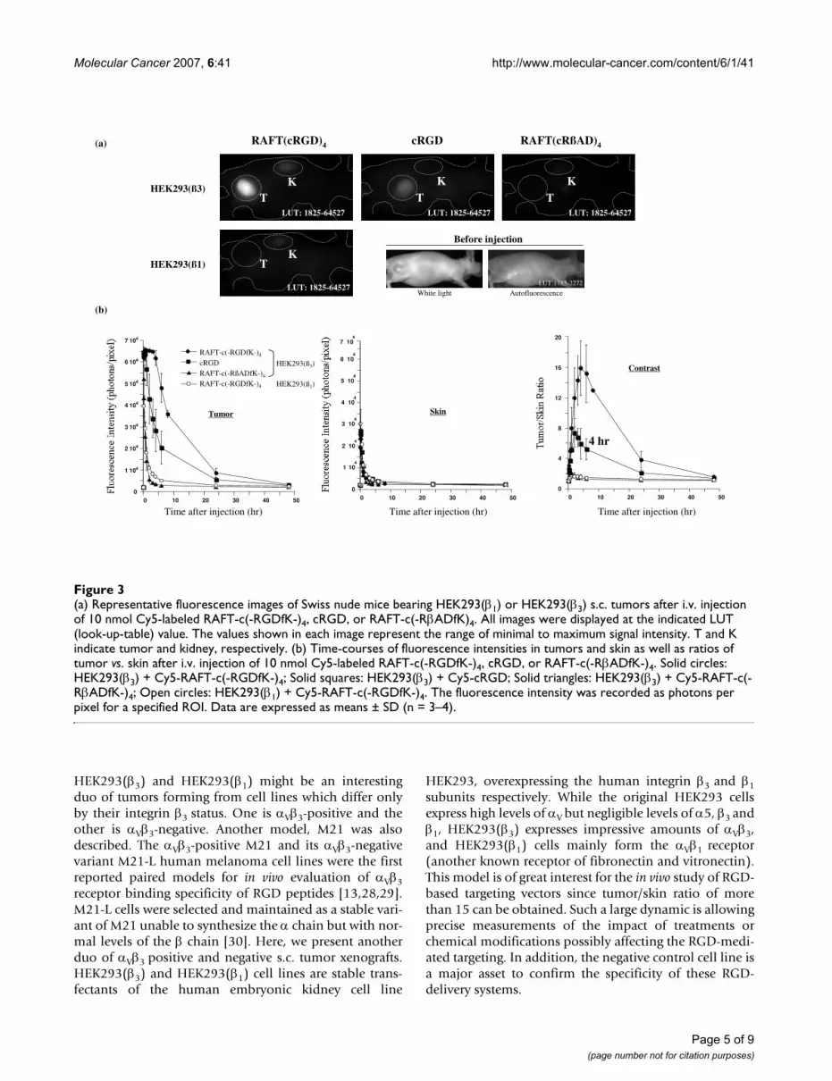
Whole body optical imagingNude mice bearing s.c. tumor xenografts of HEK293(β3)or HEK293(β1) cell line received an i.v. injection of 10nmol Cy5-labeled RAFT-c(-RGDfK-)4, cRGD, or RAFT-c(-RβADfK-)4 and were imaged at different time points dur-ing 2 days. As shown in Fig. 3a, four hours after injectiona stronger tumor uptake was observed for RAFT-c(-RGDfK-)4 than for cRGD, while the control probe RAFT-c(-RβADfK-)4 was not retained in the tumor. Interestingly,the αVβ3-negative HEK293(β1) tumors did not take-up theRAFT-c(-RGDfK-)4 peptide, demonstrating the specificityof RAFT-c(-RGDfK-)4 for the αVβ3 integrin. The quantita-tive analysis also showed that the tetramer and the mono-mer reached similar maximal tumor uptake 5 to 30 minpostinjection (p.i.) (Fig. 3b). Between 30 min an 4 hr p.i.,the tetramer's signal remained very elevated in the tumor(65 472 ± 90 to 61 875 ± 3434 photons/pixel) while amarked decrease (from 63 744 ± 3 031 to 28 349 ± 9 727photons/pixel) was measured with the cRGD. At latertime points RAFT-c(-RGDfK-)4 always showed a bettertumor accumulation than the monomer. The negativecontrol probe RAFT-c(-RβADfK-)4 was rapidly washed-outfrom the tumors. In normal skin, all 3 probes exhibitedsimilar kinetic curves, except at early time points (5 minto 1 hr) where cRGD showed a somewhat stronger non-specific diffusion (Fig. 3b). Finally, the tumor contrast (T/S ratio) was markedly enhanced with RAFT-c(-RGDfK-)4.Four hr p.i. the T/S ratio reached the value of 15.9 ± 3.6with RAFT-c(-RGDfK-)4. This was significantly higher thanthat of the monomeric cRGD (5.9 ± 2.0), or the 1.4 ± 0.1
Page 2 of 9(page number not for citation purposes)

Molecular Cancer 2007, 6:41 http://www.molecular-cancer.com/content/6/1/41
ratio obtained for the control probe. Importantly, RAFT-c(-RGDfK-)4 did not accumulate in the αVβ3-negativeHEK293(β1) xenografts. Indeed, the measured signalobtained with RAFT-c(-RGDfK-)4 in HEK293(β1) tumorswas similar to that of RAFT-c(-RβADfK-)4 in HEK293(β3)tumors.
Confocal microscopic observation of RGD-Cy5 conjugate distributionTumors of mice treated as mentioned above were excised3 or 24 hr p.i, and analyzed by CLSM imaging (Fig. 4).Cy5-RAFT-c(-RGDfK-)4 was massively internalized bytumor cells as shown at a higher magnification in theinsert (Fig. 4B). While virtually each tumor cell wasstrongly labeled at 3 hr, it was still easily detectable in alarge proportion of tumor cells after 24 hr (data notshown). A similar pattern was obtained with the mono-meric cRGD although the intensity of the signal was lower(Fig. 4C). No specific fluorescence was found with thecontrol peptide Cy5-RAFT-c(-RβADfK-)4 (Fig 4D).
Blocking studyIn order to further establish the in vivo specificity of Cy5-RGD conjugates, 10 nmol Cy5-RAFT-c(-RGDfK-)4 or Cy5-cRGD were coinjected with 300 nmol unlabeled tetra-meric RGD or 1200 nmol unlabeled monomeric cRGD.The differences in the injected doses of unlabeled mole-cules were calculated in order to maintain equal concen-trations of the competing RGD motifs. As shown in Fig.5A, the tumor uptake of Cy5-RAFT-c(-RGDfK-)4 was sig-nificantly reduced in the presence of ''cold'' (unlabeled)monomer and this effect was more obvious when the''cold'' tetramer was used. As an example, at 3 hr p.i. thesignal intensities were significantly decreasing (p <0.0001) from 65 472 ± 80 without competitor to 34 339± 6 402 in the presence of ''cold'' cRGD (reduction of50%) and down to 12 894 ± 2 504 when the ''cold''tetramer was in excess (reduction of 80%). This blockingeffect was obvious on the corresponding images (Fig. 5A,left panel). In addition, it is important to note that thestrong decrease of the signal in the tumors was observed
(A) Western blot analysis of expressions of integrin subunits αV, β1 and β3 in HEK293(β1) and HEK293(β3) cell linesFigure 1(A) Western blot analysis of expressions of integrin subunits αV, β1 and β3 in HEK293(β1) and HEK293(β3) cell lines. (B) Con-focal laser scanning microscopic images of HEK293(β1) and HEK293(β3) cells incubated for 30 min at 37°C in the presence of 0.1 μM Cy5-labeled RAFT-c(-RGDfK-)4, cRGD, or RAFT-c(-RβADfK)4. Nuclei were stained with Hoechst 33342 (blue), and fluorescence signal from Cy5 was pseudocolored red. Original objective: Plan-Neofluar 40x/1.30 Oil ph3.
HEK293(ß3)HEK293(ß1)
cRGD
RAFT-c(-RßADfK-)4
RAFT-c(-RGDfK-)4
A B
HEK
ß1
ß3
αv
ß1
HEK
ß3
HSP70
Page 3 of 9(page number not for citation purposes)

Molecular Cancer 2007, 6:41 http://www.molecular-cancer.com/content/6/1/41
while the kidneys were showing identical intensities. Thisindicated that, as expected, the non-specific renal uptakeof the tetrameric RGD was not affected by the presence ofthe different competitors. Similarly, a reduction of at least50 to 60 % was obtained when Cy5-cRGD was used forlabeling (Fig. 5B). The blocking effect of both competitorswas very strong even if RAFT-c(-RGDfK-)4 was slightlymore efficient.
DiscussionRGD-based peptides are certainly the most frequentlyused molecules for tumor targeting and are currently inuse for selective drug delivery and tumor imaging in pre-clinical models or in clinical trials. In this study wepresent evidences that presenting four copies of the cRGDmotif on our RAFT carrier greatly improves cRGD-medi-ated tumor targeting in vivo of αVβ3-positive tumors.
In vitro and in vivo, the αVβ3-positive HEK293(β3) cells andtumors were very strongly recognized by Cy5-RAFT-c(-RGDfK-)4 but not by the negative control RAFT-c(-RβADfK-)4. In addition, the αVβ3-negative HEK293(β1)samples remained negative after staining with the RGD orRβAD-based peptides. Furthermore, fluorescence imagesof both cultured cells and excised tumors clearly demon-
strate the stronger labeling of HEK293(β3) cells by Cy5-RAFT-c(-RGDfK-)4 as compared to its monomeric ana-logue, confirming the enhanced receptor bindingachieved when multiple RGD motifs are presented by asingle template. The tetrameric RGD exhibited alsostronger signal intensity in tumors, longer retention andmuch better contrast as compared to its monomeric ana-logue. Such effects could be explained by its augmentedreceptor-binding affinity due to the polyvalency effect[17,19,20] and increased molecular size which certainlydelays the circulation and tumor retention time of theCy5-RAFT-c(-RGDfK-)4. Finally, the active internalizationof the Cy5-RAFT-c(-RGDfK-)4 probe may also contributeto its improved accumulation in the tumor cells. Asshown on the tumor sections, the internalization was verystrong since most of the signal was coming from the cyto-plasm of the target cells. This suggests that such vectorcould be highly efficient to deliver drugs intracellularly.Multivalent presentation of ligands is improving signifi-cantly the targeting of tumors and several highly efficienttargeting molecules allowing a multivalent presentationof RGD have been described [21-27]. Nonetheless, forsome of these molecules the chemical formulation ispoorly characterized and thus the number of ligandmotifs being added on a polymer is random and cannotbe controlled. In addition, the conformation of these mol-ecules is not constrained. It is thus impossible to separatespatially the different biological functions presented by asingle molecule nor it is possible to know its exact struc-ture. These problems are avoided using RAFT-c(-RGDfK-)4 because the chemistry we use is regio- and chemo-selec-tive. Thus the synthesis and purification of the final mol-ecules are perfectly controlled even at gram scale. Inaddition, the RAFT architecture allows a spatial separationbetween the targeting and "drug-delivery" domains.Finally, RAFT is also interesting because its geometryallows a presentation of four RGD motifs at a very highdensity on its small surface.
To further confirm the receptor binding specificity of theCy5-labeled RGD tetramer, blocking experiments wereperformed in vivo. In agreement with other reports usingthe monomeric cRGD [13,14,16], we observed an almostcomplete inhibition of cRGD accumulation in the pres-ence of an excess of "cold" cRGD or RAFT-c(-RGDfK-)4.More interestingly the opposite experiment showed thatwhile cRGD was able to block roughly 50% of Cy5-RAFT-c(-RGDfK-)4 accumulation, the presence of an excess ofunlabeled tetramer was reducing by more than 80% theCy5-RAFT-c(-RGDfK-)4 signal in the tumor. Finally Cy5-RAFT-c(-RGDfK-)4 shows higher renal uptake than Cy5-cRGD. This was observed in either tumor-bearing or nor-mal mice. This renal retention is likely to be non-specificsince it was not modified by the presence of an excess ofunlabeled tetramer or monomer.
Hematoxylin-eosin (H.E.) staining and immunohistochemical staining with anti-CD31 and anti-αVβ3 in HEK293(β1) or HEK293(β3) s.c. xenografts. Original magnification: × 20 objective lensesFigure 2Hematoxylin-eosin (H.E.) staining and immunohistochemical staining with anti-CD31 and anti-αVβ3 in HEK293(β1) or HEK293(β3) s.c. xenografts. Original magnification: × 20 objective lenses.
H.E.
CD31
ααααVββββ3
HEK293(ß3)HEK293(ß1)
A D
B E
C F
Page 4 of 9(page number not for citation purposes)
Molecular Cancer 2007, 6:41 http://www.molecular-cancer.com/content/6/1/41
HEK293(β3) and HEK293(β1) might be an interestingduo of tumors forming from cell lines which differ onlyby their integrin β3 status. One is αVβ3-positive and theother is αVβ3-negative. Another model, M21 was alsodescribed. The αVβ3-positive M21 and its αVβ3-negativevariant M21-L human melanoma cell lines were the firstreported paired models for in vivo evaluation of αVβ3receptor binding specificity of RGD peptides [13,28,29].M21-L cells were selected and maintained as a stable vari-ant of M21 unable to synthesize the α chain but with nor-mal levels of the β chain [30]. Here, we present anotherduo of αVβ3 positive and negative s.c. tumor xenografts.HEK293(β3) and HEK293(β1) cell lines are stable trans-fectants of the human embryonic kidney cell line
HEK293, overexpressing the human integrin β3 and β1subunits respectively. While the original HEK293 cellsexpress high levels of αV but negligible levels of α5, β3 andβ1, HEK293(β3) expresses impressive amounts of αVβ3,and HEK293(β1) cells mainly form the αVβ1 receptor(another known receptor of fibronectin and vitronectin).This model is of great interest for the in vivo study of RGD-based targeting vectors since tumor/skin ratio of morethan 15 can be obtained. Such a large dynamic is allowingprecise measurements of the impact of treatments orchemical modifications possibly affecting the RGD-medi-ated targeting. In addition, the negative control cell line isa major asset to confirm the specificity of these RGD-delivery systems.
(a) Representative fluorescence images of Swiss nude mice bearing HEK293(β1) or HEK293(β3) s.c. tumors after i.v. injection of 10 nmol Cy5-labeled RAFT-c(-RGDfK-)4, cRGD, or RAFT-c(-RβADfK)4Figure 3(a) Representative fluorescence images of Swiss nude mice bearing HEK293(β1) or HEK293(β3) s.c. tumors after i.v. injection of 10 nmol Cy5-labeled RAFT-c(-RGDfK-)4, cRGD, or RAFT-c(-RβADfK)4. All images were displayed at the indicated LUT (look-up-table) value. The values shown in each image represent the range of minimal to maximum signal intensity. T and K indicate tumor and kidney, respectively. (b) Time-courses of fluorescence intensities in tumors and skin as well as ratios of tumor vs. skin after i.v. injection of 10 nmol Cy5-labeled RAFT-c(-RGDfK-)4, cRGD, or RAFT-c(-RβADfK-)4. Solid circles: HEK293(β3) + Cy5-RAFT-c(-RGDfK-)4; Solid squares: HEK293(β3) + Cy5-cRGD; Solid triangles: HEK293(β3) + Cy5-RAFT-c(-RβADfK-)4; Open circles: HEK293(β1) + Cy5-RAFT-c(-RGDfK-)4. The fluorescence intensity was recorded as photons per pixel for a specified ROI. Data are expressed as means ± SD (n = 3–4).
LUT:1785-2272
cRGDRAFT(cRGD)4 RAFT(cRßAD)4
HEK293(ß3)
HEK293(ß1)
Before injection
White light Autofluorescence
0
1 104
2 104
3 104
4 104
5 104
6 104
7 104
0 10 20 30 40 50
Tumor
(a)
0
7 104
0 10 20 30 40 50
Time after injection (hr)
Skin
(b)
0
4
8
12
16
20
0 10 20 30 40 50
Contrast
RAFT-c(-RGDfK-)4
cRGDRAFT-c(-RßADfK-)4
RAFT-c(-RGDfK-)4
HEK293(ß3)
HEK293(ß1)
Time after injection (hr) Time after injection (hr)
LUT: 1825-64527
4 hr
LUT: 1825-64527 LUT: 1825-64527
LUT: 1825-64527
TK
TK
TK
TK
6 104
5 104
4 104
3 104
2 104
104
1
Page 5 of 9(page number not for citation purposes)

Molecular Cancer 2007, 6:41 http://www.molecular-cancer.com/content/6/1/41
ConclusionUsing such paired tumor models, we demonstrated thatRAFT-c(-RGDfK-)4 is specific for the αVβ3 receptor andinternalized. In addition, due to its multifunctional back-bone, it can carry multiple biological functions on a sin-gle, spatially and chemically defined molecule. Finally,the production of large quantities of perfectly controllablebatches makes of RAFT-c(-RGDfK-)4 a powerful and versa-tile synthetic vector for clinical applications like targeted-drug delivery or molecular imaging of cancer. Ultimately,our goal will be to combine these two applications and touse RAFT-c(-RGDfK-)4 for imaging and quantification ofits targeted-drug delivery efficiency.
MethodsRGD-Peptides Synthesis and Fluorescent LabelingThe detailed protocol for synthesis of RGD peptides wasreported previously [20]. Here, a brief description wasgiven below for the strategy of RGD multimerization andfluorescence labeling. RAFT is a cyclic decapeptide (c [-Lys(Boc)-Lys(Alloc)-Lys(Boc)-Pro-Gly-Lys(Boc)-Lys(Alloc)-Lys(Boc)-Pro-Gly-]) with up to six lysine resi-dues. Protection of the lysine in position 1, 3, 6, or 8 andof the two in positions 2 and 7 results in RAFT moleculeshaving two orthogonally addressable domains pointingon either side of the cyclopeptide backbone. On the upperface, four copies of the c [-RGDfK-] peptide were grafted
Confocal laser scanning microscopic images of HEK293(β3) s.c. tumors dissected at 3 hr after i.v. injection of 10 nmol Cy5-labeled RAFT-c(-RGDfK-)4 (A, B), cRGD (C), or RAFT-c(-RβADfK-)4 (D)Figure 4Confocal laser scanning microscopic images of HEK293(β3) s.c. tumors dissected at 3 hr after i.v. injection of 10 nmol Cy5-labeled RAFT-c(-RGDfK-)4 (A, B), cRGD (C), or RAFT-c(-RβADfK-)4 (D). Paraformaldehyde-fixed cryosections were incu-bated with Hoechst 33342 for nuclear staining (blue). Signal from Cy5 was pseudocolored red. Original objective: Plan-Apochromat 63x/1.4 Oil; an additional zoom of 4x was added for the insert in B.
RAFT-c(-RGDfK-)4
cRGD RAFT-c(-RßADfK-)4
3 hr post-injection 3 hr post-injection zoom 4x
3 hr post-injection 3 hr post-injection
A B
C D
Page 6 of 9(page number not for citation purposes)

Molecular Cancer 2007, 6:41 http://www.molecular-cancer.com/content/6/1/41
via an oxime bond (R1-O-N = C-R2) for recognition of theintegrin. On the opposite side of RAFT, Cy5 mono NHS(N-hydroxysuccinimide) ester (Amersham Biosciences,Uppsala, Sweden) was added on the lysine chain (c [-KKKPGKAKPG-]) [17]. As a negative control probe, Cy5-labeled RAFT-c(-RβADfK-)4 was also synthesized in a sim-ilar way. Changing the G amino-acid by a β-Ala abolishesRGD-mediated affinity for the integrins. All the probeswere dissolved in phosphate-buffered saline (PBS) for thein vitro and in vivo application.
Cell Lines and Culture ConditionsHEK293(β3) and HEK293(β1) cells, stable transfectants ofhuman β3 and β1 subunit, respectively, from the humanembryonic kidney cell line (kindly provided by J-F. Gour-vest, Aventis, France) were cultured in DMEM enrichedwith 4.5 g.L-1 glucose and supplemented with 1%
glutamine, 10% fetal bovine serum (FBS), 50 units/mlpenicillin, 50 μg/ml streptomycin and 700 μg/mlGeneticin (G418 sulfate, Gibco, Paisley, UK). The 2 celllines were cultured at 37°C in a humidified 95% air:5%CO2 atmosphere.
Western blot analysis of integrin subunit expressionCells were lysed in RIPA lysis buffer (50 mM Tris-HCl, pH7.5, 150 mM NaCl, 0.5% sodium deoxycholate, 0.1%SDS, 1% Nonidet P-40, 1 mM NaF, 1 mM Na3VO4, 0.5mM phenylmethylsulfonyl fluoride, 10 μg/ml for each ofleupeptin, aprotinin, and pepstatin) for 30 min on ice,and then the lysates were centrifuged at 17000 × g for 15minutes at 4°C. The protein concentration of the superna-tant was quantified using a protein assay kit (Bio RadLabs., Richmond, CA). Aliquots of protein (40 μg) weresubjected to electrophoresis on 7–10% polyacrylamide
Blocking of Cy5-labeled RGD peptide accumulation in HEK293(β3) s.c. tumors by coinjection with unlabeled RGD peptideFigure 5Blocking of Cy5-labeled RGD peptide accumulation in HEK293(β3) s.c. tumors by coinjection with unlabeled RGD peptide. (A) Tumor-bearing mice received i.v. injection of 10 nmol Cy5-RAFT-c(-RGDfK-)4 alone (a), or coinjected with 1200 nmol cRGD (b) or 300 nmol RAFT-c(-RGDfK-)4 (c). (B) Tumor-bearing mice received i.v. injection of 10 nmol Cy5-cRGD alone (a), or coinjected with 1200 nmol cRGD (b) or 300 nmol RAFT-c(-RGDfK-)4 (c). Left panel: representative fluorescence images at 2 hr p.i.; Right panel: kinetics of fluorescence intensities in tumors. T, K and Int indicate tumor, kidney and intestine, respectively. Data are expressed as means ± SD (n = 2–4).
(B)
a
TInt
c
T Int
b
TInt
0
1 104
2 104
3 104
4 104
5 104
6 104
7 104
0 5 10 15 20 25
Time after Injection (hr)
(A)
a
T
K
b
T
K
c
T
K
0
1 104
2 104
3 104
4 104
5 104
6 104
7 104
0 5 10 15 20 25
Time after Injection (hr)
SalinecRGD RAFT-c(-RGDfK-)4
Labeling with Cy5-RAFT-c(-RGDfK-)4, competition with:
SalinecRGD RAFT-c(-RGDfK-)4
Labeling with Cy5-cRGD, competition with:
Page 7 of 9(page number not for citation purposes)
Molecular Cancer 2007, 6:41 http://www.molecular-cancer.com/content/6/1/41
gels containing 0.1% SDS, followed by electrophoretictransfer onto PVDF-membranes, Hybond™-P (AmershamBiosciences UK Limited, Little Chalfont, Buckingham-shire, UK). The membranes were then incubated with pri-mary antibody: rabbit anti-human integrin αV polyclonalantibody (1:5000; Chemicon International, Inc., Temec-ula, CA), rabbit anti-integrin β1 tail serum (1:1500; kindlyprovided by Dr C. Albiges-Rizo, Grenoble, France) ormouse anti-human β3 monoclonal antibody (clone VI-PL2, 1:100; BD Biosciences PharMingen, San Diego, CA).To monitor equal protein loading, membranes were alsoprobed for actin using rabbit anti-actin polyclonal anti-body (1:1000; Sigma) or for HSP70 using mouse anti-HSP70 monoclonal antibody (1:5000; Affinity BioRea-gents Inc.). For visualization, horseradish peroxidase(HRP)-conjugated secondary antibodies, followed byECL™ immunodetection (Amersham Biosciences UK Lim-ited) were used.
Animal, Tumor Models and HistochemistryAnimal procedures were in agreement with the EEC guide-lines. Female athymic Swiss nude mice, purchased fromJanvier (Le Genest Saint Isle, France) at 6–8 weeks of agewere used and maintained under specific pathogen-freeconditions. Subcutaneous (s.c.) injection of 20 × 106
HEK293(β3) or HEK293(β1) cells suspended in 200 μl ofPBS into the right flank of mice resulted in formation of6–8 mm-diameter tumors after 4–6 weeks. Immunostain-ing with mouse anti-human integrin αVβ3 monoclonalantibody, clone LM609 (1:100; Chemicon) was per-formed on acetone-fixed cryosections using M.O.M.immunodetection (peroxidase) Kit (Vector laboratories,Inc., Burlingame, CA). Rat anti-mouse CD31 monoclonalantibody, clone MEC13.3 (1:3000; BD BiosciencesPharMingen) staining was performed on methanol-fixedcryosections using Strept-. ABComplex/HRP immunode-tection Kit (DakoCytomation). The nuclei were counter-stained with hematoxylin.
In Vitro StudiesCells were seeded on sterilized 18-mm-diameter glass cov-erslips in 12-well plates (3 × 10⁵ cells per well), and incu-bated overnight at 37°C. Afterwards, the cells werewashed with PBS and incubated at 37°C in the presenceof Cy5-labeled peptides RAFT-c(-RGDfK-)4, cRGD orRAFT-c(-RβADfK-)4 at final concentration of 0.1 μM for 30min. They were then washed with PBS, fixed with 2%paraformaldehyde at room temperature for 10 min. Thenuclei were stained with 5 μM Hoechst 33342, and thecoverslips were inverted onto glass slides using Mowiol(Calbiochem, San Diego, CA) mounting medium. Theslides were observed with a confocal laser scanning micro-scopy (CLSM) (LSM510, Zeiss, France).
In Vivo Optical Imaging of Tumor-bearing MiceThe mice bearing s.c. HEK293(β3) or HEK293(β1) tumorsat diameter of 6–8 mm were used for imaging experi-ments. They received intravenous (i.v.) injection of Cy5-labeled peptides RAFT-c(-RGDfK-)4, cRGD or RAFT-c(-RβADfK-)4 at 10 nmol for each mouse (n = 3–4 for eachprobe). For the blocking experiments, s.c. HEK293(β3)tumor-bearing mice (n = 2–4 for each group) receivedcoinjection of Cy5 labeled RAFT-c(-RGDfK-)4 or cRGD(10 nmol/mouse) together with unlabeled RAFT-c(-RGDfK-)4 (300 nmol/mouse) or cRGD (1200 nmol/mouse). Four times higher molar concentration of cRGDwas used than that of tetramer to have same number ofcRGD motifs.
Fluorescence reflectance imaging was performed using aHamamatsu optical imaging system described previously[17,18]. In brief, imaging was carried out in a dark box,and anesthetized animal was illuminated with a mono-chromatic 633 nm light (50 μW.cm-2). The re-emitted flu-orescence was filtered using a colored glass filter RG 665(optical density > 5 at the excitation wavelength 633 nm)and collected with a cooled (-70°C) digital charge-cou-pled device (CCD) camera (Hamamatsu digital cameraC4742-98-26LWGS, Hamamatsu Photonics K.K., Japan).All fluorescence images were acquired using 100 ms ofexposure time, with other related parameters kept con-stant throughout the experiment. Images were acquired as16-bit TIFF files which can provide a dynamic of up to65535 grey levels. Image processing used in this study,including setting LUT (look-up-table) range and measure-ment of the fluorescence intensity for each region of inter-est (ROI), were performed using the Wasabi software(Hamamatsu). It is also important to note that all theimages are presented without background subtraction.For quantifying tumor contrast, the mean fluorescenceintensities of the tumor area (T) and that of the distantskin area (S) were calculated; dividing T by S produced theratio between tumor tissues and background level.
Histological Distribution of RGD-peptides in TumorsAt 3 and/or 24 hr after i.v. injection of 10 nmol of Cy5-labeled RAFT-c(-RGDfK-)4, cRGD or RAFT-c(-RβADfK-)4,the mice were euthanized and tumors were excised, frozenin liquid nitrogen and stored at -80°C. Sections of 20–30μm thickness were fixed with 2% paraformaldehyde atroom temperature for 10 min. The nuclei were stainedwith 5 μM Hoechst 33342, and the coverslips weremounted using Mowiol and kept at 4°C in the dark untilobservation using CLSM.
Statistical AnalysisAll the data are given as mean ± standard (SD) of n inde-pendent measurements. Statistical analysis was performed
Page 8 of 9(page number not for citation purposes)

Molecular Cancer 2007, 6:41 http://www.molecular-cancer.com/content/6/1/41
using two-tailed nonparametric Mann-Whitney t-test. Sta-tistical significance was assigned for values of p < 0.05.
Authors' contributionsZJ and JLC were in charge of the experiments. VJ ran the invivo imaging. SF, DB and PD were in charge of the synthe-sis of the molecules, MCF and JLC designed the study. Allauthors read and approved the final manuscript.
AcknowledgementsThis work was supported by the Institut National de la Sante Et de la Recherche Medicale (INSERM), the Centre National de la Recherche Sci-entifique (CNRS), the programme Interdisciplinaire 2001 – 2004 << Imagerie du Petit Animal >>, the Canceropole National << Angiogenese >> and the Institut Universitaire de France (IUF) and the Association for Research on Cancer (ARC), the Agence Nationale pour la Recherche (ANR) and the EMIL and N2L NoEs of the 6th PCRD. We greatly acknowl-edge "la Région Rhône-Alpes" (France) for financial support to Dr Zhao-Hui Jin. We are grateful to Dr. J-F. Gourvest (Aventis, France) for HEK293(β3 and β1) cells. We are also very grateful to Philippe Rizo (CEA Leti, France) for offering optical imaging system. We thank Corine Tenaud and Dominique Desplanques in our lab for technical assistance.
References1. Ruoslahti E: RGD and other recognition sequences for
integrins. Annu Rev Cell Dev Biol 1996, 12:697-715.2. Takagi J: Structural basis for ligand recognition by RGD (Arg-
Gly-Asp)-dependent integrins. Biochem Soc Trans 2004,32(Pt3):403-406.
3. Hynes RO: Integrins: a family of cell surface receptors. Cell1987, 48(4):549-554.
4. Hynes RO: Integrins: versatility, modulation, and signaling incell adhesion. Cell 1992, 69(1):11-25.
5. Haubner R, Gratias R, Diefenbach B, Goodman SL, Jonczyck A, Kes-sler H: Structural and functional aspects of RGD-containingcyclic pentapeptides as highly potent and selective integrinαVβ3 antagonists. Journal of American Chemical Society 1996,118:7461-7472.
6. Stromblad S, Cheresh DA: Integrins, angiogenesis and vascularcell survival. Chem Biol 1996, 3(11):881-885.
7. Gladson CL: Expression of integrin alpha v beta 3 in smallblood vessels of glioblastoma tumors. J Neuropathol Exp Neurol1996, 55(11):1143-1149.
8. Gladson CL, Cheresh DA: Glioblastoma expression of vitronec-tin and the alpha v beta 3 integrin. Adhesion mechanism fortransformed glial cells. J Clin Invest 1991, 88(6):1924-1932.
9. Gehlsen KR, Davis GE, Sriramarao P: Integrin expression inhuman melanoma cells with differing invasive and meta-static properties. Clin Exp Metastasis 1992, 10(2):111-120.
10. Seftor RE, Seftor EA, Gehlsen KR, Stetler-Stevenson WG, Brown PD,Ruoslahti E, Hendrix MJ: Role of the alpha v beta 3 integrin inhuman melanoma cell invasion. Proc Natl Acad Sci U S A 1992,89(5):1557-1561.
11. Filardo EJ, Brooks PC, Deming SL, Damsky C, Cheresh DA: Require-ment of the NPXY motif in the integrin beta 3 subunit cyto-plasmic tail for melanoma cell migration in vitro and in vivo.J Cell Biol 1995, 130(2):441-450.
12. Kwon S, Ke S, Houston JP, Wang W, Wu Q, Li C, Sevick-Muraca EM:Imaging dose-dependent pharmacokinetics of an RGD-fluo-rescent dye conjugate targeted to alpha v beta 3 receptorexpressed in Kaposi's sarcoma. Mol Imaging 2005, 4(2):75-87.
13. Wang W, Ke S, Wu Q, Charnsangavej C, Gurfinkel M, Gelovani JG,Abbruzzese JL, Sevick-Muraca EM, Li C: Near-infrared opticalimaging of integrin alphavbeta3 in human tumor xenografts.Mol Imaging 2004, 3(4):343-351.
14. Chen X, Conti PS, Moats RA: In vivo near-infrared fluorescenceimaging of integrin alphavbeta3 in brain tumor xenografts.Cancer Res 2004, 64(21):8009-8014.
15. Gurfinkel M, Ke S, Wang W, Li C, Sevick-Muraca EM: Quantifyingmolecular specificity of alphavbeta3 integrin-targeted opti-cal contrast agents with dynamic optical imaging. J BiomedOpt 2005, 10(3):34019.
16. Cheng Z, Wu Y, Xiong Z, Gambhir SS, Chen X: Near-infrared flu-orescent RGD peptides for optical imaging of integrinalphavbeta3 expression in living mice. Bioconjug Chem 2005,16(6):1433-1441.
17. Garanger E, Boturyn D, Jin Z, Dumy P, Favrot MC, Coll JL: New mul-tifunctional molecular conjugate vector for targeting, imag-ing, and therapy of tumors. Mol Ther 2005, 12(6):1168-1175.
18. Jin ZH, Josserand V, Razkin J, Garanger E, Boturyn D, Favrot MC,Dumy P, Coll JL: Non-invasive optical imaging of ovarianmetastases using Cy5-labeled RAFT-c(-RGDfK-)4. Mol Imag-ing 2006, 5(3):188-197.
19. Wu Y, Zhang X, Xiong Z, Cheng Z, Fisher DR, Liu S, Gambhir SS,Chen X: microPET imaging of glioma integrin{alpha}v{beta}3 expression using (64)Cu-labeled tetramericRGD peptide. J Nucl Med 2005, 46(10):1707-1718.
20. Boturyn D, Coll JL, Garanger E, Favrot MC, Dumy P: Templateassembled cyclopeptides as multimeric system for integrintargeting and endocytosis. J Am Chem Soc 2004,126(18):5730-5739.
21. Weissleder R, Kelly K, Sun EY, Shtatland T, Josephson L: Cell-spe-cific targeting of nanoparticles by multivalent attachment ofsmall molecules. Nat Biotechnol 2005, 23(11):1418-1423.
22. Shukla R, Thomas TP, Peters J, Kotlyar A, Myc A, Baker Jr JR: Tumorangiogenic vasculature targeting with PAMAM dendrimer-RGD conjugates. Chem Commun (Camb) 2005:5739-5741.
23. Schraa AJ, Kok RJ, Moorlag HE, Bos EJ, Proost JH, Meijer DK, de LeijLF, Molema G: Targeting of RGD-modified proteins to tumorvasculature: a pharmacokinetic and cellular distributionstudy. Int J Cancer 2002, 102(5):469-475.
24. Harbottle RP, Cooper RG, Hart SL, Ladhoff A, McKay T, Knight AM,Wagner E, Miller AD, Coutelle C: An RGD-oligolysine peptide: aprototype construct for integrin-mediated gene delivery.Hum Gene Ther 1998, 9(7):1037-1047.
25. Bibby DC, Talmadge JE, Dalal MK, Kurz SG, Chytil KM, Barry SE,Shand DG, Steiert M: Pharmacokinetics and biodistribution ofRGD-targeted doxorubicin-loaded nanoparticles in tumor-bearing mice. Int J Pharm 2005, 293(1-2):281-290.
26. Ye Y, Bloch S, Xu B, Achilefu S: Design, synthesis, and evaluationof near infrared fluorescent multimeric RGD peptides fortargeting tumors. J Med Chem 2006, 49(7):2268-2275.
27. Liu S, Hsieh WY, Jiang Y, Kim YS, Sreerama SG, Chen X, Jia B, WangF: Evaluation of a (99m)Tc-labeled cyclic RGD tetramer fornoninvasive imaging integrin alpha(v)beta3-positive breastcancer. Bioconjug Chem 2007, 18(2):438-446.
28. Haubner R, Bruchertseifer F, Bock M, Kessler H, Schwaiger M,Wester HJ: Synthesis and biological evaluation of a (99m)Tc-labelled cyclic RGD peptide for imaging the alphavbeta3expression. Nuklearmedizin 2004, 43(1):26-32.
29. Li C, Wang W, Wu Q, Ke S, Houston J, Sevick-Muraca E, Dong L,Chow D, Charnsangavej C, Gelovani JG: Dual optical and nuclearimaging in human melanoma xenografts using a single tar-geted imaging probe. Nucl Med Biol 2006, 33(3):349-358.
30. Cheresh DA, Spiro RC: Biosynthetic and functional propertiesof an Arg-Gly-Asp-directed receptor involved in humanmelanoma cell attachment to vitronectin, fibrinogen, andvon Willebrand factor. J Biol Chem 1987, 262(36):17703-17711.
Page 9 of 9(page number not for citation purposes)


