Embed Size (px)
Citation preview

For Peer Review O
nly
Impaired emotional processing in a patient with a left
posterior insula-SII lesion
Journal: Neurocase
Manuscript ID: NCS-OA 11-072.R1
Manuscript Type: Original Article
Date Submitted by the Author: n/a
Complete List of Authors: BORG, CELINE; CHU NORD Saint-Etienne, Neuropsychology/neurology Bedoin, Nathalie; UMR CNRS 5596, Peyron, Roland; INSERM U879, Bogey, Soline; CHU Saint-Etienne nord, laurent, bernard; CHU Nord, Thomas-Anterion, C.; CHU Saint-Etienne nord,
Keywords: insula, emotion, disgust, implicit processing, explicit processing
URL: http:/mc.manuscriptcentral.com/nncs Email: [email protected]
Neurocase

For Peer Review O
nly
1
Impaired emotional processing in a patient with a left posterior insula-SII lesion
Running head: emotion and insula.
Céline Borg1, Nathalie Bedoin
2, Roland Peyron
1,3, Soline Bogey
1, Bernard Laurent
1, and
Catherine Thomas-Antérion1
1 Neurology/Neuropsychology, CMRR Unit, Hospital Nord, 42270 Saint-Priest-en-Jarez, France
2 Laboratoire Dynamique du Langage, UMR CNRS 5596 and University of Lyon 2, France
3 INSERM U879; University of Lyon; UJM St-Etienne, France
Corresponding author:
Céline Borg
Neurology/Neuropsychology, CMRR Unit
Hospital Nord
Avenue Albert Raimond
42270 Saint-Priest-en-Jarez, France.
Tel: +33 (0)477128882
Fax: +33 (0)477120543
E-mail: [email protected]
Page 1 of 30
URL: http:/mc.manuscriptcentral.com/nncs Email: [email protected]
Neurocase
123456789101112131415161718192021222324252627282930313233343536373839404142434445464748495051525354555657585960
For Peer Review O
nly
2
Abstract
The present case-report investigated the influence of a lesion in the left posterior insula-SII
cortices on the processing of emotions. MB and 16 normal controls explicitly rated the
valence and the intensity of both facial expressions and emotional words. In addition, they
had to perform a number comparison task and a lexical decision task without focusing their
attention on emotional components of stimuli. MB identified the valence of emotional words
as well as the control group. Nevertheless, she provided higher intensity scores for disgusted
words and her responses in the lexical decision task were significantly delayed for these
stimuli. In addition, MB’s response times were not differently influenced by the presence of
irrelevant emotional faces. However, she explicitly identified fewer facial expressions of
disgust and she assessed them as significantly less intense. This pattern of results contributes
to highlight the psychological and behavioural disorders observed after a left posterior insular
stroke.
Key words: insula, emotion, disgust, implicit processing, explicit processing.
Page 2 of 30
URL: http:/mc.manuscriptcentral.com/nncs Email: [email protected]
Neurocase
123456789101112131415161718192021222324252627282930313233343536373839404142434445464748495051525354555657585960

For Peer Review O
nly
3
1. Introduction
Although the insula cortex is clearly associated with the processing of disgust (Calder, Keane,
Manes, Antoun, & Young, 2000; Hennenlotter et al., 2004; Jabbi, Bastiaansen, & Keysers,
2008; Krolak-Salmon, Hénaff, Bretrand, Mauguière, & Vighetto, 2006; Krolak-Salmon et al.,
2003; Phillips et al., 1997, 2004; Shapira et al., 2003; Sprengelmeyer et al., 1998; Stark et al.,
2007; Wicker et al., 2003), the specificity of its relation to the processing of this emotion is
debated. This cerebral area has been found to respond to other emotions such as fear, anger or
sadness (Damasio et al., 2000; Lane, Reiman, Ahern, Schwartz, & Davidson, 1997; Liotti et
al., 2000; Schienle et al., 2002). In a magnetoencephalography study, a strong activation has
been observed in the right insula 200 msec after the presentation of the stimulus, regardless of
whether it induced disgust or happiness. However, a second discriminative response occurred
about 350 msec after the stimulus, and it was stronger for disgust than for happy faces (Chen
et al., 2009). Therefore, it seems likely that the insula is specifically involved in disgust
processes, but it also seems crucial for a more general “interoceptive awareness” that may
contribute to decode our bodily states (Craig, 2009; Phan, Wager, Taylor, & Liberzon, 2002).
Accordingly, an anatomical posterior-to-anterior gradient has been described in the insula
cortex (Craig, 2009) from the primary interoceptive representations (posterior) to the ultimate
representation of one’s feeling (anterior).
Assuming that the insula contributes to the regulation of emotions, the present study
investigated a unique patient (MB) complaining about emotional disturbance after a brain
lesion involving the left posterior insular and SII cortices. The emotional dysfunction reported
by the patient in her daily life could be explained by the existence of two kinds of emotional
processes: integration and reactivity (Coan & Alleen, 2007; Levenson & Miller, 2007).
Emotional integration or explicit processing of emotions refers to elaborate responses
occurring at late post-perceptual stages and results from the explicit recognition of emotions
Page 3 of 30
URL: http:/mc.manuscriptcentral.com/nncs Email: [email protected]
Neurocase
123456789101112131415161718192021222324252627282930313233343536373839404142434445464748495051525354555657585960

For Peer Review O
nly
4
in self and others. Impaired emotional integration is considered as inducing errors in the
identification and naming of facial or vocal expressions (Calder et al., 2000; Lavenu,
Pasquier, Lebert, Petit, & Van der Linden, 1999) or yet as generating a lack of self-conscious
emotions (Evers, Kilander, & Lindau, 2007). Emotional reactivity or implicit processing of
emotions is defined as a standardized and automatic reaction to unexpected emotional stimuli.
The finality of this automatic vigilance process is to provide a defensive reaction and is
considered to interrupt the ongoing cognitive activity. Typically, emotional reactivity induces
a slowdown in response latencies when participants had to ignore the emotional content and
to explicitly direct their attention to other features of the stimulus. The classical methods that
have been used to assess this emotional bias are the emotional Stroop task and the lexical
decision task (e.g., Algom, Chajut, & Ley, 2004; Estes & Verges, 2008; MacKay et al., 2004).
Emotional reactivity increases response times when the central task is performed in the
context of emotion inducing stimuli (particularly negative emotions), although the emotional
content has not to be explicitly processed (Larsen, 2009; Vuilleumier, 2002). Disturbance in
emotional reactivity may result in the increase or the disappearance of such a negative bias.
Therefore, emotional functioning encompasses explicit and implicit processes, which
may be differently impaired by specific pathologies. For instance, patients with fronto-
temporal dementia (FTD) produce intact physiological and behavioural responses in reaction
to emotional contexts. For instance, they correctly reacted to unexpected loud noises (Sturm,
Levenson, Rosen, Allison, & Miller, 2006). In contrast, they exhibited reduced performance
when the task explicitly required higher level of emotional processes. For example, they failed
to identify negative emotions (Rosen et al., 2004) and did not exhibit the usual signs of
embarrassment (e.g., nervous laughter, embarrassment smiling), which typically follow
irrelevant emotional reactivity (Sturm et al., 2006). In addition, Wieser et al. (2006) showed
that patients with Parkinson’s disease (PD) explicitly rated the emotional content of pictures
Page 4 of 30
URL: http:/mc.manuscriptcentral.com/nncs Email: [email protected]
Neurocase
123456789101112131415161718192021222324252627282930313233343536373839404142434445464748495051525354555657585960

For Peer Review O
nly
5
as less exciting than healthy controls did. However, ERPs provided evidence for an automatic
reactivity to the emotional content of the pictures. Therefore, emotional deficits in patients
with PD may reflect blunted emotional responses, which could be partly due to disturbance in
executive functioning, whereas the automatic emotional sensitivity of the patients is spared.
In this research, we investigated the implicit and explicit emotional processes. We
used experimental tests to specifically assess each aspect of emotional processing for disgust,
fear and happiness, with the aim to characterize the consequences of a lesion in the left
posterior insula in MB. In accordance with the literature, we expected atypical emotional
effects of disgust, and deficiencies were also suspected regarding other emotions.
Additionally, this issue was addressed through both facial expressions and printed words.
Emotional responses could be genetically predetermined for facial expressions, but not for
words, which probably entail more sophisticated emotional understanding and higher cortical
processing. Therefore, in a series of experiments, we aimed to examine if MB shows selective
impairments from emotions conveyed by facial expressions and emotional words.
In order to investigate the explicit emotional functioning, participants were required to
indicate the valence of facial expressions and emotional words. Then, they had to rate the
intensity of the emotional content of the stimuli on a five-point scale. According to our first
hypothesis, the patient may be impaired in recognizing emotions, due to her lesion in the
insular cortex and to the crucial role for this cortical area in “interoceptive awareness” and
decoding of bodily states (Craig, 2009; Phan et al., 2002). This may reflect dysfunctions in
emotional integration. In other words, the percentage of incorrect responses especially in
labelling emotions was expected to be higher in MB than in control participants. In addition,
impaired emotional awareness may result in inaccurate explicit evaluation of the intensity of
emotions induced by the stimuli. Therefore, we also predicted that MB would rate emotional
stimuli as less intense than control participants.
Page 5 of 30
URL: http:/mc.manuscriptcentral.com/nncs Email: [email protected]
Neurocase
123456789101112131415161718192021222324252627282930313233343536373839404142434445464748495051525354555657585960
For Peer Review O
nly
6
However, emotional reactivity can be spared in spite of obvious impairment of
emotional integration, for instance in fronto-temporal dementia with extreme self-neglect
(Bedoin, Thomas-Antérion, Dorey, & Lebert, 2009; Sturm et al., 2006; Werner et al., 2007) or
in PD (Wieser et al., 2006). In the present research, emotional reactivity was investigated by a
number comparison task in which participants had to compare two numbers and to ignore the
emotional facial expressions. In this condition, emotional processing could be considered as
implicit and automatic. We also used a lexical decision task in which participants had to
classify words and pseudo-words while ignoring the emotional content of the words. The
maintenance of emotional reactivity in the patient would result in longer response times for
stimuli with emotional content than for neutral stimuli in both experiments. In contrast, if MB
suffered from a decrease in emotional reactivity, response times should not differ between
emotional and neutral stimuli, contrary to the pattern of results in the control group. Finally, a
potential exaggerated emotional reactivity in MB could be reflected by an excessive slow
down in response times for emotional stimuli as compared to controls, because impaired
ability to modulate emotional reactivity.
2. Methods
All participants gave their written informed consent before participation and this research was
conducted in accordance with the Helsinki Declaration.
2.1 Case-report
At onset, this 36-year-old right handed patient (MB) had, on February 2007, an acute
idiopathic stroke in the left sylvian artery, with a right hemiparesia and a partial conduction
aphasia. MRI was performed two days later, showing a recent infarct located in the left
posterior insula and extended to the adjacent operculum, including SII. This lesion was unique
and of small size as shown on figure 1. However, on the delayed MRI the lesion appeared as
Page 6 of 30
URL: http:/mc.manuscriptcentral.com/nncs Email: [email protected]
Neurocase
123456789101112131415161718192021222324252627282930313233343536373839404142434445464748495051525354555657585960

For Peer Review O
nly
7
bifocal, involving the posterior portion of the insular cortex and the lower and medial part of
SII.
(Figure 1 about here)
She was examined one year later in February 2008 at the University Hospital of Saint-
Etienne, France. She had a deficit of motor strength in the right hand, mainly for the
prehension of objects (Jamar scale equal 4 for the right and 22 for the left hand) and a right
hemianesthesia to warmth, heat, pinprick, including the right face. She had severe chronic
neuropathic pain with permanent spontaneous burning sensations, paresthesiae and electrical
discharges, mainly on the right upper limb. In addition, she had a severe allodynia with a
withdrawal reaction to brush, contact and non-noxious cold stimuli. She did not have any
disturbance for taste. She spontaneously reported emotional disturbance resulting in changes
in her behaviour: she was still able to identify emotions but she evaluated their intensity as
less intense compared with what she had been used to feel before her lesion. For example,
when she visited her family three months after the stroke, everybody was crying at the airport,
she understood her family’s emotion, but she was unable to feel happy herself, even though
she was able to correctly identify an emotionally happy situation. In the same way, she
realized that during the travel, her fear of takeoff disappeared and she was not affected by the
fact of leaving her five-year old son behind. She also reported a decrease of her libido and a
complete change of activity occurred with a compulsive drive to paint while she was never
interested in art before the stroke (Thomas-Antérion, Créac’h, Dionet, Borg, & Peyron, 2010).
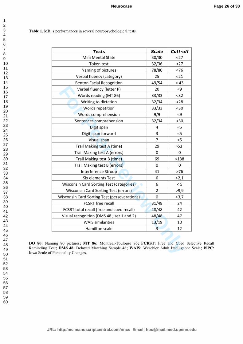
MB’s overall cognitive abilities were normal with only minor executive and verbal
working memory dysfunctions (Table 1). A facial processing test battery (the Benton Facial
Recognition Test) was also administered. MB’s score indicated that she could successfully
process the perceptual features of faces.
Page 7 of 30
URL: http:/mc.manuscriptcentral.com/nncs Email: [email protected]
Neurocase
123456789101112131415161718192021222324252627282930313233343536373839404142434445464748495051525354555657585960

For Peer Review O
nly
8
(Table 1 about here)
2.2. Controls
We compared MB’s performance with that of a control group of 16 healthy adult females,
who were matched for age (mean age: 29.8 years +/- 1.9).
2.3. Materials for emotional investigations
Behavioural symptoms were objectively assessed in MB with the IOWA Scale of Personality
Changes (ISPC, Barrash, Anderson, Jones, & Tranel, 1997; French translation from Juillerat
& Peter-Favre, 1999). This 7-level scale consists in 26 aspects of emotional behaviours and
personality. Actual behaviour (after the lesion) is compared with premorbid behaviour (before
the lesion) (Meulemans, Van der Linden, Seron, & Juillerat, 2000). A quotation of 3 points
corresponds to a “normal” behaviour and higher quotations suggest behavioural dysfunctions.
The ISPC contains four additional dimensions of control (avarice, manipulation, vanity and
behaviour of type “A”) allowing to detect possible biases (e.g., exaggeration or minimization
of the troubles). The IOWA Scale of Personality was filled by a patient’s family member
before carrying out experiments with MB.
For the present study, we used photographs of facial expressions (happiness, fear,
disgust and neutral), most of them were taken from the standardized test battery (Nim
Tottenham, The MacBrain Face Stimulus Set, John D. and Catherine T. MacArthur
Foundation Research Network on Early Experience and Brain Development). The remainder
of the pictures came from a standardized battery elaborated by Baudoin (see Baudoin &
Humphreys, 2006). All photographs, 20 male and 20 female Caucasian actors, were adjusted
to the same size and presented on a white background. This assortment of pictures has been
previously validated in a pilot study conducted with 20 young adults to ensure that each
emotion was identified according to the batteries’ norms. Therefore, a first selection of facial
Page 8 of 30
URL: http:/mc.manuscriptcentral.com/nncs Email: [email protected]
Neurocase
123456789101112131415161718192021222324252627282930313233343536373839404142434445464748495051525354555657585960
For Peer Review O
nly
9
expressions was made among 120 pictures (30 for each emotion): only those with a score of
identification that was shared by at least 80% of the participants were considered. Then, to
adjust the intensity of the emotions, we used a five level scale (1: low intensity to 5: high
intensity) and selected expressions with a similar score of intensity (mean intensity for
disgust: 3.5 (± 0.8); 3.5 (± 0.6) for fear; 3.3 (± 0.5) for happiness; 2.7 (± 0.2) for neutral
expressions).
We also used a set of 64 French words, which were divided into four emotional
valence conditions: 16 were pleasant (e.g., humour), 16 were fear-inducing (e.g., scary), 16
were disgust-inducing (e.g., spit) and 16 were neutral. Emotional and neutral words were
matched for lexical frequency from the data base BRULEX (Content, Mousty, & Radeau,
1990). The number of their letters, phonemes and syllables was also equated. Additionally,
the list contained 64 pseudo-words, which were paired with the words according to the
number of their letters, phonemes and syllables. The 20 participants of the pilot study for the
pictures were also required to categorize and evaluate the intensity of the emotions induced by
the words. As aforementioned for facial expressions, the same rule was used to select 16
words for each emotion among a data base of 120 emotional words (mean intensity for disgust
3.1 (± 0.5); 3.4 (± 0.6) for fear; 3.3 (± 0.3) for happiness; 1.7 (± 0.2) for neutral words).
2.4. Procedure
2.4.1. Explicit processing of emotion
The 40 facial expressions were displayed in a pseudo-randomized order in one block and the
64 words in another block. The order of presentation of the blocks was counterbalanced
between participants. The same timing and sequence of events was used for words and
pictures. Each target was preceded by a fixation cross during 500 msec and lasted until the
participant’s response. Firstly, the participant indicated the valence of the stimulus by
Page 9 of 30
URL: http:/mc.manuscriptcentral.com/nncs Email: [email protected]
Neurocase
123456789101112131415161718192021222324252627282930313233343536373839404142434445464748495051525354555657585960

For Peer Review O
nly
10
choosing one of the proposed labels: happiness, fear, disgust, neutral or “others” (i.e., anger,
surprise…). Then, the participant rated the intensity of the emotional content of the stimulus
on a five-point scale. The list of labels and the scale remained on the screen until the response.
In order to exclude an experimental bias, this explicit task was systematically performed after
the implicit experiments had been completed.
For the explicit task, we carried out analyses on the percentages of correct valence
identification of emotional faces and words and on the mean intensity rating scores. The case-
controls comparisons were carried out using the Q’ test (Michael, 2007; Michael, Garcia,
Bussy, Lion-François, & Guibaud, 2009), with the sole factor being the tested valence
(disgust, fear, happy and neutral items). The Q’ test offers the possibility to investigate the
main effects and interactions by comparing a single case with the control group. This test
requires the transformation of the mean percentage of correct responses and the mean scores
of each condition into z values on the basis of the mean and SD of the controls and the
submitting of the corresponding points to estimate an analysis of proportions.
2.4.2. Implicit processing of emotions with a number comparison task
The same pictures of facial expressions as in the explicit experiment were used here.
Two digits were displayed 18 cm far from each other, one on the right and the other on the
left side of the screen. Each trial started with a centred fixation point displayed for 500 msec
and immediately followed by the presentation of a facial expression at the center of the
screen. The digits were displayed on either side of the picture 100 msec later. Participants
were instructed to focus on the digits and to detect the largest by pressing the corresponding
right or left key. They were asked to perform the task as quickly and accurately as possible
and had no instructions regarding the facial expressions. The inter-stimulus-interval (ISI) was
1500 msec.
Page 10 of 30
URL: http:/mc.manuscriptcentral.com/nncs Email: [email protected]
Neurocase
123456789101112131415161718192021222324252627282930313233343536373839404142434445464748495051525354555657585960

For Peer Review O
nly
11
2.4.3. Implicit processing of emotions with a lexical decision task
The same words as in the explicit experiment were used here. Each trial began with a
500 msec-centred fixation cross, immediately followed either by a letter string or a word.
Participants had to decide whether it was a word or a pseudo-word by pressing on the
corresponding button, as quickly and accurately as possible. The ISI was 1500 msec. The
assignment of the number comparison task and the lexical decision task as the first to be
performed was counterbalanced across participants.
As regard to the number comparison and the lexical decision tasks, we carried out
analysis on the response times (RT). RTs out of 2 standard deviations (SD) were excluded
from the analyses. Discarded trials accounted for less than 5% of the total number of trials, in
the control group as in the patient. The mean error percentage was not considered in the
analysis because it represented less than 2%. The case-controls comparison was carried out
using the Q’ test as aforementioned for the explicit task.
3. Results
3.1. The IOWA Scale of Personality Changes
According to the Wilcoxon test, significant differences appeared in 9 of 26 dimensions of the
IOWA scale (p < .05) for MB. The most striking changes were loss of sensitivity, lack of
resistance, impassiveness, mood changes, irritability, impulsiveness, and also the increased
risk to be easily overwhelmed and to feel emotions in an inadequate way (see table 2).
(Table 2 about here)
Page 11 of 30
URL: http:/mc.manuscriptcentral.com/nncs Email: [email protected]
Neurocase
123456789101112131415161718192021222324252627282930313233343536373839404142434445464748495051525354555657585960
For Peer Review O
nly
12
3.2. Explicit processing task
3.2.1. Valence assessment
Facial expressions. The effect of the emotional valence significantly differed between MB
and the control group (Q’ (3) = 40.3; p < .0001), and it was mainly due to a selective
reduction of disgust identification by MB (Q’ = 4.4; p < .0001, Figure 2a). In addition,
multiple corrected q’ comparisons showed that the difference between neutral and disgusted
facial expressions was significantly higher in MB than in controls (q’ = 4.04; p < .001).
Finally, differences between disgusting facial expressions and fear-inducing or pleasant
expressions were also significantly higher in MB than in the control group (respectively, q’ =
6.17; p < .0001 and q’ = 5; p < .0001). In other words, MB was specifically impaired at
recognizing disgust from facial expressions, compared with the controls.
Emotional words. The performance pattern of MB was not significantly different from that of
the control group. MB judged the valence of words as well as the control group.
3.2.2. Intensity assessment
Facial expressions. Intensity rating differed significantly between MB and the control group
(Q’ (3) = 19.69; p < .0002, Figure 2b). The patient scored the facial expressions of disgust (Q’
= 4.45; p < .0001), fear (Q’ = 4.45; p < .0001) and happiness (Q’ = 4.45; p < .0001), as less
intense than controls, while intensity scores were normal for neutral expressions as compared
to controls. Specifically, and compared with controls, a significantly larger decrease in the
intensity score appeared between neutral expressions on the one hand, and happiness, disgust,
or fear expressions on the other hand (respectively, q’ = 4.29; p < .0001, q’ = 4; p < .001, q’ =
4,07; p < .001).
Emotional words. The effects of emotional intensity differed between MB and the control
group (Q’ (3) = 25.97; p < .001, Figure 2c). The intensity of disgust elicited by words was
Page 12 of 30
URL: http:/mc.manuscriptcentral.com/nncs Email: [email protected]
Neurocase
123456789101112131415161718192021222324252627282930313233343536373839404142434445464748495051525354555657585960

For Peer Review O
nly
13
judged as significantly higher by MB, than by the healthy controls (Q’ = 4.39; p < .0001). A
similar trend was observed for words suggesting fear and happiness but it did not reach
significance. Intensity scores were normal for neutral words as compared to controls.
Compared to all other emotions, disgusting words resulted in significantly higher intensity
scores in MB performances (disgust vs. neutral: q’ = 4.55; p < .0001, disgust vs. fear: q’ =
4.25; p < .0001, and disgust vs. pleasant: q’ = 5.03; p < .0001).
(Figures 2 a, b and c about here)
3.3. Implicit processing task: Number comparison task
No significant effect of facial expression was observed on MB’s RTs, nor on the RTs of the
control group. Precisely, negative and pleasant pictures did not slow down reaction times
when participants had to detect the highest digit by pressing the corresponding key.
3.4. Implicit processing task: Lexical decision task
The effect of emotional words was significantly different in MB and in the control group (Q’
(3) = 31.44; p < .0001). MB was significantly longer to respond than the control group,
whatever the emotional word (p < .002, Figure 3). However, post hoc multiple corrected q’
comparisons revealed that the specific pattern of MB’s results was mainly due to delayed
responses for disgust-inducing words as compared with neutral (q’ = 4.04; p < .001), fear-
inducing (q’ = 2.96; p < .03) and pleasant (q’ = 4.04; p < .001) words. Therefore, MB mainly
differed from the control group by exhibiting greater reactivity to disgust than to other
emotions elicited by the semantic content of words.
In addition, an analysis of variance was performed on correct RTs of the control group
with the emotion factor (disgust, fear, happy and neutral) as the within-subjects factor and
revealed a significant effect of emotion, F(3,45) = 10.45, p < .0001. Post hoc comparison
showed that RTs were longer in the disgust condition (712 ms) than in the neutral condition
Page 13 of 30
URL: http:/mc.manuscriptcentral.com/nncs Email: [email protected]
Neurocase
123456789101112131415161718192021222324252627282930313233343536373839404142434445464748495051525354555657585960

For Peer Review O
nly
14
(664 ms); This suggests that disgusting words induce emotional reactivity in healthy controls,
even if this bias is less important than in MB.
(Figure 3 about here)
4. Discussion
MB felt a decrease in her emotional feelings, but she did not suffer from abnormalities
in social interactions and she had no frontal lobe dysfunction. The ISPC score confirmed
emotional changes in the fields of irritability, impulsiveness, lability, fatigability,
insensitivity, impassiveness, and pointed out inadequate emotions, which was consistent with
her post-stroke complaint. We investigated these changes by using tasks, which assessed
explicit and implicit processing of various emotions. Taken together, the results of our study
provided arguments for MB’s selective deficit in emotional processing, since the
abnormalities did not concern the recognition of neutral facial expressions and neutral words
but only the emotional ones.
Firstly and as it was suggested, MB was impaired in explicit identification of disgust
from facial expressions, while she did not differ from the control group in identifying this
emotion from the content of words. The involvement of the insula in the processing of facial
expression of disgust has largely been reported (Calder et al., 2000; Hennenlotter et al., 2004;
Jabbi et al., 2008; Krolak-Salmon et al., 2003, 2006; Phillips et al., 1997, 2004; Suzuki,
Hoshini, Shigemasu, & Kawamura, 2006; Wicker et al., 2003). The dissociation observed
between the understanding of disgusted facial expressions and the recognition of disgust from
the semantic content of words is consistent with the neuropsychological study of a patient,
NK, whose brain injury affected the anterior and posterior insula cortices and the putamen
(Calder et al., 2000). NK successfully identified emotions from pictures of scenes, involving
both verbal and non-verbal processing, but he was impaired in recognizing disgust from faces,
and he incorrectly labelled this emotion when it was conveyed by non-verbal emotional
Page 14 of 30
URL: http:/mc.manuscriptcentral.com/nncs Email: [email protected]
Neurocase
123456789101112131415161718192021222324252627282930313233343536373839404142434445464748495051525354555657585960

For Peer Review O
nly
15
human sounds or prosodic cues. Accordingly, it appears that a selective impairment in the
identification of social cues of disgust extracted from facial or prosodic speech signals could
be associated with a lesion of the posterior insula. Contrary to the understanding of the
semantic content of emotional words, the understanding of facial emotions may be
phylogenetically predetermined, which may entail the involvement of specific cerebral areas
and produce a selective deficit of emotional integration of facial expressions.
Secondly and as it was hypothesized, MB assessed every expression of emotion as less
intense than the control group did. According to MB’s scores in the valence assessment task,
the decrease in her explicit sensitivity to facial expressions cannot be attributed to the inability
to identify them (except, perhaps, in the case of disgust). She may rather suffer from a general
impairment in interoceptive awareness and had difficulties in evaluating bodily states
associated with emotion feeling, a problem which has been attributed to cerebral damage to
the insula (Craig, 2009; Phan et al., 2002). As expected, cerebral lesion of the insula produces
major, but not exclusive, impairment in disgust processing. Other studies have similarly
reported deficits in the processing of various facial expressions after lesions in SI, SII and
insular areas (Adolph, Damasio, Tranel, Cooper, & Damasio, 2000), that is consistent with the
selective lesions of MB. In addition, a body of studies recently showed that the insula is
involved in the integration of various emotions (Chen et al., 2009) and in the representation of
“interoceptive states” (Craig, 2009). It is finally possible that MB’s neuropathic pain plays a
role in modifying her “interoceptive” sensitivity since the integrity of the somatosensory
system may contribute to the process of facial emotions. For example, a relation between the
intensity of the sensorial deficiencies and the extent of the impairment in recognizing facial
expressions has been documented elsewhere (Olausson et al. 2002). If the sensitivity to facial
expressions partly depends on the ability to simulate the body state associated with the
expression of this emotion, then the integrity of the somatosensory system is decisive for the
Page 15 of 30
URL: http:/mc.manuscriptcentral.com/nncs Email: [email protected]
Neurocase
123456789101112131415161718192021222324252627282930313233343536373839404142434445464748495051525354555657585960

For Peer Review O
nly
16
recognition of facial emotions (Keysers et al., 2004). MB’s sensory deficit was not strong
enough to prevent her from identifying facial expressions, but it could be important enough to
attenuate the mechanism of simulation and reduce the intensity of her feelings.
Previous results raise the question of whether alexithymia can explain MB’s inability
to feel different emotions. Functional neuroimaging studies showed that insula is a brain
region associated with alexithymia (e.g., Karlsson, Näätänen, & Stenman, 2008; Reker et al.,
2010) and a deficit in interoceptive awareness has been described in alexithymic individuals
(Herbert, Herbert, & Pollatos, 2011). Furthermore, previous studies have suggested that
alexithymic individuals process facial expressions differently from non-alexithymic
individuals (e.g., Pandey & Mandal, 1997; Parker, Prkachin, & Prkachin, 2005; Taylor &
Bagby, 1988). Therefore, the question whether MB presents an alexithymia can be discussed.
Clinically, it is essential to specify that alexithymic individuals are characterized by an
inability to find words to describe feelings or emotions. When they talk about an emotional
situation, their comments are limited to describe an overall impression of their feelings.
Experimentally speaking, even if alexithymia is associated with impaired verbal and non
verbal recognition of emotional stimuli (Lane et al., 1996), studies strongly suggested an
important role of verbal ability on emotional recognition in alexithymia (Kokkonen et al.,
2003). From a clinical point of view, MB fails to feel if she was sad, disappointed, angry or
guilty but she correctly identified and distinguished emotional situations by verbally
expressing them with a rich vocabulary. Besides, in the present study, MB identified the
correct valence of stimuli (except disgust), regardless of the nature of the stimuli, words or
facial expressions. In addition, she correctly rated the intensity of emotional words, certainly
because she based her responses on semantic cues to properly assess emotion. Knowledge
about emotional events can be derived directly from experience, or indirectly, from general
knowledge. On the contrary, facial expressions brought no semantic cues so that MB tended
Page 16 of 30
URL: http:/mc.manuscriptcentral.com/nncs Email: [email protected]
Neurocase
123456789101112131415161718192021222324252627282930313233343536373839404142434445464748495051525354555657585960

For Peer Review O
nly
17
to assign them a low intensity, since she did not feel, herself, the intensity of emotions. MB
seemed to have no physical sensations but she was still able to verbally associate relevant
emotions to events. In addition, alexithymia is an inability to associate one's visual
representations, thoughts and fantasies with a specific emotion. In contrast, MB had rich and
emotional fantasies. She began to paint six months after her stroke. She had never before nor
drawn or painted and had no interest in art. She was, however, a hairdresser with a
specialization as a colorist and loved fashion. She therefore went to buy canvases, brushes and
oil paints and painted her first picture that was reproduced in the paper of Thomas-Antérion
(2009). The composition of her paintings is very rich with a combination of colors and she
varies the themes. Today, she continues to paint. She can paint while emotionally, she feels
blunted. Nevertheless, she finds physical sensations while she paints (Thomas-Antérion,
2009). From all these strong arguments, we cannot conclude that MB is alexithymic.
Regarding the processing of words, we observed two anomalies in MB’s performance.
She explicitly judged disgusting words as more intense than the controls. This result suggests
an atypical increase in her sensitivity to the intensity of this emotion. This effect cannot be
ascribed to lower understanding of the semantic content of words, since the patient did not
differ from the controls in the explicit identification of emotions from words. Nevertheless,
the overestimation of intensity for disgust-inducing words by MB is consistent with the
observed effect exerted by this emotional content on her lexical decisions. In other words, one
of our main results was MB’s very high reactivity to the emotional content of disgusting
words, which yielded delayed responses for these words in the lexical decision task. Such a
result provides evidence for the preservation of emotional reactivity, in spite of difficulties in
regulating the magnitude of emotional responses. Theories of emotion processing suggest that
negative stimuli automatically attract more attention than neutral stimuli (e.g., Carretié et al.,
2001; Huang & Luo, 2006; Smith et al., 2003). This emotional bias is revealed by slower
Page 17 of 30
URL: http:/mc.manuscriptcentral.com/nncs Email: [email protected]
Neurocase
123456789101112131415161718192021222324252627282930313233343536373839404142434445464748495051525354555657585960
For Peer Review O
nly
18
responses to negative-inducing stimuli in several cognitive tasks, such as the lexical decision
task (e.g., Algom et al., 2004; Estes & Verges, 2008). This pattern of results was replicated
here in the control group, and it was very pronounced in MB. This means that the patient
reacted to disgust, but with atypically high intensity, which suggests deficiencies in the
emotional regulation of reactions to disgusting words. Unfortunately, we cannot infer any
conclusion about MB’s emotional sensitivity to facial expressions from performance in the
number comparison task, since we failed to show any reactivity to emotions in this task both
in the controls and in the patient (see Pessoa, McKenna, Gutierrez, & Ungerleider, 2002).
Is the role of the insula restricted to the processing of disgust? This issue could depend
on the processing level (emotional reactivity and integration), which is required by the
situation. The present case-report may contribute to the literature on cognitive/behavioural
changes after small brain injury. It confirms the role of the posterior insula-SII in explicit
evaluation of the intensity of emotions expressed by faces, in understanding the social cues
for disgust, and in regulating reactions to this emotion when conveyed by words. It also
provides new evidence for the independence of these emotional mechanisms. These results
are consistent with the emotional changes experienced by MB in social life. Her impairment
in understanding and regulating emotions conveyed by faces could partly explain the changes
that she described in family meeting, her deficit in empathy, and her difficulty in evaluating
the signs of her own feelings. These feeling states are sometimes assumed to involve the
representation of bodily responses. The imbalance of MB’s sensitivity to the intensity of
emotions may be partly associated with the modification of her somatosensory system and her
internal bodily responses.
Conflict of interests and financial support
The authors have declared that no conflict of interest exists. The present research protocol has
received no financial support.
Page 18 of 30
URL: http:/mc.manuscriptcentral.com/nncs Email: [email protected]
Neurocase
123456789101112131415161718192021222324252627282930313233343536373839404142434445464748495051525354555657585960
For Peer Review O
nly
19
REFERENCES
Algom, D., Chajut, E., & Ley, S. (2004). A rational look at the emotional Stroop
phenomenon: A generic slowdown, not a Stroop effect. Journal of Experimental Psychology:
General, 133, 323-338.
Adolph, R., Damasio, H., Tranel, D., Cooper, G., & Damasio, A.R. (2000). A role for
somatosensory cortices in the visual recognition of emotion as revealed by Three-dimensional
lesion mapping. The Journal of Neuroscience, 20(7), 2683-2690.
Barrash, J., Anderson, S.W., Jones, R., & Tranel, D. (1997). The IOWA Ratings Scale
of Personality: Reliability and validity. Journal on the International Neuropsychological
Society, 3, 27-28.
Baudouin, J.Y., & Humphreys, G.W. (2006). Compensatory strategies in processing
facial emotions: Evidence from prosopagnosia. Neuropsychologia, 44, 1361-1369.
Bedoin, N., Thomas-Antérion, C., Dorey, J.M., & Lebert, F. (2009). Implicit
sensitivity to disgust-inducing stimuli in self-neglect fronto-temporal dementia patients.
Cognitive and Behavioral Neurology, 22 (4), 236-241.
Calder, A.J., Keane, J., Manes, F., Antoun, A., & Young, A.W. (2000). Impaired
recognition and experience of disgust following brain injury. Nature Neuroscience, 3(11),
1077-1078.
Carretié, L., Martin-Loeches, M., Hinojosa, J.A., et al. (2001). Emotion and attention
interaction studied through event-related potentials. Journal of Cognitive Neuroscience, 13,
1109-1128.
Chen, Y.H., Dammers, J., Boers, F., Leiberg, S., Edgar, J.C ., Roberts, T.P., &
Mathiak, K. (2009). The temporal dynamics of insula activity to disgust and happy facial
expressions: a magnetoencephalography study. Neuroimage, 47(4), 1921-1928.
Page 19 of 30
URL: http:/mc.manuscriptcentral.com/nncs Email: [email protected]
Neurocase
123456789101112131415161718192021222324252627282930313233343536373839404142434445464748495051525354555657585960
For Peer Review O
nly
20
Coan, J.A., & Allen, J.J.B. (2007). Handbook of Emotion Elicitation and Assessment.
New York: Oxford University Press.
Content, A., Mousty, P., & Radeau, M. (1990). Brulex. Une base de données lexicales
informatisées pour le français écrit et parlé. L’année Psychologique, 90, 551-566.
Craig, A.D. (2009). How do you feel-now? The anterior insula and human awareness.
Nature Reviews Neuroscience, 10, 59-70.
Damasio, A.R., Grabowski, T.J., Bechara, A., Damasio, H., Ponto, L.L.B., Parvizi, J.,
& Hichwa, R.D. (2000). Subcortical and cortical brain activity during the feeling of self-
generated emotions. Nature Neuroscience, 3, 1049-1056.
Estes, Z., & Verges, M. (2008). Freeze or flee? Negative stimuli elicit selective
responding. Cognition, 108, 557-565.
Evers, K., Kilander, L., & Lindau, M. (2007). Insight in frontotemporal dementia
conceptual analysis and empirical evaluation of the consensus criterion “loss of Insight” in
Frontotemporal dementia. Brain & Cognition, 63, 13-23.
Hennenlotter, A., Schoeder, U., Erhard, P., et al. (2004). Neural correlates associated
with impaired disgust processing in pre-symptomatic Huntington’s disease. Brain, 127, 1446-
1453.
Herbert, B.M., Herbert, C., & Pollatos, O. (2011). On the relationship between
interoceptive awareness and alexithymia: Is interoceptive awareness related to emotional
awareness. Journal of Personality, 79(5), 1149-1175.
Huang, Y.X., & Luo, Y.J. (2006). Temporal course of emotional negativity bias: An
ERP study. Neuroscience Letters, 398, 91-96.
Jabbi, M., Bastiaansen, J., & Keysers, C. (2008). A common anterior insula
representation of disgust observation, experience and imagination shows divergent functional
connectivity pathways. PloS ONE, 3(8), e 2939.
Page 20 of 30
URL: http:/mc.manuscriptcentral.com/nncs Email: [email protected]
Neurocase
123456789101112131415161718192021222324252627282930313233343536373839404142434445464748495051525354555657585960
For Peer Review O
nly
21
Juillerat, A.C., & Peter-Favre, C. (1999). Adaptation française de l’échelle d’Iowa des
changements de personnalité.
Karlsson, H., Näätänen, P., & Stenman, H. (2008). Cortical activation in alexithymia
as a response to emotional stimuli. The British Journal of Psychiatry, 192(1), 32-38.
Keysers, C., Wickers, B., Gazzola, V., Anton, J.L., Fogassi, L., & Gallese, V. (2004).
A Touching Sight: SII/PV Activation during the Observation and Experience of Touch.
Neuron, 42, 1-20.
Kokkonen, P., Veijola, J., Karvonen, J.T., et al. (2003). Ability to speak at the age of 1
year and alexithymia 30 years later. Journal of Psychosomatic Research, 54, 491-495.
Krolak-Salmon, P., Hénaff, M.A., Bertrand, O., Mauguière, F., & Vighetto, A. (2006).
Les visages et leurs émotions. Partie II : la reconnaissance des expressions faciales. Revue
neurologique, 11, 1047-1058.
Krolak-Salmon, P., Hénaff, M.A., Isnard, J., Tallon-Baudry, C., Guenot, M., Vighetto,
A., Bertrand, O., & Mauguière, F. (2003). An attention modulated response to disgust in
human ventral anterior insula. Annals of Neurology, 53, 446-453.
Lane, R.D., Reiman, E.M., Ahern, G.L., Schwartz, G.E., Davidson, R.J. (1997).
Neuroanatomical correlated of happiness, sadness, and disgust. American Journal of
Psychiatry, 154(7), 926-933.
Lane, R.D., Sechrest, L., Reidel, R., et al. (1996). Impaired verbal and nonverbal
emotion recognition in alexithymia. Psychosomatic Medicine, 58, 203-210.
Larsen, R. (2009). The contributions of positive and negative affect to emotional
well-being. Psychological Topics 18, 2, 247-266.
Lavenu, I., Pasquier, F., Lebert, F., Petit, H., & Van der Linden, M. (1999). Perception
of emotion in frontotemporal dementia and Alzheimer’s disease. Alzheimer Disease and
Associated Disorders, 13, 96-101.
Page 21 of 30
URL: http:/mc.manuscriptcentral.com/nncs Email: [email protected]
Neurocase
123456789101112131415161718192021222324252627282930313233343536373839404142434445464748495051525354555657585960
For Peer Review O
nly
22
Levenson, R.W., & Miller, B.L. (2007). Loss of cells. Current Directions in
Psychological Science, 16, 289-294.
Liotti, M., Mayberg, H.S., Brannan, S.K., McGinnis, S., Jerabek, P., & Fox, P.T.
(2000). Differential limbic-cortical correlated of sadness and anxiety in healthy subjects:
implications for affective disorders. Biological Psychiatry, 48, 30-42.
MacKay, D.G., Shafto, M., Taylor, J., Marian, D.E., Abrams, L., & Dyer, J.R. (2004).
Relations between emotion, memory, and attention: Evidence from taboo stroop, lexical
decision, and immediate memory tasks. Memory and Cognition, 32, 474-488.
Meulemans, T., Van der Linden, M., Seron, X., & Juillerat, A.C. (2000). L’évaluation
des conduites émotionnelles, de la personnalité et de la motivation. In X. Seron and M. Van
der Linden (Eds.), Traité de neuropsychologie clinique, (pp. 301-317). Paris: Solal.
Michael, G. (2007). A significance test of interaction in 2×K designs with proportions.
Tutorials in Quantitative Methods for Psychology, 3, 1-7.
Michael, G., Garcia, S., Bussy, G., Lion-François, L., & Guibaud, L. (2009).
Reactivity to visual signals and the cerebellar vermis: Evidence form a rare case with
rhombencephalosynapsis. Behavioral Neuroscience, 123, 86-96.
Olausson, H., Lamarre, Y., Backlund, H., Morin, C., Wallin, B.G., Starck, G.,
Ekholm, S., Strigo, I., Worsley, K., Vallbo, A.B., & Bushnell, M.C. (2002). Unmyelinated
tactile afferents signal touch and project to insular cortex. Nature Neuroscience, 5, 900-904.
Pandey, R., & Mandal, M.K. (1997). Processing of facial expression of emotion and
alexithymia. British Journal of Clinical Psychology, 3, 631-633.
Parker, P.D., Prkachin, K.M., & Prkachin, G.C. (2005). Processing of facial
expressions of negative emotion in alexithymia: The influence of temporal constraint. Journal
of Personality, 73(4), 1087-1107.
Page 22 of 30
URL: http:/mc.manuscriptcentral.com/nncs Email: [email protected]
Neurocase
123456789101112131415161718192021222324252627282930313233343536373839404142434445464748495051525354555657585960
For Peer Review O
nly
23
Pessoa, L., McKenna, M., Gutierrez, E., & Ungerleider, L.G. (2002). Neural
processing of emotional faces requires attention. Proceeding of The National Academy of
Sciences, USA, 99, 11458-11463.
Phan, K.L., Wager, T., Taylor, S.F., & Liberzon, I. (2002). Functional neuroanatomy
of emotion: a meta-analysis of emotion activation studies in PET and fRMI. NeuroImage, 16,
331-341.
Phillips, M.L., Williams, L.M., Heining, M., Herba, C.M., Russel, T., Andrew, C., et
al. (2004). Differential neural responses to overt and covert presentations of facial expressions
of fear and disgust. NeuroImage, 21, 1484-1496.
Phillips, M.L., Young, A.W., Senior, C., Brammer, M., Andrews, C., Calder, A.J.,
Bullmore, E.T., Perrett, D.I., Rowland, D., Williams, S.C., Gray, J.A., & David, A.S. (1997).
A specific neural substrate for perceiving facial expressions of disgust. Nature, 389, 495-498.
Reker, M., Ohrmann, P., Rauch, A.V., Kugel, H., Bauer, J., Dannlowski, U., Arolt, V.,
Heindel, W., & Suslow, T. (2010). Individual differences in alexithymia and brain response to
masked emotion faces. Cortex, 46(5), 658-667.
Rosen, H.J., Pace-Savitsky, K., Parry, R.J., Kramer, J.H., Miller, B.L., & Levenson,
R.W. (2004). Recognition of emotion in the forntal and temporal variants of fronto-temporal
dementia. Dementia and Geriatric Cognitive Disorders, 17, 277-281.
Schienle, A., Stark, R., Walter, B., Blecker, C., Ott, U., Kirsch, P., Sammer, G., &
Vaitl, D. (2002). The insula is not specifically involved in disgust processing: an fRMI study.
Neuroreport, 13, 2023-2026.
Shapira, N.A., Liu, Y., He, A.G., Bradley, M.M., Lessig, M.C., James, G.A., et al.
(2003). Brain activation by disgust-inducing pictures in obsessive-compulsive disorder.
Biological Psychiatry, 54, 751-756.
Page 23 of 30
URL: http:/mc.manuscriptcentral.com/nncs Email: [email protected]
Neurocase
123456789101112131415161718192021222324252627282930313233343536373839404142434445464748495051525354555657585960

For Peer Review O
nly
24
Smith, N.K., Cacioppo, J.T., Larsen, T.L., et al. (2003). May I have your attention,
please: Electrocortical responses to positive and negative stimuli. Neuropsychologia, 41, 171-
183.
Sprengelmeyer, R., Raush, M., Eysel, U.T., et al. (1998). Neural structures associated
with recognition of facial expressions of basic emotions. Proc R Soc Lond B Biol Sci, 265,
1927-1931.
Stark, R., Zimmermann, M., Kagerer, S., et al. (2007). Hemodynamic brain correlates
of disgust and fear ratings. NeuroImage, 37, 663-673.
Sturm, V.E., Levenson, R.W., Rosen, H.J., Allison, S.C., & Miller, B.L. (2006).
Preserved simple emotion and diminshed self-conscious elituib ub frontotemporal lobar
degeneration. Brain, 129, 2508-2516.
Suzuki, A., Hoshini, T., Shigemasu, K., & Kawamura, M. (2006). Disgust specific
impairment of facial expression recognition in Parkinson’s disease. Brain, 129, 707-717.
Taylor, G.J., & Bagby, R.M. (1988). Measurement of alexithymia: recommendations
for clinical practice and future research. Psychiatric Clinic of North America, 11, 351-366.
Thomas-Antérion, C. (2009). Emergence of artist talent and neurology: three cases.
Revue de neuropsychologie. 1(3), 221-228.
Thomas-Antérion, C., Créac’h, C., Dionet, E., Borg, C., & Peyron, R. (2010). De novo
artistic activity following insular-SII ischemia. Pain, 150, 121-127.
Vuilleumier, P. (2002). Facial expression and selective attention. Current Opinion in
Psychiatry, 15, 291-300.
Wicker, B., Keysers, C., Plailly, J., Royet, J.P., Gallese, V., & Rizzolatti, G. (2003).
Both of us disgusted in my insula: The common neural basis of seeing and feeling disgust.
Neuron, 40, 655-664.
Page 24 of 30
URL: http:/mc.manuscriptcentral.com/nncs Email: [email protected]
Neurocase
123456789101112131415161718192021222324252627282930313233343536373839404142434445464748495051525354555657585960
For Peer Review O
nly
25
Werner, K.H., Roberts, N.A., Rosen, H.J., et al. (2007). Emotional reactivity and
emotion recognition in frontotemporal lobar degeneration. Neurology, 69, 148-155.
Page 25 of 30
URL: http:/mc.manuscriptcentral.com/nncs Email: [email protected]
Neurocase
123456789101112131415161718192021222324252627282930313233343536373839404142434445464748495051525354555657585960
For Peer Review O
nly
Table 1. MB’ s performances in several neuropsychological tests.
Tests Scale Cutt-off
Mini Mental State 30/30 <27
Token test 32/36 <27
Naming of pictures 78/80 <76
Verbal fluency (category) 25 <21
Benton Facial Recognition 49/54 < 43
Verbal fluency (letter P) 20 <9
Words reading (MT 86) 33/33 <32
Writing to dictation 32/34 <28
Words repetition 33/33 <30
Words comprehension 9/9 <9
Sentences comprehension 32/34 <30
Digit span 4 <5
Digit span forward 3 <5
Visual span 7 <5
Trail Making test A (time) 29 >53
Trail Making test A (errors) 0 0
Trail Making test B (time) 69 >138
Trail Making test B (errors) 0 0
Interference Stroop 41 >76
Six elements Test 6 >2,1
Wisconsin Card Sorting Test (categories) 6 < 5
Wisconsin Card Sorting Test (errors) 2 >9,9
Wisconsin Card Sorting Test (perseverations) 0 >3,7
FCSRT free recall 31/48 24
FCSRT total recall (free and cued recall) 48/48 42
Visual recognition (DMS 48 ; set 1 and 2) 48/48 47
WAIS similarities 13/19 10
Hamilton scale 3 12
DO 80: Naming 80 pictures; MT 86: Montreal-Toulouse 86; FCRST: Free and Cued Selective Recall
Reminding Test; DMS 48: Delayed Matching Sample 48; WAIS: Weschler Adult Intelligence Scale; ISPC:
Iowa Scale of Personality Changes.
Page 26 of 30
URL: http:/mc.manuscriptcentral.com/nncs Email: [email protected]
Neurocase
123456789101112131415161718192021222324252627282930313233343536373839404142434445464748495051525354555657585960

For Peer Review O
nly
Table 2. Exploration of emotional and behaviour disturbances by means of the IOWA Scale of Personality
(ISPC): items presenting in bold and with ** represent the most significant changes.
items Now Before Change
Irritability ** 6 1 5
Lack of initiative 1 1 0
Perseveration 1 1 0
Depression 3 1 2
Impulsiveness ** 5 1 4
Mania 5 5 0
Moody character ** 5 2 3
Lack of persistence 1 1 0
Lack of resistance ** 5 1 4
Abilities of planning 2 1 1
Obstinacy 1 1 0
Abilities of judgement 1 1 0
Anxiety 4 2 2
Insensitivity ** 7 2 5
Social behavior 1 1 0
Dependence 2 1 1
Impatience 3 3 0
Behavior of type « A » 6 3 3
Impassiveness ** 7 1 6
Social withdrawal 3 1 2
Aggressiveness 3 1 2
Indecision 1 1 0
Vanity 1 1 0
Mistrust 1 1 0
Apathy 1 3 2
Avarice 1 1 0
Inadequate emotions ** 7 1 6
Manipulation 5 5 0
Risk to be overwhelmed ** 5 1 4
Conscience of troubles 3 3 0
Total 97 50 52
Page 27 of 30
URL: http:/mc.manuscriptcentral.com/nncs Email: [email protected]
Neurocase
123456789101112131415161718192021222324252627282930313233343536373839404142434445464748495051525354555657585960
For Peer Review O
nly
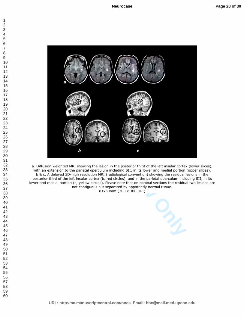
a. Diffusion weighted MRI showing the lesion in the posterior third of the left insular cortex (lower slices), with an extension to the parietal operculum including SII, in its lower and medial portion (upper slices). b & c. A delayed 3D-high resolution MRI (radiological convention) showing the residual lesions in the
posterior third of the left insular cortex (b, red circles), and in the parietal operculum including SII, in its lower and medial portion (c, yellow circles). Please note that on coronal sections the residual two lesions are
not contiguous but separated by apparently normal tissue. 81x60mm (300 x 300 DPI)
Page 28 of 30
URL: http:/mc.manuscriptcentral.com/nncs Email: [email protected]
Neurocase
123456789101112131415161718192021222324252627282930313233343536373839404142434445464748495051525354555657585960
For Peer Review O
nly
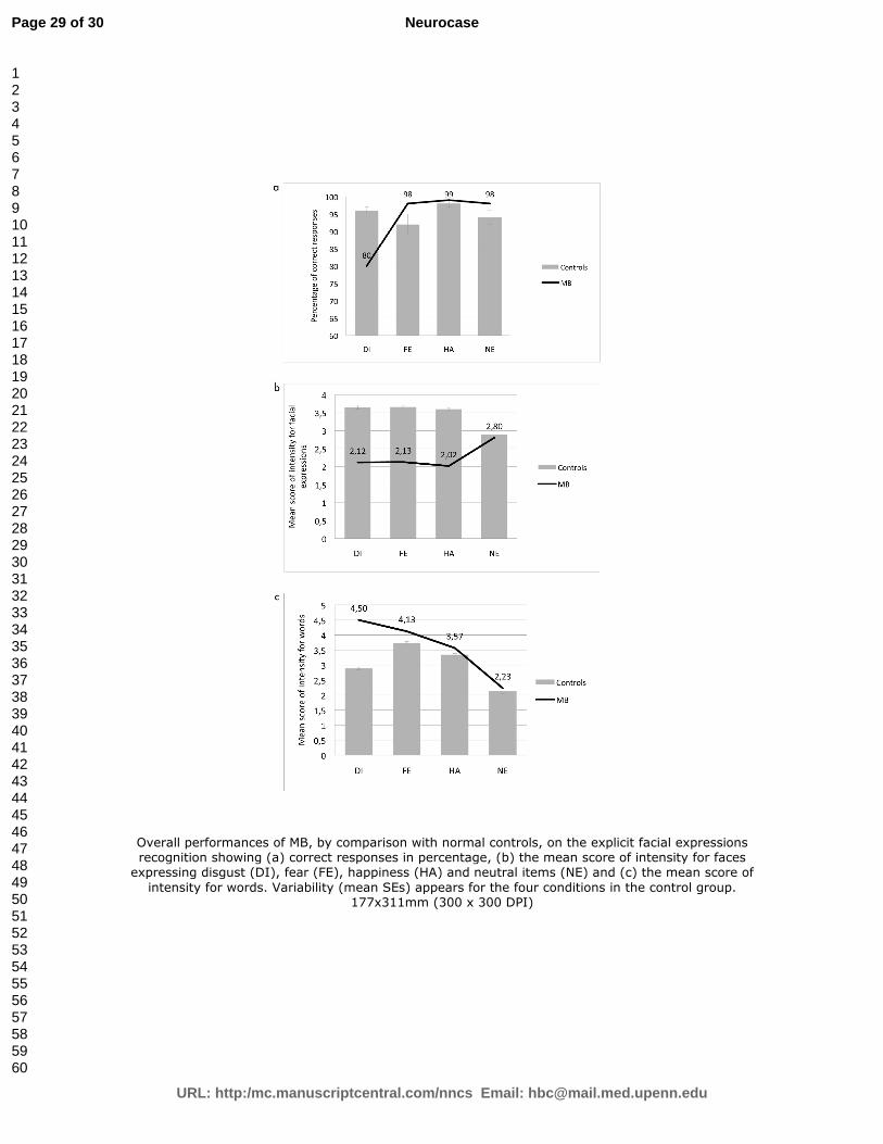
Overall performances of MB, by comparison with normal controls, on the explicit facial expressions recognition showing (a) correct responses in percentage, (b) the mean score of intensity for faces
expressing disgust (DI), fear (FE), happiness (HA) and neutral items (NE) and (c) the mean score of
intensity for words. Variability (mean SEs) appears for the four conditions in the control group. 177x311mm (300 x 300 DPI)
Page 29 of 30
URL: http:/mc.manuscriptcentral.com/nncs Email: [email protected]
Neurocase
123456789101112131415161718192021222324252627282930313233343536373839404142434445464748495051525354555657585960

For Peer Review O
nly
Mean response times of MB and controls in the lexical decision task for words expressing disgust (DI), fear (FE), happiness (HA) and for neutral items (NE). Variability (mean SEs) appears for the four conditions in
the control group. 62x38mm (300 x 300 DPI)
Page 30 of 30
URL: http:/mc.manuscriptcentral.com/nncs Email: [email protected]
Neurocase
123456789101112131415161718192021222324252627282930313233343536373839404142434445464748495051525354555657585960


