Embed Size (px)
Citation preview

0021
doi:1
Corr
Immunohistochemical Distinction betweenPreclinical Bovine Spongiform Encephalopathy
and Scrapie Infection in Sheep
C. M. A. Thuring, L. J. M. van Keulen*, J. P. M. Langeveld*,M. E. W. Vromans*, F. G. van Zijderveld* and T. Sweeney
Department of Animal Husbandry and Production, Faculty of Veterinary Medicine, University College Dublin,Ballsbridge, Dublin 4, Ireland and *Department of Bacteriology and TSEs, Central Institutefor Animal Disease Control (CIDC), P.O. Box 2004, 8203 AA Lelystad, The Netherlands
Summary
-99
0
es
Sheep are susceptible experimentally to bovine spongiform encephalopathy (BSE), the clinical signsbeing indistinguishable from those of scrapie. Because of the possibility of natural ovine BSE infection,laboratory tests are needed to distinguish between scrapie and BSE infection. The objectives of thisstudy were to determine whether (1) PrPSc accumulates in biopsy samples of the tonsil or third eyelid, orboth, of BSE-infected sheep before the appearance of clinical disease, and (2) such samples from BSE-and scrapie-infected sheep differ in respect of PrPSc accumulations. Homozygous ARQ sheep (nZ10)were dosed orally at 4–5 months of age with a brain homogenate from BSE-infected cattle. Third eyelidand tonsillar biopsy samples were taken at %6 monthly intervals post-infection and examinedimmunohistochemically for PrPSc. Third eyelid protuberances were difficult to identify, resulting inmany unsuitable samples; however, third eyelid samples shown to contain lymphoid follicles wereinvariably negative for PrPSc. In contrast, tonsillar biopsy samples became positive for PrPSc from 11 to 20months post-infection. Consistent differences in the morphology of PrPSc granules in tingible bodymacrophages (TBMs) between BSE- and scrapie-infected sheep were detected with anti-peptideantibodies directed towards amino acids 93–106 of the ovine prion protein: thus, PrPSc appearedas single granules in TBMs of tonsillar sections from BSE-infected sheep, whereas clusters of PrPSc
granules were observed within TBMs in the tonsils of scrapie-infected sheep. In contrast, antibodiesagainst epitopes situated N- and C-terminally from the 93–106 region of the ovine prion protein revealedno differences between BSE- and scrapie-infected sheep in terms of PrPSc granules in TBMs.
q 2004 Elsevier Ltd. All rights reserved.
Keywords: bovine spongiform encephalopathy; BSE; prion protein; PrPSc; scrapie; sheep; third eyelid; tingible body macrophages; tonsil;viral infection
Introduction
With increasing understanding of the way in whichthe bovine spongiform encephalopathy (BSE)epidemic began in the United Kingdom, anawareness arose that BSE might also have spreadin the sheep population, as a result of the ingestionof BSE-contaminated meat and bone meal
75/$ - see front matter
.1016/j.jcpa.2004.06.004
pondence to: L.J.M. van Keulen.
(Baylis et al., 2002; Gravenor et al., 2003). Giventhat the BSE agent is probably capable of infectinghuman beings via the oral route, resulting invariant Creutzfeldt–Jacob disease (Bruce et al.,1997), BSE-infected sheep tissues would seem apotential threat to public health. This threat mightbe even greater than that posed by the ingestion ofmeat from BSE-infected cattle, since previousstudies showed that in sheep experimentallyinfected with BSE, the agent accumulated in both
J. Comp. Path. 2005, Vol. 132, 59–69
www.elsevier.com/locate/jcpa
q 2004 Elsevier Ltd. All rights reserved.

C.M.A. Thuring et al.60
the lymphoid and neural tissues (Foster et al.,1996), whereas in natural BSE infections in cattle,infectivity has been detected almost exclusively inneural tissues and not in lymphoid tissues(Fraser and Foster, 1993). In experimentallyinfected cattle, however, infectivity was also foundin the gut-associated lymphoid tissue of the ileum(Wells et al., 1998).
Clinical signs of experimental BSE in sheepinclude ataxia and pruritus (Foster et al., 1994,2001; Houston and Gravenor, 2003). Since thesesigns resemble those of natural scrapie in sheep,it would be impossible to distinguish field cases ofovine BSE from natural scrapie cases by clinicalexamination. So far no test has been developed todistinguish between these spongiform encephalo-pathies in live sheep. At present, there are tests todiagnose scrapie in the live animal based on PrPSc
detection in biopsy samples of the tonsil or thirdeyelid (Schreuder et al., 1996, 1998; O’Rourke et al.,1998, 2000), but it is not known whether these testscan be used for the preclinical detection of BSE-infected animals or to distinguish between BSE andscrapie infection.
Hence, the first objective of this study was todetermine whether there is a preclinical presenceof PrPSc in tonsils and third eyelids of sheepexperimentally infected with BSE. The secondobjective was to identify potential differences inPrPSc accumulations in tingible body macrophages(TBMs) or follicular dendritic cells (FDCs) of thetonsil and third eyelid that might be used todistinguish between scrapie and BSE infection inlive sheep.
Materials and Methods
Sheep and Experimental Procedures
BSE-infected sheep. Texel or Texel cross ewes weremaintained and mated in a closed environmentwhere no scrapie-affected ewes had lambed in thepast 10 years. Offspring were weaned, selected onthe basis of their PrP genotype (determined by fullDNA sequencing of the open reading frame of thePrP gene) and transferred to a containment level 3unit at 4–5 months of age. This group consisted of10 ARQ/ARQ lambs (five castrated males and fivefemales) and one female ARR/ARR lamb. (The PrPgenotype is expressed as the triplet sequencepresent at amino acids 136, 154 and 171 of thePrP protein.) These lambs were dosed orally with5 g of a BSE brain homogenate, administered inthe back of the mouth with a disposable 25-mlsyringe containing a 20% (w/v) brain homogenate.
The homogenate was prepared from brainstems ofBritish cattle with confirmed BSE infection.Samples of the same brainstem homogenate poolhad been used in transmission experiments byJeffrey et al. (2001a,b).
Tonsils and third eyelids of all lambs werebiopsied at % 6-monthly intervals post-infection(p.i.) until PrPSc was detected, as describedpreviously (Schreuder et al., 1996, 1997, 1998;Thuring et al., 2002). Examination of threelymphoid follicles was regarded as the minimumbasis on which to judge the presence or absence ofPrPSc in biopsy samples (Schreuder et al., 1998).One ARQ/ARQ lamb was sampled and killed at 6months p.i. No PrPSc could be demonstrated in thetonsillar biopsy of this animal. Five ARQ/ARQlambs were killed in the preclinical phase of BSEinfection after PrPSc had been detected in tonsillarbiopsies. The remaining four BSE-infected ARQ/ARQ sheep were left to develop clinical signs(ataxia, head tremors and positive nibbling reflex)at 20, 21, 23 or 24 months p.i. These sheep werethen killed within 14 days. The method ofeuthanasia was exsanguination after anaesthesiawith sodium pentobarbital given intravenously(Nembutal; Ceva Sante Animale BV, Libourne,France).
Two negative control lambs, one male and onefemale, both ARQ/ARQ, were left undosed andmaintained outside in scrapie-free premises. Oneundosed lamb died at the age of 12 months ofunknown causes; the absence of PrPSc in theobex and tonsil was confirmed immunohisto-chemically. The remaining undosed ARQ/ARQsheep and the dosed ARR/ARR sheep were stillalive and healthy at the time of writing(43 months p.i.)Sheep with natural scrapie. Archived material con-sisted of (1) tonsillar biopsy samples from 72 pre-clinical (17 ARQ/ARQ, 32 VRQ/ VRQ and 23VRQ/ARQ), and (2) complete tonsils taken atnecropsy from 113 clinical cases (36 ARQ/ARQ,39 VRQ/VRQ and 38 VRQ/ARQ) (the PrPgenotype was determined by Taqman analysis ofthe codons 136, 154 and 171 of the PrP gene, whichdoes not discriminate between Q171 and H171.)Sheep with experimental scrapie. Two VRQ/ARQ sheepwere orally infected immediately after birth with10 ml of a 10% (w/v) brain homogenate originat-ing from a brainpool of VRQ/ARQ sheep in whichscrapie had been diagnosed (McElroy et al., 2002).They were killed at 16 and 20 months of age whenshowing clinical signs, and their tonsils werecollected at necropsy.
Distinction between BSE and Scrapie in Sheep 61
Immunohistochemistry (IHC)
Dissected whole tonsils and biopsy samples fromtonsils and third eyelids were dehydrated andembedded in paraffin wax by routine procedures.Tissue sections (4 mm) were cut, placed on2-amino-propyl-triethoxysilane-coated glass slides(Sigma Chemical Co, St Louis, MO, USA) anddried overnight at 60 8C.Single immunolabelling. After dewaxing and inacti-vation of endogenous peroxidase activity, sectionswere pretreated by immersion in formic acid for30 min and autoclaving in citrate buffer (pH 6.0)for 5 min. Immunohistochemical labelling wasperformed with polyclonal antibodies or mono-clonal antibodies directed against various epitopesdistributed along the ovine PrP. Secondary anti-body coupled to horseradish peroxidase (HRP)consisted of Mouse or Rabbit Envision HRP (DakoDiagnostics B.V., Glostrup, Denmark), and diami-nobenzidine (DAB; Sigma) was used as substrate.Negative control reagents for the polyclonalantisera and monoclonal antibodies consisted,respectively, of pre-immune sera and the mono-clonal antibody solution pre-absorbed with thepeptides or protein used for immunization.Double immunolabelling. After incubation with anti-PrP antibodies, sections were incubated with asecondary antibody coupled to alkaline phospha-tase (Powervision AP; ImmunoVision Technol-ogies, Daly City, CA, USA) and colour wasdeveloped with 5-bromo-4-chloro-3-indoxyl phos-phate and nitro blue tetrazolium chloride(BCIP/NBT; Dako) for 10 min. After rinsing withdistilled water, the second primary antibody(mouse-anti-human CD68; Serotec, Oxford,
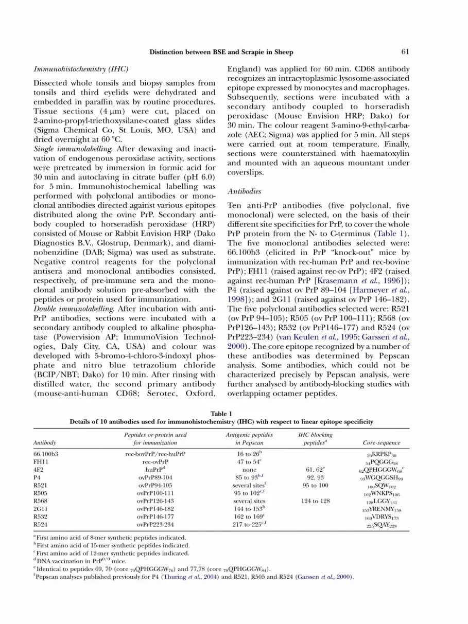
TableDetails of 10 antibodies used for immunohistochemis
Antibody
Peptides or protein used
for immunization
66.100b3 rec-bovPrP/rec-huPrPFH11 rec-ovPrP4F2 huPrPd
P4 ovPrP89-104R521 ovPrP94-105R505 ovPrP100-111R568 ovPrP126-1432G11 ovPrP146-182R532 ovPrP146-177R524 ovPrP223-234
a First amino acid of 8-mer synthetic peptides indicated.b First amino acid of 15-mer synthetic peptides indicated.c First amino acid of 12-mer synthetic peptides indicated.d DNA vaccination in PrP0/0 mice.e Identical to peptides 69, 70 (core 70QPHGGGW76) and 77,78 (core 7f Pepscan analyses published previously for P4 (Thuring et al., 2004) an
England) was applied for 60 min. CD68 antibodyrecognizes an intracytoplasmic lysosome-associatedepitope expressed by monocytes and macrophages.Subsequently, sections were incubated with asecondary antibody coupled to horseradishperoxidase (Mouse Envision HRP; Dako) for30 min. The colour reagent 3-amino-9-ethyl-carba-zole (AEC; Sigma) was applied for 5 min. All stepswere carried out at room temperature. Finally,sections were counterstained with haematoxylinand mounted with an aqueous mountant undercoverslips.
Antibodies
Ten anti-PrP antibodies (five polyclonal, fivemonoclonal) were selected, on the basis of theirdifferent site specificities for PrP, to cover the wholePrP protein from the N- to C-terminus (Table 1).The five monoclonal antibodies selected were:66.100b3 (elicited in PrP “knock-out” mice byimmunization with rec-human PrP and rec-bovinePrP); FH11 (raised against rec-ov PrP); 4F2 (raisedagainst rec-human PrP [Krasemann et al., 1996]);P4 (raised against ov PrP 89–104 [Harmeyer et al.,1998]); and 2G11 (raised against ov PrP 146–182).The five polyclonal antibodies selected were: R521(ov PrP 94–105); R505 (ov PrP 100–111); R568 (ovPrP126–143); R532 (ov PrP146–177) and R524 (ovPrP223–234) (van Keulen et al., 1995; Garssen et al.,2000). The core epitope recognized by a number ofthese antibodies was determined by Pepscananalysis. Some antibodies, which could not becharacterized precisely by Pepscan analysis, werefurther analysed by antibody-blocking studies withoverlapping octamer peptides.
1try (IHC) with respect to linear epitope specificity
Antigenic peptides
in Pepscan
IHC blocking
peptidesa Core-sequence
16 to 26b26KRPKP30
47 to 54c54PQGGG58
none 61, 62e62QPHGGGW68
e
85 to 93b,f 92, 93 93WGQGGSH99
several sitesf 95 to 100 100SQW102
95 to 102c,f102WNKPS106
several sites 124 to 128 128LGGY131
144 to 153b153YRENMY158
162 to 169c169VDRYS173
217 to 225c,f225SQAY228
8QPHGGGW84).d R521, R505 and R524 (Garssen et al., 2000).

C.M.A. Thuring et al.62
Pepscan analysis. To detect linear epitope specificityof the antibodies used, Pepscan analyses wereperformed in an ELISA-like procedure on solid-phase synthetic peptides bound to plastic surfaces,as described previously (Geysen et al., 1984; Garssenet al., 2000). Complete sets of overlapping 12- or 15-mer peptides were synthesized on the basis of theamino-acid sequences of the ovine PrP (Goldmannet al., 1990). A site was considered positive (i.e.antigenic) if the absorbance values of two or moreconsecutive peptides were at least four times that ofthe background. Background was calculated as theaverage absorbance value measured for 20 con-secutive low-reactive peptides where the standarddeviation amounted to %50% of the averageabsorbance value. The core was defined as thesequence of amino acids common to all antigenicpeptides in a site.Antibody blocking with synthetic peptides. Antibodiesthat could not be characterized precisely byPepscan analysis were further analysed by thecapacity of overlapping 8-mer peptides to blockthe immunohistochemical signal. In short, over-lapping octamer peptides were manufactured,covering the whole region of the immunogenused to raise the antibody. Peptides to a sequenceoutside the specific immunogen region served asnegative control peptides. Peptides were syn-thesized on the basis of the amino-acid sequenceof the ovine PrP (Goldmann et al., 1990) withacetylated and amidated termini, with Fmocchemistry as previously described (van Keulenet al., 1995). The products were checked foridentity by molecular mass spectrometry andbrought to over 80% purity by high performance
Fig. 1. Mapping of antigenic sites on the ovine PrP sequence for diare presented as a complete set of overlapping 15-mer peppeptides (monoclonal antibody FH11 and polyclonal antibofor (about 50) peptides around a mapped site are included
liquid chromatography as evaluated by 215-nmlight absorption analysis. Separate aliquots of theantibodies were pre-incubated overnight at 4 8C,each with one of the synthetic peptides(1 mg peptide/ml of antibody solution). Beforeapplication on the tissue sections, the antibody-peptide mixtures were further diluted 1 in 10 to theworking dilution used in IHC. The blockingcapacity of each peptide was determined byanalysing the PrPSc signal in IHC on tonsil andbrain sections of scrapie-affected sheep. The coresequence was defined as the sequence of aminoacids that was common to all peptides withblocking properties.
Results
Antibody Characterization
Pepscan analysis. A single antigenic peak wasdetected for three murine monoclonal antibodies,66.100B3, FH11 and 2G11, as well as for thepolyclonal rabbit anti-peptide antibodies, R505.2,R532.7 and R524.7 (Fig. 1). The antigenic pep-tides detected and the corresponding core-sequences of the antigenic sites bound by theseantibodies are given in Table 1. The resultsshowed that each of these antibodies had speci-ficity for a different site of ovine PrP, that thesesites were located over the whole length of PrP(from the N-terminus to C-terminus), and thatthe length of the core-sequence varied from 3 to 7amino acids. Monoclonal antibody P4 had pre-viously been shown to bind to the core-sequence93WGQGGSH99 (Thuring et al., 2004).
fferent PrP-specific antibodies by Pepscan analysis. The analysestides (monoclonal antibodies 66.100B3 and 2G11) and 12-merdies R505.2, R532.7 and R524.7). For each antibody, the signalsin the graphs.

Distinction between BSE and Scrapie in Sheep 63
Antibody blocking with synthetic peptides. The anti-bodies that could not be fully characterized by thePepscan method were further analysed by antibodyblocking with synthetic peptides (Table 1). Anti-body 4F2 was pre-incubated with overlappingsynthetic octamers covering the ovine PrP region54–95. Only pre-incubation with the octamersGQPHGGGW (starting at the amino acids 61, 69and 77) and QPHGGGWG (starting at the aminoacids 62, 70 and 78) blocked the IHC signal of 4F2,resulting in the antigenic core sequenceQPHGGGW (present in the ovine PrP at 62–68 inthe second octa-repeat, at 70–76 in the thirdocta-repeat and at 78–84 in the fourth octa-repeat).Antibodies P4 and R521 were pre-incubatedwith the peptides covering the PrP region 90–105.P4 was successfully blocked with the octamers
92GWGQGGSH and 93WGQGGSHS, resulting inthe core antigenic site 93WGQGGSH99. R521 wasblocked with the octamers starting at aminoacids 95 to 100, resulting in the antigenic coresequence 100SQW102. R568 was pre-incubated with
Fig. 2. Detection of PrPSc in tonsil and third eyelid biopsy sam
octapeptides covering the region 120–144. Octa-peptides 124 to 128 blocked the immunohisto-chemical signal resulting in the core sequence of128LGGY131.
Expression of PrPSc in Biopsy Samples of Third Eyelidand Tonsils in BSE-infected Sheep
Protuberances (Thuring et al., 2000) on thepalpebral side of the nictitating membrane werenot readily detectable in live BSE-infected sheep. Inall, 27 third eyelid biopsy samples were taken fromthe sheep housed in the isolation unit, with only 14samples containing at least three lymphoid fol-licles. PrPSc was not observed in any of thesesamples, which were taken at time points, rangingfrom 6 to 19 months p.i. (Fig. 2). The survivingmember of the pair of uninfected ARQ/ARQ sheep(controls), maintained outside in the scrapie-freepremises, had easily detectable protuberances onthird eyelids. The two biopsy samples taken fromthis animal at the age of 12 and 18 months each
ples of homozygous ARQ sheep orally infected with BSE.

C.M.A. Thuring et al.64

3
Distinction between BSE and Scrapie in Sheep 65
contained four lymphoid follicles, but no PrPSc wasdetected.
In tonsillar biopsy samples of ARQ/ARQ sheeporally infected with BSE, PrPSc was first demon-strated in the preclinical phase at 11 (nZ3), 12(nZ2), 13 (nZ1), 16 (nZ1),18 (nZ1) and 20(nZ1) months p.i. (Fig. 2). All tonsillar biopsysamples of the control ARQ/ARQ sheep and theBSE infected ARR/ARR sheep were immunohisto-chemically negative for PrPSc up to 43 months p.i.
Distribution and Morphology of PrPSc Deposits in theTonsil: Comparison of BSE- and Scrapie-infected Animals
The panel of selected antibodies (Table 1) wasapplied to serial sections of tonsillar biopsy samplesand whole tonsils from both BSE- and scrapie-infected sheep. No differences in PrPSc accumu-lations were seen between scrapie-infected sheepcarrying different PrP genotypes (irrespective ofthe stage of disease) or between sheep with naturalor experimental scrapie. However, the followingdifferences were observed between tonsillar tissuesof BSE- and scrapie-infected sheep in both thepreclinical and clinical phases of disease.Follicular dendritic cells (FDCs). With all 10 antibodiesin the panel, a reticular network of PrPSc labellingwas observed in the germinal centre of thelymphoid follicles in all TSE-infected sheep,corresponding with PrPSc accumulation at follicu-lar dendritic cells. In general, the FDCs in theBSE-infected sheep were less intensely labelledthan those in the scrapie-infected sheep. In allsheep, FDC labelling intensity varied strikinglybetween individual animals and between germinalcentres of the same animal. Even within a germinalcentre there was sometimes incomplete labelling ofthe FDC network, resulting in separated branchesof PrPSc interspersed between the lymphoid cells.Tingible body macrophages (TBMs). No PrPSc accumu-lations in TBMs were immunolabelled by the PrP N-terminal antibody 66.110b3 in the tonsillar tissue ofeither BSE-infected or scrapie-infected sheep; thenear N-terminal antibodies FH11 and 4F2, however,labelled a single large PrPSc granule in thecytoplasm of TBMs located mainly in the FDC
Fig. 3. a–h. Comparison of the PrPSc labelling in the germinal censcrapie-infected sheep (right). The core epitope sequencesections of a germinal centre in the tonsil of a BSE-infecte(c) R505, (d) 2G11. Weak labelling of the FDC network withTBMs in the sections stained with FH11 and R505. Multipleantibody. (e–h) Serial sections of a germinal centre in(e) 66.100b3, (f) FH11, (g) R505, (h) 2G11. Note the stroBSE-infected sheep. A single PrPSc granule in a TBM is visibBSE-infected sheep (c), a scrapie-infected sheep (g) is shoPrPSc granules, comparable with labelling by 2G11. Envisio
network (Fig. 3a,b,e,f). Double immunolabellingshowed co-localization of these single PrPSc gran-ules with CD68, the cytoplasmic marker formacrophages (Fig. 4). Single large TBM granuleswere also visible in tonsils from BSE-infected sheepafter immunolabelling with the antibodies P4,R521 and R505 (Fig. 3c). In contrast, theseantibodies labelled a cluster of PrPSc granules inthe cytoplasm of TBMs in the tonsils of all scrapie-infected sheep (Fig. 3g). Immunolabelling with PrPC-terminal specific antibodies (R568, 2G11, R532and R524) demonstrated clusters of TBM granulesin the tonsils of both BSE-infected and scrapie-infected sheep (Fig. 3d and h).
Discussion
The results of this study demonstrated that, as inscrapie, PrPSc was present in the palatine tonsil ofBSE-infected sheep before the onset of clinicaldisease. The earliest time at which PrPSc wasdetected in tonsillar biopsy samples ranged from11 to 20 months p.i. In a similar study (Jeffrey et al.,2001b), the same BSE brain pool was used for theoral inoculation of homozygous ARQ Romneysheep; when the animals were sequentially killedat 4, 10 or 16 months p.i., tonsillar PrPSc was firstdetected in one of four sheep at 16 months. In thepresent study, at 16 months p.i. seven of nine sheephad already shown PrPSc labelling in tonsillarbiopsy samples.
PrPSc was not detected in the lymphoid folliclesof any of the third eyelid biopsy samples collectedfrom sheep experimentally infected with BSE. Infact, third eyelid protuberances, previously shownto be a suitable sampling site for lymphoid follicles(O’Rourke et al., 1998, 2000; Thuring et al., 2000,2002), were scarcely identifiable in the sheephoused in the containment level 3 unit. Even serialsectioning of a dissected third eyelid at necropsyresulted in the detection of only a few singlelymphoid follicles. In conventional housing facili-ties it is quite likely that dust particles or micro-organisms continuously stimulate conjunctivaldefence mechanisms. In contrast, the conditions
tre of lymphoid follicles in the tonsil of BSE-infected (left) andof the antibody used is indicated in the middle. (a–d) Serial
d sheep. PrPSc immunolabelling with (a) 66.100b3, (b) FH11,all antibodies. Note the single large PrPSc granule (arrows) in
granules are demonstrated in TBMs with the 2G11 monoclonalthe tonsil of a scrapie-infected sheep immunolabelled withng labelling of the FDC network as compared with that of thele only in the section stained with FH11 (arrow). In contrast town by R505 immunolabelling to possess TBMs with clusters ofn-HRP/DAB. Bar, 50 mm.

Fig. 4. Tonsil of BSE-infected sheep. Double immunolabelling for PrPSc (blue) and CD68 (red). Granule of PrPSc (white arrow) in acell identified as a macrophage by the positive labelling for CD68. Note the nuclear fragment (black arrow) within thecytoplasm of the macrophage, hence the name tingible body macrophage (TBM). PrPSc labelling with R521 and PowervisionAP/BCIP/NBT, CD68 with Mouse Envision HRP/ AEC. Bar, 20 mm.
C.M.A. Thuring et al.66
within a containment level 3 unit include dust-freebedding material, a pelleted diet and absence ofmost micro-organisms. Such an environment prob-ably fails to activate the conjunctival defencemechanisms, thus resulting in no more thanrudimentary lymphoid follicles in third eyelidprotuberances. The finding that third eyelidprotuberances were easily recognizable in theuninfected ARQ/ARQ sheep kept outdoors sup-ports this theory.
Immunohistochemical PrPSc profiles of tonsillartissues from BSE- and scrapie-infected sheep wereexamined by comparing the PrPSc labelling pat-terns produced by a panel of anti-peptide anti-bodies directed against epitopes of the PrPSc
molecule from the N-terminus to the C-terminalend. Antibody 66.100b3, which recognizes an endN-terminal epitope, immunolabelled only thefollicular dendritic network and not TBMs ofBSE-infected and scrapie-infected sheep. Labellingwith antibodies FH11 and 4F2 (which recognizeepitopes further “downsteam” from the N-termi-nus) revealed isolated large cytoplasmic granules ofPrPSc in tonsillar TBMs of both BSE-infected andscrapie- infected sheep. Other antibodies(P4, R521, R505, R568, 2G11, R532 and R524)labelled clusters of PrPSc granules within tonsillarTBMs of scrapie-infected sheep. However, in tonsilsof BSE-infected sheep, antibodies P4, R521 andR505 showed single large PrPSc TBM granules,
while clusters of PrPSc granules were seen only withantibodies recognizing epitopes downstream fromamino acid 106 (R568, 2G11 R532 and R524).
From analysis of these morphological PrPSc
patterns in serial tonsillar sections from the BSE-and scrapie-infected sheep we conclude that(1) FDCs contain full length PrPSc at their cellmembranes in both BSE- and scrapie-infectedsheep, (2) TBMs within the FDC network cancontain a single PrPSc granule composed of almostfull length PrPSc, in both BSE- and scrapie-infectedsheep, and (3) TBMs contain multiple granules ofdegraded PrPSc that lack the amino-acid sequence93–106 in BSE-infected sheep (in contrast toscrapie-infected sheep, in which this region is stillpresent at the N-terminus). We suggest that theseclusters of granules in the cytoplasm of TBMsrepresent lysosomes in which phagocytized PrPSc isdegraded into smaller fragments. BSE-derivedPrPSc is probably degraded more completely(beyond amino acid 106, but no further thanamino acid 128) by the lysosomal enzymes than isthe scrapie-derived PrPSc (no further degradationbeyond amino acid 92). The single granules ofPrPSc composed of almost full length PrPSc mayrepresent recently fused phagolysosomes which areformed after the uptake by TBMs of extracellular(full length) PrPSc released from FDC processes.In these phagolysosomes, degradation of PrPSc
will have just started and only a small part of

Distinction between BSE and Scrapie in Sheep 67
the N-terminus will have been removed, resultingin negative immunolabelling with antibody66.100b3 (core epitope 26–30). These phagolyso-somes would be expected to be present mainly inTBMs that are actively scavenging the dendriticprocesses within the FDC network.
The possiblity that a different conformation ofthe BSE-derived PrPSc protein is responsible for thelack of binding with antibodies in the 93–106region must also be considered. This seems lesslikely because of the extensive denaturing effects ofthe pretreatment method used, and because theseantibodies recognize the BSE-derived PrPSc proteinat the FDCs and in the phagolysosomes of TBMs.Any conformational difference of the BSE-derivedPrPSc protein would then presumably have to beinduced within the lysosomal compartment of thecell only.
Lymphoid tissues originating from sheepexperimentally infected with BSE were analysedpreviously (Jeffrey et al., 2001a), in a study inwhich 10 different antibodies were applied tolymphoid tissues and the intensity of labelling wasjudged. Of the 10 antibodies, four (FH11, R521,P4 and R505) were similar to those used in thepresent study. The main finding was that antibodyFH11 failed to label PrPSc in TBMs, both inscrapie- and BSE-infected sheep. Antibodies R521or R505 applied to BSE-infected sheep lymphoidtissues resulted in a trace of light brown TBMlabelling. However, the use of R521 or R505 onlymphoid tissue sections from scrapie-infectedsheep resulted in a more intense PrPSc labellingof TBMs. Antibody P4 did not reveal distinctdifferences between lymphoid tissues from BSE-infected and scrapie-infected sheep. In a follow-up study (Jeffrey et al., 2003) the observationsdescribed were stated to occur only in the darkzone and not in the light zone of the germinalcentres; moreover, in contrast to the earlier study,P4 immunolabelling gave results similar to thoseobtained with R521 and R505 immunolabelling.In the study described here, the antisera P4, R521and R505 (directed toward the 93-106 region ofthe PrP) revealed differences in PrPSc granules inTBMs irrespective of their localization in thegerminal centre. As compared with TBMs in thelight zone, those in the dark zone were morenumerous and often showed a more abundantaccumulation of PrPSc with C-terminal antibodies,but the multigranular pattern of PrPSc accumu-lation was the same. The reasons for thedifferences between our study and the two studiesof Jeffrey et al. (2001a, 2003) are unclear. Jeffrey etal. (2003) attributed the differences between their
two studies to subtle differences in pretreatmentand equipment performance, suggesting that themorphology of the PrPSc granule formationsdiffers as a result of variations in epitopeunmasking, brought about by modifications inimmunohistochemical technique. However, in ourexperience PrPSc immunohistochemistry is veryreliable and we have observed no changes inPrPSc morphology over a period of many years inwhich immunohistochemical apparatus and pre-treatment procedures have been renewed andupdated.
In conclusion, this study showed that PrPSc
could be detected in tonsillar biopsy samplestaken during the preclinical phase of experimen-tal BSE infection in sheep. In addition, the useof antipeptide antibodies recognizing epitopes inregion 93–106 of the ovine PrP revealed aconsistent difference between BSE- and scrapie-infected sheep in terms of PrPSc morphology intonsillar biopsy samples and whole tonsils. Itshould be borne in mind, however, that thisstudy was based on sheep of only one breed(Texel cross) and one genotype (ARQ/ARQ).Further studies on other breeds and genotypesare therefore needed. In the meantime, however,the differences in PrPsc degradation revealed byantibodies binding to epitopes in the region93–106 may be useful in selecting potentiallyBSE-infected sheep for strain typing studiesin mice.
Acknowledgments
We thank C. Birkett (IAH, Compton, UK),G.Hunsmann (Deutsches Primatenzentrum,Gottingen, Germany), J. Grassi, (CEA, Gif-sur-Yvette, France), M. Groschup (FRCVD, InselRiems, Germany) and J. Grosclaude (INRA, Jouy-en-Josas, France) for kindly providing the mono-clonal antibodies FH11, 4F2, P4 and 2G11. Inrelation to raising monoclonal antibody 66.100b3,we thank C. Weissmann (Zurich, Switzerland) forthe generous gift of PrP0/0 mice and T. Sklaviadis(Thessaloniki, Greece) for human recombinantPrP. This work was supported by the DutchMinistry of Agriculture, Nature Conservation andFisheries, by the Irish Department of Agricultureand Food (National Development Plan 2000–2006Food Institutional Research Measure: Projectref. No. 00/R&D/D/132), and by the EuropeanUnion (projects CT98-6013 and CT98-7006).

C.M.A. Thuring et al.68
References
Baylis, M., Houston, F., Kao, R. R., McLean, A. R.,Hunter, N. and Gravenor, M. B. (2002). BSE—a wolfin sheep’s clothing? Trends in Microbiology, 10,563–570.
Bruce, M. E., Will, R. G., Ironside, J. W. W., McConnell, I.,Drummond, D., Suttie, A., McCardle, L., Chree, A.,Hope, J., Birkett, C. R., Cousens, S. N., Fraser, H. andBostock, C. J. (1997). Transmissions to mice indicatethat ‘new variant’ CJD is caused by the BSE agent.Nature, 389, 498–501.
Foster, J. D., Bruce, M. E., McConnell, I., Chree, A. andFraser, H. (1996). Detection of BSE infectivity in brainand spleen of experimentally infected sheep. Veter-inary Record, 138, 546–548.
Foster, J. D., Hope, J., McConnell, I., Bruce, M. E. andFraser, H. (1994). Transmission of bovinespongiform encephalopathy to sheep, goats, andmice. Annals of the New York Academy of Sciences, 724,300–303.
Foster, J. D., Parnham, D., Chong, A., Goldmann, W. andHunter, N. (2001). Clinical signs, histopathology andgenetics of experimental transmission of BSE andnatural scrapie to sheep and goats. Veterinary Record,148, 165–171.
Fraser, H. and Foster, J.D. (1993). Transmission to micesheep and goats and bioassay of bovine tissues. In:Transmissible Spongiform Encephalopathies. A Consul-tation on BSE with the Scientific Veterinary Committee ofthe Commission of the European Communities held inBrussels, 14–15 September, R. Bradley and B. Marchant,Eds, Brussels, pp. 145–159
Garssen, G. J., Van Keulen, L. J., Farquhar, C. F., Smits,M. A., Jacobs, J. G., Bossers, A., Meloen, R. H. andLangeveld, J. P. (2000). Applicability of three anti-PrPpeptide sera including staining of tonsils and brain-stem of sheep with scrapie. Microscopy Research andTechnique, 50, 32–39.
Geysen, H. M., Meloen, R. H. and Barteling, S. J. (1984).Use of peptide synthesis to probe viral antigens forepitopes to a resolution of a single amino acid.Proceedings of the National Academy of Sciences of the USA,81, 3998–4002.
Goldmann, W., Hunter, N., Foster, J. D., Salbaum, J. M.,Beyreuther, K. and Hope, J. (1990). Two alleles of aneural protein gene linked to scrapie in sheep.Proceedings of the National Academy of Sciences of theUSA, 87, 2476–2480.
Gravenor, M. B., Ryder, S. J., Gubbins, S., Hunter, N.,Baylis, M. and Kao, R. R. (2003). Searching for BSE insheep: interpreting the results so far. Veterinary Record,152, 298–299.
Harmeyer, S., Pfaff, E. and Groschup, M. H. (1998).Synthetic peptide vaccines yield monoclonal anti-bodies to cellular and pathological prion proteinsof ruminants. Journal of General Virology, 79,937–945.
Houston, E. F. and Gravenor, M. B. (2003). Clinical signsin sheep experimentally infected with scrapie andBSE. Veterinary Record, 152, 333–334.
Jeffrey, M., Martin, S. and Gonzalez, L. (2003). Cell-associated variants of disease-specific prion proteinimmunolabelling are found in different sources ofsheep transmissible spongiform encephalopathy.Journal of General Virology, 84, 1033–1045.
Jeffrey, M., Martin, S., Gonzalez, L., Ryder, S. J.,Bellworthy, S. J. and Jackman, R. (2001). Differentialdiagnosis of infections with the bovine spongiformencephalopathy (BSE) and scrapie agents in sheep.Journal of Comparative Pathology, 125, 271–284.
Jeffrey, M., Ryder, S., Martin, S., Hawkins, S. A., Terry, L.,Berthelin-Baker, C. and Bellworthy, S. J. (2001). Oralinoculation of sheep with the agent of bovinespongiform encephalopathy (BSE). 1. Onset anddistribution of disease-specific PrP accumulation inbrain and viscera. Journal of Comparative Pathology, 124,280–289.
Krasemann, S., Groschup, M. H., Harmeyer, S., Huns-mann, G. and Bodemer, W. (1996). Generation ofmonoclonal antibodies against human prion proteinsin PrP0/0 mice. Molecular Medicine, 2, 725–734.
McElroy, M. C., Parada, M. G., Church, A., Monks, E.,Weavers, E., Breslin, P., Healy, A. M., Doherty, M. L.,Collins, J. D., O’Doherty, E., Roche, J. F. and Sweeney,T. (2002). The establishment of a tissue bank ofnaturally-occurring ovine scrapie cases in the Repub-lic of Ireland. Irish Veterinary Journal, 55, 31–34.
O’Rourke, K. I., Baszler, T. V., Besser, T. E., Miller, J. M.,Cutlip, R. C., Wells, G. A., Ryder, S. J., Parish, S. M.,Hamir, A. N., Cockett, N. E., Jenny, A. and Knowles,D. P. (2000). Preclinical diagnosis of scrapieby immunohistochemistry of third eyelidlymphoid tissue. Journal of Clinical Microbiology, 38,3254–3259.
O’Rourke, K. I., Baszler, T. V., Parish, S. M. and Knowles,D. P. (1998). Preclinical detection of PrPSc innictitating membrane lymphoid tissue of sheep.Veterinary Record, 142, 489–491.
Schreuder, B. E. C., van Keulen, L. J. M., Smits, M. A.,Langeveld, J. P. M. and Stegeman, J. A. (1997).Control of scrapie eventually possible? VeterinaryQuarterly, 19, 105–113.
Schreuder, B. E. C., van Keulen, L. J. M., Vromans,M. E. W., Langeveld, J. P. M. and Smits, M. A. (1996).Preclinical test for prion diseases. Nature, 381, 563.
Schreuder, B. E. C., van Keulen, L. J. M., Vromans,M. E. W., Langeveld, J. P. M. and Smits, M. A. (1998).Tonsillar biopsy and PrPSc detection in the preclinicaldiagnosis of scrapie. Veterinary Record, 142, 564–568.
Thuring, C. M., McElroy, M. C., Sweeney, T. and Weavers,E. (2000). Suitability of protuberances on the thirdeyelids of sheep as a biopsy site for lymphoid follicles.Veterinary Record, 147, 631–632.
Thuring, C. M. A., Crowe, M. A., McAllister, H., Earley, B.,Roche, J. F. and Sweeney, T. (2002). Evaluation of

Distinction between BSE and Scrapie in Sheep 69
peripheral lymphoreticular biopsy techniques andtheir clinical side effects in sheep. Veterinary Record,150, 97–102.
Thuring, C. M. A., Erkens, J. H. F., Jacobs, J. G., Bossers,A., van Keulen, L. J. M., Garssen, G. J., van Zijderveld,F. G., Ryder, S. J., Groschup, M. H., Sweeney, T. andLangeveld, J. P. M. (2004). Discrimination betweenscrapie and bovine spongiform encephalopathy insheep by molecular size, immunoreactivity, andglycoprofile of prion protein. Journal of ClinicalMicrobiology, 42, 972–980.
van Keulen, L. J. M., Schreuder, B. E. C., Meloen, R. H.,Mooij-Harkes, G., Poelen-van den Berg, M., Vromans,M. E. W. and Langeveld, J. P. M. (1995).
Immunohistochemical detection and localization ofprion protein in brain tissue of sheep with naturalscrapie. Veterinary Pathology, 32, 299–308.
Wells, G. A. H., Hawkins, S. A. C., Green, R. B., Austin,A. R., Dexter, I., Spencer, Y. I., Chaplin, M. J., Stack,M. J. and Dawson, M. (1998). Preliminary obser-vations on the pathogenesis of experimental bovinespongiform encephalopathy (BSE): an update. Veter-inary Record, 142, 103–106.
Received;December 3rd; 2003
Accepted ; June 21st; 2004
� �


