Embed Size (px)
Citation preview

Immunoelectron Microscopic Localization of Neural Cell Adhesion Molecules (L1, N-CAM, and MAG) and Their Shared Carbohydrate Epitope and Myelin Basic Protein in Developing Sciatic Nerve Rudol f Mar t in i a n d Meli t ta Schachner
Department of Neurobiology, University of Heidelberg, 69 Heidelberg, Federal Republic of Germany
Abstract. The cellular and subcellular localization of the neural cell adhesion molecules L1, N-CAM, and myelin-associated glycoprotein (MAG), their shared carbohydrate epitope L2/HNK-1, and the myelin basic protein (MBP) were studied by pre- and post- embedding immunoelectron microscopic labeling procedures in developing mouse sciatic nerve. L1 and N-CAM showed a similar staining pattern. Both were localized on small, non-myelinated, fasciculating axons and axons ensheathed by non-myelinating Schwann cells. Schwann cells were also positive for L1 and N-CAM in their non-myelinating state and at the onset of myelination, when the Schwann cell processes had turned ~1.5 loops. Thereafter, neither axon nor Schwann cell could be detected to express the L1 anti- gen, whereas N-CAM was found in the periaxonal area and, more weakly, in compact myelin of myelin- ated fibers. Compact myelin, Schmidt-Lanterman in- cisures, paranodal loops, and finger-like processes of Schwann cells at nodes of Ranvier were Ll-negative. At the nodes of Ranvier, the axolemma was also al- ways L1- and N-CAM-negative. The L2/HNK-1 carbo- hydrate epitope coincided in its cellular and subcellu- lar localization most closely to that observed for L1. MAG appeared on Schwann cells at the time L1 ex- pression ceased. MAG was then expressed at sites of axon-myelinating Schwann cell apposition and non-
compacted loops of developing myelin. When compac- tion of myelin occurred, MAG remained present only at the axon-Schwann cell interface; Schmidt-Lanterman incisures, inner and outer mesaxons, and paranodal loops, but not at finger-like processes of Schwann cells at nodes of Ranvier or compacted myelin. All three adhesion molecules and the L2/HNK-1 epitope could be detected in a non- uniform staining pattern in basement membrane of Schwann cells and collagen fibrils of the endoneurium. MBP was detectable in compacted myelin, but not in Schmidt-Lanterman incisures, inner and outer mesaxon, paranodal loops, and finger-like processes at nodes of Ranvier, nor in the periaxonal regions of my- elinated fibers, thus showing a complementary distri- bution to MAG. These studies show that axon- Schwann cell interactions are characterized by the se- quential appearance of cell adhesion molecules and MBP apparently coordinated in time and space. From this sequence it may be deduced that L1 and N-CAM are involved in fasciculation, initial axon-Schwann cell interaction, and onset of myelination, with MAG to follow and MBP to appear only in compacted myelin. In contrast to L1, N-CAM may be further involved in the maintenance of compact myelin and axon-myelin apposition of larger diameter axons.
URING development of peripheral nerves, cell-to-cell interactions follow a sequence of events that involve surface contacts of axons with the mesenchymal en-
vironment and peripheral glia, the Schwann cells, which are derived from the neural crest. Proliferating Schwann cells have been observed to migrate along outgrowing axons of pe- ripheral nerves and withdraw from the mitotic cycle to form the myelin sheath or assume their roles as non-myelin-form- ing Schwann cells (Peters et al., 1976). Formation of myelin occurs around axons of larger diameter, whereas small di- ameter axons remain unmyelinated either as fascicules or en- sheathed singly by Schwann cell processes. Myelin forma-
tion involves the ensheathment of the axon by loops of a Schwann cell process whose cytoplasmic faces of mem- branes are initially separated by Schwann cell cytoplasm but, upon extrusion of the cytoplasm and reduction of extracellu- lar space to 2-2.5 nm between surface membranes, form the tightly apposed membranes of the adult compact myelin (Peters et al., 1976). The formation of compact myelin is the basis for rapid propagation of action potentials in the axon. Propagation of action potentials is saltatory at the nodes of Ranvier, where the internodal myelin is interrupted leaving the axolemma covered loosely by the finger-like processes of adjacent Schwann cells. The cellular and molecular signals
© The Rockefeller University Press, 0021-9525/86/12/2439/10 $1.00 The Journal of Cell Biology, Volume 103 (No. 6, Pt. 1), Dec. 1986 2439-2448 2439
on February 5, 2016
jcb.rupress.orgD
ownloaded from
Published December 1, 1986

Table L List of Primary Antibodies
Concentration or dilution used for
Antibody* against Reference for production and specificity Pre-embedding Post-embedding
Mouse L1 Rathjen and Schachner, 1984; Faissner et al., 1984 0.3 mg IgG/ml 0.6 mg IgG/ml Mouse L1, immunoafiinity purified See Materials and Methods 0 .2-0.6 mg IgG/ml Mouse N-CAM Goridis et al., 1983 1:50, 1:100, 1:25, 1:50* Mouse MAG Poltorak M., G. Keilhauer, A. Meyer, C. Landa, 1:50, 1:25, 1:50,
and M. Schachner, manuscript submitted for publication
Mouse MBP Bologa-Sandru et al., 1981 1:25, 1:25, Human hemoglobin See Materials and Methods 1:100, 1:25, 1:50* L2 epitope Kruse et al., 1984 1:50~ 1:25§ HNK-1 epitope Kruse et al., 1984 1:50§ -
* All antibodies were polyclonal antibodies prepared in rabbits except for monoclonal antibody L2, which is from rat, and HNK-I, which is from mouse. * Dilution of antiserum. § Dilution of hybridoma clone supernatant after (NH,)~SO4 precipitation and dialysis against PBS. The dialysate was approximately five times more concentrated than the supernatant.
that underlie the association of Schwann cells with axonal processes, fasciculation of small diameter axons, formation of compact myelin, and maintenance of unmyelinated axo- lemma at the nodes of Ranvier have remained obscure.
To study cell-surface interactions between neurons and glia during development of peripheral nerves, we have previ- ously analyzed the expression of the neural cell adhesion molecules L1 and N-CAM and their common carbohydrate epitope L2/HNK-1 in the mouse sciatic nerve (Nieke and Schachner, 1985). These cell surface constituents have been of interest, since, in the central nervous system, they have been found to mediate cell-to-cell interactions involving the fasciculation of neurites and the migration of granule cells in the developing mouse cerebellar cortex (Fischer et al., 1986; Lindner et al., 1983, 1986). Both cell surface mole- cules are involved in Ca++-independent adhesion and aggre- gation of neural cell bodies (Edelman, 1985; Schachner et al., 1985, for reviews). L1, which is immunochemically identical to the nerve growth factor-inducible large external protein NILE (Bock et al., 1985), which in turn has been shown to be the mammalian equivalent of chicken Ng-CAM (Friedlander et al., 1986), has been found in the central ner- vous system to be expressed only by post-mitotic neurons and to mediate neuron-to-neuron but not neuron-to-glia interac- tions (Keilhauer et al., 1985; Lindner et al., 1983; Rathjen and Schachner, 1984). On the other hand, N-CAM has been observed not only on pre- and post-mitotic neurons, but also on predominantly immature astrocytes and oligodendrocytes (Keilhauer et al., 1985). It is involved in neuron-to-neuron, neuron-to-astrocyte, and astrocyte-to-astrocyte adhesion (Keilhauer et al., 1985). Despite their different molecular properties, L1 and N-CAM share a common carbohydrate epitope consisting of a sulfated glucnronic acid that is de- tected by the monoclonal antibodies L2 and HNK-I (Chou et al., 1985; Noronha et al., 1986). These antibodies also re- act with an unknown number of glycoproteins including the novel cell adhesion molecule J1 (Kruse et al., 1985) and the myelin-associated glycoprotein MAG 1 (Kruse et al., 1984). In agreement with our previous hypothesis that all glycopro- teins expressing the L2/HNK-1 epitope are cell adhesion molecules, MAG has recently been shown to mediate neu-
1. Abbreviations used in this paper: MAG, myelin-associated glycoprotein; MBP, myelin basic protein; N-CAM, neural cell adhesion molecule.
ron-to-oligodendrocyte and oligodendrocyte-to-oligoden- drocyte interaction (Poltorak, M., G. Keilhauer, A. Meyer, C. Landa, and M. Schachner, manuscript submitted for pub- lication). MAG had previously been suggested to mediate axon-myelinating cell interactions in the central and periph- eral nervous system (Quarles, 1983/84, for review). Not all L1, N-CAM, and MAG molecules express the L2/HNK-1 epitope, which is thought to be itself a ligand in adhesion (Keilhauer et al., 1985).
The aim of the present study was to gain insight into the molecular signals that underlie cell-to-cell interactions in pe- ripheral nerves. We chose to study the three neural cell adhe- sion molecules L1, N-CAM, and MAG and their shared car- bohydrate epitope L2/HNK-1 during development of the mouse sciatic nerve. Myelin basic protein (MBP) was also investigated as an antigen that appears late developmentally and does not belong to the L2/HNK-1 family. A detailed analysis of the developmental expression of cell adhesion molecules in the three-dimensional context of the intact tis- sue appears to be essential for our understanding of morpho- genesis in the peripheral nervous system.
Materials and Methods
Animals and Tissue Processing
NMR.I mice (newborn, 8- and 21-d-old) were anesthetized by intraperitoneal injection of an aqueous solution of chloral hydrate (3 %, 0.01 ml/g body weight) and perfused through the heart with ",,50-100 mi 4 % paraformalde- hyde and 0.25% glutaraldehyde in 0.12 M Palay buffer (Palay and Chan- Palay, 1974). Sciatic nerves were then removed and immersed in 4% paraformaldehyde in 0.12 M Palay buffer for at least 2 h at 4°C. Postfixation could be extended up to 24 h with no influence on tissue integrity and preser- vation of antigen reactivity. Sciatic nerves were embedded in 3 % agar agar (E. Merck, Darmstadt, FRG) in phosphate-buffered saline, pH 7.3 (PBS). Sections 40-100-I.tm thick were cut on an Oxford vibratome (Ted Pella, Inc., Irvine, CA) and rinsed for at least 2 h in PBS containing 0.1% bovine serum albumin (PBS/BSA). For some experiments a blocking solution of 1% BSA in PBS was used with similar results.
Antibodies
Except for the monoclonal antibodies to the L2/HNK-1 epilope, polyclonal primary antibodies were used, since they stained more strongly than the cor- responding monoclonal ones. Production and specificity of the primary an- tibodies have been described and the pertinent information is summarized in Table I. Immunoaffinity purification of polyclonal rabbit antibodies to mouse L1 was performed by passing hyperimmune serum over a Sepharose
The Journal of Cell Biology, Volume 103, 1986 2440
on February 5, 2016
jcb.rupress.orgD
ownloaded from
Published December 1, 1986

4B column conjugated to immunoaffinity-purified L1 antigen by cyanogen bromide activation and elution with glycine buffer at pH 2.5. Antibodies to human hemoglobin (Sigma Chemical GmbH, Munich, FRG) were prepared in rabbits by three injections of 1 mg hemoglobin each, in complete Freund's adjuvant for the first immunization, incomplete Freund's adjuvant for the second immunization, and no adjuvant for the third immunization. The first two injections were subcutaneous and the third intraperitoneal. Animals were bled 7 d after the last immunization.
For pre-embedding staining, polyclonal antibodies were visualized by peroxidase-coupled protein A (Sigma Chemical GmbH) at a dilution of 0.75 gg/ml. The monoclonal antibodies to the L2 and HNK-I epitopes were visualized by peroxidase-coupled goat antibodies to rat and mouse IgG, respectively (72 lag/ml; Cappel Laboratories, Cochranville, PA). For post- embedding staining antibodies adsorbed to colloidal gold (5 and 15-nm diam; Janssen Pharmaceutica, Beerse, Belgium) were used at dilutions of 1:25. Polyclonal antibodies were visualized by goat antibodies to rabbit IgG and monoclonal antibodies by goat antibodies to rat IgG.
Pre-embedding Staining
Vibratome sections were treated with 0.1 M NaIO4 in PBS followed by NaBH4 reduction (10 mg/ml PBS) and 5% dimethyl sulfoxide in PBS at room temperature as described (Schachner et al., 1977). After washing in PBS/BSA the vibratome sections were incubated for 2 h with primary anti- bodies at room temperature. Control sections were immersed in PBS/BSA or with rabbit anti-hemoglobin antiserum diluted in PBS/BSA. Sections were washed for 60 min at room temperature in PBS/BSA and incubated for 2 h with peroxidase-coupled protein A or peroxidase-coupled antibodies to rat or mouse IgG. After washing in PBS for 60 rain at room temperature sections were fixed with 1% glutaraldehyde in 0.01 M phosphate buffer, pH 7.2. Sections were washed again in PBS overnight at 4°C and immersed in a solution of 2 nag diaminobenzidine hydrochloride (DAB; Sigma Chemical GmbH) in l0 ml 0.04 M Tris-buffer, pH 7.6, for 30 min. Treatment with the same DAB solution containing 0.005 % H202 followed for 15 min. In- cubations with DAB were performed in the clark at room temperature. Sec- tions were postfixed with 2% OsO4 in 0.01 M phosphate buffer, pH 7.2, dehydrated in an ascending series of acetone, and embedded in Spurr's medium. Thin sections, 50-70-nm thick, were cut on an LKB III ultramicro- tome and mounted on formvar-coated copper grids. Some sections were counterstained with lead citrate for 3 min and uranyl acetate for 15 min at room temperature. Sections were examined in a Zeiss EM 10C.
Post-embedding Staining
Vibratome sections were dehydrated in an ascending series of ethanol at low temperatures (30% at 0°C for 30 min; 50% at - 2 0 ° C for 60 min; 70% at - 3 5 ° C for 60 min; 100% at - 3 5 ° C twice for 60 rain). Infiltration with Lowicryl and polymerization by ultraviolet light (UV) was carried out for 24 h at -30°C. UV irradiation was continued for 2-3 d at room temperature to improve the sectioning properties (Roth et al., 1981). Ultrathin sections, 50-60-nm thick, were cut, mounted on formvar-coated copper grids, and rinsed in PBS containing 1% BSA for 2 h. After incubation with the first antibodies for 2 h the sections were washed for 2 h and incubated for 30 min with the second antibodies complexed with colloidal gold. All incuba- tions were carried out at room temperature. Control sections were treated as described under "Pre-embedding Staining." Sections were washed and counterstained with an aqueous solution of uranyl acetate for 10 rain at room temperature.
Results
Sciatic nerves of newborn and 8- and 21-d-old mice were taken for pre- and post-embedding immunoelectron micro- scopic procedures. At each age, fasciculating, unmyelinated axons, axons with compact and uncompacted myelin, and non-myelinating and myelinating Schwann cells could be observed. However, compact myelin was more abundant in the 21-d-old nerve, whereas unmyelinated axons, non-myelin forming Schwann cells, and uncompacted myelin were less frequently seen. Mitotic Schwann cells, which were always found associated with axons, were only seen in newborn mice.
L1 and N-CAM
Since both antigens showed a very similar localization at non-myelinated stages, they will be described together.
Detectable levels of both antigens were seen on small~ non- myelinated, fasciculating axons (Figs. 1 a and 2 c). Axons separated from each other by intervening Schwann cell processes also expressed L1 and N-CAM (Figs. 1 b and 2 c). In their non-myelinating state, Schwann cells expressed the antigens at the sites of axonal apposition (Figs. 1 and 2 c). Mitotic Schwann cells (in metaphase) recognized by the ab- sence of nuclear membrane around chromatin expressed L1 and N-CAM at the site of neuronal contact (Fig. 2 c, inset). Large axons were only positive at early developmental stages, when they were surrounded by the mesaxon by not more than '~1.5 loops (Fig. 1 c, inset). Schwann cells also remained L1- and N-CAM-positive until they had turned around the axon for ,~1.5 loops. Thereafter, L1 could not be detected on either axon or Schwann cell (Fig. 1 a). Compact myelin, Schmidt-Lanterman incisures, paranodal loops, and finger-like processes of Schwann cells at the nodes of Ran- vier were always Ll-negative (Figs. 1, b and c). In myelinated fibers N-CAM was, in contrast to L1, detectable at the peri- axonal region, and, more weakly, in compact myelin (Fig. 2 d), particularly when smaller gold particles were used as second antibody label. It is our impression that the larger di- ameter myelinated axons express N-CAM in their periaxonal region and in compact myelin more than the smaller myelinated ones. At the nodes of Ranvier, the axolemma was always L1- and N-CAM-negative (Fig. 1 b). These results were the same in pre- and post-embedding staining proce- dures. In general, the density of iinmunoreactive material was higher and more continuous in pre-embedding than in post-embedding procedures, except for compact myelin, which was never stained in pre-embedding techniques.
In 8- and 21-d-old mice, staining with L1 and N-CAM was also found in the basement membranes and collagen fibrils surrounding the myelinating and non-myelinating Schwann cells, when compared with sections that had been treated ei- ther with antibodies to human hemoglobin, MBP, (see be- low) or no primary antibodies at all (Figs. 1, a and b and 2, a and b). However, immunostaining was not uniformly dis- tributed around Schwann cells, so that regions with high and low immunoreactivity could be seen adjacent to each other in one and the same section (Figs 1, a and b). This uneven immunostaining was more obvious in sections obtained by pre- than by post-embedding procedures, but could defini- tively be observed also by post-embedding methods, thus precluding artifactual staining by diffusion of peroxidase reaction product in pre-embedding methods (Fig. 1 c). In' newborn mice staining of the extracellular matrix was either very weak or absent.
The L2/HNK-1 Carbohydrate Epitope This carbohydrate epitope showed a cellular and subcellular distribution similar to the one observed for L1 and N-CAM (Fig. 3, a and b). It was consistently detectable on some, but not all small, fasciculating axons, axons surrounded by Schwann cells, and axons at the onset of myelin formation. It was present on axon and adjacent Schwann cell before completion of '~1.5-2 loops of Schwann cell membrane, but disappeared thereafter (Fig. 3 b). No coincident localization
Martini and Schachner CAMs in Developing Peripheral Nerve 2441
on February 5, 2016
jcb.rupress.orgD
ownloaded from
Published December 1, 1986
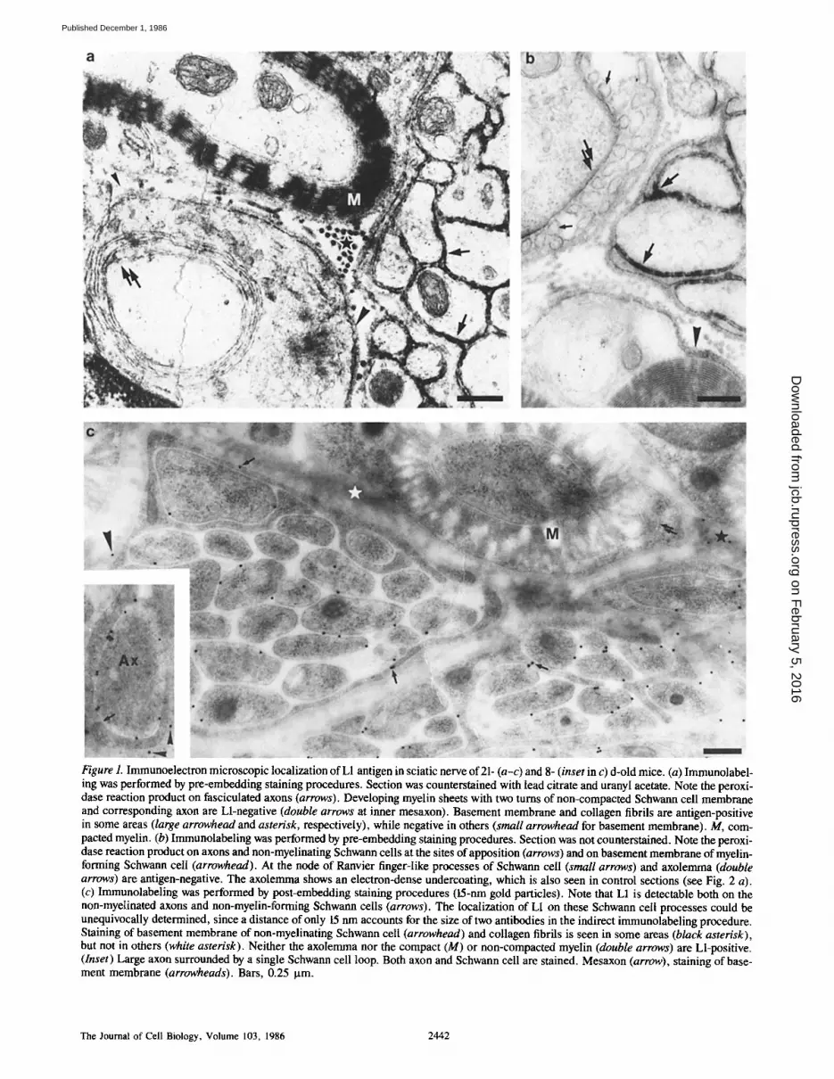
Figure 1. Immunoelectron microscopic localization of L1 antigen in sciatic nerve of 21- (a-c) and 8- (inset in c) d-old mice. (a) Immunolabel- ing was performed by pre-embedding staining procedures. Section was counterstained with lead citrate and uranyl acetate. Note the peroxi- dase reaction product on fasciculated axons (arrows). Developing myelin sheets with two turns of non-compacted Schwann cell membrane and corresponding axon are Ll-negative (double arrows at inner mesaxon). Basement membrane and collagen fibrils are antigen-positive in some areas (large arrowhead and asterisk, respectively), while negative in others (small arrowhead for basement membrane). M, com- pacted myelin. (b) Immunolabeling was performed by pre-embedding staining procedures. Section was not counterstained. Note the peroxi- dase reaction product on axons and non-myelinating Schwann cells at the sites of apposition (arrows) and on basement membrane of myelin- forming Schwann cell (arrowhead). At the node of Ranvier finger-like processes of Schwann cell (small arrows) and axolemma (double arrows) are antigen-negative. The axolemma shows an electron-dense undercoating, which is also seen in control sections (see Fig. 2 a). (c) Immunolabeling was performed by post-embedding staining procedures (15-nm gold particles). Note that L1 is detectable both on the non-myelinated axons and non-myelin-forming Schwann cells (arrows). The localization of L1 on these Schwann cell processes could be unequivocally determined, since a distance of only 15 nm accounts for the size of two antibodies in the indirect immunolabeling procedure. Staining of basement membrane of non-myelinating Schwann cell (arrowhead) and collagen fibrils is seen in some areas (black asterisk), but not in others (white asterisk). Neither the axolemma nor the compact (M) or non-compacted myelin (double arrows) are Ll-positive. (Inset) Large axon surrounded by a single Schwann cell loop. Both axon and Schwann cell are stained. Mesaxon (arrow), staining of base- ment membrane (arrowheads). Bars, 0.25 p.m.
The Journal of Cell Biology, Volume 103, 1986 2442
on February 5, 2016
jcb.rupress.orgD
ownloaded from
Published December 1, 1986
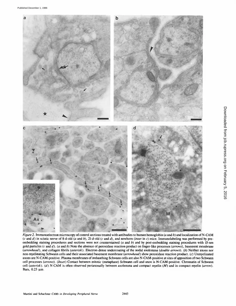
Figure 2. Immunoelectron microscopy of control sections treated with antibodies to human hemoglobin (a and b) and localization of N-CAM (c and d) in sciatic nerve of 8-d-old (a and b), 21-d-old (c and d), and newborn (inset in c) mice. Immunolabeling was performed by pre- embedding staining procedures and sections were not counterstained (a and b) and by post-embedding staining procedures with 15-nm gold particles (c and d). (a and b) Note the absence of peroxidase reaction product on finger-like processes (arrows), basement membrane (arrowhead), and collagen fibrils (asterisk). Electron-dense undercoating of the nodal axolemma (double arrows). (b) Neither axons nor non-myelinating Schwann cells and their associated basement membrane (arrowhead) show peroxidase reaction product. (c) Unmyelinated axons are N-CAM-positive. Plasma membranes of insheathing Schwann cells are also N-CAM-positive at sites of apposition of two Schwann cell processes (arrows). (Inset) Contact between mitotic (metaphase) Schwann cell and axon is N-CAM-positive. Chromatin of Schwann cell (asterisk). (d) N-CAM is often observed periaxonally between axolemma and compact myelin (M) and in compact myelin (arrow). Bars, 0.25 ~tm.
Martini and Schachner CAMs in Developing Peripheral Nerve 2443
on February 5, 2016
jcb.rupress.orgD
ownloaded from
Published December 1, 1986

Figure 3. Immunoelectron microscopic localization of the L2 epitope in sciatic nerve of 8-d-old mice. (a) Immunolabeling was performed by pre-embedding staining procedures. Section was not counterstained. Non-fasciculating axons and insheathing Schwann cell show im- munoperoxidase reaction product as observed for L1 or N-CAM. Note, however, that the labeling intensities vary between axons. Labeling of basement membranes is also not uniform, with labeling seen in some areas (large arrowhead), but not in others (small arrowhead). Collagen fibrils can be epitope-positive (large asterisk) and epitope-negative (small asterisk). M, compacted myelin. (b) Immunolabeling was performed by post-embedding staining procedures with 15-nm gold particles. Some axons are immunogold labeled, while others are not. Compact myelin (M) and periaxonal regions of myelinated fibers are never stained. On this micrograph no labeling of basement mem- branes is seen. Bars, 0.5 Ixm.
of the carbohydrate epitope was observed with MAG or MBP using the post-embedding staining method (see below). Basement membranes of Schwann cells and collagen fibrils were intensely stained by L2 and HNK-1 antibodies at both ages examined (Fig. 3 a). Both antibodies gave similar stain- ing patterns.
MAG
MAG was not detectable on non-myelinating Schwann cells. It first appeared on the myelinating membranes of Schwann cells when the mesaxon had formed '~1.5-2 loops (Fig. 4 a). When compaction of myelin occurred, MAG disappeared from the compacting membranes and was confined to the periaxonal region and to non-compacted structures, i.e., the Schmidt-Lanterman incisures, inner and outer mesaxon, and paranodal loops (Fig. 4, b and c). The finger-like pro- cesses at the nodes of Ranvier were also antigen-negative in the fully myelinated nerve. In rarer cases, however, these finger-like processes were transiently stained at early stages of myelination. MAG immunoreactivity was observed on basement membranes of the myelin-forming and non-my- elinating Schwann cells and on collagen fibrils (Fig. 4, b and c). This staining pattern was particularly prominent in the pre-embedding technique.
MAG could be detected reliably only by the post-embed- ding method. However, MAG immunoreactivity in the ex- tracellular matrix and transient appearance on finger-like processes at the node of Ranvier could be seen also by pre-
embedding methods. Furthermore, when the tissue had been damaged or the myelin was abnormally vacuolated, MAG was detected at the axon-Schwann cell interface by pre- embedding staining procedures.
M B P
MBP was reliably detectable also only by post-embedding staining procedures. In contrast to MAG it was amply pres- ent in compact myelin (Fig. 4 d), but absent from Schmidt- Lanterman incisures, inner and outer mesaxon, paranodal loops, and finger-like processes at nodes of Ranvier. It was never seen in periaxonal regions of myelinated nerves. Dur- ing myelin formation it was first seen in the compacting membranes. It was never seen on basement membranes or collagen fibrils.
Discussion
In this study we have shown that the developmental appear- ance of three cell adhesion molecules (L1, N-CAM, and MAG) and MBP, an intracellularly localized protein, follows a particular pattern that seems to be coordinated in time and space. L1 and N-CAM are the first molecules, detectable during development, with an almost identical cellular and subcellular localization. The fasciculating axons and the non-myelinating Schwann cells are L1- and N-CAM-positive at the time neurons and Schwann cells have made contact with each other. Both antigens are expressed on Schwann
The Journal of Cell Biology, Volume 103, 1986 2444
on February 5, 2016
jcb.rupress.orgD
ownloaded from
Published December 1, 1986

Figure 4. Immunoelectron microscopic localization of MAG (a-c) and MBP (d) in sciatic nerve of 8-d-old (a and b) and 21-d-old (c and d) mice. lmmunolabeling was performed by the post-embedding staining procedures with 15-rim gold particles. (a) Axon (Ax) is not MAG- positive at axolemma (arrows). Loops of Schwann cells are antigen-positive. The basement membrane is also stained (small arrowhead). Outer mesaxon (large arrowhead). (b) Uncompacted myelin is heavily stained by MAG antibodies, whereas compact myelin (M) is not stained. Note the antigen-positivity in the extracellular matrix. Outer mesaxon (large arrowhead). (c) MAG is detectable in the periaxonal region of a myelinated axon (compact myelin, M). Inner mesaxon (large arrow) and uncompacted myelin near the outer mesaxon (small arrows) are also MAG-positive. Basement membrane is also labeled (arrowhead). Compact myelin (M) is not MAG-positive. (d) MBP is only present in compact myelin (M). Bars, 0.25 lam.
Martini and Schachner CAMs in Developing Peripheral Nerve 2445
on February 5, 2016
jcb.rupress.orgD
ownloaded from
Published December 1, 1986

cells during mitosis, when the cells remain in surface contact with axons (Martin and Webster, 1973; Peters et al., 1976). L1 ceases to be expressed on both axon and myelinating Schwann cell, when the Schwann cell processes have turned for '~1.5 loops around the axon. N-CAM expression is re- duced when myelination proceeds (see also Covault and Sanes, 1986; Mirsky et al., 1986; Nieke and Schachner, 1985; Noble et al., 1985), but is still detected periaxonally in mature fibers. It is at the stage, when L1 disappears, that MAG is first detectable not only at the axon-Schwann cell interface, but also on the opposing cell surface membranes of the turning glial loops. At more advanced developmental stages, MAG remains expressed in non-compacted myelin and at sites of axon apposition, but is lost from the compact- ing Schwann cell membranes. This loss coincides with the appearance of MBP, which is only detectable in the compact myelin. This finding, however, is in contrast to the results of Schwob et al. (1985) and Omlin et al. (1982), who observed MBP not only in the major dense line, but also diffusely in the cytoplasm of developing processes of oligodendroglia or Schwann cells. It is possible that our failure to detect MBP in the cytoplasm is due to a different fixation protocol (Stern- berger, 1984, for review).
Interestingly, the L2/HNK-1 carbohydrate epitope shows coincident localization with L1 and N-CAM at the three stages examined, with the exception of the periaxonal region, where N-CAM but no L2/HNK-1 epitope is detectable. Since we do not know which other members of the L2/HNK-1 fam- ily are also expressed in sciatic nerve, the molecules carry- ing the epitope remain unidentified. Coincident labeling with MAG was, however, never observed, suggesting that the epi- tope is not involved in the activity of MAG in myelination of the sciatic nerve. The L2/HNK-I epitope is also never de- tectable in the finger-like processes of Schwann cells at the nodal region. It is therefore unlikely that an L2/HNK-I epi- tope carrying molecule on these processes assumes a similar function as the cell adhesion molecule J1 on astrocytic processes, which are thought to play a role in the main- tenance of nodal architecture in the adult rat optic nerve (ffrench-Constant et al., 1986). It should be noted also that not all non-myelinated axons express the L2/HNK-1 epitope, thus pointing to a cellular heterogeneity that has been noted previously in the peripheral nervous system for other carbo- hydrate structures (Dodd and Jessell, 1985).
The presence of L1, N-CAM, MAG, and L2/HNK-1 epi- tope on basement membranes and collagen fibrils is detect- able in 8- and 21-d-old, but not in newborn mice. This local- ization is considered specific, since it is not seen in control sections and sections treated with antiserum to MBP. Fur- thermore, these structures are also immunostained in the post-embedding staining procedure, where leakage of the peroxidase reaction product is avoided. In light of these ob- servations, the localization of L1 at the nodes of Ranvier in the sciatic nerve of the rat at the light microscopic level (Mirsky et al., 1986) can now be attributed to the particularly strong expression of L1 in basement membranes of the rat peripheral nervous system (unpublished observations) and renders a localization of L1 on the surface membrane of the nodal finger-like processes unlikely.
Our present observations are in agreement with previous results on the expression of L1 and N-CAM during develop- ment of the mouse sciatic nerve at the light microscopic level
(Nieke and Schachner, 1985). However, at this level of res- olution, it was difficult to see the localization of L1 and N-CAM on fasciculating nerves and the overlap of the local- ization of the shared carbohydrate epitope L2/HNK-1 with L1 and N-CAM. Since the L2/HNK-1 epitope is most strongly localized in basement membranes and in collagen fibrils, a less prominent staining in other structures may have been overlooked. On the other hand, in previous studies the HNK-1 epitope has been described to be associated with neu- rons of peripheral nerves (McGarry et al., 1985; Schuller- Petrovic et al., 1983).
Of particular importance is the observation that MAG is not present in compact myelin, but is unequivocally ob- served at the myelin-axon interface, Schmidt-Lanterman in- cisures, outer and inner mesaxon, and paranodal loops. These findings are in agreement with those by Trapp and Quarles (1982, 1984) and Trapp et al. (1984), but contradict results obtained by Webster et al. (1983, 1985) and Favilla et al. (1984), who observed MAG in compact myelin of oligodendroglia. We feel that the present study gives the first unequivocal evidence of the absence of MAG in compact my- elin and thus settles a controversy. Also, in contrast to a previous immunocytochemical study in the peripheral ner- vous system of the chicken, we have not found detectable lev- els of MAG on neurons (Omlin et al., 1985; Philippe et al., 1986).
Although functional relationships in cell interactions can- not be deduced from morphological data, our observations suggest that LI and N-CAM are involved in axon fascicula- tion as it has been observed previously in the central nervous system (Fischer et al., 1986) and mediate initial contacts be- tween axons and Schwann cells. In agreement with this no- tion, both L1 and N-CAM antibodies have recently been shown to interfere with adhesion of neurons to Schwann cells (Seilheimer, B., A. Faissner, G. Keilhauer, and M. Schach- ner, manuscript submitted for publication). It is, further- more, conceivable that N-CAM and L1 mediate surface in- teractions during the initial turnings of apposing Schwann cell loops. MAG appears to take over Schwann cell-axon and Schwann cell-Schwann cell interactions initiated by L1 and N-CAM. It is interesting in this context that, in the central nervous system, MAG has been shown to mediate not only neuron-oligodendrocyte, but also oligodendrocyte-oligo- dendrocyte adhesion (Poltorak, M., G. Keilhauer, A. Meyer, C. Landa, and M. Schachner, manuscript submitted for pub- lication). Since in the adult sciatic nerve the localization of MBP in compact myelin is a virtual mirror image of MAG, we would like to propose that MBP is involved in the apposi- tion of the cytoplasmic sites of the Schwann cell membranes (Schwob et al., 1985), whereas MAG is characteristic of sur- face contacts at sites where the cytoplasm is retained (Quarles, 1983/84).
It may be worthwhile to point out that other than "homo- philic" binding partners for the cell adhesion molecules have to be postulated from our observation that N-CAM, L1, and MAG are all localized in basement membranes and collagen fibrils. It is intriguing that the intensity of the immunostain- ing reaction is not uniform within one section, particularly for the collagen fibrils. Since staining artifacts can be ex- cluded in our study we would like to suggest that the ad- hesion molecules are secreted by unevenly localized cellu- lar sources and retained by the extracellular matrix, where
The Journal of Cell Biology, Volume 103, 1986 2446
on February 5, 2016
jcb.rupress.orgD
ownloaded from
Published December 1, 1986

they subserve yet unknown functions. It has been observed that the basement membrane or extracellular matrix mate- rial guides regrowing axons under regenerative conditions (Longo et al., 1984; Schwab and Thoenen, 1985). It is there- fore interesting that the adhesion molecule J1 has been ob- served on collagen fibrils in the proximity of denervated, but not innervated neuromuscular junctions (Sanes et al., 1986). It is worth noting here that a rigorous classification of adhe- sion molecules into those that mediate interactions with other cells and those mediating interactions with the ex- tracellular matrix becomes futile, as adhesion molecules are now found both as integral membrane constituents and as part of the extracellular matrix.
It is still not known which molecular signals lead an axon-Schwann cell interaction to myelination in one case, but to no myelination in the other. MAG and MBP expression seem to be the consequence of the commitment to myelina- tion, while L1 and N-CAM expression appear to precede this decision. The signals that determine the realization of these distinct differentiation pathways may involve surface bound or diffusible signals between axon and Schwann cells. Ex- pression of L1 and N-CAM could therefore provide a basis for axon-Schwann cell relationships upon which other mod- ulating events may be superimposed that trigger a Schwann cell to myelinate or remain non-myelinating.
The authors are grateful to Beate Berger and Cornelia Heuser for technical assistance, Andreas Faissner, Thomas Fahrig, Bernd Gehrig, Christo Goridis, Norbert Herschkowitz, Volker Kiinemund, Carlos Landa, Maciej Poltorak, and Richard Reynolds for antibodies, and Christine Grund for help with the Lowicryl method.
This work was supported by Deutsche Forschungsgemeinschaft (SFB 317).
Received for publication 6 August 1986, and in revised form 28 August 1986.
References
Bock, E., C. Richter-Landsberg, A. Faissner, and M. Schachner. 1985. Demonstration of immunochemical identity between the nerve growth factor- inducible large external (NILE) glycoprotein and the cell adhesion molecule L1. EMBO (Eur. Mol. Biol. Organ.) J. 4:2765-2768.
Bologa-Sandru, L., H. P. Siegrist, A. Z'Graggen, K. Hofmann, U. Wies- mann, D. Dahl, and N. Herschkowitz. 1981. Expression of antigenic markers during the development of oligodendrocytes in mouse brain cell cultures. Brain Res. 210:217-229.
Chou, K. H., A. A. Ilyas, J. E. Evans, R. H. Quarles, and F. B. Jungalwala. 1985. Structure ofa glycolipid reacting with monoclonal IgM in neuropathy and with HNK-1. Biochem. Biophys. Res. Commun. 128:383-388.
Covault, J., and J. R. Sanes. 1986. Distribution of N-CAM in synaptic and extrasynaptic portions of developing and adult skeletal muscle. J. Cell Biol. 102:716-730.
Dodd, J., and T. M. Jessel. 1985. Lactoseries carbohydrates specify subsets of dorsal root ganglion neurons projecting to the superficial dorsal horn of rat spinal cord. J. Neurosci. 5:3278-3294.
Edelman, G. M. 1985. Specific cell adhesion in histogenesis and morphogen- esis. In The Cell in Contact. G. M. Edelman and J.-P. Thiery, editors. John Wiley & Sons, Inc., New York. 139-168.
Faissner, A., J. Kruse, C. Goridis, E. Bock, and M. Schachner. 1984. The neural cell adhesion molecule LI is distinct from the N-CAM related group of surface antigens BSP-2 and D2. EMBO (Fur. Mol. Biol. Organ.) J. 3:733-737.
Favilla, J. T., D. E. Frail, C. G. Palkovits, G. L. Stoner, P. E. Braun, and H. deF. Webster. 1984. Myelin-associated glycoprotein (MAG) distribution in human central nervous tissue studied immunocytochemically with monoclonal antibody. J. NeuroimmunoL 6: I9-30.
ffrench-Constant, Ch., R. H. Miller, J. Kruse, M. Schachner, and M. Raft. 1986. Molecular specialization ofastrocyte processes at nodes of Ranvier in rat optic nerve. J. Cell Biol. 102:844-952.
Fischer, G., V. Kiinemund, and M. Schachner. 1986. Neurite out-growth patterns in cerebellar microexplant cultures are affected by antibodies to the cell surface glycoprotein L1. J. Neurosci. 6:602-612.
Friedlander, D. R., M. Grumet, and G. M. Edelman. 1986. Nerve growth
factor enhances expression of neuron-gila cell adhesion molecule in PC 12 cells. J. Cell BioL 102:413--419.
Goridis, C., H. Deagostini-Bazin, M. Him, M. R. Hirsch, G. Rougon, R. Sadoul, O. K. Langley, G. Gombos, and J. Finne. 1983. Neural surface anti- gens during nervous system development. Cold Spring Harbor Symp. Quant. Biol. 48:527-538.
Keilhauer, G., A. Faissner, and M. Schachner. 1985. Differential inhibition of neurone-neurone, neurone-astrocyte and astrocyte-astrocyte adhesion by L1, L2 and N-CAM antibodies. Nature (Lond.). 316:728-730.
Kruse, J., R. Mallhammer, H. Wernecke, A. Falssner, I. Sommer, C. Goridis, and M. Schachner. 1984. Neural cell adhesion molecules BSP-2 and L1 share a common carbohydrate moiety recognized by monoclonal antibodies L2 and HNK-1. Nature (Load.). 311:153-155.
Kruse, J., G. Keilhaner, A. Falssner, R. Timpl, and M. Schachner. 1985. The J1 glyeoprotein-a novel nervous system cell adhesion molecule of the L2/HNK-1 family. Nature (Load.). 316:146-148.
Lindner, J., F. G. Rathjen, and M. Schachner. 1983. L1 mono- and poly- clonal antibodies modify cell migration in early postnatal mouse cerebellum. Nature (Lond. ). 305:427-430.
Lindner, J., G. Zinser, W. Werz, C. Goridis, B. Bizzini, and M. Schachner. 1986. Experimental modification of postnatal cerebellar granule cell migration in vitro. Brain Res. 377:298-304.
Longo, F. M., E. G. Hayman, G. E. Davis, E. Ruoslahti, E. Engvall, M. Manthorpe, and S. Varon. 1984. Neurite-promoting factors and extracellular matrix components accumulating in vivo within nerve regeneration chambers. Brain Res. 309:105-117.
Martin, J. R., and H.de F. Webster. 1973. Mitotic Schwann cells in develop- ing nerve: their changes in shape, fine structure, and axon relationships. Dev. Biol. 32:417-431.
McGarry, R. C., R. J. Riopelle, D. E. Frail, A. M. Edwards, P. E. Braun, and J. C. Roder. 1985. The characterization and cellular distribution of a family of antigens related to myelin associated glycoprotein in the developing nervous system. J. Neuroimmunol. 10:101-114.
Mirsky, R., K. R. Jessen, M. Schachner, and C. Goridis. 1986. Distribution of the adhesion molecules N-CAM and L1 on peripheral neurons and gila in adult rats. J. NeurocytoL In press.
Nieke, J., and M. Schachner. 1985. Expression of the neural cell adhesion molecules L1 and N-CAM and their common carbohydrate epitope L2/HNK-1 during development and after transection of mouse sciatic nerve. Differentia- tion. 30:141-151.
Noble, M., M. Albrechtsen, C. M¢ller, J. Lyles, E. Bock, C. Goridis, M. Watanabe, and U. Rutishauser. 1985. Glial cells express N-CAM/D2-CAM- like polypeptides in vitro. Nature (Load.). 316:725-728.
Noronha, A. B., A. llyas, H. Antonicek, M. Schachner, and R. H. Quarles. 1986. Molecular specificity of L2 monoclonal antibodies that bind to carbohy- drate determinants of neural cell adhesion molecules and their resemblance to other monoclonal antibodies recognizing the myelin-associated glycoprotein. Brain Res. In press.
Omlin, F. X., H. deF. Webster, C. G. Palkovits, and S. R. Cohen. 1982. Immunocytochemical localization of basic protein in major dense line regions of central and peripheral myelin. J. Cell Biol. 95:242-248.
Omlin, F. X., J.-M. Matthieu, E. Philippe, J. M. Roch, and B. Droz. 1985. Expression of myelin-associated glycoprotein by small neurons of the dorsal root ganglion in chickens. Science (Wash. DC). 227:1359-1360.
Palay, S. L., and V. Chan-Palay. 1974. Cerebellar Cortex. Cytology and Organization. Springer Verlag, New York, Heidelberg, Berlin. 348 pp.
Peters, A., S. L. Palay, and H. deF. Webster. 1976. The Fine Structure of the Nervous System: The Neurons and Supporting Cells. W. B. Saunders Co., Philadelphia, London, Toronto. 406 pp.
Philippe, E., F. X. Omlin, and B. Droz. 1986. Myelin-associated glycopro- tein immunoreactive material: an early neuronal marker of dorsal root ganglion cells during chick development. Dev. Brain Res. 27:275-277.
Quarles, R. H. 1983/84. Myelin-associated glycoproteln in development and disease. Dev. Neurosci. 6:285-303.
Rathjen, F. G., and M. Schachner. 1984. Immunocytological and biochemi- cal characterization of a new neuronal cell surface component (L1 antigen) which is involved in cell adhesion. EMBO (Eur. Mol. Biol. Organ.) J. 3:1-10.
Roth, J., M. Bendayan, E. Carlemalm, W. Villiger, M. Garavito. 1981. En- hancement of structural preservation and immunocytochemical staining in low temperature embedded pancreatic tissue. J. Histochem. Cytochem. 29:663- 671.
Sanes, J. R., M. Schachner, and J. Covault. 1986. Expression of several adhesive macromolecules (N-CAM, LI, J1, NILE, uvomorulin, laminin, fibronectin and heparan sulfate proteoglycan) in embryonic, adult, and dener- vated adult skeletal muscle. J. Cell Biol. 102:420-431.
Schachner, M., E. T. Hedley-Whyte, D. W. Hsu, G. Schoonmaker, and A. Bignami. 1977. Ultrastructural localization of glial fibrillary acidic protein in mouse cerebellum by immunoperoxidase labeling. J. Celt Biol. 75:67-73.
Schachner, M., A. Falssner, G. Fischer, G. Keilhauer, J. Kruse, V. Ktine- mund, J. Lindner, and H. Wernecke. 1985. Functional and structural aspects of the cell surface in mammalian nervous system development. In The Cell in Contact. G. M. Edelman and J.-P. Thiery, editors. John Wiley & Sons, Inc., New York. 257-275.
Schuller-Petrovic, S., W. Gebhart, H. Lassmann, H. Rumpold, and D. Kraft. 1983. A shared antigenic determinant between natural killer cells and
Martini and Schachner CAMs in Developing Peripheral Nerve 2447
on February 5, 2016
jcb.rupress.orgD
ownloaded from
Published December 1, 1986

nervous tissue. Nature (Lond.). 306:t79-181. Schwab, M. E., and H. Thoenen. 1985. Dissociated neurons regenerate into
sciatic but not optic nerve explants in culture irrespective of neurotrophic fac- tors. J. Neurosci. 5:2415-2423.
Schwob, V. S., H. B. Clark, D. Agrawal, and H. C. Agrawal. 1985. Elec- tron microscopic immunocytochemical localization of myelin proteolipid pro- tein and myelin basic protein to oligodendrocytes in rat brain during myelina- tion. J. Neurochem. 45:559-571.
Sternberger, N. H. 1984. Patterns of oligodendrocyte function seen by im- munocytochemistry. In Oligodendroglia. Advances in Neurochemistry. W. T. Norton, editor. Plenum Publishing Corp., New York. 125-173.
Trapp, B. D., and R. H. Quafles. 1982. Presence of the myelin-associated glycoprotein correlates with alterations in the periodicity of the peripheral my- elin. J. Cell Biol. 92:877-882.
Trapp, B. D., and R. H. Quarles. 1984. Immunocytochemical localization
of the myelin-associated glycoprotein. Fact or artifact? J. NeuroimmunoL 6:231-249.
Trapp, B. D., R. H. Quarles, and K. Suzuki. 1984. lmmunocytochemical studies of quaking mice support a role for the myelin-associated glycoprotein in forming and maintaining the periaxonal space and periaxonal cytoplasmic collar of myelinating Schwann cells. J. Cell Biol. 99:594-606.
Webster, H. deF., C. G. Palkovits, G. L. Stoner, J. T. Favilla, D. E. Frail, and P. E. Braun. 1983. Myelin-associated glycoprotein: electron microscopic immunocytochemical localization in compact developing and adult central ner- vous system myelin. J. Neurochem. 41:1469-1479.
Webster, H. deF., H. Shii, and H. Lassmann. 1985. Immunocytochemical study of myelin-associated glycoprotein (MAG), basic protein (BP), and glial fibriltary acidic protein (GFAP) in chronic relapsing experimental allergic en- cephalomyelitis (EAE). Acta Neuropathol. 65:177-189.
The Journal of Cell Biology, Volume 103, 1986 2448
on February 5, 2016
jcb.rupress.orgD
ownloaded from
Published December 1, 1986


