Embed Size (px)
Citation preview
Image Features in Medical Vibro-acoustography: In Vitro and inVivo Results
Azra Alizad, MD1, Dana H. Whaley, MD2, James F. Greenleaf, PhD1, and Mostafa Fatemi,PhD1
1Department of Physiology and Biomedical Engineering, Mayo Clinic College of Medicine, 200 1st St. SW,Rochester, MN, 55905, USA
2Department of Radiology, Mayo Clinic College of Medicine, 200 1st St. SW, Rochester, MN, 55905, USA
AbstractVibro-acoustography is an imaging method based on audio-frequency harmonic vibrations inducedin the object by the radiation force of focused ultrasound. The purpose of this study is to investigatefeatures of vibro-acoustography images and manifestation of various tissue structures andcalcifications in such images. Our motivation for this study is to pave the way for further in vitro andin vivo applications of vibro-acoustography. Here, vibro-acoustography images of excised prostateand in vivo breast are presented and compared with images obtained with other modalities. Resultingvibro-acoustography images obtained with a 3 MHz ultrasound transducer and at a vibrationfrequency of 50–60 kHz show soft tissue structures, tissue borders, and microcalcifications with highcontrast, high resolution, and no speckle. It is concluded that vibro-acoustography offers featuresthat may be valuable for diagnostic purposes.
KeywordsVibro-acoustography; radiation force; ultrasound; breast imaging; calcification
A. IntroductionUltrasonography is one of the most common medical imaging modalities. However, there aresome limitations to this method. Namely, ultrasound images suffer from a speckle artifact. Asa result, small objects, such as microcalcifications are hard to detect with this method. Specklealso can reduce the sensitivity of the imaging system in detection of masses in tissue. For theseand other reasons, investigators have been seeking alternative non-invasive imaging methodsthat can offer higher quality images as well as new information about tissue.
Vibro-acoustography is an imaging method based on the radiation force of ultrasound [1,2].In this method, the image is formed from the acoustic response of the object to the oscillatingradiation force of the amplitude modulated ultrasound. In vibro-acoustography, twointersecting continuous-wave ultrasound beams at slightly different frequencies of f1 and f2 =
Corresponding author: Azra Alizad, MD, Fax: +1 507 266 0361, E-mail address: [email protected]'s Disclaimer: This is a PDF file of an unedited manuscript that has been accepted for publication. As a service to our customerswe are providing this early version of the manuscript. The manuscript will undergo copyediting, typesetting, and review of the resultingproof before it is published in its final citable form. Please note that during the production process errors may be discovered which couldaffect the content, and all legal disclaimers that apply to the journal pertain.PACS Codes: 43.25.Qp, 43.60.Lq, 43.80.Qf, 43.80.Vj, 43.80.Jz
NIH Public AccessAuthor ManuscriptUltrasonics. Author manuscript; available in PMC 2009 November 1.
Published in final edited form as:Ultrasonics. 2008 November ; 48(6-7): 559–562. doi:10.1016/j.ultras.2008.04.014.
NIH
-PA Author Manuscript
NIH
-PA Author Manuscript
NIH
-PA Author Manuscript
f1+Δf, where Δf≪f1, are used. The two ultrasound beams are focused, and they are aligned tointersect at their respective focal regions. At this intersection region, which is normally a smallvolume, the combined ultrasound field energy density is sinusoidally modulated at Δf, thus thefield generates a highly localized oscillatory radiation force at the difference frequency whenit interacts with the object. The harmonic force vibrates the object at Δf. The vibration resultsin a secondary acoustic field that propagates in the object. This acoustic field, which is atfrequency Δf, is detected by an audio hydrophone. This signal is then filtered by a band passfilter centered at Δf to reject noise and any interfering signal. As the ultrasound beam is scannedacross the object, the filtered hydrophone signal is recorded and its amplitude is mapped intoan image.
To understand the interaction of two ultrasound fields at different frequencies and generationof a third acoustic field at the difference frequency, one needs to solve the nonlinear waveequation. The general theory of nonlinear wave propagation has been investigated extensively,for example see references [3] and [4]. A detailed theoretical and simulation study of wavepropagation and interaction of two ultrasound beams, as it applies to vibro-acoustography, ispresented in another paper in this issue [5]. Therefore, we do not elaborate on the theoreticaldetails of wave propagation in this paper, instead refer the reader to [5].
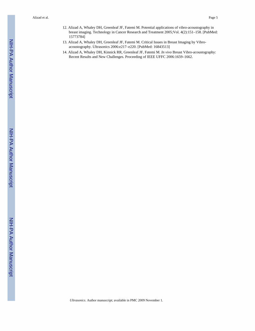
A comparison of conventional B-mode ultrasound imaging and vibro-acoustography isillustrated in Figure 1. Ultrasound imaging is based on linear reflection or scattering ofultrasound. That is, the echo, which is used to construct the image, is at the frequency of thesource ultrasound. In contrast, the secondary acoustic field that is used for making the vibro-acoustography image is at the difference frequency Δf, which is typically two order ofmagnitude smaller than the incident ultrasound frequency. This frequency conversion is theresult of a nonlinear process that is central to vibro-acoustography methodology. The frequencyconversion enriches the vibro-acoustography image with additional information not present inconventional ultrasound image.
A vibro-acoustography image contains two types of information: ultrasonic properties of theobject, such as the scattering and power absorption characteristics, and the dynamiccharacteristics of the object at frequency Δf, which relates to tissue stiffness, boundaryconditions, and coupling to the surrounding medium [6]. The former properties are those thatare also present in conventional ultrasound imaging. The latter properties, which can bedescribed in terms of object’s mechanical parameters at Δf, are not available from conventionalultrasound. Another feature of vibro-acoustography relates to image speckle. Speckle is thesnowy pattern seen in conventional ultrasound images. Speckle results from randominterference of the scattered ultrasound field. Speckle reduces the contrast of ultrasound imagesand often limits detection of small structures, such as microcalcifications in tissue. Becausevibro-acoustography uses the secondary acoustic field, this modality is practically speckle free,resulting in high contrast images that allow small structures to be visible. This feature makesvibro-acoustography particularly suitable for detection of breast microcalcifications.
Vibro-acoustography has been tested on various human tissues [6–14]. A comparative studyof vibro-acoustography with other radiation force methods for tissue elasticity imaging ispresented in [7]. The spatial resolution of vibro-acoustography is in the sub-millimeter range,making the technique suitable for high-resolution imaging [9,11].
In this paper, we present some experimental results and discuss some features of vibro-acoustography images and their potential applications.
Alizad et al. Page 2
Ultrasonics. Author manuscript; available in PMC 2009 November 1.
NIH
-PA Author Manuscript
NIH
-PA Author Manuscript
NIH
-PA Author Manuscript
B. MethodsWe examine images of ex-vivo and in vivo human tissues acquired by three methods: x-ray,ultrasound, and vibro-acoustography. Ex-vivo tissue samples are fixed in formaldehyde beforethe experiments. Vibro-acoustography scans of tissue samples are conducted in a water tank.Ultrasound images of ex-vivo tissue samples are obtained by a clinical ultrasound scanner (GEVivid 7).
The in vivo experiments are conducted on human breast. The breast imaging system consistsof a stereotactic mammography (x-ray) machine combined with a vibro-acoustographyscanner. This system allows us to take matching x-ray and vibro-acoustography images of thesame region of breast within a 5×5 cm window. The system uses a 3 MHz transducer, whichprovides sufficient penetration and suitable spatial resolution of 0.7 mm for our purpose. Thedifference frequency Δf is set at either 50 or 60 kHz, which is suitable for most experiments.The image contrast changes with Δf for a given object, as the dynamics of the object and theacoustic environment depend on frequency. The spatial increment for raster scanning is set at0.2 mm. To acquire an image, the patient lies in prone position on the examination bed with abreast hanging down through a hole in the bed. The breast is slightly compressed between twopanels. The x-ray detector is behind the breast and the audio hydrophone for vibro-acoustography is placed in contact with the side of the breast.
Three features of vibro-acoustography images will be discussed: speckle pattern, detection ofcalcifications in tissue, and definition of tissue borders. The x-ray and ultrasound images willprovide helpful references in discussing vibro-acoustography image features.
C. Experimental ResultsFigure 2 displays images of an excised human prostate. Panel (a) in this figure is the x-rayimage, which shows the general view of the prostate. The bright spot at the center is acalcification. Tissue structures are not clearly visible in this image. Panel (b) displays theultrasound image of the same sample. This image is overwhelmed with speckle, which makesit hard to distinguish tissue structures or the calcification. Panel (c) is the vibro-acoustographyimages of the prostate with the ultrasound focused at the depth of 10 mm from the top surfaceof the sample. The vibro-acoustography image shows detailed tissue structures as well as thecalcification. Comparing the vibro-acoustography and ultrasound images, it is evident thatvibro-acoustography provides speckle-free and clear images of tissue, which allows one toidentify tissue structures and the small (< 1 mm) calcification.
Figure 3 displays images of another excised human prostate sample. Panel (a) displays the x-ray image of this sample, which indicates the presence of a cluster of microcalcifications onthe right side and two larger calcifications at the center. Panel (b) shows the vibro-acoustography image of the prostate with the ultrasound beam focused at 10 mm depth fromthe top surface of the sample. The speckle-free vibro-acoustography image clearly shows thecluster of micro calcifications as well as the larger calcifications. Besides, the vibro-acoustography image displays a clear border between the prostate tissue and the prostatic fataround the sample.
Figure 4 displays in vivo images of a human breast. Panel (a) displays a clinical x-raymammography of the entire breast. The dark regions indicate that this breast contains asignificant amount of fat. Panel (b) displays the vibro-acoustography image of a region in thebreast. In this case the ultrasound is focused at 15 mm from the skin. The vibro-acoustographyimage shows detailed structures of the breast with well-defined borders. The linear structuresmay include ducts, vessels, and Cooper’s ligaments.
Alizad et al. Page 3
Ultrasonics. Author manuscript; available in PMC 2009 November 1.
NIH
-PA Author Manuscript
NIH
-PA Author Manuscript
NIH
-PA Author Manuscript
Figure 5 displays an in vivo image of a human breast. Panel (a) shows x-ray mammographywith an obscured mass. Panel (b) displays vibro-acoustography image of this breast, whichreveals the mass as a bright region with irregular border. Biopsy results indicated infiltratingductal carcinoma, Nottingham grade I (of III).
D. Discussion and ConclusionsThe imaging method described here is a noninvasive method that utilizes ultrasound energy,but the images are constructed from a low-frequency acoustic field. Unlike conventionalultrasound images, vibro-acoustography images are speckle-free, which increases imagecontrast and allows detection of mass lesions and small (sub-millimeter) details, such asmicrocalcifications. In addition, vibro-acoustography shows tissue borders with appreciableclarity as demonstrated in the breast images. Detection of small calcifications is an importantissue, especially in breast, as they may be associated with some types of cancers. Detection oftissue borders can be important in sizing mass lesions. Also, it is important to be able to seetissue borders when conducting image guided procedures in prostate. Further investigation isneeded to fully explore the potentials of vibro-acoustography for clinical imaging.
AcknowledgementsThe authors are grateful to the following individuals for their valuable work during the course of this study: Dr. MatthewUrban for processing vibro-acoustography images, Randall R. Kinnick for laboratory support and scanning tissues,Thomas M. Kinter for software support, our study coordinator Lori Johnson, and Joyce Rahn for her help withmammography. This research was supported in part by grant BCTR0504550 from the Susan G. Komen Breast CancerFoundation and Grants CA91956, EB00535, CA127235 and CA121579 from NIH. Disclosure of conflict of interest:Vibro-acoustography technique is patented by Mayo Clinic and some of the authors (MF and JFG).
References1. Fatemi M, Greenleaf JF. Ultrasound stimulated vibro-acoustic spectroscopy. Science 1998;Vol.
280:82–85. [PubMed: 9525861]2. Fatemi M, Greenleaf JF. Vibro-acoustography. An imaging modality based on ultrasound stimulated
acoustic emission. Proc. Natl. Acad. Sci. USA 1999;Vol. 96:6603–6608. [PubMed: 10359758]3. Novikov, BK.; Rudenko, OV.; Timoshenko, VI. Acoustic Detection and Detectors Acoustic. In: Beyer,
RT., editor. Nonlinear Underwater Acoustics. Originally published New, 1981. New: AcousticalSociety of America; 1987. corporate author
4. Beyer, RT. Nonlinear Acoustics. Vol. 1st edition. published by Naval Sea Systems Command; 1974.p. 91-157.chapter III
5. Malcolm AE, Reitich F, Yang J, Greenleaf JF, Fatemi M. A combined parabolic-integral equationapproach to the acoustic simulation of vibro-acoustic imaging. Ultrasonics. 2008In press
6. Greenleaf, JF.; Ehman, RL.; Fatemi, M.; Muthupillai, R. Imaging elastic properties of tissue. In: Duck,FA.; Baker, AC.; Starrit, HC., editors. Ultrasound in Medicine (Medical Science Series). Bristol,England: Institute of Physics Publishing; 1998. p. 263-277.
7. Fatemi M, Greenleaf JF. Probing the dynamics of tissue at low frequencies with the radiation force ofultrasound. Phys. Med. Biol 2000;Vol. 45:1449–1464. [PubMed: 10870703]
8. Fatemi M, Manduca A, Greenleaf JF. Imaging elastic properties of biological tissues by low-frequencyharmonic vibration. Proc. of IEEE, 91 2003;Vol. 10:1503–1517.
9. Fatemi M, Wold LE, Alizad A, et al. Vibro-acoustic tissue mammography. IEEE Trans. Med. Imag2002;Vol. 21(1):1–8.
10. Alizad A, Fatemi M, Wold LE, et al. Performance of vibro-acoustography in detecting ofmicrocalcifications in excised human breast tissue: a study on 74 breast tissue samples. IEEE Trans.Med. Imag 2004;Vol. 23(3):307–312.
11. Fatemi, M.; Greenleaf, JF. Imaging the viscoelastic properties of tissue. In: Fink, M.; Montagner, J-P.; Tourin, A., editors. Topics in Applied Physic. Vol. Vol. 84. Berlin: Springer, Verlog, Heidelberg;2002. p. 257-275.
Alizad et al. Page 4
Ultrasonics. Author manuscript; available in PMC 2009 November 1.
NIH
-PA Author Manuscript
NIH
-PA Author Manuscript
NIH
-PA Author Manuscript
12. Alizad A, Whaley DH, Greenleaf JF, Fatemi M. Potential applications of vibro-acoustography inbreast imaging. Technology in Cancer Research and Treatment 2005;Vol. 4(2):151–158. [PubMed:15773784]
13. Alizad A, Whaley DH, Greenleaf JF, Fatemi M. Critical Issues in Breast Imaging by Vibro-acoustography. Ultrasonics 2006:e217–e220. [PubMed: 16843513]
14. Alizad A, Whaley DH, Kinnick RR, Greenleaf JF, Fatemi M. In vivo Breast Vibro-acoustography:Recent Results and New Challenges. Proceeding of IEEE UFFC 2006:1659–1662.
Alizad et al. Page 5
Ultrasonics. Author manuscript; available in PMC 2009 November 1.
NIH
-PA Author Manuscript
NIH
-PA Author Manuscript
NIH
-PA Author Manuscript
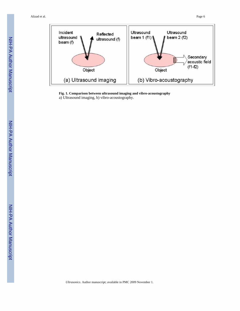
Fig. 1. Comparison between ultrasound imaging and vibro-acoustographya) Ultrasound imaging, b) vibro-acoustography.
Alizad et al. Page 6
Ultrasonics. Author manuscript; available in PMC 2009 November 1.
NIH
-PA Author Manuscript
NIH
-PA Author Manuscript
NIH
-PA Author Manuscript
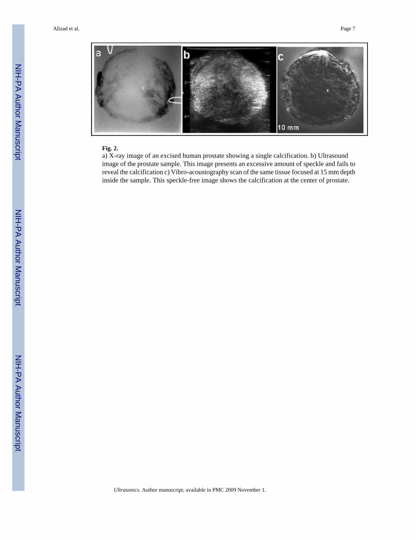
Fig. 2.a) X-ray image of an excised human prostate showing a single calcification. b) Ultrasoundimage of the prostate sample. This image presents an excessive amount of speckle and fails toreveal the calcification c) Vibro-acoustography scan of the same tissue focused at 15 mm depthinside the sample. This speckle-free image shows the calcification at the center of prostate.
Alizad et al. Page 7
Ultrasonics. Author manuscript; available in PMC 2009 November 1.
NIH
-PA Author Manuscript
NIH
-PA Author Manuscript
NIH
-PA Author Manuscript
Fig. 3.a) X-ray image of an excised human prostate. b) Vibro-acoustography scan of the same prostateat 15mm depth. This image shows a cluster as well as a single calcification as confirmed bythe x-ray. c) Ultrasound image of the prostate. The ultrasound image fails to reveal thecalcifications.
Alizad et al. Page 8
Ultrasonics. Author manuscript; available in PMC 2009 November 1.
NIH
-PA Author Manuscript
NIH
-PA Author Manuscript
NIH
-PA Author Manuscript

Fig. 4. Breast images of a normal volunteer(a) Clinical x-ray mammography of the entire breast, (b) vibro-acoustography focused at 2 cmdepth from the skin at Δf = 50 kHz.
Alizad et al. Page 9
Ultrasonics. Author manuscript; available in PMC 2009 November 1.
NIH
-PA Author Manuscript
NIH
-PA Author Manuscript
NIH
-PA Author Manuscript
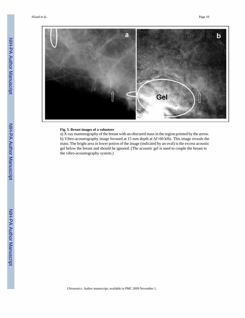
Fig. 5. Breast images of a volunteera) X-ray mammography of the breast with an obscured mass in the region pointed by the arrow.b) Vibro-acoustography image focused at 15 mm depth at Δf=60 kHz. This image reveals themass. The bright area in lower potion of the image (indicated by an oval) is the excess acousticgel below the breast and should be ignored. (The acoustic gel is used to couple the breast tothe vibro-acoustography system.)
Alizad et al. Page 10
Ultrasonics. Author manuscript; available in PMC 2009 November 1.
NIH
-PA Author Manuscript
NIH
-PA Author Manuscript
NIH
-PA Author Manuscript