Embed Size (px)
Citation preview

Identification of subject specific and functional consistent ROIs using semi-supervised learning
Yuhui Dua,b, Hongming Lia, Hong Wuc, Yong Fan*a
aNational Laboratory of Pattern Recognition, Institute of Automation, Chinese Academy of Sciences, Beijing 100190, China;
bCollege of Information and Communication Engineering, North University of China, Taiyuan 030051, China;
cSchool of Computer Science and Engineering, University of Electronic Science and Technology of China, Chengdu 611731, China
ABSTRACT
Regions of interests (ROIs) for defining nodes of brain network are of great importance in brain network analysis of fMRI data. The ROIs are typically identified using prior anatomical information, seed region based correlation analysis, clustering analysis, region growing or ICA based methods. In this paper, we propose a novel method to identify subject specific and functional consistent ROIs for brain network analysis using semi-supervised learning. Specifically, a graph theory based semi-supervised learning method is adopted to optimize ROIs defined using prior knowledge with a constraint of local and global functional consistency, yielding subject specific ROIs with enhanced functional connectivity. Experiments using simulated fMRI data have demonstrated that functional consistent ROIs can be identified effectively from data with different signal to noise ratios (SNRs). Experiments using resting state fMRI data of 25 normal subjects for identifying ROIs of the default mode network have demonstrated that the proposed method is capable of identifying subject specific ROIs with stronger functional connectivity and higher consistency across subjects than existing alternative techniques, indicating that the proposed method can better identify brain network ROIs with intrinsic functional connectivity.
Keywords: fMRI, regions of interests, semi-supervised learning, functional connectivity
1. INTRODUCTION Brain functional network analysis has been an important method in neuroimaging studies of neuroscience and clinical problems1-5. To construct the brain functional network, regions of interests (ROIs) for defining network nodes have to be properly identified. Once the brain network’s nodes are defined, functional connectivity among different nodes can be measured by correlation coefficient between representative functional signals of the network nodes. The identification of reliable, reproducible and accurate ROIs for individuals is of great importance since functional connectivity is vulnerable to the selection of ROIs due to the fact that a change of ROIs in their locations, sizes, or shapes might dramatically change its functional connectivity pattern6.
ROIs are typically defined based upon prior anatomical information of cytoarchitectonic structure or anatomical landmarks7. However, different anatomical parcellation schemes have significant influences on network topological properties8, and the anatomical volume of known regions may vary across subjects, especially in the presence of neurological disease9. Although defining ROIs based on the location of brain activation under specifically designed tasks might lead to better subject specific ROIs, the analysis of task related fMRI data is subject to a variety of parameters, including smoothing, which might affects the accuracy of detected ROIs10. Seed region based method has also been widely applied to the identification of ROIs of the same functional network. For instance, precuneus/posterior cingulate cortex (P/PCC) is often used to detect the nodes of DMN using correlation analysis due to that PCC is a central hub and not expected to have major differences between subjects11. However, seed region based method is sensitive to the seed region selected and the threshold used for extracting ROIs. Other data-driven methods for ROI identification include clustering
* [email protected]; [email protected]
Medical Imaging 2012: Image Processing, edited by David R. Haynor, Sébastien Ourselin, Proc. of SPIE Vol. 8314, 83144S · © 2012 SPIE · CCC code: 0277-786X/12/$18 · doi: 10.1117/12.911248
Proc. of SPIE Vol. 8314 83144S-1
Downloaded from SPIE Digital Library on 10 Apr 2012 to 159.226.19.146. Terms of Use: http://spiedl.org/terms

analysis, region growing method12, and independent component analysis (ICA) based methods13, 14. The region growing method is to maximize the homogeneity of voxels within an ROI by incrementally adding neighboring voxels into the ROI under consideration. However, clustering analysis and region growing methods only consider local information of single ROI. ICA based method can potentially extract ROIs of a specific functional network simultaneously through thresholding the spatial independent components. However, the number of independent components used in the ICA computation and the threshold for extracting ROIs are difficult to be properly determined.
It is known that different regions of the human brain are both structurally and functionally specialized and brain regions are organized into functional networks to support various cognitive functions. Many brain networks have been investigated, such as the default mode network (DMN)15 and the attention control network16. DMN roughly consists of posterior cingulate/precuneus, bilateral temporoparietal junction, medial prefrontal, bilateral superior frontal, inferior temporal, cerebellar tonsils, and bilateral parahippocampal regions. The attention control network is comprised of the bilateral intraparietal sulcus, bilateral dorsolateral prefrontal cortex, precentral sulcus including frontal eye fields, bilateral anterior superior insula, bilateral middle temporal and bilateral anterior cingulated, and supplementary motor area regions. However, it is difficult to define accurately subject-specific regions of networks due to the functional variability across subjects. It is believed that ROIs belonging to a specific functional network should be defined as sets of strongly interacting and functional consistent regions, and voxels in these ROIs should show coherent temporal activities. Based on the hypothesis, we propose a new approach for extracting subject specific and functional consistent ROIs of the same network using semi-supervised learning techniques. Specifically, the extraction of ROIs is achieved by a graph theory based semi-supervised learning method with a guidance of reliable seeds.
2. METHODS The proposed method for ROIs extraction consists of two steps. First, reliable initial seeds are determined based on prior information such as anatomical information or meta analysis. Then a graph theory based semi-supervised learning method is adopted to simultaneously refine ROIs based on the initial seeds. In particular, Pearson correlation coefficient between fMRI time series is used to measure similarity of voxels.
2.1 Determination of reliable initial seeds
To get reliable seeds for identifying ROIs, initial seeds are selected based on prior information. Firstly, a small number of voxels that belong to the ROIs according to prior knowledge are adopted as initial seeds of ROIs. Then, voxels that are spatially far away from the ROIs and have dissimilar temporal information with that of voxels in ROIs are selected as seeds of the background regions.
2.2 ROIs extraction using semi-supervised learning method
Based on obtained seeds, a graph theory based semi-supervised learning algorithm is adopted to extract ROIs, which models the fMRI image from which ROIs are to be extracted as a graph , where each node of is a voxel of the image and each edge of connecting a pair of voxels is typically associated with their similarity. With the graph theory based image representation, the ROIs extraction problem is solved by assigning different labels to voxels (graph nodes), belonging to either the ROIs or the background.
Many graph theory based semi-supervised learning methods have been proposed17, including mincuts method18, Gaussian fields and harmonic function method19, local and global consistency method (LGC)20, and the graph transduction via alternating minimization method21. Given initial labels of a few voxels, labels of unlabeled voxels can be predicted by exploiting the similarity between voxels. In LGC method, the labeling problem can be solved by minimizing a cost function within a regularization framework20 ∑ ∑, , (1)
where is the weight of edge, is the label vector of the node , ∑ is the degree of the node , is the initial label vector of the node ., and is the number of nodes. The first term of eqn.(1) is a local consistency constraint to encourage similar voxels to have the same labels, and the second term measures the consistency between the labeling result and the initial labeling information. These two terms are balanced by the parameter to achieve labeling with local and global consistency. A matrix form of eqn.(1) is given as:
, (2)
Proc. of SPIE Vol. 8314 83144S-2
Downloaded from SPIE Digital Library on 10 Apr 2012 to 159.226.19.146. Terms of Use: http://spiedl.org/terms

where I is an identity matrix, ⁄ ⁄ is the normalized edge weight matrix, , is the classification function matrix, is the initial label matrix, c is the class number. The minimization of can be achieved using an iterative procedure that has been demonstrated to converge to the optimal solution20 1 , (3)
where is the updated label information at the m-th iteration, is equivalent to , and 0 1 is a parameter related to . This iterative procedure can be regarded as propagation of label information. At each iteration, every voxel absorbs the label information from other voxels and retains partial label information of its initial state. The label information is updated until convergence and each voxel is assigned to the class from which it receives the most information.
Due to that LGC method tends to prefer the class with the majority of initial labels, so a graph transduction via alternation minimization method (GTAM) has been proposed21. Specifically, a label regularizer method is used to handle the label imbalance issue by explicitly optimizing both the classification function matrix and a binary label matrix with an objective function , , (4) where is a binary label matrix with 1 if the node belongs to the class j and 0 otherwise. The matrix is a node regularizer to balance the influence of labels from different classes. In particular, ∑ ·· , where the symbol denotes Hadamard product and the column vector 1 1,1, 1 . In this paper,
we use the GTAM method for semi-supervised learning due to its robustness to the initial seeds.
2.3 Edge weight measurement between voxels
In our method, affinity of pair-wise voxels is measured by similarity of fMRI time series to reflect the functional consistency. Particularly, the similarity between voxels and is computed by , when , (5)
where refers to the BOLD time series of the voxel , ·,· is a function for computing Pearson correlation coefficient. 0 . After obtaining the affinity matrix, the graph construction is performed in two steps: graph sparsification and edge weighting. Sparsity is important to ensure that a graph-based algorithm remains efficient and robust to noise, and the sparsity of graph can be obtained through thresholding the affinity matrix or constructing a k-nearest graph. In this paper, edge weight is set to 0 for any pair of voxels when the Pearson correlation value is negative or its p value is not smaller than 0.01.
2.4 Framework of the proposed method
The main procedure of our method for ROIs extraction is summarized as:
a) Determine the initial label matrix based on the prior information.
b) Compute the similarity measurement for all pair-wise voxels according to eqn.(5), and then construct the graph.
c) Determine the parameter of empirically, and then implement the semi-supervised learning method until convergence.
d) Extract ROIs according to the final labels of voxels.
3. EXPERIMENTAL RESULTS The efficacy of the proposed method has been evaluated using both simulated data and real fMRI data. Experiments with simulated data were performed to evaluate the robustness of algorithms under different SNRs. Experiments using resting state fMRI data of 25 normal subjects were performed for identifying ROIs of the default mode network (DMN) and compared with region growing, sphere ROIs using standardized coordinates, and ICA based approach. The performance of different methods was assessed using within-subject functional connectivity among ROIs and the consistency of the functional connectivity pattern across subjects.
Proc. of SPIE Vol. 8314 83144S-3
Downloaded from SPIE Digital Library on 10 Apr 2012 to 159.226.19.146. Terms of Use: http://spiedl.org/terms
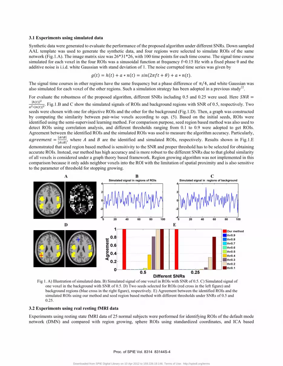
3.1 Experiments using simulated data
Synthetic data were generated to evaluate the performance of the proposed algorithm under different SNRs. Down sampled AAL template was used to generate the synthetic data, and four regions were selected to simulate ROIs of the same network (Fig.1.A). The image matrix size was 26*31*26, with 100 time points for each time course. The signal time course simulated for each voxel in the four ROIs was a sinusoidal function at frequency f=0.15 Hz with a fixed phase θ and the additive noise is i.i.d. white Gaussian with stand deviation of 1. The noise corrupted time series was given by 2 .
The signal time courses in other regions have the same frequency but a phase difference of π 4⁄ , and white Gaussian was also simulated for each voxel of the other regions. Such a simulation strategy has been adopted in a previous study22.
For evaluate the robustness of the proposed algorithm, different SNRs including 0.5 and 0.25 were used. Here | || | . Fig.1.B and C show the simulated signals of ROIs and background regions with SNR of 0.5, respectively. Two seeds were chosen with one for objective ROIs and the other for the background (Fig.1.D). Then, a graph was constructed by computing the similarity between pair-wise voxels according to eqn. (5). Based on the initial seeds, ROIs were identified using the semi-supervised learning method. For comparison purpose, seed region based method was also used to detect ROIs using correlation analysis, and different thresholds ranging from 0.1 to 0.9 were adopted to get ROIs. Agreement between the identified ROIs and the simulated ROIs was used to measure the algorithm accuracy. Particularly, | || |, where and are the identified and simulated ROIs, respectively. Results shown in Fig.1.E demonstrated that seed region based method is sensitivity to the SNR and proper threshold has to be selected for obtaining accurate ROIs. Instead, our method has high accuracy and is more robust to the different SNRs due to that global similarity of all voxels is considered under a graph theory based framework. Region growing algorithm was not implemented in this comparison because it only adds neighbor voxels into the ROI with the limitation of spatial proximity and is also sensitive to the parameter of threshold for stopping growing.
A
B C
D
E
Fig 1. A) Illustration of simulated data. B) Simulated signal of one voxel in ROIs with SNR of 0.5. C) Simulated signal of one voxel in the background with SNR of 0.5. D) Two seeds selected for ROIs (red cross in the left figure) and background regions (blue cross in the right figure), respectively. E) Agreement between the identified ROIs and the simulated ROIs using our method and seed region based method with different thresholds under SNRs of 0.5 and 0.25.
3.2 Experiments using real resting fMRI data
Experiments using resting state fMRI data of 25 normal subjects were performed for identifying ROIs of the default mode network (DMN) and compared with region growing, sphere ROIs using standardized coordinates, and ICA based
Proc. of SPIE Vol. 8314 83144S-4
Downloaded from SPIE Digital Library on 10 Apr 2012 to 159.226.19.146. Terms of Use: http://spiedl.org/terms
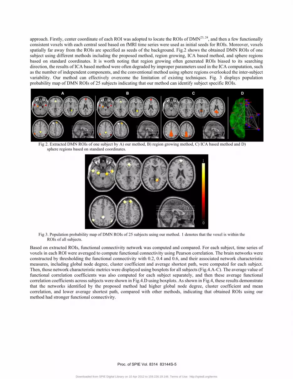
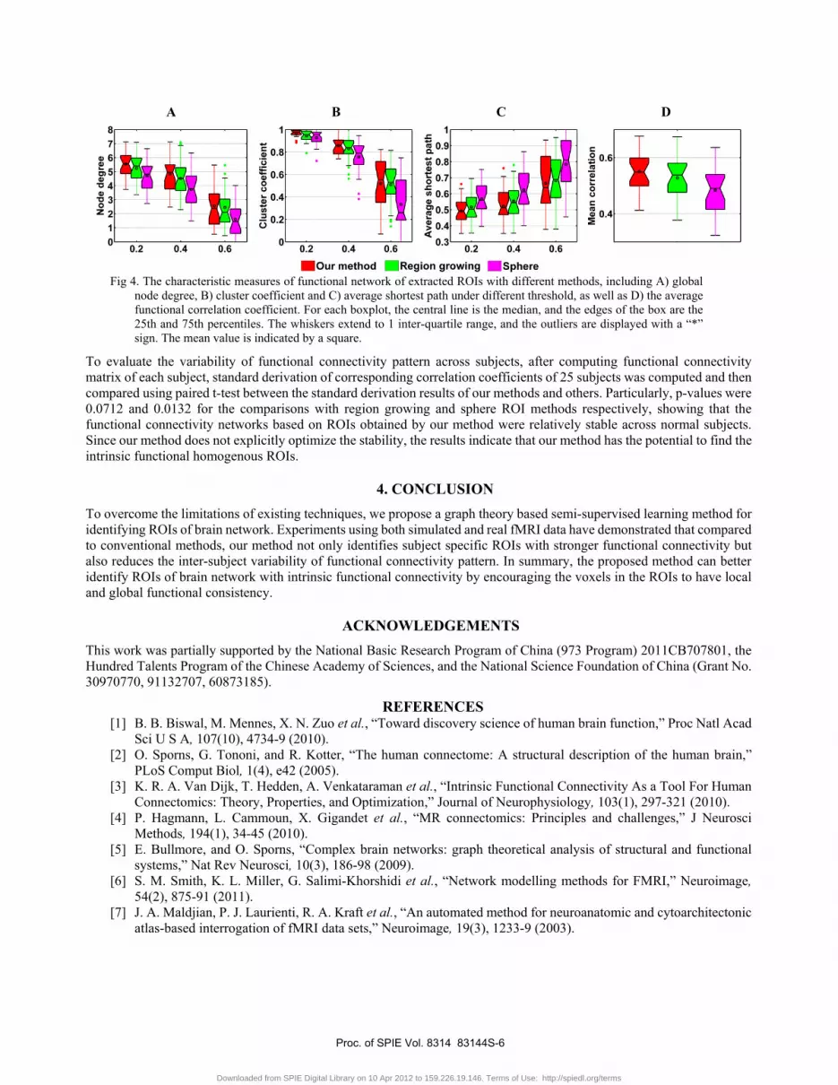
approach. Firstly, center coordinate of each ROI was adopted to locate the ROIs of DMN23, 24, and then a few functionally consistent voxels with each central seed based on fMRI time series were used as initial seeds for ROIs. Moreover, voxels spatially far away from the ROIs are specified as seeds of the background. Fig.2 shows the obtained DMN ROIs of one subject using different methods including the proposed method, region growing, ICA based method, and sphere regions based on standard coordinates. It is worth noting that region growing often generated ROIs biased to its searching direction, the results of ICA based method were often degraded by improper parameters used in the ICA computation, such as the number of independent components, and the conventional method using sphere regions overlooked the inter-subject variability. Our method can effectively overcome the limitation of existing techniques. Fig. 3 displays population probability map of DMN ROIs of 25 subjects indicating that our method can identify subject specific ROIs.
Fig 2. Extracted DMN ROIs of one subject by A) our method, B) region growing method, C) ICA based method and D)
sphere regions based on standard coordinates.
Fig 3. Population probability map of DMN ROIs of 25 subjects using our method. 1 denotes that the voxel is within the
ROIs of all subjects.
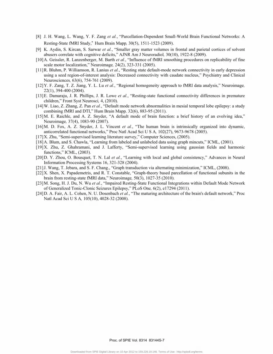
Based on extracted ROIs, functional connectivity network was computed and compared. For each subject, time series of voxels in each ROI were averaged to compute functional connectivity using Pearson correlation. The brain networks were constructed by thresholding the functional connectivity with 0.2, 0.4 and 0.6, and their associated network characteristic measures, including global node degree, cluster coefficient and average shortest path, were computed for each subject. Then, those network characteristic metrics were displayed using boxplots for all subjects (Fig.4.A-C). The average value of functional correlation coefficients was also computed for each subject separately, and then these average functional correlation coefficients across subjects were shown in Fig.4.D using boxplots. As shown in Fig.4, these results demonstrate that the networks identified by the proposed method had higher global node degree, cluster coefficient and mean correlation, and lower average shortest path, compared with other methods, indicating that obtained ROIs using our method had stronger functional connectivity.
Proc. of SPIE Vol. 8314 83144S-5
Downloaded from SPIE Digital Library on 10 Apr 2012 to 159.226.19.146. Terms of Use: http://spiedl.org/terms
A B C D
Fig 4. The characteristic measures of functional network of extracted ROIs with different methods, including A) global node degree, B) cluster coefficient and C) average shortest path under different threshold, as well as D) the average functional correlation coefficient. For each boxplot, the central line is the median, and the edges of the box are the 25th and 75th percentiles. The whiskers extend to 1 inter-quartile range, and the outliers are displayed with a “*” sign. The mean value is indicated by a square.
To evaluate the variability of functional connectivity pattern across subjects, after computing functional connectivity matrix of each subject, standard derivation of corresponding correlation coefficients of 25 subjects was computed and then compared using paired t-test between the standard derivation results of our methods and others. Particularly, p-values were 0.0712 and 0.0132 for the comparisons with region growing and sphere ROI methods respectively, showing that the functional connectivity networks based on ROIs obtained by our method were relatively stable across normal subjects. Since our method does not explicitly optimize the stability, the results indicate that our method has the potential to find the intrinsic functional homogenous ROIs.
4. CONCLUSION To overcome the limitations of existing techniques, we propose a graph theory based semi-supervised learning method for identifying ROIs of brain network. Experiments using both simulated and real fMRI data have demonstrated that compared to conventional methods, our method not only identifies subject specific ROIs with stronger functional connectivity but also reduces the inter-subject variability of functional connectivity pattern. In summary, the proposed method can better identify ROIs of brain network with intrinsic functional connectivity by encouraging the voxels in the ROIs to have local and global functional consistency.
ACKNOWLEDGEMENTS This work was partially supported by the National Basic Research Program of China (973 Program) 2011CB707801, the Hundred Talents Program of the Chinese Academy of Sciences, and the National Science Foundation of China (Grant No. 30970770, 91132707, 60873185).
REFERENCES [1] B. B. Biswal, M. Mennes, X. N. Zuo et al., “Toward discovery science of human brain function,” Proc Natl Acad
Sci U S A, 107(10), 4734-9 (2010). [2] O. Sporns, G. Tononi, and R. Kotter, “The human connectome: A structural description of the human brain,”
PLoS Comput Biol, 1(4), e42 (2005). [3] K. R. A. Van Dijk, T. Hedden, A. Venkataraman et al., “Intrinsic Functional Connectivity As a Tool For Human
Connectomics: Theory, Properties, and Optimization,” Journal of Neurophysiology, 103(1), 297-321 (2010). [4] P. Hagmann, L. Cammoun, X. Gigandet et al., “MR connectomics: Principles and challenges,” J Neurosci
Methods, 194(1), 34-45 (2010). [5] E. Bullmore, and O. Sporns, “Complex brain networks: graph theoretical analysis of structural and functional
systems,” Nat Rev Neurosci, 10(3), 186-98 (2009). [6] S. M. Smith, K. L. Miller, G. Salimi-Khorshidi et al., “Network modelling methods for FMRI,” Neuroimage,
54(2), 875-91 (2011). [7] J. A. Maldjian, P. J. Laurienti, R. A. Kraft et al., “An automated method for neuroanatomic and cytoarchitectonic
atlas-based interrogation of fMRI data sets,” Neuroimage, 19(3), 1233-9 (2003).
Proc. of SPIE Vol. 8314 83144S-6
Downloaded from SPIE Digital Library on 10 Apr 2012 to 159.226.19.146. Terms of Use: http://spiedl.org/terms
[8] J. H. Wang, L. Wang, Y. F. Zang et al., “Parcellation-Dependent Small-World Brain Functional Networks: A Resting-State fMRI Study,” Hum Brain Mapp, 30(5), 1511-1523 (2009).
[9] K. Aydin, S. Kircan, S. Sarwar et al., “Smaller gray matter volumes in frontal and parietal cortices of solvent abusers correlate with cognitive deficits,” AJNR Am J Neuroradiol, 30(10), 1922-8 (2009).
[10] A. Geissler, R. Lanzenberger, M. Barth et al., “Influence of fMRI smoothing procedures on replicability of fine scale motor localization,” Neuroimage, 24(2), 323-331 (2005).
[11] R. Bluhm, P. Williamson, R. Lanius et al., “Resting state default-mode network connectivity in early depression using a seed region-of-interest analysis: Decreased connectivity with caudate nucleus,” Psychiatry and Clinical Neurosciences, 63(6), 754-761 (2009).
[12] Y. F. Zang, T. Z. Jiang, Y. L. Lu et al., “Regional homogeneity approach to fMRI data analysis,” Neuroimage, 22(1), 394-400 (2004).
[13] E. Damaraju, J. R. Phillips, J. R. Lowe et al., “Resting-state functional connectivity differences in premature children,” Front Syst Neurosci, 4, (2010).
[14] W. Liao, Z. Zhang, Z. Pan et al., “Default mode network abnormalities in mesial temporal lobe epilepsy: a study combining fMRI and DTI,” Hum Brain Mapp, 32(6), 883-95 (2011).
[15] M. E. Raichle, and A. Z. Snyder, “A default mode of brain function: a brief history of an evolving idea,” Neuroimage, 37(4), 1083-90 (2007).
[16] M. D. Fox, A. Z. Snyder, J. L. Vincent et al., “The human brain is intrinsically organized into dynamic, anticorrelated functional networks,” Proc Natl Acad Sci U S A, 102(27), 9673-9678 (2005).
[17] X. Zhu, “Semi-supervised learning literature survey,” Computer Sciences, (2005). [18] A. Blum, and S. Chawla, “Learning from labeled and unlabeled data using graph mincuts,” ICML, (2001). [19] X. Zhu, Z. Ghahramani, and J. Lafferty, “Semi-supervised learning using gaussian fields and harmonic
functions,” ICML, (2003). [20] D. Y. Zhou, O. Bousquet, T. N. Lal et al., “Learning with local and global consistency,” Advances in Neural
Information Processing Systems 16, 321-328 (2004). [21] J. Wang, T. Jebara, and S. F. Chang., “Graph transduction via alternating minimization,” ICML, (2008). [22] X. Shen, X. Papademetris, and R. T. Constable, “Graph-theory based parcellation of functional subunits in the
brain from resting-state fMRI data,” Neuroimage, 50(3), 1027-35 (2010). [23] M. Song, H. J. Du, N. Wu et al., “Impaired Resting-State Functional Integrations within Default Mode Network
of Generalized Tonic-Clonic Seizures Epilepsy,” PLoS One, 6(2), e17294 (2011). [24] D. A. Fair, A. L. Cohen, N. U. Dosenbach et al., “The maturing architecture of the brain's default network,” Proc
Natl Acad Sci U S A, 105(10), 4028-32 (2008).
Proc. of SPIE Vol. 8314 83144S-7
Downloaded from SPIE Digital Library on 10 Apr 2012 to 159.226.19.146. Terms of Use: http://spiedl.org/terms


