Embed Size (px)
Citation preview
Hyperelastic modeling of location-dependent human distal femoralcartilage mechanics
Jessica M. Deneweth a,n, Ellen M. Arruda b,c,d, Scott G. McLean a
a School of Kinesiology, University of Michigan, 401 Washtenaw Avenue, Ann Arbor, MI 48109, USAb Department of Mechanical Engineering, University of Michigan, 2350 Hayward Ave., Ann Arbor, MI 48109, USAc Department of Biomedical Engineering, University of Michigan, 2200 Bonisteel Blvd., Ann Arbor, MI 48109, USAd Program in Macromolecular Science & Engineering, University of Michigan, 2300 Hayward Ave., Ann Arbor, MI 48109, USA
a r t i c l e i n f o
Article history:Received 27 January 2014Received in revised form24 June 2014Accepted 29 June 2014
Keywords:Eight-chain networkHyperelasticModelingCartilage mechanicsKneeOsteoarthritis
a b s t r a c t
Knee articular cartilage exhibits complex mechanical behavior, even under high strain rates, which posesa challenge to developing accurate and efficient cartilage models. In particular, the tissue's stress–strainresponse is non-linear and the stiffness of the response is location-dependent. Hyperelastic models suchas those of Alan Gent and others have increasingly found use in soft tissue biomechanics. Recently, ahyperelastic statistical chain network model representing the transverse isotropy of the collagen matrixin the superficial tangential zone has been developed. The model successfully simulated the 100% strain/s unconfined compression response of human proximal tibial cartilage. Moreover, spatial variations inthe tangent modulus to the nominal stress–strain curve taken at 10% strain were reflected in thevariability of a single parameter of the model. Given the success of the model, we desired to determinewhether these outcomes are equally applicable to healthy human distal femoral cartilage so that acomplete model of tibiofemoral joint cartilage can be developed. The transversely isotropic model wasemployed along with two other hyperelastic chain network models to determine which model bestsimulated unconfined compression data for healthy distal femoral cartilage. The transversely isotropicmodel fit the data excellently (R2¼0.999). The model was subsequently simplified to depend on a singleparameter and reapplied to the dataset. The modified model maintained an excellent fit to the data(R2¼0.999), and its single parameter varied in a statistically similar regional pattern (po0.05) to theexperimentally-obtained elastic modulus of the tissue. Outcomes suggest that this model is suitable formodeling the spatially-varying, non-linear mechanics of healthy human distal femoral cartilage.Implementation of this constitutive relation within computational models of the knee will providenovel insight into the relationship between joint mechanics, cartilage loading, and knee osteoarthritisdevelopment.
& 2014 Elsevier Ltd. All rights reserved.
1. Introduction
Knee osteoarthritis (OA)1 afflicts 14% of the US population overthe age of 26 and 37% of the population over age 60, yet it remainsa poorly understood disease [1]. Computational knee modelsafford a powerful research tool for investigating how the diseaseinitiates and progresses [2–4]. Computational studies in whichjoint kinematics and kinetics are systematically varied and theeffect on cartilage stress is determined can indicate which loading
patterns are most likely to initiate and/or promote OA. However,the effectiveness of these models depends on the accuracy of theconstitutive relations describing the many structures making upthe knee. In the case of OA, in which the articular cartilage (AC)2 isheavily affected, the AC material model is particularly important[5].
Selection of a material model requires balancing multiplecriteria, such as correct mechanical response for loading condi-tions of interest, use of parameters with physical meaning thatreveal insights into underlying mechanisms, and computationalefficiency, e.g, short running time and minimal number of
Contents lists available at ScienceDirect
journal homepage: www.elsevier.com/locate/nlm
International Journal of Non-Linear Mechanics
http://dx.doi.org/10.1016/j.ijnonlinmec.2014.06.0130020-7462/& 2014 Elsevier Ltd. All rights reserved.
n Corresponding author. Tel.: þ1 248 568 6369; fax: þ1 734 764 5237.E-mail address: [email protected] (J.M. Deneweth).1 OA: osteoarthritis. 2 AC: articular cartilage.
Please cite this article as: J.M. Deneweth, et al., Hyperelastic modeling of location-dependent human distal femoral cartilage mechanics,International Journal of Non-Linear Mechanics (2014), http://dx.doi.org/10.1016/j.ijnonlinmec.2014.06.013i
International Journal of Non-Linear Mechanics ∎ (∎∎∎∎) ∎∎∎–∎∎∎
parameters [6,7]. For the study of OA, appropriate loading condi-tions are those associated with walking and other activities ofdaily living [8,9]. During level-ground walking knee cartilageundergoes compressive strains at a relatively high strain rate of1 strain/s and peak strains near 20% [10,11], which can bechallenging deformation parameters for popular AC models, e.g.,the linear biphasic model and its derivatives [6,12,13].
The interaction of the three primary constituents of AC—collagenfibrils, large proteoglycan aggregates, and water containing mobileions—gives rise to the tissue's mechanical response [14,15]. At equili-brium, the negatively-charged proteoglycans create a swelling pres-sure in the tissue, which is counteracted by tensile forces in thecollagen network [16,17]. When compressed, fluid stress rises causingfluid to flow out of the tissue until equilibrium is re-established[18,19]. Fluid flow is resisted by electrostatic interactions with theproteoglycans and the physical impediments of the collagen matrix,producing a complex viscous response [18,20]. However, for the veryshort loading times that characterize typical human activity negligiblefluid flow occurs. In this case, the tissue deforms nearly isochorically,with the amount of deformation governed by the mechanics of thecollagen matrix [13,21–23]. Thus, the high strain-rate loading responseof healthy AC may be successfully modeled by focusing on the short-term elasticity of the collagen network, without incorporating viscouseffects or the contribution of proteoglycans.
The collagen network is characterized by non-linear elasticity[24], suggesting that a non-linear elastic model, such as a hyper-elastic statistical chain network model, could be used to model thehigh strain-rate loading response of AC. Current linear elasticmodel are limited in their ability to fully represent the collagennetwork [19,25]. In contrast, hyperelastic statistical chain networkmodels have successfully modeled high strain-rate, finite defor-mations of biological tissues containing collagen, such as aorticvalve, skin, myocardium, tendon, and ligament [26–29]. Thephysical structure of these models is analogous to the collagennetwork that forms the backbone of the cartilage matrix [30,31].These models require a small number of physically-meaningfulparameters, enabling them to be executed in short computationaltimes [26,32–34]. Taken together, these findings indicate that astatistical chain network model would be a viable model for high-strain rate loading of human knee cartilage. Other similarapproaches to non-linear elasticity requiring a limited number ofparameters to capture the full three-dimensional response of softtissues are also becoming increasingly popular in soft tissuebiomechanics. Although there are several notable models in thisgroup, it behooves us to mention Alan Gent's elegant 1996 model[35].
Recently, it was determined that a transversely isotropic eight-chain network with freely jointed chains could simulate the1 strain/s uniaxial compression response of healthy human tibialplateau cartilage with high accuracy (R2¼0.999) [32]. The trans-verse isotropy of the model reflects the anisotropy of the super-ficial tangential zone (STZ)3 of cartilage [31,36], which is the zonethat most influences the mechanical response of the AC matrix[21]. High accuracy was maintained when the model was reducedto dependence on a single parameter that related to the volumedensity of the solid collagen matrix. Furthermore, experimentally-determined regional variations in the tissue's linear elastic mod-ulus at 10% strain were captured by this single parameter. Conse-quently this material model would be highly useful for evaluatingthe role of regional loading patterns on the development of OA[9,37,38]. It is plausible that a similar model would be equallyeffective at modeling the femoral cartilage of the knee.
With the above facts in mind, we currently aimed to evaluatethree statistical chain-network models, including the transverselyisotropic eight-chain network of freely-jointed chains, againsthealthy human distal femoral cartilage. The goal was to providea complete material model for healthy human tibiofemoral jointcartilage that can be readily implemented into whole-knee com-putational modeling schemes. The first aim of the study was todetermine which of three statistical chain network models couldsuccessfully model the uniaxial compression response of distalfemoral AC. We hypothesized that the transversely-isotropic eight-chain network model, which was successful in representingregional tibial cartilage behaviors, would be the most successfulof the three models. The second aim was to determine whetherthe model that best fit the data could represent the regionalmechanical properties of the femoral AC via a single parameter.We hypothesized that variations in CR, the parameter that reflectsthe chain density of the material, would match documentedregional variations in the elastic tangent modulus at 10% nominalstrain of the AC.
2. Methods
2.1. Material models
Three material models were evaluated for this study: the eight-chain isotropic network with freely-jointed chains (FJC)4 [39], theeight-chain isotropic network with MacKintosh chains (MAC)5
[33,40], and the eight-chain transversely isotropic network withfreely-jointed chains (TI)6 [32]. Each model can be described by(1) the constitutive relation used to model a single chain molecule,i.e., a single collagen fibril, and (2) the manner in which the chainsare assembled together to the material network, i.e., the cartilagesolid matrix. The following sections briefly describe two chainmodels (the freely-jointed chain and the MacKintosh chain), twonetwork models (the eight-chain isotropic network and the eight-chain transversely isotropic network), how they were combinedinto the three chain-network models (FJC, MAC, TI) of interest, andhow those models were implemented for the case of uniaxialcompression.
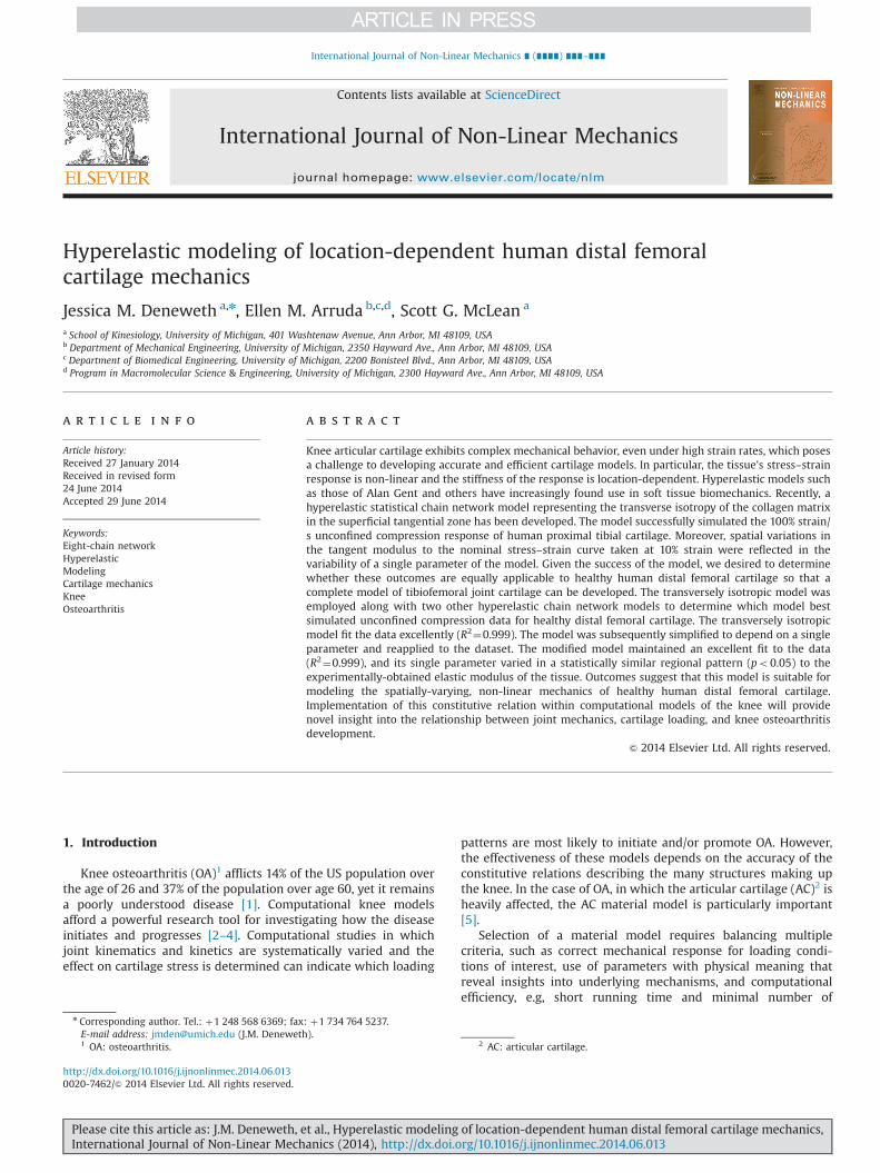
2.1.1. Mechanical response of single collagen fibrilThe freely-jointed chain [41] and the MacKintosh chain [40]
represent two common chain models. These chains are suitable forthe large, non-linear deformations that typically occur in biologi-cal tissues. The chains are considered entropy springs: each chainseeks the conformation that results in maximal entropy. Elongat-ing the chain decreases its entropy and increases its strain energy.The freely-jointed chain can be modeled as N rigid links of length l(Fig. 1a). One end of the chain is fixed at the origin and the otherend occupies a volume dv at a location r with probability p(r):
ln pðrÞ ¼ p0�NrNlβrþ ln
βr
sinh βr
� �ð1Þ
where p0 is a constant, r¼ |r|, βr ¼ℒ�1ðr=NlÞ, and ℒðxÞ ¼coth x�1=x is the Langevin function. The inverse Langevin iscommonly computed from the Padé approximation [42]:
ℒ�1ðxÞ ¼ xð3�x2Þð1�x2ÞþOðx6Þ ð2Þ
3 STZ: superficial tangential zone.
4 FJC: isotropic eight-chain network with freely-jointed chains.5 MAC: isotropic eight-chain network with MacKintosh chains.6 TI: transversely-isotropic eight-chain network with freely-jointed chains.
J.M. Deneweth et al. / International Journal of Non-Linear Mechanics ∎ (∎∎∎∎) ∎∎∎–∎∎∎2
Please cite this article as: J.M. Deneweth, et al., Hyperelastic modeling of location-dependent human distal femoral cartilage mechanics,International Journal of Non-Linear Mechanics (2014), http://dx.doi.org/10.1016/j.ijnonlinmec.2014.06.013i
Elongating the freely-jointed chain to vector length r requires atensile force, fchain [39]:
f chain ¼kΘlβr ð3Þ
where the Padé approximation has been used to simplify theinverse Langevin, k is Boltzmann's constant, 1.38065�10�23 J K�1,andΘ is the absolute temperature. Please refer to Arruda [39] for acomplete derivation. In the freely-jointed chain the position of onelink relative to the previous link is uncorrelated, i.e., all link angleshave equal probability. Consequently the chain is highly flexibleand its resting end-to-end length r0 is much less than its contourlength L¼Nl (Fig. 1a). The chain force remains low for small strainsand then rapidly increases as the strain approaches the lockingstretch,
ffiffiffiffiN
p.
The MacKintosh model, in contrast, enforces a smooth, con-tinuous curvature to the chain (Fig. 1b). Therefore, the chain issemiflexible and its initial length, r0, is on the same order ofmagnitude as its maximum length, L. To enforce the curvatureconstraint, the model incorporates an additional parameter tothose of the freely-jointed chain, the persistence length (lP), whichcontrols the chain's bending rigidity. As lP increases, the moleculebecomes less flexible and r0 approaches L. The average length ofthe MacKintosh chain under no applied tension, rf¼0, can be
related to L and lP via [33]:
rf ¼ 0 ¼ L 1� L6lP
� �ð4Þ
Extending the chain to vector length r requires a tensile force,fchain:
f chain ¼kΘlp
14ð1�ðr=LÞÞ2
!ðL=lpÞ�6ð1�ðr=LÞÞðL=lpÞ�2ð1�ðr=LÞÞ
� �ð5Þ
where all constants are as defined previously. For a completederivation, please refer to Palmer [33].
2.1.2. Mechanical response of the networkThe eight-chain network [39] has been previously implemen-
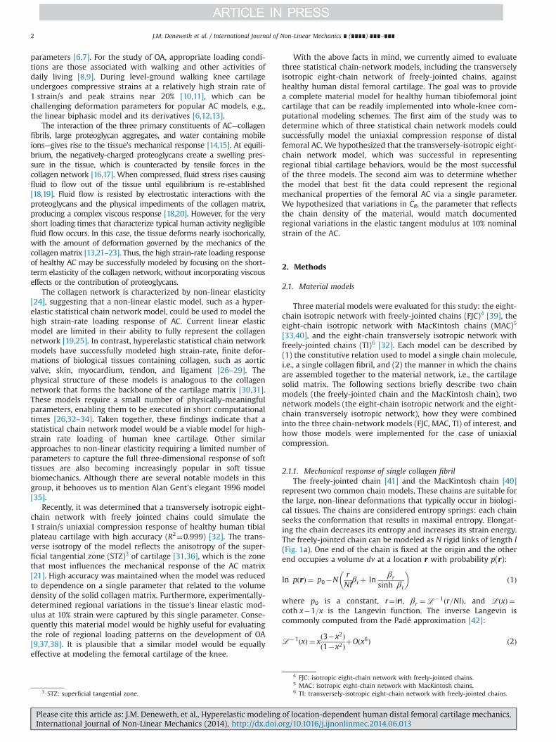
ted with freely-jointed chains and with MacKintosh chains tomodel various biological materials, including skin, cardiac wall,breast, actin filament network, ligament, and cartilage[27,29,33,43–45]. In its isotropic form, the model can be visualizedas a unit cube with eight chains emanating from the center of thecube and extending to each corner (Fig. 2). The chains stretch androtate as the cube is deformed. If the principal directions ofdeformation remain constant throughout the deformation, thesides of the cube will maintain alignment with the principalstretch directions.
For the case of freely-jointed chains, Eq. (3) is incorporated intothe isotropic eight-chain network to obtain the strain energy, UFJC,for the FJC model:
UFJC ¼ nkΘNλchainffiffiffiffi
Np βchainþ ln
βchain
sinh βchain
� �ð6Þ
where n is the chain density, λchain ¼ ðλ21þλ22þλ23Þ1=2=ffiffiffi3
p, λi is the
principal stretch in the ith direction (i¼1, 2, 3), andβchain ¼ℒ�1ðλchain=
ffiffiffiffiN
pÞ [39].
For uniaxial compression, the nominal stress in the axialdirection becomes [39]
To1FJC ¼nkθ3
ffiffiffiffiN
p
λchainℒ�1 λchainffiffiffiffi
Np
� �ðλ�ð1=λ2ÞÞ ð7Þ
where λ is the applied axial stretch and λchain¼[(λ2þ2/λ)/3]1/2.The model assumes incompressibility of the material, which isacceptable for healthy AC subjected to high strain rates becauserelative motion of the fluid with respect to the solid and fluidexudation are negligible [45–48]. Two material constants dictatethe stress response: CR¼nkθ, which reflects the chain density ofthe material and influences the initial modulus of linear elasticity
Fig. 1. Schematics of the (a) freely-jointed chain and (b) MacKintosh chain. Thehighly-flexible freely-jointed chain is composed of N rigid segments of length l. Incontrast, the MacKintosh chain is constrained to have a smooth curvature asdetermined by the persistence length, lp, and is less flexible than the freely-jointedchain. Both chains have a contour length, L. Adapted from Deneweth [32].
Fig. 2. The isotropic eight-chain network in its reference configuration (left) and deformed in uniaxial compression (right). The statistical chains rotate and stretch withapplied deformations.
J.M. Deneweth et al. / International Journal of Non-Linear Mechanics ∎ (∎∎∎∎) ∎∎∎–∎∎∎ 3
Please cite this article as: J.M. Deneweth, et al., Hyperelastic modeling of location-dependent human distal femoral cartilage mechanics,International Journal of Non-Linear Mechanics (2014), http://dx.doi.org/10.1016/j.ijnonlinmec.2014.06.013i

(i.e., the slope of the stress–strain curve as the strain approacheszero), and N, which dictates the locking stretch of the network.
Likewise, implementation of MacKintosh chains into the iso-tropic eight-chain network yields the strain energy for the MACmodel [33]:
UMac ¼ nkΘL
4lpyþ lnðyÞ� lnðL�2lpyÞ
� �ð8Þ
y¼ 1� rL¼ 1�r0λchain
Lð9Þ
where n, k, Θ, and λchain are defined identically to the FJC model.Assuming r0¼rf¼0, the axial nominal stress is
To1MAC ¼nkΘ3lp
rf ¼ 0
λchain1
4ð1�ðrf ¼ 0λchain=LÞÞ2
" #
� ðL=lpÞ�6ð1�ðrf ¼ 0λchain=LÞÞðL=lpÞ�2ð1�ðrf ¼ 0λchain=LÞÞ
" #ðλ�1=λ2Þ ð10Þ
Since rf¼0 is a function of lp and L (Eq. (2)), the responsedepends on only three material constants: CR¼nkθ, lp, and L.
Transversely isotropic [28,32] and orthotropic [26,49] eight-chain networks have recently been introduced. A transverselyisotropic eight-chain network with freely-jointed chains has beenshown to better simulate the uniaxial compression response inhuman tibial plateau cartilage than isotropic eight-chain networkmodels [32]. This transversely isotropic model was a modificationof the orthotropic eight-chain model of Bischoff [26] and will beused in the current study. The transversely isotropic case isderived by modifying the structure of the isotropic eight-chainmodel from a cube to a rectangular prism (Fig. 3). The side alignedwith the principal compression axis has dimension a, which hasbeen normalized by dividing by l. The remaining two sides, whichlie in the plane perpendicular to the compression axis, havenormalized dimension b. The ratio b:a indicates the degree ofanisotropy of the tissue, with b:a¼1 occuring for the isotropiccase. The direction corresponding to the largest dimension willalso exhibit the highest tensile modulus [26,49]. Additionally,incompressibility is not assumed but a bulk compressibility termis included to maintain near-incompressibility. The total strainenergy of the transversely isotropic eight-chain configuration offreely jointed chains (TI) is given as [26,32]
UTI ¼U0þnkΘ4
N ∑4
i ¼ 1
ρðiÞ
NβðiÞρ þ ln
βðiÞρ
sinh βðiÞρ
24
35
0@
� βPffiffiffiffiN
p ln ½λa2a λ2b2
b ��þ Bα2 cosh ½αðJ�1Þ��1� � ð11Þ
where U0 is a constant; βðiÞρ ¼ℒ�1ðρðiÞ=NÞ; βP ¼ℒ�1ðP=NÞ; P ¼
12
ffiffiffiffiffiffiffiffiffiffiffiffiffiffiffiffiffiffia2þ2b2
p¼
ffiffiffiffiN
pis the undeformed chain length; ρ(i) is the
deformed length of the ith chain; λa and λb are the stretches
along the axis of compression and the two axes perpendicular tothe axis of compression, respectively; J¼λ1λ2λ3 is the ratio of thedeformed volume to the original volume; B controls the bulkcompressibility near J¼1; and α is a constant that governs thecurvature of the hydrostatic pressure versus volume curve forlarge volume changes.
The nominal axial stress for uniaxial deformation of the nearlyincompressible transversely isotropic freely jointed eight-chain(TI) model is
T01TI ¼nkθ4λ1
a2λ21βρρ
� βPffiffiffiffiN
p" #
�b2λ22βρρ
� βPffiffiffiffiN
p" # !
ð12Þ
whereρ¼ 1
2
ffiffiffiffiffiffiffiffiffiffiffiffiffiffiffiffiffiffiffiffiffiffiffiffiffiffiffia2λ21þ2b2λ22
q; P ¼
ffiffiffiffiN
p¼ 1
2
ffiffiffiffiffiffiffiffiffiffiffiffiffiffiffiffiffiffia2þ2b2
p; and λ2 ¼
ffiffiffiffiffiffiffiffiffiJ=λ1
p. Small
volume changes were assumed to agree with the near incompres-sibility of the tissue so α was set equal to unity [48]. The TI modelrequires three independent parameters in addition to CR¼nkθ: a,b, and J.
2.2. Experimental data
Each model was simulated against uniaxial compression datausing a method described previously [37]. Cylindrical, full-thickness explants of healthy AC were procured from 29 standar-dized tests sites of seven human femurs. Femurs were obtainedfrom cadaveric Caucasian female donors whose cause of death wasunlikely to compromise the knee joint (mean7standard deviationage: 5073 years). Knees were stored at �20 1C until sampleswere procured. The femoral AC surface was isolated by thawingthe knee overnight to room temperature, removing all soft tissueto expose the tibiofemoral and patellofemoral joints, and disarti-culating the femur from the tibia and patella. Standardized gridpatterns were fitted onto the trochlear (3 cells mediolateral�3cells proximodistal, typical cell size: 10 mm�13 mm) and con-dylar (2 cells mediolateral�5 cells proximodistal, typical cell size:12 mm�15 mm) surfaces such that a maximum AC surface areafell under the grid (Fig. 4) [32,50]. Cartilage samples were obtainedfrom the center of each grid cell, which resulted in 9 trochlearsamples and 20 condylar samples per knee. A 4-mm diameterround-hole hand punch was used to isolate the sample, and asurgical scalpel was employed to carefully separate the AC fromthe subchondral bone [51]. Each sample was stored in phosphatebuffered saline solution at �20 1C until mechanical testing wasperformed. Freezing and thawing AC at this temperature has beenfound to preserve the samples’ mechanical properties [52,53]. AnIndia ink test was conducted prior to extraction to identify surfacefibrillation [54]. Samples with surface damage were excluded fromtesting.
Explants were subjected to three trials of unconfined compres-sion from 0% to 20% peak nominal strain at 1 strain/s, which
Fig. 3. The transversely isotropic model replicates the architecture of the collagen matrix in the STZ. The dimension a is aligned perpendicular to the cartilage surface, whilethe remaining two sides, each with dimension b, are parallel to the cartilage surface. As b increases relative to a, the orientation of the chains rotate to become more parallelto the cartilage surface. Correspondingly the tensile modulus in plane defined by the b dimensions increases while the tensile modulus along the a dimension decreases.
J.M. Deneweth et al. / International Journal of Non-Linear Mechanics ∎ (∎∎∎∎) ∎∎∎–∎∎∎4
Please cite this article as: J.M. Deneweth, et al., Hyperelastic modeling of location-dependent human distal femoral cartilage mechanics,International Journal of Non-Linear Mechanics (2014), http://dx.doi.org/10.1016/j.ijnonlinmec.2014.06.013i

replicates the AC strain and strain rate associated with level-ground gait [10,11]. Testing was conducted with a custom high-rate uniaxial tester [37]. It contained a fixed dynamic load cell(Dytran Instruments, Chatsworth, CA; sensitivity: 120 mV/N) and ahorizontal high-speed electric linear actuator (SMAC, Carlsbad, CA;positional accuracy: 70.001 mm) fixed at opposite ends of a PBSsolution bath. Compression plates were mounted on the load celland actuator (Dytran Instruments, Chatsworth, CA; diameter:15.78 mm). A high-speed video camera (Photron USA, San Diego,CA; maximum frame rate: 5400 frames/s) was mounted above thetesting apparatus to record deformation of the sample duringeach test.
Cartilage explants were thawed at room temperature, placedunder a minimal tare load (0.2 N), and allowed to equilibrate for10 min in the phosphate buffered saline bath prior to testing. Arandom speckle pattern was applied to the exposed explantsurface with black India ink [37,55]. The sample underwent tenpre-conditioning cycles at the prescribed strain and strain rate.Subsequently three experimental trials of compression (com-pressed from 0% to 20% peak strain followed by immediate returnto 0% strain) were conducted while synchronous force and videowere recorded at 125 Hz. Several minutes were allowed betweentrials for the specimen to re-equilibrate.
Digital image correlation software was used to calculate aver-age nominal axial strain, i.e., λ�1, via the local deformation of thespeckle pattern [37,56]. Average nominal stress was computed bydividing the current force by the undeformed cross-sectional area.Axial nominal stress–strain curves were constructed and used asinput data for model simulations. Additionally, the tangent mod-ulus to the nominal stress vs. nominal strain curve at 10% strain(E10%) was extracted and averaged across trials [37]. This strainlevel was selected because it is representative of physiologicalcartilage strain experienced during walking and running [10,11].
2.3. Model simulations
Model simulations were conducted in Matlab (Mathworks,Natick, MA) using the third experimental data trial from each AC
specimen. Inputs were the experimental strain, experimentalstress, and an initial guess at the unknown model parameters:CR and N for FJC; CR, lP, and L for MAC; and CR, a, b, and J for TI.Since all parameters represent lengths, moduli, or a volume ratio,non-negative parameter values were required. A built-in non-linear least-squares optimization routine determined the modelparameters that minimized the squared error between the experi-mental stress and model-predicted stress. Based on our previouswork [32], we additionally constrained a¼1 so that variations in balone dictated the anisotropy of the tissue, bZ1 since a and bwere normalized to l, and Jr1 [57,58]. The goodness of fit of eachmodel was determined from:
R2 ¼ 1� ∑iðσexpi �σpredi Þ2∑iðσexpi �σpredi Þ2
ð13Þ
where σexpi is the experimental stress corresponding to the ithstrain data point, σpredi is the ith predicted stress, and σpred is themean predicted stress.
2.4. Analysis of regional dependence
The best-fitting model (highest R2) was simplified so that onlyCR was a free parameter and the remaining parameters were heldconstant [32]. The optimization scheme was repeated in Matlab forall experimental trials for each AC specimen. The goodness of fitwas calculated for each simulation via Eq. (13). Simulations withexcellent fit to the data, R2Z0.97, were used for regional analysis.This high cut-off was selected so that the effect of CR could beclearly assessed. Mean CR was computed for the trochlea as awhole, the condyles as a whole, six sub-regions on the trochlea[the medial, central, and lateral frequently loaded, i.e., “weight-bearing” (WB),7 and the medial, central, and lateral less-frequentlyloaded, i.e., less weightbearing (LWB),8 sub-regions], and four sub-regions on the condyles [medial and lateral WB and LWB] (Fig. 4).
Fig. 4. (Left) Cartilage samples were extracted from the trochlea and condyles of each femur. A grid pattern was drawn onto each surface, and a sample was taken from thecenter of each grid cell (black circle). (Right) For statistical analysis, the 29 test sites (circles) were grouped by region (trochlea, Troch, n¼9; condyles, Cond, n¼20). Sampleswere grouped within region by mediolateral position and weightbearing frequency (WB/LWB). C: central trochlea, L: lateral trochlea, LC: lateral condyle, LWB: less-frequently loaded, M: medial trochlea, MC: medial condyle, WB: frequently loaded.
7 WB: frequently loaded (“weightbearing”) articular cartilage.8 LWB: less-frequently loaded (“less weightbearing”) articular cartilage.
J.M. Deneweth et al. / International Journal of Non-Linear Mechanics ∎ (∎∎∎∎) ∎∎∎–∎∎∎ 5
Please cite this article as: J.M. Deneweth, et al., Hyperelastic modeling of location-dependent human distal femoral cartilage mechanics,International Journal of Non-Linear Mechanics (2014), http://dx.doi.org/10.1016/j.ijnonlinmec.2014.06.013i
Weightbearing and less weightbearing areas represented femoralcartilage contacting the opposing joint surface between 01 and 301knee flexion and beyond 301 knee flexion, respectively [59–62].
Mean CR of the trochlea and condyle primary regions werecompared with repeated-measures analyses of variance (ANOVA).For the sub-regions, the main effect of side [medial, lateral,(central)] and contact frequency (WB, LWB) and the interactionof the two factors on CR were evaluated with repeated-measuresmixed model ANOVA. In the case of significant main effects orinteractions (po0.05), Bonferroni-adjusted pairwise comparisonswere made. For comparison to experimental data, mean E10% wascalculated for each region of interest and submitted to the samestatistical analysis. All statistical analyses were conducted withSAS 8.0 software (SAS Institute Inc., Cary, NC).
3. Results
3.1. Determination of best fitting model
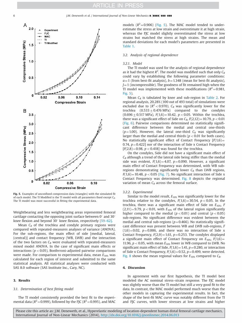
The TI model consistently provided the best fit to the experi-mental data (R2¼0.999), followed by the FJC (R2¼0.995), and MAC
models (R2¼0.906) (Fig. 5). The MAC model tended to under-estimate the stress at low strain and overestimate it at high strain,whereas the FJC model slightly overestimated the stress at lowstrains but matched the stress at high strains. The mean andstandard deviations for each model's parameters are presented inTable 1.
3.2. Analysis of regional dependence
3.2.1. ModelThe TI model was used for the analysis of regional dependence
as it had the highest R2. The model was modified such that only CRcould vary by establishing the following parameter conditions:a¼1 (from best-fit analysis), b¼1.348 (mean for best-fit analysis),J¼1 (incompressible). The goodness of fit remained high when theTI model was implemented with these modifications (R2¼0.981,Fig. 5).
Mean CR is tabulated by knee and sub-region in Table 2. Forregional analysis, 20.28% (100 out of 493 total) of simulations wereexcluded due to (R2o0.970). CR was significantly lower for thetrochlea (0.53370.476 MPa) compared to the condyles(0.69670.517 MPa), F(1,6)¼10.42, po0.05. Within the trochlea,there was a significant effect of Side on CR, F(2,12)¼10.79, po0.01(Fig. 6). Pairwise comparisons determined no statistically signifi-cant difference between the medial and central one-thirds(p¼1.00). However, the lateral one-third CR was significantlylarger than the medial and central thirds (po0.01 for both cases).No statistically significant effect of Contact Frequency [F(1,6)¼0.74, p¼0.422] nor of the interaction of Side x Contact Frequency[F(2,8)¼0.98, po0.418] was found for the trochlea.
On the condyles, Side did not have a significant main effect ofCR although a trend of the lateral side being stiffer than the medialside was evident, F(1,6)¼4.07, p¼0.090. However, a significantmain effect of Contact Frequency was determined, with WB sub-regions demonstrating significantly lower CR than LWB regions,F(1,6)¼10.48, po0.05 (Fig. 7). No significant interaction of Side xContact Frequency was determined. Fig. 8 depicts the regionalvariation of mean CR across the femoral surface.
3.2.2. ExperimentalSimilar to the model result, E10% was significantly lower for the
trochlea relative to the condyles, F(1,6)¼30.54, po0.05. In thetrochlea, there was a significant main effect of Side on E10%, F(2,11)¼9.79, po0.01, with E10% of the lateral region significantlyhigher compared to the medial (po0.01) and central (po0.05)sub-regions. No significant difference was evident between themedial and central sub-regions (p¼0.466). No statistically signifi-cant difference was present between WB and LWB sub-regions, F(1,6)¼0.02, p¼0.886, and there was no interaction of Side xContact Frequency, F(2,9)¼1.61, p¼0.253. The condyles displayeda significant main effect of Contact Frequency on E10%, F(1,6)¼11.96, po0.05, with mean E10% lower in WB compared to LWB. Nosignificant main effect of Side, F(1,6)¼1.41, p¼0.280, or interactionof Side x Contact Frequency, F(1,6)¼0.52, p¼0.499, were detected.Fig. 8 shows the mean regional values for E10% compared to CR.
4. Discussion
In agreement with our first hypothesis, the TI model bestmodeled the AC nominal stress–strain response. The FJC modelwas slightly worse than the TI model but still a very good fit to thedata. In contrast, the MAC model performed much worse than theother models in capturing the experimental results. In fact, theshape of the best-fit MAC curve was notably different from the TIand FJC curves, with lower stresses at low strains and higher
Fig. 5. Examples of unconfined compression data (triangles) with the simulated fitof each model. The TI Modified is the TI model with all parameters fixed except CR.The TI model was most successful in fitting the experimental data.
J.M. Deneweth et al. / International Journal of Non-Linear Mechanics ∎ (∎∎∎∎) ∎∎∎–∎∎∎6
Please cite this article as: J.M. Deneweth, et al., Hyperelastic modeling of location-dependent human distal femoral cartilage mechanics,International Journal of Non-Linear Mechanics (2014), http://dx.doi.org/10.1016/j.ijnonlinmec.2014.06.013i

stresses at higher strains. The MacKintosh chains of the MACmodel are quite different from the freely-jointed chains of the FJCand TI models, which likely accounts for the disparate modelperformances. The MAC model was developed for chains that aresemi-flexible [33,40], whereas the FJC and TI assume flexiblechains [26,39,41]. Additionally, the length of the undeformedconfiguration relative to its limiting extensibility differs amongmodels. The undeformed configuration of the Mackintosh chain isnear its limiting extensibility [40]. In contrast, the undeformedlength of the freely-jointed chain is much less than its limitingextensibility [39,41]. Based on the results of the model simula-tions, it appears that type II collagen, which represents the mostcommon collagen type found AC [63], is best represented by thefreely-jointed chain utilized in the FJC and TI models.
Our second hypothesis was that the TI model could representthe regional mechanical properties of femoral AC via variations inCR. The results of this study support this hypothesis. In themodified TI model a¼1, b¼1.348, J¼1, and CR was selected tooptimize the simulated stress to the experimental stress. Model
simulations against the entire experimental dataset producedvalues of CR that varied regionally within each knee in a similarfashion to regional variations in the AC elastic modulus at 10%nominal strain (E10%). Specifically, CR of the condyles was signifi-cantly higher than CR of the trochlea, which corresponds withfindings of a higher linear elastic modulus on the condylescompared to the trochlea [50,64]. Within the trochlea, CR of thelateral sub-regions were significantly higher than the central andmedial sub-regions. Within the condyles, CR was higher in theless-weightbearing regions compared to the weightbearingregions. Of note, the linear elastic modulus used as comparisonfor CR was computed at 10% nominal strain because the experi-ment was a strain-driven test and 10% is a typical AC strain duringgait. However, we additionally calculated the linear elastic moduliat 1%, 5%, and 15% nominal strain and examined the curvature ofthe stress–strain plots. From these analyses we confirmed that thetrends reported for E10% would be unaffected if the strain level forcalculating the linear modulus was changed.
Table 1Mean and standard deviation of optimized parameters for the FJC, MAC, and TI models.
FJC MAC TI
CR (MPa) N R2 CR (MPa) L lP R2 CR (MPa) b J R2
Mean 0.053 1.143 0.995 5.045 4.207 5.792 0.906 0.370 1.348 0.994 0.999SD 0.047 1.390 0.005 2.667 0.907 0.902 0.075 0.615 0.248 0.005 0.001
Table 2Mean CR (MPa) of modified TI model by knee and sub-region.
KneeTrochlea Condyles
M WB C WB L WB M LWB C LWB L LWB M WB L WB M LWB L LWB
1 0.094 0.085 0.244 0.107 0.125 0.272 0.228 0.335 0.270 0.7072 0.217 0.462 0.404 0.158 0.480 0.617 0.504 0.614 0.617 1.4653 1.349 1.658 1.532 1.389 1.811 1.620 2.163 1.658 1.8624 0.130 0.320 0.324 0.078 0.379 0.215 0.375 0.208 0.5435 0.189 0.196 0.300 0.191 0.550 0.598 0.535 0.439 0.6346 0.411 0.604 0.963 0.121 0.122 1.594 0.358 0.595 0.657 1.0087 0.314 0.194 0.521 0.144 0.404 0.733 0.256 0.483 0.607 0.332
M – medial; C – central; L – lateral; WB – weightbearing; LWB – less weightbearing.Blank cells indicate that no simulations with R240.970 were conducted.
Fig. 6. Mean CR across the six sub-regions of the trochlea. Bars indicate onestandard deviation. Hatching indicates less weightbearing regions. The lateralweightbearing and less weightbearing sub-regions had significantly higher CRcompared to the medial and central sub-regions. No statistical differences weredetermined for weightbearing versus less weightbearing regions. These findingsagree well with experimentally-determined cartilage moduli.
Fig. 7. Mean CR across the four sub-regions of the condyles. Bars indicate onestandard deviation. Hatching indicates less weightbearing regions. The weightbear-ing regions had significantly lower CR compared to the less weightbearing regions.No statistical differences were determined between the medial and lateral regions.These findings agree well with the experimentally-determined cartilage moduli.
J.M. Deneweth et al. / International Journal of Non-Linear Mechanics ∎ (∎∎∎∎) ∎∎∎–∎∎∎ 7
Please cite this article as: J.M. Deneweth, et al., Hyperelastic modeling of location-dependent human distal femoral cartilage mechanics,International Journal of Non-Linear Mechanics (2014), http://dx.doi.org/10.1016/j.ijnonlinmec.2014.06.013i
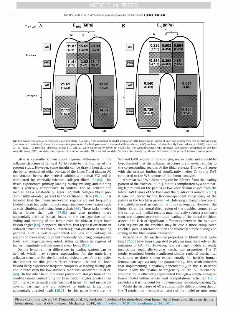
Little is currently known about regional differences in thecollagen structure of femoral AC to relate to the findings of thepresent study. However, some insight can be drawn from data onthe better-researched tibial plateau of the knee. Tibial plateau ACnot situated below the menisci exhibits a minimal STZ and isdominated by vertically-oriented collagen fibers [36,65]. Thistissue experiences primary loading during walking and runningthat is generally compressive. In contrast, the AC beneath themenisci has a substantially larger STZ, with collagen fibers pre-dominantly oriented parallel to the cartilage surface [36,65]. It isbelieved that the meniscus-covered regions are not frequentlyloaded in gait but rather in tasks requiring deep knee flexion, suchas stair climbing and rising from a chair [66]. These tasks involvehigher forces than gait [67,68] and also produce moretangentially-oriented (shear) loads on the cartilage due to thesliding and rotating of the femur over the tibia at larger kneeflexion angles [69]. It appears, therefore, that regional variations incollagen structure of tibial AC match regional variations in loadingpatterns. That is, vertically-oriented and less stiff cartilage inregions of lower magnitude but frequently occurring compressiveloads and tangentially-oriented, stiffer cartilage in regions ofhigher magnitude and infrequent shear loads [8,38].
On the femur, similar differences in loading pattern can bedefined, which may suggest expectations for the underlyingcollagen structure. For the femoral condyles, areas of the condylesthat contact the tibia joint surfaces between �51 and 301 kneeflexion likely experience frequent compressive loading during gaitand interact with the low-stiffness, meniscus-uncovered tibial AC[66]. On the other hand, the more posterior/distal portions of thecondyles make contact only for knee flexion angles greater than301, interact with much stiffer meniscal tissue [70] and meniscus-covered cartilage, and are believed to undergo large, moretangentially-directed loads [59,71]. In this paper these are the
WB and LWB regions of the condyles, respectively, and it could behypothesized that the collagen structure is somewhat similar tothe corresponding regions of the tibial plateau. This would agreewith the present finding of significantly higher CR in the LWBcompared to the WB regions of the femur condyles.
A similar WB/LWB dichotomy can be inferred from the loadingpattern of the trochlea [59,71], but it is complicated by a dominat-ing lateral pull on the patella at low knee flexion angles from thelateral soft tissues of the knee and the quadriceps muscle [72,73].It also influenced by the flexion-dependent congruency of thepatella in the trochlear groove [74]. Inferring collagen structure atthe patellofemoral articulation is thus challenging. However, thehigher CR on the lateral third region of the trochlea compared tothe central and medial regions may indirectly suggest a collagenstructure adapted to concentrated loading of the lateral trochlearaspect. The lack of significant differences between the WB andLWB regions on the trochlea may be due a more complicatedtrochlea–patella interaction than the relatively simple sliding androlling of the tibia–femur interaction.
Variations in the mechanical properties of tibiofemoral carti-lage [37,50] have been suggested to play an important role in theinitiation of OA [75]. However, few cartilage models currentlyincorporate regionally-varying mechanical properties. The TImodel examined herein manifested similar regional mechanicalvariations to those shown experimentally for healthy humanfemoral cartilage via only one parameter, CR. This result indicatesthat implementing a spatially-dependent CR in the TI networkwould allow the spatial heterogeneity of the AC mechanicalresponse to be efficiently represented through a simple collagen-based model within whole joint computational schemes. Fig. 8provides a starting point for implementing regionally-varying CR.
While the structure of AC is substantially different from that ofthe TI model, the mechanistic nature of the model permits some
Fig. 8. Comparison of E10% determined experimentally (A) and CR from Modified TI model simulations (B). Boxed areas represent each sub-region with text displaying mean(one standard deviation) values of the respective parameter. For both parameters, the medial (M) and central (C) trochlea had significantly lower values (po0.05) comparedto the lateral (L) trochlea. Likewise, mean E10% and CR were significantly lower (po0.05) for the weightbearing (WB) condylar sub-regions compared to the lessweightbearing (LWB) condylar sub-regions. LC – lateral condyle, MC – medial condyle. No other statistically significant differences were present between sub-regions.
J.M. Deneweth et al. / International Journal of Non-Linear Mechanics ∎ (∎∎∎∎) ∎∎∎–∎∎∎8
Please cite this article as: J.M. Deneweth, et al., Hyperelastic modeling of location-dependent human distal femoral cartilage mechanics,International Journal of Non-Linear Mechanics (2014), http://dx.doi.org/10.1016/j.ijnonlinmec.2014.06.013i

exploration of potential effects of the collagen matrix on the ACstress–strain response at high strain rates. In the context of kneeAC, CR could be interpreted to partially reflect the volume densityof intact collagen fibrils. Areas with fewer collagen fibrils or withincreased damage to existing fibrils (e.g., due to OA) are expectedto exhibit a lower CR. The effect of CR is reflected in the initialmodulus of the stress–strain curve; lower CR dictates a initialmodulus [39].
The ratio a:b indicates the extent of transverse isotropy of thecollagen matrix: a:b41 occurs when the collagen fibers are morefrequently aligned in the direction of compression, and a:bo1occurs when the collagen fibers are preferentially oriented in theplane perpendicular to the compression axis [26]. Collagen fibersin the STZ of knee AC primarily align parallel to the cartilagesurface (i.e., perpendicular to the compression axis) [31,36]. Theresults of this study agree well with this finding as a:b was lessthan one across all test sites. If a depth-dependent cartilageresponse is desired, the model could be extended to the entirethickness of the cartilage by allowing a:b to vary with depth: a:bo1 in the STZ, a:bE1 in the middle zone where the collagenfibers transition from their parallel orientation at the surface totheir radial orientation near the subchondral bone, and a:b41 inthe deep zone near the subchondral bone [31].
The kinematical variable J governs the compressibility of thetissue. If the tissue is incompressible, meaning that it deformswithout changing total volume, J¼1. Articular cartilage is com-posed mainly of water and thus is nearly incompressible at highstrain rates where negligible fluid flow occurs [47,48]. In thepresent study J remained nearly constant across regions and knees,which agrees well with the physiology of the tissue and withprevious findings for modeling tibial cartilage [32]. Therefore, wefeel confident that J can remain fixed near 1 for modeling healthycartilage. Likewise, changes in the water composition of the tissueor the ability of the tissue to maintain its hydrostatic pressure (e.g.,due to severe matrix damage) could be modeled with a decrease inJ, although this must first be validated.
We explicitly chose to ignore the proteoglycan content in themodel although it influences the compressive loading response ofAC [14,19,76,77]. For the short loading duration utilized in ourstudy, negligible fluid flow occurs, negating viscous effects due tothe proteoglycan aggregates interacting with fluid flowing out ofthe tissue [12]. Rather AC deformation occurs under nearlyconstant volume and is controlled by the resistance of the collagennetwork [21,78]. We acknowledge that the presence of proteogly-cans, by creating an osmotic pressure to resist collapse of thecollagen network, enables this deformation to occur [77]. How-ever, it likely does not influence the stress–strain relationship inthis case. Bader et al. [22] determined that proteoglycans governedthe viscous damping of the compression response while collagendominated the elastic response. Therefore, in the case of a singleunconfined compression loading and immediate unloading, asdone in our study, the response should be dominated by themechanical response of the collagen network. This was confirmedvia computational modeling by Li et al. [79]. With these findings inmind we chose to not explicitly consider proteoglycan content.Likewise, the very good fit of the model is possible because of theminimal influence of proteoglycan in the loading scenario utilized.Additionally, this finding is likely possible because we strictlyexamined healthy tissue. Osteoarthritic tissue is known to havehigher proteoglycan content, increased swelling, and a compro-mised collagen structure [76,80]. In this case, it is possible thatnegligible fluid flow may not be a valid assumption and proteo-glycan content must be considered.
The mean R2 for the TI model with all parameters fixed except CRwas very high, but 20% of the simulations were subsequently rejectedfor R2o0.97. Post-hoc repeated-measures ANOVA determined that
mean b for high-rejection test sites (50% or more rejected trials) was1.287, which was significantly lower than mean b for low-rejectionsites (b¼1.325), F(1,6)¼325.95, po0.001. This finding suggests thatthe model is particularly sensitive to b in regions with less anisotropythan average (i.e., lower b). Nonetheless all rejected simulationsdemonstrated R240.90, which is generally accepted as a highgoodness of fit. When the regional analysis for CR was repeated withall simulations included, observed regional variations in CR remainedthe same. Consequently, the modified TI model would be an excellentmethod for modeling the high strain-rate response of femoral AC.
Several potential limitations may impact the generalizability ofstudy outcomes. The experimental data used for this study, forexample, were taken from a restricted demographic. It is thereforeunclear as to the extent to which these results can be applied tothe general population. Further work must be done to validate themodel against datasets from other demographics and if necessarydetermine relevant parameter values for these groups. The modelhas also only been evaluated for AC loaded in unconfined com-pression, but other deformation states, e.g., shear, may also berelevant to cartilage degeneration. Eight-chain networks havesuccessfully modeled multiple deformation states for polymers[34,39], but this has not been examined with AC. Additionalresearch into the extent to which the current model parameterscan successfully simulate other important deformation statesshould now be conducted. Nonetheless, the TI model appears tobe highly valuable for modeling of AC within whole-knee compu-tational models. Several elegant non-linear, inhomogeneous, ani-sotropic cartilage models currently exist, but few modeltopographical variations in cartilage mechanics and none with asingle varying-parameter as in the TI model [57,81-88]. Modelingcartilage with the TI model will provide new insights into diseasesassociated with the non-uniform mechanical properties of thetissue without compromising the efficient dynamics of the wholemodel. If such steps are taken, efforts to implement and validatethe TI model using a region-dependent CR, within a valid kneejoint model, should initially be undertaken.
The TI model as proposed in this paper can feasibly beimplemented within any commercial finite element package thatpermits user-defined hyperelastic materials (e.g., Abaqus, DassaultSystèmes, France). The model's strain energy Eq. (11) would besupplied along with its derivatives and parameter values. Thestrain energy can be simplified by using constant values for allparameters except CR (i.e., nkθ): α,a,J,B¼1; and b¼1.3481. Tomodel the heterogeneity in human AC, CR must be defined as afunction of position across the cartilage surface (Fig. 8). The TImodel is appropriate for any material with known transverseanisotropy and a mechanical response primarily dictated by non-linear, recoverable deformation.
5. Conclusions
Three statistical chain network models have been evaluated forsimulating high strain-rate uniaxial compression of healthyhuman femoral AC. The TI model was found to most successfullyfit the data. Moreover, the model maintained an excellent fit whenits degrees of freedom were reduced from three to one, which isnot common for a mechanistic cartilage model. The reduceddegree of freedom TI model was particularly novel in that regionalvariations in its single non-constant parameter, CR, followed thesame pattern as experimentally-measured elastic modulus. Themodel incorporates parameters that could be interpreted withrespect to the collagen structure of the AC. This mechanistic naturealong with its efficient formulation make it particularly valuablefor implementing into computational models of the knee. Further-more, its ability to simulate the regionally-varying properties of
J.M. Deneweth et al. / International Journal of Non-Linear Mechanics ∎ (∎∎∎∎) ∎∎∎–∎∎∎ 9
Please cite this article as: J.M. Deneweth, et al., Hyperelastic modeling of location-dependent human distal femoral cartilage mechanics,International Journal of Non-Linear Mechanics (2014), http://dx.doi.org/10.1016/j.ijnonlinmec.2014.06.013i
healthy knee cartilage, which is currently not replicated by othermodels, will greatly enhance current investigations of knee OAinitiation and progression.
Acknowledgments
The authors gratefully acknowledge Ms. K. Newman,Ms. S. Pomeroy, and Mr. S. Sylvia for their assistance with dataacquisition. J.M.D. was supported by the Department of Defensethrough the National Defense Science & Engineering GraduateFellowship Program while conducting this research. E.M.A. wouldlike to thank Ray Ogden, Giuseppe Saccomandi, and Alan Wine-man for inviting her to contribute to this special issue in honor ofAlan Gent.
References
[1] R.C. Lawrence, D.T. Felson, C.G. Helmick, L.M. Arnold, H. Choi, R.A. Deyo,S. Gabriel, R. Hirsch, M.C. Hochberg, G.G. Hunder, J.M. Jordan, J.N. Katz, H.M. Kremers, F. Wolfe, Estimates of the prevalence of arthritis and otherrheumatic conditions in the United States. Part II, Arthritis Rheum. 58 (1)(2008) 26–35.
[2] R. Shirazi, A. Shirazi-Adl, Analysis of partial meniscectomy and ACL recon-struction in knee joint biomechanics under a combined loading, Clin. Biomech.24 (9) (2009) 755–761.
[3] J.Y. Bae, K.S. Park, J.K. Seon, D.S. Kwak, I. Jeon, E.K. Song, Biomechanical analysisof the effects of medial meniscectomy on degenerative osteoarthritis, Med.Biol. Eng. Comput. 50 (1) (2012) 53–60.
[4] E. Pena, B. Calvo, M.A. Martinez, D. Palanca, M. Doblare, Why lateralmeniscectomy is more dangerous than medial meniscectomy. A finite elementstudy, J. Orthop. Res. 24 (5) (2006) 1001–1010.
[5] W. Wilson, C.C. van Donkelaar, R. van Rietbergen, R. Huiskes, The role ofcomputational models in the search for the mechanical behavior and damagemechanisms of articular cartilage, Med. Eng. Phys. 27 (10) (2005) 810–826.
[6] Z.A. Taylor, K. Miller, Constitutive modeling of cartilaginous tissues: a review,J. Appl. Biomech. 22 (3) (2006) 212–229.
[7] M. Hubbard, Computer simulation in sport and industry, J. Biomech. 26 (Suppl.1) (1993) 53–61.
[8] T.P. Andriacchi, S. Koo, S.F. Scanlan, Gait mechanics influence healthy cartilagemorphology and osteoarthritis of the knee, J. Bone Jt. Surg. Am. 91 (2 Suppl. 1)(2009) 95–101.
[9] T.P. Andriacchi, A. Mundermann, The role of ambulatory mechanics in theinitiation and progression of knee osteoarthritis, Curr. Opin. Rheumatol. 18 (5)(2006) 514–518.
[10] F. Liu, M. Kozanek, A. Hosseini, S.K. Van de Velde, T.J. Gill, H.E. Rubash, G. Li,In vivo tibiofemoral cartilage deformation during the stance phase of gait, J.Biomech. 43 (4) (2010) 658–665.
[11] R.A. Mann, J. Hagy, Biomechanics of walking, running, and sprinting, Am. J.Sports Med. 8 (5) (1980) 345–350.
[12] C.G. Armstrong, W.M. Lai, V.C. Mow, An analysis of the unconfined compres-sion of articular cartilage, J. Biomech. Eng. 106 (2) (1984) 165–173.
[13] M.R. DiSilvestro, Q. Zhu, J.K. Suh, Biphasic poroviscoelastic simulation of theunconfined compression of articular cartilage: II—Effect of variable strainrates, J. Biomech. Eng. 123 (2) (2001) 198–200.
[14] V.C. Mow, X.E. Guo, Mechano-electrochemical properties of articular cartilage:their inhomogeneities and anisotropies, Annu. Rev. Biomed. Eng. 4 (2002) 175–209.
[15] X.L. Lu, V.C. Mow, Biomechanics of articular cartilage and determination ofmaterial properties, Med. Sci. Sports Exerc. 40 (2) (2008) 193–199.
[16] E.H. Han, S.S. Chen, S.M. Klisch, R.L. Sah, Contribution of proteoglycan osmoticswelling pressure to the compressive properties of articular cartilage, Biophys.J. 101 (4) (2011) 916–924.
[17] X.L. Lu, V.C. Mow, X.E. Guo, Proteoglycans and mechanical behavior ofcondylar cartilage, J. Dent. Res. 88 (3) (2009) 244–248.
[18] D.D. Sun, X.E. Guo, M. Likhitpanichkul, W.M. Lai, V.C. Mow, The influence ofthe fixed negative charges on mechanical and electrical behaviors of articularcartilage under unconfined compression, J. Biomech. Eng. 126 (1) (2004) 6–16.
[19] V.C. Mow, M.H. Holmes, W.M. Lai, Fluid transport and mechanical propertiesof articular cartilage: a review, J. Biomech. 17 (5) (1984) 377–394.
[20] D.L. Bader, G.E. Kempson, J. Egan, W. Gilbey, A.J. Barrett, The effects of selectivematrix degradation on the short-term compressive properties of adult humanarticular cartilage, Biochim. Biophys. Acta 1116 (2) (1992) 147–154.
[21] J. Mizrahi, A. Maroudas, Y. Lanir, I. Ziv, T.J. Webber, The instantaneousdeformation of cartilage: effects of collagen fiber orientation and osmoticstress, Biorheology 23 (4) (1986) 311–330.
[22] D.L. Bader, G.E. Kempson, The short-term compressive properties of adulthuman articular cartilage, Biomed. Mater. Eng. 4 (3) (1994) 245–256.
[23] A.I. Maroudas, Balance between swelling pressure and collagen tension innormal and degenerate cartilage, Nature 260 (5554) (1976) 808–809.
[24] E. Gentleman, A.N. Lay, D.A. Dickerson, E.A. Nauman, G.A. Livesay, K.C. Dee,Mechanical characterization of collagen fibers and scaffolds for tissue engi-neering, Biomaterials 24 (21) (2003) 3805–3813.
[25] L.P. Li, K.B. Gu, Reconsideration on the use of elastic models to predict theinstantaneous load response of the knee joint, Proc. Inst. Mech. Eng. H 225 (9)(2011) 888–896.
[26] J.E. Bischoff, E.M. Arruda, K. Grosh, A microstructurally based orthotropichyperelastic constitutive law, J. Appl. Mech.-Trans. ASME 69 (5) (2002)570–579.
[27] J.E. Bischoff, E.M. Arruda, K. Grosh, A rheological network model for thecontinuum anisotropic and viscoelastic behavior of soft tissue, Biomech.Model. Mechanobiol. 3 (1) (2004) 56–65.
[28] E. Kühl, K. Garikipati, E.M. Arruda, K. Grosh, Remodeling of biological tissue:mechanically induced reorientation of a transversely isotropic chain network,J. Mech. Phys. Solids 53 (7) (2005) 1552–1573.
[29] J. Ma, H. Narayanan, K. Garikipati, K. Grosh, E.M. Arruda, Experimental andcomputational investigation of viscoelasticity of native and engineered liga-ment and tendon, in: Iutam Symposium on Cellular, Molecular and TissueMechanics, Proceedings, Springer, New York (2010) 3–17.
[30] E.M. Hasler, W. Herzog, J.Z. Wu, W. Muller, U. Wyss, Articular cartilagebiomechanics: theoretical models, material properties, and biosyntheticresponse, Crit. Rev. Biomed. Eng. 27 (6) (1999) 415–488.
[31] A.K. Jeffery, G.W. Blunn, C.W. Archer, G. Bentley, Three-dimensional collagenarchitecture in bovine articular cartilage, J. Bone Jt. Surg. Br. 73 (5) (1991)795–801.
[32] J.M. Deneweth, S.G. McLean, E.M. Arruda, Evaluation of hyperelastic modelsfor the non-linear and non-uniform high strain-rate mechanics of tibialcartilage, J. Biomech. (2013) (published online May 12, 2013).
[33] J.S. Palmer, M.C. Boyce, Constitutive modeling of the stress–strain behavior ofF-actin filament networks, Acta Biomater. 4 (3) (2008) 597–612.
[34] M.C. Boyce, E.M. Arruda, Constitutive models of rubber elasticity: a review,Rubber Chem. Technol. 73 (3) (2000) 504–523.
[35] A.N. Gent, New constitutive relation for rubber, Rubber Chem. Technol. 69 (1)(1996) 59.
[36] J.M. Clark, Variation of collagen fiber alignment in a joint surface: a scanningelectron microscope study of the tibial plateau in dog, rabbit, and man, J.Orthop. Res. 9 (2) (1991) 246–257.
[37] J.M. Deneweth, K.E. Newman, S.M. Sylvia, S.G. McLean, E.M. Arruda, Hetero-geneity of tibial plateau cartilage in response to a physiological compressivestrain rate, J. Orthop. Res. 31 (3) (2013) 370–375.
[38] A.M. Chaudhari, P.L. Briant, S.L. Bevill, S. Koo, T.P. Andriacchi, Knee kinematics,cartilage morphology, and osteoarthritis after ACL injury, Med. Sci. SportsExerc. 40 (2) (2008) 215–222.
[39] E.M. Arruda, M.C. Boyce, A 3-dimensional constitutive model for the largestretch behavior of rubber elastic—materials, J. Mech. Phys. Solids 41 (2)(1993) 389–412.
[40] F.C. MacKintosh, J. Kas, P.A. Janmey, Elasticity of semiflexible biopolymernetworks, Phys. Rev. Lett. 75 (24) (1995) 4425–4428.
[41] W. Kühn, F. Grun, Relations between elastic constants and the strainbirefringence of high-elastic substances, Kolloid-Z. 101 (3) (1942) 248–271.
[42] A. Cohen, A Pade approximant to the inverse Langevin function, Rheol. Acta 30(3) (1991) 270–273.
[43] J.E. Bischoff, E.M. Arruda, K. Grosh, Finite element modeling of human skinusing an isotropic, nonlinear elastic constitutive model, J. Biomech. 33 (6)(2000) 645–652.
[44] Y. Liu, A. Kerdok, R. Howe, A nonlinear finite element model of soft tissueindentation, Med. Simul. (2004) 67–76.
[45] C.P. Brown, T.C. Nguyen, H.R. Moody, R.W. Crawford, A. Oloyede, Assessment ofcommon hyperelastic constitutive equations for describing normal and osteoar-thritic articular cartilage, Proc. Inst. Mech. Eng. H 223 (H6) (2009) 643–652.
[46] V.C. Mow, S.C. Kuei, W.M. Lai, C.G. Armstrong, Biphasic creep and stressrelaxation of articular cartilage in compression? Theory and experiments, J.Biomech. Eng. 102 (1) (1980) 73–84.
[47] G.A. Ateshian, B.J. Ellis, J.A. Weiss, Equivalence between short-time biphasic andincompressible elastic material responses, J. Biomech. Eng. 129 (3) (2007) 405–412.
[48] M. Wong, M. Ponticiello, V. Kovanen, J.S. Jurvelin, Volumetric changes ofarticular cartilage during stress relaxation in unconfined compression, J.Biomech. 33 (9) (2000) 1049–1054.
[49] J.E. Bischoff, E.M. Arruda, K. Grosh, Orthotropic hyperelasticity in terms of anarbitrary molecular chain model, J. Appl. Mech.-Trans. ASME, 69, 2002198–201.
[50] A.C. Swann, B.B. Seedhom, The stiffness of normal articular cartilage and thepredominant acting stress levels: implications for the aetiology of osteoar-throsis, Br. J. Rheumatol. 32 (1) (1993) 16–25.
[51] R.J. Lewis, A.K. MacFarland, S. Anandavijayan, R.M. Aspden, Material propertiesand biosynthetic activity of articular cartilage from the bovine carpo-metacarpal joint, Osteoarthr. Cartil. 6 (6) (1998) 383–392.
[52] R. Krishnan, S. Park, F. Eckstein, G.A. Ateshian, Inhomogeneous cartilageproperties enhance superficial interstitial fluid support and frictional proper-ties, but do not provide a homogeneous state of stress, J. Biomech. Eng. 125 (5)(2003) 569–577.
[53] S. Ronken, M.P. Arnold, H. Ardura Garcia, A. Jeger, A.U. Daniels, D. Wirz, Acomparison of healthy human and swine articular cartilage dynamic indenta-tion mechanics, Biomech. Model. Mechanobiol. 11 (5) (2012) 631–639.
[54] G. Meachim, Light microscopy of Indian ink preparations of fibrillatedcartilage, Ann. Rheum. Dis. 31 (6) (1972) 457–464.
J.M. Deneweth et al. / International Journal of Non-Linear Mechanics ∎ (∎∎∎∎) ∎∎∎–∎∎∎10
Please cite this article as: J.M. Deneweth, et al., Hyperelastic modeling of location-dependent human distal femoral cartilage mechanics,International Journal of Non-Linear Mechanics (2014), http://dx.doi.org/10.1016/j.ijnonlinmec.2014.06.013i
[55] J. Ma, M.J. Smietana, T.Y. Kostrominova, E.M. Wojtys, L.M. Larkin, E.M. Arruda,Three-dimensional engineered bone–ligament–bone constructs for anteriorcruciate ligament replacement, Tissue Eng. Part A 18 (1–2) (2012) 103–116.
[56] J. Ma, K. Goble, M. Smietana, T. Kostrominova, L. Larkin, E.M. Arruda,Morphological and functional characteristics of three-dimensional engineeredbone–ligament–bone constructs following implantation, J. Biomech. Eng. 131(10) (2009) 101017.
[57] S. Chegini, S.J. Ferguson, Time and depth dependent Poisson's ratio of cartilageexplained by an inhomogeneous orthotropic fiber embedded biphasic model,J. Biomech. 43 (9) (2010) 1660–1666.
[58] R.K. Korhonen, M.S. Laasanen, J. Toyras, J. Rieppo, J. Hirvonen, H.J. Helminen, J.S. Jurvelin, Comparison of the equilibrium response of articular cartilage inunconfined compression, confined compression and indentation, J. Biomech.35 (7) (2002) 903–909.
[59] J. Goodfellow, D.S. Hungerford, M. Zindel, Patello-femoral joint mechanics andpathology. 1. Functional anatomy of the patello-femoral joint, J. Bone Jt. Surg.Br. 58 (3) (1976) 287–290.
[60] A. Garg, P.S. Walker, Prediction of total knee motion using a three-dimensionalcomputer-graphics model, J. Biomech. 23 (1) (1990) 45–58.
[61] J.A. Feller, A.A. Amis, J.T. Andrish, E.A. Arendt, P.J. Erasmus, C.M. Powers,Surgical biomechanics of the patellofemoral joint, Arthroscopy 23 (5) (2007)542–553.
[62] G. Li, S.E. Park, L.E. DeFrate, M.E. Schutzer, L. Ji, T.J. Gill, H.E. Rubash, Thecartilage thickness distribution in the tibiofemoral joint and its correlationwith cartilage-to-cartilage contact, Clin. Biomech. 20 (7) (2005) 736–744.
[63] D. Eyre, Collagen of articular cartilage, Arthritis Res. 4 (1) (2002) 30–35.[64] M.K. Barker, B.B. Seedhom, The relationship of the compressive modulus of
articular cartilage with its deformation response to cyclic loading: doescartilage optimize its modulus so as to minimize the strains arising in it dueto the prevalent loading regime? Rheumatology 40 (3) (2001) 274–284.
[65] D.R. Eyre, M.A. Weis, J.J. Wu, Articular cartilage collagen: an irreplaceableframework? Eur. Cells Mater. 12 (2006) 57–63.
[66] J.T. Bingham, R. Papannagari, S.K. Van de Velde, C. Gross, T.J. Gill, D.T. Felson, H.E. Rubash, G. Li, In vivo cartilage contact deformation in the healthy humantibiofemoral joint, Rheumatology 47 (11) (2008) 1622–1627.
[67] I.G. Goudakos, C. Konig, P.B. Schottle, W.R. Taylor, N.B. Singh, I. Roberts,F. Streitparth, G.N. Duda, M.O. Heller, Stair climbing results in more challen-ging patellofemoral contact mechanics and kinematics than walking at earlyknee flexion under physiological-like quadriceps loading, J. Biomech. 42 (15)(2009) 2590–2596.
[68] B.B. Seedhom, K. Terayama, Knee forces during the activity of getting out of achair with and without the aid of arms, Biomed. Eng. 11 (8) (1976) 278–282.
[69] M.A. Lafortune, P.R. Cavanagh, H.J. Sommer III, A. Kalenak, Three-dimensionalkinematics of the human knee during walking, J. Biomech. 25 (4) (1992)347–357.
[70] H.N. Chia, M.L. Hull, Compressive moduli of the human medial meniscus in theaxial and radial directions at equilibrium and at a physiological strain rate, J.Orthop. Res. 26 (7) (2008) 951–956.
[71] T.M. van Eijden, E. Kouwenhoven, J. Verburg, W.A. Weijs, A mathematicalmodel of the patellofemoral joint, J. Biomech. 19 (3) (1986) 219–229.
[72] C.M. Powers, F.G. Shellock, M. Pfaff, Quantification of patellar tracking usingkinematic MRI, J. Magn. Reson. Imaging 8 (3) (1998) 724–732.
[73] S.K. Han, S. Federico, M. Epstein, W. Herzog, An articular cartilage contactmodel based on real surface geometry, J. Biomech. 38 (1) (2005) 179–184.
[74] V.V. Patel, K. Hall, M. Ries, C. Lindsey, E. Ozhinsky, Y. Lu, S. Majumdar, Magneticresonance imaging of patellofemoral kinematics with weight-bearing,19182033, 85-A(12), 2003, pp. 2419–2424.
[75] T.P. Andriacchi, A. Mundermann, R.L. Smith, E.J. Alexander, C.O. Dyrby, S. Koo,A framework for the in vivo pathomechanics of osteoarthritis at the knee, Ann.Biomed. Eng. 32 (3) (2004) 447–457.
[76] C.G. Armstrong, V.C. Mow, Variations in the intrinsic mechanical properties ofhuman articular cartilage with age, degeneration, and water content, J. Bone Jt.Surg. Am. 64 (1) (1982) 88–94.
[77] I.M. Basalo, R.L. Mauck, T.A. Kelly, S.B. Nicoll, F.H. Chen, C.T. Hung, G.A. Ateshian, Cartilage interstitial fluid load support in unconfined compressionfollowing enzymatic digestion, J. Biomech. Eng. 126 (6) (2004) 779–786.
[78] C.J. Moger, K.P. Arkill, R. Barrett, P. Bleuet, R.E. Ellis, E.M. Green, C.P. Winlove,Cartilage collagen matrix reorientation and displacement in response tosurface loading, J. Biomech. Eng. 131 (3) (2009) 031008.
[79] L.P. Li, M.D. Buschmann, A. Shirazi-Adl, Strain-rate dependent stiffness ofarticular cartilage in unconfined compression, J. Biomech. Eng. 125 (2) (2003)161–168.
[80] L. Hendren, P. Beeson, A review of the differences between normal andosteoarthritis articular cartilage in human knee and ankle joints, Foot 19 (3)(2009) 171–176.
[81] W. Wilson, J.M. Huyghe, C.C. van Donkelaar, A composition-based cartilagemodel for the assessment of compositional changes during cartilage damageand adaptation, Osteoarthr. Cartil. 14 (6) (2006) 554–560.
[82] W. Wilson, C.C. van Donkelaar, B. van Rietbergen, K. Ito, R. Huiskes, Stresses inthe local collagen network of articular cartilage: a poroviscoelastic fibril-reinforced finite element study, J. Biomech. 37 (3) (2004) 357–366.
[83] L.P. Li, J. Soulhat, M.D. Buschmann, A. Shirazi-Adl, Nonlinear analysis ofcartilage in unconfined ramp compression using a fibril reinforced poroelasticmodel, Clin. Biomech. 14 (9) (1999) 673–682.
[84] M.J. Askew, V.C. Mow, Biomechanical function of the collagen fibril ultra-structure of articular cartilage, J. Biomech. Eng. 100 (3) (1978) 105–115.
[85] G.A. Ateshian, V. Rajan, N.O. Chahine, C.E. Canal, C.T. Hung, Modeling thematrix of articular cartilage using a continuous fiber angular distributionpredicts many observed phenomena, J. Biomech. Eng. 131 (6) (2009) 061003.
[86] N.M. Bachrach, V.C. Mow, F. Guilak, Incompressibility of the solid matrix ofarticular cartilage under high hydrostatic pressures, J. Biomech. 31 (5) (1998)445–451.
[87] L.P. Li, W. Herzog, R.K. Korhonen, J.S. Jurvelin, The role of viscoelasticity ofcollagen fibers in articular cartilage: axial tension versus compression, Med.Eng. Phys. 27 (1) (2005) 51–57.
[88] M.A. Soltz, G.A. Ateshian, A Conewise Linear Elasticity mixture model for theanalysis of tension-compression nonlinearity in articular cartilage, J. Biomech.Eng. 122 (6) (2000) 576–586.
J.M. Deneweth et al. / International Journal of Non-Linear Mechanics ∎ (∎∎∎∎) ∎∎∎–∎∎∎ 11
Please cite this article as: J.M. Deneweth, et al., Hyperelastic modeling of location-dependent human distal femoral cartilage mechanics,International Journal of Non-Linear Mechanics (2014), http://dx.doi.org/10.1016/j.ijnonlinmec.2014.06.013i


![[Management of ipsilateral femoral neck and shaft fractures]](https://img.dokumen.tips/doc/110x75/6356c196debc1859f6037d64/management-of-ipsilateral-femoral-neck-and-shaft-fractures.jpg)


