Embed Size (px)
Citation preview
Molecular Psychiatry (2021) 26:4864–4883https://doi.org/10.1038/s41380-020-0839-9
ARTICLE
Human forebrain endothelial cell therapy for psychiatric disorders
Debkanya Datta1,2,3 ● Sivan Subburaju1,2,3● Sarah Kaye1,3 ● Jugajyoti Baruah1,2,3
● Yong Kee Choi1,2,3 ● Yeqi Nian4,5●
Jahan S. Khalili6 ● Sangmi Chung 7● Abdallah Elkhal4,5 ● Anju Vasudevan 1,2,3
Received: 23 September 2019 / Revised: 23 June 2020 / Accepted: 3 July 2020 / Published online: 13 July 2020© The Author(s) 2020. This article is published with open access
AbstractAbnormalities of or reductions in GABAergic interneurons are implicated in the pathology of severe neuropsychiatricdisorders, for which effective treatments are still elusive. Transplantation of human stem cell-derived interneurons is apromising cell-based therapy for treatment of these disorders. In mouse xenograft studies, human stem cell-derived-interneuron precursors could differentiate in vivo, but required a prolonged time of four to seven months to migrate from thegraft site and integrate with the host tissue. This poses a serious roadblock for clinical translation of this approach. Fortransplantation to be effective, grafted neurons should migrate to affected areas at a faster rate. We have previously shownthat endothelial cells of the periventricular vascular network are the natural substrates for GABAergic interneurons in thedeveloping mouse forebrain, and provide valuable guidance cues for their long-distance migration. In addition,periventricular endothelial cells house a GABA signaling pathway with direct implications for psychiatric disease origin.In this study we translated this discovery into human, with significant therapeutic implications. We generated humanperiventricular endothelial cells, using human pluripotent stem cell technology, and extensively characterized its molecular,cellular, and functional properties. Co-culture of human periventricular endothelial cells with human interneuronssignificantly accelerated interneuron migration in vitro and led to faster migration and wider distribution of graftedinterneurons in vivo, compared to neuron-only transplants. Furthermore, the co-transplantation strategy was able to rescueabnormal behavioral symptoms in a pre-clinical model of psychiatric disorder, within 1 month after transplantation. Weanticipate this strategy to open new doors and facilitate exciting advances in angiogenesis-mediated treatment of psychiatricdisorders.
Introduction
Abnormal migration, positioning, and reduction inGABAergic interneurons during the critical prenataldevelopmental period results in dysfunctional cortical neu-ronal synchrony implicated in brain diseases such as autism,epilepsy, and schizophrenia [1–5], conditions awaitingmore effective treatments. Cell transplantation is a powerfultool to introduce new cells with intrinsic plasticity toovercome cellular deficits and initiate repair and regenera-tion. To be successful, grafted cells should possess theability to migrate and disperse through affected areas, dif-ferentiate into fully mature neurons, functionally integrate,and modulate circuitry activity in the damaged host brain. Abetter comprehension of the cellular and molecularmechanisms of neuronal development has led to use of theirprecursors in transplantation [6–8]. While the origin andspecification of cortical GABAergic interneurons was wellestablished [9–11], mechanisms that underlined theirmigration were not fully understood. Our studies served to
* Anju [email protected]
1 Angiogenesis and Brain Development Laboratory, HuntingtonMedical Research Institutes (HMRI), 686 S Fair Oaks Avenue,Pasadena, CA 91105, USA
2 Department of Psychiatry, Harvard Medical School, Boston, MA02215, USA
3 Division of Basic Neuroscience, McLean Hospital, 115 MillStreet, Belmont, MA 02478, USA
4 Department of Surgery, Harvard Medical School, Boston, MA02115, USA
5 Division of Transplantation, Brigham and Women’s Hospital, 221Longwood Avenue, EBRC 309, Boston, MA 02115, USA
6 Personal Peptides LLC, Houston, TX 77002, USA7 Department of Cell biology and Anatomy, New York Medical
College, Valhalla, NY 10595, USA
Supplementary information The online version of this article (https://doi.org/10.1038/s41380-020-0839-9) contains supplementarymaterial, which is available to authorized users.
1234
5678
90();,:
1234567890();,:
address this critical gap by showing that embryonic fore-brain vascular networks are strategically positioned to pro-vide physical support and critical guidance cues forGABAergic interneuron migration in the developing tele-ncephalon [12–14]. In addition, our work has establishednovel autonomous links between the periventricular vas-cular network and the origin of psychiatric disorders, fromthe earliest developmental time points [13, 15]. Under-standing brain development thus begins with an apprecia-tion of all of its cellular components. Deeper insights intothe anatomy, origin, molecular regulation, function, anddysfunction of the periventricular vascular network in thelast decade [12–15] were crucial for understanding its directsignificance for psychiatric disorders.
Due to the restricted availability of human fetal tissue forcell therapy, human pluripotent stem cell (hPSC) technol-ogy provides an unprecedented opportunity to study diseasemechanisms [16–22]. Multiple groups have successfullyderived human interneuron/interneuron progenitors fromhPSCs [23–26] and transplantation of interneurons/inter-neuron progenitors has emerged as a promising treatmentoption for psychiatric disorders [27–32]. When transplantedin mouse [33] and rat [34] models of epilepsy, hPSC-derived-interneuron precursors, survived well, fired actionpotentials, formed functional synaptic connections andcould reduce abnormal seizure activities. Though showinggreat promise, one issue that needs improvement is themigration efficiency of transplanted cells. At 2 weeks posttransplantation, transplanted interneurons displayed mini-mal migration, and it was only at 4–7 months post trans-plantation, that some migration and integration into hostbrain was observed [23, 25, 33, 34]. Therefore, the bene-ficial effects of interneuron graft-in-disease models wereobserved only several months after transplantation. Thispresents an obstacle for the clinical translation ofinterneuron-based therapy, especially for very sick orseverely affected patients. Another drawback that has beendescribed with GABA producing cell types after trans-plantation is their transient effects, due to reductions inGABA levels [35, 36]. A decrease in GABA-mediatedinhibition is a critical contributing factor for hyperexcit-ability and seizure initiation and increased secretion ofGABA, by grafted cells, is important for increasing theseizure threshold. Thus, at present, while transplantation ofGABAergic interneurons represents the most promisingcell-based therapeutic alternative for GABA-related dis-eases, there are difficulties that need to be overcome.
Our key fundamental discovery that pre-formed vascularnetworks are the natural guides for GABAergic neuronalmigration from the earliest developmental time pointassumes a new significance here that can serve to improvehPSC-derived GABAergic neuronal migration. The peri-ventricular vascular network not only acts as a physical
substrate for neuronal migration in the embryonic forebrain,but also has a very unique gene expression profile, unlikeendothelial cells from other brain regions or organs [12–15].Periventricular endothelial cells (PVECs) show enrichedexpression of cell surface marker, GABAA receptorβ3 subunit (GABRB3) as opposed to pial endothelial cellsor control endothelial cells prepared from midbrain andhindbrain [15]. Therefore, GABRB3 serves as a valuabletool to selectively sort human endothelial cells, which areakin to mouse PVECs. In addition, we know that PVECsexpress and release GABA that promotes rapid and exten-sive long-distance migration of GABAergic interneurons[15]. Neuronal GABA cannot compensate for the roles ofendothelial GABA and interneurons stall in their migrationin the absence of endothelial GABA [15]. This close neu-rovascular interaction during embryonic brain developmentthat is sealed by space and time is the key missing link ininterneuron-based therapy. Interestingly, it has been repor-ted that transplanted neuronal precursors align and migratealong the surface of host blood vessels [37, 38] as though insearch of a missing or lost counterpart. All of this funda-mental knowledge provided us with strong rationale togenerate human PVECs from human embryonic stem cellsand to tap into the potential of these endothelial cells toimprove human GABAergic interneuron migration in vitroand in vivo.
Our results show that human PVECs can faithfullymimic the functional aspects of mouse periventricularangiogenesis and promote long-distance migration ofhuman GABAergic interneurons in culture and after trans-plantation into multiple mouse brain regions. In addition,this co-transplantation approach markedly improved cellmigration and dispersion, stabilized GABA release levelsand improved behavioral outcome in a pre-clinical model ofpsychiatric disorder, within 1 month after transplantation, asopposed to interneuron-only transplantation, which did notshow any rescue in the same time frame. Notably, this workshows for the first time how prenatal forebrain angiogen-esis, when tapped into correctly, has a remarkable potentialfor repair and regeneration in the adult brain that can serveto ameliorate abnormal psychiatric behaviors.
Results
Efficient generation of human PVECs from humanembryonic stem cells
Brain endothelial cells have been derived from hPSC pre-viously [39–42], but their analysis, gene expression studiesand functions have largely focused on the blood–brainbarrier attributes. Generation of embryonic forebrain spe-cific PVECs has not been reported so far. PVECs show
Human forebrain endothelial cell therapy for psychiatric disorders 4865
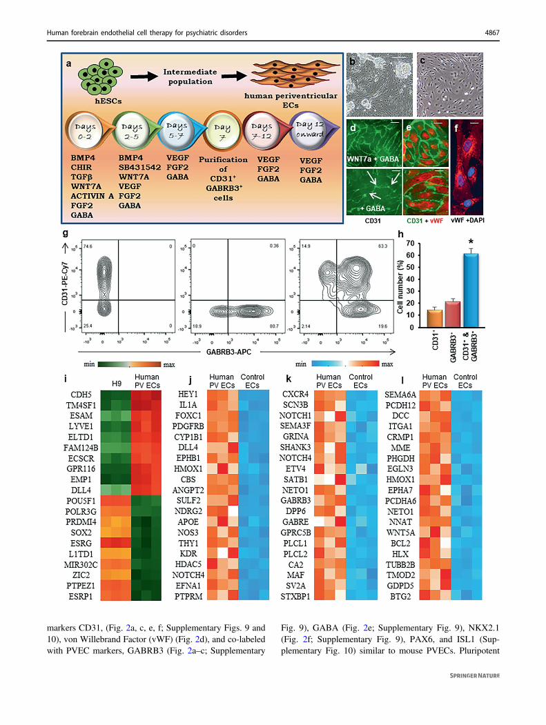
enrichment in genes controlling neurogenesis, neuronalmigration, chemotaxis, and axon guidance and can be dis-tinguished from other endothelial cells by their specifictranscription factor expression, signaling molecules andextracellular receptors [12–15]. Indeed, this specific popu-lation of endothelial cells houses a novel GABA signalingpathway that is distinct from the classical neuronal GABAsignaling pathway in the embryonic forebrain. Vasculatureis autonomous with respect to anatomy, patterning, geneexpression, developmental regulation, and function in dif-ferent organs and/or regions at different time points.Developing strategies to generate embryonic forebrain-likevasculature will augment its region-specific vessel growthand function after transplantation and will be important forclinical applications. Based on our knowledge of geneexpression of mouse PVECs of the embryonic forebrain, wefirst established a differentiation strategy to generate humanPVECs from human ES cell lines (Fig. 1a–c; Supplemen-tary Figs. 1–4). The strategy was initially implementedusing the human ES cell line H9, and robustness of theprotocol was subsequently validated with the human ES cellline H1. From day 0–2 of differentiation, mesodermal cellfate was induced in H9 cells by culturing them in E8 mediasupplemented with bone morphogenetic protein 4 (BMP4),Activin A, and CHIR 99021 (small molecule activator ofWNT pathway). From days 2–5, vascular induction waspromoted by inhibiting TGFβ signaling (using the smallmolecular inhibitor SB431542) and by addition of vascularendothelial growth factor-A (VEGF-A). Since PVECsexpress Wnt7a and GABA, we added these components tothe differentiating medium. A combination of WNT7Aprotein and low concentration of GABA (5 μM) from days 0to 5 in the differentiating medium was critical for tightjunction formation and production of endogenous GABA inthese endothelial cells (Supplementary Figs. 1 and 2).Interestingly, higher levels of GABA (50 or 100 µM) dis-rupted the localization of tight junction proteins Claudin 5and ZO-1, alluding to a correlation between GABA leveland tight junction formation in PVECs (SupplementaryFig. 1d–i). Addition of WNT7A and GABA also sig-nificantly increased expression of transcription factorsPax6, Dlx1, Dlx2, Nkx2.1, tight junction component Clau-din 5, Vegf receptor Flk1, cell-cell adhesion molecule Cd31and chemokine 12 or Cxcl12 (also known as stromal cell-derived factor 1 or SDF-1), and SDF-1 receptor Cxcr4,(Supplementary Fig. 3). Immunocytochemistry and westernblotting data illustrate the importance of WNT7A andGABA co-addition in the differentiating medium for CD31and VE-Cadherin expression (Fig. 1d, SupplementaryFig. 4) in PVECs. From day 5 of differentiation,endothelial-like cells were observed in the differentiatingcell population that expressed markers CD31 and vWF(Fig. 1e, f). Of importance, on day 7, differentiated CD31+
GABRB3+ cells (>60% of total differentiated cells) wereisolated by fluorescence-activated cell sorting (FACS)(Fig. 1g, h) and were further maintained in endothelial cellculture medium containing VEGF, FGF2, and GABA.
To test the efficacy and specificity of our differentiationprotocol, we performed microarray analyses of H9 ES cells,human PVECs (Fig. 1i–l; Supplementary Fig. 5) andcommercially available human-iPSC-derived human aorticendothelial cell (HAEC)-like cells (see “Materials andmethods” and Supplementary Fig. 6) as control. Weextracted RNA from cells of these three groups and per-formed microarray hybridization and analysis. A compar-ison of gene expression between H9 cells and humanPVECs showed a distinct upregulation of angiogenesisrelated genes in human PVECs only, while H9 cellsdepicted an upregulation of embryonic stem cell-relatedgenes or markers of pluripotency (Fig. 1i). GSEA analysisalso revealed an enrichment of angiogenesis gene sets inonly PVECs (Supplementary Fig. 5), indicating the effec-tiveness of the differentiation. Next, we compared the geneexpression of human PVECs with control endothelial cells(Fig. 1j–l). This resulted in an upregulation of 1947 genesand a downregulation of 1873 genes in human PVECsversus controls. Specific angiogenesis-related genes weresignificantly upregulated in PVECs versus control endo-thelial cells (Fig. 1j), further indicating the distinct nature ofthe two endothelial cell types. In addition, gene expressionthat is specific to the central theme of forebrain develop-ment like GABA neuron development (Fig. 1k) and neu-rogenesis (Fig. 1l) was enriched in PVECs versus controlendothelial cells, reflective of PVECs’ unique molecularsignature [12–15]. A clear separation between PVECs andcontrol endothelial cells was observed along the PCA axis(Supplementary Fig. 6a) with respect to gene expression(Supplementary Fig. 6b, c) and gene ontology categoriesdepicting biological processes (Supplementary Fig. 6d).Fate mapping further confirmed the cardiac fate of controlendothelial cells (Supplementary Fig. 6e). A violin plotportrays differential gene expression of top genes in severalcategories (angiogenesis, GABA receptor, transcriptionfactor, tight junction and Wnt signaling) in PVECs versuscontrol endothelial cells (Supplementary Fig. 7). We alsocompared the gene expression profile of human PVECswith mouse PVECs and found a significant overlap incommon blood vessel development and GABA pathway-related genes (Supplementary Fig. 8).
Cellular and functional characterization of humanPVECs
We further assessed the expression of PVEC markers inpurified human PVEC cultures by immunocytochemistry.All cells in culture (100%) expressed endothelial cell
4866 D. Datta et al.
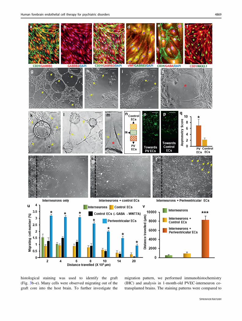
markers CD31, (Fig. 2a, c, e, f; Supplementary Figs. 9 and10), von Willebrand Factor (vWF) (Fig. 2d), and co-labeledwith PVEC markers, GABRB3 (Fig. 2a–c; Supplementary
Fig. 9), GABA (Fig. 2e; Supplementary Fig. 9), NKX2.1(Fig. 2f; Supplementary Fig. 9), PAX6, and ISL1 (Sup-plementary Fig. 10) similar to mouse PVECs. Pluripotent
Human forebrain endothelial cell therapy for psychiatric disorders 4867
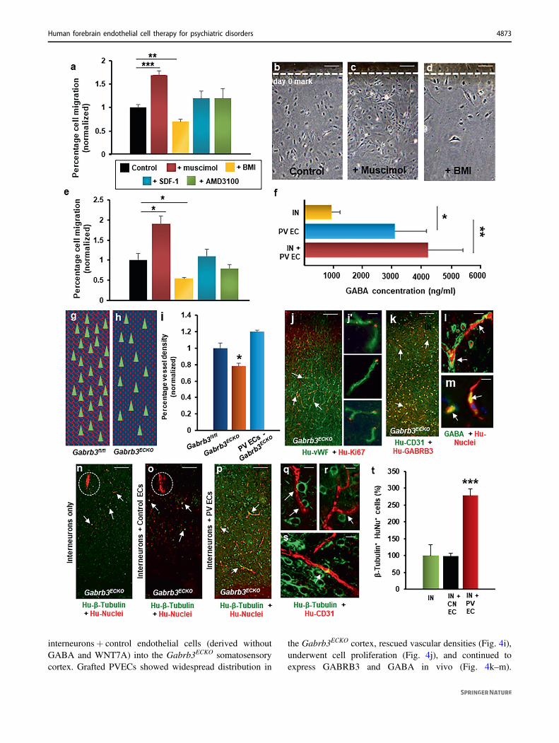
markers OCT4 and TRA1-60 were not present in any cell,confirming the absence of undifferentiated cells in thePVEC population (Supplementary Fig. 11). Effective pro-liferation, migration, sprouting, alignment, branching,lumen formation, and anastomosis are key elements ofangiogenesis. Therefore, we performed in vitro assays todemonstrate the angiogenic properties of human PVECs.These endothelial cells formed tubular networks within 24 hof seeding on Matrigel (Fig. 2g), demonstrating highangiogenic capacity. In addition, human PVECs formedtubular networks in a three-dimensional milieu when cul-tured within fibrin gel matrix demonstrating budding,branching, and lumen formation (Fig. 2h–j). Later stages ofendothelial cell branching and fusion of vessels (anasto-mosis) were also observed (Fig. 2h–j). Sprouting of newcapillaries from existing blood vessels is another hallmarkof angiogenesis. We performed a fibrin gel bead assay forsprouting, in which PVECs were coated on cytodex beadsand embedded in three-dimensional fibrin gels. PVECsstarted budding and sprouting within 2 days (Fig. 2k).Within the next days, long tubular vessels with clearintercellular lumens were formed (Fig. 2l, m). In addition togeneral angiogenic properties, PVECs possess some uniqueproperties. First, PVECs themselves have the ability tomigrate long distance. Second, PVECs induce long-distancemigration of GABAergic interneurons. We tested theseproperties in human PVECs using in vitro chemoattractivityand migration assays. We used human GABAergic inter-neurons (Cellular Dynamics; Supplementary Fig. 12) thathave been extensively characterized with respect to theirmorphological, electrophysiological and molecular char-acteristics in several neuroscience research models, as asource of neurons for these assays. Using three-well culture
inserts (ibidi GmbH), we seeded human interneurons in asmall rectangular patch on a 35 mm poly-ornithine/laminincoated culture dish. Equal number of PVECs and controlHAEC-like endothelial cells (that do not haveperiventricular-specific gene expression; Fig. 1j–l; Supple-mentary Figs. 6–8) were seeded as patches on either side ofthe neuronal patch, with the gap between each patch being500 µm (Fig. 2n). The number of interneurons that migratedtoward PVECs versus control endothelial cells was quan-titated after 36 h. Interneurons showed significantly higherchemo-attractive response toward PVECs compared tocontrol endothelial cells (Fig. 2o–q), confirming that humaninterneurons respond selectively to chemo-attractive cuessecreted by human PVECs. Next, we performed cellmigration assays to test the long-distance migratory poten-tial of human PVECs and its role in guiding human inter-neuron migration (Fig. 2r–v). We compared the migrationof PVECs with human interneurons, and with two types ofcontrol endothelial cells: (a) HAEC-like human endothelialcells and (b) endothelial cells derived from H9 cells in theabsence of GABA and WNT7A (Supplementary Figs. 2–4).Human PVECs seeded alone, traveled further distancecompared to human interneurons or either set of controlendothelial cells (Fig. 2u), confirming their cell-intrinsiccapacity for long-distance migration. Also, for the samedistance range, a higher percentage of PVECs migrated outthan interneurons or control endothelial cells (Fig. 2u). Totest the ability of human PVECs in facilitating humaninterneuron migration, we performed a co-culture migrationassay where interneurons were co-seeded with humanPVECs. Migration of co-seeded interneurons were com-pared to interneurons, which were seeded alone, or seededalong with control human endothelial cells. When seededalong with PVECs, interneurons migrated significantly,both in terms of cell number and distance when compared tointerneurons cultured along with control endothelial cells orinterneurons alone (Fig. 2r–t, v). Collectively, these dataconfirm the high angiogenic potential of human PVECs andits specificity with regard to instructing and promotingmigration of human GABAergic interneurons.
Co-transplantation of human PVECs with GABAergicinterneurons successfully enhanced neuronalmigration in vivo
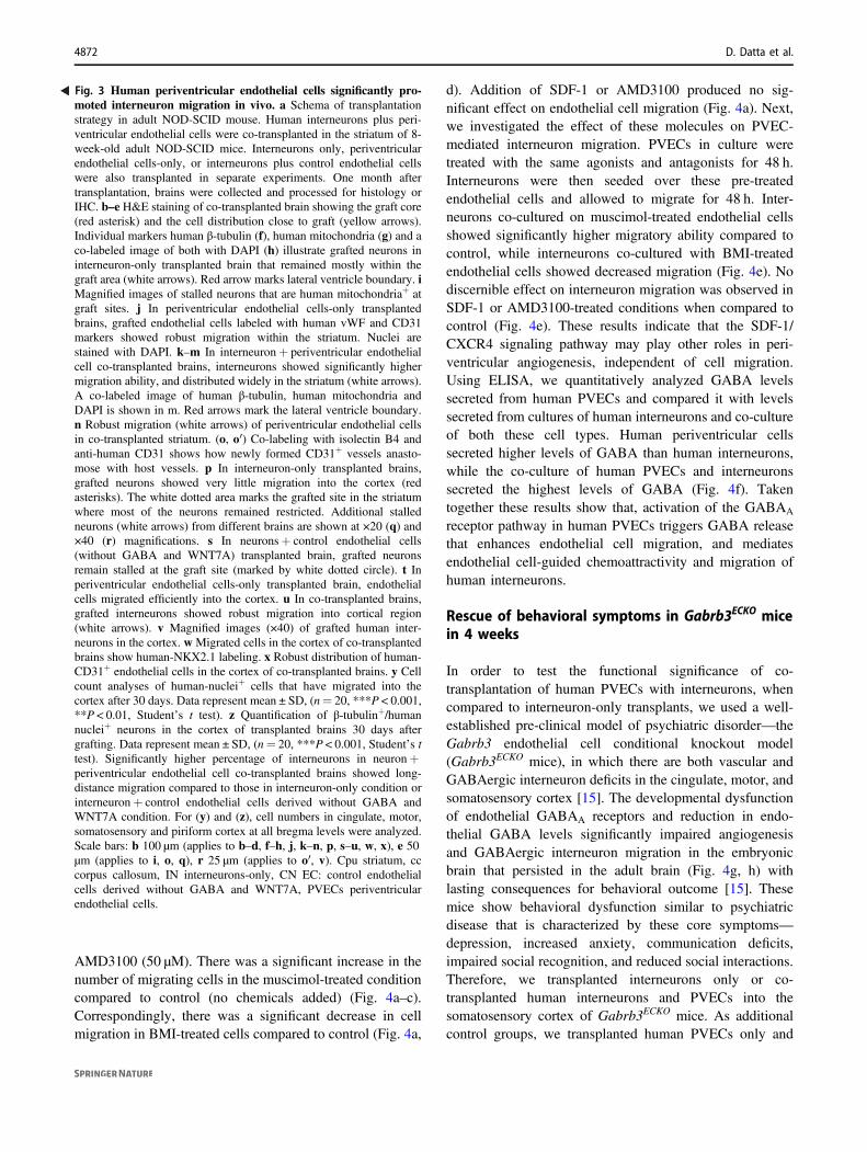
Next, we investigated whether human PVECs could facil-itate human interneuron migration in vivo. To this end, wetransplanted human PVECs along with human interneuronsin a ratio of 1:1 into the striatum of adult NOD-SCID mice,on each side of the brain (Fig. 3a). The striatum provides apotentially powerful experimental system for studying cellmigration and dispersion, with optimal cell survival aftertransplantation. One month after transplantation,
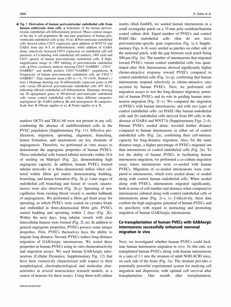
Fig. 1 Derivation of human periventricular endothelial cells fromhuman embryonic stem cells. a Schematic of the human periven-tricular endothelial cell differentiation protocol. Phase contrast imagesof the day 6 cell population (b) and pure population of human peri-ventricular endothelial cells at day 10 (c). d Periventricular endothelialcells showed robust CD31 expression upon addition of WNT7A andGABA from day 0–5 of differentiation, while addition of GABAalone, selectively increased CD31 expression on endothelial cell–celljunctions. e Co-labeling with endothelial cell markers, vWF (red) andCD31 (green) of human periventricular endothelial cells. f High-magnification image of vWF labeling of periventricular endothelialcells. g Flow cytometry analysis showing CD31+GABRB3−, CD31−
GABRB3+ and double positive CD31+GABRB3+ populations. hFrequencies of human periventricular endothelial cells are CD31+/GABRB3+. Data represent mean ± SD (n= 6, *P < 0.05, Student’s ttest). i Heatmap showing top 20 differentially expressed genes in H9cells versus H9-derived periventricular endothelial cells (PV ECs),indicating efficient endothelial cell differentiation. Heatmaps showingtop 20 upregulated genes in H9-derived periventricular endothelialcells versus control endothelial cells in three different categories:angiogenesis (j), GABA pathway (k) and neurogenesis (l) categories.Scale bars: b 100 µm (applies to c), d 50 µm (applies to e, f).
4868 D. Datta et al.
histological staining was used to identify the graft(Fig. 3b–e). Many cells were observed migrating out of thegraft core into the host brain. To further investigate the
migration pattern, we performed immunohistochemistry(IHC) and analysis in 1-month-old PVEC-interneuron co-transplanted brains. The staining patterns were compared to
Human forebrain endothelial cell therapy for psychiatric disorders 4869
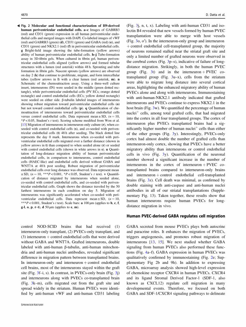
control NOD-SCID brains that had received (1)interneuron-only transplant, (2) PVECs-only transplant, and(3) interneuron+ control endothelial cells that were derivedwithout GABA and WNT7A. Grafted interneurons, doublelabeled with anti-human β-tubulin, anti-human mitochon-dria and anti-human nuclei antibodies, revealed significantdifference in migration pattern between transplanted brains.In interneuron-only and interneuron+ control endothelialcell brains, most of the interneurons stayed within the graftsite (Fig. 3f–i, s). In contrast, in PVECs-only brain (Fig. 3j)and interneurons along with PVECs co-transplanted brain(Fig. 3k–m), cells migrated out from the graft site andspread widely in the striatum. Human PVECs were identi-fied by anti-human vWF and anti-human CD31 labeling
(Fig. 3j, n, t, x). Labeling with anti-human CD31 and iso-lectin B4 revealed that new vessels formed by human PVECtransplantation were able to merge with host vessels(Fig. 3o, o′). In the interneuron-only group and interneuron+ control endothelial cell-transplanted group, the majorityof neurons remained stalled near the striatal graft site andonly a limited number of grafted neurons were observed inthe cerebral cortex (Fig. 3p–s), indicative of failure of long-distance migration. Strikingly, in both the human PVECgroup (Fig. 3t) and in the interneuron+ PVEC co-transplanted group (Fig. 3u–x), cells from the striatumwere able to migrate long distance into several corticalareas, highlighting the enhanced migratory ability of humanPVECs alone and along with interneurons. Immunostainingwith anti-human-NKX2.1 antibody indicated that graftedinterneurons and PVECs continue to express NKX2.1 in thehost brain (Fig. 3w). We quantified the percentage of humannuclei+ cells, among total grafted cells, that had migratedinto the cortex in all four transplanted groups. The cortex ofinterneuron plus PVECs transplanted brains had a sig-nificantly higher number of human nuclei+ cells than eitherof the other groups (Fig. 3y). Interestingly, PVECs-onlycortex had almost double the number of grafted cells thaninterneuron-only cortex, showing that PVECs have a bettermigratory ability than interneurons or control endothelialcells in vivo (Fig. 3y). Quantification of the neuronalnumber showed a significant increase in the number ofinterneurons in the cortex of interneuron+ PVEC co-transplanted brains compared to interneuron-only brainsand interneuron+ control endothelial cell-transplantedbrains (Fig. 3z). Cell death was minimal, as confirmed bydouble staining with anti-caspase and anti-human nucleiantibodies in all of our striatal transplantations (Supple-mentary Fig. 13). Taken together, these results show thathuman interneurons require human PVECs for long-distance migration in vivo.
Human PVEC-derived GABA regulates cell migration
GABA secreted from mouse PVECs plays both autocrineand paracrine roles. It enhances the migration of PVECs,triggers angiogenesis, and promotes robust migration ofinterneurons [13, 15]. We next studied whether GABAsignaling from human PVECs also performed these func-tions (Fig. 4a–f). GABA expression in human PVECs wasqualitatively confirmed by immunostaining (Fig. 2e; Sup-plementary Fig 2b and 9h). In addition to expressingGABA, microarray analysis showed high-level expressionof chemokine receptor CXCR4 in human PVECs. CXCR4and its ligand Stromal Derived Factor-1 (SDF-1, alsoknown as CXCL12) regulate cell migration in manydevelopmental events. Therefore, we focused on bothGABA and SDF-1/CXCR4 signaling pathways to delineate
Fig. 2 Molecular and functional characterization of H9-derivedhuman periventricular endothelial cells. a–c Images of GABRB3(red) and CD31 (green) expression in all human periventricular endo-thelial cells and merged images with DAPI. Co-labeled images of vWF(red) and GABRB3 (green) (d), CD31 (green) and GABA (red) (e), andCD31 (green) and NKX2.1 (red) (f) in periventricular endothelial cells.g Bright-field image showing the tube-formation (yellow arrows)ability of human periventricular endothelial cells. h–j Tube-formationassay in 3D-fibrin gels. When cultured in fibrin gel, human periven-tricular endothelial cells aligned (yellow arrows) and formed tubularstructures with a lumen (red asterisk) within 48 h. Sprouting and tubeformation in fibrin gels. Nascent sprouts (yellow arrows) are observedon day 2 (k) that continue to proliferate, migrate, and form intercellulartubes (yellow arrows in l) with a clear lumen (red asterisk, m). nSchematic of the chemoattraction assay. Using a three-well cultureinsert, interneurons (IN) were seeded in the middle (green dotted rec-tangle), while periventricular endothelial cells (PV ECs; orange dottedrectangle) and control endothelial cells (ECs; yellow dotted rectangle)were seeded on either side. β-tubulin labeled images of interneuronsshowing robust migration toward periventricular endothelial cells (o)but not toward control endothelial cells (p). q Quantification of che-moattraction of interneurons toward periventricular endothelial cellsversus control endothelial cells. Data represent mean ± SD, (n= 10,*P < 0.05, Student’s t test). Scoring scheme modified from Won et al.[13] Migration of interneurons in interneuron-only culture (r), when co-seeded with control endothelial cells (s), and co-seeded with periven-tricular endothelial cells (t) 48 h after seeding. The black dotted linerepresents the day 0 mark. Interneurons when co-seeded with peri-ventricular endothelial cells migrated over a farther distance (shown inyellow arrows in t) than compared to when seeded alone (r) or seededwith control endothelial cells (shown in white arrows in s). u Quanti-tation of long-distance migration ability of human periventricularendothelial cells, in comparison to interneurons, control endothelialcells (HAEC-like) and endothelial cells derived without GABA andWNT7A at 48 h post seeding. Robust migration of periventricularendothelial cells over long distance was observed. Data represent mean± SD, (n= 10, ***P < 0.001, *P < 0.05, Student’s t test). v Quantifi-cation of distance migrated by interneurons when seeded alone,co-seeded with control endothelial cells, and co-seeded with periven-tricular endothelial cells. Graph shows the distance traveled by the 50farthest interneurons in each condition on day 5. Migration ofinterneurons was significantly accelerated when co-seeded with peri-ventricular endothelial cells. Data represent mean ± SD, (n= 10,***P < 0.001, Student’s t test). Scale bars: a 100 µm (applies to b, c, f,g-l, o, p, r–t), d 50 µm (applies to e, m).
4870 D. Datta et al.
their role in the migratory events. To study their function inhuman PVEC migration, we performed a long-distancemigration assay with endothelial cells in the presence of
GABAA receptor agonist muscimol (100 µM), GABAA
receptor antagonist bicuculline methiodide (BMI, 100 µM),human SDF-1 (40 nM), and CXCR4 receptor antagonist
Human forebrain endothelial cell therapy for psychiatric disorders 4871
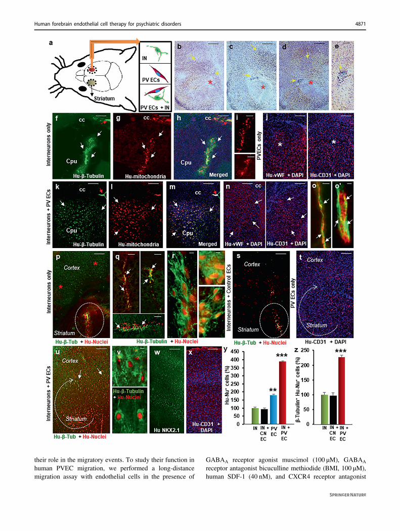
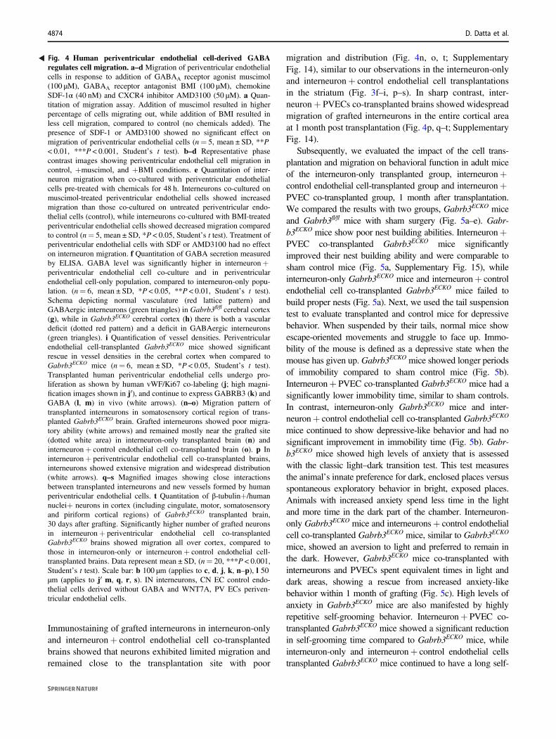
AMD3100 (50 µM). There was a significant increase in thenumber of migrating cells in the muscimol-treated conditioncompared to control (no chemicals added) (Fig. 4a–c).Correspondingly, there was a significant decrease in cellmigration in BMI-treated cells compared to control (Fig. 4a,
d). Addition of SDF-1 or AMD3100 produced no sig-nificant effect on endothelial cell migration (Fig. 4a). Next,we investigated the effect of these molecules on PVEC-mediated interneuron migration. PVECs in culture weretreated with the same agonists and antagonists for 48 h.Interneurons were then seeded over these pre-treatedendothelial cells and allowed to migrate for 48 h. Inter-neurons co-cultured on muscimol-treated endothelial cellsshowed significantly higher migratory ability compared tocontrol, while interneurons co-cultured with BMI-treatedendothelial cells showed decreased migration (Fig. 4e). Nodiscernible effect on interneuron migration was observed inSDF-1 or AMD3100-treated conditions when compared tocontrol (Fig. 4e). These results indicate that the SDF-1/CXCR4 signaling pathway may play other roles in peri-ventricular angiogenesis, independent of cell migration.Using ELISA, we quantitatively analyzed GABA levelssecreted from human PVECs and compared it with levelssecreted from cultures of human interneurons and co-cultureof both these cell types. Human periventricular cellssecreted higher levels of GABA than human interneurons,while the co-culture of human PVECs and interneuronssecreted the highest levels of GABA (Fig. 4f). Takentogether these results show that, activation of the GABAA
receptor pathway in human PVECs triggers GABA releasethat enhances endothelial cell migration, and mediatesendothelial cell-guided chemoattractivity and migration ofhuman interneurons.
Rescue of behavioral symptoms in Gabrb3ECKO micein 4 weeks
In order to test the functional significance of co-transplantation of human PVECs with interneurons, whencompared to interneuron-only transplants, we used a well-established pre-clinical model of psychiatric disorder—theGabrb3 endothelial cell conditional knockout model(Gabrb3ECKO mice), in which there are both vascular andGABAergic interneuron deficits in the cingulate, motor, andsomatosensory cortex [15]. The developmental dysfunctionof endothelial GABAA receptors and reduction in endo-thelial GABA levels significantly impaired angiogenesisand GABAergic interneuron migration in the embryonicbrain that persisted in the adult brain (Fig. 4g, h) withlasting consequences for behavioral outcome [15]. Thesemice show behavioral dysfunction similar to psychiatricdisease that is characterized by these core symptoms—depression, increased anxiety, communication deficits,impaired social recognition, and reduced social interactions.Therefore, we transplanted interneurons only or co-transplanted human interneurons and PVECs into thesomatosensory cortex of Gabrb3ECKO mice. As additionalcontrol groups, we transplanted human PVECs only and
Fig. 3 Human periventricular endothelial cells significantly pro-moted interneuron migration in vivo. a Schema of transplantationstrategy in adult NOD-SCID mouse. Human interneurons plus peri-ventricular endothelial cells were co-transplanted in the striatum of 8-week-old adult NOD-SCID mice. Interneurons only, periventricularendothelial cells-only, or interneurons plus control endothelial cellswere also transplanted in separate experiments. One month aftertransplantation, brains were collected and processed for histology orIHC. b–e H&E staining of co-transplanted brain showing the graft core(red asterisk) and the cell distribution close to graft (yellow arrows).Individual markers human β-tubulin (f), human mitochondria (g) and aco-labeled image of both with DAPI (h) illustrate grafted neurons ininterneuron-only transplanted brain that remained mostly within thegraft area (white arrows). Red arrow marks lateral ventricle boundary. iMagnified images of stalled neurons that are human mitochondria+ atgraft sites. j In periventricular endothelial cells-only transplantedbrains, grafted endothelial cells labeled with human vWF and CD31markers showed robust migration within the striatum. Nuclei arestained with DAPI. k–m In interneuron+ periventricular endothelialcell co-transplanted brains, interneurons showed significantly highermigration ability, and distributed widely in the striatum (white arrows).A co-labeled image of human β-tubulin, human mitochondria andDAPI is shown in m. Red arrows mark the lateral ventricle boundary.n Robust migration (white arrows) of periventricular endothelial cellsin co-transplanted striatum. (o, o′) Co-labeling with isolectin B4 andanti-human CD31 shows how newly formed CD31+ vessels anasto-mose with host vessels. p In interneuron-only transplanted brains,grafted neurons showed very little migration into the cortex (redasterisks). The white dotted area marks the grafted site in the striatumwhere most of the neurons remained restricted. Additional stalledneurons (white arrows) from different brains are shown at ×20 (q) and×40 (r) magnifications. s In neurons+ control endothelial cells(without GABA and WNT7A) transplanted brain, grafted neuronsremain stalled at the graft site (marked by white dotted circle). t Inperiventricular endothelial cells-only transplanted brain, endothelialcells migrated efficiently into the cortex. u In co-transplanted brains,grafted interneurons showed robust migration into cortical region(white arrows). v Magnified images (×40) of grafted human inter-neurons in the cortex. w Migrated cells in the cortex of co-transplantedbrains show human-NKX2.1 labeling. x Robust distribution of human-CD31+ endothelial cells in the cortex of co-transplanted brains. y Cellcount analyses of human-nuclei+ cells that have migrated into thecortex after 30 days. Data represent mean ± SD, (n= 20, ***P < 0.001,**P < 0.01, Student’s t test). z Quantification of β-tubulin+/humannuclei+ neurons in the cortex of transplanted brains 30 days aftergrafting. Data represent mean ± SD, (n= 20, ***P < 0.001, Student’s ttest). Significantly higher percentage of interneurons in neuron+periventricular endothelial cell co-transplanted brains showed long-distance migration compared to those in interneuron-only condition orinterneuron+ control endothelial cells derived without GABA andWNT7A condition. For (y) and (z), cell numbers in cingulate, motor,somatosensory and piriform cortex at all bregma levels were analyzed.Scale bars: b 100 µm (applies to b–d, f–h, j, k–n, p, s–u, w, x), e 50µm (applies to i, o, q), r 25 µm (applies to o′, v). Cpu striatum, cccorpus callosum, IN interneurons-only, CN EC: control endothelialcells derived without GABA and WNT7A, PVECs periventricularendothelial cells.
4872 D. Datta et al.
interneurons+ control endothelial cells (derived withoutGABA and WNT7A) into the Gabrb3ECKO somatosensorycortex. Grafted PVECs showed widespread distribution in
the Gabrb3ECKO cortex, rescued vascular densities (Fig. 4i),underwent cell proliferation (Fig. 4j), and continued toexpress GABRB3 and GABA in vivo (Fig. 4k–m).
Human forebrain endothelial cell therapy for psychiatric disorders 4873
Immunostaining of grafted interneurons in interneuron-onlyand interneuron+ control endothelial cell co-transplantedbrains showed that neurons exhibited limited migration andremained close to the transplantation site with poor
migration and distribution (Fig. 4n, o, t; SupplementaryFig. 14), similar to our observations in the interneuron-onlyand interneuron+ control endothelial cell transplantationsin the striatum (Fig. 3f–i, p–s). In sharp contrast, inter-neuron+ PVECs co-transplanted brains showed widespreadmigration of grafted interneurons in the entire cortical areaat 1 month post transplantation (Fig. 4p, q–t; SupplementaryFig. 14).
Subsequently, we evaluated the impact of the cell trans-plantation and migration on behavioral function in adult miceof the interneuron-only transplanted group, interneuron+control endothelial cell-transplanted group and interneuron+PVEC co-transplanted group, 1 month after transplantation.We compared the results with two groups, Gabrb3ECKO miceand Gabrb3fl/fl mice with sham surgery (Fig. 5a–e). Gabr-b3ECKO mice show poor nest building abilities. Interneuron+PVEC co-transplanted Gabrb3ECKO mice significantlyimproved their nest building ability and were comparable tosham control mice (Fig. 5a, Supplementary Fig. 15), whileinterneuron-only Gabrb3ECKO mice and interneuron+ controlendothelial cell co-transplanted Gabrb3ECKO mice failed tobuild proper nests (Fig. 5a). Next, we used the tail suspensiontest to evaluate transplanted and control mice for depressivebehavior. When suspended by their tails, normal mice showescape-oriented movements and struggle to face up. Immo-bility of the mouse is defined as a depressive state when themouse has given up. Gabrb3ECKO mice showed longer periodsof immobility compared to sham control mice (Fig. 5b).Interneuron+ PVEC co-transplanted Gabrb3ECKO mice had asignificantly lower immobility time, similar to sham controls.In contrast, interneuron-only Gabrb3ECKO mice and inter-neuron+ control endothelial cell co-transplanted Gabrb3ECKO
mice continued to show depressive-like behavior and had nosignificant improvement in immobility time (Fig. 5b). Gabr-b3ECKO mice showed high levels of anxiety that is assessedwith the classic light–dark transition test. This test measuresthe animal’s innate preference for dark, enclosed places versusspontaneous exploratory behavior in bright, exposed places.Animals with increased anxiety spend less time in the lightand more time in the dark part of the chamber. Interneuron-only Gabrb3ECKO mice and interneurons+ control endothelialcell co-transplanted Gabrb3ECKO mice, similar to Gabrb3ECKO
mice, showed an aversion to light and preferred to remain inthe dark. However, Gabrb3ECKO mice co-transplanted withinterneurons and PVECs spent equivalent times in light anddark areas, showing a rescue from increased anxiety-likebehavior within 1 month of grafting (Fig. 5c). High levels ofanxiety in Gabrb3ECKO mice are also manifested by highlyrepetitive self-grooming behavior. Interneuron+ PVEC co-transplanted Gabrb3ECKO mice showed a significant reductionin self-grooming time compared to Gabrb3ECKO mice, whileinterneuron-only and interneuron+ control endothelial cellstransplanted Gabrb3ECKO mice continued to have a long self-
Fig. 4 Human periventricular endothelial cell-derived GABAregulates cell migration. a–d Migration of periventricular endothelialcells in response to addition of GABAA receptor agonist muscimol(100 µM), GABAA receptor antagonist BMI (100 µM), chemokineSDF-1α (40 nM) and CXCR4 inhibitor AMD3100 (50 µM). a Quan-titation of migration assay. Addition of muscimol resulted in higherpercentage of cells migrating out, while addition of BMI resulted inless cell migration, compared to control (no chemicals added). Thepresence of SDF-1 or AMD3100 showed no significant effect onmigration of periventricular endothelial cells (n= 5, mean ± SD, **P< 0.01, ***P < 0.001, Student’s t test). b–d Representative phasecontrast images showing periventricular endothelial cell migration incontrol, +muscimol, and +BMI conditions. e Quantitation of inter-neuron migration when co-cultured with periventricular endothelialcells pre-treated with chemicals for 48 h. Interneurons co-cultured onmuscimol-treated periventricular endothelial cells showed increasedmigration than those co-cultured on untreated periventricular endo-thelial cells (control), while interneurons co-cultured with BMI-treatedperiventricular endothelial cells showed decreased migration comparedto control (n= 5, mean ± SD, *P < 0.05, Student’s t test). Treatment ofperiventricular endothelial cells with SDF or AMD3100 had no effecton interneuron migration. f Quantitation of GABA secretion measuredby ELISA. GABA level was significantly higher in interneuron+periventricular endothelial cell co-culture and in periventricularendothelial cell-only population, compared to interneuron-only popu-lation. (n= 6, mean ± SD, *P < 0.05, **P < 0.01, Student’s t test).Schema depicting normal vasculature (red lattice pattern) andGABAergic interneurons (green triangles) in Gabrb3fl/fl cerebral cortex(g), while in Gabrb3ECKO cerebral cortex (h) there is both a vasculardeficit (dotted red pattern) and a deficit in GABAergic interneurons(green triangles). i Quantification of vessel densities. Periventricularendothelial cell-transplanted Gabrb3ECKO mice showed significantrescue in vessel densities in the cerebral cortex when compared toGabrb3ECKO mice (n= 6, mean ± SD, *P < 0.05, Student’s t test).Transplanted human periventricular endothelial cells undergo pro-liferation as shown by human vWF/Ki67 co-labeling (j; high magni-fication images shown in j′), and continue to express GABRB3 (k) andGABA (l, m) in vivo (white arrows). (n–o) Migration pattern oftransplanted interneurons in somatosensory cortical region of trans-planted Gabrb3ECKO brain. Grafted interneurons showed poor migra-tory ability (white arrows) and remained mostly near the grafted site(dotted white area) in interneuron-only transplanted brain (n) andinterneuron+ control endothelial cell co-transplanted brain (o). p Ininterneuron+ periventricular endothelial cell co-transplanted brains,interneurons showed extensive migration and widespread distribution(white arrows). q–s Magnified images showing close interactionsbetween transplanted interneurons and new vessels formed by humanperiventricular endothelial cells. t Quantitation of β-tubulin+/humannuclei+ neurons in cortex (including cingulate, motor, somatosensoryand piriform cortical regions) of Gabrb3ECKO transplanted brain,30 days after grafting. Significantly higher number of grafted neuronsin interneuron+ periventricular endothelial cell co-transplantedGabrb3ECKO brains showed migration all over cortex, compared tothose in interneuron-only or interneuron+ control endothelial cell-transplanted brains. Data represent mean ± SD, (n= 20, ***P < 0.001,Student’s t test). Scale bar: b 100 μm (applies to c, d, j, k, n–p), l 50µm (applies to j′ m, q, r, s). IN interneurons, CN EC control endo-thelial cells derived without GABA and WNT7A, PV ECs periven-tricular endothelial cells.
4874 D. Datta et al.
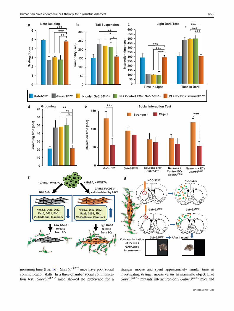
grooming time (Fig. 5d). Gabrb3ECKO mice have poor socialcommunication skills. In a three-chamber social communica-tion test, Gabrb3ECKO mice showed no preference for a
stranger mouse and spent approximately similar time ininvestigating stranger mouse versus an inanimate object. LikeGabrb3ECKO mutants, interneuron-only Gabrb3ECKO mice and
g
Gabrb3ECKO Gabrb3ECKO
After 1 monthGabrb3ECKO
NOD-SCID NOD-SCID
Co-transplanta�onof PV ECs + GABAergic
interneurons
Gabrb3ECKO IN only: Gabrb3ECKOGabrb3fl/fl IN + PV ECs: Gabrb3ECKOIN + Control ECs: Gabrb3ECKO
0
50
100
150
Inte
ract
ion
time
(sec
)
Grooming
)ces(e
mitgni
moorG
Social Interaction Test
Gabrb3ECKOGabrb3fl/fl
Stranger 1 Object
Neurons onlyGabrb3ECKO
Neurons + ECs Gabrb3ECKO
***
***
0
10
20
30
40
50
60
70 ****
*
Neurons + Control ECs Gabrb3ECKO
0
50
100
150
200
250
300
Imm
obili
ty (s
ec)
Tail Suspension
****
0
1
2
3
4
5
6
Time in Light Time in Dark
erocSgnitse
NNest Building
***
Inte
ract
ion
time
(sec
)
050
100150200250300350400450500550600
Light Dark Test
*********
*****
*********
a c
d
b
e
f+ GABA, + WNT7A
GABRB3+/CD31+
cells isolated by FACS
- GABA, - WNT7A
No FACS
Nkx2.1, Dlx1, Dlx2, Pax6, Cd31, Flk1
VE-Cadherin, Claudin 5
Low GABA release
from ECs
High GABA release
from ECs
Nkx2.1, Dlx1, Dlx2, Pax6, Cd31, Flk1
VE-Cadherin, Claudin 5
Human forebrain endothelial cell therapy for psychiatric disorders 4875
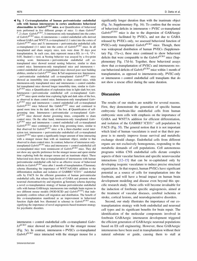
interneuron+ control endothelial cells co-transplanted Gabr-b3ECKO mice showed no preference for the stranger mouse(Fig. 5e). In contrast, interneuron+ PVECs co-transplantedGabrb3ECKO mice interacted with the stranger mouse for a
significantly longer duration than with the inanimate object(Fig. 5e; Supplementary Fig. 16). To confirm that the rescueof behavioral deficits in interneuron+ PVEC co-transplantedGabrb3ECKO mice is due to the dispersion of GABAergicinterneurons facilitated by PVECs, and not due to GABAreleased by PVECs only, we assessed behavioral function ofPVECs-only transplanted Gabrb3ECKO mice. Though, therewas widespread distribution of human PVECs (Supplemen-tary Fig. 17a–c), these mice continued to show behavioraldeficits that were comparable to the Gabrb3ECKO mice (Sup-plementary Fig. 17d–h). Together, these behavioral assaysshow that co-transplantation of PVECs and interneurons res-cue behavioral deficits of Gabrb3ECKO mice within 1 month oftransplantation, as opposed to interneuron-only, PVEC-onlyor interneuron+ control endothelial cell transplants that donot show a rescue effect during the same duration.
Discussion
The results of our studies are notable for several reasons.First, they demonstrate the generation of specific humanembryonic forebrain-like endothelial cells from humanembryonic stem cells with emphasis on the importance ofGABA and WNT7A addition for efficient differentiation,and isolation of the GABRB3+/CD31+ cell population byFACS (Fig. 5f). The general notion that it is not importantwhich kind of human vasculature is used or that their pur-pose is to merely improve tissue survival and metabolicexchange should change. Endothelial cells from differentorgans are not exclusively homogenous, responding to themetabolic demands of cell populations. Cell autonomousprograms within CNS endothelial cells dictate complexaspects of their vascular function and specific neurovascularinteractions [12–15] that can be re-capitulated only bydeveloping isogenic vasculature to induce precise structuralorganization. In that respect, human PVECs have significantpotential as a source of cells for transplantation into theforebrain, and will have a broad impact on human braindevelopment modeling and disease even beyond this spe-cific research study. These cells will become invaluable forthe induction of forebrain specific angiogenesis, aimed atthe treatment of vascular diseases, cerebral ischemia orstroke, cortical lesions, and neurodegenerative disorders.
Second, our study illustrates the importance of our co-transplantation strategy with both endothelial and neuronalcell types and its significant benefits for brain repair. Theidentification of the molecular components involved inforebrain GABAergic interneuron development triggeredthe efficient generation of GABAergic neuronal populationsbased on ES cell engineering. However, these GABAergicinterneurons have been used in transplantation without theirnatural substrate or guide, causing them to stall at
Fig. 5 Co-transplantation of human periventricular endothelialcells with human interneurons in cortex ameliorates behavioralabnormalities in Gabrb3ECKO mice. a–e Quantification of behavioraltests performed in five different groups of mice: 1) sham Gabrb3fl/fl, 2) sham Gabrb3ECKO, 3) interneurons only transplanted into the cortexof Gabrb3ECKO mice, 4) interneurons+ control endothelial cells derivedwithout GABA and WNT7A co-transplanted (1:1 ratio) into the cortex ofGabrb3ECKO mice and 5) interneuron+ periventricular endothelial cellsco-transplanted (1:1 ratio) into the cortex of Gabrb3ECKO mice. In alltransplanted and sham surgery mice, tests were done 30 days posttransplantation. In each case, data represent mean ± SD, (n= 6, *P <0.05, Student’s t test). a Nesting behavior was assayed by a five-pointnesting score. Interneuron+ periventricular endothelial cell co-transplanted mice showed normal nesting behavior, similar to shamcontrol mice. Interneuron-only transplanted mice and interneuron+control endothelial cell co-transplanted mice showed poor nest buildingabilities, similar to Gabrb3ECKO mice. b Tail suspension test. Interneuron+ periventricular endothelial cell co-transplanted Gabrb3ECKO miceshowed an immobility time comparable to sham control mice, whileinterneuron-only transplanted mice and interneurons+ control endothe-lial transplanted mice showed longer immobility time, similar to Gabr-b3ECKO mice. c Quantification of exploration time in light–dark box test.Interneuron+ periventricular endothelial cell co-transplanted Gabr-b3ECKO mice spent similar time exploring light and dark sides of the box,as observed in sham control mice. Interneuron-only transplanted Gabr-b3ECKO mice and interneuron+ control endothelial cell co-transplantedGabrb3ECKO mice behaved like Gabrb3ECKO mice and continued tospend more time in the dark side of the box. d Quantification of self-grooming time. Interneuron+ periventricular co-transplanted Gabr-b3ECKO mice showed shorter grooming times, comparable to shamcontrol mice. On the other hand, interneuron-only transplanted Gabr-b3ECKO mice and interneuron+ control endothelial cell co-transplantedGabrb3ECKO mice continued to show long grooming times, similar tothat observed for Gabrb3ECKO mice. e In a three-chamber social inter-action test, interneuron+ periventricular endothelial cell co-transplantedGabrb3ECKO mice spent a significantly longer amount of time interactingwith a stranger mouse than with an inanimate object, similar to thatobserved in sham control mice. In contrast, behavior of interneuron only-transplanted Gabrb3ECKO mice and interneuron+ control endothelial cellco-transplanted mice were reminiscent of Gabrb3ECKO mice. They didnot show any specific preference for the stranger mouse and spent similartime exploring both the stranger mouse and an inanimate object. Thesebehavioral tests show that co-transplantation of interneurons with humanperiventricular endothelial cells led to an effective rescue of behavioraldeficits in Gabrb3ECKO mice after 1 month of transplantation. f Summaryschema illustrating the importance of WNT7A/GABA addition to thedifferentiation medium and isolation of GABRB3+/CD31+ endothelialcells by FACS for the efficient generation of human periventricularendothelial cells, that release high levels of GABA and promote robustneuronal chemoattractivity and migration. g Summary schema depictinga novel co-transplantation strategy of human periventricular endothelialcells with human GABAergic interneurons into multiple brain regions intwo different mouse models (NOD-SCID and Gabrb3ECKO) that inde-pendently confirm robust cell migration and distribution. The benefit ofthis co-transplantation manifested as an improvement in behavioralfunction (light–dark box illustrated in schema) in Gabrb3ECKO mice,signifying the importance of novel angiogenesis-based treatment strategyfor psychiatric disorders.
4876 D. Datta et al.
transplantation sites with an inability to migrate into regionsthat require new neurons [23, 25, 33, 34]. Our resultsdemonstrate how grafted human PVECs provide a migra-tion promoting corridor that help human GABAergicinterneurons migrate long distances within shorter periodsof time to integrate themselves with the host tissue, therebyproviding greater significance for faster repair of braindamage. Interestingly, when transplanted into the adultstriatum, both endothelial cells and interneurons dispersedsignificantly and migrated both tangentially and long dis-tance into the cerebral cortex, recapitulating the embryonicsituation. This finding constitutes an important step towardthe rational use of embryonic stem cell-derived PVECs inbrain repair strategies targeting different regions of theforebrain (Fig. 5g).
Third, human PVECs may be of significance in anemerging field of brain organoid technology that is makingremarkable progress. However, lack of blood vessels withingrowing brain organoids limits their application, both withrespect to disease modeling and in the context of clinicaltransplantation. The current technique available for vascu-larization of organoids involves complex in vivo grafting oforganoids [43]. Co-culture with PVECs is likely to improveforebrain organoid development, compartmentalization, andstructure as well as minimize organoid to organoid growthand variability. Fourth, human PVECs can be generatedfrom patient-derived iPSCs in diverse disease scenarios andmay provide novel insights into disease etiology andpathogenesis.
Finally, our work emphasizes the importance of forebrainendothelial cell therapy for the improvement of behavioralfunction. The Gabrb3ECKO mice have reductions in bothblood vessels and GABAergic interneurons; therefore,transplantation of interneurons only, did not rescue theabnormal behavioral symptoms. Co-transplantation of bothcell types was significant for the behavioral rescue. Thisillustrates the need for a greater understanding of both cell-type and region-specific defects with respect to psychiatricdisorders for designing treatment strategies. It will beinteresting in the future to extend this co-transplantationstrategy to new disease models and to fine-tune the therapywith respect to the affected brain region, different neuronalcell types and long-term graft survival and safety. Thus, weexpect our study to herald a new era in angiogenesis-specific treatment avenues for psychiatric disorders.
Materials and methods
Endothelial cell differentiation and culture
H9 cells (WiCell, Madison, WI) were maintained in E8media (Thermo Fisher Scientific) on Matrigel (BD
Biosciences)-coated plates and passaged once a week with0.5 mM EDTA (Thermo Fisher Scientific) in PBS. On day 0of differentiation, H9 cells were dissociated with Accutase(Sigma), and plated at a density of 105 cells/cm2 onMatrigel-coated plates. Cells were cultured for 2 days in E8medium supplemented with BMP4 (5 ng/ml, Peprotech),Activin (25 ng/ml, Peprotech), CHIR 99021 (1 µM,)WNT7A (500 ng/ml, Peprotech), and GABA (5 μM,Sigma). Rock Inhibitor, Y-27632 (10 μM, Selleck Chemi-cals) was added for first 24 h to improve cell survival. Onday 2, cells were switched to vascular inducing mediumcomposed of E6 medium (Thermo Fisher Scientific) con-taining BMP4 (50 ng/ml), SB431542 (5 μM, CaymanChemicals), GABA (5 μM), WNT7A (500 ng/ml), FGF2(100 ng/ml, Peprotech), and VEGF-A (50 ng/ml, Pepro-tech). On day 5, cells were split at 1:6 ratio using Accutaseand plated on Matrigel-coated plates in PVEC mediumconsisting of E6 with VEGF-A (50 ng/ml), FGF2 (100 ng/ml), and GABA (5 μM). On day 7, PVECs were isolatedfrom the mixed population by FACS and seeded onMatrigel-coated plates in PVECs medium at a density of6 × 105 cells/cm2. For routine culturing, PVECs were dis-sociated using Accutase, and seeded on Matrigel-coatedplates at a density of 6 ×10⁵ cells/cm2 (high density seeding)or 1.2 × 10⁵cells/cm2 (low density seeding), with mediumchange every alternate day. PVECs were cryopreservedafter at least one passaging in freezing medium composedof 90% PVECs medium and 10% DMSO. As a source ofneurons, we used human iPSC-derived GABAergic neuronsfrom Cellular Dynamics (Madison, WI, Cat# R1013). Ascontrol for microarray analysis and for cell migrationassays, we used human-iPSC-derived endothelial cells fromCellular Dynamics (Madison WI, Cat# R1022), whose geneexpression profile is similar to HAECs (https://fujifilmcdi.com/assets/CDI130925MIPTEC04.pdf). The humanGABAergic neurons and human HAEC-like endothelialcells were cultured using the manufacturer’s protocol. Celllines were routinely tested for mycoplasma contaminationusing a Mycoplasma Detection Kit (InvivoGen, San Diego,CA). Cells used in this study were verified to be myco-plasma free before undertaking any experiment with them.
PVEC isolation by FACS
On day 7 of differentiation, cells were dissociated withAccutase and filtered through a 35 μm nylon mesh cellstrainer cap to obtain a single cell suspension. Cell suspen-sion was washed with ice-cold FACS buffer (2% FCS and0.1% NaN3 in PBS) and incubated with Fcγ blocker (BDBiosciences Pharmingen, 1 µg/ml) for 30 min. Cells werethen washed in ice-cold FACS buffer and stained with PE/Cy7 anti-human CD31 antibody (BioLegend, Cat # 303117)and anti-human GABRB3 antibody (Creative Diagnostics,
Human forebrain endothelial cell therapy for psychiatric disorders 4877
Cat # DCABH-10376) conjugated with APC using anantibody conjugation kit (Abcam) for 1 h. Cells werewashed in ice-cold FACS buffer and CD31+GABRB3+
endothelial cells were isolated by FACS using a BD FACSAria-II flow cytometer (BD Biosciences, San Jose, CA).
Gene expression profile analysis
Total RNA was extracted using the RNeasy plus minikit(Qiagen) according to the manufacturer’s instruction. RNAquality was determined using nanodrop and an Agilent 2100bioanalyzer. Microarray hybridization was performed usingthe Human Gene Array 2.0 ST gene chip (Affymetrix) at theBoston University Microarray and Sequencing ResourceCore, Boston, MA. Principal component analysis (PCA) wasperformed after normalizing gene-level expression valuesfrom CEL files of Affymetrix human gene 2.0 ST arrays byusing the implementation of the Robust Multiarray Averagein the Affymetrix transcriptome analysis console (TAC)(v4.0.1, Applied Biosystem, Foster City, CA, USA). Forexploratory group analysis, a Volcano plot, and a hier-archical clustering heatmap using TAC software were cre-ated after curating with a threshold parameter, 1.5-foldexpression, P < 0.05, FDRq < 0.1. Relative Log Expressionand Normalized Unscaled Standard Error using the affyPLMpackage (version 1.34.0) and differential expression wereassessed using the moderated (empirical Bayesian) t testimplemented in the limma package (v 3.14.4) [44]. TheHeatmap visualization was performed using Morpheus(Broad Institute, Boston, MA, USA). Violin plot visualiza-tion for expression level comparison of samples was gen-erated with the log2 expression value using GraphPad Prismv8.0 (GraphPad Software, La Jolla, CA, USA). The geneontology for gene enrichment study was performed in threeGO TERM annotation categories by using the Database forAnnotation, Visualization, and Integrated Discovery v6.8with the modified Fisher’s exact test [45] and was visualizedusing GraphPad Prism software.
Tube-formation assay
For the Matrigel-based tube-formation assay, 105 PVECs (atpassage number P2 or P3) were suspended in 400 µl ofPVECs medium and seeded in one well of 24-well cultureplates precoated with growth factor-reduced Matrigel (BDBiosciences). Cells formed tubular structures within 24 h,which were imaged using a light microscope. The ability ofPVECs to form tubes in 3D was assessed using the FibrinGel In Vitro Angiogenesis Assay Kit (Chemicon). A total of5 × 105 cells/ml of medium were seeded in one well of a 24-well plate coated with a fibrin matrix as described by themanufacturer. After 24 h, cells were covered with a secondlayer of fibrin and fresh medium added. Capillary tube
networks formed inside the fibrin gel within 2 days andwere imaged using a light microscope.
Sprouting assay
Sprouting of PVECs was observed using a fibrin gel beadassay. Briefly, 106 PVECs (at passage P2 or P3) werecoated onto 2500 Cytodex beads and allowed to attachovernight. Next day, beads were embedded in fibrin gels in24-well culture plates (500 beads/ml), and human primarylung fibroblasts (ATCC) were plated on top of the gel(20,000 fibroblasts/well). Budding and sprouting of endo-thelial cells from the beads were observed from day 2onward and lumen formation was visible from day 4.
Long-distance migration assay
In preparation for migration assays, two-well silicone cultureinserts (from ibidi GmbH, Cat# 80209) were converted intoone-well inserts by cutting with a sharp, sterile blade. Indi-vidual one-well inserts were placed in the middle of a 35-mm dish coated with poly-ornithine and laminin. Theboundary of the insert was marked on the back of each dishwith a thin marker. For this assay, PVECs, control endo-thelial cells and endothelial cells derived without GABA andWNT7A were used at passage number P2 or P3, whileGABA interneurons cultured for 6 weeks were used. In total,104 PVECs or endothelial cells derived without GABA andWNT7A were seeded in each insert in PVEC mediumwithout GABA. Same number of GABAergic interneuronsor control endothelial cells were seeded per insert in theirrespective manufacturer’s recommended medium. The insertwas removed after 48 h and cells cultured for 5 days. After5 days, cells were fixed and fluorescently labeled with ananti-human CD31 antibody (for endothelial cells) or an anti-human β-tubulin antibody (for neurons) and DAPI. Thedistance between each cell body and the edge of the insertboundary was measured using ImageJ.
Co-culture migration assay
PVECs or control endothelial cells at passage number P2 orP3, and GABA interneurons cultured for 6 weeks were usedfor this assay. A total of 3 × 10⁴ GABAergic interneuronsand 3 × 10⁴ PVECs were co-suspended in 70 µl of co-culture medium (50% PVEC medium without GABA and50% GABA neuron maintenance medium from CellularDynamics) and seeded in a one-well insert (prepared asdescribed above) in a poly-Ornithine/Laminin coated 35-mm dish. As control, 3 × 10⁴ GABAergic interneurons onlyor 3 × 10⁴ GABAergic interneurons and the same number ofcontrol endothelial cells were co-seeded. Inserts wereremoved after 2 days, and co-culture was maintained for
4878 D. Datta et al.
5 days. After 5 days, cells were fixed and double labeledwith anti-human CD31 and anti-human β-tubulin anti-bodies. Neuronal migration was assessed by measuring thedistance traveled by β-tubulin+ neurons from the day 0mark using ImageJ software.
Chemoattractivity assay
For this assay PVECs at passage number P2 or P3, controlendothelial cells at passage P2, and GABA interneuronscultured for 6 weeks were used. Three-well culture inserts(ibidi GmbH, Cat # 80369) were placed in the center ofpoly-Ornithine/laminin coated 35 mm dish. A total of 3 ×10⁴ GABA neurons were seeded in the center well. Equalnumber (104 cells) of PVECs and control endothelial cellswere seeded in the two-side wells. The inner edge of theneuronal well was demarcated at the back of the dish usinga thin sharpie. Inserts were removed one day post-seeding,and cells were fixed after 36 h. Cells were double labeledwith anti-human CD31 and anti-human β-tubulin antibodiesand imaged. The chemo-attractive response of β-tubulin+
neurons toward endothelial cells in each experiment wereimaged and quantified using a scoring scheme modifiedfrom Won et al. [13].
Migration assays with chemicals
To assess the roles of GABA or SDF-1/CXCL12 signalingon migration of human PVECs, 104 cells were seeded inone-well insert and allowed to migrate in the presence ofrespective agonist or antagonist in PVEC medium (withoutGABA). After 5 days, cells were fixed, stained with anti-human CD31 antibody (Millipore), imaged and the distancemigrated was calculated using ImageJ. To examine theeffect of endothelial GABA or endothelial SDF-1/CXCL12 signaling on interneuron migration, humanPVECs were seeded in 35-mm dish (105 cells/cm2) andincubated with respective chemical for a period of 48 h. Atotal of 3 × 10⁴ GABAergic interneurons were seeded ontop of the PVECs using a one-well insert, and allowed tomigrate over the pre-incubated PVECs for 2 days. Migra-tion of neurons was assayed by staining with anti-human β-tubulin antibody (Biolegend) and imaged. The concentra-tion of chemicals used in the assays are as follows: mus-cimol (Sigma) 100 µM, BMI (Sigma) 100 µM, AMD3100(Sigma) 50 µM, recombinant human SDF-1α (Peprotech)40 nM. The chemicals were kept at −20° as concentratedstock solutions and diluted on the day of the experiment.
Animals
Adult NOD-SCID mice (8 weeks old) were purchased fromCharles River Laboratories, MA. Tie2-cre mice and Gabrb3
floxed (Gabrb3fl/fl) mice were obtained from Jackson Labs.The Tie2-cre transgene is known for uniform expression ofcre-recombinase in endothelial cells during embryogenesisand adulthood [14, 15]. To selectively delete Gabrb3 inendothelial cells, Tie2-cre transgenic mice (males) werecrossed to Gabrb3fl/fl mice (females) to generate Tie2-cre;Gabrb3fl/+ mice (males). These were further crossed withGabrb3fl/fl mice (females) to obtain the Gabrb3 conditionalknockout (Tie2-cre; Gabrb3fl/fl mice). Animal experimentswere in full compliance with the NIH Guide for Care andUse of Laboratory Animals and were approved bythe HMRI and McLean Institutional Animal Care Com-mittees (IACUC).
Stereotaxic surgery and cell transplantation
NOD-SCID mice, Gabrb3fl/fl mice and Gabrb3ECKO micewere housed on a 12 h light/dark cycle and had free accessto food and water throughout the study. Eight-week-oldmice were used for all transplantations. Cells for trans-plantation were suspended in transplantation mediumcomposed of DMEM/F-12 with no phenol red (ThermoFisher Scientific), BDNF (10 ng/ml, Peprotech), GDNF (10ng/ml, Peprotech), Rock-Inhibitor (10 uM), and Boc-Asp(OMe) fluoromethyl ketone (20-µM, Cayman Chemicals).For co-transplantation, PVECs or control endothelial cellsderived without WNT7A and GABA (at passage P2 orP3; 50,000 cells/µl) and GABAergic interneurons (at6 weeks of differentiation; 50,000 cells/µl) were suspendedin a 1:1 ratio. For interneuron-only or endothelial cell-onlytransplants, cells were suspended at a concentration of50,000 cells/µl. Before surgery, mice were anesthetizedwith 4% isoflurane, and kept under 2% isoflurane gasthroughout the procedure. All microinjections were per-formed through a pulled borosilicate glass pipette with along, gently tapering shank using an UMP microsyringepump (World Precision Instruments) and a Kopf stereotaxicframe (Kopf Instruments, CA). One microliter of cellsolution was injected at a rate of 0.125 μl/min. After eachinjection, the microcannula remained in position for 5 minbefore withdrawing slowly to avoid back-flow of cells.Injection coordinates were as follows: for striatum–bregma:0.49 mm, ventral: −3.0 mm, lateral: −1.8 mm; for neo-cortex-bregma: 0.49 mm, ventral: 1.8 mm, lateral: 2.0 mm.Transplanted Gabrb3ECKO mice received subcutaneousinjections of cyclosporine (35 µl of 50 mg/ml stock, Per-rigo), beginning two days before surgery, and continuingevery day until mice were sacrificed. Transplanted micewere terminally anesthetized with a Ketamine/Xylazinecocktail (100 and 10 mg/kg, respectively) and perfusedintracardially with cold 4% formaldehyde for cryo-processing and IHC, or with a zinc fixative (BD Bios-ciences Pharmingen) for paraffin histology and IHC.
Human forebrain endothelial cell therapy for psychiatric disorders 4879
Immunohistochemistry (IHC)
For frozen section IHC, PFA-perfused brains were post-fixed in cold 4% formaldehyde for 48 h, cryo-protected in asucrose gradient, flash frozen in dry ice, and cryo-sectionedinto 40 µm coronal sections. For immunostaining, sectionswere washed once with PBS, blocked in FBS containing0.5% Triton X100 for 1 h, and incubated with the primaryantibody overnight at 4 °C. The following day, slides werewashed six times with PBS at room temperature, incubatedwith secondary antibodies (Alexa-568 and Alexa-488,1:400) for 2 h at room temperature, washed with PBS sixtimes, and mounted onto slides with a DAPI-containingmounting medium (Vectashield). For paraffin IHC, brainswere post fixed in Zinc fixative (BD Biosciences Pharmin-gen) for 48 h, dehydrated in an alcohol gradient (70, 80, 95,100%), cleared in Xylene, embedded in paraffin wax, andsectioned into 8-µm coronal sections. Prior to immunos-taining of paraffin sections, tissue was deparaffinized andantigen retrieval was performed in a pH 9 solution (DAKO)at 96 °C. Primary antibodies used for IHC were as follows:anti-human CD31 (1:100, Biolegend), anti-human vWF(1:100, Sigma), anti-human nuclei (1:100, Rockland), anti-human mitochondria (1:100, Millipore), anti-human β-TUBULIN (1:2000, Biolegend), anti-GABA (1:1000,Sigma), anti-GABRB3 (1:200, Sigma), anti-Caspase (1:50,Millipore), anti-Claudin 5 (1:200, Sigma), anti-human Ki67(1:50, Thermo Fisher Scientific), anti- ZO-1 (1:400, ThermoFisher Scientific), isolectin B4 (1:50, Sigma), anti-NKX2.1(1:250, Abcam), anti-OCT4 (1:200, SCBT), ant-TRA1-60(1:200, Millipore). The secondary antibodies used wereAlexa-594 and Alexa-488 (1:400, Thermo Fisher Scientific).
Immunocytochemistry
Cells were grown to 70–80% confluency on coverslips,fixed in 4% PFA for 15 min at room temperature, blocked inblocking solution (PBS supplemented with 1% BovineSerum and 0.25% Triton X) for 1 h at room temperature,and incubated with the primary antibodies overnight in 4 °C. Next day, cells were stained with a secondary antibody(Alexa-594 or Alexa-488, 1:500, Thermo Fisher Scientific)for 2 h at room temperature and mounted using ProLong™Diamond Antifade Mountant with DAPI (Thermo FisherScientific). The primary antibodies were same as that forIHC and used at same dilutions (mentioned above), exceptfor anti-human CD31 which was purchased from Milliporeand used at a dilution of 1:100.
H and E staining
Brains were post fixed in a Zinc fixative (BD BiosciencesPharmingen) for 48 h, dehydrated in an alcohol gradient
(70, 80, 95, 100%), cleared in Xylene, embedded in paraffinwax, and sectioned into 8 µm coronal sections. Briefly,slides were deparaffinized in Xylene, hydrated in ethanolgradient (100, 95, 70%), rinsed in tap water, stained withHematoxylin for 1 min, rinsed again in tap water, stained inEosin for 30 s, dehydrated in an ethanol series (70, 95,100%) followed by xylene, and mounted onto glass slides inpermount (Sigma).
Microscopic analysis and cell counting
Twenty sections from each brain were used for IHC andhistology experiments. All low and high magnificationimages were obtained with a FSX100 microscope (Olym-pus). Counting of human nuclei+ cells in each type oftransplant was performed using ImageJ. The number oftransplanted neurons that had migrated into the cerebralcortex was obtained by counting human nuclei+ and β-tubulin+ cells using the ImageJ software. Blinding wasperformed during cell counting analysis.
Behavioral experiments
All behavioral tests were done during the light phase of thelight/dark cycle. Before behavioral testing, mice wereacclimatized to the testing room for 1 h. Behavioral assayswere performed according to established protocols refer-enced here: nest building with shredded paper [46], self-grooming [47], light–dark box [48], tail suspension test[49], and three-chamber social interaction test [50]. Bothmales and females were used for all behavioral assays.Experimenters scoring behaviors were blinded to the gen-otypes. Results were analyzed using Student’s t test, andone-way ANOVA. Data are presented as mean ± standarddeviation. Values of p < 0.05 were considered statisticallysignificant.
ELISA
ELISA was used to detect and compare GABA levelsreleased from human interneurons only, human PVECs-only and PVECs-neuron co-culture. PVECs-only andinterneuron-only cultures were prepared by seeding cells ina 12-well plate at a density of 105 cells/cm2. The endothelialand interneuron co-culture was prepared by co-seeding bothcells at a 1:1 ratio with a final density of 105 cells/cm2.Supernatants from cells were collected after 96 h and storedat −80 °C. GABA concentrations were quantitativelydetermined by competitive ELISA according to the manu-facturers’ protocol (GABA Research ELISA kits, LaborDiagnostica Nord, Germany), and absorbance was mea-sured using a multiplate microplate fluorescence reader(Molecular Devices, CA) at 450 nm.
4880 D. Datta et al.
Quantitative real-time PCR
Total RNA was prepared using the RNeasy plus minikit(Qiagen). cDNA from total RNA was generated using theSuperScript™ III First-Strand Synthesis System (ThermoFisher Scientific) as per manufacturer’s protocol. PCRreactions were run on a CFX96 Touch Real-Time PCR(Bio-Rad) with SsoAdvanced™ Universal SYBR® GreenSupermix (Bio-Rad). Primers for qPCR were obtained fromThermo Fisher Scientific. The relative expression level foreach gene was normalized to that of GAPDH gene andsubsequent fold changes were determined according topublished methodology [51].
Western blot
Cell lysates were prepared in standard radio-immunoprecipitation buffer containing 1× protease Inhibitorcocktails, and protein was quantified using Bio-Rad proteinquantification assay. Cell lysates were then separated in a 12%gel, and immunoblotted onto a nitrocellulose membrane usingthe NOVEX SDS electrophoresis unit. Membrane wasblocked for 1 h at RT using the Odyssey blocking buffer(Licor), and incubated with primary antibodies; anti-CD31(Thermo Fisher) and anti-VE-cadherin (Sigma) and anti-GAPDH (Proteintech) overnight at 4 °C. Next day, mem-branes were washed and incubated with secondary antibodies,anti-mouse IRD 800 and anti-rabbit IRD 680 for 1 h at RT indark. Fluorescent signal was detected using Licor detectionsystem and data analyzed using the lmageLite studio software.
Statistical analysis
All statistical analyses were performed using GraphPadPrism 7 (GraphPad Software, La Jolla CA). Power analysiswas performed using Java Applets for Power and Samplesize. Simple random sampling was used. Sample sizesassigned to the same experimental groups were equal. Theexact sample size (n) for each experimental group isreported in individual figure legends. For in vitro assays, thenumber of cells examined, is mentioned in respectivemethods sections. For histology and IHC experiments,20 sections from each brain were used for each experi-mental group. Uniform penetration of antibodies or stainsthroughout the section was ascertained and quality of thestaining in each digital section was examined. Only thosesections which showed uniform labeling were included infurther analysis. Blinding was used during cell countinganalysis and behavioral experiments. No data were exclu-ded. Statistical significance of differences between groupswas analyzed by two-tailed Student’s t test (Prism; Graph-Pad software) and has been noted in individual figurelegends. Significance was reported at p < 0.05.
Acknowledgements This work was supported by awards from theNational Institute of Mental Health (R01MH110438) and NationalInstitute of Neurological Disorders and Stroke (R01NS100808) to AV.SC was supported by grants from the National Institute of MentalHealth (MH107884) and the New York State Stem Cell Science(NYSTEM) program (C32607GG).
Author contributions AV conceived and designed the study; DDperformed endothelial cell differentiation and culture, angiogenesisand cell migration assays, immunocytochemistry, imaging, andbehavioral experiments; SS performed stereotaxic surgery, cell trans-plantation, perfusion and cryo-sectioning of brains; SK performedmouse colony maintenance, genotyping, and assisted with behavioralassays; JB performed immunohistochemistry and western blotting;YKC conducted gene expression profile analysis and quantitative real-time PCR; YN and AE contributed the FACS and ELISA results; JKperformed the initial gene expression profile analysis; SC provided H9cells and suggestions for interneuron culture; DD and AV analyzeddata, prepared figures and wrote the manuscript. SS, SC and AEprovided comments on the manuscript. AV supervised and coordi-nated all aspects of the project.
Compliance with ethical standards
Conflict of interest The authors declare that they have no conflict ofinterest.
Publisher’s note Springer Nature remains neutral with regard tojurisdictional claims in published maps and institutional affiliations.
Open Access This article is licensed under a Creative CommonsAttribution 4.0 International License, which permits use, sharing,adaptation, distribution and reproduction in any medium or format, aslong as you give appropriate credit to the original author(s) and thesource, provide a link to the Creative Commons license, and indicate ifchanges were made. The images or other third party material in thisarticle are included in the article’s Creative Commons license, unlessindicated otherwise in a credit line to the material. If material is notincluded in the article’s Creative Commons license and your intendeduse is not permitted by statutory regulation or exceeds the permitteduse, you will need to obtain permission directly from the copyrightholder. To view a copy of this license, visit http://creativecommons.org/licenses/by/4.0/.
References1. Lewis DA, Levitt P. Schizophrenia as a disorder of neurodeve-
lopment. Annu Rev Neurosci. 2002;25:409–32.2. Lewis DA, Hashimoto T, Volk DW. Cortical inhibitory neurons
and schizophrenia. Nat Rev Neurosci. 2005;6:312–24.3. Marin O. Interneuron dysfunction in psychiatric disorders. Nat
Rev Neurosci. 2012;13:107–20.4. Levitt P, Eagleson KL, Powell EM. Regulation of neocortical
interneuron development and the implications for neurodevelop-mental disorders. Trends Neurosci. 2004;27:400–6.
5. Treiman DM. GABAergic mechanisms in epilepsy. Epilepsia.2001;42 Suppl 3:8–12.
6. Alvarez-Dolado M, Calcagnotto ME, Karkar KM, Southwell DG,Jones-Davis DM, Estrada RC, et al. Cortical inhibition modifiedby embryonic neural precursors grafted into the postnatal brain. JNeurosci. 2006;26:7380–9.
7. Castiglioni V, Onorati M, Rochon C, Cattaneo E. Induced plur-ipotent stem cell lines from Huntington’s disease mice undergoneuronal differentiation while showing alterations in the lysoso-mal pathway. Neurobiol Dis. 2012;46:30–40.
Human forebrain endothelial cell therapy for psychiatric disorders 4881

8. Bellin M, Marchetto MC, Gage FH, Mummery CL. Inducedpluripotent stem cells: the new patient? Nat Rev Mol Cell Biol.2012;13:713–26.
9. Marin O, Rubenstein JL. A long, remarkable journey: tangentialmigration in the telencephalon. Nat Rev Neurosci. 2001;2:780–90.
10. Corbin JG, Nery S, Fishell G. Telencephalic cells take a tangent:non-radial migration in the mammalian forebrain. Nat Neurosci.2001;4:1177–82.
11. Wonders CP, Anderson SA. The origin and specification of cor-tical interneurons. Nat Rev Neurosci. 2006;7:687–96.
12. Vasudevan A, Long JE, Crandall JE, Rubenstein JL, Bhide PG.Compartment-specific transcription factors orchestrate angiogen-esis gradients in the embryonic brain. Nat Neurosci.2008;11:429–39.
13. Won C, Lin Z, Kumar TP, Li S, Ding L, Elkhal A, et al.Autonomous vascular networks synchronize GABA neuronmigration in the embryonic forebrain. Nat Commun. 2013;4:2149.
14. Li S, Haigh K, Haigh JJ, Vasudevan A. Endothelial VEGF sculptscortical cytoarchitecture. J Neurosci. 2013;33:14809–15.
15. Li S, Kumar TP, Joshee S, Kirschstein T, Subburaju S, Khalili JS,et al. Endothelial cell-derived GABA signaling modulates neu-ronal migration and postnatal behavior. Cell Res.2018;28:221–48.
16. Matsui T, Akamatsu W, Nakamura M, Okano H. Regeneration ofthe damaged central nervous system through reprogrammingtechnology: basic concepts and potential application for cellreplacement therapy. Exp Neurol. 2014;260:12–18.
17. Brennand KJ, Simone A, Jou J, Gelboin-Burkhart C, Tran N,Sangar S, et al. Modelling schizophrenia using human inducedpluripotent stem cells. Nature. 2011;473:221–5.
18. Lee G, Ramirez CN, Kim H, Zeltner N, Liu B, Radu C, et al.Large-scale screening using familial dysautonomia induced plur-ipotent stem cells identifies compounds that rescue IKBKAPexpression. Nat Biotechnol. 2012;30:1244–8.
19. Egawa N, Kitaoka S, Tsukita K, Naitoh M, Takahashi K,Yamamoto T, et al. Drug screening for ALS using patient-specificinduced pluripotent stem cells. Sci Transl Med. 2012;4:145ra104.
20. Parent JM, Anderson SA. Reprogramming patient-derived cells tostudy the epilepsies. Nat Neurosci. 2015;18:360–6.
21. Ardhanareeswaran K, Mariani J, Coppola G, Abyzov A, Vac-carino FM. Human induced pluripotent stem cells for modellingneurodevelopmental disorders. Nat Rev Neurol. 2017;13:265–78.
22. Tabar V, Studer L. Pluripotent stem cells in regenerative medi-cine: challenges and recent progress. Nat Rev Genet.2014;15:82–92.
23. Kim TG, Yao R, Monnell T, Cho JH, Vasudevan A, Koh A, et al.Efficient specification of interneurons from human pluripotentstem cells by dorsoventral and rostrocaudal modulation. StemCells. 2014;32:1789–804.
24. Maroof AM, Keros S, Tyson JA, Ying SW, Ganat YM, MerkleFT, et al. Directed differentiation and functional maturation ofcortical interneurons from human embryonic stem cells. Cell StemCell. 2013;12:559–72.
25. Nicholas CR, Chen J, Tang Y, Southwell DG, Chalmers N, VogtD, et al. Functional maturation of hPSC-derived forebrain inter-neurons requires an extended timeline and mimics human neuraldevelopment. Cell Stem Cell. 2013;12:573–86.
26. Liu Y, Weick JP, Liu H, Krencik R, Zhang X, Ma L, et al. Medialganglionic eminence-like cells derived from human embryonicstem cells correct learning and memory deficits. Nat Biotechnol.2013;31:440–7.
27. Zhu Q, Naegele JR, Chung S. Cortical GABAergic interneuron/progenitor transplantation as a novel therapy for intractable epi-lepsy. Front Cell Neurosci. 2018;12:167.
28. Hunt RF, Baraban SC. Interneuron transplantation as a treatmentfor epilepsy. Cold Spring Harb Perspect Med. 2015;5:a022376.
29. Shetty AK, Bates A. Potential of GABA-ergic cell therapy forschizophrenia, neuropathic pain, and Alzheimer’s and Parkinson’sdiseases. Brain Res. 2016;1638:74–87.
30. Southwell DG, Nicholas CR, Basbaum AI, Stryker MP, Krieg-stein AR, Rubenstein JL, et al. Interneurons from embryonicdevelopment to cell-based therapy. Science. 2014;344:1240622.
31. Tyson JA, Anderson SA. GABAergic interneuron transplants tostudy development and treat disease. Trends Neurosci.2014;37:169–77.
32. Spatazza J, Mancia Leon WR, Alvarez-Buylla A. Transplantationof GABAergic interneurons for cell-based therapy. Prog BrainRes. 2017;231:57–85.
33. Cunningham M, Cho JH, Leung A, Savvidis G, Ahn S, Moon M,et al. hPSC-derived maturing GABAergic interneurons ameliorateseizures and abnormal behavior in epileptic mice. Cell Stem Cell.2014;15:559–73.
34. Upadhya D, Hattiangady B, Castro OW, Shuai B, Kodali M,Attaluri S, et al. Human induced pluripotent stem cell-derivedMGE cell grafting after status epilepticus attenuates chronic epi-lepsy and comorbidities via synaptic integration. Proc Natl AcadSci USA. 2019;116:287–96.
35. Alvarez Dolado M, Broccoli V. GABAergic neuronal precursorgrafting: implications in brain regeneration and plasticity. NeuralPlast. 2011;2011:384216.
36. Nolte MW, Loscher W, Herden C, Freed WJ, Gernert M. Benefitsand risks of intranigral transplantation of GABA-producing cellssubsequent to the establishment of kindling-induced seizures.Neurobiol Dis. 2008;31:342–54.
37. Wichterle H, Garcia-Verdugo JM, Herrera DG, Alvarez-Buylla A.Young neurons from medial ganglionic eminence disperse in adultand embryonic brain. Nat Neurosci. 1999;2:461–6.
38. Lassiter CM, Gal JS, Becker S, Hartman NW, Grabel L.Embryonic stem cell-derived neural progenitors transplanted tothe hippocampus migrate on host vasculature. Stem Cell Res.2016;16:579–88.
39. Lippmann ES, Azarin SM, Kay JE, Nessler RA, Wilson HK, Al-Ahmad A, et al. Derivation of blood-brain barrier endothelial cellsfrom human pluripotent stem cells. Nat Biotechnol.2012;30:783–91.
40. Appelt-Menzel A, Cubukova A, Günther K, Edenhofer F, PiontekJ, Krause G, et al. Establishment of a human blood-brain barrierco-culture model mimicking the neurovascular unit using inducedpluri- and multipotent stem cells. Stem Cell Rep. 2017;8:894–906.
41. Ribecco-Lutkiewicz M, Sodja C, Haukenfrers J, Haqqani AS, LyD, Zachar P, et al. A novel human induced pluripotent stem cellblood-brain barrier model: applicability to study antibody-triggered receptor-mediated transcytosis. Sci Rep. 2018;8:1873.
42. Li Y, Sun X, Liu H, Huang L, Meng G, Ding Y, et al. Devel-opment of human in vitro brain-blood barrier model from inducedpluripotent stem cell-derived endothelial cells to predict thein vivo permeability of drugs. Neurosci Bull. 2019. https://doi.org/10.1007/s12264-019-00384-7.
43. Mansour AA, Gonçalves JT, Bloyd CW, Li H, Fernandes S,Quang D, et al. An in vivo model of functional and vascularizedhuman brain organoids. Nat Biotechnol. 2018;36:432–41.
44. Ritchie ME, Phipson B, Wu D, Hu Y, Law CW, Shi W, et al.limma powers differential expression analyses for RNA-sequencing and microarray studies. Nucleic Acids Res. 2015;43:e47.
45. Jiao X, Sherman BT, da. Huang W, Stephens R, Baseler MW,Lane HC, et al. DAVID-WS: a stateful web service to facilitategene/protein list analysis. Bioinformatics. 2012;28:1805–6.
46. Hess SE, Rohr S, Dufour BD, Gaskill BN, Pajor EA, Garner JP,et al. Home improvement: C57BL/6J mice given more naturalisticnesting materials build better nests. J Am Assoc Lab Anim Sci.2008;47:25–31.
4882 D. Datta et al.

47. Silverman JL, Tolu SS, Barkan CL, Crawley JN. Repetitive self-groomingbehavior in the BTBR mouse model of autism is blocked by the mGluR5antagonist MPEP. Neuropsychopharmacology. 2010;35:976–89.
48. Takao K, Miyakawa, T. Light/dark transition test for mice. J VisExp. 2006;104.
49. Can A, Dao DT, Terrillion CE, Piantadosi SC, Bhat S, Gould TD.The tail suspension test. J Vis Exp. 2012;59:e3769.
50. Moy SS, Nadler JJ, Perez A, Barbaro RP, Johns JM, MagnusonTR et al. Sociability and preference for social novelty in fiveinbred strains: an approach to assess autistic-like behavior in mice.Genes Brain Behav. 2004;3:287–302.
51. Livak KJ, Schmittgen TD. Analysis of relative gene expressiondata using real-time quantitative PCR and the 2(-Delta Delta C(T))method. Methods. 2001;25:402–8.
Human forebrain endothelial cell therapy for psychiatric disorders 4883


