Embed Size (px)
Citation preview
Behavioural Brain Research 115 (2000) 143–158
Local synaptic connections of basal forebrain neurons
Laszlo Zaborszky *, Alvaro DuqueCenter for Molecular and Beha6ioral Neuroscience, Rutgers, The State Uni6ersity of New Jersey, 197 Uni6ersity A6enue, Newark,
NJ 07102 USA
Received 16 December 1999; accepted 11 May 2000
Abstract
Single, biocytin filled neurons in combination with immunocytochemistry and retrograde tracing as well as material withtraditional double-immunolabeling were used at the light and electron microscopic levels to study the neural circuitry within thebasal forebrain. Cholinergic neurons projecting to the frontal cortex exhibited extensive local collaterals terminating onnon-cholinergic, (possible GABAergic) neurons within the basal forebrain. Elaborate axon arbors confined to the basal forebrainregion also originated from NPY, somatostatin and other non-cholinergic interneurons. It is proposed that putative interneuronstogether with local collaterals from projection neurons contribute to regional integrative processing in the basal forebrain thatmay participate in more selective functions, such as attention and cortical plasticity. © 2000 Elsevier Science B.V. All rightsreserved.
Keywords: Juxtacellular labeling; Cholinergic neurons; GABAergic neurons; Parvalbumin-containing neurons; NPY neurons; EEG
www.elsevier.com/locate/bbr
1. Introduction
Comparative anatomical studies [14,45] in the middleof the twentieth century emphasized the progressivedevelopment of the peculiar aggregated neurons in thebasal forebrain (BF) in the mammalian phylogeny.Nonetheless, this region remained largely a terra incog-nita or ‘unnamed substance’ [50,51] even in the ‘renais-sance’ of neuroanatomy beginning in the late sixties.Recent interest in BF research was surged by discover-ies in the late seventies and early eighties showing thata specific population of neurons in this region, namelythose that use acetylcholine as their transmitter andproject to the cerebral cortex, are seriously compro-mised in Alzheimer’s disease (AD) [5,24,44,81,92,93,97].Moreover, this neuronal damage could be, at least,partially responsible for the deteriorating cognitivefunctions in AD and related disorders [8,12,23,79].
A combination of single unit recordings in the BFand EEG monitoring [16,25–30,56,72,76] during vari-ous behaviors indicated that neocortical activation criti-
cally depends on basal forebrain inputs to the cortex.The neural circuitry underlying BF modulation of corti-cal activity still remained obscure, because these studiesdid not identify the recorded neurons chemically ormorphologically [26]. Basal forebrain areas, includingthe medial septum/vertical limb of the diagonal band(MS/VDB), horizontal limb of the diagonal band(HDB), sublenticular substantia innominata and palli-dal regions (ventral pallidum and globus pallidus) con-tain a heterogeneous population of neurons, wherecholinergic, non-cholinergic projection neurons, andputative interneurons are intermingled along differentascending and descending pathways [111]. In vitro elec-trophysiological studies [3,57,73] as well as in vivorecordings of single neurons [77], in combination withtransmitter identification of the recorded cells, openedthe way to determine the specific function of the indi-vidual neural elements of the BF [32]. A precise knowl-edge of input–output relationships of specific neurons,including their local synaptic interactions will addressmany questions, such as how different neuronal popu-lations of the BF may concurrently participate in gener-alized (e.g. arousal) or more selective functions,including cortical plasticity and attention (for discus-sion, see [111]). This review summarizes our preliminary
* Corresponding author. Tel.: +1-973-3531080, ext. 3181.E-mail address: [email protected] (L. Zaborszky).
0166-4328/00/$ - see front matter © 2000 Elsevier Science B.V. All rights reserved.PII: S 0 1 6 6 -4328 (00 )00255 -2
L. Zaborszky, A. Duque / Beha6ioural Brain Research 115 (2000) 143–158144
studies1 concerning local axon collaterals of identifiedBF neurons and their possible postsynaptic targets.
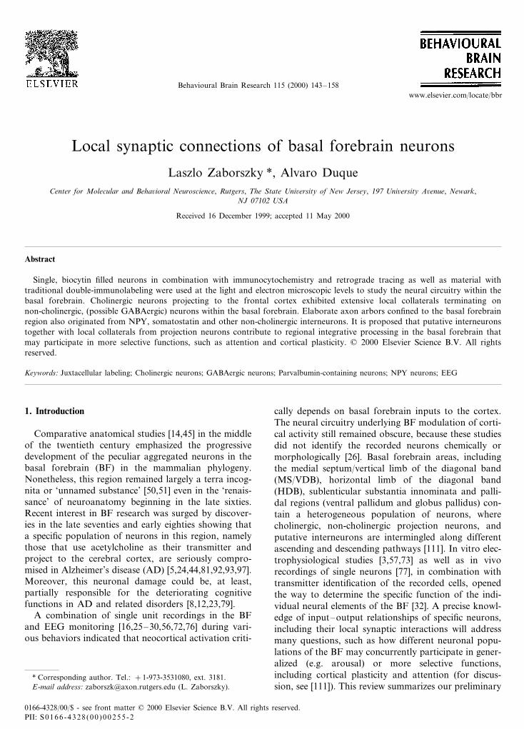
Recent studies in rats suggest that cholinergic neu-rons make up only about half of the neurons projectingto neocortical areas, the rest contain various calcium-binding proteins [111], including parvalbumin, calre-tinin and calbindin. Fig. 1 shows the three-dimensionaldistribution of the two most prevalent basalocorticalprojection systems: the cholinergic and parvalbumin-containing neurons.
2. Cholinergic neurons
Studies using multiple retrograde tracers or an-tidromic activation of basalocortical neurons suggestedthat individual cholinergic axons are highly branched intheir cortical terminal fields but seldom collateralize toinnervate different parts of the cortex [6,7,64,82]. Ourown studies [Csordas and Zaborszky, in preparation]using multiple tracers delivered into functionally relatedcortical areas tend to support this notion, in contrast tosome earlier [1,70], and more recent suggestions [57]that cholinergic or non-cholinergic neurons wouldprovide dispersed collaterals to innervate widespreadcortical regions. Using Golgi impregnation or cholineacetyl transferase (ChAT) immunostaining [13], onlythe initial segment (up to 50 mm) of the axons can beobserved. Therefore, no data are available about thelocal arborizations of cholinergic axons. A study usingintracellular iontophoresis of HRP for staining of an-tidromically identified neurons revealed that corti-copetal neurons in the BF had local axon collateralsdisplaying numerous boutons en passant [84,86], how-ever, due to the lack of transmitter identification, theclassification of the reconstructed neurons remainedopen to speculation [26].
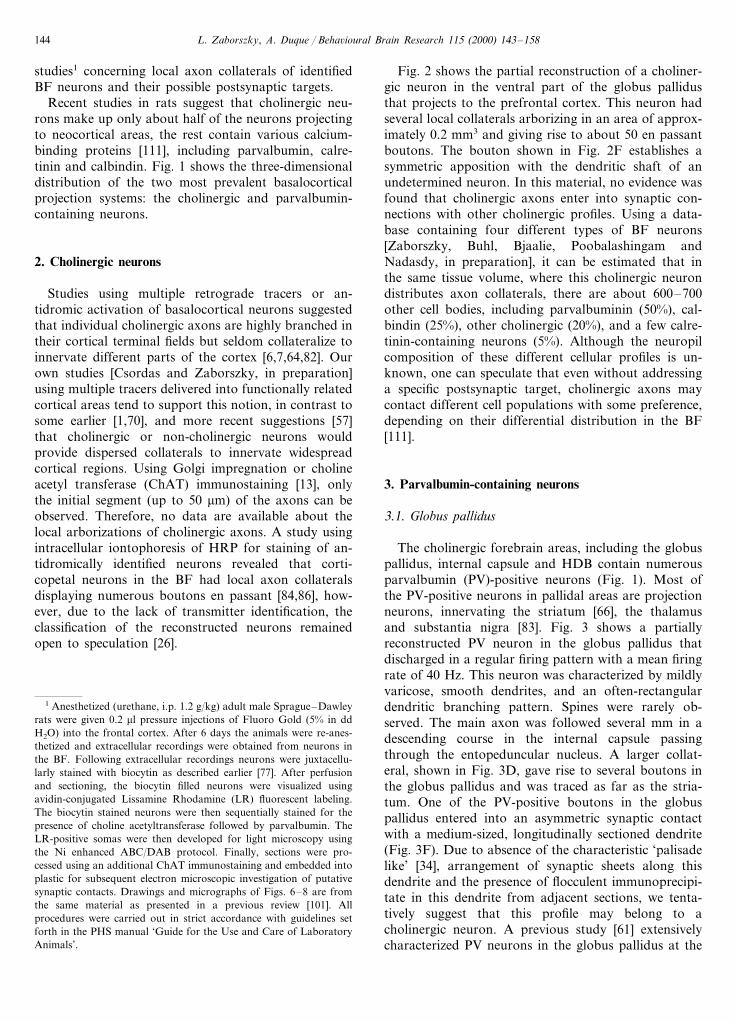
Fig. 2 shows the partial reconstruction of a choliner-gic neuron in the ventral part of the globus pallidusthat projects to the prefrontal cortex. This neuron hadseveral local collaterals arborizing in an area of approx-imately 0.2 mm3 and giving rise to about 50 en passantboutons. The bouton shown in Fig. 2F establishes asymmetric apposition with the dendritic shaft of anundetermined neuron. In this material, no evidence wasfound that cholinergic axons enter into synaptic con-nections with other cholinergic profiles. Using a data-base containing four different types of BF neurons[Zaborszky, Buhl, Bjaalie, Poobalashingam andNadasdy, in preparation], it can be estimated that inthe same tissue volume, where this cholinergic neurondistributes axon collaterals, there are about 600–700other cell bodies, including parvalbuminin (50%), cal-bindin (25%), other cholinergic (20%), and a few calre-tinin-containing neurons (5%). Although the neuropilcomposition of these different cellular profiles is un-known, one can speculate that even without addressinga specific postsynaptic target, cholinergic axons maycontact different cell populations with some preference,depending on their differential distribution in the BF[111].
3. Parvalbumin-containing neurons
3.1. Globus pallidus
The cholinergic forebrain areas, including the globuspallidus, internal capsule and HDB contain numerousparvalbumin (PV)-positive neurons (Fig. 1). Most ofthe PV-positive neurons in pallidal areas are projectionneurons, innervating the striatum [66], the thalamusand substantia nigra [83]. Fig. 3 shows a partiallyreconstructed PV neuron in the globus pallidus thatdischarged in a regular firing pattern with a mean firingrate of 40 Hz. This neuron was characterized by mildlyvaricose, smooth dendrites, and an often-rectangulardendritic branching pattern. Spines were rarely ob-served. The main axon was followed several mm in adescending course in the internal capsule passingthrough the entopeduncular nucleus. A larger collat-eral, shown in Fig. 3D, gave rise to several boutons inthe globus pallidus and was traced as far as the stria-tum. One of the PV-positive boutons in the globuspallidus entered into an asymmetric synaptic contactwith a medium-sized, longitudinally sectioned dendrite(Fig. 3F). Due to absence of the characteristic ‘palisadelike’ [34], arrangement of synaptic sheets along thisdendrite and the presence of flocculent immunoprecipi-tate in this dendrite from adjacent sections, we tenta-tively suggest that this profile may belong to acholinergic neuron. A previous study [61] extensivelycharacterized PV neurons in the globus pallidus at the
1 Anesthetized (urethane, i.p. 1.2 g/kg) adult male Sprague–Dawleyrats were given 0.2 ml pressure injections of Fluoro Gold (5% in ddH2O) into the frontal cortex. After 6 days the animals were re-anes-thetized and extracellular recordings were obtained from neurons inthe BF. Following extracellular recordings neurons were juxtacellu-larly stained with biocytin as described earlier [77]. After perfusionand sectioning, the biocytin filled neurons were visualized usingavidin-conjugated Lissamine Rhodamine (LR) fluorescent labeling.The biocytin stained neurons were then sequentially stained for thepresence of choline acetyltransferase followed by parvalbumin. TheLR-positive somas were then developed for light microscopy usingthe Ni enhanced ABC/DAB protocol. Finally, sections were pro-cessed using an additional ChAT immunostaining and embedded intoplastic for subsequent electron microscopic investigation of putativesynaptic contacts. Drawings and micrographs of Figs. 6–8 are fromthe same material as presented in a previous review [101]. Allprocedures were carried out in strict accordance with guidelines setforth in the PHS manual ‘Guide for the Use and Care of LaboratoryAnimals’.
L. Zaborszky, A. Duque / Beha6ioural Brain Research 115 (2000) 143–158 145
Fig. 1. Distribution of cholinergic and parvalbumin-containing neurons in the basal forebrain. (A) Neurons from alternate sections stained forcholine acetyltransferase (ChAT: red) and parvalbumin (PV: white) were mapped using the Neurolucida® (Microbrightfield) software. Data werethen converted into the Micro3D® (Oslo Research Park) software and visualized using an SGI computer for 3-dimensional rendering. Note thatboth cell types show higher and lower density regions (clusters) and the two markers change their medio-lateral position from rostral to caudal.The corpus callosum is visualized in blue, the outline of the brain in green. (B and C) Two sections (approximately 300 mm apart) from a seriesstained for ChAT (B) and PV (C), respectively. Abbreviations: CP, caudate putamen; f, fornix; GP, globus pallidus; HDB, horizontal limb of thediagonal band; ic, internal capsule; LV, lateral ventricle; SI, substantia innominata; sm, stria medullaris; SR, rhinal sulcus.
L. Zaborszky, A. Duque / Beha6ioural Brain Research 115 (2000) 143–158146
electron microscope level. According to that study mostof the PV-positive boutons in the globus pallidus (86%)formed symmetric synapses with somata and large den-drites, while the remaining 14% formed asymmetricsynapses with medium to small dendrites. However, theabove study was not able to determine whether PV-pos-itive terminals in the globus pallidus originate from
local axon collaterals of PV-positive pallidal neurons orderive from an outside afferent source. According tothe same study, many of the postsynaptic targets ofPV-boutons were also PV-positive neurons; however,some PV-positive boutons were seen in contact withunlabeled dendritic profiles, similar to what is shown inFig. 3. F. Bevan et al. [10] reconstructed several pal-
Fig. 2. Partial reconstruction of a juxtacellularly filled cholinergic neuron. (A) Visualization of the biocytin injected neuron using LyssamineRhodamine (LR) fluorescent marker. (B) Arrowhead points to the same neuron as filled with biocytin in (A) is stained for the presence of cholineacetyl transferase (ChAT) using FITC labeled secondary antibodies. (C) The same neuron is also retrogradely labeled with Fluoro-Gold (FG)delivered into the frontal cortex a week prior of the recording session. (D) Diagram illustrating the location of the reconstructed neuron. (E) Highmagnification view of the reconstructed cholinergic neuron. Arrowheads point to various segments of the axon. Note that the area around the cellbody is rich in axon collaterals but the varicosities present are extremely fine and not visible at this magnification. The three stippled symbolsrepresent other cholinergic cell bodies that were adjacent to labeled boutons without synaptic contacts. (F) Electron micrograph showing that abiocytin filled profile is in symmetric synaptic contact with an unlabeled dendrite. Arrow points to another dendrite containing flocculentimmunoprecipitates indicative of ChAT in this dendritic profile. Bar scale: 1 mm in (F) and 50 mm in (A). Abbreviations: see Fig. 1. Neuronsdepicted in this and subsequent figures were reconstructed using the Neurolucida® software package. The files were then edited applying Adobe®
Illustrator 8.0.
L. Zaborszky, A. Duque / Beha6ioural Brain Research 115 (2000) 143–158 147
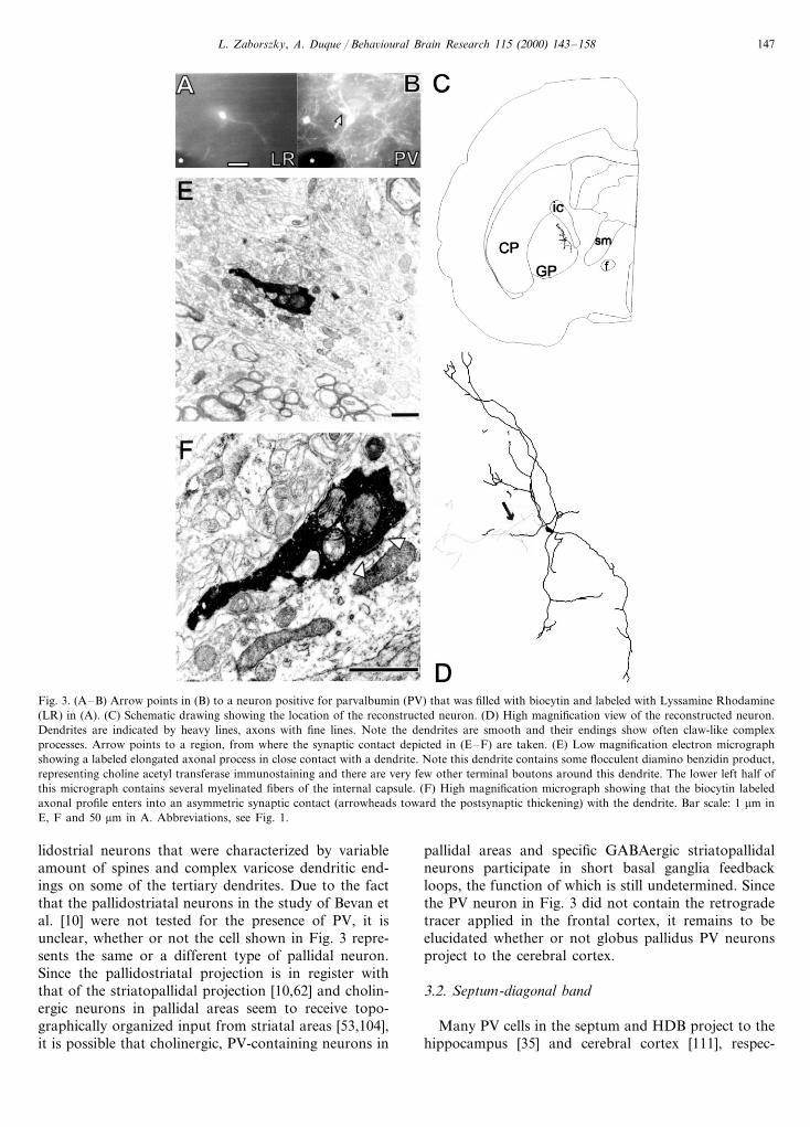
Fig. 3. (A–B) Arrow points in (B) to a neuron positive for parvalbumin (PV) that was filled with biocytin and labeled with Lyssamine Rhodamine(LR) in (A). (C) Schematic drawing showing the location of the reconstructed neuron. (D) High magnification view of the reconstructed neuron.Dendrites are indicated by heavy lines, axons with fine lines. Note the dendrites are smooth and their endings show often claw-like complexprocesses. Arrow points to a region, from where the synaptic contact depicted in (E–F) are taken. (E) Low magnification electron micrographshowing a labeled elongated axonal process in close contact with a dendrite. Note this dendrite contains some flocculent diamino benzidin product,representing choline acetyl transferase immunostaining and there are very few other terminal boutons around this dendrite. The lower left half ofthis micrograph contains several myelinated fibers of the internal capsule. (F) High magnification micrograph showing that the biocytin labeledaxonal profile enters into an asymmetric synaptic contact (arrowheads toward the postsynaptic thickening) with the dendrite. Bar scale: 1 mm inE, F and 50 mm in A. Abbreviations, see Fig. 1.
lidostrial neurons that were characterized by variableamount of spines and complex varicose dendritic end-ings on some of the tertiary dendrites. Due to the factthat the pallidostriatal neurons in the study of Bevan etal. [10] were not tested for the presence of PV, it isunclear, whether or not the cell shown in Fig. 3 repre-sents the same or a different type of pallidal neuron.Since the pallidostriatal projection is in register withthat of the striatopallidal projection [10,62] and cholin-ergic neurons in pallidal areas seem to receive topo-graphically organized input from striatal areas [53,104],it is possible that cholinergic, PV-containing neurons in
pallidal areas and specific GABAergic striatopallidalneurons participate in short basal ganglia feedbackloops, the function of which is still undetermined. Sincethe PV neuron in Fig. 3 did not contain the retrogradetracer applied in the frontal cortex, it remains to beelucidated whether or not globus pallidus PV neuronsproject to the cerebral cortex.
3.2. Septum-diagonal band
Many PV cells in the septum and HDB project to thehippocampus [35] and cerebral cortex [111], respec-

L. Zaborszky, A. Duque / Beha6ioural Brain Research 115 (2000) 143–158148
tively. PV has been found in GABAergic neurons inmany brain areas, including GABAergic local neuronsof the cerebral cortex, the hippocampus, and the neos-triatum [17,20,63,65]. Identified PV cells in the BFdischarged at 7–15 Hz, regular or in random modesand showed positive correlation in their discharge pat-tern to concurrent EEG desynchronization (Duque,Balatoni, Detari and Zaborszky, in press). SinceGABAergic basalocortical axons were found toterminate exclusively on cortical GABAergic inter-neurons [37], our finding is compatible with the notionthat GABAergic basalocortical input promotes func-
tional activation in the cerebral cortex by disinhibition[56].
4. Other non-cholinergic projection neurons
Fig. 4 shows a retrogradely labeled, juxtacellularlyfilled neuron in the anterior amygdaloid area that pro-jected to the prefrontal cortex. This neuron was testedfor the presence for ChAT and for two calcium-bindingproteins, PV and calretinin. None of these substancesturned out to be localized within this neuron. A partial
Fig. 4. (A–B) biocytin filled neuron marked with Lyssamine Rhodamine (LR) in (A) that is also retrogradely labeled from the frontal cortex usingFluoro-Gold (FG) in (B). (C) Shows the location of this neuron in the anterior amygdaloid area. (D) High magnification view of the neuron. Notethe richly arborized denritic processes that are heavily studded with spines. Arrowhead points to the origin of the axon. (E) Higher magnificationview of the boxed area in D. (F) High magnification electron micrograph showing a longitudinally cut dendrite that is contacted by a biocytinfilled axon terminal. Arrowheads point to the symmetric postsynaptic side. Bar scale, 1 mm in (F) and 50 mm in (A). Abbreviations: see Fig. 1.
L. Zaborszky, A. Duque / Beha6ioural Brain Research 115 (2000) 143–158 149
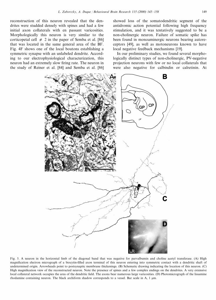
reconstruction of this neuron revealed that the den-drites were studded densely with spines and had a fewinitial axon collaterals with en passant varicosities.Morphologically this neuron is very similar to thecorticopetal cell c 2 in the paper of Semba et al. [86]that was located in the same general area of the BF.Fig. 4F shows one of the local boutons establishing asymmetric synapse with an unlabeled dendrite. Accord-ing to our electrophysiological characterization, thisneuron had an extremely slow firing rate. The neuron inthe study of Reiner et al. [84] and Semba et al. [86]
showed loss of the somatodendritic segment of theantidromic action potential following high frequencystimulation, and it was tentatively suggested to be anon-cholinergic neuron. Failure of somatic spike hasbeen found in monoaminergic neurons bearing autore-ceptors [49], as well as motoneurons known to havelocal negative feedback mechanisms [19].
In our preliminary studies, we found several morpho-logically distinct types of non-cholinergic, PV-negativeprojection neurons with few or no local collaterals thatwere also negative for calbindin or calretinin. At
Fig. 5. A neuron in the horizontal limb of the diagonal band that was negative for parvalbumin and choline acetyl transferase. (A) Highmagnification electron micrograph of a biocytin-filled axon terminal of this neuron entering into symmetric contact with a dendritic shaft ofundetermined origin. Arrowheads point to postsynaptic membrane thickenings. (B) Schematic drawing indicating the location of this neuron. (C)High magnification view of the reconstructed neuron. Note the presence of spines and a few complex endings on the dendrites. A very extensivelocal collateral network occupies the area of the dendritic field. The axons bear numerous large varicosities. (D) Photomicrograph of the lissaminerhodamine containing neuron. The black archiform shadow corresponds to a vessel. Bar scale in A, 1 mm.
L. Zaborszky, A. Duque / Beha6ioural Brain Research 115 (2000) 143–158150
present, it is unclear what type of neurotransmitter is inthese non-cholinergic corticopetal neurons and what istheir local and/or cortical postsynaptic target.
5. Putative local circuit neurons
Fig. 5 shows a sparsely spiny neuron that was nega-tive for ChAT and PV and is partially reconstructed.Around the cell body and occupying a large part of thedendritic arbor, a dense network of axon collaterals canbe seen. These collaterals bear numerous large, bulbousvaricosities. Within a space of about 0.14 mm3, 374varicosities were counted. Within this axonal arboriza-tion volume, there are about 550 other neurons, includ-ing 300 calretinin, 200 cholinergic, 40 parvalbumin and10 calbindin-containing cells. One of the axonal vari-cosities shown in Fig. 5A enters into symmetric synapsewith a dendritic shaft. The chemical characteristics ofthis postsynaptic profile have not been determined.
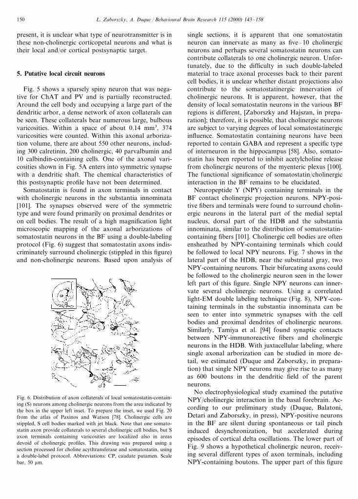
Somatostatin is found in axon terminals in contactwith cholinergic neurons in the substantia innominata[101]. The synapses observed were of the symmetrictype and were found primarily on proximal dendrites oron cell bodies. The result of a high magnification lightmicroscopic mapping of the axonal arborizations ofsomatostatin neurons in the BF using a double-labelingprotocol (Fig. 6) suggest that somatostatin axons indis-criminately surround cholinergic (stippled in this figure)and non-cholinergic neurons. Based upon analysis of
single sections, it is apparent that one somatostatinneuron can innervate as many as five–10 cholinergicneurons and perhaps several somatostatin neurons cancontribute collaterals to one cholinergic neuron. Unfor-tunately, due to the difficulty in such double-labeledmaterial to trace axonal processes back to their parentcell bodies, it is unclear whether distant projections alsocontribute to the somatostatinergic innervation ofcholinergic neurons. It is apparent, however, that thedensity of local somatostatin neurons in the various BFregions is different, [Zaborszky and Hajszan, in prepa-ration]; therefore, it is possible, that cholinergic neuronsare subject to varying degrees of local somatostatinergicinfluence. Somatostatin containing neurons have beenreported to contain GABA and represent a specific typeof interneuron in the hippocampus [58]. Also, somato-statin has been reported to inhibit acetylcholine releasefrom cholinergic neurons of the myenteric plexus [100].The functional significance of somatostatin/cholinergicinteraction in the BF remains to be elucidated.
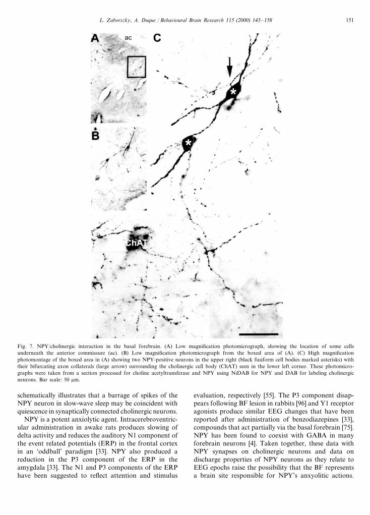
Neuropeptide Y (NPY) containing terminals in theBF contact cholinergic projection neurons. NPY-posi-tive fibers and terminals were found to surround cholin-ergic neurons in the lateral part of the medial septalnucleus, dorsal part of the HDB and the substantiainnominata, similar to the distribution of somatostatin-containing fibers [101]. Cholinergic cell bodies are oftenensheathed by NPY-containing terminals which couldbe followed to local NPY neurons. Fig. 7 shows in thelateral part of the HDB, near the substriatal gray, twoNPY-containing neurons. Their bifurcating axons couldbe followed to the cholinergic neuron seen in the lowerleft part of this figure. Single NPY neurons can inner-vate several cholinergic neurons. Using a correlatedlight-EM double labeling technique (Fig. 8), NPY-con-taining terminals in the substantia innominata can beseen to enter into symmetric synapses with the cellbodies and proximal dendrites of cholinergic neurons.Similarly, Tamiya et al. [94] found synaptic contactsbetween NPY-immunoreactive fibers and cholinergicneurons in the HDB. With juxtacellular labeling, wheresingle axonal arborization can be studied in more de-tail, we estimated (Duque and Zaborszky, in prepara-tion) that single NPY neurons may give rise to as manyas 600 boutons in the dendritic field of the parentneurons.
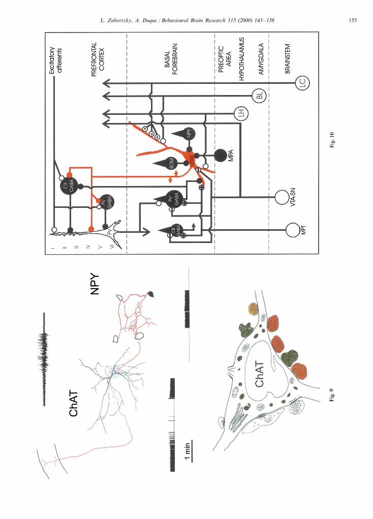
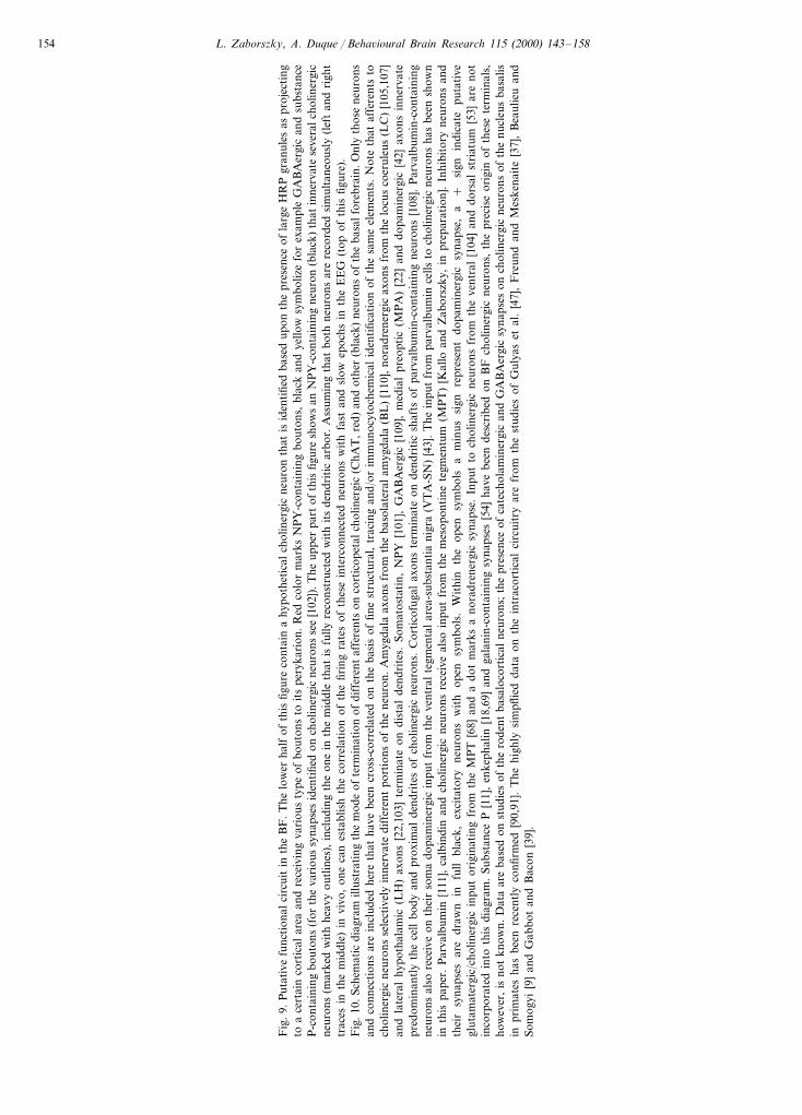
No electrophysiological study examined the putativeNPY/cholinergic interaction in the basal forebrain. Ac-cording to our preliminary study (Duque, Balatoni,Detari and Zaborszky, in press), NPY-positive neuronsin the BF are silent during spontaneous or tail pinchinduced desynchronization, but accelerated duringepisodes of cortical delta oscillations. The lower part ofFig. 9 shows a hypothetical cholinergic neuron, receiv-ing several different types of axon terminals, includingNPY-containing boutons. The upper part of this figure
Fig. 6. Distribution of axon collaterals of local somatostatin-contain-ing (S) neurons among cholinergic neurons from the area indicated bythe box in the upper left inset. To prepare the inset, we used Fig. 20from the atlas of Paxinos and Watson [78]. Cholinergic cells arestippled, S cell bodies marked with jet black. Note that one somato-statin axon provide collaterals to several cholinergic cell bodies, but Saxon terminals containing varicosities are localized also in areasdevoid of cholinergic profiles. This drawing was prepared using asection processed for choline acytltransferase and somatostatin, usinga double-label protocol. Abbreviations: CP, caudate putamen. Scalebar, 50 mm.
L. Zaborszky, A. Duque / Beha6ioural Brain Research 115 (2000) 143–158 151
Fig. 7. NPY/cholinergic interaction in the basal forebrain. (A) Low magnification photomicrograph, showing the location of some cellsunderneath the anterior commissure (ac). (B) Low magnification photomicrograph from the boxed area of (A). (C) High magnificationphotomontage of the boxed area in (A) showing two NPY-positive neurons in the upper right (black fusiform cell bodies marked asterisks) withtheir bifurcating axon collaterals (large arrow) surrounding the cholinergic cell body (ChAT) seen in the lower left corner. These photomicro-graphs were taken from a section processed for choline acetyltransferase and NPY using NiDAB for NPY and DAB for labeling cholinergicneurons. Bar scale: 50 mm.
schematically illustrates that a barrage of spikes of theNPY neuron in slow-wave sleep may be coincident withquiescence in synaptically connected cholinergic neurons.
NPY is a potent anxiolytic agent. Intracerebroventric-ular administration in awake rats produces slowing ofdelta activity and reduces the auditory N1 component ofthe event related potentials (ERP) in the frontal cortexin an ‘oddball’ paradigm [33]. NPY also produced areduction in the P3 component of the ERP in theamygdala [33]. The N1 and P3 components of the ERPhave been suggested to reflect attention and stimulus
evaluation, respectively [55]. The P3 component disap-pears following BF lesion in rabbits [96] and Y1 receptoragonists produce similar EEG changes that have beenreported after administration of benzodiazepines [33],compounds that act partially via the basal forebrain [75].NPY has been found to coexist with GABA in manyforebrain neurons [4]. Taken together, these data withNPY synapses on cholinergic neurons and data ondischarge properties of NPY neurons as they relate toEEG epochs raise the possibility that the BF representsa brain site responsible for NPY’s anxyolitic actions.

L. Zaborszky, A. Duque / Beha6ioural Brain Research 115 (2000) 143–158152
6. General discussion
Understanding of the cellular organization of thecerebral cortex, including the hippocampus, has ad-vanced in the last twenty years primarily due to theapplication of intracellular electrophysiology with rig-orous combination of correlated light and electron mi-croscopic techniques [15,36,40,48,88]. Such methods,
for technical reasons, were not adapted in their fullcapacity in the BF [67,84,86]. More recently, the appli-cation of the juxtacellular technique of Pinault [77,80]with a combination of immunostaining to identify thetransmitter content of the recorded neurons began tounravel the functional circuitry of the BF. The integra-tive capacity of the cerebral cortex largely depends onthe presence of various types of inhibitory and excita-
Fig. 8. NPY/cholinergic interaction in the basal forebrain. (A) Schematic drawing showing the location of the cholinergic neuron in the substantialinnominata (arrow in B) that was subsequently investigated with electron microscopy to identify synaptic contacts. (C) low magnification electronmicrograph of a cholinergic neuron. Note the presence of heavy immunostaining in the perikaryon which is also rich in endoplasmic reticulum,characteristic for cholinergic projection neurons. Several NPY containing varicosities approaching the cell body are labeled by arrowheads. Thebouton enclosed in the box at upper left to the cell body is enlarged in the two insets at lower right, which are taken from two adjacent thinsections. Arrowheads in these insets point to the postsynaptic membrane thickening. Bar scale in the inset 1 mm; in B, 100 mm.
L. Zaborszky, A. Duque / Beha6ioural Brain Research 115 (2000) 143–158 153
L. Zaborszky, A. Duque / Beha6ioural Brain Research 115 (2000) 143–158154
Fig
.9.
Put
ativ
efu
ncti
onal
circ
uit
inth
eB
F.
The
low
erha
lfof
this
figur
eco
ntai
na
hypo
thet
ical
chol
iner
gic
neur
onth
atis
iden
tifie
dba
sed
upon
the
pres
ence
ofla
rge
HR
Pgr
anul
esas
proj
ecti
ngto
ace
rtai
nco
rtic
alar
eaan
dre
ceiv
ing
vari
ous
type
ofbo
uton
sto
its
pery
kari
on.
Red
colo
rm
arks
NP
Y-c
onta
inin
gbo
uton
s,bl
ack
and
yello
wsy
mbo
lize
for
exam
ple
GA
BA
ergi
can
dsu
bsta
nce
P-c
onta
inin
gbo
uton
s(f
orth
eva
riou
ssy
naps
esid
enti
fied
onch
olin
ergi
cne
uron
sse
e[1
02])
.The
uppe
rpa
rtof
this
figur
esh
ows
anN
PY
-con
tain
ing
neur
on(b
lack
)th
atin
nerv
ate
seve
ralc
holin
ergi
cne
uron
s(m
arke
dw
ith
heav
you
tlin
es),
incl
udin
gth
eon
ein
the
mid
dle
that
isfu
llyre
cons
truc
ted
wit
hit
sde
ndri
tic
arbo
r.A
ssum
ing
that
both
neur
ons
are
reco
rded
sim
ulta
neou
sly
(lef
tan
dri
ght
trac
esin
the
mid
dle)
invi
vo,
one
can
esta
blis
hth
eco
rrel
atio
nof
the
firin
gra
tes
ofth
ese
inte
rcon
nect
edne
uron
sw
ith
fast
and
slow
epoc
hsin
the
EE
G(t
opof
this
figur
e).
Fig
.10.
Sche
mat
icdi
agra
mill
ustr
atin
gth
em
ode
ofte
rmin
atio
nof
diff
eren
taf
fere
nts
onco
rtic
opet
alch
olin
ergi
c(C
hAT
,red
)an
dot
her
(bla
ck)
neur
ons
ofth
eba
salf
oreb
rain
.Onl
yth
ose
neur
ons
and
conn
ecti
ons
are
incl
uded
here
that
have
been
cros
s-co
rrel
ated
onth
eba
sis
offin
est
ruct
ural
,tr
acin
gan
d/or
imm
unoc
ytoc
hem
ical
iden
tific
atio
nof
the
sam
eel
emen
ts.
Not
eth
ataf
fere
nts
toch
olin
ergi
cne
uron
sse
lect
ivel
yin
nerv
ate
diff
eren
tpo
rtio
nsof
the
neur
on.
Am
ygda
laax
ons
from
the
baso
late
ral
amyg
dala
(BL
)[1
10],
nora
dren
ergi
cax
ons
from
the
locu
sco
erul
eus
(LC
)[1
05,1
07]
and
late
ral
hypo
thal
amic
(LH
)ax
ons
[22,
103]
term
inat
eon
dist
alde
ndri
tes.
Som
atos
tati
n,N
PY
[101
],G
AB
Aer
gic
[109
],m
edia
lpr
eopt
ic(M
PA
)[2
2]an
ddo
pam
iner
gic
[42]
axon
sin
nerv
ate
pred
omin
antl
yth
ece
llbo
dyan
dpr
oxim
alde
ndri
tes
ofch
olin
ergi
cne
uron
s.C
orti
cofu
gal
axon
ste
rmin
ate
onde
ndri
tic
shaf
tsof
parv
albu
min
-con
tain
ing
neur
ons
[108
].P
arva
lbum
in-c
onta
inin
gne
uron
sal
sore
ceiv
eon
thei
rso
ma
dopa
min
ergi
cin
put
from
the
vent
ral
tegm
enta
lar
ea-s
ubst
anti
ani
gra
(VT
A-S
N)
[43]
.T
hein
put
from
parv
albu
min
cells
toch
olin
ergi
cne
uron
sha
sbe
ensh
own
inth
ispa
per.
Par
valb
umin
[111
],ca
lbin
din
and
chol
iner
gic
neur
ons
rece
ive
also
inpu
tfr
omth
em
esop
onti
nete
gmen
tum
(MP
T)
[Kal
loan
dZ
abor
szky
,in
prep
arat
ion]
.In
hibi
tory
neur
ons
and
thei
rsy
naps
esar
edr
awn
infu
llbl
ack,
exci
tato
ryne
uron
sw
ith
open
sym
bols
.W
ithi
nth
eop
ensy
mbo
lsa
min
ussi
gnre
pres
ent
dopa
min
ergi
csy
naps
e,a
+si
gnin
dica
tepu
tati
vegl
utam
ater
gic/
chol
iner
gic
inpu
tor
igin
atin
gfr
omth
eM
PT
[68]
and
ado
tm
arks
ano
radr
ener
gic
syna
pse.
Inpu
tto
chol
iner
gic
neur
ons
from
the
vent
ral
[104
]an
ddo
rsal
stri
atum
[53]
are
not
inco
rpor
ated
into
this
diag
ram
.Su
bsta
nce
P[1
1],
enke
phal
in[1
8,69
]an
dga
lani
n-co
ntai
ning
syna
pses
[54]
have
been
desc
ribe
don
BF
chol
iner
gic
neur
ons,
the
prec
ise
orig
inof
thes
ete
rmin
als,
how
ever
,is
not
know
n.D
ata
are
base
don
stud
ies
ofth
ero
dent
basa
loco
rtic
alne
uron
s;th
epr
esen
ceof
cate
chol
amin
ergi
can
dG
AB
Aer
gic
syna
pses
onch
olin
ergi
cne
uron
sof
the
nucl
eus
basa
lisin
prim
ates
has
been
rece
ntly
confi
rmed
[90,
91].
The
high
lysi
mpfl
ied
data
onth
ein
trac
orti
cal
circ
uitr
yar
efr
omth
est
udie
sof
Gul
yas
etal
.[4
7],
Fre
und
and
Mes
kena
ite
[37]
,B
eaul
ieu
and
Som
ogyi
[9]
and
Gab
bot
and
Bac
on[3
9].
L. Zaborszky, A. Duque / Beha6ioural Brain Research 115 (2000) 143–158 155
tory local circuit neurons that address with often re-markable specificity projection or other local neurons.According to our preliminary studies, non-cholinergicprojection neurons seem to have sparse-to-moderatelocal collaterals. On the other hand, cholinergic, NPY,and some other interneurons with unidentified transmit-ter via their rich local axon collaterals may significantlyimpact on local processing in the BF. According to invitro studies, cholinergic and GABAergic cells in theBF display robust intrinsic pacemaker mechanismswhich allow them to discharge in rhythmic trains [3]. Ithas been hypothesized that ligand- or voltage-depen-dent oscillations of interneurons via GABAergicsynapses may be critical for cooperative ensemble func-tion of the hippocampus [88]. Whether local synapticinteractions in the basal forebrain contribute to syn-chronized activity in BF neuronal populations remainsto be elucidated.
In the absence of precise cellular data on synapticdistributions and correlated in vitro recording, we donot know under what conditions these putative in-terneurons are active and how their inputs affect actionpotential generation in cholinergic and GABAergic pro-jection neurons. Using bundles of microwires in the BFof cats, under chloralose anesthesia, Detari et al. [29]registered different types of synaptic interactions, in-cluding inhibition and excitation. The distance betweentwo units that were in inhibitory relationship oftenreached as long as 2 mm [Detari, personal communica-tion]. At present, it is unknown whether state-relatedmodulatory inputs (noradrenaline, serotonin) innervateNPY or other local neurons; however, a neuron like theone depicted in Fig. 5 would be in a strategicallyexcellent position to distribute and amplify such modu-latory input to a basal forebrain tissue ‘block’ contain-ing at least 600–800 cells.
The wire diagram of Fig. 10 depicts the complicatedarray of local and projection neurons in the BF withtheir major afferent sources and their potential corticaltarget. Our preliminary studies did not allow us to drawa firm conclusion as to whether the local collateralsaddress different neurons randomly, or with some spe-cificity. Also, it is unknown whether or not they contactstrategically different portions of their target cells.Therefore, the connections involving locally, arborizingaxons shown in Fig. 10 should be regarded as an initialattempt at summarizing available data. On the otherhand, a considerable amount of evidence exists regard-ing the synaptic topography of long-range afferents tocholinergic neurons, which are included in this sche-matic circuit diagram.
This complicated local circuitry is paralleled by asimilar complex receptor mechanism. For example,GABA elicits via GABAA receptors Cl− dependentpostsynaptic currents in cultured basal forebrain neu-rons [2] and basal forebrain GABAA receptors are
involved in behavior-induced acetylcholine release inthe cortex, including the hippocampus [74,85]. In addi-tion, in situ hybridization and immunocytochemicalstudies suggest that various BF neurons expressGABAA receptors with different subunit compositions:cholinergic neurons are typically characterized by thesubunit composition a3/b2,3/g2, whereas most of theparvalbumin-positive GABAergic neurons expresseither a1/b2/g2 or composition a1/a3/b2/g2 [31,38,41,52,98]. GABAB receptors have been localized in theBF of pigeons [95]. Its cellular localization in rodents isunknown, and, consequently, its function remains spec-ulative [2]. Acetylcholine elicits depolarizing or hyper-polarizing responses in different, locally arborizingpostsynaptic neurons via nicotinic and muscarinic re-ceptors as has been shown in cortical slice preparations[71,99]. Since muscarinic and nicotinic receptors havebeen localized in BF, acetylcholine may have a complexeffect in local processing similar to its effect in corticalinformation processing [21,46,57,59,60,89].
Although noradrenergic and dopaminergic axonscontact cholinergic neurons in extensive portions of theBF [42,105], the majority of afferents, including corti-cal, striatal, hypothalamic, and various peptidergicfibers, appear to have a preferential distribution todifferent regions of the BF [22,53,87,106]. In addition,we have shown recently that prefrontal axons withintheir distribution area exclusively contact non-choliner-gic neurons of the BF, including parvalbumin-contain-ing and other undetermined cell populations [108].Since the number of putative GABAergic inputs tocholinergic cell bodies varies between different regionsin the BF [102] and cholinergic dendrites show markedregionally specific orientation (Zaborszky, Nadasdyand J. Somogyi, in preparation), it is likely that cholin-ergic neurons process different types of informationaccording to their location in the BF. Preliminary datapresented in this paper suggest that collaterals of pro-jection or local circuit neurons could contact only alimited number of specific neurons, depending on theparticular location. To what extent local circuit neuronsand collaterals of projection neurons together with themore restricted type of afferents mentioned above con-tribute to regionally specific information processing inthe basal forebrain remains to be investigated in futurestudies.
Acknowledgements
The research summarized in this paper was sup-ported by NIH grant No. NS23945 (L.Z), S06GM08223 and NSF-BIR-9413198 (A.D). We thank DrJ.M. Tepper for allowing us to use his equipment (NIHgrant NS-34865). Special thanks are due to Dr Wei Lu,

L. Zaborszky, A. Duque / Beha6ioural Brain Research 115 (2000) 143–158156
Derek Buhl and Elizabeth Rommer for their experttechnical assistance.
References
[1] Adams CE, Cepeda C, Boylan MK, et al. Basal forebrainneurons have axon collaterals that project to widely divergentcortical areas in the cat. Brain Res 1986;397:365–71.
[2] Akaike N, Harata N, Ueno S, Tateishi N. GABAergic synapticcurrent in dissociated nucleus basalis of Meynert neurons of therat. Brain Res 1992;570:102–8.
[3] Alonso A. Intrinsic electroresponsiviness of basal forebraincholinergic and non-cholinergic neurons. In: Lydic R, Bagh-doyan HA, editors. Handbook of Behavioral State Control –Cellular and Molecular Mechanisms. New York: CRC Press,1999:297–309.
[4] Aoki C, Pickel VM. Neuropeptide Y in the cerebral cortex andthe caudate-putamen nuclei: ultrastructural basis for interac-tions with GABAergic and non-GABAergic neurons. J Neu-rosci 1989;9:4333–54.
[5] Armstrong DM, Saper CB, Levey AI, Wainer BH, Terry RD.Distribution of cholinergic neurons in rat brain: demonstratedby the immunocytochemical localization of choline acetyltrans-ferase. J Comp Neurol 1983;216:53–68.
[6] Aston-Jones G, Shaver R, Dinan T. Cortically projecting nu-cleus basalis neurons in rat are physiologically heterogeneous.Neurosci Lett 1984;46:19–24.
[7] Aston-Jones G, Shaver R, Dinan TG. Nucleus basalis neuronsexhibit axonal branching with decreased impulse conductionvelocity in rat cerebrocortex. Brain Res 1985;325:271–85.
[8] Bartus RT, Flicker C, Dean RL, Fisher S, Pontecorvo M,Figueiredo J. Behavioral and biochemical effects of nucleusbasalis magnocellularis lesions: implications and possible rele-vance to understanding or treating Alzheimers disease. ProgBrain Res 1986;70:345–61.
[9] Beaulieu C, Somogyi P. Enrichment of cholinergic synapticterminals on GABAergic neurons and coexistence of im-munoreactive GABA and choline acetyltransferase in the samesynaptic terminals in the striate cortex of the cat. J CompNeurol 1991;304:666–80.
[10] Bevan MD, Booth PA, Eaton SA, Bolam JP. Selective innerva-tion of neostriatal interneurons by a subclass of neuron in theglobus pallidus of the rat. J Neurosci 1998;18:9438–52.
[11] Bolam JP, Ingham CA, Izzo PN, Levey AI, Rye DB, SmithAD, Wainer BH. Substance P-containing terminals in synapticcontact with cholinergic neurons in the neostriatum and basalforebrain: a double immunocytochemical study in the rat. BrainRes 1986;397:279–89.
[12] Bowen DM, Smith CB, White P, Davison AN. Neurotransmit-ter-related enzymes and indices of hypoxia in senile dementiaand other abiotrophies. Brain 1976;99:459–96.
[13] Brauer K, Schober W, Werner L, Winkelmann E, Lungwit W,Hajdu F. Neurons in the basal forebrain complex of the rat: aGolgi study. J Hirnforsch 1988;29:43–71.
[14] Brockhaus H. Vergleichend-anatomische Untersuchungen uberden Basalkernkomplex. J Psychol Neurol 1942;51:57–95.
[15] Buhl EH, Halasy K, Somogyi P. Diverse sources of hippocam-pal unitary inhibitory postsynaptic potentials and the numberof synaptic release sites. Nature 1994;368:823–8.
[16] Buzsaki G, Bickford RG, Ponomareff G, Thal LJ, Mandel R,Gage FH. Nucleus basalis and thalamic control of neocorticalactivity in the freely moving rat. J Neurosci 1988;8:4007–26.
[17] Celio MR. Calbindin D-28k and parvalbumin in the rat ner-vous system. Neuroscience 1990;35:375–475.
[18] Chang HT, Penny GR, Kitai ST. Enkephalinergic-cholinergicinteraction in the rat globus pallidus: a pre-embedding double-labeling immunocytochemistry study. Brain Res 1987;426:197–203.
[19] Coombs JS, Curtis DR, Eccles JC. The interpretation of spikepotentials of motoneurons. J Physiol 1957;139:198–231.
[20] Cowan RL, Wilson CJ, Emson PC, Heizmann CW. Parvalbu-min-containing GABAergic interneurons in the rat neostriatum.J Comp Neurol 1990;302:197–205.
[21] Csillik B, Rakic P, Knyihar-Csillik E. Peptidergic innervationand the nicotinic acetylcholine receptor in the primate basalnucleus. Eur J Neurosci 1998;10:573–85.
[22] Cullinan WE, Zaborszky L. Organization of ascending hypo-thalamic projections to the rostral forebrain with special refer-ence to the innervation of cholinergic projection neurons. JComp Neurol 1991;306:631–67.
[23] Davies P, Maloney AJ. Selective loss of central cholinergicneurons in Alzheimer’s disease. Lancet 1976;2:1403.
[24] de Lacalle S, Saper CB. The cholinergic system in the primatebrain: Basal forebrain and pontine-tegmental cell groups. In:F.E. Bloom, A. Bjorklund, T. Hokfelt, editors. Handbook ofChemical Neuroanatomy. The Primate Nervous System, Part I,New York: Elsevier, 1997, pp. 217–252.
[25] Detari L, Juhasz G, Kukorelli T. Firing properties of cat basalforebrain neurones during sleep–wakefulness cycle. Electroen-cephalogr Clin Neurophysiol 1984;58:362–8.
[26] Detari L, Rasmusson DD, Semba K. The role of basal fore-brain neurons in tonic and phasic activation of the cerebralcortex. Prog Neurobiol 1999;58:249–77.
[27] Detari L, Semba K, Rasmusson DD. Responses of corticalEEG-related basal forebrain neurons to brainstem and sensorystimulation in urethane-anaesthetized rats. Eur J Neurosci1997;9:1153–61.
[28] Detari L, Vanderwolf CH. Activity of identified cortically pro-jecting and other basal forebrain neurons during large slowwaves and cortical activation in anaesthetized rats. Brain Res1987;437:1–8.
[29] Detari L, Vanderwolf CH, Kukorelli T. Inhibitory connectionsin the basal forebrain: a possible explanation for the ambiguousrole of the BF in the regulation of sleep and wakefulness. In:Mancia M, Marini G, editors. The Diencephanlon and Sleep,New York: Raven Press, 1990, pp. 355–359.
[30] Dringenberg HC, Vanderwolf CH. Involvement of direct andindirect pathways in electrocorticographic activation. NeurosciBiobehav Rev 1998;22:243–57.
[31] Duncan GE, Breese GR, Criswell HE, McCown TJ, HerbertJS, Devaud LL, Morrow AL. Distribution of [3H]-zolpidembinding sites in relation to messenger RNA encoding the alpha1, beta 2 and gamma 2 subunits of GABAA receptors in ratbrain. Neuroscience 1995;64:1113–28.
[32] Duque A., Balatoni B., Detari L., Zaborszky L. EEG correla-tion of the discharge properties of identified neurons in thebasal forebrain. J. Neurophysiol. 2000; in press.
[33] Ehlers CL, Somes C, Lopez A, Kirby D, Rivier JE. Electro-physiological actions of neuropeptide Y and its analogs: newmeasures for anxiolytic therapy? Neuropsychopharmacology1997;17:34–43.
[34] Fox CA, Andrade AN, Lu Qui IJ, Rafols JA. The primateglobus pallidus: a Golgi and electron microscopic study. JHirnforsch 1974;15:75–93.
[35] Freund TF, Antal M. GABA-containing neurons in the septumcontrol inhibitory interneurons in the hippocampus. Nature1988;336:170–3.
[36] Freund TF, Martin KA, Whitteridge D. Innervation of catvisual areas 17 and 18 by physiologically identified X- andY-type thalamic afferents I. Arborization patterns and quanti-tative distribution of postsynaptic elements. J Comp Neurol1985;242:263–74.
L. Zaborszky, A. Duque / Beha6ioural Brain Research 115 (2000) 143–158 157
[37] Freund TF, Meskenaite V. Gamma-Aminobutyric acid-con-taining basal forebrain neurons innervate inhibitory interneu-rons in the neocortex. Proc Natl Acad Sci 1992;89:738–42.
[38] Fritschy JM, Mohler H. GABAA-receptor heterogeneity in theadult rat brain: differential regional and cellular distribution ofseven major subunits. J Comp Neurol 1995;359:154–94.
[39] Gabbott PL, Bacon SJ. Local circuit neurons in the medialprefrontal cortex (areas 24a,b,c, 25 and 32) in the monkey: I.Cell morphology and morphometrics. J Comp Neurol1996;364:567–608.
[40] Gabbott PL, Martin KA, Whitteridge D. Connections betweenpyramidal neurons in layer 5 of cat visual cortex (area 17). JComp Neurol 1987;259:364–81.
[41] Gao B, Hornung JP, Fritschy JM. Identification of distinctGABAA-receptor subtypes in cholinergic and parvalbumin-pos-itive neurons of the rat and marmoset medial septum–diagonalband complex. Neuroscience 1995;65:101–17.
[42] Gaykema RP, Zaborszky L. Direct catecholaminergic–cholin-ergic interactions in the basal forebrain. II. Substantia nigra-ventral tegmental area projections to cholinergic neurons. JComp Neurol 1996;374:555–77.
[43] Gaykema RP, Zaborszky L. Parvalbumin-containing neuronsin the basal forebrain receive direct input from the substantianigra-ventral tegmental area. Brain Res 1997;747:173–9.
[44] Geula C, Mesulam MM. Cholinergic systems and related neu-ropathological predilection patterns in Alzheimer disease. In:Terry RD, Katzman R, Bick KL, editors. Alzheimer Disease.New York: Raven Press, 1994:263–91.
[45] Gorry JD. Studies on the comparative anatomy of the ganglionbasale de Meynert. Acta Anat 1963;55:51–104.
[46] Gotti C, Fornasari D, Clementi F. Human neuronal nicotinicreceptors. Prog Neurobiol 1997;53:199–237.
[47] Gulyas AI, Hajos N, Freund TF. Interneurons containingcalretinin are specialized to control other interneurons in the rathippocampus. J Neurosci 1996;16:3397–411.
[48] Gulyas AI, Miles R, Sik A, Toth K, Tamamaki N, Freund TF.Hippocampal pyramidal cells excite inhibitory neurons througha single release site. Nature 1993;366:683–7.
[49] Guyenet PG, Aghajanian GK. Antidromic identification ofdopaminergic and other output neurons of the rat substantianigra. Brain Res 1978;150:69–84.
[50] Heimer L, de Olmos J, Alheid GF, Zaborszky L. ’Perestroika’in the basal forebrain: opening the border between neurologyand psychiatry. Prog Brain Res 1991;87:109–65.
[51] Heimer L, de Olmos, JS, Alheid GF, et al. The human basalforebrain. Part II. In: Bloom FE, Bjorklund A, Hokfelt T,editors. Handbook of Chemical Neuroanatomy, The PrimateNervous System, Part III, vol. 15 New York: Elsevier, 1999, pp.57–226.
[52] Henderson Z. Expression of GABAA receptor subunit messen-ger RNA in non-cholinergic neurons of the rat basal forebrain.Neuroscience 1995;65:1077–86.
[53] Henderson Z. The projection from the striatum to the nucleusbasalis in the rat: an electron microscopic study. Neuroscience1997;78:943–55.
[54] Henderson Z, Morris N. Galanin-immunoreactive synaptic ter-minals on basal forebrain cholinergic neurons in the rat. JComp Neurol 1997;383:82–93.
[55] Hillyard SA, Kutas M. Electrophysiology of cognitive process-ing. Annu Rev Psychol 1983;34:33–61.
[56] Jimenez-Capdeville ME, Dykes RW, Myasnikov AA. Differen-tial control of cortical activity by the basal forebrain in rats: arole for both cholinergic and inhibitory influences. J CompNeurol 1997;381:53–67.
[57] Jones BE, Muhlethaler M. Cholinergic and GABAergic neu-rons of the basal forebrain: role in cortical activation. In: LydicR, Baghdoyan HA, editors. Handbook of Behavioral State
Control – Cellular and Molecular Mechanisms, New York:CRC Press, 1999, pp. 213–234.
[58] Katona I, Acsady L, Freund TF. Postsynaptic targets of so-matostatin-immunoreactive interneurons in the rat hippocam-pus. Neuroscience 1999;88:37–55.
[59] Khateb A, Fort P, Williams S, Serafin M, Jones BE, Muh-lethaler M. Modulation of cholinergic nucleus basalis neuronsby acetylcholine and N-methyl-D-aspartate. Neuroscience1997;81:47–55.
[60] Khateb A, Fort P, Williams S, Serafin M, Muhlethaler M,Jones BE. GABAergic input to cholinergic nucleus basalisneurons. Neuroscience 1998;86:937–47.
[61] Kita H. Parvalbumin-immunopositive neurons in rat globuspallidus: a light and electron microscopic study. Brain Res1994;657:31–41.
[62] Kita H. Two pathways between the cortex and the basalganglia output nuclei and the globus pallidus. In: Ohye et al.editors. The Basal Ganglia V, New York: Plenum Press, 1996,pp. 77–95.
[63] Kita H, Kosaka T, Heizmann CW. Parvalbumin-immunoreac-tive neurons in the rat neostriatum: a light and electron micro-scopic study. Brain Res 1990;536:1–15.
[64] Koliatsos VE, Martin LJ, Walker LC, Richardson RT, DeLongMR, Price DL. Topographic, non-collateralized basal forebrainprojections to amygdala, hippocampus, and anterior cingulatecortex in the rhesus monkey. Brain Res 1988;463:133–9.
[65] Kosaka T, Katsumaru H, Hama K, Wu JY, Heizmann CW.GABAergic neurons containing the Ca2+-binding protein par-valbumin in the rat hippocampus and dentate gyrus. Brain Res1987;419:119–30.
[66] Kuo H, Chang HT. Ventral pallido-striatal pathway in the ratbrain: a light and electron microscopic study. J Comp Neurol1992;321:626–36.
[67] Lavin A, Grace AA. Physiological properties of rat ventralpallidal neurons recorded intracellularly in vivo. J Neurophysiol1996;75:1432–43.
[68] Lavoie B, Parent A. Pedunculopontine nucleus in the squirrelmonkey: cholinergic and glutamatergic projections to the sub-stantia nigra. J Comp Neurol 1994;344:232–41.
[69] Martinez-Murillo R, Blasco I, Alvarez FJ, Villalba R, SolanoML, Montero-Caballero MI, Rodrigo J. Distribution ofenkephalin-immunoreactive nerve fibres and terminals in theregion of the nucleus basalis magnocellularis of the rat: a lightand electron microscopic study. J Neurocytol 1988;17:361–76.
[70] McKinney M, Coyle JT, Hedreen JC. Topographic analysis ofthe innervation of the rat neocortex and hippocampus by thebasal forebrain cholinergic system. J Comp Neurol1983;217:103–21.
[71] Metherate R, Ashe JH. Synaptic interactions involving acetyl-choline, glutamate, and GABA in rat auditory cortex. ExpBrain Res 1995;107:59–72.
[72] Metherate R, Cox CL, Ashe JH. Cellular bases of neocorticalactivation: modulation of neural oscillations by the nucleusbasalis and endogenous acetylcholine. J Neurosci1992;12:4701–11.
[73] Momiyama T, Sim JA, Brown DA. Dopamine D1-like recep-tor-mediated presynaptic inhibition of excitatory transmissiononto rat magnocellular basal forebrain neurones. J PhysiolLond 1996;495:97–106.
[74] Moor E, Schirm E, Jacso J, Westerink BH. Involvement ofmedial septal glutamate and GABAA receptors in behaviour-in-duced acetylcholine release in the hippocampus: a dual probemicrodialysis study. Brain Res 1998;789:1–8.
[75] Moore H, Sarter M, Bruno JP. Bidirectional modulation ofcortical acetylcholine efflux by infusion of benzodiazepine re-ceptor ligands into the basal forebrain. Neurosci Lett1995;189:31–4.
L. Zaborszky, A. Duque / Beha6ioural Brain Research 115 (2000) 143–158158
[76] Nunez A. Unit activity of rat basal forebrain neurons: relation-ship to cortical activity. Neuroscience 1996;72:757–66.
[77] Pang K, Tepper JM, Zaborszky L. Morphological and electro-physiological characteristics of noncholinergic basal forebrainneurons. J Comp Neurol 1998;394:186–204.
[78] Paxinos G, Watson C. The Rat Brain in Stereotaxic Coordi-nates. New York: Academic Press, 1998.
[79] Perry EK, Gibson PH, Blessed G, Perry RH, Tomlinson BE.Neurotransmitter enzyme abnormalities in senile dementia.Choline acetyltransferase and glutamic acid decarboxylase ac-tivities in necropsy brain tissue. J Neurol Sci 1977;34:247–65.
[80] Pinault D. A novel single-cell staining procedure performed invivo under electrophysiological control: morpho-functional fea-tures of juxtacellularly labeled thalamic cells and other centralneurons with biocytin or neurobiotin. J Neurosci Methods1996;65:113–36.
[81] Price DL, Whitehouse PJ, Struble RG. Cellular pathology inAlzheimer’s and Parkinson’s diseases. Trends Neurosci1986;9:29–33.
[82] Price JL, Stern R. Individual cells in the nucleus basalis-diago-nal band complex have restricted axonal projections to thecerebral cortex in the rat. Brain Res 1983;269:352–6.
[83] Rajakumar N, Elisevich K, Flumerfelt BA. Parvalbumin-con-taining GABAergic neurons in the basal ganglia output systemof the rat. J Comp Neurol 1994;350:324–36.
[84] Reiner PB, Semba K, Fibiger HC, McGeer EG. Physiologicalevidence for subpopulations of cortically projecting basal fore-brain neurons in the anesthetized rat. Neuroscience1987;20:629–36.
[85] Sarter M, Bruno JP, Dudchenko P. Activating the damagedbasal forebrain cholinergic system: tonic stimulation versussignal amplification. Psychopharmacology 1990;101:1–17.
[86] Semba K, Reiner PB, McGeer EG, Fibiger HC. Morphology ofcortically projecting basal forebrain neurons in the rat asrevealed by intracellular iontophoresis of horseradish perox-idase. Neuroscience 1987;20:637–51.
[87] Semba K, Reiner PB, McGeer EG, Fibiger HC. Brainstemafferents to the magnocellular basal forebrain studied by axonaltransport, immunohistochemistry, and electrophysiology in therat. J Comp Neurol 1988;267:433–53.
[88] Sik A, Penttonen M, Ylinen A, Buzsaki G. Hippocampal CA1interneurons: an in vivo intracellular labeling study. J Neurosci1995;15:6651–65.
[89] Smiley JF, Levey AI, Mesulam MM. M2 muscarinic receptorimmunolocalization in cholinergic cells of the monkey basalforebrain and striatum. Neuroscience 1999;90:803–14.
[90] Smiley JF, Mesulam MM. Cholinergic neurons of the nucleusbasalis of Meynert receive cholinergic, catecholaminergic andGABAergic synapses: an electron microscopic investigation inthe monkey. Neuroscience 1999;88:241–55.
[91] Smiley JF, Subramanian M, Mesulam MM. Monoaminergic-cholinergic interactions in the primate basal forebrain. Neuro-science 1999;93:817–29.
[92] Sofroniew MV, Eckenstein F, Thoenen H, Cuello AC. Topog-raphy of choline acetyltransferase-containing neurons in theforebrain of the rat. Neurosci Lett 1982;33:7–12.
[93] Swaab DF. Neurobiology and neurophatology of the humanhypothalamus. In: Bloom FE, Bjorklund A, Hokfelt T, editors.The Primate Nervous System, Part I. Handbook of ChemicalNeuroanatomy, vol. 13, New York: Elsevier, 1997, pp. 39–118.
[94] Tamiya R, Hanada M, Inagaki S, Takagi H. Synaptic relation
between neuropeptide Y axons and cholinergic neurons in therat diagonal band of Broca. Neurosci Lett 1991;122:64–6.
[95] Veenman CL, Albin RL, Richfield EK, Reiner A. Distributionsof GABAA, GABAB, and benzodiazepine receptors in theforebrain and midbrain of pigeons. J Comp Neurol1994;344:61–189.
[96] Wang Y, Nakashima K, Shiraishi Y, Kawai Y, Ohama E,Takahashi K. P300-like potential disappears in rabbits withlesions in the nucleus basalis of Meynert. Exp Brain Res1997;114:288–92.
[97] Whitehouse PJ, Price DL, Struble RG, Clark AW, Coyle JT,Delong MR. Alzheimer’s disease and senile dementia: loss ofneurons in the basal forebrain. Science 1982;215:1237–9.
[98] Wisden W, Laurie DJ, Monyer H, Seeburg PH. The distribu-tion of 13 GABAA receptor subunit mRNAs in the rat brain. I.Telencephalon, diencephalon, mesencephalon. J Neurosci1992;12:1040–62.
[99] Xiang Z, Huguenard JR, Prince DA. Cholinergic switchingwithin neocortical inhibitory networks. Science 1998;281:985–8.
[100] Yau WM, Lingle PF, Youther ML. Modulation of cholinergicneurotransmitter release from myenteric plexus by somato-statin. Peptides 1983;4:49–53.
[101] Zaborszky L. Afferent connections of the forebrain cholinergicprojection neurons, with special reference to monoaminergicand peptidergic fibers. In: Frotscher M, Misgeld U, editors.Central Cholinergic Synaptic Transmission. Basel: Birkhauser,1989:12–32.
[102] Zaborszky L. Synaptic organization of basal forebrain choliner-gic neurons. In: Levin E, Decker M, Butcher L, editors. Neuro-transmitter Interactions and Cognitive Functions. Boston:Birkhauser, 1992:27–65.
[103] Zaborszky L, Cullinan WE. Hypothalamic axons terminate onforebrain cholinergic neurons: an ultrastructural double-label-ing study using PHA-L tracing and ChAT immunocytochem-istry. Brain Res 1989;479:177–84.
[104] Zaborszky L, Cullinan WE. Projections from the nucleus ac-cumbens to cholinergic neurons of the ventral pallidum: acorrelated light and electron microscopic double- immunolabel-ing study in rat. Brain Res 1992;570:92–101.
[105] Zaborszky L, Cullinan WE. Direct catecholaminergic-choliner-gic interactions in the basal forebrain. I. Dopamine-beta-hy-droxylase- and tyrosine hydroxylase input to cholinergicneurons. J Comp Neurol 1996;374:535–54.
[106] Zaborszky L, Cullinan WE, Braun A. Afferents to basal fore-brain cholinergic projection neurons: an update. Adv Exp MedBiol 1991;295:43–100.
[107] Zaborszky L, Cullinan WE, Luine VN. Catecholaminergic–cholinergic interaction in the basal forebrain. Prog Brain Res1993;98:31–49.
[108] Zaborszky L, Gaykema RP, Swanson DJ, Cullinan WE. Corti-cal input to the basal forebrain. Neuroscience 1997;79:1051–78.
[109] Zaborszky L, Heimer L, Eckenstein F, Leranth C. GABAergicinput to cholinergic forebrain neurons: an ultrastructural studyusing retrograde tracing of HRP and double immunolabeling. JComp Neurol 1986;250:282–95.
[110] Zaborszky L, Leranth C, Heimer L. Ultrastructural evidence ofamygdalofugal axons terminating on cholinergic cells of therostral forebrain. Neurosci Lett 1984;52:219–25.
[111] Zaborszky L, Pang K, Somogyi J, Nadasdy Z, Kallo I. Thebasal forebrain corticopetal system revisited. Ann NY Acad Sci1999;877:339–67.


