Embed Size (px)
Citation preview

HOW SAFE IS YOUR
DATA? Locking cybercriminals out
Issue 7April - June 2017
NO MORE RED LIGHTSUsing maths to smooth traffic
TRAPPING LIGHTClever engineering for brighter LEDs
A SPOONFUL OF SUGARMaking the medicine go down
AUTISM HOPE“Gold mine” revives dream of an effective drug

Download the A*STAR Research app
Clear. Concise. Convenient.
www.research.a-star.edu.sg/mobile
ap-p.indd 1 17/3/15 12:36 PM
www.astar-research.com
A*STAR Research is a publication of the Agency for Science, Technology and Research (A*STAR) — Singapore’s lead government agency for fostering world-class scientific research.
A*STAR Research is published quarterly, presenting research highlights and feature articles. All articles are first published online on the A*STAR Research website and app, and available free to all readers. Register online to receive the table of contents by email as each biweekly online issue is published.
© 2017 A*STAR. This publication may be reproduced in its original form for personal use only. Modification or commercial use without prior permission from the copyright holder is prohibited.
ASTAR Research is published for A*STAR by the Partnership and Custom Media unit of Nature Research, part of Springer Nature.
EditorialAgency for Science, Technology and Research1 Fusionopolis Way, #20-10 Connexis North Tower Singapore 138632, Singapore
Editor-in-Chief David LaneEditorial Board Huck Hui Ng Colin Stewart Evan Newell Keith Carpenter Chandra Verma David Wu Boris Luk’yanchukManaging Editor Adeline ShamAdministrative Assistant Lay Hoon Tan
ISSN 2010-0531
[TABLE OF CONTENTS]
03 EDITORIAL
Notes from the editors
04 RESEARCH HIGHLIGHTS
04 Keeping egg cells on ice
05 Floating fields for fine fabrication
06 Animal growth in action
07 Graphene chills out
08 How the heart is made
09 The perfect pattern to trap light
1 0 Laying the foundations for hybrid silicon lasers
1 1 Looking for safer corrosion treatments
12 Making brighter protein predictions
13 Getting binding pockets out of hiding
14 TB consumption
15 Attachments that push the envelope
16 FEATURES & INNOVATIONS
How safe is your data?
4 8 15
www.astar-research.com 1 A*STAR RESEARCH
2 ISSUE 7 | APRIL – JUNE 2017A*STAR RESEARCH www.astar-research.com PB A*STAR RESEARCH
20 RESEARCH HIGHLIGHTS
20 Stem cell ‘canaries’
2 1 Translating the ribosome’s grim role
22 Getting the skinny on different shades of fat
23 Tumor diversity could lead to bespoke treatments
24 Disease severity forecast
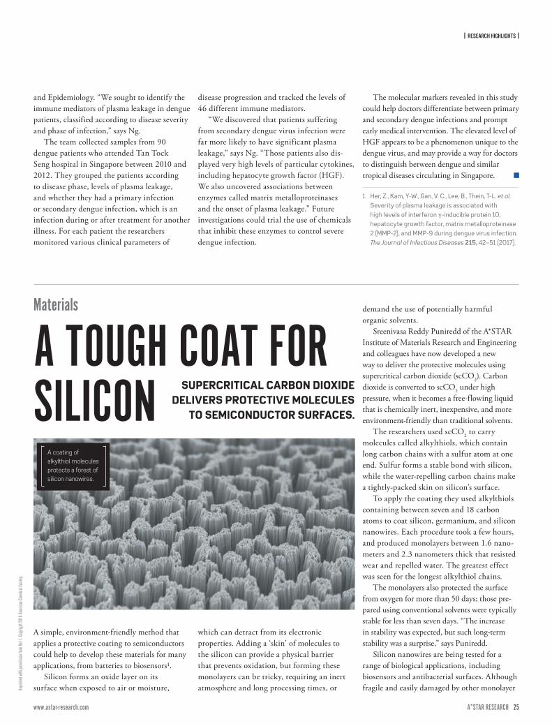
25 A tough coat for silicon
26 A new spin on data storage
27 Immunity barriers stay strong
28 Strength in diversity
29 Supporting bone repair
30 Liver cells work better under pressure
3 1 Optimizing drill design when the chips are down
32 MAGLEV FOR MANUFACTURING
A platform that floats
33 RESEARCH HIGHLIGHTS
33 High-fidelity
34 Charting T cell territory
35 Damping gives a faster switch
36 An inflammatory gene
3 7 Counting microbes on a smartphone
38 Tiny additions yield stronger parts
39 Making wires of polymers chains
40 New thought for food
4 1 Hydrogen freed by nanoparticle duo
4 2 More open to coercion
4 3 Growing blood in the lab
4 4 A human gene atlas for virus replication
4 5 A closer look at cell types
46 FEATURES & INNOVATIONS
The sugars we can’t live without
50 RESEARCH HIGHLIGHTS
50 A quick on/off switch for genes
5 1 Protein prompts a primordial proliferation
52 A signal of safe therapy
53 Better profiling our immune defenders
54 A crossroads for intersections
55 Enzymes engineered using direct evolution
56 Two-step security for the Internet of Things
57 New order of fish that can smell fear
58 $10 test detects TB in minutes
59 Probes lose their attachment issues
60 Keeping immune cells alive
6 1 A universal language for immunological sentinels
62 Reviving the dream of a universal autism drug
63 Altered metabolism extends beyond cancer cells
64 Drug discovery fast track
5838
50
| EDITORIAL |
PB ISSUE 7 | APRIL – JUNE 2017A*STAR RESEARCH www.astar-research.com 3 A*STAR RESEARCH
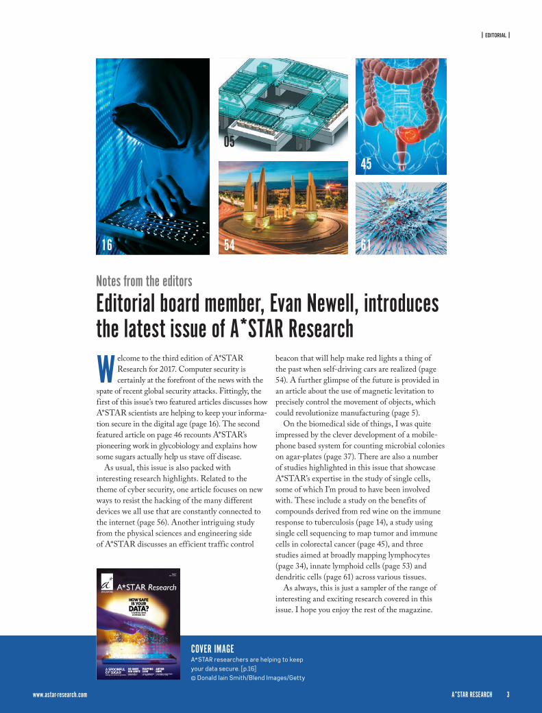
W elcome to the third edition of A*STAR Research for 2017. Computer security is certainly at the forefront of the news with the
spate of recent global security attacks. Fittingly, the first of this issue’s two featured articles discusses how A*STAR scientists are helping to keep your informa-tion secure in the digital age (page 16). The second featured article on page 46 recounts A*STAR’s pioneering work in glycobiology and explains how some sugars actually help us stave off disease.
As usual, this issue is also packed with interesting research highlights. Related to the theme of cyber security, one article focuses on new ways to resist the hacking of the many different devices we all use that are constantly connected to the internet (page 56). Another intriguing study from the physical sciences and engineering side of A*STAR discusses an efficient traffic control
beacon that will help make red lights a thing of the past when self-driving cars are realized (page 54). A further glimpse of the future is provided in an article about the use of magnetic levitation to precisely control the movement of objects, which could revolutionize manufacturing (page 5).
On the biomedical side of things, I was quite impressed by the clever development of a mobile-phone based system for counting microbial colonies on agar-plates (page 37). There are also a number of studies highlighted in this issue that showcase A*STAR’s expertise in the study of single cells, some of which I’m proud to have been involved with. These include a study on the benefits of compounds derived from red wine on the immune response to tuberculosis (page 14), a study using single cell sequencing to map tumor and immune cells in colorectal cancer (page 45), and three studies aimed at broadly mapping lymphocytes (page 34), innate lymphoid cells (page 53) and dendritic cells (page 61) across various tissues.
As always, this is just a sampler of the range of interesting and exciting research covered in this issue. I hope you enjoy the rest of the magazine.
Notes from the editors
Editorial board member, Evan Newell, introduces the latest issue of A*STAR Research
COVER IMAGEA*STAR researchers are helping to keep your data secure. [p.16]
© Donald Iain Smith/Blend Images/Getty
16
www.astar-research.com 3 A*STAR RESEARCH
54
05
61
45
4 ISSUE 7 | APRIL – JUNE 2017A*STAR RESEARCH www.astar-research.com 5 A*STAR RESEARCH
| RESEARCH HIGHLIGHTS |
© SC
IEPRO
/Scie
nce P
hoto
Librar
y/Ge
tty
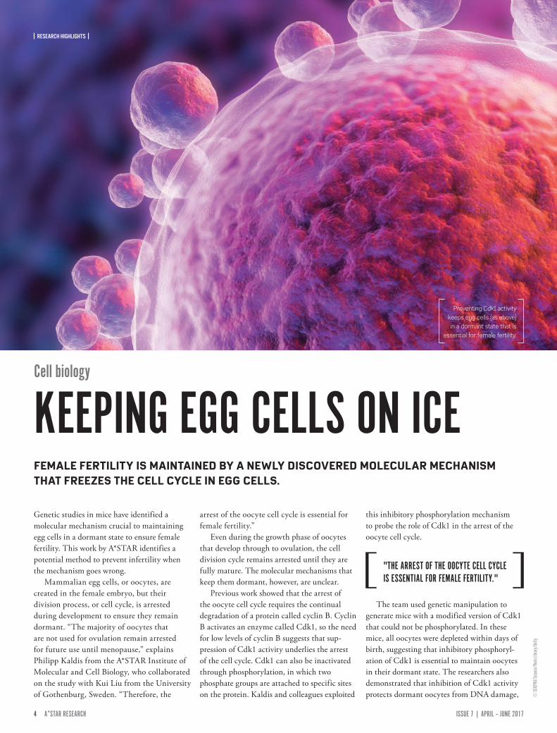
Genetic studies in mice have identified a molecular mechanism crucial to maintaining egg cells in a dormant state to ensure female fertility. This work by A*STAR identifies a potential method to prevent infertility when the mechanism goes wrong.
Mammalian egg cells, or oocytes, are created in the female embryo, but their division process, or cell cycle, is arrested during development to ensure they remain dormant. “The majority of oocytes that are not used for ovulation remain arrested for future use until menopause,” explains Philipp Kaldis from the A*STAR Institute of Molecular and Cell Biology, who collaborated on the study with Kui Liu from the University of Gothenburg, Sweden. “Therefore, the
arrest of the oocyte cell cycle is essential for female fertility.”
Even during the growth phase of oocytes that develop through to ovulation, the cell division cycle remains arrested until they are fully mature. The molecular mechanisms that keep them dormant, however, are unclear.
Previous work showed that the arrest of the oocyte cell cycle requires the continual degradation of a protein called cyclin B. Cyclin B activates an enzyme called Cdk1, so the need for low levels of cyclin B suggests that sup-pression of Cdk1 activity underlies the arrest of the cell cycle. Cdk1 can also be inactivated through phosphorylation, in which two phosphate groups are attached to specific sites on the protein. Kaldis and colleagues exploited
this inhibitory phosphorylation mechanism to probe the role of Cdk1 in the arrest of the oocyte cell cycle.
The team used genetic manipulation to generate mice with a modified version of Cdk1 that could not be phosphorylated. In these mice, all oocytes were depleted within days of birth, suggesting that inhibitory phosphoryl-ation of Cdk1 is essential to maintain oocytes in their dormant state. The researchers also demonstrated that inhibition of Cdk1 activity protects dormant oocytes from DNA damage,
Cell biology
KEEPING EGG CELLS ON ICEFEMALE FERTILITY IS MAINTAINED BY A NEWLY DISCOVERED MOLECULAR MECHANISM THAT FREEZES THE CELL CYCLE IN EGG CELLS.
Preventing Cdk1 activity keeps egg cells (as above) in a dormant state that is
essential for female fertility.
"THE ARREST OF THE OOCYTE CELL CYCLE IS ESSENTIAL FOR FEMALE FERTILITY."
| RESEARCH HIGHLIGHTS |
4 ISSUE 7 | APRIL – JUNE 2017A*STAR RESEARCH www.astar-research.com 5 A*STAR RESEARCH
© 20
17 A*
STAR
Sing
apore
Insti
tute o
f Man
ufactu
ring T
echn
ology
and that growing oocytes with active Cdk1 had DNA damage that caused them to die.
“Our findings show that inhibitory phos-phorylation of Cdk1 is important for preserving the oocyte pool and that prematurely activating Cdk1 leads to cell death and ultimately to female infertility,” explains Kaldis.
The insight also identifies a mechanism that could be targeted with drugs.
“Inhibitory phosphorylation of Cdk1 is controlled by the enzyme Wee1, the inhibiting of which could be achieved by a drug which is now in clinical trials,” says Kaldis. “One could envision that this drug
could be tried in cases where oocytes fail to arrest.”
1. Adhikari, D., Busayavalasa, K., Zhang, J., Hu, M., Risal, S. et al. Inhibitory phosphorylation of Cdk1 mediates prolonged prophase I arrest in female germ cells and is essential for female reproductive lifespan. Cell Research 26, 1212–1225 (2016).
Magnetic levitation
FLOATING FIELDS FOR FINE FABRICATION
Magnetic levitation (maglev) is well known for its use in high-speed rail net-works, but could also be applied at smaller scales in medicine and electronics. To do so, researchers must be able to precisely control electromagnetic fields so that they can move and rotate objects without touching them.
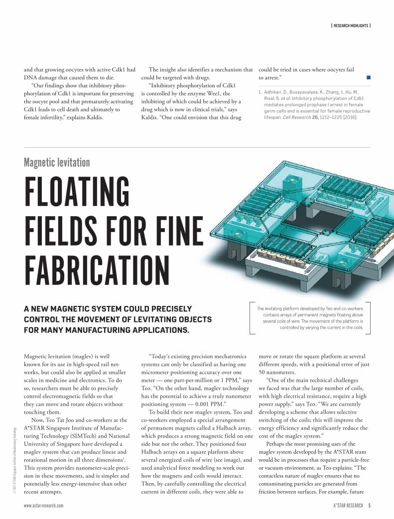
Now, Teo Tat Joo and co-workers at the A*STAR Singapore Institute of Manufac-turing Technology (SIMTech) and National University of Singapore have developed a maglev system that can produce linear and rotational motion in all three dimensions1. This system provides nanometer-scale preci-sion in these movements, and is simpler and potentially less energy-intensive than other recent attempts.
“Today’s existing precision mechatronics systems can only be classified as having one micrometer positioning accuracy over one meter — one part-per-million or 1 PPM,” says Teo. “On the other hand, maglev technology has the potential to achieve a truly nanometer positioning system — 0.001 PPM.”
To build their new maglev system, Teo and co-workers employed a special arrangement of permanent magnets called a Halbach array, which produces a strong magnetic field on one side but not the other. They positioned four Halbach arrays on a square platform above several energized coils of wire (see image), and used analytical force modeling to work out how the magnets and coils would interact. Then, by carefully controlling the electrical current in different coils, they were able to
move or rotate the square platform at several different speeds, with a positional error of just 50 nanometers.
“One of the main technical challenges we faced was that the large number of coils, with high electrical resistance, require a high power supply,” says Teo. “We are currently developing a scheme that allows selective switching of the coils; this will improve the energy efficiency and significantly reduce the cost of the maglev system.”
Perhaps the most promising uses of the maglev system developed by the A*STAR team would be in processes that require a particle-free or vacuum environment, as Teo explains: “The contactless nature of maglev ensures that no contaminating particles are generated from friction between surfaces. For example, future
The levitating platform developed by Teo and co-workers contains arrays of permanent magnets floating above several coils of wire. The movement of the platform is
controlled by varying the current in the coils.
A NEW MAGNETIC SYSTEM COULD PRECISELY CONTROL THE MOVEMENT OF LEVITATING OBJECTS FOR MANY MANUFACTURING APPLICATIONS.
| RESEARCH HIGHLIGHTS |
6 ISSUE 7 | APRIL – JUNE 2017A*STAR RESEARCH www.astar-research.com 7 A*STAR RESEARCH
Repro
duce
d from
Ref. 1
and l
icens
ed un
der C
C BY 4
.0 ©
2016
M. P
erez-C
amps
et al
.
wafer lithography processes such as extreme UV lithography, which operates in a vacuum, will require a maglev system to handle the wafer.”
Teo also suggests that maglev technology could replace conventional conveyor
belts in factories. Unlike traditional con-veyors that can only move objects on pre-defined tracks, maglev could move several objects simultaneously to different desired locations.
1. Zhu, H., Teo, T. J. & Pang, C. K. Design and modeling of a six-degree-of-freedom magnetically levitated positioner using square coils and 1-D Halbach arrays. IEEE Transactions on Industrial Electronics 64, 440–450 (2017).
A team from A*STAR’s Institute of Medical Biology and Institute of Molecular and Cell Biology in Singapore have shown how the gene-regulating proteins Pou5f3 and Nanog determine the organization of body structures in zebrafish embryos1. Their work shows how precise the orchestration of molecular events behind normal embryonic development is, and why it can easily go wrong.
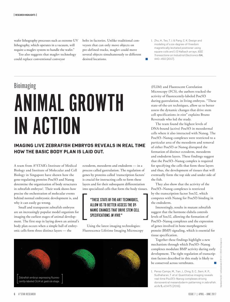
Small and transparent zebrafish embryos are an increasingly popular model organism for imaging the earliest stages of animal develop-ment. The first step in laying down an animal’s body plan occurs when a simple ball of embry-onic cells form three distinct layers — the
ectoderm, mesoderm and endoderm — in a process called gastrulation. The regulation of genes by proteins called ‘transcription factors’ is crucial for instructing cells to form these layers and for their subsequent differentiation into specialized cells that form the body tissues.
Using the latest imaging technologies: Fluorescence Lifetime Imaging Microscopy
(FLIM) and Fluorescent Correlation Microscopy (FCS), the authors tracked the activity of f luorescently-labeled Pou5f3 during gastrulation, in living embryos. “These state-of-the-art techniques, allow us to better assess the dynamic changes that drive stem cell specifications in vivo” explains Bruno Reversade who led the study.
The team found the highest levels of DNA-bound (active) Pou5f3 in mesodermal cells where it also interacted with Nanog. The Pou5f3–Nanog complexes were restricted to a particular area of the mesoderm and removal of either Pou5f3 or Nanog disrupted the formation of distinct ectoderm, mesoderm and endoderm layers. These findings suggest that the Pou5f3–Nanog complex is required for specifying the cells that form these layers and thus, the development of tissues that will eventually form the top side and under side of the fish.
They also show that the activity of the Pou5f3–Nanog complexes is restricted by the transcription factor Sox32, which competes with Nanog for Pou5f3 binding in the endoderm.
Interestingly, results in mutant zebrafish suggest that the hormone elabela controls levels of Sox32, allowing the formation of Pou5f3–Nanog complexes and the expression of genes involved in bone morphogenetic protein (BMP) signaling, which is essential for tissue specification.
Together these findings highlight a new mechanism through which Pou5f3–Nanog complexes modulate BMP activity during early development. The tight regulation of transcrip-tion factors described in this study is likely to be conserved across vertebrates.
1. Perez-Camps, M., Tian, J., Chng, S. C., Sem, K. P., Sudhaharan, T. et al. Quantitative imaging reveals real-time Pou5f3–Nanog complexes driving dorsoventral mesendoderm patterning in zebrafish. eLife 5, e11475 (2016).
Bioimaging
ANIMAL GROWTH IN ACTIONIMAGING LIVE ZEBRAFISH EMBRYOS REVEALS IN REAL TIME HOW THE BASIC BODY PLAN IS LAID OUT.
"THESE STATE-OF-THE-ART TECHNIQUES, ALLOW US TO BETTER ASSESS THE DY-NAMIC CHANGES THAT DRIVE STEM CELL SPECIFICATIONS IN VIVO."
Zebrafish embryo expressing fluores-cently-labeled Oct4 at gastrula stage.
| RESEARCH HIGHLIGHTS |
6 ISSUE 7 | APRIL – JUNE 2017A*STAR RESEARCH www.astar-research.com 7 A*STAR RESEARCH
© M
ARK G
ARLIC
K/SC
IENCE
PHOT
O LIBR
ARY/
Getty
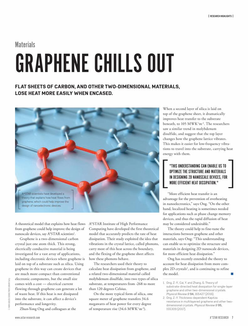
A theoretical model that explains how heat flows from graphene could help improve the design of nanoscale devices, say A*STAR scientists1.
Graphene is a two-dimensional carbon crystal just one atom thick. This strong, electrically conductive material is being investigated for a vast array of applications, including electronic devices where graphene is laid on top of a substrate such as silica. Using graphene in this way can create devices that are much more compact than conventional electronic components, but the small size comes with a cost — electrical current f lowing through graphene can generate a lot of waste heat. If this heat is not dissipated into the substrate, it can affect a device’s performance and longevity.
Zhun-Yong Ong and colleagues at the
A*STAR Institute of High Performance Computing have developed the first theoretical model that accurately predicts the rate of heat dissipation. Their study exploited the idea that vibrations in the crystal lattice, called phonons, carry most of this heat across the boundary, and the flexing of the graphene sheet affects how these phonons behave.
The researchers used their theory to calculate heat dissipation from graphene, and a related two-dimensional material called molybdenum disulfide, into two types of silica substrate, at temperatures from -268 to more than 120 degrees Celsius.
On the more typical form of silica, one square meter of graphene transfers 34.6 megawatts of heat power for every degree of temperature rise (34.6 MWK-1m-2).
When a second layer of silica is laid on top of the graphene sheet, it dramatically improves heat transfer to the substrate beneath, to 105 MWK-1m-2. The researchers saw a similar trend in molybdenum disulfide, and suggest that the top layer changes how the graphene lattice vibrates. This makes it easier for low-frequency vibra-tions to travel into the substrate, carrying heat energy with them.
“More efficient heat transfer is an advantage for the prevention of overheating in nanoelectronics,” says Ong. “On the other hand, localized heating is sometimes needed for applications such as phase change memory devices, and thus the rapid diffusion of heat may be considered undesirable.”
The theory could help to fine-tune the interactions between graphene and other materials, says Ong: “This understanding can enable us to optimize the structure and materials in designing 2D nanoscale devices, for more efficient heat dissipation.”
Ong has recently extended the theory to account for heat dissipation from more com-plex 2D crystals2, and is continuing to refine the model.
1. Ong, Z.-Y., Cai, Y. and Zhang, G. Theory of substrate-directed heat dissipation for single-layer graphene and other two-dimensional crystals. Physical Review B 94, 165427 (2016).
2. Ong, Z.-Y. Thickness-dependent Kapitza resistance in multilayered graphene and other two-dimensional crystals. Physical Review B 95, 155309 (2017).
"THIS UNDERSTANDING CAN ENABLE US TO OPTIMIZE THE STRUCTURE AND MATERIALS IN DESIGNING 2D NANOSCALE DEVICES, FOR MORE EFFICIENT HEAT DISSIPATION."
Materials
GRAPHENE CHILLS OUTFLAT SHEETS OF CARBON, AND OTHER TWO-DIMENSIONAL MATERIALS, LOSE HEAT MORE EASILY WHEN ENCASED.
A*STAR scientists have developed a theory that explains how heat flows from graphene, which could help improve the design of nanoelectronic devices.
| RESEARCH HIGHLIGHTS |
8 ISSUE 7 | APRIL – JUNE 2017A*STAR RESEARCH www.astar-research.com 9 A*STAR RESEARCH
Imag
e cred
it: Ph
ilipp K
eller,
Bill L
emon
, Yina
n Wan
and K
ristin
Bran
son,
Janeli
a Farm
Rese
arch C
ampu
s, Ho
ward
Hugh
es M
edica
l Insti
tute,
Ashb
urn, V
a.
Heart development
HOW THE HEART IS MADE
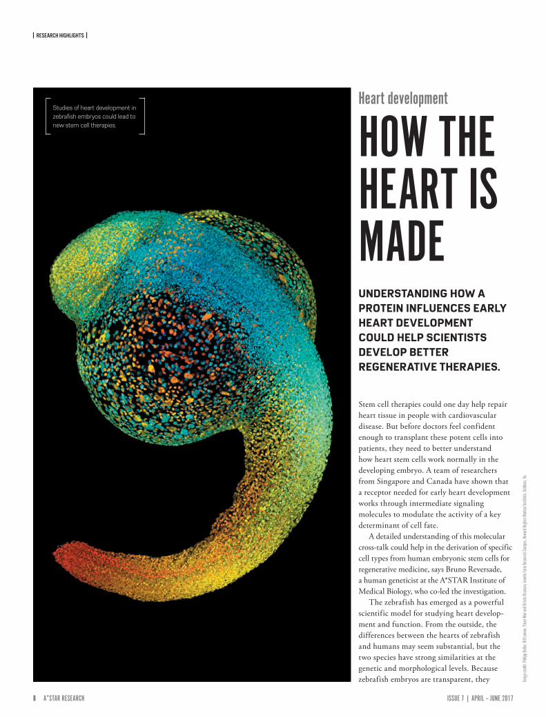
Stem cell therapies could one day help repair heart tissue in people with cardiovascular disease. But before doctors feel confident enough to transplant these potent cells into patients, they need to better understand how heart stem cells work normally in the developing embryo. A team of researchers from Singapore and Canada have shown that a receptor needed for early heart development works through intermediate signaling molecules to modulate the activity of a key determinant of cell fate.
A detailed understanding of this molecular cross-talk could help in the derivation of specific cell types from human embryonic stem cells for regenerative medicine, says Bruno Reversade, a human geneticist at the A*STAR Institute of Medical Biology, who co-led the investigation.
The zebrafish has emerged as a powerful scientific model for studying heart develop-ment and function. From the outside, the differences between the hearts of zebrafish and humans may seem substantial, but the two species have strong similarities at the genetic and morphological levels. Because zebrafish embryos are transparent, they
Studies of heart development in zebrafish embryos could lead to new stem cell therapies.
UNDERSTANDING HOW A PROTEIN INFLUENCES EARLY HEART DEVELOPMENT COULD HELP SCIENTISTS DEVELOP BETTER REGENERATIVE THERAPIES.
| RESEARCH HIGHLIGHTS |
8 ISSUE 7 | APRIL – JUNE 2017A*STAR RESEARCH www.astar-research.com 9 A*STAR RESEARCH
Repro
duce
d from
Ref. 1
and l
icens
ed un
der C
C BY 4
.0 ©
2016
G. Al
agap
pan &
C. E.
Png
provide a handy system for watching heart development in action.
Reversade teamed up with Ian Scott from the University of Toronto to examine the link between zebrafish born with no heart due to a mutation in a gene that encodes a cell-surface G protein-coupled receptor called the Apelin receptor. They knew that these fish had defec-tive heart stem cells, but it wasn’t clear why.
The researchers looked into the expression of genes targeted by another protein called Nodal, a known master regulator of cell fate. They found that zebrafish without a working Apelin
receptor had lower levels of Nodal target gene expression at the stage of embryonic formation when Nodal activity would normally induce heart stem cells to form. However, experimen-tally elevating levels of the Apelin receptor increased the expression of these same targets. What’s more, directly boosting Nodal activity in zebrafish that lacked the Apelin receptor was sufficient to help them develop beating hearts.
The Apelin receptor and Nodal don’t seem to be working in the same cells, though. As Reversade and Scott showed, the Apelin receptor modulated Nodal signaling through
two Nodal ligands, called Squint and Cyclops. Thus, the Apelin receptor seems to serve as kind of a distant control knob for fine-tuning the Nodal pathway during the earliest stages of heart development.
Future research will help determine how turning the knob on the expression level of the Apelin receptor can aid human patients with congenital heart diseases.
1. Deshwar, A. R., Chng, S. C., Ho, L., Reversade, B. & Scott, I. C. The Apelin receptor enhances Nodal/TGFβ signaling to ensure proper cardiac development. eLife 5, e13758 (2016).
Brighter LEDs and more efficient solar cells are two potential applications for A*STAR's research into lattice structures that can slow or trap light.
Harnessing wave energy by localizing it and suppressing its propagation through a medium is a powerful technique. Now, Gandhi Alagappan and Ching Eng Jason Png from the A*STAR Institute of High Performance Com-puting have calculated a design that localizes light in tiny loops, within a two-dimensional
structure created by merging two lattices of slightly differing periodicities1.
The new technique is not limited to light, and may enable the design of systems that can precisely control wave energy in any realm and at any scale — sound, thermal, water, or even matter waves such as in Bose-Einstein condensates.
For light-based devices the new insights could be used to build more efficient photonic components, said Alagappan.
“If you pattern the surface of an LED with merged lattices it will assist with getting the light out efficiently,” said Alagappan. “For a solar cell, however merged lattices will help light to enter better so that more energy can be harvested.”
The ability to create resonators in which light is localized on the surface of a device also has applications in quantum computing components based on light, such as defects in diamond.
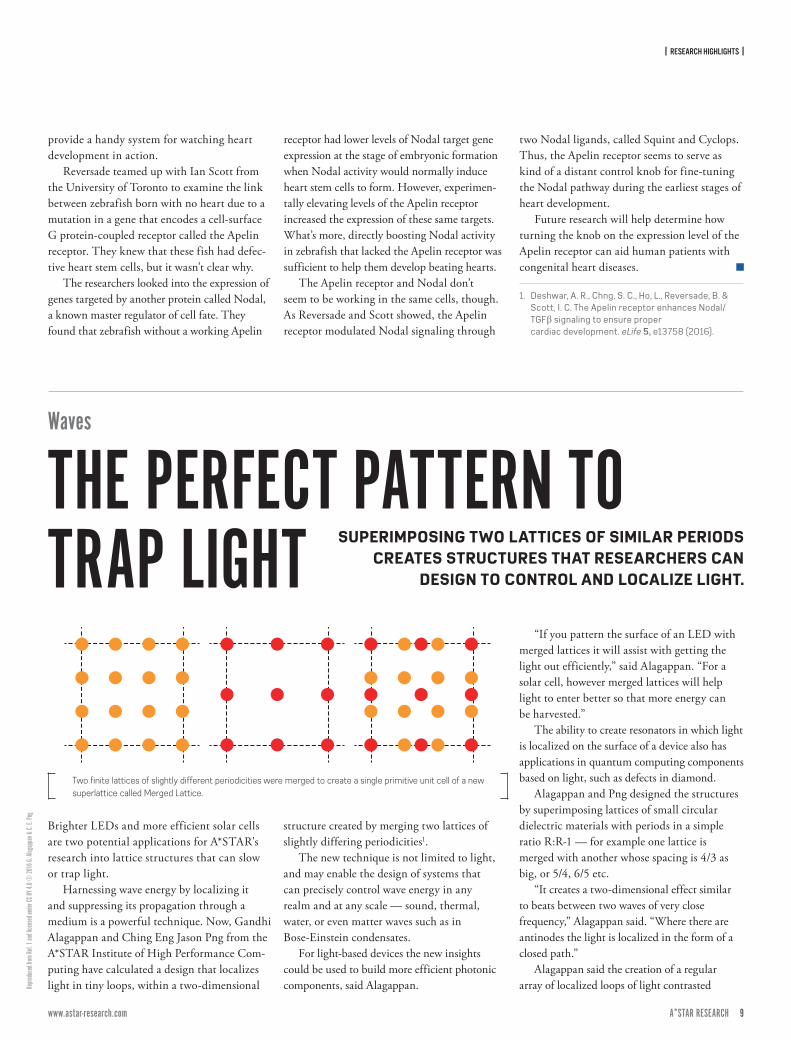
Alagappan and Png designed the structures by superimposing lattices of small circular dielectric materials with periods in a simple ratio R:R-1 — for example one lattice is merged with another whose spacing is 4/3 as big, or 5/4, 6/5 etc.
“It creates a two-dimensional effect similar to beats between two waves of very close frequency,” Alagappan said. “Where there are antinodes the light is localized in the form of a closed path.”
Alagappan said the creation of a regular array of localized loops of light contrasted
Two finite lattices of slightly different periodicities were merged to create a single primitive unit cell of a new superlattice called Merged Lattice.
Waves
THE PERFECT PATTERN TO TRAP LIGHT SUPERIMPOSING TWO LATTICES OF SIMILAR PERIODS
CREATES STRUCTURES THAT RESEARCHERS CAN DESIGN TO CONTROL AND LOCALIZE LIGHT.
| RESEARCH HIGHLIGHTS |
10 ISSUE 7 | APRIL – JUNE 2017A*STAR RESEARCH www.astar-research.com 11 A*STAR RESEARCH
© 20
17 A*
STAR
Data
Storag
e Ins
titute
with Anderson Localization, which arises from randomness in a structure. “This is a systematic way of creating a large number of loops,” Alagappan said.
Alagappan and Png ran numerical simulations of the propagation of light in a
range of wavelengths slightly below that of the lattice spacing, and calculated the energy band structure. They found that as R increased, there emerged a large number of energy bands whose light had a group velocity of zero, the signature of light localized within the crystal.
Alagappan said merged lattices would also provide a way for researchers to explore topolog-ical properties, such as protected edge modes.
1. Alagappan, G, & Png, C. E. Localization of waves in merged lattices. Scientific Reports 6, 31620 (2016).
Producing semiconductor lasers on a silicon wafer is a long-held goal for the electronics industry, but their fabrication has proved challenging. Now, researchers at A*STAR have developed an innovative way to manufacture them that is cheap, simple and scalable1.
Hybrid silicon lasers combine the light-emit-ting properties of group III–V semiconductors — alloys containing elements from group III and group V on the periodic table — like gallium arsenide and indium phosphide, with the matu-rity of silicon manufacturing techniques. These lasers are attracting considerable attention as they promise inexpensive, mass-producible optical devices that can integrate with photonic and microelectronic elements on a single silicon chip. They have potential in a wide range of applica-tions, from short-distance data communication to high-speed, long-distance optical transmission.
In the current production process, however, lasers are fabricated on separate III–V semicon-ductor wafers before being individually aligned to each silicon device — a time-consuming,
costly process that limits the number of lasers that can be placed on a chip.
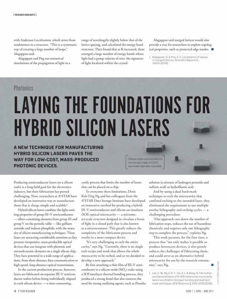
To overcome these limitations, Doris Keh-Ting Ng and her colleagues from the A*STAR Data Storage Institute have developed an innovative method for producing a hybrid III–V semiconductor and silicon-on-insulator (SOI) optical microcavity — a microme-ter-scale structure designed to circulate a beam of light in a closed path that is also known as a microresonator. This greatly reduces the complexity of the fabrication process and results in a more compact device.
“It’s very challenging to etch the entire cavity,” says Ng. “Currently, there is no single etch recipe and mask that allows the whole microcavity to be etched, and so we decided to develop a new approach.”
By first attaching a thin film of III–V semi-conductor to a silicon oxide (SiO2) wafer using a SOI interlayer thermal bonding process, they produced a strong bond that also removes the need for strong oxidizing agents, such as Piranha
solution (a mixture of hydrogen peroxide and sulfuric acid) or hydrofluoric acid.
And by using a dual hard-mask technique to etch the microcavity that confined etching to the intended layer, they eliminated the requirement to use multiple overlay lithography and etching cycles — a challenging procedure.
“Our approach cuts down the number of fabrication steps, reduces the use of hazardous chemicals, and requires only one lithography step to complete the process,” explains Ng.
This work presents, for the first time, a process that “not only makes it possible to produce heterocore devices, it also greatly reduces the challenges of fabricating them, and could serve as an alternative hybrid microcavity for use by the research commu-nity,” says Ng.
1. Lee, C.-W., Ng, D. K.-T., Tan, A. L. & Wang, Q. Fabrication and demonstration of III−V/Si heterocore microcavity lasers via ultrathin interlayer bonding and dual hard mask techniques. ACS Photonics 3, 2191–2196 (2016).
Photonics
LAYING THE FOUNDATIONS FOR HYBRID SILICON LASERSA NEW TECHNIQUE FOR MANUFACTURING HYBRID SILICON LASERS PAVES THE WAY FOR LOW-COST, MASS-PRODUCED PHOTONIC DEVICES.
Oblique angle scanning electron microscopy image of a 500 nanometer diameter microdisk.

| RESEARCH HIGHLIGHTS |
10 ISSUE 7 | APRIL – JUNE 2017A*STAR RESEARCH www.astar-research.com 11 A*STAR RESEARCH
© Av
alon_
Studio
/E+/
Getty
In the search for corrosion-resistant treatments for carbon steel that are non-toxic, A*STAR researchers have developed a technique for investi-gating the effectiveness of a corrosion inhibitor that is safer and environmentally friendly.1
Carbon steel, an alloy made from iron and carbon, is the single largest class of alloys in use today. It’s used to make a range of products from fences and springs to steel wires and pipelines, and for structural support in
buildings, bridges, as well as nuclear power and fossil fuel power plants.
The corrosion of carbon steel, however, is a huge cost to industry and is of enormous practical importance. One common corrosion inhibitor used in the construction industry, cal-cium nitrite, is quite toxic to humans, impairing the ability of red blood cells to transport oxygen.
Seeking safer corrosion inhibitors, Yong Teck Tan and colleagues from the National
University of Singapore and Singapore Institute of Manufacturing Technology investigated molybdate as a potential alternative and devel-oped a technique to determine its suitability.
Molybdate is non-toxic, and protects the carbon steel from corrosion by competitive adsorption against chloride on the passive film surface, and, in the presence of calcium cations, can also deposit a layer of calcium molybdate.
“Our aim was to first determine the suitability of molybdate as a corrosion inhibitor for carbon steel in alkaline environments, and then to investigate its effect on the passivation of carbon steel,” says Tan. Passivation refers to the coating of a surface with a material to make it less chemically reactive.
“Previous studies using electrochemical techniques have focused on corrosion inhi-bition efficiency at a particular time, which provides a snapshot of the level of corrosion at that instant,” explains Tan. “Depending on whether it was assessed over short or long timescales, different conclusions were drawn.”
So the research team took a longer look. They used an electrochemical method for estimating the extent of corrosion over the entire duration of the investigation, and could assess the overall effectiveness of molybdate.
“Even though molybdate resulted in a slightly higher passive current in the later stages, faster passivation in the early stages resulted in a lower overall level of corrosion,” says Tan.
The researchers found that incomplete coverage of the carbon steel by the cal-cium molybdate led to slightly higher corrosion rates compared with untreated surfaces. By controlling the composition of the molybdate solution, however, the calcium molybdate film covered the entire surface, resulting in improved corrosion resistance.
“Overall, molybdate proved to be an effective corrosion inhibitor,” says Tan. “We will now explore its effectiveness in solutions containing other ions.”
1. Tan, Y. T., Wijesinghe, S. L. & Blackwood, D. J. Effect of molybdate on the passivation of carbon steel in alkaline solutions under open-circuit conditions. Journal of The Electrochemical Society 163, C649–C658 (2016).
"OVERALL, MOLYBDATE PROVED TO BE AN EFFECTIVE CORROSION INHIBITOR."
Materials
LOOKING FOR SAFER CORROSION TREATMENTSA NEW TECHNIQUE FOR INVESTIGATING THE ACTION OF MOLYBDATE ON CARBON STEEL COULD LEAD TO SAFER TREATMENTS FOR PROTECTING METAL ALLOYS.
A*STAR researchers have developed a method to analyse corrosion inhibitors for carbon steel that may one day be used for reinforcement bars in the construction industry.
12 ISSUE 7 | APRIL – JUNE 2017A*STAR RESEARCH www.astar-research.com 13 A*STAR RESEARCH
© Po
bytov
/Digi
talVis
ion Ve
ctors/
Getty
| RESEARCH HIGHLIGHTS |
Most methods for the structural characteri-zation of biomolecules, such as X-ray crystal-lography or electron microscopy, require static or crystallized samples. Attaching fluorescent molecules to protein surfaces, however, enables direct imaging of dynamic biomolecular inter-actions using light. This could be improved, say A*STAR researchers, with predictive modeling of fluorescence lifetimes1.
Fluorescence normally involves single molecules that spontaneously absorb light and then re-emit it as a different color. But under
the right conditions, an absorbed photon can hop from a donor molecule to a nearby acceptor compound that also fluoresces. Researchers have recently exploited the fact that this effect is strongly dependent on distance to produce ‘spectroscopic rulers’ that measure the nanoscale dynamics between donor and acceptor probes attached to different parts of a protein backbone.
A key challenge is to make spectroscopic rulers with acceptable accuracy. Conventional fluorophores have large, flexible structures that press against proteins in multiple ways,
making it tricky to gauge the ruler’s length. So to seek alternatives, Tsz Sian Chwee and colleagues from the A*STAR Institute of High Performance Computing investigated whether they could calculate the fluorescence of stiff and small molecules known as syn-bimanes, and then use such theories for probe design.
Typical quantum chemistry approaches, how-ever, have trouble computing properties when a molecule absorbs a photon and enters an excited state. Chwee and his team hoped to overcome these inaccuracies using time-dependent density
Fluorescent probes
MAKING BRIGHTER PROTEIN PREDICTIONSSUPERCOMPUTER SIMULATIONS SHORTEN DEVELOPMENT TIME OF RIGID FLUORESCENT MOLECULES USED TO CLARIFY PROTEIN STRUCTURE AND DYNAMICS.
Computer models that describe how the vibrations of fluorescent probes influence light absorption and emission could lead to more accurate protein measurements.
| RESEARCH HIGHLIGHTS |
12 ISSUE 7 | APRIL – JUNE 2017A*STAR RESEARCH www.astar-research.com 13 A*STAR RESEARCH
Repro
duce
d from
Ref. 1
and l
icens
ed un
der C
C BY 4
.0 ©
2016
Y. S.
Tan e
t al.
functional theory that treats the problem of excited electrons with an ‘exchange–correlation’ algorithm derived partly from experiments.
“Time-dependent density functional theory is used by the scientific community to study phenomenon such as absorption and emission, but the full potential of this approach hasn’t been harnessed yet,” says Chwee.
Using fluorescence lifetimes as a test param-eter, the researchers compared how different exchange–correlation theories simulated syn-bimanes in realistic, solvent-filled situations.
These trials revealed that models incorporating vibronic interactions — the synchronized coupling of molecular vibrations to electronic excitations — provided the most accurate predictions of fluorescent lifetimes. They dis-covered several exchange–correlation functions that are capable of handling these equations at minimal computational cost.“Vibronic aspects have largely been overlooked, even though they play decisive roles in the photophysics of fluorescent molecules,” notes Chwee. “While we carried out our calculations on supercomputers,
the computational resources are modest enough they could have been completed on a modern workstation in a couple of weeks.”
Chwee anticipates that rapid analysis using density functional theories might be better at spotting rare f luorescent probe candidates with strong absorption and tunable emission properties.
1. Wong, Z. C., Fan, W. Y., Chwee, T. S. & Sullivan, M. B. Modelling fluorescence lifetimes with TD-DFT: A case study with syn-bimanes. RSC Advances 6, 87237–87245 (2016).
Drug design
GETTING BINDING POCKETS OUT OF HIDING BENZENE-BASED PROBES HIGHLIGHT TWO HIDDEN
BINDING SITES ON AN ANTICANCER DRUG TARGET IN A MULTIDISCIPLINARY STRATEGY.
In the quest for new cancer therapies, A*STAR researchers have devised a computational strategy that unearths any previously unknown binding sites or ‘pockets’ on drug targets.¹
More effective cancer treatments are likely to emerge from the drug development pipe-line. Cancer drug discovery hinges on identi-fying and characterizing binding pockets in target proteins. Typically, this evaluation uses computational techniques that rely on static
protein structures. However, proteins have an inherent f lexibility that causes a tendency to change shape upon contact with the drugs. Certain binding pockets remain undetectable unless they interact with an appropriate substance and, therefore, are missed by conventional simulations. These hidden pockets, however, are usually water-repelling, or hydrophobic, sites that only open when there are low polarity substances.
To tackle this, Yaw Sing Tan and Chandra Verma from the Bioinformatics Institute have developed a probe-based method called ligand-mapping molecular dynamics (LMMD). They used this technique to seek hidden binding pockets in the anticancer target protein MDM2. The resulting predictions were experimentally validated by long-standing collaborators from A*STAR's p53 Laboratory and Institute of Chemical and Engineering Sciences as well as structural biologists from Newcastle University, UK.
Tan explains that initially he had designed this probe-based method for another target protein and successfully used it to find a hidden binding pocket that stayed closed in conventional simulations. “We then decided to apply this approach to MDM2 to see if we could discover any previously unknown binding sites that could enhance the potency of existing MDM2 inhibitors,” he adds.
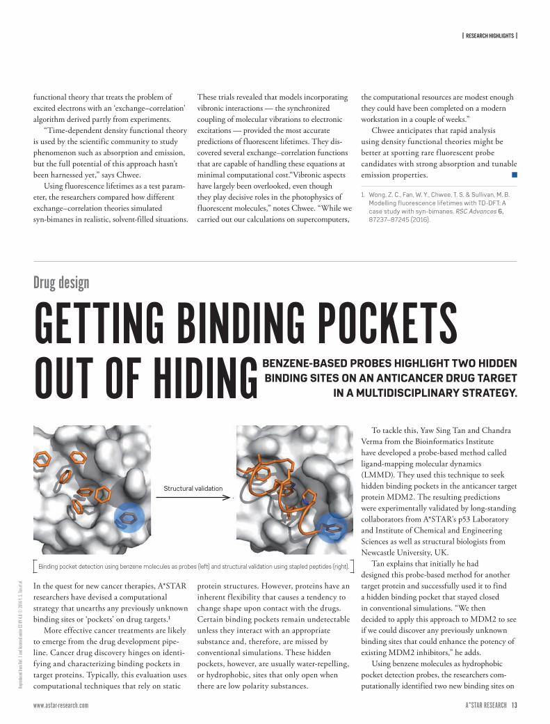
Using benzene molecules as hydrophobic pocket detection probes, the researchers com-putationally identified two new binding sites on
Structural validation
Binding pocket detection using benzene molecules as probes (left) and structural validation using stapled peptides (right).
| RESEARCH HIGHLIGHTS |
14 ISSUE 7 | APRIL – JUNE 2017A*STAR RESEARCH www.astar-research.com 15 A*STAR RESEARCH
© KA
TERY
NA KO
N/SC
IENCE
PHOT
O LIBR
ARY/
Getty
and ©
bert_
phan
tana/
iStoc
k/Ge
tty
MDM2. “We were excited to see that these sites lie very close to the binding pocket of the tumor suppressor protein p53,” says Tan.
Furthermore, the researchers expect the newly found sites to lead to more potent stapled peptides — these are amino acid helices chemically stabilized by a hydrocarbon chain that have recently emerged as powerful p53 activators. Consequently, they created
stapled peptides from analogs known to tightly bind MDM2 and reactivate p53, and determined the affinity of these peptides to MDM2. Their simulations showed that the peptides bound MDM2 more strongly than p53 in the pockets and matched biophysical and X-ray crystallography experiments.
“This method could be used to interrogate other anticancer protein targets to uncover
novel binding sites that could be targeted for inhibition,” says Tan. The team is now working to expand the reach of LMMD probes to other ligand types.
1. Tan, Y. S., Reeks, J., Brown, C. J., Thean, D., Gago, F. J. F. et al. Benzene probes in molecular dynamics simulations reveal novel binding sites for ligand design. The Journal of Physical Chemistry Letters 7, 3452–3457(2016).
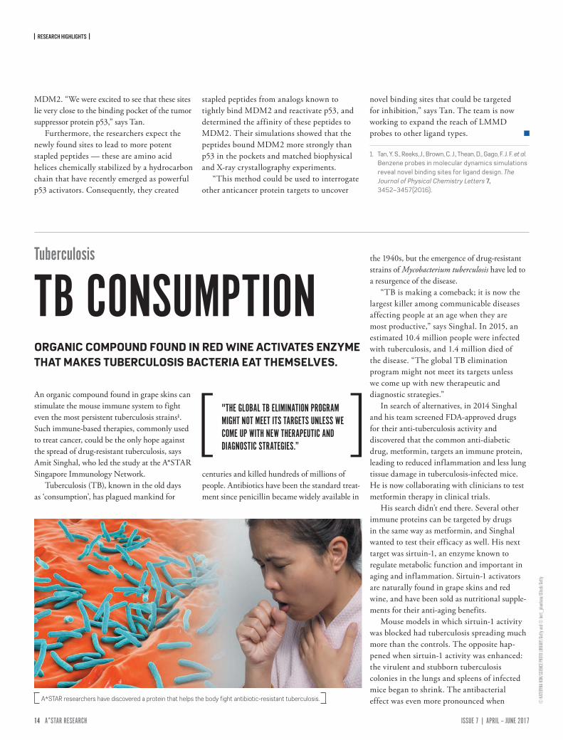
An organic compound found in grape skins can stimulate the mouse immune system to fight even the most persistent tuberculosis strains1. Such immune-based therapies, commonly used to treat cancer, could be the only hope against the spread of drug-resistant tuberculosis, says Amit Singhal, who led the study at the A*STAR Singapore Immunology Network.
Tuberculosis (TB), known in the old days as ‘consumption’, has plagued mankind for
centuries and killed hundreds of millions of people. Antibiotics have been the standard treat-ment since penicillin became widely available in
the 1940s, but the emergence of drug-resistant strains of Mycobacterium tuberculosis have led to a resurgence of the disease.
“TB is making a comeback; it is now the largest killer among communicable diseases affecting people at an age when they are most productive,” says Singhal. In 2015, an estimated 10.4 million people were infected with tuberculosis, and 1.4 million died of the disease. “The global TB elimination program might not meet its targets unless we come up with new therapeutic and diagnostic strategies.”
In search of alternatives, in 2014 Singhal and his team screened FDA-approved drugs for their anti-tuberculosis activity and discovered that the common anti-diabetic drug, metformin, targets an immune protein, leading to reduced inflammation and less lung tissue damage in tuberculosis-infected mice. He is now collaborating with clinicians to test metformin therapy in clinical trials.
His search didn’t end there. Several other immune proteins can be targeted by drugs in the same way as metformin, and Singhal wanted to test their efficacy as well. His next target was sirtuin-1, an enzyme known to regulate metabolic function and important in aging and inflammation. Sirtuin-1 activators are naturally found in grape skins and red wine, and have been sold as nutritional supple-ments for their anti-aging benefits.
Mouse models in which sirtuin-1 activity was blocked had tuberculosis spreading much more than the controls. The opposite hap-pened when sirtuin-1 activity was enhanced: the virulent and stubborn tuberculosis colonies in the lungs and spleens of infected mice began to shrink. The antibacterial effect was even more pronounced when A*STAR researchers have discovered a protein that helps the body fight antibiotic-resistant tuberculosis.
Tuberculosis
TB CONSUMPTIONORGANIC COMPOUND FOUND IN RED WINE ACTIVATES ENZYME THAT MAKES TUBERCULOSIS BACTERIA EAT THEMSELVES.
"THE GLOBAL TB ELIMINATION PROGRAM MIGHT NOT MEET ITS TARGETS UNLESS WE COME UP WITH NEW THERAPEUTIC AND DIAGNOSTIC STRATEGIES.”
| RESEARCH HIGHLIGHTS |
14 ISSUE 7 | APRIL – JUNE 2017A*STAR RESEARCH www.astar-research.com 15 A*STAR RESEARCH
© SC
IEPRO
/Scie
nce P
hoto
Librar
y/Ge
tty
sirtuin-1 treatment was combined with a standard antibiotic.
Closer examination of the lung tissue revealed less damage and inflammation under sirtuin-1 enhancement, as compared with untreated controls. Gene expression analysis found that the enzyme worked by inducing the
tuberculosis bacteria to devour themselves, a process known as autophagy.
Singhal is now testing sirtuinin-1 activators on monkey models of tuberculosis. He is also looking into whether they can be combined with metformin for a more powerful therapy.
“We now have two candidates to further
expand our studies and we may even find something else.”
1. Cheng, C. Y., Gutierrez, N. M., Marzuki, M. B., Lu, X., Foreman, T. W., Paleja, B. et al. Host sirtuin 1 regulates mycobacterial immunopathogenesis and represents a therapeutic target against tuberculosis. Science Immunology 2, eaaj1789 (2017).
Infertility
ATTACHMENTS THAT PUSH THE ENVELOPEDETAILED UNDERSTANDING OF THE CELL DIVISIONS THAT GIVE RISE TO SPERM AND EGGS COULD LEAD TO INFERTILITY TREATMENTS.
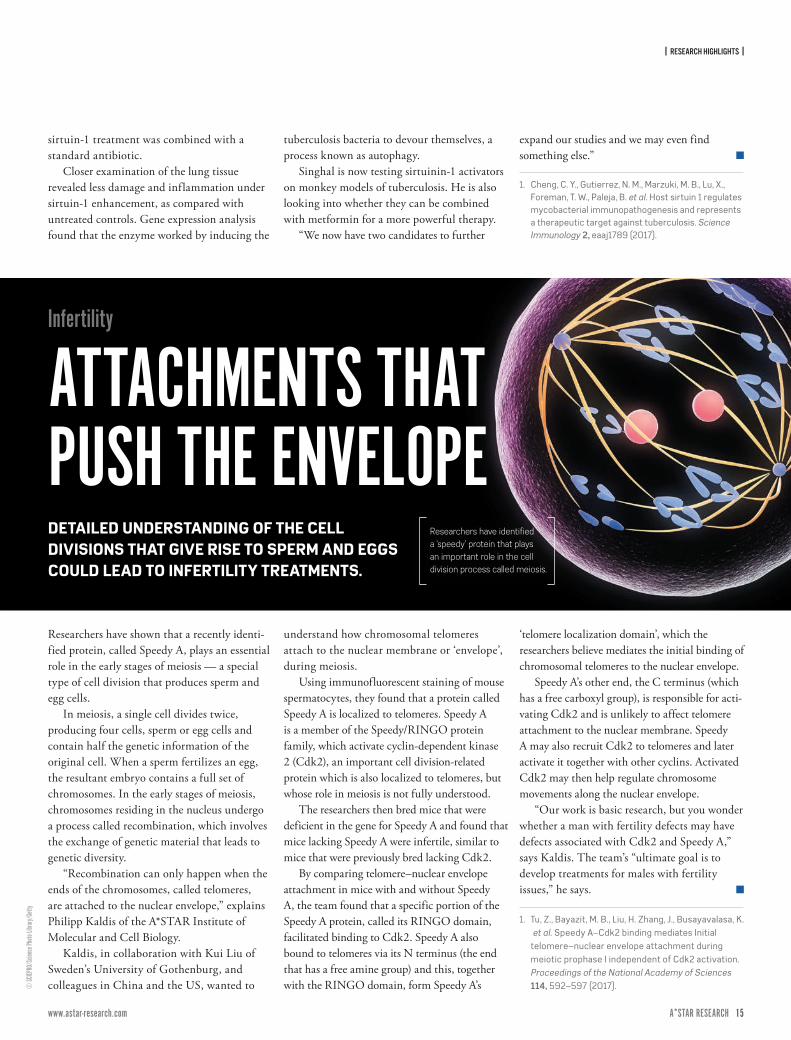
Researchers have shown that a recently identi-fied protein, called Speedy A, plays an essential role in the early stages of meiosis — a special type of cell division that produces sperm and egg cells.
In meiosis, a single cell divides twice, producing four cells, sperm or egg cells and contain half the genetic information of the original cell. When a sperm fertilizes an egg, the resultant embryo contains a full set of chromosomes. In the early stages of meiosis, chromosomes residing in the nucleus undergo a process called recombination, which involves the exchange of genetic material that leads to genetic diversity.
“Recombination can only happen when the ends of the chromosomes, called telomeres, are attached to the nuclear envelope,” explains Philipp Kaldis of the A*STAR Institute of Molecular and Cell Biology.
Kaldis, in collaboration with Kui Liu of Sweden’s University of Gothenburg, and colleagues in China and the US, wanted to
understand how chromosomal telomeres attach to the nuclear membrane or ‘envelope’, during meiosis.
Using immunofluorescent staining of mouse spermatocytes, they found that a protein called Speedy A is localized to telomeres. Speedy A is a member of the Speedy/RINGO protein family, which activate cyclin-dependent kinase 2 (Cdk2), an important cell division-related protein which is also localized to telomeres, but whose role in meiosis is not fully understood.
The researchers then bred mice that were deficient in the gene for Speedy A and found that mice lacking Speedy A were infertile, similar to mice that were previously bred lacking Cdk2.
By comparing telomere–nuclear envelope attachment in mice with and without Speedy A, the team found that a specific portion of the Speedy A protein, called its RINGO domain, facilitated binding to Cdk2. Speedy A also bound to telomeres via its N terminus (the end that has a free amine group) and this, together with the RINGO domain, form Speedy A’s
‘telomere localization domain’, which the researchers believe mediates the initial binding of chromosomal telomeres to the nuclear envelope.
Speedy A’s other end, the C terminus (which has a free carboxyl group), is responsible for acti-vating Cdk2 and is unlikely to affect telomere attachment to the nuclear membrane. Speedy A may also recruit Cdk2 to telomeres and later activate it together with other cyclins. Activated Cdk2 may then help regulate chromosome movements along the nuclear envelope.
“Our work is basic research, but you wonder whether a man with fertility defects may have defects associated with Cdk2 and Speedy A,” says Kaldis. The team’s “ultimate goal is to develop treatments for males with fertility issues,” he says.
1. Tu, Z., Bayazit, M. B., Liu, H. Zhang, J., Busayavalasa, K. et al. Speedy A–Cdk2 binding mediates Initial telomere–nuclear envelope attachment during meiotic prophase I independent of Cdk2 activation. Proceedings of the National Academy of Sciences 114, 592–597 (2017).
Researchers have identified a ‘speedy’ protein that plays an important role in the cell division process called meiosis.

| FEATURES & INNOVATIONS |
16 ISSUE 7 | APRIL – JUNE 2017A*STAR RESEARCH www.astar-research.com 17 A*STAR RESEARCH
HOW SAFEIS YOUR DATA?
A diverse group of researchers is leading efforts to ensure that digital information remains secure, however and wherever it is used.
16 ISSUE 7 | APRIL – JUNE 2017A*STAR RESEARCH www.astar-research.com 17 A*STAR RESEARCH
| FEATURES & INNOVATIONS |
© Bi
ll Hint
on/M
omen
t/Gett
y
Every day we store and transfer sensitive digital data, post personal infor-mation on social media, and provide valuable
details to companies when we use their services. Keeping secure the 2.5 quintillion (2.5 million billion) bytes of data created every day from outside attack is a mammoth task. The potential for breaching security is vast, due to a plethora of available services and the many weak links that appear in the chain whenever data is moved. A further consideration is who should have access to data, taking the issue beyond technology into the social and political realm.
These challenges demand a huge global effort from computer technicians and researchers across the world. Research groups at A*STAR are using their technical expertise to monitor online services, identify vulnerable areas of data management, and develop software and hardware that keep data secure. Their work is not only defending data against attack, but also maintaining easy access to it for authorized users.
MANAGING MOBILESArguably, the first line of defense against data misuse should be implemented in the Global System for Mobile Communications (GSM), the world’s most widely-used wireless telephony technology. With a 90 per cent share of the market, around 4.5 billion customers rely on the security of GSM to protect their communications.
“GSM was first deployed 25 years ago and has become the global standard for mobile communications,” says Jiqiang Lu at the A*STAR Institute for Infocomm Research (I2R).
The A5/1 stream cipher, the encryption scheme that GSM uses to protect data, has been
successfully attacked before to test its security, but almost all the attacks were hypothetical in the sense of their impact on the real-world security of GSM — they either required a large amount of complex data or had a long attack time, meaning they could be mitigated and blocked by existing GSM security protocols. Lu and co-workers decided to investigate whether a detailed and fast-acting attack on the GSM A5/1 cipher could reveal fundamental weaknesses in the system. Using a computer setup costing just US$15,000 in 2013, the researchers employed a powerful algorithm to explore the A5/1 cryptosystem, and obtained 984 gigabytes of information about the system structure over 55 days. They used this information to launch attacks that pulled data from the GSM in just 9 seconds — usually too quick for interception by security protocols — and illustrated that A5/1 would be vulnerable if it were to be attacked by sufficiently skilled hackers.
“The GSM should immediately upgrade its encryption algorithm to a stronger one,” says Lu.
CONTAINING THE CLOUDWhile Lu’s team continue to protect our data as it flies around the global mobile network, another group at I2R which includes researcher Jia Xu is examining the cloud storage providers that have revolutionized how we archive and share data. By entrusting large organizations to store multiple copies of our data on cloud servers around the world, we are freed from worrying about our phone, laptop or USB drive being lost, stolen or broken. But how can we be sure that these organizations will keep our data secure?
Xu and co-workers have designed cryptographic algo-rithms for cloud storage that
| FEATURES & INNOVATIONS |
18 ISSUE 7 | APRIL – JUNE 2017A*STAR RESEARCH www.astar-research.com 19 A*STAR RESEARCH
not only protect the integrity of data, but also control who can access it.
“The core challenge in cloud storage is to balance three factors: efficiency, security, and usability,” says Xu. “Cloud providers would like their services to be almost as efficient and low-cost as when no security features are implemented, while customers want the user interface to be as simple to use as possible.” The research com-munity is attempting to identify security vulnerabilities in existing cloud services, and to design new hardware and software solutions to resolve them.
Some security weaknesses arise from so-called deduplication tech-niques, which identify and remove duplicated copies of the same file, allowing cloud providers such as
Dropbox to save server storage space and network bandwidth. Xu and co-workers identified severe security vulnerabilities in certain types of deduplication that could be exploited using attacking software.
Dropbox disabled cross-user deduplication in 2012. However, the new algorithms developed by Xu and the team will allow deduplication to be used alongside robust encryption, thereby improving efficiency while pro-tecting data stored in the cloud.
THE VALUE OF OUR DATAMost of us have made large amounts of information available to organizations through shopping online and posting on social media. These activities have created extremely large datasets,
“The ‘big’ keep accumulatingmore and more data about the ‘small’.”
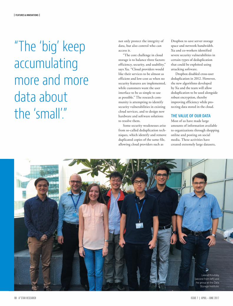
Leonid Krivitsky (second from left) and
his group at the Data Storage Institute.
| FEATURES & INNOVATIONS |
18 ISSUE 7 | APRIL – JUNE 2017A*STAR RESEARCH www.astar-research.com 19 A*STAR RESEARCH
known as Big Data, which can be analyzed to reveal human behavior patterns and trends. This valuable information is often sold to other organizations.
“Companies are hungry for more data, to enable them to better understand and profile users,” says Lux Anantharaman who heads the Business Analytics Translation center in I2R. “They know the power of Big Data to provide tar-geted ads, known as personalized marketing, but profiling can also lead to price discrimination called personalized pricing, which most users are not aware of. For example, some airline websites price tickets differently based on the user’s device operating system — Mac OS users get charged more.”
Anantharaman is concerned that most users are not aware of the value of their data, or the fact that when they use ‘free’ online services they are actually ‘paying’ for them with their data. Companies then explore the data with analytical computing tools and use the information, along with the latest insights on human behavior from social scientists and economists, to shape the choices offered to their customers.
“The ‘big’ keep accumulating more and more data about the ‘small’,” says Anantharaman. “We, the small, are slowly becoming aware of this fact, but generally we feel helpless and resigned about it. Moreover, government regulations haven’t kept pace with technology, and often take the side of big organizations, doing a disservice to the users. For example, recent US government measures allow internet service providers to access a user’s browsing history without the user’s permission.”
Anantharaman is adamant that the best way to overcome these difficulties is by educating users and improving government regulation. “This might sound
odd coming from a technology person, but Big Data is not just about technology, it is about how data are used, which is a legal and social issue,” he says. “For this reason, our research focuses not just on technological mechanisms, but also explores how regulations and education can help users better understand the power and pitfalls of Big Data privacy.”
QUANTUM COMPLICATIONSWhile we grapple with data safety in the computing systems that we already use, other scientists are developing technology that could completely transform the field of data security for the devices of tomorrow. In contrast to ordinary computers whose logical ‘bits’ can only take values of 0 or 1, quantum computers use ‘qubits’ that can have values of 0, 1 or a combination of both values. This capability opens up an entirely new domain of logic and mathematics, allowing quantum computers to solve complex problems in a fraction of the time it would take a conventional machine. This revolution will arrive with great benefits, but will bring its own problems, as Leonid Krivitsky at the A*STAR Data Storage Institute explains:
“Many cryptography systems rely on hard problems such as prime factorization — the fact that it is very difficult to figure out the prime factors of a given number. However, theoretical work has shown that the factorization problem could be solved very quickly using a quantum computer. So, once a universal quantum computer is built, it could hack ciphers which were previously thought to be unbreakable.”
This might seem alarming, but there is no reason to panic. Functional quantum computers are still a long way off, and to counteract the potential threats,
many groups around the world are contributing to the growing field of quantum cryptography, which will redefine our protocols of secure communication. In fact, the new cryptography algorithms made available by quantum computers could provide ultra-high data security long before any risks become a concern.
“I foresee the use of a quantum communication channel as a backup resource for highly sensitive transactions, where security is more important than the transmission speed,” says Krivitsky.
For now, though, the chal-lenge is to physically build a stable quantum computer. Krivitsky and co-workers are exploring the possibility of using tiny defects in synthetic diamonds to act as nodes which process and store quantum information.
“We place several diamonds on a single chip and communicate with optical links, similar to those which form the background of the internet,” says Krivitsky. “Our innovations will enable transmis-sion of quantum information over long distances and contribute to the development of a worldwide quantum network.”
SAFEGUARDING THE FUTUREThe task of keeping our data secure is clearly a complicated and inter-disciplinary challenge. A*STAR researchers are not only developing new technical initiatives, but also working at the forefront of global efforts to raise awareness of data security. By looking for chinks in the armor of global systems like GSM and cloud storage, educating the public about the commercial value of their data, and planning for the future paradigm shift that might be brought about by quantum computers, it is reas-suring to know that the brightest minds at A*STAR are focused on keeping our data safe.
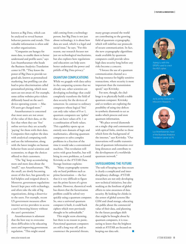
Jiqiang Lu with the GPGPU used to crack the A5/1 cipher.
Jia Xu at the Institute for Infocomm Research.
Lux Anantharaman, head of the Business Analytics Translation center at I2R.
© 20
17 A*
STAR
Data
Storag
e Ins
titute
© 20
17 A*
STAR
Insti
tute f
or Inf
ocom
m Re
searc
h
| RESEARCH HIGHLIGHTS |
20 ISSUE 7 | APRIL – JUNE 2017A*STAR RESEARCH www.astar-research.com 21 A*STAR RESEARCH
© M
onty
Raku
sen/
Cultu
ra/Ge
tty
Severe illnesses sometimes require treatment regimens carrying grave risks, including organ failure. Now, a non-invasive technique developed at A*STAR could help predict patient vulnerability to potentially toxic drugs.
Therapeutics can induce organ damage via mechanisms that vary between individuals. These idiosyncratic drug reactions are a common reason for the withdrawal of new drugs, and can be a significant problem during disease treatment.
Research led by Min-Han Tan and Hanry Yu from the Institute of Bioengineering and Nanotechnology, and National Cancer Centre shows how cells derived from a patient’s blood offer the first opportunity to test an individu-al’s susceptibility to idiosyncratic liver damage,
known as hepatotoxicity; in this case, from the cancer drug, pazopanib.
Currently there is no easy way to predict idio-syncratic harm from the drug, “Pazopanib causes idiosyncratic hepatotoxicity, and liver biopsies are not commonly undertaken due to their invasive nature and potential risks,” says Tan.
The researchers took white blood cells from five patients receiving pazopanib for metastatic renal cell cancer, three of whom exhibited hepatotoxicity. They converted these
white blood cells into stem cells, and then into ‘hepatocyte-like cells’ (HLCs). This created a population of cells that retained the genetics and morphology of each patient’s native liver cells, without the risks of a biopsy. The stem cells were then treated with pazopanib.
After 24 hours, the HLCs taken from the three patients exhibiting hepatotoxicity also experienced significantly more cell death than those from the two patients without liver damage. This validated that the test can model the patient-specific effects of pazopanib on the liver.
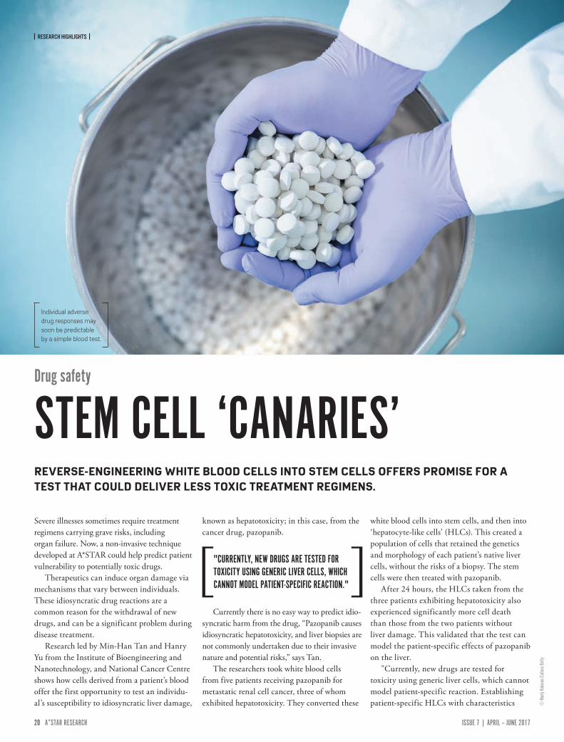
"Currently, new drugs are tested for toxicity using generic liver cells, which cannot model patient-specific reaction. Establishing patient-specific HLCs with characteristics
Drug safety
STEM CELL ‘CANARIES’REVERSE-ENGINEERING WHITE BLOOD CELLS INTO STEM CELLS OFFERS PROMISE FOR A TEST THAT COULD DELIVER LESS TOXIC TREATMENT REGIMENS.
Individual adverse drug responses may soon be predictable by a simple blood test.
"CURRENTLY, NEW DRUGS ARE TESTED FOR TOXICITY USING GENERIC LIVER CELLS, WHICH CANNOT MODEL PATIENT-SPECIFIC REACTION."
| RESEARCH HIGHLIGHTS |
20 ISSUE 7 | APRIL – JUNE 2017A*STAR RESEARCH www.astar-research.com 21 A*STAR RESEARCH
Repro
duce
d from
Ref. 1
and l
icens
ed un
der C
C BY 4
.0 (h
ttps:/
/crea
tivec
ommo
ns.or
g/lic
ense
s/by
/4.0/
) © 20
16 J.
Carle
varo-
Fita e
t al.
that are representative of genetic variation will be valuable for pharmaceutical drug testing,” says Yu.
The team also discovered the mechanism by which pazopanib causes injury by evaluating the changes in HLC gene expression following drug administration. In cells from both groups of patients, gene expression changes indicated a response to drug-induced stress. HLCs from hepatotoxicity-susceptible individuals, however,
also showed evidence of differential iron metabolism as well as other genetic variations from non-susceptible HLCs. This probably contributes to the greater levels of cellular damage and death and provides the first exper-imental evidence of pazopanib’s mechanism of action in idiosyncratic hepatotoxicity.
Tan hopes his team’s research could be used in future to predict an individual’s response to a proposed treatment. “We plan to expand the
approach to different drugs and organs, and determine the nature of drug toxicity,” explains Tan. “Our ultimate goal is to benefit patients and clinicians by gaining a better understanding of toxicity.”
1. Choudhury, Y., Toh, Y. C., Xing, J., Qu, Y., Poh, J. et al. Patient-specific hepatocyte-like cells derived from induced pluripotent stem cells model pazopanib-mediated hepatotoxicity. Scientific Reports 7, 41238 (2017).
A large number of long noncoding RNAs (lncRNA) have been found associating with the ribosome, the protein-making machinery in the cytoplasm. What the so-called ‘noncoding’ RNAs are doing on the ribosome, whose main job is to translate RNA into protein, has puzzled the A*STAR researchers who discovered them.
The answer could be macabre, suggest cell biologist, Leah Vardy, at the A*STAR Institute
of Medical Biology and bioinformatician, Rory Johnson, at the University of Bern, Switzerland. Translation by the ribosome could lead to the degradation of these lncRNAs, says Vardy. “The ribosome may be acting as a garbage dump or graveyard for lncRNAs at the end of their life.”
In recent years, researchers have found that many RNAs previously labeled as junk, actually play an important role in causing a wide range of
diseases, from cancers to metabolic disease and neurodegeneration. Still, many questions remain about their basic biology, with studies covering less than one per cent of the tens of thousands of lncRNAs in our genomes. “LncRNAs are one of the most promising avenues for understanding disease today,” says Johnson.
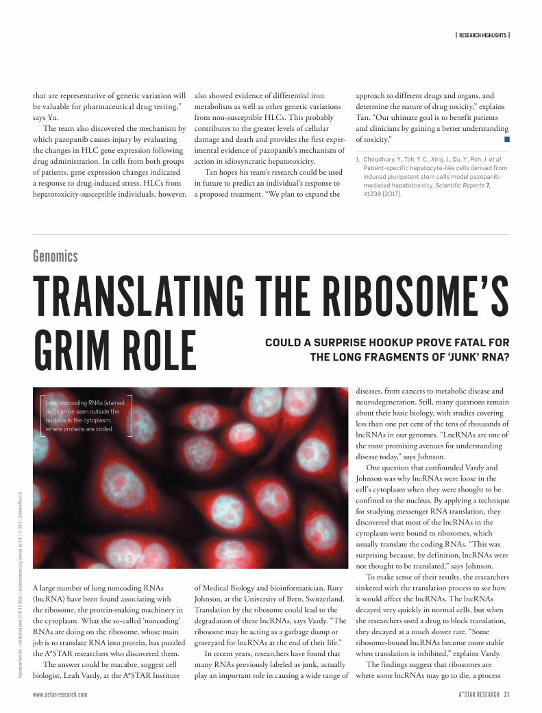
One question that confounded Vardy and Johnson was why lncRNAs were loose in the cell's cytoplasm when they were thought to be confined to the nucleus. By applying a technique for studying messenger RNA translation, they discovered that most of the lncRNAs in the cytoplasm were bound to ribosomes, which usually translate the coding RNAs. “This was surprising because, by definition, lncRNAs were not thought to be translated,” says Johnson.
To make sense of their results, the researchers tinkered with the translation process to see how it would affect the lncRNAs. The lncRNAs decayed very quickly in normal cells, but when the researchers used a drug to block translation, they decayed at a much slower rate. “Some ribosome-bound lncRNAs become more stable when translation is inhibited,” explains Vardy.
The findings suggest that ribosomes are where some lncRNAs may go to die, a process
Long noncoding RNAs (stained red) can be seen outside the nucleus in the cytoplasm, where proteins are coded.
Genomics
TRANSLATING THE RIBOSOME’S GRIM ROLE COULD A SURPRISE HOOKUP PROVE FATAL FOR
THE LONG FRAGMENTS OF ‘JUNK’ RNA?
| RESEARCH HIGHLIGHTS |
22 ISSUE 7 | APRIL – JUNE 2017A*STAR RESEARCH www.astar-research.com 23 A*STAR RESEARCH
Modifi
ed fr
om Re
f. 1 an
d lice
nsed
unde
r CC B
Y 4.0
(http
s://c
reativ
ecom
mons
.org/
licen
ses/
by/4
.0/) ©
2017
U. S.
Dinis
h et a
l.
similar to the well-known mechanism of ‘nonsense mediated decay’, in which ribosomes promote the degradation of malformed messenger RNAs. The researchers found that ribosome-bound lncRNAs look more like messenger RNA than their free-floating counterparts in the cytoplasm. “In some ways,
lncRNAs look like malformed mRNAs, and for this reason may enter the nonsense mediated decay pathway,” explains Johnson.
To confirm their hypotheses about the fatal affair between noncoding RNAs and protein-coding ribosomes, Vardy and Johnson plan to study the relationship at an individual
level to understand how they are regulated and whether they perform both coding and non-coding functions.
1. Carlevaro-Fita, J., Rahim, A., Guigó, R., Vardy, L. A. & Johnson, R. Cytoplasmic long noncoding RNAs are frequently bound to and degraded at ribosomes in human cells. RNA 22, 867–882 (2016).
A technique that uses light imaging to monitor whether one type of fat tissue has converted to another has been employed by A*STAR researchers to better understand conditions such as diabetes and obesity. The technique could underpin a fast and cost-effective approach to monitoring this conversion.
The two main types of adipose tissue have very different properties: white (WAT) stores excess energy and its accumulation is linked to obesity, while brown (BAT) has immense energy burning capabilities. Some WAT can be converted to BAT-like tissue by a process known as browning which, in rodents at least, has anti-obesity and anti-diabetic functions.
Current methods for monitoring browning are time-consuming and crude. Two teams at the A*STAR Singapore Bioimaging Consortium led by Shigeki Sugii and Malini Olivo collaborated to improve the process.
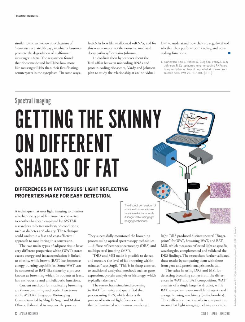
They successfully monitored the browning process using optical spectroscopy techniques — diffuse reflectance spectroscopy (DRS) and multispectral imaging (MSI).
“DRS and MSI made it possible to detect and measure the level of fat browning within minutes,” says Sugii. “This is in sharp contrast to traditional analytical methods such as gene expression, protein analysis or histology, which typically take days.”
The researchers stimulated browning in WAT from mice and quantified the process using DRS, which detects the pattern of scattered light from a sample that is illuminated with narrow wavelength
light. DRS produced distinct spectral “finger-prints” for WAT, browning WAT, and BAT. MSI, which measures reflected light at specific wavelengths, complemented and validated the DRS findings. The researchers further validated these results by comparing them with those from gene and protein analysis methods.
The value in using DRS and MSI for detecting browning comes from the differ-ences in WAT and BAT composition. WAT consists of a single large fat droplet, while BAT comprises many small fat droplets and energy burning machinery (mitochondria). This difference, particularly in composition, means that light imaging techniques can be
The distinct composition of white and brown adipose tissues make them easily distinguishable using light imaging techniques.
DIFFERENCES IN FAT TISSUES’ LIGHT REFLECTING PROPERTIES MAKE FOR EASY DETECTION.
Spectral imaging
GETTING THE SKINNY ON DIFFERENT SHADES OF FAT
| RESEARCH HIGHLIGHTS |
22 ISSUE 7 | APRIL – JUNE 2017A*STAR RESEARCH www.astar-research.com 23 A*STAR RESEARCH
© SC
IEPRO
/Scie
nce P
hoto
Librar
y/Ge
tty
used to tell them apart. “Optical spectroscopy can fingerprint the intrinsic differences in tissue optical characteristics, thereby differ-entiating between classical brown and white fat tissue, and white fat during the browning process,” explains Olivo.
The researchers anticipate that these tech-niques can be developed for studying browning in live animals and humans. “These techniques will facilitate the study of associations between obesity and fat browning capacities, and the potential development of therapeutic
approaches to enhance browning for tackling obesity,” says Sugii.
1. Dinish, U. S., Wong, C. L., Sriram, S., Ong, W. K., Balasundaram, G. et al. Diffuse optical spectroscopy and imaging to detect and quantify adipose tissue browning. Scientific Reports 7, 41357 (2017).
Liver cancer
TUMOR DIVERSITY COULD LEAD TO BESPOKE TREATMENTSINSIGHTS INTO THE GROWTH OF LIVER CANCER TUMORS AND THEIR GENETIC DIVERSITY COULD INFORM FUTURE PERSONALIZED CANCER THERAPIES.
Liver cancer tumors are genetically diverse and therefore difficult to treat, a Singapo-rean research team reports1. The genetic differences found in each patient may one day enable personal, targeted therapies to treat the disease.
Hepatocellular carcinoma (HCC), the most common type of liver cancer, remains the second most common cause of cancer-related death worldwide. This is partly because HCC tumors are often not discovered until they reach an advanced stage, and treatments for the disease are limited because scientists do not fully understand how the cancer evolves.
“One problem with developing effective cancer treatments is that tumors change and spread very rapidly and resistance to treatment evolves quickly; this is certainly the case for liver cancer,” says Weiwei Zhai of the A*STAR Genome Institute of Singapore, who led the research with Roger Foo, also of A*STAR, and Pierce Chow of the National Cancer Centre.
“Determining the level of heterogeneity — or genetic diversity — within each tumor is key to understanding tumor evolution, disease progression and treatment response.”
Zhai’s team and colleagues used next-generation DNA sequencing technology to analyze 66 tumor samples from nine HCC patients whose cancers stemmed from different causes. The samples were taken from multiple sites within each tumor, and from different tumors within each liver — this differs from current HCC-diagnosis biopsies, which only take a single sample from one part of one tumor.
“We discovered that there is considerable genetic diversity both within and between tumors in a single individual,” says Zhai. “The spatial growth pattern of the tumors was rather like the growth rings inside a tree trunk, suggesting HCC tumors grow by expanding outwards in a sequence. This is very different from what researchers have found in colorectal cancers, for instance. The next challenge is to
consider how to target this spatial pattern in future treatments.”
The high tumor heterogeneity helps explain why targeting HCC tumors with a single drug has had limited success. The team’s next challenge is to unravel the natural history of tumor evolution and build a full picture of disease progression, from primary tumor surgery to patient relapse.
“We have received a NMRC Translation and Clinical Research grant to conduct the most comprehensive and methodical study of HCC to date,” says Zhai. “Led by Pierre Chow, the project will be multidisciplinary with researchers from A*STAR, the National Cancer Center of Singapore, SingHealth Translational Immunology and Inflammation Centre, and the Cancer Science Institute of Singapore.”
1. Zhai, W., Lim, T. K-H., Zhang, T., Phang, S-T., Tiang, Z. et al. The spatial organization of intra-tumour heterogeneity and evolutionary trajectories of metastases in hepatocellular carcinoma. Nature Communications 8, 4565 (2017).

| RESEARCH HIGHLIGHTS |
24 ISSUE 7 | APRIL – JUNE 2017A*STAR RESEARCH www.astar-research.com 25 A*STAR RESEARCH
© SC
IEPRO
/Scie
nce P
hoto
Librar
y/Ge
tty
Dengue virus
DISEASE SEVERITY FORECAST
Dengue virus infection threatens more than half of the world’s population. With millions of cases each year, scientists are working hard to fully understand the disease and bring it under control. Now, A*STAR researchers have uncov-ered several molecular markers whose levels are elevated during dengue infection and provide a measure of the severity of the disease.
While the majority of dengue virus infections result in a mild, self-limiting fever, some cases develop into the more severe, life-threatening dengue shock syndrome. The key process that determines disease severity is plasma leakage — the amount of blood leaking from capillaries. During the first week of infection, scientists believe that the cell dysfunction inside blood vessels, coupled with increasing levels of small proteins called cytokines, combine to increase the permeability of the vascular system, resulting in varying degrees of plasma leakage.
“Dengue fever has been a problem in Singapore for more than three decades, but in recent years more severe forms have been on the rise, such as plasma leakage and hemorrhagic shock,” explains Lisa Ng from the Singapore Immunology Network. Ng co-led the project together with Yee-Sin Leo and clinicians at the Institute of Infectious Diseases
A*STAR researchers have uncov-ered several molecular markers that could help doctors identify patients at risk of severe plasma leakage during dengue virus infection.
INDICATOR PROTEINS COULD HELP IDENTIFY PATIENTS AT RISK OF COMPLICATIONS DURING DENGUE FEVER INFECTION.
| RESEARCH HIGHLIGHTS |
24 ISSUE 7 | APRIL – JUNE 2017A*STAR RESEARCH www.astar-research.com 25 A*STAR RESEARCH
Repri
nted w
ith pe
rmiss
ion fr
om Re
f 1. C
opyri
ght 2
016 A
meric
an Ch
emica
l Soc
iety.
and Epidemiology. “We sought to identify the immune mediators of plasma leakage in dengue patients, classified according to disease severity and phase of infection,” says Ng.
The team collected samples from 90 dengue patients who attended Tan Tock Seng hospital in Singapore between 2010 and 2012. They grouped the patients according to disease phase, levels of plasma leakage, and whether they had a primary infection or secondary dengue infection, which is an infection during or after treatment for another illness. For each patient the researchers monitored various clinical parameters of
disease progression and tracked the levels of 46 different immune mediators.
“We discovered that patients suffering from secondary dengue virus infection were far more likely to have significant plasma leakage,” says Ng. “Those patients also dis-played very high levels of particular cytokines, including hepatocyte growth factor (HGF). We also uncovered associations between enzymes called matrix metalloproteinases and the onset of plasma leakage.” Future investigations could trial the use of chemicals that inhibit these enzymes to control severe dengue infection.
The molecular markers revealed in this study could help doctors differentiate between primary and secondary dengue infections and prompt early medical intervention. The elevated level of HGF appears to be a phenomenon unique to the dengue virus, and may provide a way for doctors to distinguish between dengue and similar tropical diseases circulating in Singapore.
1. Her, Z., Kam, Y-W., Gan, V. C., Lee, B., Thein, T-L. et al. Severity of plasma leakage is associated with high levels of interferon γ-inducible protein 10, hepatocyte growth factor, matrix metalloproteinase 2 (MMP-2), and MMP-9 during dengue virus infection. The Journal of Infectious Diseases 215, 42–51 (2017).
A simple, environment-friendly method that applies a protective coating to semiconductors could help to develop these materials for many applications, from batteries to biosensors1.
Silicon forms an oxide layer on its surface when exposed to air or moisture,
which can detract from its electronic properties. Adding a ‘skin’ of molecules to the silicon can provide a physical barrier that prevents oxidation, but forming these monolayers can be tricky, requiring an inert atmosphere and long processing times, or
demand the use of potentially harmful organic solvents.
Sreenivasa Reddy Puniredd of the A*STAR Institute of Materials Research and Engineering and colleagues have now developed a new way to deliver the protective molecules using supercritical carbon dioxide (scCO2). Carbon dioxide is converted to scCO2 under high pressure, when it becomes a free-flowing liquid that is chemically inert, inexpensive, and more environment-friendly than traditional solvents.
The researchers used scCO2 to carry molecules called alkylthiols, which contain long carbon chains with a sulfur atom at one end. Sulfur forms a stable bond with silicon, while the water-repelling carbon chains make a tightly-packed skin on silicon’s surface.
To apply the coating they used alkylthiols containing between seven and 18 carbon atoms to coat silicon, germanium, and silicon nanowires. Each procedure took a few hours, and produced monolayers between 1.6 nano-meters and 2.3 nanometers thick that resisted wear and repelled water. The greatest effect was seen for the longest alkylthiol chains.
The monolayers also protected the surface from oxygen for more than 50 days; those pre-pared using conventional solvents were typically stable for less than seven days. “The increase in stability was expected, but such long-term stability was a surprise,” says Puniredd.
Silicon nanowires are being tested for a range of biological applications, including biosensors and antibacterial surfaces. Although fragile and easily damaged by other monolayer
A coating of alkylthiol molecules protects a forest of silicon nanowires.
Materials
A TOUGH COAT FOR SILICON SUPERCRITICAL CARBON DIOXIDE
DELIVERS PROTECTIVE MOLECULES TO SEMICONDUCTOR SURFACES.
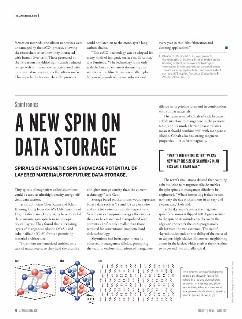
| RESEARCH HIGHLIGHTS |
26 ISSUE 7 | APRIL – JUNE 2017A*STAR RESEARCH www.astar-research.com 27 A*STAR RESEARCH
Repri
nted f
rom Re
f. 1, C
opyri
ght 2
017,
with
perm
ission
from
Else
vier.
formation methods, the silicon nanowires were undamaged by the scCO2 process, allowing the researchers to test how they interacted with human liver cells. Those protected by the 18-carbon alkylthiol significantly reduced cell growth on the nanowires, compared with unprotected nanowires or a flat silicon surface. This is probably because the cells’ proteins
could not latch on to the monolayer’s long carbon chains.
“This scCO2 technology can be adopted for many kinds of inorganic surface modification,” says Puniredd. “The technology is not only scalable, but also enhances the quality and stability of the film. It can potentially replace billions of pounds of organic solvents used
every year in thin-film fabrication and cleaning applications.”
1. Bhartia, B., Puniredd, S. R., Jayaraman, S., Gandhimathi, C., Sharma, M. et al. Highly stable bonding of thiol monolayers to hydrogen-terminated Si via supercritical carbon dioxide: Towards a super hydrophobic and bio-resistant surface. ACS Applied Materials & Interfaces 8, 24933–24945 (2016).
Tiny spirals of magnetism called skyrmions could be used as ultrahigh density energy-effi-cient data carriers.
Jarvis Loh, Gan Chee Kwan and Khoo Khoong Hong from the A*STAR Institute of High Performance Computing have modeled these minute spin spirals in nanoscopic crystal layers. They found that alternating layers of manganese silicide (MnSi) and cobalt silicide (CoSi) forms a promising material architecture.
“Skyrmions are nanosized entities, only tens of nanometers, so they hold the promise
of higher storage density than the current technology,” said Gan.
Storage based on skyrmions would represent binary data such as ‘1’s and ‘0’s as clockwise and anticlockwise spin spirals, respectively. Skyrmions can improve energy efficiency as they can be created and manipulated with currents significantly smaller than those required for conventional magnetic hard disk technology.
Skyrmions had been experimentally observed in manganese silicide, prompting the team to explore simulations of manganese
silicide in its pristine form and in combination with similar materials.
The team selected cobalt silicide because cobalt sits close to manganese in the periodic table, and its similar lattice characteristics mean it should combine well with manganese silicide. Cobalt also has strong magnetic properties — it is ferromagnetic.
The team’s simulations showed that coupling cobalt silicide to manganese silicide enables the spin spirals in manganese silicide to be engineered. “What’s interesting is that we can now vary the size of skyrmions in an easy and elegant way,” Loh said.
In the skyrmion’s center the magnetic spin of the atoms is flipped 180 degrees relative to the spin on its outside edge; between the edge and the center the spins progressively tilt between the two extremes. The size of skyrmions depends on the ability of the material to support high relative tilt between neighboring atoms in the lattice, which enables the skyrmion to be packed into a smaller spiral.
2
1 Along[111]1 2
(a) (c)(b)
Two different views of manganese silicide are shown in (a) and (b), where the red and blue spheres represent manganese and silicon respectively. A larger scale view of manganese silicide showing spiraling atomic spins is shown in (c).
Spintronics
A NEW SPIN ON DATA STORAGESPIRALS OF MAGNETIC SPIN SHOWCASE POTENTIAL OF LAYERED MATERIALS FOR FUTURE DATA STORAGE.
"WHAT’S INTERESTING IS THAT WE CAN NOW VARY THE SIZE OF SKYRMIONS IN AN EASY AND ELEGANT WAY."
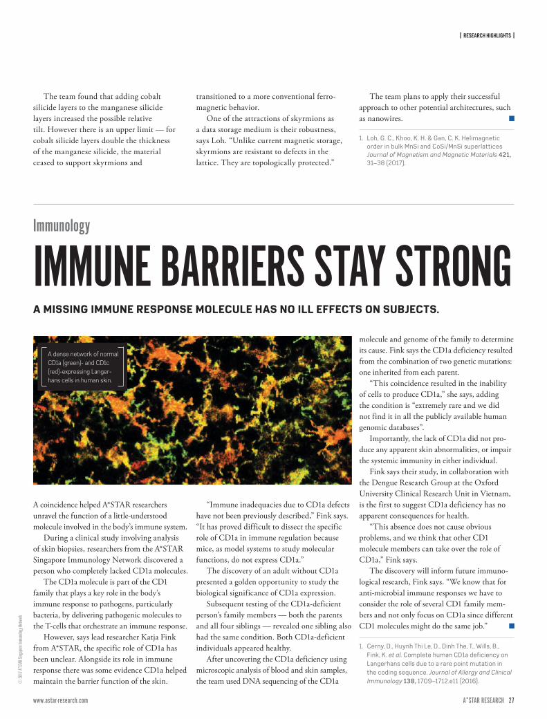
| RESEARCH HIGHLIGHTS |
26 ISSUE 7 | APRIL – JUNE 2017A*STAR RESEARCH www.astar-research.com 27 A*STAR RESEARCH
© 20
17 A*
STAR
Sing
apore
Immu
nolog
y Netw
ork
A coincidence helped A*STAR researchers unravel the function of a little-understood molecule involved in the body’s immune system.
During a clinical study involving analysis of skin biopsies, researchers from the A*STAR Singapore Immunology Network discovered a person who completely lacked CD1a molecules.
The CD1a molecule is part of the CD1 family that plays a key role in the body’s immune response to pathogens, particularly bacteria, by delivering pathogenic molecules to the T-cells that orchestrate an immune response.
However, says lead researcher Katja Fink from A*STAR, the specific role of CD1a has been unclear. Alongside its role in immune response there was some evidence CD1a helped maintain the barrier function of the skin.
“Immune inadequacies due to CD1a defects have not been previously described,” Fink says. “It has proved difficult to dissect the specific role of CD1a in immune regulation because mice, as model systems to study molecular functions, do not express CD1a.”
The discovery of an adult without CD1a presented a golden opportunity to study the biological significance of CD1a expression.
Subsequent testing of the CD1a-deficient person’s family members — both the parents and all four siblings — revealed one sibling also had the same condition. Both CD1a-deficient individuals appeared healthy.
After uncovering the CD1a deficiency using microscopic analysis of blood and skin samples, the team used DNA sequencing of the CD1a
molecule and genome of the family to determine its cause. Fink says the CD1a deficiency resulted from the combination of two genetic mutations: one inherited from each parent.
“This coincidence resulted in the inability of cells to produce CD1a,” she says, adding the condition is “extremely rare and we did not find it in all the publicly available human genomic databases”.
Importantly, the lack of CD1a did not pro-duce any apparent skin abnormalities, or impair the systemic immunity in either individual.
Fink says their study, in collaboration with the Dengue Research Group at the Oxford University Clinical Research Unit in Vietnam, is the first to suggest CD1a deficiency has no apparent consequences for health.
“This absence does not cause obvious problems, and we think that other CD1 molecule members can take over the role of CD1a,” Fink says.
The discovery will inform future immuno-logical research, Fink says. “We know that for anti-microbial immune responses we have to consider the role of several CD1 family mem-bers and not only focus on CD1a since different CD1 molecules might do the same job.”
1. Cerny, D., Huynh Thi Le, D., Dinh The, T., Wills, B., Fink, K. et al. Complete human CD1a deficiency on Langerhans cells due to a rare point mutation in the coding sequence. Journal of Allergy and Clinical Immunology 138, 1709–1712.e11 (2016).
Immunology
IMMUNE BARRIERS STAY STRONGA MISSING IMMUNE RESPONSE MOLECULE HAS NO ILL EFFECTS ON SUBJECTS.
A dense network of normal CD1a (green)- and CD1c (red)-expressing Langer-hans cells in human skin.
The team found that adding cobalt silicide layers to the manganese silicide layers increased the possible relative tilt. However there is an upper limit — for cobalt silicide layers double the thickness of the manganese silicide, the material ceased to support skyrmions and
transitioned to a more conventional ferro-magnetic behavior.
One of the attractions of skyrmions as a data storage medium is their robustness, says Loh. “Unlike current magnetic storage, skyrmions are resistant to defects in the lattice. They are topologically protected.”
The team plans to apply their successful approach to other potential architectures, such as nanowires.
1. Loh, G. C., Khoo, K. H. & Gan, C. K. Helimagnetic order in bulk MnSi and CoSi/MnSi superlattices Journal of Magnetism and Magnetic Materials 421, 31–38 (2017).

| RESEARCH HIGHLIGHTS |
28 ISSUE 7 | APRIL – JUNE 2017A*STAR RESEARCH www.astar-research.com 29 A*STAR RESEARCH
© Im
age S
ource
/Digi
talVis
ion/
A research collaboration between A*STAR and the University of Oxford has generated a simple and efficient approach for assembling organic molecules that show promise as ther-apeutic drugs. The team drew on a strategy favored by the pharmaceutical industry, devising a synthetic route to an advanced intermediate that, late in the synthesis, could be diversified into five target molecules.
“Late stage divergence from a versatile intermediate enables rapid access to a diverse range of drug-like molecules for drug discovery,” explains Jayasree Seayad from the A*STAR Institute of Chemical and Engineering Sciences, who co-led the work alongside Darren Dixon from Oxford.
The team used an iridium-catalyzed reaction to synthesize five diverse members of a family of naturally occurring molecules — aspidosperma alkaloids — derived from a flowering plant endemic to South America. Their method enabled each target molecule to be synthesized in fewer than ten steps from simple, readily available materials.
Pivotal to the synthesis was the creation of a suitably advanced intermediate. The team
chose a nitrogen- and oxygen-containing cyclic compound known as a δ-lactam. Treatment with the iridium catalyst and a reducing agent, removed the oxygen atom from this stable compound, helping convert it into a highly reactive enamine. The enamine molecule then reacted with its own tail, triggering a cascade of bond-forming reactions to produce a pen-tacyclic target molecule called minovine and a quadracyclic natural product vincaminorine.
“The enamine intermediate undergoes two different reaction pathways in a cascade manner to form two skeletally distinct natural alkaloids in a single-pot.” Seayad says.
By making simple, one-step changes to the stable δ-lactam advanced intermediate before converting it into the reactive enamine,
Organic chemistry
STRENGTH IN DIVERSITYA TRANSITION METAL CATALYST TRIGGERS THE TRANSFORMATION OF AN ADVANCED SYNTHETIC INTERMEDIATE INTO A WHOLE FAMILY OF NATURALLY OCCURRING MOLECULES.
A*STAR and University of Oxford researchers used a transition metal catalyst to trigger the synthesis of naturally-occurring molecules that show promise as therapeutic drugs.
“THE ENAMINE INTERMEDIATE UNDERGOES TWO DIFFERENT REACTION PATHWAYS IN A CASCADE MANNER TO FORM TWO SKELETALLY DISTINCT NATURAL ALKALOIDS IN A SINGLE-POT.”
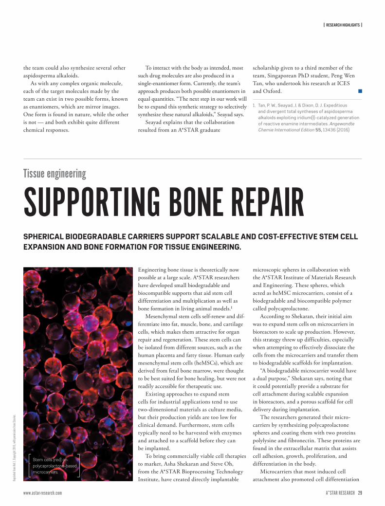
| RESEARCH HIGHLIGHTS |
28 ISSUE 7 | APRIL – JUNE 2017A*STAR RESEARCH www.astar-research.com 29 A*STAR RESEARCH
Repri
nted f
rom Re
f.1, C
opyri
ght 2
016,
with
perm
ission
from
Else
vier.
the team could also synthesize several other aspidosperma alkaloids.
As with any complex organic molecule, each of the target molecules made by the team can exist in two possible forms, known as enantiomers, which are mirror images. One form is found in nature, while the other is not — and both exhibit quite different chemical responses.
To interact with the body as intended, most such drug molecules are also produced in a single-enantiomer form. Currently, the team’s approach produces both possible enantiomers in equal quantities. “The next step in our work will be to expand this synthetic strategy to selectively synthesize these natural alkaloids,” Seayad says.
Seayad explains that the collaboration resulted from an A*STAR graduate
scholarship given to a third member of the team, Singaporean PhD student, Peng Wen Tan, who undertook his research at ICES and Oxford.
1. Tan, P. W., Seayad, J. & Dixon, D. J. Expeditious and divergent total syntheses of aspidosperma alkaloids exploiting iridium(I)-catalyzed generation of reactive enamine intermediates. Angewandte Chemie International Edition 55, 13436 (2016)
Tissue engineering
SUPPORTING BONE REPAIREngineering bone tissue is theoretically now possible at a large scale. A*STAR researchers have developed small biodegradable and biocompatible supports that aid stem cell differentiation and multiplication as well as bone formation in living animal models.¹
Mesenchymal stem cells self-renew and dif-ferentiate into fat, muscle, bone, and cartilage cells, which makes them attractive for organ repair and regeneration. These stem cells can be isolated from different sources, such as the human placenta and fatty tissue. Human early mesenchymal stem cells (heMSCs), which are derived from fetal bone marrow, were thought to be best suited for bone healing, but were not readily accessible for therapeutic use.
Existing approaches to expand stem cells for industrial applications tend to use two-dimensional materials as culture media, but their production yields are too low for clinical demand. Furthermore, stem cells typically need to be harvested with enzymes and attached to a scaffold before they can be implanted.
To bring commercially viable cell therapies to market, Asha Shekaran and Steve Oh, from the A*STAR Bioprocessing Technology Institute, have created directly implantable
microscopic spheres in collaboration with the A*STAR Institute of Materials Research and Engineering. These spheres, which acted as heMSC microcarriers, consist of a biodegradable and biocompatible polymer called polycaprolactone.
According to Shekaran, their initial aim was to expand stem cells on microcarriers in bioreactors to scale up production. However, this strategy threw up difficulties, especially when attempting to effectively dissociate the cells from the microcarriers and transfer them to biodegradable scaffolds for implantation.
“A biodegradable microcarrier would have a dual purpose,” Shekaran says, noting that it could potentially provide a substrate for cell attachment during scalable expansion in bioreactors, and a porous scaffold for cell delivery during implantation.
The researchers generated their micro-carriers by synthesizing polycaprolactone spheres and coating them with two proteins polylysine and fibronectin. These proteins are found in the extracellular matrix that assists cell adhesion, growth, proliferation, and differentiation in the body.
Microcarriers that most induced cell attachment also promoted cell differentiation
Stem cells (red) on polycaprolactone-based microcarriers.
SPHERICAL BIODEGRADABLE CARRIERS SUPPORT SCALABLE AND COST-EFFECTIVE STEM CELL EXPANSION AND BONE FORMATION FOR TISSUE ENGINEERING.
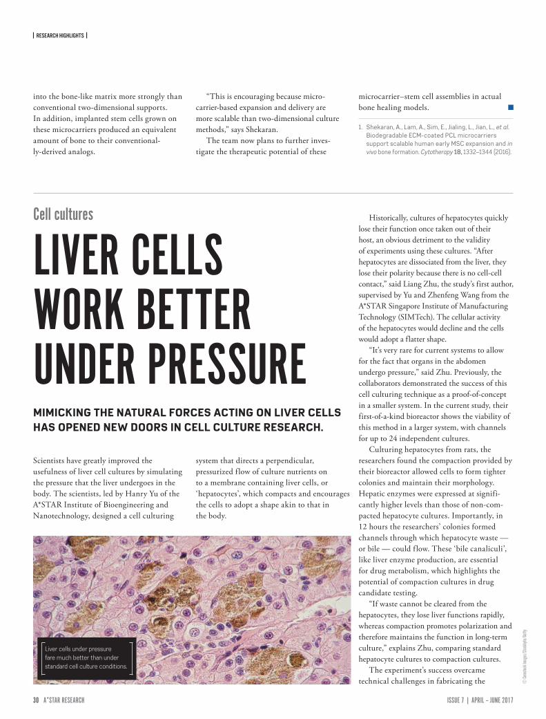
| RESEARCH HIGHLIGHTS |
30 ISSUE 7 | APRIL – JUNE 2017A*STAR RESEARCH www.astar-research.com 31 A*STAR RESEARCH
© Co
mstoc
k Ima
ges/
Stock
byte/
Getty
into the bone-like matrix more strongly than conventional two-dimensional supports. In addition, implanted stem cells grown on these microcarriers produced an equivalent amount of bone to their conventional-ly-derived analogs.
“This is encouraging because micro-carrier-based expansion and delivery are more scalable than two-dimensional culture methods,” says Shekaran.
The team now plans to further inves-tigate the therapeutic potential of these
microcarrier–stem cell assemblies in actual bone healing models.
1. Shekaran, A., Lam, A., Sim, E., Jialing, L., Jian, L., et al. Biodegradable ECM-coated PCL microcarriers support scalable human early MSC expansion and in vivo bone formation. Cytotherapy 18, 1332–1344 (2016).
Scientists have greatly improved the usefulness of liver cell cultures by simulating the pressure that the liver undergoes in the body. The scientists, led by Hanry Yu of the A*STAR Institute of Bioengineering and Nanotechnology, designed a cell culturing
system that directs a perpendicular, pressurized f low of culture nutrients on to a membrane containing liver cells, or ‘hepatocytes’, which compacts and encourages the cells to adopt a shape akin to that in the body.
Historically, cultures of hepatocytes quickly lose their function once taken out of their host, an obvious detriment to the validity of experiments using these cultures. “After hepatocytes are dissociated from the liver, they lose their polarity because there is no cell-cell contact,” said Liang Zhu, the study’s first author, supervised by Yu and Zhenfeng Wang from the A*STAR Singapore Institute of Manufacturing Technology (SIMTech). The cellular activity of the hepatocytes would decline and the cells would adopt a flatter shape.
“It’s very rare for current systems to allow for the fact that organs in the abdomen undergo pressure,” said Zhu. Previously, the collaborators demonstrated the success of this cell culturing technique as a proof-of-concept in a smaller system. In the current study, their first-of-a-kind bioreactor shows the viability of this method in a larger system, with channels for up to 24 independent cultures.
Culturing hepatocytes from rats, the researchers found the compaction provided by their bioreactor allowed cells to form tighter colonies and maintain their morphology. Hepatic enzymes were expressed at signifi-cantly higher levels than those of non-com-pacted hepatocyte cultures. Importantly, in 12 hours the researchers’ colonies formed channels through which hepatocyte waste — or bile — could f low. These ‘bile canaliculi’, like liver enzyme production, are essential for drug metabolism, which highlights the potential of compaction cultures in drug candidate testing.
“If waste cannot be cleared from the hepatocytes, they lose liver functions rapidly, whereas compaction promotes polarization and therefore maintains the function in long-term culture,” explains Zhu, comparing standard hepatocyte cultures to compaction cultures.
The experiment’s success overcame technical challenges in fabricating the
Cell cultures
LIVER CELLS WORK BETTER UNDER PRESSUREMIMICKING THE NATURAL FORCES ACTING ON LIVER CELLS HAS OPENED NEW DOORS IN CELL CULTURE RESEARCH.
Liver cells under pressure fare much better than under standard cell culture conditions.
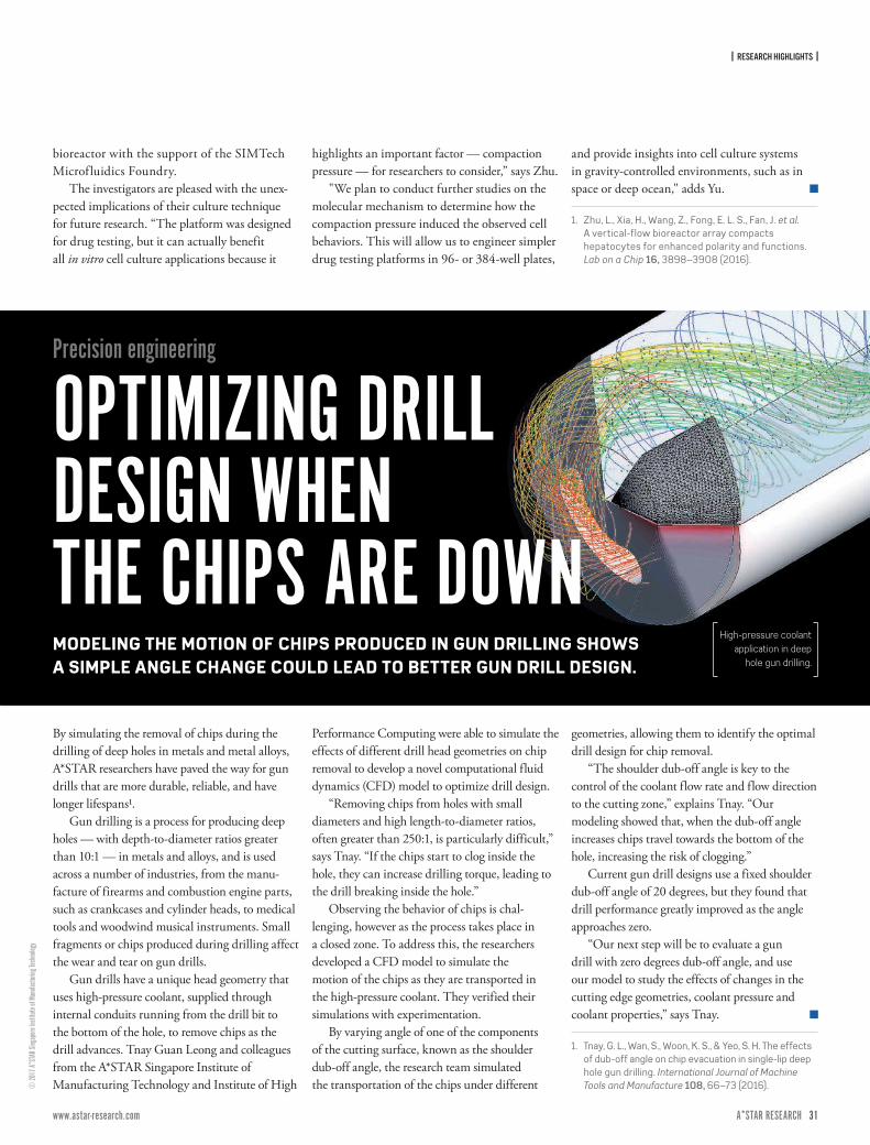
| RESEARCH HIGHLIGHTS |
30 ISSUE 7 | APRIL – JUNE 2017A*STAR RESEARCH www.astar-research.com 31 A*STAR RESEARCH
bioreactor with the support of the SIMTech Microfluidics Foundry.
The investigators are pleased with the unex-pected implications of their culture technique for future research. “The platform was designed for drug testing, but it can actually benefit all in vitro cell culture applications because it
highlights an important factor — compaction pressure — for researchers to consider,” says Zhu.
"We plan to conduct further studies on the molecular mechanism to determine how the compaction pressure induced the observed cell behaviors. This will allow us to engineer simpler drug testing platforms in 96- or 384-well plates,
and provide insights into cell culture systems in gravity-controlled environments, such as in space or deep ocean," adds Yu.
1. Zhu, L., Xia, H., Wang, Z., Fong, E. L. S., Fan, J. et al. A vertical-flow bioreactor array compacts hepatocytes for enhanced polarity and functions. Lab on a Chip 16, 3898–3908 (2016).
© 20
17 A*
STAR
Sing
apore
Insti
tute o
f Man
ufactu
ring T
echn
ology
Precision engineering
OPTIMIZING DRILL DESIGN WHEN THE CHIPS ARE DOWNMODELING THE MOTION OF CHIPS PRODUCED IN GUN DRILLING SHOWS A SIMPLE ANGLE CHANGE COULD LEAD TO BETTER GUN DRILL DESIGN.
By simulating the removal of chips during the drilling of deep holes in metals and metal alloys, A*STAR researchers have paved the way for gun drills that are more durable, reliable, and have longer lifespans1.
Gun drilling is a process for producing deep holes — with depth-to-diameter ratios greater than 10:1 — in metals and alloys, and is used across a number of industries, from the manu-facture of firearms and combustion engine parts, such as crankcases and cylinder heads, to medical tools and woodwind musical instruments. Small fragments or chips produced during drilling affect the wear and tear on gun drills.
Gun drills have a unique head geometry that uses high-pressure coolant, supplied through internal conduits running from the drill bit to the bottom of the hole, to remove chips as the drill advances. Tnay Guan Leong and colleagues from the A*STAR Singapore Institute of Manufacturing Technology and Institute of High
Performance Computing were able to simulate the effects of different drill head geometries on chip removal to develop a novel computational fluid dynamics (CFD) model to optimize drill design.
“Removing chips from holes with small diameters and high length-to-diameter ratios, often greater than 250:1, is particularly difficult,” says Tnay. “If the chips start to clog inside the hole, they can increase drilling torque, leading to the drill breaking inside the hole.”
Observing the behavior of chips is chal-lenging, however as the process takes place in a closed zone. To address this, the researchers developed a CFD model to simulate the motion of the chips as they are transported in the high-pressure coolant. They verified their simulations with experimentation.
By varying angle of one of the components of the cutting surface, known as the shoulder dub-off angle, the research team simulated the transportation of the chips under different
geometries, allowing them to identify the optimal drill design for chip removal.
“The shoulder dub-off angle is key to the control of the coolant flow rate and flow direction to the cutting zone,” explains Tnay. “Our modeling showed that, when the dub-off angle increases chips travel towards the bottom of the hole, increasing the risk of clogging.”
Current gun drill designs use a fixed shoulder dub-off angle of 20 degrees, but they found that drill performance greatly improved as the angle approaches zero.
“Our next step will be to evaluate a gun drill with zero degrees dub-off angle, and use our model to study the effects of changes in the cutting edge geometries, coolant pressure and coolant properties,” says Tnay.
1. Tnay, G. L., Wan, S., Woon, K. S., & Yeo, S. H. The effects of dub-off angle on chip evacuation in single-lip deep hole gun drilling. International Journal of Machine Tools and Manufacture 108, 66–73 (2016).
High-pressure coolant application in deep
hole gun drilling.
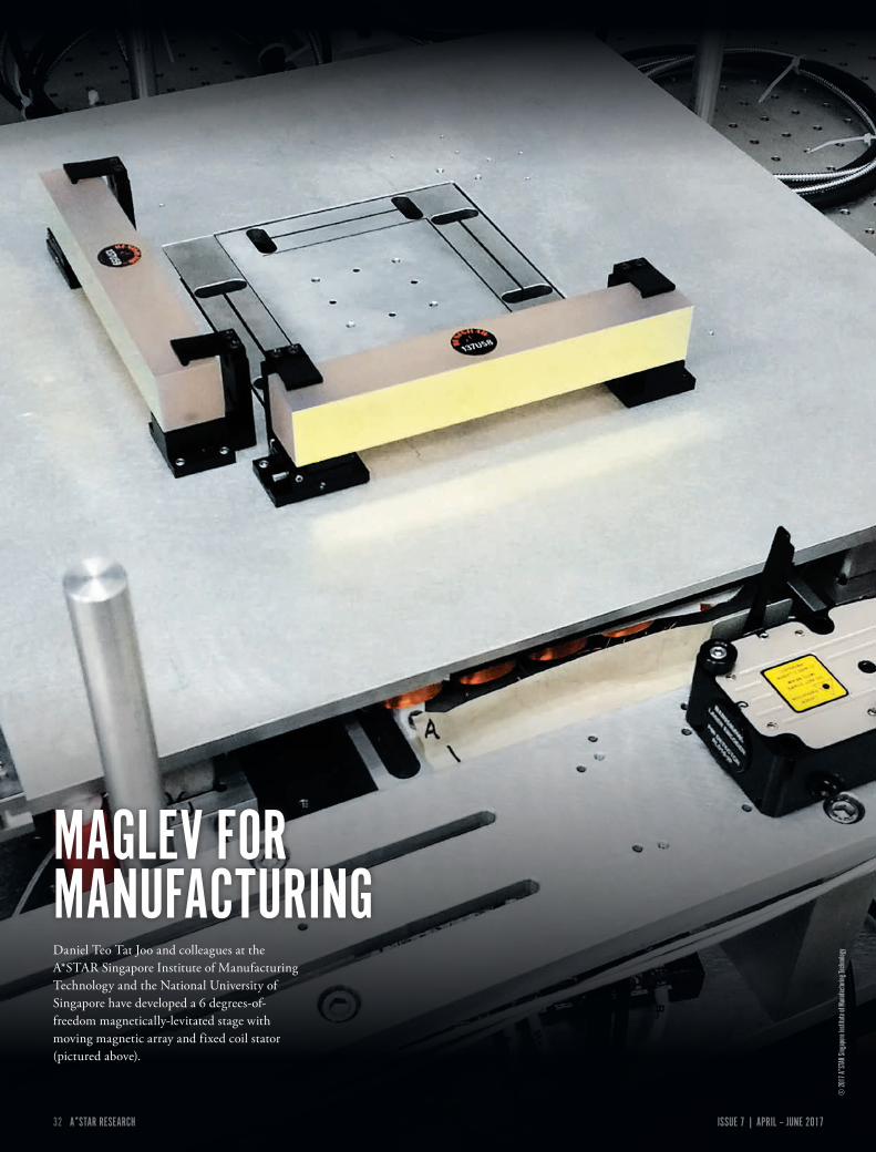
32 ISSUE 7 | APRIL – JUNE 2017A*STAR RESEARCH www.astar-research.com 33 A*STAR RESEARCH
© 20
17 A*
STAR
Sing
apore
Insti
tute o
f Man
ufactu
ring T
echn
ologyDaniel Teo Tat Joo and colleagues at the
A*STAR Singapore Institute of Manufacturing Technology and the National University of Singapore have developed a 6 degrees-of-freedom magnetically-levitated stage with moving magnetic array and fixed coil stator (pictured above).
MAGLEV FOR MANUFACTURING

32 ISSUE 7 | APRIL – JUNE 2017A*STAR RESEARCH www.astar-research.com 33 A*STAR RESEARCH
| RESEARCH HIGHLIGHTS |
© An
drew
Broo
kes/
Cultu
ra/Ge
tty
Critical components of modern communi-cations systems could be vastly improved by A*STAR research. Previously independent models of the components have been brought together for the first time, opening the door to improved electro-optical circuits.
Light offers particular advantages over con-ventional electronics — it can be transmitted with high fidelity over long distances, and can carry much more information. Optical fiber networks exploit these advantages for fast and efficient data communications. The devices at each end of an optical fiber, however, are usually built on conventional electronics, and the performance of this electro-optical interface is a factor that limits the rate of data transmission.
Much research has focused on the devel-opment of faster and smaller electro-optical components that can be integrated into conventional silicon-based electronic circuits and microchips. But progress has been hindered by the complexity of simulating both electronic and optical effects in the same device.
Soon Thor Lim and colleagues from the A*STAR Institute of High Performance Computing found a way to combine electronic
and optical effects into a single numerical simulation model. They now demonstrate that it can significantly increase the performance of a silicon optical modulator.
“Optical modulators are electro-optical devices that modify the propagating light by applying electrical pulses,” says Lim. “They are used in optical communication systems to encode electronic information into laser beams.”
While there are many fabrication parameters for silicon modulators, there are also many fabrication constraints, and so finding the optimal set of parameters requires painstaking computation.
“The problem is that two types of simulation must usually be performed for
Photonics
HIGH-FIDELITYSIMULTANEOUSLY SIMULATING ELECTRICAL AND OPTICAL INPUT ACHIEVES UNPRECEDENTED PERFORMANCE IN ELECTRO-OPTICAL INTERFACES.
Combining optical and electronic effects into a single simulation model could help researchers enhance the performance of electro-optical circuits in modern communication systems.
“OUR IN-HOUSE CODE PERFORMS BOTH ELECTRI-CAL AND OPTICAL SIMULATION IN ONE SINGLE PLATFORM WITH NO LOSS IN DATA FIDELITY.”
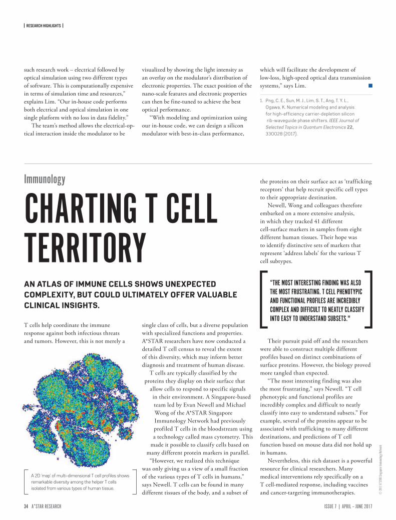
| RESEARCH HIGHLIGHTS |
34 ISSUE 7 | APRIL – JUNE 2017A*STAR RESEARCH www.astar-research.com 35 A*STAR RESEARCH
© 20
17 A*
STAR
Sing
apore
Immu
nolog
y Netw
ork
such research work – electrical followed by optical simulation using two different types of software. This is computationally expensive in terms of simulation time and resources,” explains Lim. “Our in-house code performs both electrical and optical simulation in one single platform with no loss in data fidelity.”
The team’s method allows the electrical-op-tical interaction inside the modulator to be
visualized by showing the light intensity as an overlay on the modulator’s distribution of electronic properties. The exact position of the nano-scale features and electronic properties can then be fine-tuned to achieve the best optical performance.
“With modeling and optimization using our in-house code, we can design a silicon modulator with best-in-class performance,
which will facilitate the development of low-loss, high-speed optical data transmission systems,” says Lim.
1. Png, C. E., Sun, M. J., Lim, S. T., Ang, T. Y. L., Ogawa, K. Numerical modeling and analysis for high-efficiency carrier-depletion silicon rib-waveguide phase shifters. IEEE Journal of Selected Topics in Quantum Electronics 22, 330028 (2017).
A 2D 'map' of multi-dimensional T cell profiles shows remarkable diversity among the helper T cells isolated from various types of human tissue.
Immunology
CHARTING T CELL TERRITORY
T cells help coordinate the immune response against both infectious threats and tumors. However, this is not merely a
single class of cells, but a diverse population with specialized functions and properties. A*STAR researchers have now conducted a detailed T cell census to reveal the extent of this diversity, which may inform better diagnosis and treatment of human disease.
T cells are typically classified by the proteins they display on their surface that
allow cells to respond to specific signals in their environment. A Singapore-based team led by Evan Newell and Michael Wong of the A*STAR Singapore Immunology Network had previously profiled T cells in the bloodstream using a technology called mass cytometry. This
made it possible to classify cells based on many different protein markers in parallel.
“However, we realized this technique was only giving us a view of a small fraction of the various types of T cells in humans,” says Newell. T cells can be found in many different tissues of the body, and a subset of
the proteins on their surface act as ‘trafficking receptors’ that help recruit specific cell types to their appropriate destination.
Newell, Wong and colleagues therefore embarked on a more extensive analysis, in which they tracked 41 different cell-surface markers in samples from eight different human tissues. Their hope was to identify distinctive sets of markers that represent ‘address labels’ for the various T cell subtypes.
Their pursuit paid off and the researchers were able to construct multiple different profiles based on distinct combinations of surface proteins. However, the biology proved more tangled than expected.
“The most interesting finding was also the most frustrating,” says Newell. “T cell phenotypic and functional profiles are incredibly complex and difficult to neatly classify into easy to understand subsets.” For example, several of the proteins appear to be associated with trafficking to many different destinations, and predictions of T cell function based on mouse data did not hold up in humans.
Nevertheless, this rich dataset is a powerful resource for clinical researchers. Many medical interventions rely specifically on a T cell-mediated response, including vaccines and cancer-targeting immunotherapies.
AN ATLAS OF IMMUNE CELLS SHOWS UNEXPECTED COMPLEXITY, BUT COULD ULTIMATELY OFFER VALUABLE CLINICAL INSIGHTS.
“THE MOST INTERESTING FINDING WAS ALSO THE MOST FRUSTRATING. T CELL PHENOTYPIC AND FUNCTIONAL PROFILES ARE INCREDIBLY COMPLEX AND DIFFICULT TO NEATLY CLASSIFY INTO EASY TO UNDERSTAND SUBSETS."
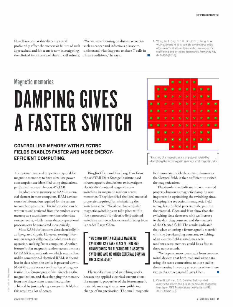
| RESEARCH HIGHLIGHTS |
34 ISSUE 7 | APRIL – JUNE 2017A*STAR RESEARCH www.astar-research.com 35 A*STAR RESEARCH
© 20
17 A*
STAR
Data
Storag
e Ins
titute
Magnetic memories
DAMPING GIVES A FASTER SWITCH
The optimal material properties required for magnetic memories to have ultra-low power consumption are identified using simulations performed by researchers at A*STAR.
Random access memory, or RAM, is a cru-cial element in most computers. RAM devices store the information required for the system to complete processes. This information can be written to and retrieved from the random-access memory at a much faster rate than other data storage media, which means that computational processes can be completed more quickly.
Most RAM devices store data electrically in an integrated circuit. However, storing infor-mation magnetically could enable even faster operation, making faster computers. Another feature is that magnetic random access memory (MRAM) is non-volatile — which means that, unlike conventional electrical RAM, it doesn’t lose its data when the device is powered down. MRAM store data as the direction of magnet-ization in a ferromagnetic film. Switching the magnetization, and thus changing the memory from one binary state to another, can be achieved by just applying a magnetic field, but this requires a lot of power.
BingJin Chen and Guchang Han from the A*STAR Data Storage Institute used micromagnetic simulations to investigate electric-field assisted magnetization switching in magnetic random access memories. They identified the ideal material properties required for minimizing the switching time. “We show that a reliable magnetic switching can take place within five nanoseconds for electric-field assisted switching and no other external driving force is needed,” says Chen.
Electric-field assisted switching works because the applied electrical current alters the magnetic properties of the ferromagnetic material, making it more susceptible to a change of magnetization. The small magnetic
field associated with the current, known as the Oersted field, is then sufficient to switch the magnetization.
The simulations indicated that a material property known as magnetic damping was important in optimizing the switching time. Damping is a reduction in magnetic field strength as the field penetrates deeper into the material. Chen and Han show that the switching time decreases with an increase in the damping constant and the strength of the Oersted field. The results indicated that when choosing a ferromagnetic material with the best damping constant, switching of an electric-field assisted magnetic random access memory could be as fast as three nanoseconds.
“We hope to move our study from two-ter-minal devices that both read and write data using the same connections to more stable three-terminal memory structures where these two paths are separated,” says Chen.
1. Chen B. J. & Han, G. C. Oersted field-guided electric field switching in perpendicular magnetic free layer. IEEE Transactions on Magnetics 52, 3401906 (2016).
Switching of a magnetic bit is computer-simulated by discretizing the ferromagnetic layer into small magnetic cells.
CONTROLLING MEMORY WITH ELECTRIC FIELDS ENABLES FASTER AND MORE ENERGY-EFFICIENT COMPUTING.
"WE SHOW THAT A RELIABLE MAGNETIC SWITCHING CAN TAKE PLACE WITHIN FIVE NANOSECONDS FOR ELECTRIC-FIELD ASSISTED SWITCHING AND NO OTHER EXTERNAL DRIVING FORCE IS NEEDED."
Newell notes that this diversity could profoundly affect the success or failure of such approaches, and his team is now investigating the clinical importance of these T cell subsets.
“We are now focusing on disease scenarios such as cancer and infectious disease to understand what happens to these T cells in those conditions,” he says.
1. Wong, M. T., Ong, D. E. H., Lim, F. S. H., Teng, K. W. W., McGovern, N. et al. A high-dimensional atlas of human T cell diversity reveals tissue-specific trafficking and cytokine signatures. Immunity 45, 442 –456 (2016).
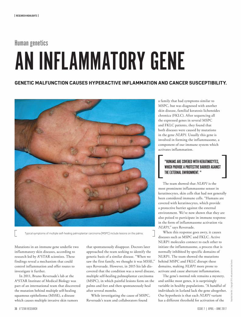
| RESEARCH HIGHLIGHTS |
36 ISSUE 7 | APRIL – JUNE 2017A*STAR RESEARCH www.astar-research.com 37 A*STAR RESEARCH
Repri
nted f
rom Re
f. 1, C
opyri
ght 2
016,
with
perm
ission
from
Else
vier.
Mutations in an immune gene underlie two inflammatory skin diseases, according to research led by A*STAR scientists. These findings reveal a mechanism that could control inflammation and offer routes to investigate it further.
In 2011, Bruno Reversade’s lab at the A*STAR Institute of Medical Biology was part of an international team that discovered the mutation behind multiple self-healing squamous epithelioma (MSSE), a disease which causes multiple invasive skin tumors
that spontaneously disappear. Doctors later approached the team seeking to identify the genetic basis of a similar disease. “When we saw the first family, we thought it was MSSE,” says Reversade. However, in 2015 his lab dis-covered that the condition was a novel disease, multiple self-healing palmoplantar carcinoma (MSPC), in which painful lesions form on the palms and feet and then spontaneously heal after several months.
While investigating the cause of MSPC, Reversade’s team and collaborators found
a family that had symptoms similar to MSPC, but was diagnosed with another skin disease, familial keratosis lichenoides chronica (FKLC). After sequencing all the expressed genes in several MSPC and FKLC patients, they found that both diseases were caused by mutations in the gene NLRP1. Usually this gene is involved in forming the inflammasome, a component of our immune system which activates inflammation.
The team showed that NLRP1 is the most prominent inflammasome sensor in keratinocytes, skin cells that had not generally been considered immune cells. “Humans are covered with keratinocytes, which provide a protective barrier against the external environment. We’ve now shown that they are also poised to participate in immune response in the form of inflammasome activation via NLRP1,” says Reversade.
When this response goes awry, it causes diseases such as MSPC and FKLC. Active NLRP1 molecules connect to each other to initiate the inflammasome, a process that is normally inhibited by specific domains of NLRP1. The team showed the mutations behind MSPC and FKLC disrupt these domains, making NLRP1 more prone to activate and cause aberrant inflammation.
The gene’s normal role remains a mystery, and unlike most genes, it is surprisingly variable in healthy populations. “A handful of individuals in Iceland lack the gene altogether. Our hypothesis is that each NLRP1 variant has a different threshold for activation of the
Typical symptoms of multiple self-healing palmoplantar carcinoma (MSPC) include lesions on the palms.
"HUMANS ARE COVERED WITH KERATINOCYTES, WHICH PROVIDE A PROTECTIVE BARRIER AGAINST THE EXTERNAL ENVIRONMENT. "
Human genetics
AN INFLAMMATORY GENEGENETIC MALFUNCTION CAUSES HYPERACTIVE INFLAMMATION AND CANCER SUSCEPTIBILITY.
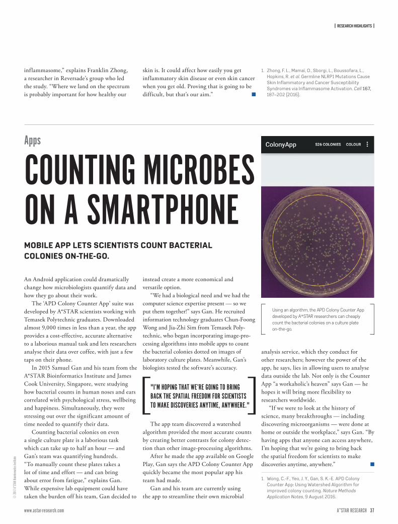
| RESEARCH HIGHLIGHTS |
36 ISSUE 7 | APRIL – JUNE 2017A*STAR RESEARCH www.astar-research.com 37 A*STAR RESEARCH
© 20
17 A*
STAR
Bioin
forma
tics I
nstit
ute
An Android application could dramatically change how microbiologists quantify data and how they go about their work.
The ‘APD Colony Counter App’ suite was developed by A*STAR scientists working with Temasek Polytechnic graduates. Downloaded almost 9,000 times in less than a year, the app provides a cost-effective, accurate alternative to a laborious manual task and lets researchers analyse their data over coffee, with just a few taps on their phone.
In 2015 Samuel Gan and his team from the A*STAR Bioinformatics Institute and James Cook University, Singapore, were studying how bacterial counts in human noses and ears correlated with psychological stress, wellbeing and happiness. Simultaneously, they were stressing out over the significant amount of time needed to quantify their data.
Counting bacterial colonies on even a single culture plate is a laborious task which can take up to half an hour — and Gan’s team was quantifying hundreds. “To manually count these plates takes a lot of time and effort — and can bring about error from fatigue,” explains Gan. While expensive lab equipment could have taken the burden off his team, Gan decided to
instead create a more economical and versatile option.
“We had a biological need and we had the computer science expertise present — so we put them together!” says Gan. He recruited information technology graduates Chun-Foong Wong and Jia-Zhi Sim from Temasek Poly-technic, who began incorporating image-pro-cessing algorithms into mobile apps to count the bacterial colonies dotted on images of laboratory culture plates. Meanwhile, Gan’s biologists tested the software’s accuracy.
The app team discovered a watershed algorithm provided the most accurate counts by creating better contrasts for colony detec-tion than other image-processing algorithms.
After he made the app available on Google Play, Gan says the APD Colony Counter App quickly became the most popular app his team had made.
Gan and his team are currently using the app to streamline their own microbial
analysis service, which they conduct for other researchers; however the power of the app, he says, lies in allowing users to analyse data outside the lab. Not only is the Counter App “a workaholic’s heaven” says Gan — he hopes it will bring more f lexibility to researchers worldwide.
“If we were to look at the history of science, many breakthroughs — including discovering microorganisms — were done at home or outside the workplace,” says Gan. “By having apps that anyone can access anywhere, I’m hoping that we’re going to bring back the spatial freedom for scientists to make discoveries anytime, anywhere.”
1. Wong, C.-F., Yeo, J. Y., Gan, S. K.-E. APD Colony Counter App: Using Watershed Algorithm for improved colony counting. Nature Methods Application Notes, 9 August 2016.
Using an algorithm, the APD Colony Counter App developed by A*STAR researchers can cheaply count the bacterial colonies on a culture plate on-the-go.
Apps
COUNTING MICROBES ON A SMARTPHONEMOBILE APP LETS SCIENTISTS COUNT BACTERIAL COLONIES ON-THE-GO.
inflammasome,” explains Franklin Zhong, a researcher in Reversade’s group who led the study. “Where we land on the spectrum is probably important for how healthy our
skin is. It could affect how easily you get inflammatory skin disease or even skin cancer when you get old. Proving that is going to be difficult, but that’s our aim.”
1. Zhong, F. L., Mamaï, O., Sborgi, L., Boussofara, L., Hopkins, R. et al. Germline NLRP1 Mutations Cause Skin Inflammatory and Cancer Susceptibility Syndromes via Inflammasome Activation. Cell 167, 187–202 (2016).
"I’M HOPING THAT WE’RE GOING TO BRING BACK THE SPATIAL FREEDOM FOR SCIENTISTS TO MAKE DISCOVERIES ANYTIME, ANYWHERE."
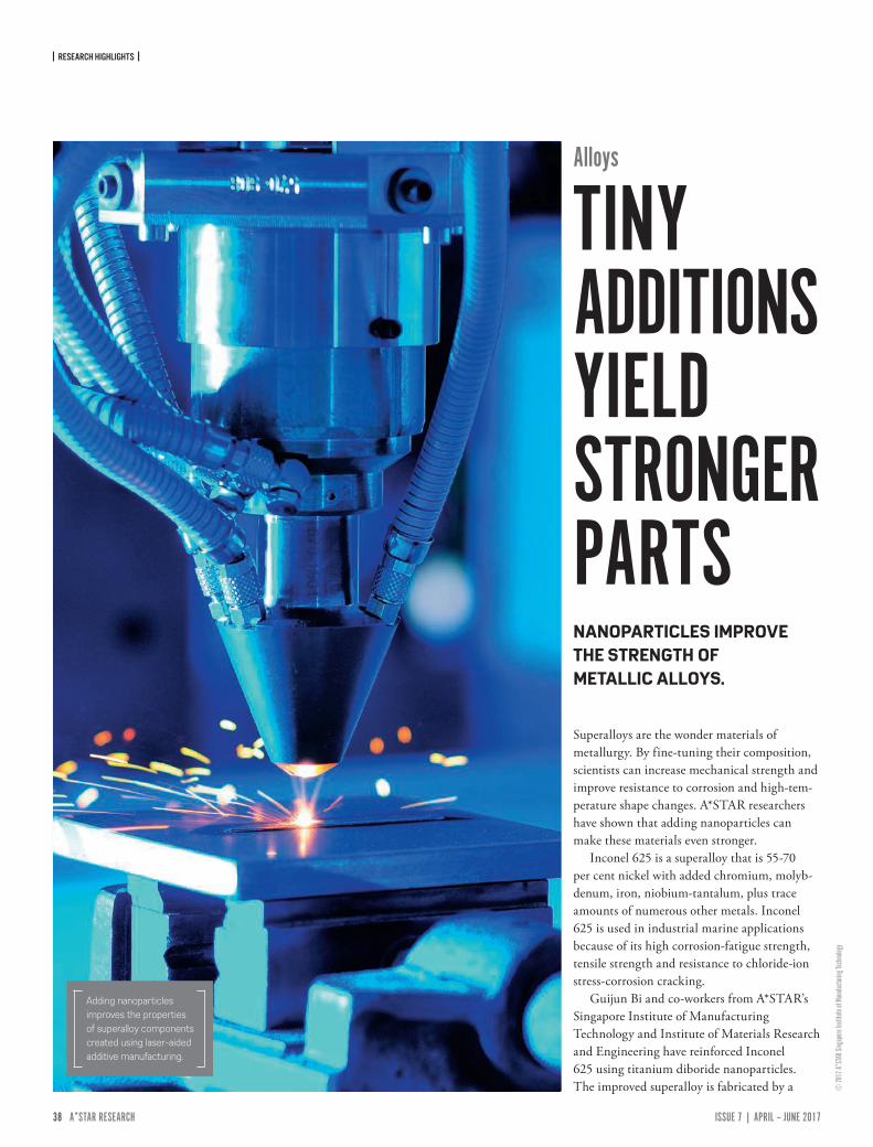
| RESEARCH HIGHLIGHTS |
38 ISSUE 7 | APRIL – JUNE 2017A*STAR RESEARCH www.astar-research.com 39 A*STAR RESEARCH
© 20
17 A*
STAR
Sing
apore
Insti
tute o
f Man
ufactu
ring T
echn
ology
Alloys
TINY ADDITIONS YIELD STRONGER PARTS
Superalloys are the wonder materials of metallurgy. By fine-tuning their composition, scientists can increase mechanical strength and improve resistance to corrosion and high-tem-perature shape changes. A*STAR researchers have shown that adding nanoparticles can make these materials even stronger.
Inconel 625 is a superalloy that is 55-70 per cent nickel with added chromium, molyb-denum, iron, niobium-tantalum, plus trace amounts of numerous other metals. Inconel 625 is used in industrial marine applications because of its high corrosion-fatigue strength, tensile strength and resistance to chloride-ion stress-corrosion cracking.
Guijun Bi and co-workers from A*STAR’s Singapore Institute of Manufacturing Technology and Institute of Materials Research and Engineering have reinforced Inconel 625 using titanium diboride nanoparticles. The improved superalloy is fabricated by a
NANOPARTICLES IMPROVE THE STRENGTH OF METALLIC ALLOYS.
Adding nanoparticles improves the properties of superalloy components created using laser-aided additive manufacturing.
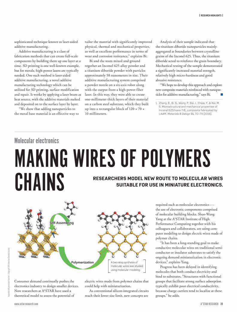
| RESEARCH HIGHLIGHTS |
38 ISSUE 7 | APRIL – JUNE 2017A*STAR RESEARCH www.astar-research.com 39 A*STAR RESEARCH
Repri
nted w
ith pe
rmiss
ion fr
om Re
f 1. C
opyri
ght 2
016 A
meric
an Ch
emica
l Soc
iety
sophisticated technique known as laser-aided additive manufacturing.
Additive manufacturing is a class of fabrication methods that can create full-scale components by building them up one layer at a time. 3D printing is one well-known example, but for metals, high-power lasers are typically needed. One such method is laser-aided additive manufacturing, a novel additive manufacturing technology which can be utilized for 3D printing, surface modification and repair. It works by applying a laser beam as heat source, with the additive materials melted and deposited on to the surface layer by layer.
“We show that adding nanoparticles to the metal base material is an effective way to
tailor the material with significantly improved physical, thermal and mechanical properties, as well as excellent performance in terms of wear and corrosion resistance,” explains Bi.
Bi and the team mixed and ground together an Inconel 625 alloy powder and a titanium diboride powder with particles approximately 58 nanometers in size. Their additive manufacturing system comprised a powder nozzle on a six-axis robot along with the output from a high-power fiber laser. In this way, they were able to create one-millimeter thick layers of their material on a carbon steel substrate, which they built up into a rectangular block of 120 × 70 × 10 millimeters.
Analysis of their sample indicated that the titanium diboride nanoparticles mainly aggregated at boundaries between crystalline grains of the Inconel 625. Thus, the titanium diboride acted to reinforce the grain boundary. Mechanical testing of the sample demonstrated a significantly increased material strength, relatively high micro-hardness and good abrasive resistance.
“We hope to develop this approach and explore new composite materials reinforced with nanopar-ticles for additive manufacturing,” says Bi.
1. Zhang, B., Bi, G., Wang, P., Bai, J., Chew, Y., & Nai, M. S. Microstructure and mechanical properties of Inconel 625/nano-TiB2 composite fabricated by LAAM. Materials & Design 11, 70–79 (2016).
Consumer demand continually pushes the electronics industry to design smaller devices. Now researchers at A*STAR have used a theoretical model to assess the potential of
electric wires made from polymer chains that could help with miniaturization.
As conventional silicon-integrated circuits reach their lower size limit, new concepts are
required such as molecular electronics — the use of electronic components comprised of molecular building blocks. Shuo-Wang Yang at the A*STAR Institute of High Performance Computing together with his colleagues and collaborators, are using com-puter modeling to design electric wires made of polymer chains.
“It has been a long-standing goal to make conductive molecular wires on traditional semi-conductor or insulator substrates to satisfy the ongoing demand miniaturization in electronic devices,” explains Yang.
Progress has been delayed in identifying molecules that both conduct electricity and bind to substrates. “Structures with functional groups that facilitate strong surface adsorption typically exhibit poor electrical conductivity, because charge carriers tend to localize at these groups,” he adds.
Polymerization
Self-AssemblingSelf-Assembling
Polymerization A two-step synthesis of molecular wires was studied using molecular modeling.
Molecular electronics
MAKING WIRES OF POLYMERS CHAINS RESEARCHERS MODEL NEW ROUTE TO MOLECULAR WIRES
SUITABLE FOR USE IN MINIATURE ELECTRONICS.
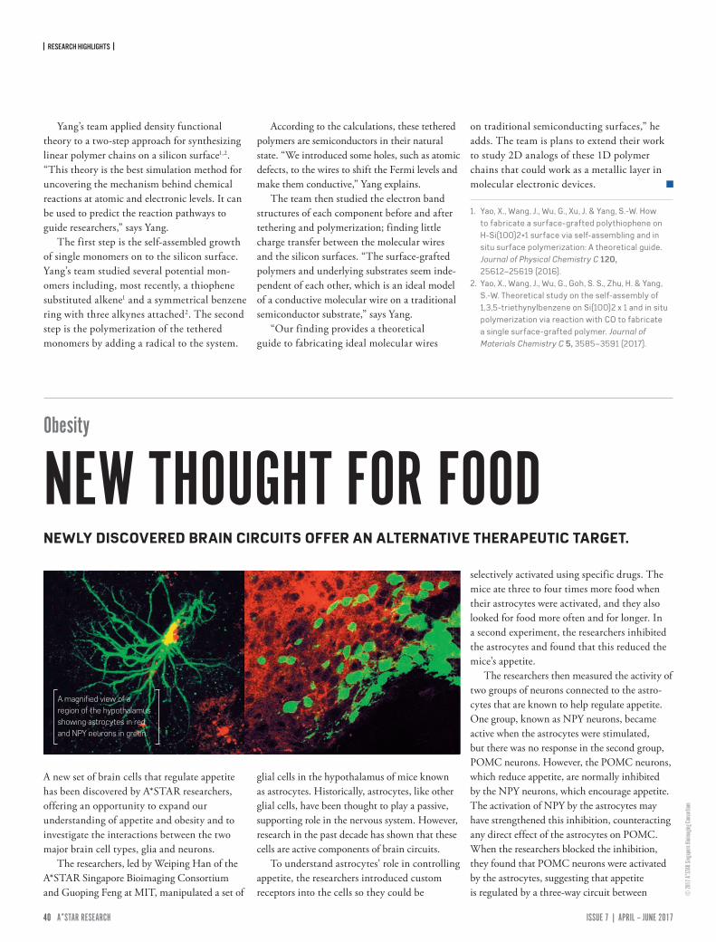
| RESEARCH HIGHLIGHTS |
40 ISSUE 7 | APRIL – JUNE 2017A*STAR RESEARCH www.astar-research.com 41 A*STAR RESEARCH
© 20
17 A*
STAR
Sing
apore
Bioim
aging
Cons
ortium
Yang’s team applied density functional theory to a two-step approach for synthesizing linear polymer chains on a silicon surface1,2. “This theory is the best simulation method for uncovering the mechanism behind chemical reactions at atomic and electronic levels. It can be used to predict the reaction pathways to guide researchers,” says Yang.
The first step is the self-assembled growth of single monomers on to the silicon surface. Yang’s team studied several potential mon-omers including, most recently, a thiophene substituted alkene1 and a symmetrical benzene ring with three alkynes attached2. The second step is the polymerization of the tethered monomers by adding a radical to the system.
According to the calculations, these tethered polymers are semiconductors in their natural state. “We introduced some holes, such as atomic defects, to the wires to shift the Fermi levels and make them conductive,” Yang explains.
The team then studied the electron band structures of each component before and after tethering and polymerization; finding little charge transfer between the molecular wires and the silicon surfaces. “The surface-grafted polymers and underlying substrates seem inde-pendent of each other, which is an ideal model of a conductive molecular wire on a traditional semiconductor substrate,” says Yang.
“Our finding provides a theoretical guide to fabricating ideal molecular wires
on traditional semiconducting surfaces,” he adds. The team is plans to extend their work to study 2D analogs of these 1D polymer chains that could work as a metallic layer in molecular electronic devices.
1. Yao, X., Wang, J., Wu, G., Xu, J. & Yang, S.-W. How to fabricate a surface-grafted polythiophene on H-Si(100)2×1 surface via self-assembling and in situ surface polymerization: A theoretical guide. Journal of Physical Chemistry C 120, 25612–25619 (2016).
2. Yao, X., Wang, J., Wu, G., Goh, S. S., Zhu, H. & Yang, S.-W. Theoretical study on the self-assembly of 1,3,5-triethynylbenzene on Si(100)2 x 1 and in situ polymerization via reaction with CO to fabricate a single surface-grafted polymer. Journal of Materials Chemistry C 5, 3585–3591 (2017).
A new set of brain cells that regulate appetite has been discovered by A*STAR researchers, offering an opportunity to expand our understanding of appetite and obesity and to investigate the interactions between the two major brain cell types, glia and neurons.
The researchers, led by Weiping Han of the A*STAR Singapore Bioimaging Consortium and Guoping Feng at MIT, manipulated a set of
glial cells in the hypothalamus of mice known as astrocytes. Historically, astrocytes, like other glial cells, have been thought to play a passive, supporting role in the nervous system. However, research in the past decade has shown that these cells are active components of brain circuits.
To understand astrocytes’ role in controlling appetite, the researchers introduced custom receptors into the cells so they could be
selectively activated using specific drugs. The mice ate three to four times more food when their astrocytes were activated, and they also looked for food more often and for longer. In a second experiment, the researchers inhibited the astrocytes and found that this reduced the mice’s appetite.
The researchers then measured the activity of two groups of neurons connected to the astro-cytes that are known to help regulate appetite. One group, known as NPY neurons, became active when the astrocytes were stimulated, but there was no response in the second group, POMC neurons. However, the POMC neurons, which reduce appetite, are normally inhibited by the NPY neurons, which encourage appetite. The activation of NPY by the astrocytes may have strengthened this inhibition, counteracting any direct effect of the astrocytes on POMC. When the researchers blocked the inhibition, they found that POMC neurons were activated by the astrocytes, suggesting that appetite is regulated by a three-way circuit between
A magnified view of a region of the hypothalamus showing astrocytes in red and NPY neurons in green.
Obesity
NEW THOUGHT FOR FOODNEWLY DISCOVERED BRAIN CIRCUITS OFFER AN ALTERNATIVE THERAPEUTIC TARGET.
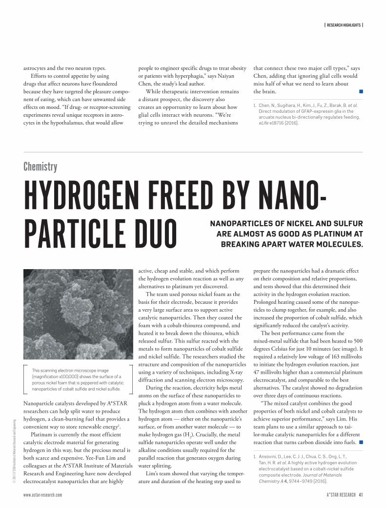
| RESEARCH HIGHLIGHTS |
40 ISSUE 7 | APRIL – JUNE 2017A*STAR RESEARCH www.astar-research.com 41 A*STAR RESEARCH
© 20
17 A*
STAR
Insti
tute o
f Mate
rials
Rese
arch a
nd En
ginee
ring
astrocytes and the two neuron types.Efforts to control appetite by using
drugs that affect neurons have floundered because they have targeted the pleasure compo-nent of eating, which can have unwanted side effects on mood. “If drug- or receptor-screening experiments reveal unique receptors in astro-cytes in the hypothalamus, that would allow
people to engineer specific drugs to treat obesity or patients with hyperphagia,” says Naiyan Chen, the study’s lead author.
While therapeutic intervention remains a distant prospect, the discovery also creates an opportunity to learn about how glial cells interact with neurons. “We're trying to unravel the detailed mechanisms
that connect these two major cell types,” says Chen, adding that ignoring glial cells would miss half of what we need to learn about the brain.
1. Chen, N., Sugihara, H., Kim, J., Fu, Z., Barak, B. et al. Direct modulation of GFAP-expressin glia in the arcuate nucleus bi-directionally regulates feeding. eLife e18716 (2016).
Chemistry
HYDROGEN FREED BY NANO-PARTICLE DUO NANOPARTICLES OF NICKEL AND SULFUR
ARE ALMOST AS GOOD AS PLATINUM AT BREAKING APART WATER MOLECULES.
Nanoparticle catalysts developed by A*STAR researchers can help split water to produce hydrogen, a clean-burning fuel that provides a convenient way to store renewable energy1.
Platinum is currently the most efficient catalytic electrode material for generating hydrogen in this way, but the precious metal is both scarce and expensive. Yee-Fun Lim and colleagues at the A*STAR Institute of Materials Research and Engineering have now developed electrocatalyst nanoparticles that are highly
active, cheap and stable, and which perform the hydrogen evolution reaction as well as any alternatives to platinum yet discovered.
The team used porous nickel foam as the basis for their electrode, because it provides a very large surface area to support active catalytic nanoparticles. Then they coated the foam with a cobalt-thiourea compound, and heated it to break down the thiourea, which released sulfur. This sulfur reacted with the metals to form nanoparticles of cobalt sulfide and nickel sulfide. The researchers studied the structure and composition of the nanoparticles using a variety of techniques, including X-ray diffraction and scanning electron microscopy.
During the reaction, electricity helps metal atoms on the surface of these nanoparticles to pluck a hydrogen atom from a water molecule. The hydrogen atom then combines with another hydrogen atom — either on the nanoparticle’s surface, or from another water molecule — to make hydrogen gas (H2). Crucially, the metal sulfide nanoparticles operate well under the alkaline conditions usually required for the parallel reaction that generates oxygen during water splitting.
Lim’s team showed that varying the temper-ature and duration of the heating step used to
prepare the nanoparticles had a dramatic effect on their composition and relative proportions, and tests showed that this determined their activity in the hydrogen evolution reaction. Prolonged heating caused some of the nanopar-ticles to clump together, for example, and also increased the proportion of cobalt sulfide, which significantly reduced the catalyst’s activity.
The best performance came from the mixed-metal sulfide that had been heated to 500 degrees Celsius for just 10 minutes (see image). It required a relatively low voltage of 163 millivolts to initiate the hydrogen evolution reaction, just 47 millivolts higher than a commercial platinum electrocatalyst, and comparable to the best alternatives. The catalyst showed no degradation over three days of continuous reactions.
“The mixed catalyst combines the good properties of both nickel and cobalt catalysts to achieve superior performance,” says Lim. His team plans to use a similar approach to tai-lor-make catalytic nanoparticles for a different reaction that turns carbon dioxide into fuels.
1. Ansovini, D., Lee, C. J. J., Chua, C. S., Ong, L. T., Tan, H. R. et al. A highly active hydrogen evolution electrocatalyst based on a cobalt-nickel sulfide composite electrode. Journal of Materials Chemistry A 4, 9744–9749 (2016).
This scanning electron microscope image (magnification x100,000) shows the surface of a porous nickel foam that is peppered with catalytic nanoparticles of cobalt sulfide and nickel sulfide.
42 ISSUE 7 | APRIL – JUNE 2017A*STAR RESEARCH www.astar-research.com 43 A*STAR RESEARCH
© se
norja
ckso
n/E+
/Gett
ye
| RESEARCH HIGHLIGHTS |
Magnetic materials are a vital ingredient in the components that store information in computers and mobile phones. Now, A*STAR researchers have developed a material that could help these magnetic-based memory devices to store and retrieve data faster while using less power.
Memory devices work when a small magnetic field is applied to the storage medium to align atomic-level magnets known as spins. This spin alignment, or magnetization, in one region of the magnetic material can represent one ‘bit’ of information, which can be ‘read’ back again using a magnet. Scientists are trying to improve the performance of magnetic memories by reducing both the energy required to change the magnetization and unwanted noise.
One approach is to use a magnetic material with a property known as negative magnetocrystalline anisotropy. This means
that less energy is required to align the spins in one direction then another, and so the material is generally easier to magnetize and demagnetize. This low coercivity is useful because this so-called ‘soft’ magnetic material can guide a magnetic field onto the storage layer, thus lowering the intensity of field that must be applied to alter the ‘hard’ material’s magnetization.
Tiejun Zhou and co-workers from the A*STAR Data Storage Institute found a way
to further reduce the coercivity of a soft mate-rial called cobalt iridium by adding rhodium.
The team created their magnetic material with a technique known as direct current magnetron sputtering. Cobalt, iridium and rhodium were simultaneously ejected from sep-arate solid sources in a vacuum chamber and deposited on a silicon substrate. By changing the power supplied to each of the sources, the researchers could control the composition of the final material, increasing the amount of rhodium at the expense of iridium. Measure-ments of the magnetic properties of CoIr-Rh films demonstrated that the introduction of this rhodium reduced the coercivity and the damping constant by more than a half of that of unmodified cobalt iridium.
“When used in a device, such negative magnetocrystalline anisotropy materials
Data storage
MORE OPEN TO COERCIONA NOVEL ‘SOFT’ MAGNETIC MATERIAL COULD ENABLE FASTER COMPUTER MEMORY.
"BY FINE TUNING THE COMPOSITION, WE CAN CONTINUOUSLY IMPROVE THE PROPERTIES OF MAGNETIC MATERIALS TO MEET THE CRITERIA REQUIRED FOR INDUSTRY-LEVEL APPLICATIONS."
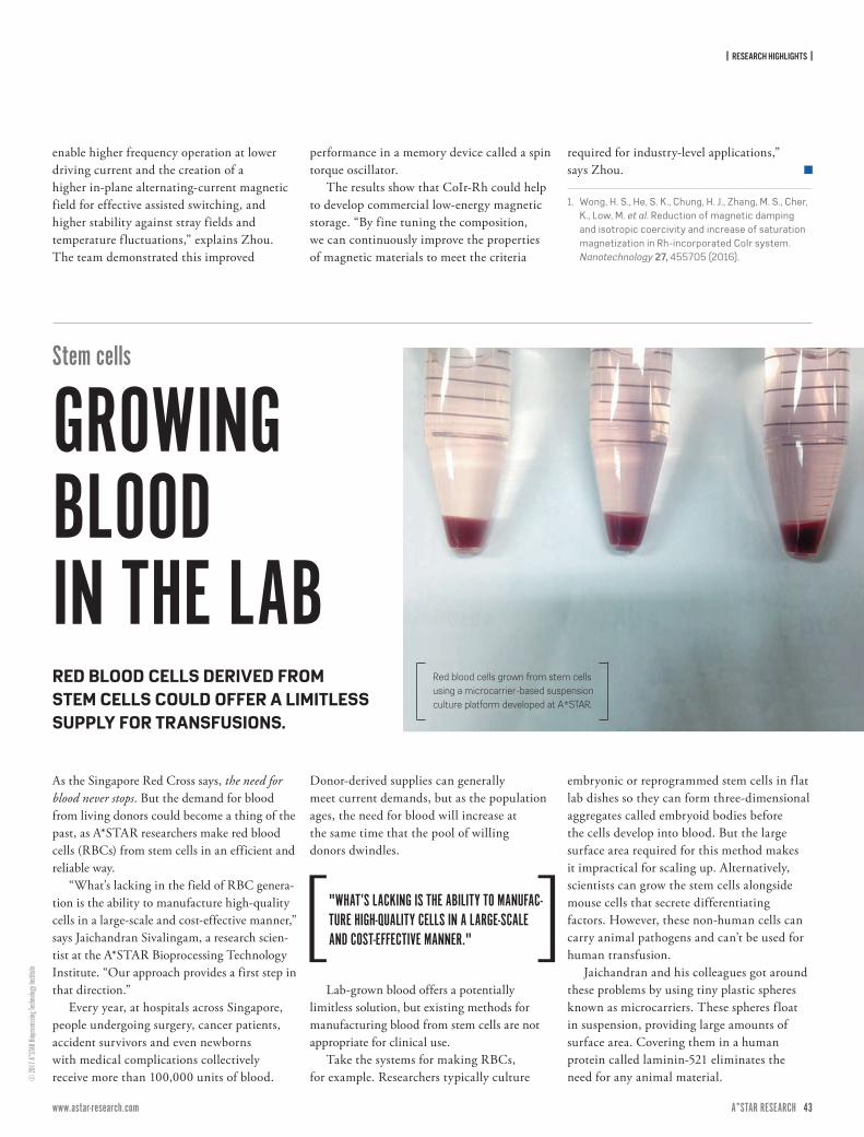
| RESEARCH HIGHLIGHTS |
42 ISSUE 7 | APRIL – JUNE 2017A*STAR RESEARCH www.astar-research.com 43 A*STAR RESEARCH
© 20
17 A*
STAR
Biop
roces
sing T
echn
ology
Insti
tute
enable higher frequency operation at lower driving current and the creation of a higher in-plane alternating-current magnetic field for effective assisted switching, and higher stability against stray fields and temperature f luctuations,” explains Zhou. The team demonstrated this improved
performance in a memory device called a spin torque oscillator.
The results show that CoIr-Rh could help to develop commercial low-energy magnetic storage. “By fine tuning the composition, we can continuously improve the properties of magnetic materials to meet the criteria
required for industry-level applications,” says Zhou.
1. Wong, H. S., He, S. K., Chung, H. J., Zhang, M. S., Cher, K., Low, M. et al. Reduction of magnetic damping and isotropic coercivity and increase of saturation magnetization in Rh-incorporated CoIr system. Nanotechnology 27, 455705 (2016).
Stem cells
GROWING BLOOD IN THE LAB
As the Singapore Red Cross says, the need for blood never stops. But the demand for blood from living donors could become a thing of the past, as A*STAR researchers make red blood cells (RBCs) from stem cells in an efficient and reliable way.
“What’s lacking in the field of RBC genera-tion is the ability to manufacture high-quality cells in a large-scale and cost-effective manner,” says Jaichandran Sivalingam, a research scien-tist at the A*STAR Bioprocessing Technology Institute. “Our approach provides a first step in that direction.”
Every year, at hospitals across Singapore, people undergoing surgery, cancer patients, accident survivors and even newborns with medical complications collectively receive more than 100,000 units of blood.
Donor-derived supplies can generally meet current demands, but as the population ages, the need for blood will increase at the same time that the pool of willing donors dwindles.
Lab-grown blood offers a potentially limitless solution, but existing methods for manufacturing blood from stem cells are not appropriate for clinical use.
Take the systems for making RBCs, for example. Researchers typically culture
embryonic or reprogrammed stem cells in f lat lab dishes so they can form three-dimensional aggregates called embryoid bodies before the cells develop into blood. But the large surface area required for this method makes it impractical for scaling up. Alternatively, scientists can grow the stem cells alongside mouse cells that secrete differentiating factors. However, these non-human cells can carry animal pathogens and can’t be used for human transfusion.
Jaichandran and his colleagues got around these problems by using tiny plastic spheres known as microcarriers. These spheres f loat in suspension, providing large amounts of surface area. Covering them in a human protein called laminin-521 eliminates the need for any animal material.
Red blood cells grown from stem cells using a microcarrier-based suspension culture platform developed at A*STAR.
RED BLOOD CELLS DERIVED FROM STEM CELLS COULD OFFER A LIMITLESS SUPPLY FOR TRANSFUSIONS.
"WHAT’S LACKING IS THE ABILITY TO MANUFAC-TURE HIGH-QUALITY CELLS IN A LARGE-SCALE AND COST-EFFECTIVE MANNER."
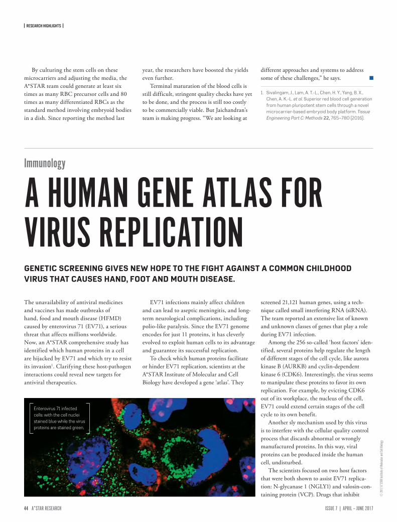
| RESEARCH HIGHLIGHTS |
44 ISSUE 7 | APRIL – JUNE 2017A*STAR RESEARCH www.astar-research.com 45 A*STAR RESEARCH
© 20
17 A*
STAR
Insti
tute o
f Mole
cular
and C
ell Bi
ology
By culturing the stem cells on these microcarriers and adjusting the media, the A*STAR team could generate at least six times as many RBC precursor cells and 80 times as many differentiated RBCs as the standard method involving embryoid bodies in a dish. Since reporting the method last
year, the researchers have boosted the yields even further.
Terminal maturation of the blood cells is still difficult, stringent quality checks have yet to be done, and the process is still too costly to be commercially viable. But Jaichandran’s team is making progress. “We are looking at
different approaches and systems to address some of these challenges,” he says.
1. Sivalingam, J., Lam, A. T.-L., Chen, H. Y., Yang, B. X., Chen, A. K.-L. et al. Superior red blood cell generation from human pluripotent stem cells through a novel microcarrier-based embryoid body platform. Tissue Engineering Part C: Methods 22, 765–780 (2016).
The unavailability of antiviral medicines and vaccines has made outbreaks of hand, food and mouth disease (HFMD) caused by enterovirus 71 (EV71), a serious threat that affects millions worldwide. Now, an A*STAR comprehensive study has identified which human proteins in a cell are hijacked by EV71 and which try to resist its invasion1. Clarifying these host-pathogen interactions could reveal new targets for antiviral therapeutics.
EV71 infections mainly affect children and can lead to aseptic meningitis, and long-term neurological complications, including polio-like paralysis. Since the EV71 genome encodes for just 11 proteins, it has cleverly evolved to exploit human cells to its advantage and guarantee its successful replication.
To check which human proteins facilitate or hinder EV71 replication, scientists at the A*STAR Institute of Molecular and Cell Biology have developed a gene ‘atlas’. They
screened 21,121 human genes, using a tech-nique called small interfering RNA (siRNA). The team reported an extensive list of known and unknown classes of genes that play a role during EV71 infection.
Among the 256 so-called ‘host factors' iden-tified, several proteins help regulate the length of different stages of the cell cycle, like aurora kinase B (AURKB) and cyclin-dependent kinase 6 (CDK6). Interestingly, the virus seems to manipulate these proteins to favor its own replication. For example, by evicting CDK6 out of its workplace, the nucleus of the cell, EV71 could extend certain stages of the cell cycle to its own benefit.
Another sly mechanism used by this virus is to interfere with the cellular quality control process that discards abnormal or wrongly manufactured proteins. In this way, viral proteins can be produced inside the human cell, undisturbed.
The scientists focused on two host factors that were both shown to assist EV71 replica-tion: N-glycanase 1 (NGLY1) and valosin-con-taining protein (VCP). Drugs that inhibit
Enterovirus 71 infected cells: with the cell nuclei stained blue while the virus proteins are stained green.
Immunology
A HUMAN GENE ATLAS FOR VIRUS REPLICATIONGENETIC SCREENING GIVES NEW HOPE TO THE FIGHT AGAINST A COMMON CHILDHOOD VIRUS THAT CAUSES HAND, FOOT AND MOUTH DISEASE.

| RESEARCH HIGHLIGHTS |
44 ISSUE 7 | APRIL – JUNE 2017A*STAR RESEARCH www.astar-research.com 45 A*STAR RESEARCH
© 20
17 A*
STAR
Geno
me In
stitut
e of S
ingap
ore
these two host factors also reduce the number of EV71-infected cells. VCP is probably held inside vesicular structures used by the virus to copy its genome, but it remains unknown how EV71 benefits from NGLY1.
“This is the first genome-wide siRNA screening for EV71-human factors interaction
and reveals the complex interplay between the virus and the proteins of a specific human cell line,” points out Justin Jang Hann Chu, lead author of the study. “Some host factors we found are shared with picornaviruses and enteroviruses infections, while others are completely new and need to be further
explored. This information opens a new chapter in the development of antiviral strategies for HFMD.”
1. Wu, K. X., Phuektes, P., Kumar, P., Goh, G. Y., Moreau, D. et al. Human genome-wide RNAi screen reveals host factors required for enterovirus 71 replication. Nature Communications 7, 13150 (2016).
Bowel cancer
A CLOSER LOOK AT CELL TYPESAn algorithm developed by A*STAR which analyzes which genes are turned on and off in individual cancer cells within a tumor is being used to examine the different cell types in bowel cancers1. The findings may challenge an estab-lished understanding of cancer development.
The differences between cells in tumors is a major obstacle in cancer therapy, explains Shyam Prabhakar from the A*STAR Genome Institute of Singapore who collaborated with the National Cancer Center Singapore and other research centers. “We hope that our novel approach to understanding the basic biology of tumors will lead to new ideas for treatment,” says Prabhakar, adding that he anticipates this approach could be applied to other types of cancer in the future.
The raw data for the algorithm comes from RNA sequencing, which characterizes the RNA molecules that control protein synthesis. While this technique has previously been used to study cancer, most researchers have focused on mixed cell samples, missing the crucial dis-tinctions between different types of cell in one tumor. Looking at single cells provides much
finer detail about the subtypes of cells present, and may offer the knowledge clinicians need to determine the most appropriate treatments for particular patients.
“We were surprised that existing algorithms for defining different cell types did not work well,” says Prabhakar. This led the researchers to develop a new algorithm which substantially improved the accuracy of clustering cells into specific types based on their gene activities.
The next surprise was that the results chal-lenged an established dogma of cancer biology which suggests that tumor development often involves epithelial cells changing into mesenchymal cells. “We found no evidence that this was happening,” says Prabhakar, explaining that the results show that in some cases the genes in pre-existing non-cancerous mesenchymal cells simply become more active, rather than epithelial cells undergoing the
textbook “epithelial-mesenchymal transition”. If this insight applies to other types of cancers, then it will rewrite current textbook versions of cancer development.
“We would like to connect what we are seeing at the single cell level to something that clinicians really care about,” says Prabhakar. He hopes that single-cell analysis might reveal why some cells are resistant to chemotherapy or immunotherapy and why some cells metasta-size and spread to other parts of the body.
“Drug resistance and metastasis are the killer aspects of cancer,” Prabhakar adds. Understanding these processes is the crucial first step toward preventing them.
1. Li, H., Courtois, E. T., Sengupta, D., Tan, Y., Chen, K. H. et al. Reference component analysis of single-cell transcriptomes elucidates cellular heterogeneity in human colorectal tumors. Nature Genetics 49, 708–718 (2017).
ANALYZING GENE ACTIVITY IN SINGLE CELLS OFFERS A CLEARER VIEW OF BOWEL CANCER.
Bowel cancer (colorectal cancer) develops in the lower region of the gastrointestinal tract.

| FEATURES & INNOVATIONS |
46 ISSUE 7 | APRIL – JUNE 2017A*STAR RESEARCH www.astar-research.com 47 A*STAR RESEARCH
THE SUGARSThe body relies on thousands of sugar–protein complexes to stay healthy.
we can’t live without
Despite sugar bashing in the media, not all sugars are bad for us. Some - such as glycans - are essential in keeping us fighting fit.

| FEATURES & INNOVATIONS |
46 ISSUE 7 | APRIL – JUNE 2017A*STAR RESEARCH www.astar-research.com 47 A*STAR RESEARCH
© kn
ape/
E+/G
etty
ver two weeks in 2004, Song Zhiwei witnessed the slow death of a colony of cells.
Song, a bioengineer at the A*STAR Bioprocessing Technology Institute (BTI), had bathed a plate of Chinese hamster ovary cells (CHO) with lectin, a toxic protein derived from plants. He then observed as the millions of cells were reduced to a dozen survivors. They looked average, but Song knew they had superpowers. The secret was hidden in sugars.
Sugars are essential for life. Among the most important classes of sugars are those that are chemically attached to proteins. These glycoproteins are involved in everything from recognizing immune system invaders to lubri-cating membranes and stimulating the thyroid. They also fuel a booming pharmaceutical industry — many household drugs contain glycoproteins, and biotech compa-nies invest significant resources in optimizing the sugaring of these proteins to improve their bioactivity and therapeutic potency.
Lectin is known to bind to sugars dangling on the ends of glycoproteins found on the cell surface, eventually leading to cell death. In Song's experiment, only mutant CHO cells that did not pro-duce those sugars could survive the lectin treatment. This approach of 'seeing what sticks' is an established method of identifying mutants that can subsequently be mass-produced by the biotech industry.
Song spent the next two years conducting cell culture experiments, molecular biology studies and genetic tests to prove that the cells were actually mutants. Determining the exact structure of
the mutant glycoproteins required help from his colleague Lee May May, who headed the analytics group at BTI. Lee used mass spectrometry tools to determine the exact biochemical structure of the proteins produced by Song’s mutant cells, revealing that they lacked key sugars. Song had created the first sugar-mutant cell lines applicable to biotech manufacturing.
The collaboration has since expanded into a globally renowned partnership between bioengineers and bioanalysts at A*STAR, advancing understanding of the role of sugars in disease.
SWEET TALKSugars are the smallest and simplest carbohydrates, made of single or connected molecular units of carbon, hydrogen and oxygen. Our blood contains hundreds of types of sugars: some floating freely, but many more attached to proteins, like icicles on a window ledge. Almost 70 per cent of the proteins in our body are glycosylated, which means that they won’t function without their sugary accoutrement. The specific arrangement of sugars, or glycans, on a glycoprotein determines how a protein folds and interacts with other molecules, alters its solubility and sometimes even the messages it transmits to cells. “The cell expends an enormous amount of energy to put sugars on proteins,” says Pauline Rudd, a veteran in the field of glycobiology, who joined the BTI analytics team in 2015. “If you didn’t have sugars, you wouldn’t survive.”
Researchers first discovered the critical role of glycoproteins in the early 1900s. An Austrian physician, Karl Landsteiner, noticed that human blood mixed with the blood of animals, or even other
O
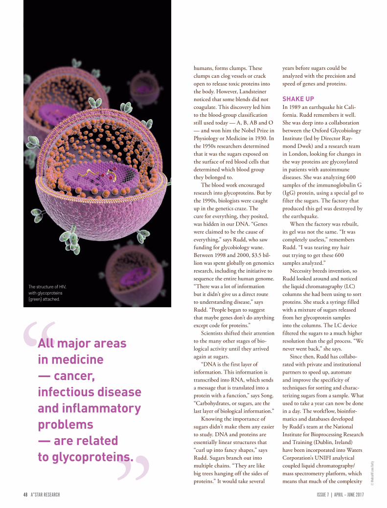
| FEATURES & INNOVATIONS |
48 ISSUE 7 | APRIL – JUNE 2017A*STAR RESEARCH www.astar-research.com 49 A*STAR RESEARCH
© M
edica
lRF.co
m/Ge
tty
humans, forms clumps. These clumps can clog vessels or crack open to release toxic proteins into the body. However, Landsteiner noticed that some blends did not coagulate. This discovery led him to the blood-group classification still used today — A, B, AB and O — and won him the Nobel Prize in Physiology or Medicine in 1930. In the 1950s researchers determined that it was the sugars exposed on the surface of red blood cells that determined which blood group they belonged to.
The blood work encouraged research into glycoproteins. But by the 1990s, biologists were caught up in the genetics craze. The cure for everything, they posited, was hidden in our DNA. “Genes were claimed to be the cause of everything,” says Rudd, who saw funding for glycobiology wane. Between 1998 and 2000, $3.5 bil-lion was spent globally on genomics research, including the initiative to sequence the entire human genome. “There was a lot of information but it didn’t give us a direct route to understanding disease,” says Rudd. “People began to suggest that maybe genes don’t do anything except code for proteins.”
Scientists shifted their attention to the many other stages of bio-logical activity until they arrived again at sugars.
“DNA is the first layer of information. This information is transcribed into RNA, which sends a message that is translated into a protein with a function,” says Song. “Carbohydrates, or sugars, are the last layer of biological information.”
Knowing the importance of sugars didn’t make them any easier to study. DNA and proteins are essentially linear structures that “curl up into fancy shapes,” says Rudd. Sugars branch out into multiple chains. “They are like big trees hanging off the sides of proteins.” It would take several
years before sugars could be analyzed with the precision and speed of genes and proteins.
SHAKE UPIn 1989 an earthquake hit Cali-fornia. Rudd remembers it well. She was deep into a collaboration between the Oxford Glycobiology Institute (led by Director Ray-mond Dwek) and a research team in London, looking for changes in the way proteins are glycosylated in patients with autoimmune diseases. She was analyzing 600 samples of the immunoglobulin G (IgG) protein, using a special gel to filter the sugars. The factory that produced this gel was destroyed by the earthquake.
When the factory was rebuilt, its gel was not the same. “It was completely useless,” remembers Rudd. “I was tearing my hair out trying to get these 600 samples analyzed.”
Necessity breeds invention, so Rudd looked around and noticed the liquid chromatography (LC) columns she had been using to sort proteins. She stuck a syringe filled with a mixture of sugars released from her glycoprotein samples into the columns. The LC device filtered the sugars to a much higher resolution than the gel process. “We never went back,” she says.
Since then, Rudd has collabo-rated with private and institutional partners to speed up, automate and improve the specificity of techniques for sorting and charac-terizing sugars from a sample. What used to take a year can now be done in a day. The workflow, bioinfor-matics and databases developed by Rudd's team at the National Institute for Bioprocessing Research and Training (Dublin, Ireland) have been incorporated into Waters Corporation's UNIFI analytical coupled liquid chromatography/mass spectrometry platform, which means that much of the complexity
All major areas in medicine — cancer, infectious disease and inflammatory problems — are related to glycoproteins.
The structure of HIV, with glycoproteins (green) attached.
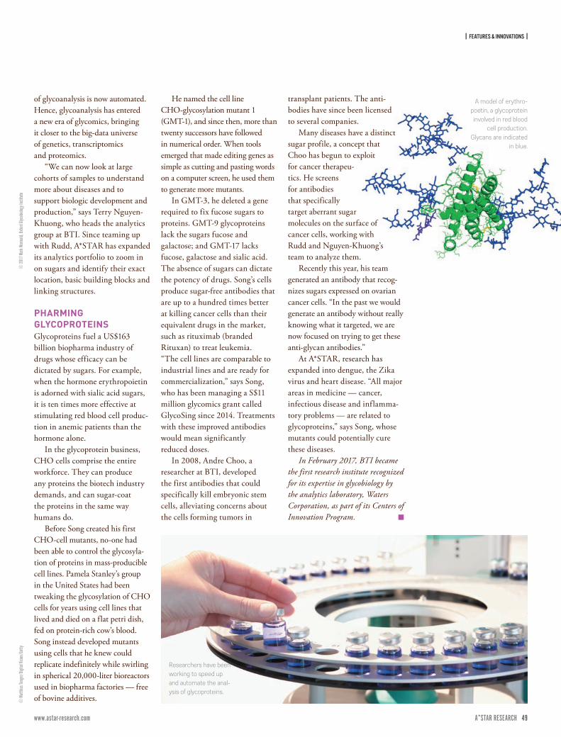
| FEATURES & INNOVATIONS |
48 ISSUE 7 | APRIL – JUNE 2017A*STAR RESEARCH www.astar-research.com 49 A*STAR RESEARCH
of glycoanalysis is now automated. Hence, glycoanalysis has entered a new era of glycomics, bringing it closer to the big-data universe of genetics, transcriptomics and proteomics.
“We can now look at large cohorts of samples to understand more about diseases and to support biologic development and production,” says Terry Nguyen-Khuong, who heads the analytics group at BTI. Since teaming up with Rudd, A*STAR has expanded its analytics portfolio to zoom in on sugars and identify their exact location, basic building blocks and linking structures.
PHARMING GLYCOPROTEINSGlycoproteins fuel a US$163 billion biopharma industry of drugs whose efficacy can be dictated by sugars. For example, when the hormone erythropoietin is adorned with sialic acid sugars, it is ten times more effective at stimulating red blood cell produc-tion in anemic patients than the hormone alone.
In the glycoprotein business, CHO cells comprise the entire workforce. They can produce any proteins the biotech industry demands, and can sugar-coat the proteins in the same way humans do.
Before Song created his first CHO-cell mutants, no-one had been able to control the glycosyla-tion of proteins in mass-producible cell lines. Pamela Stanley’s group in the United States had been tweaking the glycosylation of CHO cells for years using cell lines that lived and died on a flat petri dish, fed on protein-rich cow’s blood. Song instead developed mutants using cells that he knew could replicate indefinitely while swirling in spherical 20,000-liter bioreactors used in biopharma factories — free of bovine additives.
He named the cell line CHO-glycosylation mutant 1 (GMT-1), and since then, more than twenty successors have followed in numerical order. When tools emerged that made editing genes as simple as cutting and pasting words on a computer screen, he used them to generate more mutants.
In GMT-3, he deleted a gene required to fix fucose sugars to proteins. GMT-9 glycoproteins lack the sugars fucose and galactose; and GMT-17 lacks fucose, galactose and sialic acid. The absence of sugars can dictate the potency of drugs. Song’s cells produce sugar-free antibodies that are up to a hundred times better at killing cancer cells than their equivalent drugs in the market, such as rituximab (branded Rituxan) to treat leukemia. “The cell lines are comparable to industrial lines and are ready for commercialization,” says Song, who has been managing a S$11 million glycomics grant called GlycoSing since 2014. Treatments with these improved antibodies would mean significantly reduced doses.
In 2008, Andre Choo, a researcher at BTI, developed the first antibodies that could specifically kill embryonic stem cells, alleviating concerns about the cells forming tumors in
transplant patients. The anti-bodies have since been licensed to several companies.
Many diseases have a distinct sugar profile, a concept that Choo has begun to exploit for cancer therapeu-tics. He screens for antibodies that specifically target aberrant sugar molecules on the surface of cancer cells, working with Rudd and Nguyen-Khuong’s team to analyze them.
Recently this year, his team generated an antibody that recog-nizes sugars expressed on ovarian cancer cells. “In the past we would generate an antibody without really knowing what it targeted, we are now focused on trying to get these anti-glycan antibodies.”
At A*STAR, research has expanded into dengue, the Zika virus and heart disease. “All major areas in medicine — cancer, infectious disease and inflamma-tory problems — are related to glycoproteins,” says Song, whose mutants could potentially cure these diseases.
In February 2017, BTI became the first research institute recognized for its expertise in glycobiology by the analytics laboratory, Waters Corporation, as part of its Centers of Innovation Program.
Researchers have been working to speed up and automate the anal-ysis of glycoproteins.
A model of erythro-poetin, a glycoprotein involved in red blood
cell production. Glycans are indicated
in blue.
© 20
17 M
ark W
ormald
, Oxfo
rd Gly
cobio
logy I
nstit
ute©
Matt
hias T
unge
r/Digi
tal Vi
sion/
Getty
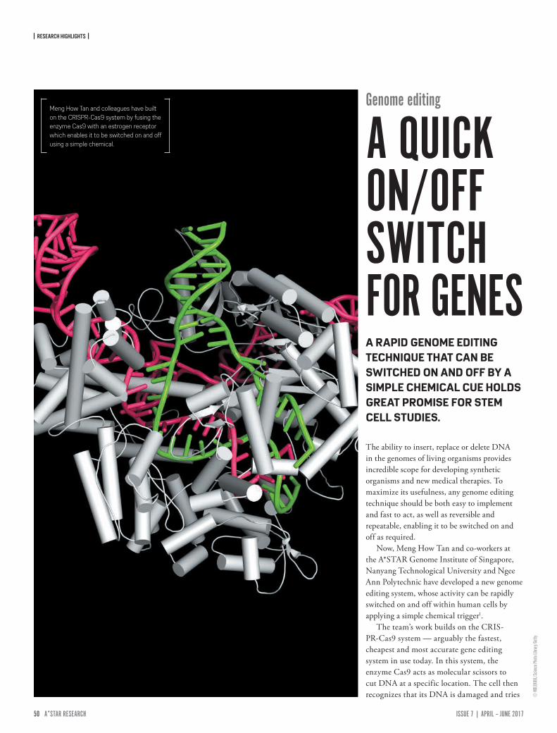
| RESEARCH HIGHLIGHTS |
50 ISSUE 7 | APRIL – JUNE 2017A*STAR RESEARCH www.astar-research.com 51 A*STAR RESEARCH
© M
OLEK
UUL/
Scien
ce Ph
oto Li
brary/
Getty
Genome editing
A QUICK ON/OFF SWITCH FOR GENES
The ability to insert, replace or delete DNA in the genomes of living organisms provides incredible scope for developing synthetic organisms and new medical therapies. To maximize its usefulness, any genome editing technique should be both easy to implement and fast to act, as well as reversible and repeatable, enabling it to be switched on and off as required.
Now, Meng How Tan and co-workers at the A*STAR Genome Institute of Singapore, Nanyang Technological University and Ngee Ann Polytechnic have developed a new genome editing system, whose activity can be rapidly switched on and off within human cells by applying a simple chemical trigger1.
The team’s work builds on the CRIS-PR-Cas9 system — arguably the fastest, cheapest and most accurate gene editing system in use today. In this system, the enzyme Cas9 acts as molecular scissors to cut DNA at a specific location. The cell then recognizes that its DNA is damaged and tries
Meng How Tan and colleagues have built on the CRISPR-Cas9 system by fusing the enzyme Cas9 with an estrogen receptor which enables it to be switched on and off using a simple chemical.
A RAPID GENOME EDITING TECHNIQUE THAT CAN BE SWITCHED ON AND OFF BY A SIMPLE CHEMICAL CUE HOLDS GREAT PROMISE FOR STEM CELL STUDIES.
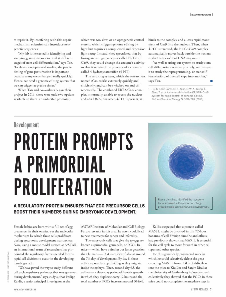
| RESEARCH HIGHLIGHTS |
50 ISSUE 7 | APRIL – JUNE 2017A*STAR RESEARCH www.astar-research.com 51 A*STAR RESEARCH
© JE
SPER
KLAU
SEN /
SCIEN
CE PH
OTO L
IBRAR
Y/Ge
tty
to repair it. By interfering with this repair mechanism, scientists can introduce new genetic sequences.
“My lab is interested in identifying and studying genes that are essential at different stages of stem cell differentiation,” says Tan. “In these developmental studies, the precise timing of gene perturbation is important because many events happen really quickly. Hence, we need a genome editing system that we can trigger at precise times.”
When Tan and co-workers began their project in 2014, there were only two options available to them: an inducible promoter,
which was too slow, or an optogenetic control system, which triggers genome editing by light but requires a complicated and expensive light setup. Instead, they speculated that by fusing an estrogen receptor called ERT2 to Cas9, they could change the enzyme’s activity so that it required the presence of a chemical called 4-hydroxytamoxifen (4-HT).
The resulting system, which the researchers named iCas, works extremely quickly and efficiently, and can be switched on and off repeatedly. The combined ERT2-Cas9 com-plex is normally unable to access the nucleus and edit DNA, but when 4-HT is present, it
binds to the complex and allows rapid move-ment of Cas9 into the nucleus. Then, when 4-HT is removed, the ERT2-Cas9 complex automatically moves back outside the nucleus so the Cas9 can’t cut DNA any more.
“As well as using our system to study stem cell differentiation more precisely, we can use it to study the reprogramming, or transdif-ferentiation, of one cell type into another,” says Tan.
1. Liu, K. I., Bin Ramli, M. N., Woo, C. W. A., Wang, Y., Zhao, T. et al. A chemical-inducible CRISPR–Cas9 system for rapid control of genome editing. Nature Chemical Biology 9, 980–987 (2016).
Development
PROTEIN PROMPTS A PRIMORDIAL PROLIFERATIONFemale babies are born with a full set of egg precursors in their ovaries, yet the molecular mechanism by which these cells proliferate during embryonic development was unclear. Now, using a mouse model created at A*STAR, an international team of researchers has pin-pointed the regulatory factors needed for this rapid cell division to occur in the developing female gonad.
“We have paved the way to study different cell cycle regulatory pathways that may go awry during development,” says study author Philipp Kaldis, a senior principal investigator at the
A*STAR Institute of Molecular and Cell Biology. Future research in this area, he notes, could lead to new treatments for cancer and infertility.
The embryonic cells that give rise to eggs are known as primordial germ cells, or PGCs. In mice — which have a similar but faster gestation than humans — PGCs are identifiable at around the 7th day of development. By day 8, these cells temporarily stop dividing as they migrate inside the embryo. Then, around day 9.5, the cells enter a three-day period of frenetic growth in which they duplicate every 12 hours and the total number of PGCs increases around 50-fold.
Kaldis suspected that a protein called MASTL might be involved in this 72-hour bonanza of cell division since he and others had previously shown that MASTL is essential for the cell cycle to move forward in other cell types and other species.
He thus genetically engineered mice in which he could selectively delete the gene encoding MASTL from PGCs. Kaldis then sent the mice to Kiu Liu and Sanjiv Risal at the University of Gothenberg in Sweden, and collectively they showed that the PGCs in these mice could not complete the anaphase step in
Researchers have identified the regulatory factors involved in the production of egg precursor cells during embryonic development.A REGULATORY PROTEIN ENSURES THAT EGG PRECURSOR CELLS
BOOST THEIR NUMBERS DURING EMBRYONIC DEVELOPMENT.
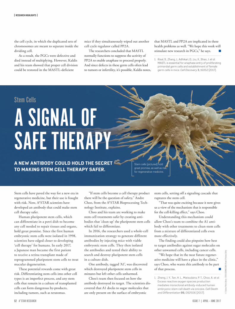
| RESEARCH HIGHLIGHTS |
52 ISSUE 7 | APRIL – JUNE 2017A*STAR RESEARCH www.astar-research.com 53 A*STAR RESEARCH
© Sc
ience
Photo
Libra
ry - S
TEVE
GSCH
MEISS
NER/
Bran
d X Pi
ctures
/Gett
y
the cell cycle, in which the duplicated sets of chromosomes are meant to separate inside the dividing cell.
As a result, the PGCs were defective and died instead of multiplying. However, Kaldis and his team showed that proper cell division could be restored in the MASTL-deficient
mice if they simultaneously wiped out another cell cycle regulator called PP2A.
The researchers concluded that MASTL normally functions to suppress the activity of PP2A to enable anaphase to proceed properly. And since defects in these germ cells often lead to tumors or infertility, it’s possible, Kaldis notes,
that MASTL and PP2A are implicated in these health problems as well. “We hope this work will stimulate new research in PGCs,” he says.
1. Risal, S., Zhang, J., Adhikari, D., Liu, X., Shao, J. et al. MASTL is essential for anaphase entry of proliferating primordial germ cells and establishment of female germ cells in mice. Cell Discovery 3, 16052 (2017).
Stem Cells
A SIGNAL OF SAFE THERAPYA NEW ANTIBODY COULD HOLD THE SECRET TO MAKING STEM CELL THERAPY SAFER.
Stem cells have paved the way for a new era in regenerative medicine, but their use is fraught with risk. Now, A*STAR scientists have developed an antibody that could make stem cell therapy safer.
Human pluripotent stem cells, which can differentiate in a petri dish to become any cell needed to repair tissues and organs, hold great promise. Since the first human embryonic stem cells were isolated in 1998, scientists have edged closer to developing ‘cell therapy’ for humans. In early 2017, a Japanese man became the first patient to receive a retina transplant made of reprogrammed pluripotent stem cells to treat macular degeneration.
These potential rewards come with great risk. Differentiating stem cells into other cell types is an imperfect process, and any stem cells that remain in a culture of transplanted cells can form dangerous by-products, including tumors, such as teratomas.
“If stem cells become a cell therapy product there will be the question of safety,” Andre Choo, from the A*STAR Bioprocessing Tech-nology Institute, explains.
Choo and his team are working to make stem cell treatments safer by creating anti-bodies that ‘clean up’ the pluripotent stem cells which fail to differentiate.
In 2016, the researchers used a whole-cell immunization strategy to generate different antibodies by injecting mice with viable embryonic stem cells. They then isolated the antibodies and tested their ability to search and destroy pluripotent stem cells in a culture dish.
One antibody, tagged ‘A1’, was discovered which destroyed pluripotent stem cells in minutes but left other cells unharmed.
Choo’s team then focused on how the antibody destroyed its target. The scientists dis-covered that A1 docks to sugar molecules that are only present on the surface of embryonic
stem cells, setting off a signaling cascade that ruptures the stem cell.
“That was quite exciting because it now gives us a view of the mechanism that is responsible for the cell-killing effect,” says Choo.
Understanding this mechanism could allow Choo’s team to combine the A1 anti-body with other treatments to clean stem cells from a mixture of differentiated cells even more effectively.
The finding could also pinpoint how best to target antibodies against sugar molecules on other unwanted cells, including cancer cells.
“We hope that in the near future regener-ative medicine will have a place in the clinic,” says Choo, who wants this antibody to be part of that process.
1. Zheng, J. Y., Tan, H. L., Matsudaira, P. T., Choo, A. et al. Excess reactive oxygen species production mediates monoclonal antibody-induced human embryonic stem cell death via oncosis. Cell Death and Differentiation 99, 092506 (2017).
Stem cells (pictured) hold great promise, as well as risk, for regenerative medicine.
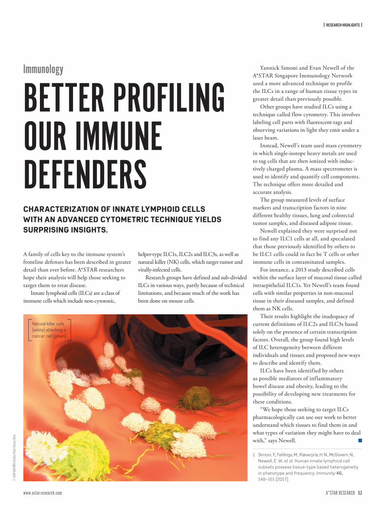
| RESEARCH HIGHLIGHTS |
52 ISSUE 7 | APRIL – JUNE 2017A*STAR RESEARCH www.astar-research.com 53 A*STAR RESEARCH
© JU
AN GA
RTNE
R/Sc
ience
Photo
Libra
ry/Ge
tty
A family of cells key to the immune system’s frontline defenses has been described in greater detail than ever before. A*STAR researchers hope their analysis will help those seeking to target them to treat disease.
Innate Iymphoid cells (ILCs) are a class of immune cells which include non-cytotoxic,
helper-type ILC1s, ILC2s and ILC3s, as well as natural killer (NK) cells, which target tumor and virally-infected cells.
Research groups have defined and sub-divided ILCs in various ways, partly because of technical limitations, and because much of the work has been done on mouse cells.
Yannick Simoni and Evan Newell of the A*STAR Singapore Immunology Network used a more advanced technique to profile the ILCs in a range of human tissue types in greater detail than previously possible.
Other groups have studied ILCs using a technique called flow cytometry. This involves labeling cell parts with fluorescent tags and observing variations in light they emit under a laser beam.
Instead, Newell’s team used mass cytometry in which single-isotope heavy metals are used to tag cells that are then ionized with induc-tively charged plasma. A mass spectrometer is used to identify and quantify cell components. The technique offers more detailed and accurate analysis.
The group measured levels of surface markers and transcription factors in nine different healthy tissues, lung and colorectal tumor samples, and diseased adipose tissue.
Newell explained they were surprised not to find any ILC1 cells at all, and speculated that those previously identified by others to be ILC1 cells could in fact be T cells or other immune cells in contaminated samples.
For instance, a 2013 study described cells within the surface layer of mucosal tissue called intraepithelial ILC1s. Yet Newell’s team found cells with similar properties in non-mucosal tissue in their diseased samples, and defined them as NK cells.
Their results highlight the inadequacy of current definitions of ILC2s and ILC3s based solely on the presence of certain transcription factors. Overall, the group found high levels of ILC heterogeneity between different individuals and tissues and proposed new ways to describe and identify them.
ILCs have been identified by others as possible mediators of inflammatory bowel disease and obesity, leading to the possibility of developing new treatments for these conditions.
“We hope those seeking to target ILCs pharmacologically can use our work to better understand which tissues to find them in and what types of variation they might have to deal with,” says Newell.
1. Simoni, Y., Fehlings, M., Kløverpris, H. N., McGovern, N., Newell, E. W. et al. Human innate lymphoid cell subsets possess tissue-type based heterogeneity in phenotype and frequency. Immunity 46, 148–161 (2017).
Natural killer cells (white) attacking a cancer cell (green).
Immunology
BETTER PROFILING OUR IMMUNE DEFENDERSCHARACTERIZATION OF INNATE LYMPHOID CELLS WITH AN ADVANCED CYTOMETRIC TECHNIQUE YIELDS SURPRISING INSIGHTS.

54 ISSUE 7 | APRIL – JUNE 2017A*STAR RESEARCH www.astar-research.com 55 A*STAR RESEARCH
© Hn
oomP
atcha
/Mom
ent/G
etty
| RESEARCH HIGHLIGHTS |
Sick of waiting at traffic lights? The semi-au-tonomous driving aids being fitted to many new cars could consign the red light to history, A*STAR researchers report. According to their modeling, a system in which each car crosses the intersection in its own virtual bubble of safe space, modulating its speed using adaptive cruise control, will result in smooth traffic flow in each direction.
In the city of the near future, traffic lights would be complemented by and then replaced with a communication beacon, explains Bo Yang from the A*STAR Institute of High Performance Computing, who led the work.
The beacon gathers and transmits data about the distance and approach speed of vehicles nearing the intersection, which each car feeds into an algorithm that plots a safe course through without having to stop.
At the algorithm’s heart is the concept of “adaptive repulsive force”. The closer two cars’ trajectory would bring them at an intersection, the stronger their repulsion and the greater the speed adjustment they make to pass each other safely.
Yang set out to reduce barriers to adoption of this system. “One of our most interesting findings is that the rules governing the
necessary repulsion between vehicles is rather simple,” he says. The result is a system that does not require much computing power at the beacon or in the vehicle itself.
Cars need not be fully self-driving, but rather simply able to brake and accelerate autonomously – which cars fitted with smart cruise control can already do. The driver gives up control of the car’s speed through the intersection but remains in charge of steering.
In Yang’s simulations, the algorithm worked effectively even for relatively complex intersec-tions. “In most cases, pre-emptive deceleration only slightly lowered the vehicle velocity,
Intelligent transport
A CROSSROADS FOR INTERSECTIONSCARS COULD SOON NEGOTIATE SMART INTERSECTIONS WITHOUT EVER HAVING TO STOP.
Autonomous driving aids will help city traffic flow more freely.
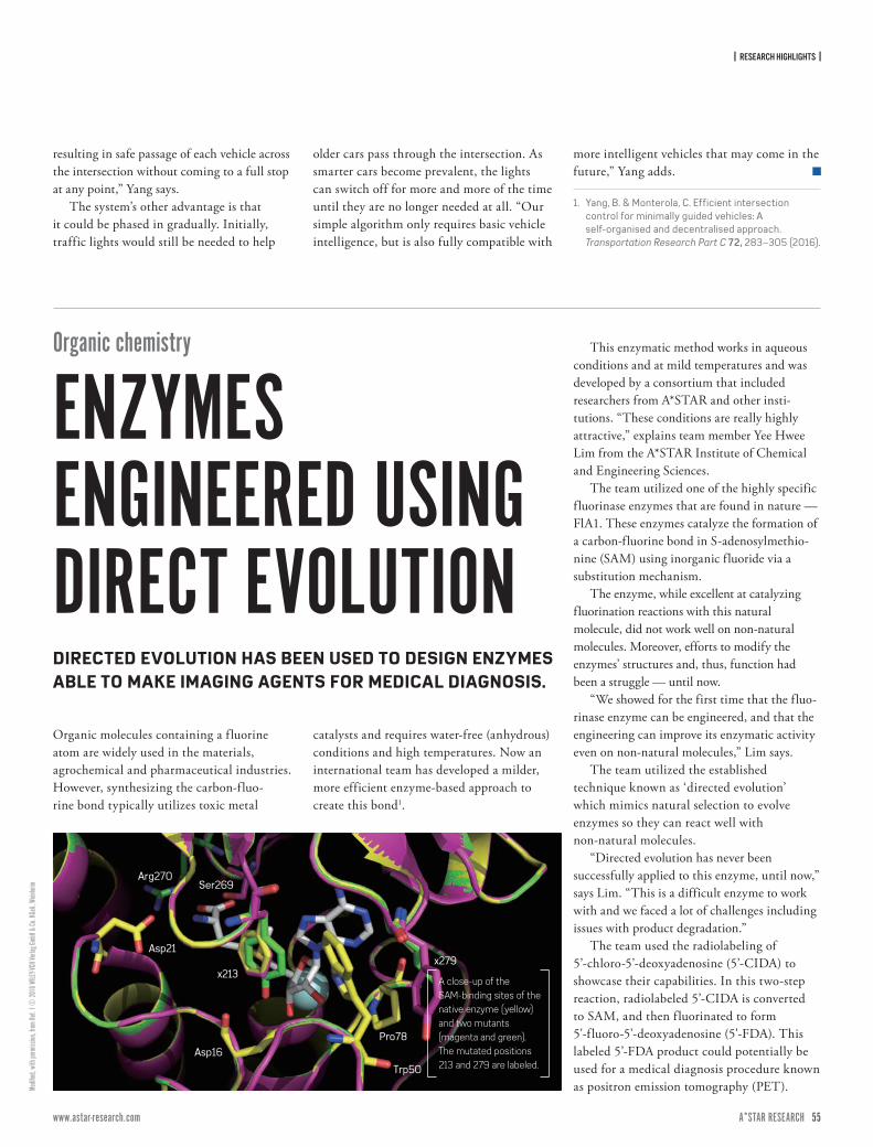
| RESEARCH HIGHLIGHTS |
54 ISSUE 7 | APRIL – JUNE 2017A*STAR RESEARCH www.astar-research.com 55 A*STAR RESEARCH
Modifi
ed, w
ith pe
rmiss
ion, fr
om Re
f. 1 ©
2016
WILE
Y-VCH
Verla
g Gmb
H & Co
. KGa
A, We
inheim
resulting in safe passage of each vehicle across the intersection without coming to a full stop at any point,” Yang says.
The system’s other advantage is that it could be phased in gradually. Initially, traffic lights would still be needed to help
older cars pass through the intersection. As smarter cars become prevalent, the lights can switch off for more and more of the time until they are no longer needed at all. “Our simple algorithm only requires basic vehicle intelligence, but is also fully compatible with
more intelligent vehicles that may come in the future,” Yang adds.
1. Yang, B. & Monterola, C. Efficient intersection control for minimally guided vehicles: A self-organised and decentralised approach. Transportation Research Part C 72, 283–305 (2016).
Organic molecules containing a f luorine atom are widely used in the materials, agrochemical and pharmaceutical industries. However, synthesizing the carbon-fluo-rine bond typically utilizes toxic metal
catalysts and requires water-free (anhydrous) conditions and high temperatures. Now an international team has developed a milder, more efficient enzyme-based approach to create this bond1.
This enzymatic method works in aqueous conditions and at mild temperatures and was developed by a consortium that included researchers from A*STAR and other insti-tutions. “These conditions are really highly attractive,” explains team member Yee Hwee Lim from the A*STAR Institute of Chemical and Engineering Sciences.
The team utilized one of the highly specific fluorinase enzymes that are found in nature — FlA1. These enzymes catalyze the formation of a carbon-fluorine bond in S-adenosylmethio-nine (SAM) using inorganic fluoride via a substitution mechanism.
The enzyme, while excellent at catalyzing fluorination reactions with this natural molecule, did not work well on non-natural molecules. Moreover, efforts to modify the enzymes’ structures and, thus, function had been a struggle — until now.
“We showed for the first time that the fluo-rinase enzyme can be engineered, and that the engineering can improve its enzymatic activity even on non-natural molecules,” Lim says.
The team utilized the established technique known as ‘directed evolution’ which mimics natural selection to evolve enzymes so they can react well with non-natural molecules.
“Directed evolution has never been successfully applied to this enzyme, until now,” says Lim. “This is a difficult enzyme to work with and we faced a lot of challenges including issues with product degradation.”
The team used the radiolabeling of 5’-chloro-5’-deoxyadenosine (5’-CIDA) to showcase their capabilities. In this two-step reaction, radiolabeled 5’-CIDA is converted to SAM, and then f luorinated to form 5'-f luoro-5'-deoxyadenosine (5'-FDA). This labeled 5’-FDA product could potentially be used for a medical diagnosis procedure known as positron emission tomography (PET).
Arg270Ser269
Asp21
Asp16
x213x279
Pro78
Trp50
A close-up of the SAM-binding sites of the native enzyme (yellow) and two mutants (magenta and green). The mutated positions 213 and 279 are labeled.
Organic chemistry
ENZYMES ENGINEERED USING DIRECT EVOLUTIONDIRECTED EVOLUTION HAS BEEN USED TO DESIGN ENZYMES ABLE TO MAKE IMAGING AGENTS FOR MEDICAL DIAGNOSIS.

| RESEARCH HIGHLIGHTS |
56 ISSUE 7 | APRIL – JUNE 2017A*STAR RESEARCH www.astar-research.com 57 A*STAR RESEARCH
“We show that improving an enzyme’s activity three-fold can open up more applica-tions,” says Lim. “The native enzymes have been used previously to try and label PET agents but the reaction times were long, sometimes hours. Using our enzyme we could do the reaction in 30
minutes, a more reasonable timeline given that Fluorine-18's half-life is less than two hours.”
Lim’s team is now exploring how mutating the enzyme’s structure changes its interaction with 5’-CIDA and SAM. “I’d like to engage computational biologists to better understand
more about the mutations that we have made and how those correlate with our reactions.”
1. Sun, H., Yeo, W. L., Lim, Y. H., Chew, X., Smith, D. J., et al. Directed evolution of a fluorinase for improved fluorination efficiency with a non-native substrate. Angewandte Chemie 128, 14489–14492 (2016).
© Pr
asit p
hoto/
Mome
nt/Ge
tty
Cyber security
TWO-STEP SECURITY FOR THE INTERNET OF THINGSThe power of big data is the key to a strategy developed by A*STAR to improve the security of networks of internet-connected objects, known as the Internet of Things (IoT).
More than 20 billion devices — everythign from streetlights to refrigerators— are expected to be upgraded and connected to each other by 2020. However, with IoT becoming increasingly widely adopted, developers need to guarantee its security. One hacked target could be the gateway to other parts of the network, making it vulnerable to breaches of sensitive information. This was demonstrated in October 2016, when a huge attack on IoT devices across Europe and the USA, such as CCTV cameras with easy-to-guess passwords, contributed to outages for several major websites.
Currently, a number of web services, including online banking and Google, use or offer a two-step authentication process to increase the security levels. Since passwords
can be leaked or cracked, these services require secondary secret information from the customer. This could be another code transmitted via SMS, email or a security token; or the user’s fingerprints or facial recognition.
However, the direct application of these methods to the IoT is not practical. “We want to achieve the same level of security as bank servers offer, but the resources needed are simply overkill for typical IoT devices,” explains Jun Wen Wong, one of the researchers involved in the study. “We had to think about a brand new protocol.”
The new strategy, devised by A*STAR researchers of the Institute for Infocomm Research, uses the conventional password as first step for authentication, but a second step uses the whole history of the data exchanged between the IoT device and the server.
The scientists proposed algorithms that generate and store in the IoT device a very small
piece of secret information, which can concisely represent the whole history dataset, and can be retrieved for the authentication. Thanks to this approach, taken from the big data sector and originally applied to the IoT, this security two-step system can be compatible with IoT devices with low computation and small memory.
Using the data exchanged between the device and the server has very interesting leakage-re-silience properties. As data are constantly generated by the IoT device and sent to the server, the history dataset is growing, so hackers would have to steal a considerable amount of data over an extended period of time, becoming more open to detection.
1. Chan, A. C. F., Wong, J. W., Zhou, J. & Teo, J. Scalable two-factor authentication using historical data. In: Askoxylakis I., Ioannidis S., Katsikas S., Meadows C. (eds) Computer Security – ESORICS 2016. ESORICS 2016. Lecture Notes in Computer Science 9878, pp. 91–110, Springer, Cham.
HARNESSING THE POTENTIAL OF BIG DATA TO IMPROVE THE SECURITY OF INTERNET OF THINGS DEVICES.
A*STAR researchers have developed a new two step authentication strategy incorporating data exchange history that could help protect IoT connected devices against cyber attacks.
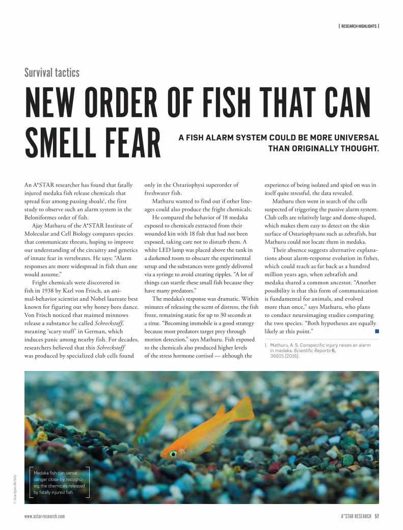
| RESEARCH HIGHLIGHTS |
56 ISSUE 7 | APRIL – JUNE 2017A*STAR RESEARCH www.astar-research.com 57 A*STAR RESEARCH
© Ka
zuo O
gawa
/Aflo
/Gett
y
An A*STAR researcher has found that fatally injured medaka fish release chemicals that spread fear among passing shoals1, the first study to observe such an alarm system in the Beloniformes order of fish.
Ajay Mathuru of the A*STAR Institute of Molecular and Cell Biology compares species that communicate threats, hoping to improve our understanding of the circuitry and genetics of innate fear in vertebrates. He says: “Alarm responses are more widespread in fish than one would assume.”
Fright chemicals were discovered in fish in 1938 by Karl von Frisch, an ani-mal-behavior scientist and Nobel laureate best known for figuring out why honey bees dance. Von Frisch noticed that maimed minnows release a substance he called Schreckstoff, meaning ‘scary stuff ’ in German, which induces panic among nearby fish. For decades, researchers believed that this Schreckstoff was produced by specialized club cells found
only in the Ostariophysi superorder of freshwater fish.
Mathuru wanted to find out if other line-ages could also produce the fright chemicals.
He compared the behavior of 18 medaka exposed to chemicals extracted from their wounded kin with 18 fish that had not been exposed, taking care not to disturb them. A white LED lamp was placed above the tank in a darkened room to obscure the experimental setup and the substances were gently delivered via a syringe to avoid creating ripples. “A lot of things can startle these small fish because they have many predators.”
The medaka’s response was dramatic. Within minutes of releasing the scent of distress, the fish froze, remaining static for up to 30 seconds at a time. “Becoming immobile is a good strategy because most predators target prey through motion detection,” says Mathuru. Fish exposed to the chemicals also produced higher levels of the stress hormone cortisol — although the
experience of being isolated and spied on was in itself quite stressful, the data revealed.
Mathuru then went in search of the cells suspected of triggering the passive alarm system. Club cells are relatively large and dome-shaped, which makes them easy to detect on the skin surface of Ostariophysans such as zebrafish, but Mathuru could not locate them in medaka.
Their absence suggests alternative explana-tions about alarm-response evolution in fishes, which could reach as far back as a hundred million years ago, when zebrafish and medaka shared a common ancestor. “Another possibility is that this form of communication is fundamental for animals, and evolved more than once,” says Mathuru, who plans to conduct neuroimaging studies comparing the two species. “Both hypotheses are equally likely at this point.”
1. Mathuru, A. S. Conspecific injury raises an alarm in medaka. Scientific Reports 6, 36615 (2016).
Survival tactics
NEW ORDER OF FISH THAT CAN SMELL FEAR A FISH ALARM SYSTEM COULD BE MORE UNIVERSAL
THAN ORIGINALLY THOUGHT.
Medaka fish can sense danger close-by, recogniz-ing the chemicals released by fatally injured fish.
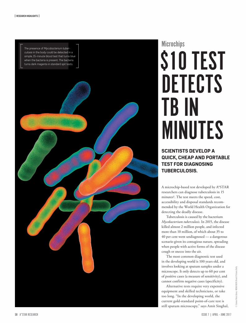
| RESEARCH HIGHLIGHTS |
58 ISSUE 7 | APRIL – JUNE 2017A*STAR RESEARCH www.astar-research.com 59 A*STAR RESEARCH
Microchips
$10 TEST DETECTS TB IN MINUTES
A microchip-based test developed by A*STAR researchers can diagnose tuberculosis in 15 minutes1. The test meets the speed, cost, accessibility and disposal standards recom-mended by the World Health Organization for detecting the deadly disease.
Tuberculosis is caused by the bacterium Mycobacterium tuberculosis. In 2015, the disease killed almost 2 million people, and infected more than 10 million, of which about 35 to 40 per cent went undiagnosed — a dangerous scenario given its contagious nature, spreading when people with active forms of the disease cough or sneeze into the air.
The most common diagnostic test used in the developing world is 100 years old, and involves looking at sputum samples under a microscope. It only detects up to 60 per cent of positive cases (a measure of sensitivity), and cannot confirm negative cases (specificity).
Alternative tests require very expensive equipment and skilled technicians, or take too long. “In the developing world, the current gold-standard point-of-care test is still sputum microscopy,” says Amit Singhal,
The presence of Mycobacterium tuber-culosis in the body could be detected in a simple, 15-minute blood test that turns blue when the bacteria is present. The bacteria turns dark magenta in standard spit tests.
SCIENTISTS DEVELOP A QUICK, CHEAP AND PORTABLE TEST FOR DIAGNOSING TUBERCULOSIS.
© Sc
ience
Photo
Libra
ry - M
EHAU
KULY
K/Br
and X
Pictu
res/G
etty
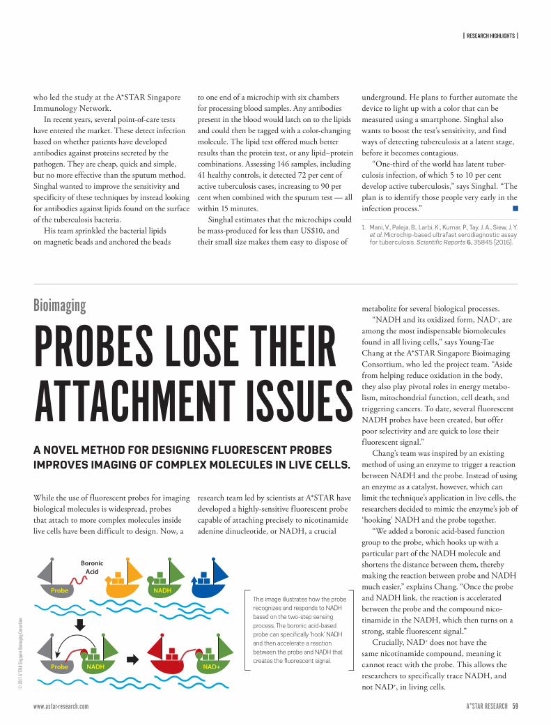
| RESEARCH HIGHLIGHTS |
58 ISSUE 7 | APRIL – JUNE 2017A*STAR RESEARCH www.astar-research.com 59 A*STAR RESEARCH
© 20
17 A*
STAR
Sing
apore
Bioim
aging
Cons
ortium
While the use of fluorescent probes for imaging biological molecules is widespread, probes that attach to more complex molecules inside live cells have been difficult to design. Now, a
research team led by scientists at A*STAR have developed a highly-sensitive fluorescent probe capable of attaching precisely to nicotinamide adenine dinucleotide, or NADH, a crucial
metabolite for several biological processes.“NADH and its oxidized form, NAD+, are
among the most indispensable biomolecules found in all living cells,” says Young-Tae Chang at the A*STAR Singapore Bioimaging Consortium, who led the project team. “Aside from helping reduce oxidation in the body, they also play pivotal roles in energy metabo-lism, mitochondrial function, cell death, and triggering cancers. To date, several fluorescent NADH probes have been created, but offer poor selectivity and are quick to lose their fluorescent signal.”
Chang’s team was inspired by an existing method of using an enzyme to trigger a reaction between NADH and the probe. Instead of using an enzyme as a catalyst, however, which can limit the technique’s application in live cells, the researchers decided to mimic the enzyme’s job of ‘hooking’ NADH and the probe together.
“We added a boronic acid-based function group to the probe, which hooks up with a particular part of the NADH molecule and shortens the distance between them, thereby making the reaction between probe and NADH much easier,” explains Chang. “Once the probe and NADH link, the reaction is accelerated between the probe and the compound nico-tinamide in the NADH, which then turns on a strong, stable fluorescent signal.”
Crucially, NAD+ does not have the same nicotinamide compound, meaning it cannot react with the probe. This allows the researchers to specifically trace NADH, and not NAD+, in living cells.
BoronicAcid
Probe
Probe
NADH
NADH NAD+
This image illustrates how the probe recognizes and responds to NADH based on the two-step sensing process. The boronic acid-based probe can specifically ‘hook’ NADH and then accelerate a reaction between the probe and NADH that creates the fluorescent signal.
who led the study at the A*STAR Singapore Immunology Network.
In recent years, several point-of-care tests have entered the market. These detect infection based on whether patients have developed antibodies against proteins secreted by the pathogen. They are cheap, quick and simple, but no more effective than the sputum method. Singhal wanted to improve the sensitivity and specificity of these techniques by instead looking for antibodies against lipids found on the surface of the tuberculosis bacteria.
His team sprinkled the bacterial lipids on magnetic beads and anchored the beads
to one end of a microchip with six chambers for processing blood samples. Any antibodies present in the blood would latch on to the lipids and could then be tagged with a color-changing molecule. The lipid test offered much better results than the protein test, or any lipid–protein combinations. Assessing 146 samples, including 41 healthy controls, it detected 72 per cent of active tuberculosis cases, increasing to 90 per cent when combined with the sputum test — all within 15 minutes.
Singhal estimates that the microchips could be mass-produced for less than US$10, and their small size makes them easy to dispose of
underground. He plans to further automate the device to light up with a color that can be measured using a smartphone. Singhal also wants to boost the test’s sensitivity, and find ways of detecting tuberculosis at a latent stage, before it becomes contagious.
“One-third of the world has latent tuber-culosis infection, of which 5 to 10 per cent develop active tuberculosis,” says Singhal. “The plan is to identify those people very early in the infection process.”
1. Mani, V., Paleja, B., Larbi, K., Kumar, P., Tay, J. A., Siew, J. Y. et al. Microchip-based ultrafast serodiagnostic assay for tuberculosis. Scientific Reports 6, 35845 (2016).
Bioimaging
PROBES LOSE THEIR ATTACHMENT ISSUESA NOVEL METHOD FOR DESIGNING FLUORESCENT PROBES IMPROVES IMAGING OF COMPLEX MOLECULES IN LIVE CELLS.
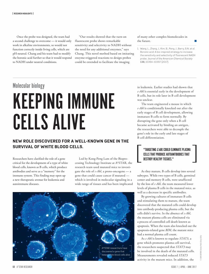
| RESEARCH HIGHLIGHTS |
60 ISSUE 7 | APRIL – JUNE 2017A*STAR RESEARCH www.astar-research.com 61 A*STAR RESEARCH
Once the probe was designed, the team had a second challenge to overcome — it would only work in alkaline environments, so would not function correctly inside living cells, which are pH-neutral. Chang and his team had to modify the boronic acid further so that it would respond to NADH under neutral conditions.
“Our results showed that the turn-on f luorescent probe shows remarkable sensitivity and selectivity to NADH without the need for any additional enzymes,” says Chang. This novel method based on imitating enzyme-triggered reactions to design probes could be extended to facilitate the imaging
of many other complex biomolecules in the future.
1. Wang, L., Zhang, J., Kim, B., Peng, J., Berry, S.N. et al. Boronic acid: A bio-inspired strategy to increase the sensitivity and selectivity of fluorescent NADH probe. Journal of the American Chemical Society 138, 10394–10397 (2017).
Researchers have clarified the role of a gene critical for the development of a type of white blood cells, known as B cells, which produce antibodies and serve as a “memory” for the immune system. This finding may open up a new therapeutic avenue for leukemia and autoimmune diseases.
Led by Kong-Peng Lam of the Biopro-cessing Technology Institute at A*STAR, the research team used mutated mice to investi-gate the role of c-Abl, a proto-oncogene — a gene that could cause cancer if mutated — which is involved in molecular signaling in a wide range of tissues and has been implicated
in leukemia. Earlier studies had shown that c-Abl is essential early in the development of B cells, but its role later in B cell development was unclear.
The team engineered a mouse in which c-Abl is conditionally knocked out after the early stages of B cell development, allowing immature B cells to form normally. By disrupting the gene only when a B cell became activated by binding an antigen, the researchers were able to decouple the gene’s role in the early and late stages of B cell differentiation.
As they mature, B cells develop into several subtypes. While two types of B cells, germinal center and memory B cells, were unaffected by the loss of c-Abl, the team measured lower levels of plasma B cells in the mutated mice, as well as a decrease in specific antibodies.
By growing cultures of immature B cells and stimulating them to mature, the team discovered that the mutated cells could develop into antibody-producing plasma cells, but the cells didn’t survive. In the absence of c-Abl, the mutant plasma cells are eliminated via a process of controlled cell death known as apoptosis. When the team also knocked out the apoptosis-related gene BIM, the mutant mice had a normal plasma cell count.
As c-Abl is known to regulate STAT3, a gene which promotes plasma cell survival, the researchers suspected that STAT3 may be involved in the death of the mutated cells. Measurements revealed reduced STAT3 activity in the mutant mice. In addition, the
"TARGETING C-ABL COULD ELIMINATE PLASMA CELLS THAT PRODUCE AUTOANTIBODIES THAT DESTROY HEALTHY TISSUES."
A*STAR researchers have discovered a gene that keeps white blood cells alive.
© JU
AN GA
RTNE
R/Sc
ience
Photo
Libra
ry/Ge
tty
Molecular biology
KEEPING IMMUNE CELLS ALIVENEW ROLE DISCOVERED FOR A WELL-KNOWN GENE IN THE SURVIVAL OF WHITE BLOOD CELLS.
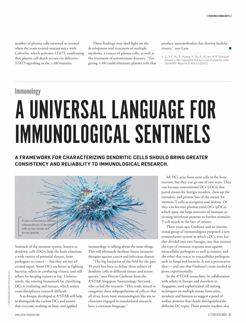
| RESEARCH HIGHLIGHTS |
60 ISSUE 7 | APRIL – JUNE 2017A*STAR RESEARCH www.astar-research.com 61 A*STAR RESEARCH
© ST
EVE G
SCHM
EISSN
ER/S
CIENC
E PHO
TO LI
BRAR
Y/Ge
tty
number of plasma cells returned to normal when the team treated mutant mice with Colivelin, which activates STAT3, confirming that plasma cell death occurs via defective STAT3 signaling in the c-Abl mutants.
These findings may shed light on the development and treatment of multiple myeloma, a cancer of plasma cells, as well as the treatment of autoimmune diseases. “Tar-geting c-Abl could eliminate plasma cells that
produce autoantibodies that destroy healthy tissues,” says Lam.
1. Li, Y. F., Xu, S., Huang, Y., Ou, X., & Lam, K. P. Tyrosine kinase c-Abl regulates the survival of plasma cells. Scientific Reports 7, 40133 (2017).
Immunology
A UNIVERSAL LANGUAGE FOR IMMUNOLOGICAL SENTINELSA FRAMEWORK FOR CHARACTERIZING DENDRITIC CELLS SHOULD BRING GREATER CONSISTENCY AND RELIABILITY TO IMMUNOLOGICAL RESEARCH.
Sentinels of the immune system, known as dendritic cells (DCs), help the body eliminate a wide variety of potential threats, from pathogens to cancer — but they are not all created equal. Some DCs are better at fighting bacteria, others at combating viruses, and still others for keeping tumors at bay. Unfortu-nately, the existing framework for classifying DCs is confusing and inexact, which makes cross-disciplinary research difficult.
A technique developed at A*STAR will help to distinguish the various DCs and ensure that everyone working on basic and applied
immunology is talking about the same things. This will ultimately facilitate future immuno-therapies against cancer and infectious disease.
“The big limitation of the field for the past 10 years has been to define these subsets of dendritic cells in different tissues and across species,” says Florent Ginhoux from the A*STAR Singapore Immunology Network, who co-led the research. “This study aimed to categorize these subpopulations of cells so that all of us, from basic immunologists like me to clinicians engaged in translational research, have a common language.”
All DCs arise from stem cells in the bone marrow, but they can go one of two ways. They can become conventional DCs (cDCs) that patrol tissues for foreign invaders, chew up the intruders, and present bits of the enemy for immune T cells to recognize and destroy. Or they can become plasmacytoid DCs (pDCs), which spew out large amounts of immune-ac-tivating interferon proteins to further stimulate T-cell attack in the face of viruses.
Three years ago, Ginhoux and an interna-tional group of immunologists proposed a new nomenclature system in which cDCs were fur-ther divided into two lineages, one that mounts the type of immune response seen against intracellular pathogens as well as tumors, and the other that reacts to extracellular pathogens such as fungi and bacteria. It was a provocative idea — and one that Ginhoux’s team needed to prove experimentally.
So the A*STAR researchers, in collaboration with others in Europe and elsewhere in Singapore, used sophisticated cell sorting techniques on multiple tissues from mice, monkeys and humans to suggest a panel of surface proteins that clearly distinguished the different DC types. These protein markers also
A new nomenclature defines subpopulations of dendritic cells across tissues and across species.
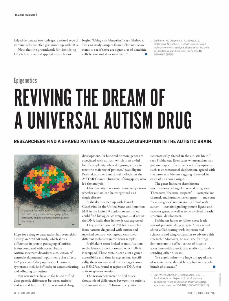
| RESEARCH HIGHLIGHTS |
62 ISSUE 7 | APRIL – JUNE 2017A*STAR RESEARCH www.astar-research.com 63 A*STAR RESEARCH
helped demarcate macrophages, a related type of immune cell that often gets mixed up with DCs.
Now that the groundwork for identifying DCs is laid, the real applied research can
begin. “Using this blueprint,” says Ginhoux, “we can study samples from different disease states to see if there are signatures of dendritic cells before and after treatment.”
1. Guilliams, M., Dutertre, C. A., Scott, C. L., McGovern, N., Sichien, D. et al. Unsupervised high-dimensional analysis aligns dendritic cells across tissues and species. Immunity 45, 669–684 (2016).
© 20
17 So
nish A
zam
Hope for a drug to treat autism has been rekin-dled by an A*STAR study, which showsdifferences in protein packaging of autisticbrains compared with normal brains. Autism spectrum disorder is a collection of neurodevelopmental impairments that affects 1–2 per cent of the population. Common symptoms include difficulty in communicating and adhering to routines.
But researchers have so far failed to find clear genetic differences between autistic and normal brains. This has stymied drug
development. “A hundred or more genes are associated with autism, which is an awful lot of complexity when designing a drug to treat the majority of patients,” says Shyam Prabhakar, a computational biologist at the A*STAR Genome Institute of Singapore, who led the analysis.
This diversity has caused some to question whether autism can be categorized as a single disease.
Prabhakar teamed up with Daniel Geschwind in the United States and Jonathan Mill in the United Kingdom to see if they could find biological convergence — if not in the DNA itself, then in how it was expressed.
They studied around 250 brain samples from patients diagnosed with autism and matched controls, each group examined different molecules in the brain samples.
Prabhakar’s team looked at modifications to the histone proteins around which DNA is wrapped. These proteins can alter a gene’s accessibility and thus its expression. Specifi-cally, the team analyzed histone tags known as H3K27ac, found at regions of DNA that activate gene expression.
The researchers were thrilled to see thousands of differences between the autistic and normal tissue. “Histone acetylation is
systematically altered in the autistic brain,” says Prabhakar. Even cases where autism was just one aspect of a broader set of symptoms, such as chromosomal duplication, agreed with the pattern of histone tagging observed in cases of unknown origin.
The genes linked to these histone modifications belonged to several categories. There were “the usual suspects” — synaptic, ion channel, and immune system genes — and some “new categories” not previously linked with autism — certain signaling-protein ligand and receptor genes, as well as some involved in early structural development.
Prabhakar hopes to follow these leads toward potential drug targets. “I'm excited about collaborating with experimental scientists and drug companies to advance this research.” Moreover, he says, the findings demonstrate the effectiveness of histone acetylome-wide association studies for under-standing other diseases.
“It’s a gold mine — a huge untapped area of research that should be applied to a whole bunch of diseases.”
1. Sun, W., Poschmann, J., del Rosario, R. C.-H., Parikshak, N. N., Hajan, H. S. et al. Histone acetylome-wide association study of autism spectrum disorder. Cell 167, 1385–1397 (2016).
Epigenetics
REVIVING THE DREAM OF A UNIVERSAL AUTISM DRUGRESEARCHERS FIND A SHARED PATTERN OF MOLECULAR DISRUPTION IN THE AUTISTIC BRAIN.
A graphical representation of autism, depicting the brain using puzzle pieces signifying the complexity and lack of understanding about the disease.
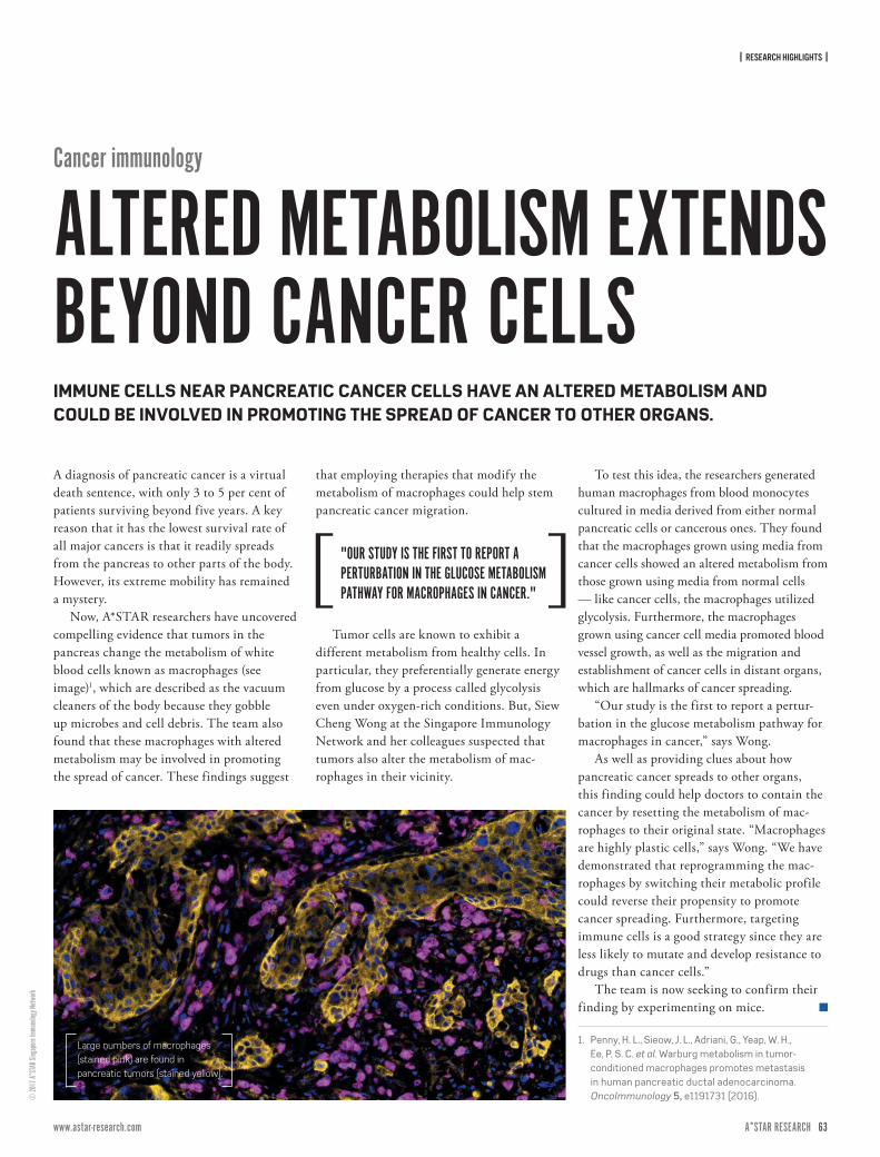
| RESEARCH HIGHLIGHTS |
62 ISSUE 7 | APRIL – JUNE 2017A*STAR RESEARCH www.astar-research.com 63 A*STAR RESEARCH
© 20
17 A*
STAR
Sing
apore
Immu
nolog
y Netw
ork
Cancer immunology
ALTERED METABOLISM EXTENDSBEYOND CANCER CELLSIMMUNE CELLS NEAR PANCREATIC CANCER CELLS HAVE AN ALTERED METABOLISM AND COULD BE INVOLVED IN PROMOTING THE SPREAD OF CANCER TO OTHER ORGANS.
A diagnosis of pancreatic cancer is a virtual death sentence, with only 3 to 5 per cent of patients surviving beyond five years. A key reason that it has the lowest survival rate of all major cancers is that it readily spreads from the pancreas to other parts of the body. However, its extreme mobility has remained a mystery.
Now, A*STAR researchers have uncovered compelling evidence that tumors in the pancreas change the metabolism of white blood cells known as macrophages (see image)1, which are described as the vacuum cleaners of the body because they gobble up microbes and cell debris. The team also found that these macrophages with altered metabolism may be involved in promoting the spread of cancer. These findings suggest
that employing therapies that modify the metabolism of macrophages could help stem pancreatic cancer migration.
Tumor cells are known to exhibit a different metabolism from healthy cells. In particular, they preferentially generate energy from glucose by a process called glycolysis even under oxygen-rich conditions. But, Siew Cheng Wong at the Singapore Immunology Network and her colleagues suspected that tumors also alter the metabolism of mac-rophages in their vicinity.
To test this idea, the researchers generated human macrophages from blood monocytes cultured in media derived from either normal pancreatic cells or cancerous ones. They found that the macrophages grown using media from cancer cells showed an altered metabolism from those grown using media from normal cells — like cancer cells, the macrophages utilized glycolysis. Furthermore, the macrophages grown using cancer cell media promoted blood vessel growth, as well as the migration and establishment of cancer cells in distant organs, which are hallmarks of cancer spreading.
“Our study is the first to report a pertur-bation in the glucose metabolism pathway for macrophages in cancer,” says Wong.
As well as providing clues about how pancreatic cancer spreads to other organs, this finding could help doctors to contain the cancer by resetting the metabolism of mac-rophages to their original state. “Macrophages are highly plastic cells,” says Wong. “We have demonstrated that reprogramming the mac-rophages by switching their metabolic profile could reverse their propensity to promote cancer spreading. Furthermore, targeting immune cells is a good strategy since they are less likely to mutate and develop resistance to drugs than cancer cells.”
The team is now seeking to confirm their finding by experimenting on mice.
1. Penny, H. L., Sieow, J. L., Adriani, G., Yeap, W. H., Ee, P. S. C. et al. Warburg metabolism in tumor-conditioned macrophages promotes metastasis in human pancreatic ductal adenocarcinoma. OncoImmunology 5, e1191731 (2016).
Large numbers of macrophages (stained pink) are found in pancreatic tumors (stained yellow).
"OUR STUDY IS THE FIRST TO REPORT A PERTURBATION IN THE GLUCOSE METABOLISM PATHWAY FOR MACROPHAGES IN CANCER."
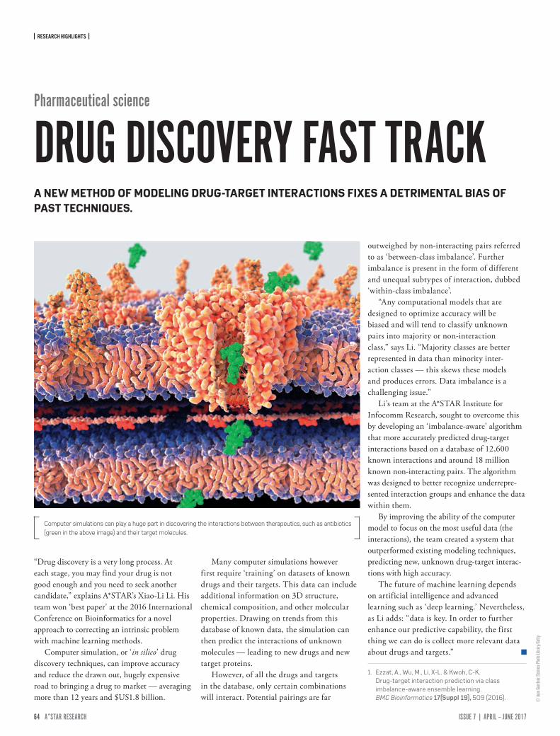
| RESEARCH HIGHLIGHTS |
64 ISSUE 7 | APRIL – JUNE 2017A*STAR RESEARCH www.astar-research.com PB A*STAR RESEARCH
© Ju
an Ga
ertne
r/Scie
nce P
hoto
Librar
y/Ge
tty
“Drug discovery is a very long process. At each stage, you may find your drug is not good enough and you need to seek another candidate,” explains A*STAR’s Xiao-Li Li. His team won ‘best paper’ at the 2016 International Conference on Bioinformatics for a novel approach to correcting an intrinsic problem with machine learning methods.
Computer simulation, or ‘in silico’ drug discovery techniques, can improve accuracy and reduce the drawn out, hugely expensive road to bringing a drug to market — averaging more than 12 years and $US1.8 billion.
Many computer simulations however first require ‘training’ on datasets of known drugs and their targets. This data can include additional information on 3D structure, chemical composition, and other molecular properties. Drawing on trends from this database of known data, the simulation can then predict the interactions of unknown molecules — leading to new drugs and new target proteins.
However, of all the drugs and targets in the database, only certain combinations will interact. Potential pairings are far
outweighed by non-interacting pairs referred to as ‘between-class imbalance’. Further imbalance is present in the form of different and unequal subtypes of interaction, dubbed ‘within-class imbalance’.
“Any computational models that are designed to optimize accuracy will be biased and will tend to classify unknown pairs into majority or non-interaction class,” says Li. “Majority classes are better represented in data than minority inter-action classes — this skews these models and produces errors. Data imbalance is a challenging issue.”
Li’s team at the A*STAR Institute for Infocomm Research, sought to overcome this by developing an ‘imbalance-aware’ algorithm that more accurately predicted drug-target interactions based on a database of 12,600 known interactions and around 18 million known non-interacting pairs. The algorithm was designed to better recognize underrepre-sented interaction groups and enhance the data within them.
By improving the ability of the computer model to focus on the most useful data (the interactions), the team created a system that outperformed existing modeling techniques, predicting new, unknown drug-target interac-tions with high accuracy.
The future of machine learning depends on artificial intelligence and advanced learning such as ‘deep learning.’ Nevertheless, as Li adds: “data is key. In order to further enhance our predictive capability, the first thing we can do is collect more relevant data about drugs and targets.”
1. Ezzat, A., Wu, M., Li, X-L. & Kwoh, C-K. Drug-target interaction prediction via class imbalance-aware ensemble learning. BMC Bioinformatics 17(Suppl 19), 509 (2016).
Pharmaceutical science
DRUG DISCOVERY FAST TRACKA NEW METHOD OF MODELING DRUG-TARGET INTERACTIONS FIXES A DETRIMENTAL BIAS OF PAST TECHNIQUES.
Computer simulations can play a huge part in discovering the interactions between therapeutics, such as antibiotics (green in the above image) and their target molecules.

Advanced Remanufacturing and Technology Centre (ARTC)Bioinformatics Institute (BII)Bioprocessing Technology Institute (BTI)Clinical Imaging Research Centre (CIRC)Data Storage Institute (DSI)Experimental Power Grid Centre (EPGC)Experimental Therapeutics Centre (ETC)Genome Institute of Singapore (GIS)Institute of Bioengineering and Nanotechnology (IBN)Institute of Chemical and Engineering Sciences (ICES)Institute of High Performance Computing (IHPC)Institute for Infocomm Research (I2R) Institute of Materials Research and Engineering (IMRE)Institute of Medical Biology (IMB)Institute of Microelectronics (IME)Institute of Molecular and Cell Biology (IMCB)National Metrology Centre (NMC)Singapore Bioimaging Consortium (SBIC)Singapore Institute for Clinical Sciences (SICS)Singapore Institute of Manufacturing Technology (SIMTech)Singapore Immunology Network (SIgN)
The Agency for Science, Technology and Research (A*STAR) is Singapore’s lead government agency dedicated to fostering world-class scientific research and talent for a vibrant knowledge-based economy.
A*STAR actively nurtures public-sector research and development in biomedical sciences, physical sciences and engineering, and spurs growth in Singapore’s key economic clusters by providing human, intellectual and industrial capital to our partners in industry and the healthcare sector.
A*STAR currently oversees the following research institutes, consor-tia and centers and supports extramural research with universities, hospital research centers, and other local and international partners.

www.astar-research.com